


Abstract
Emerging evidence indicates that low-grade chronic systemic inflammation is a key contributor to the onset and progression of numerous chronic diseases. Vanilla planifolia, a globally valued spice recognized for its characteristic sweet aroma and flavor, is primarily derived from its beans and widely utilized in culinary, therapeutic, and medicinal contexts. Beyond its traditional use, vanilla provides essential nutrients and bioactive compounds, including B vitamins such as vitamin B6, niacin, and riboflavin, which are integral to metabolic regulation. In addition to its applications in food and pharmaceuticals, vanilla exhibits complementary medicinal properties. Vanillin, the principal bioactive constituent of vanilla, imparts its distinctive flavor and aroma and is accompanied by other phenolic compounds with notable antioxidant activity. This review highlights the potential of vanillin as a therapeutic agent, shifting its perception from a conventional flavoring compound to a promising bioactive molecule with relevance in chronic disease prevention. Furthermore, the applications of vanillin within the food industry are discussed.
1. Introduction
Emerging research evidence has suggested that low-grade chronic systemic inflammation plays a vitally important role in modulating several chronic diseases such as cancer, cardiovascular disease (CVD), metabolic syndrome, and type 2 diabetes [1,2], which is known as “inflammatory diseases” [3]. Although the underlying molecular inflammatory process involved in each disease may vary from other diseases, the basic mode of action of the inflammatory mediators and proinflammatory cytokines is similar.
Oxidative stress is closely linked to chronic inflammation [4], and additional factors may further exacerbate the accumulation of proinflammatory cytokines, perpetuating a “vicious cycle” [5]. In cultured adipocytes, reactive oxygen species (ROS) enhance the production of monocyte chemotactic protein-1 (MCP-1) and upregulate interleukin-6 (IL-6) expression [1]. Within adipose tissue, ROS facilitate macrophage infiltration, thereby fostering a proinflammatory microenvironment [6]. ROS also activate intracellular signaling cascades, predominantly via NF-κB, which in turn stimulate the expression of IL-6 and tumor necrosis factor-α (TNF-α). Furthermore, oxidative stress can induce cellular senescence, particularly in adipocytes, through oxidative damage. Senescent adipocytes may subsequently recruit macrophages and amplify proinflammatory cytokine production [3]. Substantial evidence indicates that excessive macronutrient intake promotes oxidative stress, thereby driving inflammation through NF-κB-mediated signaling pathways [7].
Vanilla planifolia is a perennial, climbing tropical plant of the Orchidaceae family [8]. The term “Vanilla” derives from the Spanish word Vainilla, a diminutive of Vaina meaning “sheath” or “pod,” which aptly reflects the morphology of the vanilla bean [9]. Native to Central and South America and the Caribbean, vanilla was first cultivated by the Totonacs along Mexico’s eastern coast [9]. Vanilla contains essential nutrients and bioactive compounds that underpin its culinary, therapeutic, and medicinal applications, including B vitamins that support metabolic functions and alleviate pain in conditions such as arthritis [9]. Vanillin, the primary bioactive compound, imparts its characteristic flavor and aroma, while phenolic compounds provide antioxidant activity that mitigates oxidative stress and cellular damage [10]. Beyond antioxidant effects, vanilla demonstrates anti-inflammatory, antimicrobial, and anti-carcinogenic properties, and in Ayurvedic medicine it is traditionally used to relieve stress and promote well-being [9,11].
The rising prevalence of chronic diseases such as diabetes, cancer, and obesity underscores the need for alternative bioactive compounds. Vanilla planifolia emerges as a promising source of plant-derived molecules with potential roles in disease prevention and management, warranting exploration beyond its traditional use as a flavoring agent. This review highlights its bioactive properties, therapeutic applications in chronic disease prevention, and potential utilization in functional foods and nutraceuticals.
2. Method
A comprehensive literature search was conducted using electronic databases including PubMed, Scopus, Web of Science, and Google Scholar. The search covered studies published from January 1998 to December 2025. The following keywords and their combinations were used: “vanillin”, “oxidative stress”, “antioxidant activity”, “genotoxicity”, “DNA damage”, “anticancer”, “antidiabetic”, “anti-inflammatory”, “neuroprotective”, “antimicrobial”, “apoptosis”, “flavoring agent”, “safety”, “toxicity”, “mitochondrial dysfunction”, and “cell cycle arrest”. Boolean operators (AND, OR) were applied to refine the search strategy. Additional relevant studies were identified by manually screening the reference lists of selected articles.
3. Oxidative Stress and Chronic Diseases
This review focuses on oxidative-stress-related diseases with well-established mechanistic links to redox imbalance, including cardiovascular diseases, type 2 diabetes mellitus, and cancer. These conditions were selected because oxidative stress plays a central role in the pathogenesis via mechanisms, including mitochondrial dysfunction, chronic inflammation, DNA damage, and impaired cellular signalling. In addition, these diseases represent major contributors to global morbidity and mortality, making them highly relevant from both mechanistic and public health perspectives. By concentrating on these representative conditions, this review aims to provide a comprehensive overview of the pathological consequences of oxidative stress and therapeutic implications.
3.1. Cardiovascular Disease
Cardiovascular disease (CVD) encompasses a range of disorders affecting the heart and blood vessels, and it is a leading cause of morbidity and mortality worldwide. The term cardiovascular disease broadly refers to conditions such as coronary heart disease (CHD), stroke, peripheral artery disease (PAD), and aortic disease [12]. CHD, characterized by the build-up of plaque in the coronary arteries, often leads to angina or myocardial infarction (heart attack). Stroke occurs when blood flow to the brain is obstructed or reduced, leading to brain cell damage. PAD involves narrowing or blockage of the peripheral arteries, typically in the legs, while aortic diseases affect the body’s largest blood vessel, the aorta. A common condition is an aortic aneurysm, where the aorta weakens and bulges, which can further cause life-threatening bleeding if it ruptures [12].
The prevalence of CVD is alarmingly high, making it a global health crisis. According to the World Health Organization [12], CVD represents the leading cause of mortality worldwide, accounting for an estimated 17.9 million deaths annually. This statistic underscores the significant impact of CVD on public health and highlights the urgent need for effective prevention and treatment strategies. CVD affects individuals across all demographics, though risk factors such as age, sex, lifestyle, and genetics contribute to its development and progression.
The underlying mechanisms of CVD are complex and multifaceted, involving a combination of genetic predispositions, environmental, and lifestyle factors. A primary mechanism in the development of CVD is the process of atherosclerosis, where fatty deposits (atheromas) build up on the arterial walls, leading to reduced blood flow and increased risk of clot formation [13]. This process is often exacerbated by factors such as high cholesterol levels, hypertension, smoking, and diabetes. Oxidative stress plays a critical role in the pathogenesis of CVD. It arises from an imbalance between the production of ROS and the body’s ability to neutralise them with antioxidants, causing cellular and molecular disruptions that can lead to cardiac dysfunction [14]. ROS can damage endothelial cells lining the blood vessels, leading to inflammation and endothelial dysfunction. This damage promotes the progression of atherosclerosis and contributes to the development of other cardiovascular conditions. Oxidative stress not only affects the endothelial cells but also impacts various lipoproteins and cellular components, further aggravating cardiovascular damage [15]. Elevated levels of oxidised low-density lipoprotein (LDL) cholesterol, often referred to as “bad cholesterol,” for instance, are a significant risk factor for atherosclerosis. Chronic oxidative stress can lead to persistent inflammation, which compounds endothelial injury and accelerates the development of cardiovascular diseases [1]. Thus, understanding the mechanisms of oxidative stress and its impact on cardiovascular health is crucial for developing targeted interventions to reduce the burden of CVD.
3.2. Type 2 Diabetes Mellitus
The chronic metabolic disease known as type 2 diabetes mellitus (T2DM) is characterised by decreased insulin production and insulin resistance. Type 2 diabetes is characterised by increased blood glucose levels due to the body’s inability to utilise insulin efficiently, in contrast to type 1 diabetes, which is caused by the body’s inability to make insulin. This disease usually manifests at maturity, while it is increasingly being identified in younger populations. It is linked to obesity, sedentary lifestyles, and genetic predispositions [16]. Targeting oxidative stress in treatment techniques is crucial because of its involvement in developing insulin resistance and causing problems related to diabetes.
There are 422 million people worldwide who have diabetes; most of them live in low- and middle-income countries. Every year, the illness is directly to blame for 1.5 million deaths. Both the incidence and the number of cases of diabetes have steadily increased during the last few decades. Given that this incidence is significantly higher than in previous years, it suggests a growing public health risk. Among the causes of this trend are fast urbanisation, dietary modifications that lead to a rise in the consumption of high-calorie foods, and a decline in physical activity. As evidenced by the disease’s increasing prevalence, comprehensive public health programmes are badly needed to control and alleviate Malaysians’ burden from type 2 diabetes [17].
Oxidative stress is one of the primary causes and developments of type 2 diabetes. It is distinguished by an imbalance between the body’s antioxidant defences and the generation of ROS [18]. Chronic hyperglycemia in type 2 diabetes is a factor in the overproduction of ROS. These very reactive compounds have the potential to seriously harm DNA, proteins, and lipids in cells. Insulin signalling pathway degradation is a major mechanism via which oxidative stress contributes to type 2 diabetes. Oxidative stress contributes to insulin resistance predominantly via degradation and disruption of the insulin signalling cascade. Under physiological conditions, insulin binds to the insulin receptor (IR), leading to tyrosine phosphorylation of insulin receptor substrate (IRS-1 and IRS-2), activation of phosphatidylinositol 3-kinase (PI3K), and thereby stimulates the Akt. Akt activation promotes glucose uptake through translocation of GLUT4 to the plasma membrane. Nonetheless, excessive production of ROS activates stress-sensitive serine/threonine kinases, for instance, c-Jun N-terminal kinase (JNK), inhibitor of κB kinase β (IKKβ), and p38 MAPK. These kinases trigger serine phosphorylation of IRS proteins, which inhibits normal tyrosine phosphorylation and promotes their dissociation from the insulin receptor. Serine-phosphorylated IRS becomes a target for ubiquitination and subsequent proteasomal degradation, and subsequently decreasing IRS protein availability. Moreover, oxidative stress can impair Akt activation and enhance activity of phosphatases including PTEN, further attenuating downstream signalling. Taken together, these alterations disrupt GLUT4 translocation, reduce glucose uptake in adipose tissue and skeletal muscle, and contribute to the development of type 2 diabetes and insulin resistance [19]. ROS have the ability to alter important signalling molecules, which can interfere with cells’ normal reactions to insulin and exacerbate insulin resistance [20]. Furthermore, pancreatic beta-cell activity may be impaired by oxidative stress, which would decrease insulin output and worsen hyperglycemia. Increased oxidative stress from elevated blood sugar levels exacerbates insulin resistance and beta-cell malfunction, creating an endless cycle [21].
Moreover, oxidative stress has been connected to the development of problems associated with diabetes, including retinopathy, neuropathy, nephropathy, and cardiovascular illnesses [18]. ROS can increase the risk of cardiovascular events by damaging endothelial cells, causing inflammation, and hastening the production of atherosclerotic plaques in the vascular system. Oxidative stress in peripheral tissues can harm the kidneys and nerves, causing neuropathy and kidney damage [22]. Due to a combination of hereditary and lifestyle factors, type 2 diabetes is becoming common worldwide. Targeting oxidative stress in treatment techniques is crucial because of its involvement in developing insulin resistance and causing problems related to diabetes. Reducing oxidative stress through glycemic control enhancements, antioxidant therapy, and lifestyle modifications may lessen the consequences of type 2 diabetes and enhance the quality of life for those who have this long-term illness.
3.3. Cancer
Research has demonstrated that elevated oxidative stress contributes to cancer development, including colorectal cancer [23]. Oxidative stress is hypothesized to be closely associated with both obesity and cancer. Evidence from an obese animal model of nonalcoholic steatohepatitis supports this hypothesis, showing that adiponectin deficiency promotes hepatic tumor formation and enhances oxidative stress [24]. ROS play a pivotal role in carcinogenesis [25]. Increased ROS levels elevate mutation rates and susceptibility to mutagenic agents, thereby contributing to DNA damage during the early stages of tumorigenesis [26]. ROS also drive tumor proliferation through ligand-independent transactivation of receptor tyrosine kinases [27], facilitating metastasis and cancer cell invasion [28]. Furthermore, Semenza [29] reported that ROS stabilize hypoxia-inducible factor 1, a transcriptional regulator of vascular endothelial growth factor, thereby promoting tumor angiogenesis.
Interestingly, insulin has been identified as a proliferative factor in prostate cancer, suggesting that carbohydrate restriction may reduce serum insulin levels and slow prostate cancer progression [30]. Epidemiological studies further indicate that patients with type 2 diabetes and obesity exhibit an increased risk of liver, colorectal, breast, and pancreatic cancers [31]. These associations implicate leptin, insulin/insulin-like growth factor-1, adiponectin, and chronic inflammation as additive mediators linking type 2 diabetes and obesity to cancer. Fat accumulation is frequently associated with systemic oxidative stress via ROS elevation [32]. Elevated oxidative stress can induce chronic inflammation, which in turn contributes to the pathogenesis of cancer [33]. Mechanistically, oxidative stress activates transcription factors such as Wnt/β-catenin, NF-κB, and nuclear factor E2-related factor 2 (Nrf2), thereby stimulating inflammatory signaling pathways [25]. Collectively, these findings suggest that increased circulating or local ROS derived from adipose tissue expansion within the tumor microenvironment exacerbate oxidative stress in tumor cells, ultimately enhancing cancer progression in patients with type 2 diabetes or obesity.
The underlying mechanism of cancer involves genetic and epigenetic changes that lead to the uncontrolled proliferation of cells. These changes can be caused by various factors, including exposure to carcinogens, inherited genetic mutations, and errors during cell division [1]. One important factor in the development of cancer is oxidative stress, which can lead to DNA damage and genomic instability. ROS generation and the body’s capacity to neutralise them are out of balance, which leads to oxidative stress [34].
Numerous studies have demonstrated that natural plant-derived compounds, particularly polyphenols, flavonoids, and phenolic acids, exhibit strong antioxidant properties that can neutralize ROS [35,36]. Vanilla planifolia represents a promising candidate due to its rich profile of phenolic constituents, including vanillin and related derivatives, which may contribute to oxidative stress mitigation. This rationale provides the foundation for our subsequent investigation into the bioactivity of Vanilla planifolia extracts.
4. Vanilla planifolia
Vanilla planifolia is a perennial, climbing tropical plant belonging to the Orchidaceae family [8]. The term “vanilla” derives from the Spanish word vainilla, a diminutive of vaina meaning “sheath” or “pod,” aptly describing the morphology of the vanilla bean [9]. Indigenous to Central and South America and the Caribbean, vanilla was first cultivated by the Totonacs of Mexico’s eastern coast [9]. Following the Aztec conquest of the Totonacs in the fifteenth century, vanilla became integrated into Aztec culture [9]. Natural vanilla thrives within a 20-degree latitudinal band on either side of the Equator, with major production occurring in Indonesia and Madagascar, although the species is native to Mexico. The plant requires approximately three years to reach maturity and produce flowers, while the fruit (vanilla beans) must remain on the vine for nine months before harvest. At this stage, the beans are devoid of flavor and require post-harvest curing to develop their characteristic aroma and taste [37].
The vanillin molecule (Figure 1) comprises a benzene ring attached to a hydroxyl group, an aldehyde group, and an ether group. This chemical structure is responsible for its characteristic bioactive properties and enables modification or combination with other compounds to synthesize various derivatives [38].
Figure 1.
Molecular structure of vanillin.
Vanillin can be obtained from three primary sources including biotechnological, chemical/synthetic, and natural. Its classification as either an artificial or natural flavor depends on the method of synthesis and origin. Biotechnologically produced vanillin, as well as natural vanillin synthesized using ferulic acid as a substrate, have been recognized as food-grade additives by several international food regulatory authorities [39]. In contrast, chemically synthesized vanillin is typically derived from cost-effective precursors such as lignin, eugenol, or guaiacol, and may be supplemented with ethyl vanillin. Compared to natural vanillin, artificial vanillin generally exhibits a simpler and less complex flavor profile [40,41].
5. Bioactive Compounds of Vanilla
Limited information is available regarding the chemical composition of mature green vanilla beans. Fiber components, including cellulose, lignin, and hemicellulose, contain approximately 8% of the fresh weight, which is significantly higher than the typical 1–4% found in most fruits and vegetables [42]. Sucrose is the predominant carbohydrate, although the total carbohydrate content is lower than that of most fruits [43]. Similarly, protein content is less than 1%, comparable to that of most fruits and vegetables. The high glucosidase activity in vanilla suggests the presence of elevated levels of aromatic compounds [44].
Vanilla beans contain remarkably high lipid content, approximately 12% of fresh weight, which is substantially greater than the 0.2–0.4% typically found in fruits and vegetables. The predominant organic acids are citrate and malate, accounting for approximately 80% of total organic acids. Additionally, vanilla beans contain 470 mg of potassium and 170 mg of calcium, both of which are present in significantly higher concentrations than in most vegetables and fruits [45].
Vanilla planifolia Jacks. ex Andrews contains a notably high concentration of glucovanillin, accounting for approximately 1.7% of its fresh weight. Vanillin (4-hydroxy-3-methoxybenzaldehyde) is the principal volatile compound, comprising 80–90% of the total volatiles. The characteristic flavor and aroma of vanilla develop through a series of enzymatic reactions during the curing process. Other reported compounds include benzyl ethers, lactones, carbonyl compounds, eugenol, phenols, phenol ethers, caproic acid, vitispiranes, anisyl alcohol, anisaldehyde, anisic acid, and vanillic acid [46]. In addition, vanilla contains B-complex vitamins, 25% carbohydrates, 15% fats, and approximately 6% mineral salts, including potassium, manganese, iron, calcium, magnesium, and zinc. The water content of vanilla beans ranges from 35–40% [47]. The bioactive constituents of Vanilla planifolia, particularly vanillin, demonstrate rapid gastrointestinal absorption but undergo extensive hepatic metabolism, predominantly via glucuronidation and sulfation, resulting in limited systemic bioavailability. Pharmacokinetic investigations report rapid clearance and urinary excretion of conjugated metabolites. These pharmacokinetic characteristics constrain therapeutic applicability, and thus more research is required to elucidate the formulation strategies and delivery systems such as encapsulation or nanoparticle carriers to improve the bioavailability [48].
6. Pharmaceutical Roles of Vanillin
6.1. Anti-Inflammatory Properties
Vanillin, a bioactive compound found in Vanilla planifolia, exhibits strong antioxidant properties [9]. Studies have shown that vanillin and vanillic acid can protect brain cells from oxidative stress [9]. A study conducted on aging rats found that vanillin may help reduce inflammation and enhance antioxidant defences [8]. Due to its antioxidant properties, vanillin can prevent oxidative damage to membranes in mammalian tissues [8]. Recent experiments also suggest that vanillin can alleviate inflammation, reduce anxiety, and enhance appetite [49]. In addition, vanilla extract also recognized in Ayurveda as a stress-relieving and calming agent, possesses the ability to neutralize free radicals and oxidants [9]. This natural remedy has been employed for centuries in various cultures as a means of promoting overall well-being. According to Ayurvedic practices, the consumption of water infused with vanilla flavor is a traditional method known to mitigate inflammation. This ancient wisdom aligns with contemporary scientific understanding, as vanilla’s anti-inflammatory properties have been increasingly acknowledged [9].
6.2. Anticancer Effects
Vanillin has demonstrated antimutagenic and anticancer activities by reducing chemically induced genotoxicity in experimental models such as mouse bone marrow cells and Drosophila melanogaster, protecting against mutagens including methylmethane, mitomycin C, N-ethyl-N-nitrosourea, and bleomycin, and inhibiting proliferation of colorectal cancer cells (HT-29) through cell cycle arrest at the G2/M and G0/G1 phases and induction of apoptosis [50,51,52]. Similarly, studies on hamster lung cells indicated that vanillin reduced UV-induced DNA mutations and chromosomal damage, while enhancing DNA repair mechanisms [53]. In human cancer cell lines like lung cancer (NCI-H460), vanillin suppressed metastasis and reduced gene expression related to tumor growth [54]. Apart from that, as a potent antioxidant, vanillin neutralizes free radicals that can damage cells and DNA. Vanillin protected DNA from radiation-induced damage in both laboratory and animal models [55]. The free radical scavenging ability underscores its importance in preventing oxidative stress, a precursor to various diseases, including cancer.
A recent study showed that Vanilla planifolia extracts significantly decreased cell viability in HepG2 cells at a dosage of 5000 µg/mL for 72 h, 48 h, and 24 h. A similar effect was observed in mouse liver fibroblast (FC3H) cells [50]. In addition, ethanolic Vanilla planifolia leaf extract has also demonstrated antiproliferative activity against squamous carcinoma and breast cancer cells [56,57]. A study by Chang et al. [58] evaluated the antitumor effect of Vanilla planifolia on glioblastoma multiforme cells. The study found that the ethanol extract of Vanilla planifolia stem significantly reduced the viability and colony-forming ability of glioblastoma multiforme cells via downregulation of 14 hub DEGs and upregulation of 9 hub DEGs. Table 1 summarizes the pharmacological effects of Vanilla planifolia, vanillin, and its derivatives.
Table 1.
Pharmacological effects of Vanilla planifolia extract, vanillin, and its derivatives.
Vanillin is a natural compound with anti-neoplastic properties with minimal adverse effects [66]. Vanillin (5, 4, 3, 2, 1 μg/mL) significantly reduced B16F10 cell viability following 24 h of incubation. Melanoma tumor size was decreased after treatment at both 100 and 50 mg/kg/day of vanillin for 10 days. NF-κB is a pivotal transcription factor that regulates the expression of genes involved in immune responses and cell proliferation [2]. The previous study showed that NF-κB protein expression was downregulated in the C57BL/6 mice treated with 100 mg/kg/day vanillin for 10 days compared to the control group [59].
Of particular interest are novel metal-based complexes incorporating vanillin or its derivatives, which have demonstrated anti-inflammatory properties with potential anticancer benefits [67]. For instance, ruthenium (III), iron (III), and chromium (III) complexes incorporating ortho-vanillin derivatives exhibited significant inhibition of carrageenan-induced (1% w/v) tumor edema in the legs of Wistar rats. These effects were evaluated in comparison to the established anti-inflammatory drugs DMSO and sodium diclofenac, both administered at the same concentration [68]. Moreover, vanillin and its novel derivatives are being evaluated for their potential efficacy against breast cancer through in vitro and in vivo studies. Apoptosis induced by vanillin is a key anticancer mechanism, not only for vanillin itself but also for its derivatives. 4-hydroxy-3-methoxy benzaldehyde (vanillin) derivatives have been shown to inhibit the survival and proliferation of MCF-7 cells in a time- and dose-dependent manner (1, 2, 4, 8, 12, 16, 32, 64, 128, 256, 512 µg/mL for 72 h) by inducing apoptotic cell death through the activation of a caspase-8-mediated pathway [62]. Additionally, the vanillin-derived compound VALD-3 induces apoptosis and cell cycle arrest in human breast cancer cells by inhibiting Wnt/β-catenin signaling, highlighting its potential as a therapeutic agent for breast cancer treatment [61]. Evidence indicates that VALD-3, a Schiff base ligand synthesized from o-vanillin derivatives (10 and 20 mg/L for 48 h), promotes apoptosis by activating caspase-3 and caspase-8 and modulating the balance of Bcl-2 family proteins, thereby shifting cells toward programmed death [61]. It also interferes with cell cycle progression, inducing arrest at the S and G2/M phases in MCF-7 cells (40 mg/L for 48 h) through regulation of cyclins and cyclin-dependent kinases, which suppresses uncontrolled proliferation [61]. In addition, vanillin (5 mM for 24 h) reduces metastatic potential by inhibiting matrix metalloproteinase-9 (MMP-9) expression via inhibition of NF-κB in human hepatocellular carcinoma cells (HepG2) [69]. The anti-inflammatory effects are mediated through suppression of NF-κB signaling, leading to decreased expression of pro-inflammatory cytokines that otherwise support tumor growth and survival. Furthermore, vanillin (90 µg/mL for 24 h) decreased mitochondrial membrane potential fluorescence in MCF-7 cells, indicating that vanillin induced mitochondrial dysfunction or early apoptosis in the treated cells [70]. Collectively, these mechanisms highlight vanillin’s multifaceted role in targeting cancer cell viability, proliferation, and metastasis, providing a mechanistic foundation for its potential therapeutic application.
6.3. Antidiabetic Properties
The therapeutic efficacy of vanillin in ameliorating muscle dysmetabolism associated with T2DM was investigated in male Sprague–Dawley rats. Experimental induction of T2DM elicited a pronounced redox imbalance, characterized by reduced glutathione content, diminished activities of key antioxidant enzymes such as catalase and superoxide dismutase, and elevated levels of nitric oxide and malondialdehyde. Notably, oral administration of vanillin at low (150 mg/kg) and high (300 mg/kg) doses over a five-week period significantly attenuated metabolic and biochemical perturbations, restoring antioxidant defenses, and enhancing glycogen reserves [71]. Moreover, oral administration of vanillin at doses of 150 mg/kg and 300 mg/kg for five weeks significantly reduced blood glucose levels in diabetic rats. Beyond glycemic control, vanillin treatment enhanced pancreatic tissue and serum glutathione (GSH) concentrations, increased superoxide dismutase and catalase activities, and elevated hepatic glycogen content [72]. Collectively, these findings highlight vanillin’s capacity to reinforce antioxidant defenses and modulate metabolic regulation in the context of diabetes. A similar observation was reported by Salau et al. [73], where oral administration of ferulic acid at doses of 150 or 300 mg/kg for five weeks significantly lowered blood glucose levels and increased serum insulin levels in male Sprague–Dawley rats using the fructose–streptozotocin model. The concomitant reduction in hyperglycemia, elevation of circulating insulin, and reduction of insulin resistance in ferulic acid–treated diabetic rats collectively demonstrate the potential of this phenolic compound to enhance glucose homeostasis in T2DM.
Diabetic nephropathy represents a major microvascular complication of diabetes mellitus. Oral administration of ferulic acid (200 mg/kg for eight weeks) showed nephroprotective effects in mice with high-fat diet/streptozotocin-induced diabetic nephropathy. Treatment with ferulic acid improved lipid metabolism, as evidenced by reducing low-density lipoprotein cholesterol (LDL-C), total cholesterol, and triglycerides, along with modulating high-density lipoprotein cholesterol (HDL-C). Ferulic acid also enhanced autophagic activity via increased light chain 3 (LC3) expression, while suppressing p62 accumulation and downregulating inflammatory mediators, including NOD-like receptor family pyrin domain containing 3 (NLRP3) and interleukin-1β (IL-1β), within renal tissues [61].
6.4. Cardiovascular Health
Myocardial infarction (MI) represents a leading global health burden, contributing substantially to morbidity and mortality worldwide. Oral administration of vanillin (150 mg·kg−1·day−1) mitigated isoproterenol-induced pathological alterations while concomitantly enhancing Akt/HIF-1α/VEGF signaling, suggesting potential modulation of oxidative stress, inflammation, and apoptosis [74]. The study evaluated the anti-hypercholesterolemic potential of ethanol fruit extract of vanilla (Vanilla planifolia Andrews) in mice fed a high-fat quail egg suspension. Administration of vanilla extract (200 mg·kg−1 for 30 days) significantly reduced blood cholesterol levels compared with the negative control [75]. The current evidence evaluating the role of Vanilla planifolia in cardiovascular health remains limited, suggesting the need for further studies to elucidate its mechanisms.
6.5. Neuroprotective Effects
The progressive degeneration of neurons in the central nervous system (CNS), accompanied by their functional impairment, underlies the pathogenesis of various neurodegenerative diseases, including several neuropsychiatric disorders, multiple sclerosis, stroke, spinal cord injuries and traumatic brain, Parkinson’s disease, and Alzheimer’s disease [76]. Although the neuroprotective mechanisms of vanillin are not yet fully elucidated, they are thought to be mediated through its combined anti-inflammatory and antioxidant properties [77,78]. Oxidative stress is known to amplify the chronic secretion of pro-inflammatory cytokines, playing a pivotal role in the pathogenesis of neurodegenerative diseases [1,78]. Vanillin (100 nM for 24 h) exerts protective effects against rotenone-induced injury by inhibiting intracellular ROS formation in SH-SY5Y neuroblastoma cells [79]. In addition, vanillin exerts anti-inflammatory and neuroprotective effects by regulating the expression of genes involved in pro-inflammatory and anti-inflammatory cytokine production. Specifically, vanillin reduces the secretion of pro-inflammatory cytokines by activated microglial cells, including interleukin-1β, interleukin-6, interferon-β, and tumor necrosis factor-α, while simultaneously promoting the production of anti-inflammatory cytokines such as interleukin-4, interleukin-10, and TGF-β in tissues. Moreover, it has been shown to reduce apoptosis rates in cases of spinal cord injury [78,79,80]. Lan et al. [81] investigated the neuroprotective effects of vanillin in a hypoxic–ischemic brain damage (HIBD) model using 7-day-old Sprague–Dawley rats. Vanillin was administered at doses of 40 or 80 mg/kg every 12 h for two consecutive days. The results demonstrated that vanillin significantly promoted early neurofunctional recovery, ameliorated histomorphological injury, particularly by preserving blood–brain barrier ultrastructure and reduced neuronal damage in the cortex and hippocampal CA1 and CA3 regions.
Vanillin has been shown to provide a calming effect [37]. A study has shown that the scent of vanillin helped soothe infants who undergo blood sampling, as it reduced their stress responses and discomfort. The aroma of vanilla has been shown to reduce crying in newborns [37]. The aroma of vanilla has calming effects in adults, reducing startle reflexes and alleviating symptoms of sleep apnea, a sleep disorder characterized by repeated interruptions in breathing during sleep. Vanilla also gives mental health benefits. Animal studies have shown that vanillin can reduce anxiety and depression [37]. The study showed that smelling vanillin reduced signs of depression in rats. Intraperitoneal administration of vanillin (100 mg/kg) markedly attenuated KBrO3-induced oxidative stress, as evidenced by the preservation of brain fatty acid composition, suppression of lipid peroxidation, and restoration of antioxidant enzyme activities [82].
6.6. Antimicrobial Properties
Vanillin has demonstrated significant antimicrobial properties, making it valuable in both health and food applications [49]. Its antimicrobial activity enables it to inhibit or kill harmful microorganisms, including bacteria, fungi, and yeasts. Vanillin disrupts ion gradients like potassium ion flow, impairing respiration and energy production against bacteria such as E. coli, Lactobacillus plantarum, and Listeria innocua. Vanillin’s antifungal properties are particularly strong against various fungi, making it effective in food systems like soft drinks, fruit purées, and agar-based mediums. It also inhibits food spoilage in fungi such as Alternaria alternata. Moreover, vanillin exhibits activity against medically significant fungi, including Cryptococcus neoformans and Candida albicans, attributed to its aldehyde group, which disrupts fungal growth mechanisms. These properties make vanillin useful in preventing food spoilage and extending the shelf life of products like muffins, cookies, and soft drinks, while maintaining taste and appearance [49].
The antimicrobial mode of action primarily involves (a) interaction with the cell membrane, (b) inactivation or destruction of genetic material, or (c) inactivation of essential enzymes. Owing to their hydrophobic nature, phenolic compounds such as vanillin interact with membrane proteins and bilayer lipids, leading to the destabilization of the plasma membrane [83]. Vanillin (0.5, 1.0, 1.5, 2.0 mg/mL for 1 day) has demonstrated antimicrobial activity in vitro against E. coli O157:H7 [84]. Treatment with 80 mM vanillin for 1 min delayed the total aerobic bacteria and mold and yeast populations in fresh-cut mango [85]. Furthermore, the combination of vanillin with natural polymers such as chitosan is being explored as a non-toxic and effective method for food preservation and inhibiting the growth of harmful bacteria [86]. Vanillin derivatives exhibit antibacterial properties with potential applications in biocompatible implants, particularly in the development of antimicrobial scaffolds in reducing the risk of implant rejection and infections [87].
6.7. Other Potential Health Benefits
Traditionally, vanilla has been used in many cultures therapeutically, including as an aphrodisiac and to aid with gas relief [37]. Studies have shown that the spice’s aroma and flavor can provide certain health benefits. Interestingly, vanilla can curb sugar intake [37]. Vanilla contains fewer calories and carbohydrates than sugar, but provides natural sweetness. Vanilla serves as an excellent substitute for reducing sugar consumption in daily diets. Additionally, vanilla has practical applications in pain relief. The alcohol content in vanilla extract may provide a numbing effect for toothache pain, while its antioxidant compounds have the potential to support the healing process. A few drops of vanilla extract in the affected area can provide temporary relief [37]. Research also indicates that vanillin, a primary compound in vanilla, demonstrates promising biological activities [49]. Animal studies suggest that orally administered vanillin can alleviate certain types of pain, such as those caused by nerve damage or inflammation [37]. Exposure to vanillin has been shown to prevent bradycardia and decrease the incidence of breathing pauses, possibly through activation of the olfactory nerve and increased orbitofrontal blood flow in both adults and newborns [88].
Hydrogels are moist, flexible materials composed of 3D cross-linked macromolecular polymer chains that incorporate a significant amount of water within their network. Due to their ability to mimic human tissues, biocompatible hydrogels can be developed for wound dressings and tissue engineering scaffolds by combining polymers with natural crosslinkers [89]. Chitosan-based hydrogels crosslinked with vanillin have demonstrated efficacy in promoting wound healing [90]. When combined with polyvinyl alcohol (PVA), these hydrogels exhibit potent scavenging effects on PTIO free radicals, ABTS+, and DPPH [91]. Additionally, hydrogels made from PVA, vanillin, and carboxymethyl chitosan effectively inhibit the growth of Escherichia coli (E. coli) and Staphylococcus aureus (S. aureus) [92]. A novel hydrogel synthesized by crosslinking chitosan with the vanillin isomer 5-methoxysalicylaldehyde displayed promising physicochemical properties and effectively suppressed the fungus Candida albicans, demonstrating potential for treating various skin lesions [93]. Vanillin also exhibited cytoprotective and cytocompatibility properties, mitigating cellular damage caused by ROS. Human fibroblast and keratinocyte cells exposed to vanillin (10–500 µM) showed >80% survival. Furthermore, vanillin promoted cell migration and inhibited ROS-induced cell death, thus contributing to enhanced wound healing [65]. Ethyl vanillin is another promising candidate for hydrogel production, which facilitates the formation of hydrogels and its ability to couple with gelatin via a Schiff base reaction. The antibacterial properties of the resulting complex, which inhibits S. aureus and E. coli, alongside its non-toxic nature, suggest that new polymeric wound dressings composed of ethyl vanillin, polyvinyl alcohol, and gelatin could be effective for wound healing applications [89]. In a study on diabetic rats, a novel vanillin-derived zinc complex [Zn(phen)(van)2] (ZPV) demonstrated significant improvements in wound healing, including reduced levels of IL-1β and TNF-α, upregulated transcriptional activity of TGF-β and VEGF, reduced size of wound, and enhanced re-epithelialization, angiogenesis, and collagen deposition, indicating its potential to improve healing in diabetic conditions [94].
7. Application in Food Industry
Flavoring Agent
Vanilla, a spice renowned for its distinctive flavor and aroma, holds substantial economic significance worldwide. Its production primarily relies on extracting compounds from specific Vanilla species, which share key molecules, such as vanillin, that contribute to its characteristic flavor [95]. The main commercially cultivated species include Vanilla pompona Schiede, Vanilla × tahitensis J. W. Moore, and Vanilla planifolia Jacks. ex Andrews, with the majority of cultivation focused in Madagascar and smaller-scale production elsewhere [96]. This highlights the economic significance of vanilla and the necessity of preserving and optimizing this valuable resource.
Vanilla is a chemical extract derived from the beans of orchid species such as Vanilla planifolia Jacks. ex Andrews [45]. The flavor profile of vanilla extracts exhibits significant variations, and even within the same species, aroma and flavor can differ depending on the region of cultivation due to climatic conditions and agronomic practices. Despite being a labor-intensive product, vanilla is one of the most expensive condiments, ranking after saffron and cardamom [45].
Pure organic vanilla contains a richer and more diverse composition of fragrance and flavor compounds compared to synthetic vanilla [97]. Authentic vanilla extract, derived from vanilla beans, is highly pure and possesses an elegant flavor, attributed to the presence of over 250 organic compounds that contribute to its complex taste. Notably, natural vanilla extract has a characteristic dark brown hue and a sweet, fruity aroma. Due to the global shortage of organic vanilla, synthetic vanilla has gained popularity in the market as an alternative. However, efforts are being made to enhance the production of organic vanilla to meet international demand [98].
Vanilla is one of the most popular and expensive spices globally, serving as a premium flavoring agent and a key ingredient in the perfume industry, with high international demand [99]. Naturally, a vanilla bean contains approximately 2% vanillin, along with other compounds that enhance its overall quality [45]. Madagascar is the world’s leading producer of orchid-derived vanillin, followed by China, Indonesia, and Mexico, which are also the largest exporters of vanilla seed pods [100]. The United States is the largest importer of both vanilla seed pods and vanillin extract [97]. The demand for vanillin derived from Vanilla orchids is expected to increase in the future.
Extracts from the dried vanilla bean are widely utilized in a diverse range of food products. Industrially produced vanillin is in high demand and is used extensively in food, beverages, and confectionery [101]. The food industry is the largest consumer of vanilla, with ice cream being the primary application. Vanilla is also widely used in yogurt, cookies, brownies, and cakes.
Natural extracts from Vanilla planifolia may be utilized for specific culinary applications. In most food applications, cost-effective imitation vanilla extract is utilized to replicate the desired flavor profile. In numerous baked products, particularly those in which vanilla does not serve as the primary flavor, imitation and natural vanilla extracts are frequently indistinguishable. As a noncaloric sweet flavoring agent, vanilla may contribute to dietary strategies designed to reduce consumer sugar intake. Moreover, the utilization of vanillin extends beyond food applications, with notable use in products such as cigarettes [101].
The distinctive flavor of vanilla results from the integrated orosensory contributions of numerous aromatic volatiles formed during bean processing. Although hundreds of chemical constituents collectively shape this complex flavor profile, vanillin (4-hydroxy-3-methoxybenzaldehyde) is the predominant compound, typically present at concentrations of 1–2% (wt./wt.) in cured pods. Other key flavor compounds include vanillyl alcohol, anise alcohol, p-hydroxybenzyl alcohol, vanillic acid (4-hydroxy-3-methylbenzoic acid), p-hydroxybenzaldehyde, and p-hydroxybenzoic, along with other nonvolatile compounds, free amino acids, resins, and tannins. Both vanillic acid and vanillin are approved as food-flavoring agents [102].
8. Safety and Toxicity
The global vanilla market generates millions in annual revenue and depends primarily on Vanilla planifolia, a species with limited genetic diversity and susceptibility to pathogens. Increasing consumer demand for natural vanilla, valued for its authentic flavor profile, highlights the urgent need for sustainable production methods. Wild relatives of vanilla represent a promising resource for expanding and stabilizing supply, but their safety must be carefully evaluated to identify potential toxicity risks. Vanilla extract, oil, seed powder, and vanillin are classified as Generally Recognized as Safe (GRAS) by the U.S. Food and Drug Administration (FDA) for use as spices, natural seasonings, and flavoring agents in food (section 409 of the Federal Food, Drug, and Cosmetic Act [21 CFR 182.10; 21 CFR 182.60; CFR582.10]). Vanillic acid is also approved as a food flavoring agent by the Joint FAO/WHO Expert Committee on Food Additives under designation number 959 [103]. Long-term animal studies have shown no adverse effects of vanillin at dietary concentrations of 50 and 20 g/kg [104], and additional work confirmed its safety at 300 and 150 mg/kg administered orally and intragastrically for fourteen weeks [105].
De Oliveira et al. [106] investigated ethanolic extracts of Vanilla planifolia, Vanilla cribbiana, Vanilla chamissonis, and Vanilla bahiana. The study found mutagenic effects in Vanilla cribbiana at high doses (5000 μg/plate) in the TA98 strain without metabolic activation, but extracts were safe at lower concentrations. Araujo et al. [50] reported that Vanilla planifolia induced mutations at the highest tested concentrations (4000 and 5000 μg/plate) in the TA98 strain. These mutagenic effects may be linked to phenolic compounds and other organic molecules with antioxidant potential. At elevated concentrations, antioxidants can exhibit pro-oxidant behavior, as observed in other plant extracts with high antioxidant content [107,108]. The complex mixture of bioactive compounds in plant extracts may also interact with multiple cellular targets, leading to adverse biological effects [109]. Importantly, the doses tested in these studies are far higher than typical human exposure, reinforcing the safety of vanilla under normal dietary conditions. El-Wahab and Moram [110] reported reduced growth and decreased liver and blood antioxidant enzyme activity in rats fed vanillin at 1.25 g/kg diet for forty-two days, although the reasons for these differing responses remain unclear.
Vanillin, the major bioactive metabolite of Vanilla planifolia, has been associated with anticarcinogenic, antimutagenic, and antioxidant properties [111,112]. At relatively high concentrations, vanillin has not been linked to significant side effects [113]. Mirza and Panchal [114] demonstrated that oral administration of vanillic acid at doses 1000 mg/kg/day for fourteen days did not induce toxicity in rats. Synthetic vanillin is widely used in the food industry, but studies have reported genotoxic effects in mice [115], emphasizing the importance of distinguishing between natural and synthetic sources in safety evaluations.
Vanillin and related aromatic compounds such as vanillic acid and p-hydroxybenzaldehyde contribute to the distinctive flavor and fragrance of vanilla, which underpins its economic importance. Species with higher concentrations of these compounds have supported the long-standing popularity and global consumption of vanilla, maintaining its critical role in the food and perfumery industries [116]. Extracts of Vanilla planifolia are recognized as safe by the U.S. FDA [117]. Human exposure is typically low because vanilla is primarily used as a flavoring agent and spice. For example, most baking recipes incorporate approximately one teaspoon of vanilla extract, equivalent to about 4 g [37].
9. Limitations of the Study
This study has several limitations. Most findings are derived from animal models or cell culture experiments, and no large-scale randomized controlled trials have yet confirmed efficacy in human studies. The potential synergistic effects among bioactive compounds remain poorly understood. Moreover, the concentration of phytochemicals in Vanilla planifolia varies with geographic origin, cultivation practices, and extraction methods, which may potentially confound results. The dose–response relationship in diabetes has not been systematically evaluated. Future studies should compare moderate and high concentrations of vanillin, vanillic acid, and other phenolic compounds within the same experimental framework. Future clinical trials should evaluate vanillin, vanillic acid, and other phenolic compounds at varying concentrations to clarify their individual and combined effects.
10. Conclusions and Future Perspectives
The available research evidence showed the diverse bioactivities and potential applications of vanillin in human health. The preclinical studies have indicated its non-toxic effects, suggesting efficient assimilation and elimination. Vanillin has also predominantly been used as a flavoring and fragrance compound. Although preclinical studies have demonstrated the potential beneficial effects of vanillin, certain effects of vanillin remain controversial and require further elucidation through long-term clinical trials involving large cohorts from the general population. Additionally, further research is needed to elucidate their precise mechanisms of action under both pathological and healthy conditions, facilitating their potential integration into clinical practice. Future research on nanocarrier-based delivery systems may also enhance vanillin’s stability, bioavailability, and bioactivity. While vanillin derivatives show promise, their safety and efficacy must be validated through rigorous human clinical trials. Overall, more randomized clinical trials are needed to comprehensively assess the sustained impacts of dietary interventions.
Author Contributions
Conceptualization, B.L.T.; data curation, B.L.T.; formal analysis, B.L.T.; writing—original draft preparation, B.L.T.; writing—review and editing, B.L.T.; writing—review and editing, L.C.C. All authors have read and agreed to the published version of the manuscript.
Funding
This research was funded by MSU Publication Grant (MPG) under MSU Research Grant Scheme (MRGS) Phase 02-2025 (MPG-006-022025).
Institutional Review Board Statement
Not applicable.
Informed Consent Statement
Not applicable.
Data Availability Statement
No new data were created or analyzed in this study. Data sharing is not applicable to this article.
Conflicts of Interest
The authors declare no conflicts of interest.
Abbreviations
The following abbreviations are used in this manuscript:
| CHD | Coronary heart disease |
| CNS | Central nervous system |
| CVD | Cardiovascular disease |
| FC3H | Mouse liver fibroblast cells |
| HepG2 | Liver cancer cell |
| HT-29 | Colorectal cancer cells |
| IL-6 | Interleukin-6 |
| LDL | Low-density lipoprotein |
| MCP-1 | Monocyte chemotactic protein-1 |
| NCI-H460 | Lung cancer cell |
| PAD | Peripheral artery disease |
| RNS | Reactive nitrogen species |
| ROS | Reactive oxygen species |
| T2DM | Type 2 diabetes mellitus |
| TNF-α | Tumor necrosis factor-α |
References
- Tan, B.L.; Norhaizan, M.E.; Liew, W.-P.-P. Nutrients and oxidative stress: Friend or foe? Oxid. Med. Cell. Longev. 2018, 2018, 9719584. [Google Scholar] [CrossRef] [Scilit]
- Tan, B.L.; Norhaizan, M.E.; Liew, W.P.P.; Sulaiman Rahman, H. Antioxidant and oxidative stress: A mutual interplay in age-related diseases. Front. Pharmacol. 2018, 9, 1162. [Google Scholar] [CrossRef] [Scilit] [PubMed]
- Tan, B.L.; Norhaizan, M.E. Chapter 4 Molecular mechanisms of oxidative stress-induced diseases. In Nutrients and Oxidative Stress: Biochemistry Aspects and Pharmacological Insights; SpringerBriefs in Food, Health, and Nutrition; Springer: Cham, Switzerland, 2024. [Google Scholar] [CrossRef] [Scilit]
- Tan, B.L.; Konsue, N.; Bennett, L.L.; El-Kenawy, A.E.M. Editorial: Preventive potential of antioxidants in age-related diseases. Front. Pharmacol. 2024, 15, 1515004. [Google Scholar] [CrossRef] [Scilit] [PubMed]
- Sherwani, S.; Kan, M.W.A.; Rajendrasozhan, S.; Al-Motair, K.; Husain, Q.; Khan, W.A. The vicious cycle of chronic endometriosis and depression—An immunological and physiological perspective. Front. Med. 2024, 11, 1425691. [Google Scholar] [CrossRef] [Scilit]
- Xu, S.; Lu, F.; Gao, J.; Yuan, Y. Inflammation-mediated metabolic regulation in adipose tissue. Obes. Rev. 2024, 25, e13724. [Google Scholar] [CrossRef] [Scilit]
- Tan, B.L.; Norhaizan, M.E. Nutritionally mediated oxidative stress. In Nutrients and Oxidative Stress: Biochemistry Aspects and Pharmacological Insights; SpringerBriefs in Food, Health, and Nutrition; Springer: Cham, Switzerland, 2024. [Google Scholar] [CrossRef] [Scilit]
- Peña-Barrientos, A.; Perea-Flores, M.D.J.; Martínez-Gutiérrez, H.; Patrón-Soberano, O.A.; González-Jiménez, F.E.; Vega-Cuellar, M.Á; Dávila-Ortiz, G. Physicochemical, microbiological, and structural relationship of vanilla beans (Vanilla planifolia, Andrews) during traditional curing process and use of its waste. J. Appl. Res. Med. Aromat. Plants 2023, 32, 100445. [Google Scholar] [CrossRef] [Scilit]
- Sikarwar, P.S.; Vikram, B.; Sengupta, J.D.; Singh, A.; Kumar, N.; Mishra, A.; Gautam, D.K.; Kumar, V. Vanilla: A review of powerful herb with Ayurvedic medicinal properties. Int. J. Environ. Clim. Change 2024, 14, 809–817. [Google Scholar] [CrossRef] [Scilit]
- Khanh, T.; Salvatore, I.; Geller, J.; Emmanuelle, T.; Ullrich, L.; Chetschik, I. Molecular aroma composition of vanilla beans from different origins. J. Agric. Food Chem. 2024, 72, 19120–19130. [Google Scholar] [CrossRef] [Scilit]
- Muscolo, A.; Mariateresa, O.; Torello, G.; Russo, M. Oxidative stress: The role of antioxidant phytochemicals in the prevention and treatment of diseases. Int. J. Mol. Sci. 2024, 25, 3264. [Google Scholar] [CrossRef] [Scilit]
- World Health Organization. Cardiovascular Diseases. 2025. Available online: https://www.who.int/health-topics/cardiovascular-diseases#tab=tab_1 (accessed on 27 December 2025).
- Jebari-Benslaiman, S.; Galicia-García, U.; Larrea-Sebal, A.; Olaetxea, J.R.; Alloza, I.; Vandenbroeck, K.; Benito-Vicente, A.; Martín, C. Pathophysiology of Atherosclerosis. Int. J. Mol. Sci. 2022, 23, 3346. [Google Scholar] [CrossRef] [Scilit]
- D’Oria, R.; Schipani, R.; Leonardini, A.; Natalicchio, A.; Perrini, S.; Cignarelli, A.; Laviola, L.; Giorgino, F. The role of oxidative stress in cardiac disease: From physiological response to injury factor. Oxidative Med. Cell. Longev. 2020, 2020, 5732956. [Google Scholar] [CrossRef] [Scilit] [PubMed]
- Valaitienė, J.; Laučytė-Cibulskienė, A. Oxidative stress and its biomarkers in cardiovascular diseases. Artery Res. 2024, 30, 18. [Google Scholar] [CrossRef] [Scilit]
- Goyal, R.; Jialal, I.; Singhal, M. Type 2 diabetes. In StatPearls [Internet]; StatPearls Publishing: Treasure Island, FL, USA, 2023. Available online: https://www.ncbi.nlm.nih.gov/books/NBK513253/ (accessed on 27 December 2025).
- World Health Organization. Diabetes. 2024. Available online: https://www.who.int/health-topics/diabetes?gad_source=1&gclid=CjwKCAjwtNi0BhA1EiwAWZaANJQp1PbXl0NfTTh9SqjzT0Ol0rHt3OZUa1YL8Jsx2tmb0AZNqpbyoRoCq0gQAvD_BwE#tab=tab_1 (accessed on 24 December 2024).
- Caturano, A.; D’Angelo, M.; Mormone, A.; Russo, V.; Mollica, M.P.; Salvatore, T.; Galiero, R.; Rinaldi, L.; Vetrano, E.; Marfella, R.; et al. Oxidative stress in Type 2 diabetes: Impacts from pathogenesis to lifestyle modifications. Curr. Issues Mol. Biol. 2023, 45, 6651–6666. [Google Scholar] [CrossRef] [Scilit]
- Accili, D.; Deng, Z.; Liu, Q. Insulin resistance in type 2 diabetes mellitus. Nat. Rev. Endocrinol. 2025, 21, 413–426. [Google Scholar] [CrossRef] [Scilit] [PubMed]
- Bhatti, J.S.; Sehrawat, A.; Mishra, J.; Sidhu, I.S.; Navik, U.; Khullar, N.; Kumar, S.; Bhatti, G.K.; Reddy, P.H. Oxidative stress in the pathophysiology of type 2 diabetes and related complications: Current therapeutics strategies and future perspectives. Free. Radic. Biol. Med. 2022, 184, 114–134. [Google Scholar] [CrossRef] [Scilit]
- Eguchi, N.; Vaziri, N.D.; Dafoe, D.C.; Ichii, H. The role of oxidative stress in pancreatic β cell dysfunction in diabetes. Int. J. Mol. Sci. 2021, 22, 1509. [Google Scholar] [CrossRef] [Scilit]
- Scioli, M.G.; Storti, G.; D’Amico, F.; Rodríguez Guzmán, R.; Centofanti, F.; Doldo, E.; Céspedes Miranda, E.M.; Orlandi, A. Oxidative stress and new pathogenetic mechanisms in endothelial dysfunction: Potential diagnostic biomarkers and therapeutic targets. J. Clin. Med. 2020, 9, 1995. [Google Scholar] [CrossRef] [Scilit]
- Tan, B.L.; Norhaizan, M.E.; Huynh, K.; Yeap, S.K.; Hazilawati, H.; Roselina, K. Brewers’ rice modulates oxidative stress in azoxymethane-mediated colon carcinogenesis in rats. World J. Gastroenterol. 2015, 21, 8826–8835. [Google Scholar] [CrossRef] [Scilit]
- Vachliotis, I.D.; Valsamidis, I.; Polyzos, S.A. Tumor necrosis factor-alpha and adiponectin in nonalcoholic fatty liver disease-associated hepatocellular carcinoma. Cancers 2023, 15, 5306. [Google Scholar] [CrossRef] [Scilit]
- Tan, B.L.; Norhaizan, M.E.; Chan, L.C. ROS-mediated mitochondrial pathway is required for Manilkara zapota (L.) P. Royen leaf methanol extract inducing apoptosis in the modulation of caspase activation and EGFR/NF-κB activities of HeLa human cervical cancer cells. Evidence-Based Complement. Altern. Med. 2018, 2018, 6578648. [Google Scholar] [CrossRef] [Scilit]
- Tan, B.L.; Zulkifli, F.; Norhaizan, M.E. Dietary polyphenols as modulators of cell signaling and inflammation in colorectal carcinogenesis. Front. Nutr. 2025, 12, 1691590. [Google Scholar] [CrossRef] [Scilit]
- Lliadis, S.; Papanikolaou, N.A. Reactive oxygen species mechanisms that regulate protein–protein interactions in cancer. Int. J. Mol. Sci. 2024, 25, 9255. [Google Scholar] [CrossRef] [Scilit]
- Ueda, Y.; Kiyonaka, S.; Mori, Y.; Takahashi, N. Oxidative stress in metastatic progression. Trends Cell Biol. 2025. [Google Scholar] [CrossRef] [Scilit]
- Semenza, G.L. Defining the role of hypoxia-inducible factor 1 in cancer biology and therapeutics. Oncogene 2010, 29, 625–634. [Google Scholar] [CrossRef] [Scilit]
- Babakhanlou, R.; Gowin, K. The impact of diet and nutrition on prostate cancer—Food for thought? Curr. Oncol. Rep. 2025, 27, 278–289. [Google Scholar] [CrossRef] [Scilit]
- Pliszka, M.; Szablewski, L. Associations between diabetes mellitus and selected cancers. Int. J. Mol. Sci. 2024, 25, 7476. [Google Scholar] [CrossRef] [Scilit] [PubMed]
- Black, H.S. Oxidative stress and ROS link diabetes and cancer. J. Mol. Pathol. 2024, 5, 96–119. [Google Scholar] [CrossRef] [Scilit]
- Tan, B.L.; Norhaizan, M.E.; Chan, L.C. Manilkara zapota (L.) P. Royen leaf water extract induces apoptosis in human hepatocellular carcinoma (HepG2) cells via ERK1/2/Akt1/JNK1 signaling pathways. Evid. Based Complement. Altern. Med. 2018, 2018, 7826576. [Google Scholar] [CrossRef] [Scilit] [PubMed]
- Juan, C.A.; de la Lastra, J.M.P.; Plou, F.J.; Pérez-Lebeña, E. The chemistry of reactive oxygen species (ROS) revisited: Outlining their role in biological macromolecules (DNA, lipids and proteins) and induced pathologies. Int. J. Mol. Sci. 2021, 22, 4642. [Google Scholar] [CrossRef] [Scilit]
- Tan, B.L.; Norhaizan, M.E. Carotenoids: How effective are they to prevent age-related diseases? Molecules 2019, 24, 1801. [Google Scholar] [CrossRef] [Scilit] [PubMed]
- Raina, R.; Bhatt, R.; Hussain, A.; Afroze, N.; Hussain, A. Polyphenols reverse ROS induced epigenetic alterations in the process of carcinogenesis. Epigenetics Commun. 2025, 5, 5. [Google Scholar] [CrossRef] [Scilit]
- Whitbourne, K. Health Benefits of Vanilla. 2024. Available online: https://www.webmd.com/diet/health-benefits-vanilla (accessed on 27 December 2025).
- Gendron, D. Vanillin: A promising biosourced building block for the preparation of various heterocycles. Front. Chem. 2022, 10, 949355. [Google Scholar] [CrossRef] [Scilit]
- Arya, S.S.; Rookes, J.E.; Cahill, D.M.; Lenka, S.K. Vanillin: A review on the therapeutic prospects of a popular flavouring molecule. Adv. Tradit. Med. 2021, 21, 415–431. [Google Scholar] [CrossRef] [Scilit]
- Berenstein, N. Making a global sensation: Vanilla flavor, synthetic chemistry, and the meanings of purity. Hist. Sci. 2016, 54, 399–424. [Google Scholar] [CrossRef] [Scilit] [PubMed]
- Lahouidak, S.; Salghi, R.; Zougagh, M.; Ríos, A. Capillary electrophoresis method for the discrimination between natural and artificial vanilla flavor for controlling food frauds. Electrophoresis 2018, 39, 1628–1633. [Google Scholar] [CrossRef] [Scilit] [PubMed]
- Liu, Z. A review on the emerging conversion technology of cellulose, starch, lignin, protein and other organics from vegetable-fruit-based waste. Int. J. Biol. Macromol. 2023, 242, 124804. [Google Scholar] [CrossRef] [Scilit] [PubMed]
- Zhong, H.; Yadav, V.; Wen, Z.; Zhou, X.; Wang, M.; Han, S.; Pan, M.; Zhang, C.; Zhang, F.; Wu, X. Comprehensive metabolomics-based analysis of sugar composition and content in berries of 18 grape varieties. Front. Plant Sci. 2023, 14, 1200071. [Google Scholar] [CrossRef] [Scilit]
- Peña-Barrientos, A.; Dávila-Ortiz, G.; Martínez-Gutiérrez, H.; de Jesús Perea-Flores, M. Biochemical, micro and ultrastructural changes in vanilla pods (Vanilla planifolia, Andrews) during the curing process. Plant Physiol. Biochem. 2025, 219, 109377. [Google Scholar] [CrossRef] [Scilit]
- Iftikhar, T.; Majeed, H.; Waheed, M.; Zahra, S.S.; Niaz, M.; AL-Huqail, A.A. Vanilla. In Essentials of Medicinal and Aromatic Crops; Zia-Ul-Haq, M., Al-Huqail, A.A., Riaz, M., Gohar, U.F., Eds.; Springer: Cham, Switzerland, 2023; pp. 341–371. [Google Scholar] [CrossRef] [Scilit]
- Hassan, S.; Araceli, S.-S.; Denis, B.; de los Ángeles, V.-V.M.; Mayra, N.-G.; Delfino, R.-L. Identification of volatile compounds in cured Mexican vanilla (Vanilla planifolia G. Jackson) beans using headspace solid-phase microextraction with gas chromatography-mass spectrometry. Fruits 2016, 71, 407–418. [Google Scholar] [CrossRef] [Scilit]
- Havkin-Frenkel, D.; Belanger, F.C. Handbook of Vanilla Science and Technology; Wiley: Hoboken, NJ, USA, 2018. [Google Scholar] [CrossRef] [Scilit]
- Singletary, K.W. Vanilla: Potential health benefits. Nutr. Today 2020, 55, 186–196. [Google Scholar] [CrossRef] [Scilit]
- Olatunde, A.; Mohammed, A.; Ibrahim, M.A.; Tajuddeen, N.; Shuaibu, M.N. Vanillin: A food additive with multiple biological activities. Eur. J. Med. Chem. Rep. 2022, 5, 100055. [Google Scholar] [CrossRef] [Scilit]
- Araujo, N.G.G.; Araujo-Lima, C.F.; de Oliveira, R.T.; Macedo, A.F.; Felzenszwalb, I. In vitro cytotoxicity and genotoxicity assessment of methanolic extracts of vanillas from Brazilian biodiversity with commercial potential. Toxicol. Rep. 2024, 13, 101693. [Google Scholar] [CrossRef] [Scilit]
- Karathanos, V.T.; Mourtzinos, I.; Yannakopoulou, K.; Andrikopoulos, N.K. Study of the solubility, antioxidant activity and structure of inclusion complex of vanillin with β-cyclodextrin. Food Chem. 2007, 101, 652–658. [Google Scholar] [CrossRef] [Scilit]
- Ho, K.; Yazan, L.S.; Ismail, N.; Ismail, M. Apoptosis and cell cycle arrest of human colorectal cancer cell line HT-29 induced by vanillin. Cancer Epidemiol. 2009, 33, 155–160. [Google Scholar] [CrossRef] [Scilit]
- Keshava, C.; Keshava, N.; Ong, T.-m.; Nath, J. Protective effect of vanillin on radiation-induced micronuclei and chromosomal aberrations in V79 cells. Mutat. Res. Fundam. Mol. Mech. Mutagen. 1998, 397, 149–159. [Google Scholar] [CrossRef] [Scilit]
- Srinual, S.; Chanvorachote, P.; Pongrakhananon, V. Suppression of cancer stem-like phenotypes in NCI-H460 lung cancer cells by vanillin through an Akt-dependent pathway. Int. J. Oncol. 2017, 50, 1341–1351. [Google Scholar] [CrossRef] [Scilit]
- Maurya, D.K.; Adhikari, S.; Nair, C.K.K.; Devasagayam, T.P.A. DNA protective properties of vanillin against γ-radiation under different conditions: Possible mechanisms. Mutat. Res. Genet. Toxicol. Environ. Mutagen. 2007, 634, 69–80. [Google Scholar] [CrossRef] [Scilit]
- Kaliappan, V.; Kumaravelu, P. Antiproliferative effects of Vanilla planifolia leaf extract against breast cancer MCF-7 cells. Int. J. Basic Clin. Pharmacol. 2018, 8, 51. [Google Scholar] [CrossRef] [Scilit]
- Vijaybabu, K.; Punnagai, K. In-vitro anti-proliferative effects of ethanolic extract of Vanilla planifolia leaf extract against A431 human epidermoid carcinoma cells. Biomed. Pharmacol. J. 2019, 12, 1141–1146. [Google Scholar] [CrossRef] [Scilit]
- Chang, H.H.; Chang, A.Y.W.; Tsai, B.-C.; Chen, Y.-J.; Wu, S.-G.; Chen, L.-J.; Lin, Y.-X.; Hsueh, Y.-S. Ethanol extract of Vanilla planifolia stems reduces PAK6 expression and induces cell death in glioblastoma cells. J. Cell. Mol. Med. 2024, 28, e70065. [Google Scholar] [CrossRef] [Scilit]
- Pourhadi, M.; Ghasemi, A.; Abediny, R.; Javanmard, S.H.; Vaseghi, G. The inhibitory effects of vanillin on the growth of melanoma by reducing nuclear factor-κB activation. Adv. Biomed. Res. 2022, 11, 68. [Google Scholar] [CrossRef] [Scilit]
- Yousuf, M.; Shamsi, A.; Queen, A.; Shahbaaz, M.; Khan, P.; Hussain, A.; Alajmi, M.F.; Rizwanul Haque, Q.M.; Imtaiyaz Hassan, M. Targeting cyclin-dependent kinase 6 by vanillin inhibits proliferation of breast and lung cancer cells: Combined computational and biochemical studies. J. Cell. Biochem. 2021, 122, 897–910. [Google Scholar] [CrossRef] [Scilit]
- Li, H.; Dang, C.; Tai, X.; Xue, L.; Meng, Y.; Ma, S.; Zhang, J. VALD-3, a Schiff base ligand synthesized from o-vanillin derivatives, induces cell cycle arrest and apoptosis in breast cancer cells by inhibiting the Wnt/β-catenin pathway. Sci. Rep. 2021, 11, 14985. [Google Scholar] [CrossRef] [Scilit]
- Sahoo, C.R.; Paidesetty, S.K.; Sarathbabu, S.; Dehury, B.; Senthil Kumar, N.; Padhy, R.N. Molecular dynamics simulation, synthesis and topoisomerase inhibitory actions of vanillin derivatives: A systematic computational structural integument. J. Biomol. Struct. Dyn. 2022, 40, 11653–11663. [Google Scholar] [CrossRef] [Scilit]
- Celik, S.; Ozkok, F.; Ozel, A.E.; Cakir, E.; Akyuz, S. Synthesis, FT-IR and NMR Characterization, antibacterial and antioxidant activities, and DNA docking analysis of a new vanillin-derived imine compound. J. Mol. Struct. 2021, 1236, 130288. [Google Scholar] [CrossRef] [Scilit]
- Salahuddin, N.; Awad, S.; Elfiky, M. Vanillin-crosslinked chitosan/ZnO nanocomposites as a drug delivery system for 5-Fluorouracil: Study on the release behavior via Mesoporous ZrO2–Co3O4 nanoparticles modified sensor and antitumor activity. RSC Adv. 2022, 12, 21422–21439. [Google Scholar] [CrossRef] [Scilit]
- Sinsuebpol, C.; Burapapadh, K.; Chowjaroen, V.; Changsan, N. The radical scavenging activity of vanillin and its impact on the healing properties of wounds. J. Adv. Pharm. Technol. Res. 2023, 14, 99–104. [Google Scholar] [CrossRef] [Scilit]
- Kafali, M.; Finos, M.A.; Tsoupras, A. Vanillin and its derivatives: A critical review of their anti-inflammatory, anti-infective, wound-healing, neuroprotective, and anti-cancer health-promoting benefits. Nutraceuticals 2024, 4, 522–561. [Google Scholar] [CrossRef] [Scilit]
- Tsoupras, A.; Pafli, S.; Stylianoudakis, C.; Ladomenou, K.; Demopoulos, C.A.; Philippopoulos, A. Anti-inflammatory and antithrombotic potential of metal-based complexes and porphyrins. Compounds 2024, 4, 376–400. [Google Scholar] [CrossRef] [Scilit]
- Abdel-Rahman, L.H.; El-Khatib, R.M.; Abdel-Fatah, S.M.; Moustafa, H.; Alsalme, A.M.; Nafady, A. Novel Cr (III), Fe (III) and Ru (III) Vanillin based metallo-pharmaceuticals for cancer and inflammation treatment: Experimental and theoretical studies. Appl. Organom. Chemis. 2019, 33, e5177. [Google Scholar] [CrossRef] [Scilit]
- Liang, J.-A.; Wu, S.-L.; Lo, H.-Y.; Hsiang, C.-Y.; Ho, T.-Y. Vanillin inhibits matrix metalloproteinase-9 expression through down-regulation of nuclear factor-κB signaling pathway in human hepatocellular carcinoma cells. Mol. Pharmacol. 2009, 75, 151–157. [Google Scholar] [CrossRef] [Scilit]
- Saravanan, S.; Palanisamy, S.; Shanmugam, S.M.; Manoharan, P.; Chakravarthy, E.; Balasubramaniam, D.; Shanmugam, M. Vanillic acid exhibits potent antiproliferative and free radical scavenging effects under in vitro conditions. Int. J. Nutr. Pharmacol. Neurol. Dis. 2023, 13, 188–198. [Google Scholar] [CrossRef] [Scilit]
- Salau, V.F.; Erukainure, O.L.; Olofinsan, K.A.; Islam, M.S. Vanillin exerts therapeutic effects against hyperglycemia-altered glucose metabolism and purinergic activities in testicular tissues of diabetic rats. Reprod. Toxicol. 2021, 102, 24–34. [Google Scholar] [CrossRef] [Scilit]
- Salau, V.F.; Erukainure, O.L.; Olofinsan, K.O.; Msomi, N.Z.; Ijomone, O.M.; Islam, M.S. Vanillin improves glucose homeostasis and modulates metabolic activities linked to type 2 diabetes in fructose–streptozotocin induced diabetic rats. J. Metab. Dis. 2024, 130, 169–182. [Google Scholar] [CrossRef] [Scilit]
- Salau, V.F.; Erukainure, O.L.; Olofinsan, K.O.; Bharuth, V.; Ijomone, O.M.; Islam, M.S. Ferulic acid improves glucose homeostasis by modulation of key diabetogenic activities and restoration of pancreatic architecture in diabetic rats. Fundam. Clin. Pharmacol. 2023, 37, 324–339. [Google Scholar] [CrossRef] [Scilit]
- Elseweidy, M.M.; Ali, S.I.; Shaheen, M.A.; Abdelghafour, A.M.; Hammad, S.K. Vanillin and pentoxifylline ameliorate isoproterenol-induced myocardial injury in rats via the Akt/HIF-1α/VEGF signaling pathway. Food Funct. 2023, 14, 3067–3082. [Google Scholar] [CrossRef] [Scilit]
- Mohammad, K. Anti-hypercholesterolemic effect of ethanol fruit extract of Vanilla (Vanilla planifolia Andrews) on male mice. Eur. J. Biomed. Pharm. Sci. 2019, 6, 39–41. [Google Scholar]
- Teleanu, R.I.; Niculescu, A.-G.; Roza, E.; Vladâcenco, O.; Vladâcenco, A.M.; Teleanu, D.M. Neurotransmitters—Key factors in neurological and neurodegenerative disorders of the central nervous system. Int. J. Mol. Sci. 2022, 23, 5954. [Google Scholar] [CrossRef] [Scilit]
- Liu, Y.-N.; Kang, J.-W.; Zhang, Y.; Song, S.-S.; Cu, Q.-X.; Zhang, H.; Lu, L.; Wei, S.-W.; Liang, C.; Su, R.-W. Vanillin prevents the growth of endometriotic lesions through anti-inflammatory and antioxidant pathways in a mouse model. Food Funct. 2023, 14, 6730–6744. [Google Scholar] [CrossRef] [Scilit]
- Li, X.; Lou, X.; Xu, S.; Du, J.; Wu, J. Hypoxia inducible factor-1 (HIF-1α) reduced inflammation in spinal cord injury via miR-380-3p/NLRP3 by Circ 0001723. Biol. Res. 2020, 53, 35. [Google Scholar] [CrossRef] [Scilit]
- Dhanalakshmi, C.; Manivasagam, T.; Nataraj, J.; Justin Thenmozhi, A.; Essa, M.M. Neurosupportive role of vanillin, a natural phenolic compound, on Rotenone induced neurotoxicity in SH-SY5Y neuroblastoma cells. Evid.-Based Complement. Altern. Med. 2015, 2015, 626028. [Google Scholar] [CrossRef] [Scilit]
- Iannuzzi, C.; Liccardo, M.; Sirangelo, I. Overview of the Role of Vanillin in Neurodegenerative diseases and neuropathophysiological conditions. Int. J. Mol. Sci. 2023, 24, 1817. [Google Scholar] [CrossRef] [Scilit]
- Lan, X.-B.; Wang, Q.; Yang, J.-M.; Ma, L.; Zhang, W.-J.; Zheng, P.; Sun, T.; Niu, J.-G.; Liu, N.; Yu, J.-Q. Neuroprotective effect of Vanillin on hypoxic-ischemic brain damage in neonatal rats. Biomed. Pharmacother. 2019, 118, 109196. [Google Scholar] [CrossRef] [Scilit]
- Ben Saad, H.; Kharrat, N.; Driss, D.; Gargouri, M.; Marrakchi, R.; Jammoussi, K.; Magné, C.; Boudawara, T.; Ellouz Chaabouni, S.; Zeghal, K.M.; et al. Effects of vanillin on potassium bromate-induced neurotoxicity in adult mice: Impact on behavior, oxidative stress, genes expression, inflammation and fatty acid composition. Arch. Physiol. Biochem. 2017, 123, 165–174. [Google Scholar] [CrossRef] [Scilit]
- Fitzgerald, D.J.; Stratford, M.; Gasson, M.J.; Ueckert, J.; Bos, A.; Narbad, A. Mode of Antimicrobial action of Vanillin against Escherichia coli, Lactobacillus Plantarum and Listeria Innocua. J. Appl. Microbiol. 2004, 97, 104–113. [Google Scholar] [CrossRef] [Scilit]
- Chen, P.; Liu, Y.; Li, C.; Hua, S.; Sun, C.; Huang, L. Antibacterial mechanism of vanillin against Escherichia coli O157: H7. Heliyon 2023, 9, e19280. [Google Scholar] [CrossRef] [Scilit]
- Ngarmsak, M.; Delaquis, P.; Toivonen, P.; Ngarmsak, T.; Ooraikul, B.; Mazza, G. Antimicrobial activity of vanillin against spoilage microorganisms in stored fresh-cut mangoes. J. Food Prot. 2006, 69, 1724–1727. [Google Scholar] [CrossRef] [Scilit]
- Narasagoudr, S.S.; Hegde, V.G.; Vanjeri, V.N.; Chougale, R.B.; Masti, S.P. Ethyl Vanillin incorporated chitosan/Poly(Vinyl Alcohol) active films for food packaging applications. Carbohydr. Polym. 2020, 236, 116049. [Google Scholar] [CrossRef] [Scilit]
- Polo, L.; Díaz De Greñu, B.; Della Bella, E.; Pagani, S.; Torricelli, P.; Vivancos, J.L.; Ruiz-Rico, M.; Barat, J.M.; Aznar, E.; Martínez-Máñez, R.; et al. Antimicrobial activity of commercial calcium phosphate based materials functionalized with vanillin. Acta Biomater. 2018, 81, 293–303. [Google Scholar] [CrossRef] [Scilit]
- Edraki, M.; Pourpulad, H.; Kargar, M.; Pishva, N.; Zare, N.; Montaseri, H. Olfactory stimulation by vanillin prevents apnea in premature newborn infants. Iran. J. Pediatr. 2013, 23, 261–268. [Google Scholar]
- Hafezi Moghaddam, R.; Dadfarnia, S.; Haji Shabani, A.M.; Shirmardi, S.P.; Hafezi Moghaddam, Z. Fabrication of two hydrogels composites through the coupling of gelatin with ethyl vanillin/polyvinyl alcohol using electron beam irradiation for ciprofloxacin delivery. Polym. Bull. 2023, 80, 8407–8429. [Google Scholar] [CrossRef] [Scilit]
- Xu, C.; Zhan, W.; Tang, X.; Mo, F.; Fu, L.; Lin, B. Self-healing chitosan/vanillin hydrogels based on Schiff-base bond/hydrogen bond hybrid linkages. Polym. Test. 2018, 66, 155–163. [Google Scholar] [CrossRef] [Scilit]
- Xiong, S.; Li, R.; Ye, S.; Ni, P.; Shan, J.; Yuan, T.; Liang, J.; Fan, Y.; Zhang, X. Vanillin enhances the antibacterial and antioxidant properties of polyvinyl alcohol-chitosan hydrogel dressings. Int. J. Biol. Macromol. 2022, 220, 109–116. [Google Scholar] [CrossRef] [Scilit]
- Suneetha, M.; Hemalatha, D.; Kim, H.; Rao, K.S.V.K.; Han, S.S. Vanillin/fungal-derived carboxy methyl chitosan/polyvinyl alcohol hydrogels prepared by freeze-thawing for wound dressing applications. Int. J. Biol. Macromol. 2024, 266, 130910. [Google Scholar] [CrossRef] [Scilit]
- Iftime, M.-M.; Rosca, I.; Sandu, A.-I.; Marin, L. Chitosan crosslinking with a vanillin isomer toward self-healing hydrogels with antifungal activity. Int. J. Biol. Macromol. 2022, 205, 574–586. [Google Scholar] [CrossRef] [Scilit]
- De Aragão Tavares, E.; De Medeiros, W.M.T.Q.; De Assis Pontes, T.P.; Barbosa, M.M.; De Araújo, A.A.; De Araújo, R.F.; Figueiredo, J.G.; Leitão, R.C.; Da Silva Martins, C.; Da Silva, F.O.N.; et al. Chitosan membrane modified with a new zinc(II)-vanillin complex improves skin wound healing in diabetic rats. Front. Pharmacol. 2019, 9, 1511. [Google Scholar] [CrossRef] [Scilit]
- Correll, D.S. Vanilla-its botany, history, cultivation and economic import. Econ. Bot. 1948, 7, 291–292. [Google Scholar] [CrossRef] [Scilit]
- de Oliveira, R.T.; da Silva Oliveira, J.P.; Macedo, A.F. Vanilla beyond Vanilla planifolia and Vanilla × tahitensis: Taxonomy and Historical Notes, Reproductive Biology, and Metabolites. Plants 2022, 11, 3311. [Google Scholar] [CrossRef] [Scilit]
- Divakaran, M.; Bhai, R.S.; Garcia, R.M.; Aarthi, S.; Devasahayam, S.; Babu, K.N.; Sudarshan, M.R. Vanilla. In Handbook of Spices in India: 75 Years of Research and Development; Ravindran, P.N., Sivaraman, K., Devasahayam, S., Babu, K.N., Eds.; Springer: Singapore, 2024; pp. 2591–2685. [Google Scholar] [CrossRef] [Scilit]
- Bhattacharjee, S.K.; Shiva, K.N. VANILLA: The World’s Most Flavourful Spice Orchid of Commerce; Aavishkar Publishers: Jaipur, India, 2008. [Google Scholar]
- Spence, C. Odour hedonics and the ubiquitous appeal of vanilla. Nat. Food 2022, 3, 837–846. [Google Scholar] [CrossRef] [Scilit]
- Roy, T.; Pal, N.; Das, N. Biochemical, biosynthetic and biotechnological approaches for an orchid-derived natural flavoring and therapeutic agent vanillin—A review. In The Chemistry inside Spices & Herbs: Research and Development; Bentham Science: Singapore, 2024; Volume 3, pp. 66–108. [Google Scholar] [CrossRef] [Scilit]
- Martău, G.A.; Călinoiu, L.F.; Vodnar, D.C. Bio-vanillin: Towards a sustainable industrial production. Trends Food Sci. Technol. 2021, 109, 579–592. [Google Scholar] [CrossRef] [Scilit]
- Sinha, A.K.; Sharma, U.K.; Sharma, N. A comprehensive review on vanilla flavor: Extraction, isolation and quantification of vanillin and other constituents. Int. J. Food Sci. Nutr. 2008, 59, 299–326. [Google Scholar] [CrossRef] [Scilit]
- Joint FAO/WHO Expert Committee on Food Additives. Evaluation of Certain Food Additives and Contaminants; WHO Technical Report Series No. 909; World Health Organization: Geneva, Switzerland, 2002. [Google Scholar]
- Hagan, E.C.; Hansen, W.H.; Fitzhugh, O.G.; Jenner, P.M.; Jones, W.I.; Taylor, J.M.; Long, E.L.; Nelson, A.A.; Brouwer, J.B. Food flavourings and compounds of related structure. II. Subacute and chronic toxicity. Food Cosmet. Toxicol. 1967, 5, 141–157. [Google Scholar] [CrossRef] [Scilit]
- Ho, K.; Yazan, L.S.; Ismail, N.; Ismail, M. Toxicology study of vanillin on rats via oral and intra-peritoneal administration. Food Chem. Toxicol. 2011, 49, 25–30. [Google Scholar] [CrossRef] [Scilit]
- de Oliveira, R.T.; da Silva Oliveira, J.P.; da Silva, A.L.M.; Dantas, E.K.C.; Koblitz, M.G.B.; Bello, M.L.; Felzenszwalb, I.; Araújo-Lima, C.F.; Macedo, A.F. Vanilla from Brazilian Atlantic Forest: In vitro and in silico toxicity assessment and high-resolution metabolomic analysis of Vanilla spp. ethanolic extracts. Food Chem. 2024, 456, 139948. [Google Scholar] [CrossRef] [Scilit] [PubMed]
- Rietjens, I.M.C.M.; Boersma, M.G.; Van Der Woude, H.; Jeurissen, S.M.F.; Schutte, M.E.; Alink, G.M. Flavonoids and alkenylbenzenes: Mechanisms of mutagenic action and carcinogenic risk. Mutat. Res.—Fundam. Mol. Mech. Mutagen. 2005, 574, 124–138. [Google Scholar] [CrossRef] [Scilit]
- Araújo-Lima, C.F.; Paula da Silva Oliveira, J.; Coscarella, I.L.; Aiub, C.A.F.; Felzenszwalb, I.; Caprini Evaristo, G.P.; Macedo, A.F. Metabolomic analysis of Cyrtopodium glutiniferum extract by UHPLC-MS/MS and in vitro antiproliferative and genotoxicity assessment. J. Ethnopharmacol. 2020, 253, 112607. [Google Scholar] [CrossRef] [Scilit] [PubMed]
- Gupta, P.D.; Birdi, T.J. Development of botanicals to combat antibiotic resistance. J. Ayurveda Integr. Med. 2017, 8, 266–275. [Google Scholar] [CrossRef] [Scilit] [PubMed]
- El-Wahab, H.; Moram, G. Toxic effects of some synthetic food colorants and/or flavor additives on male rats. Toxicol. Ind. Health 2012, 29, 224–232. [Google Scholar] [CrossRef] [Scilit]
- Rakoczy, K.; Szlasa, W.; Saczko, J.; Kulbacka, J. Therapeutic role of vanillin receptors in cancer. Adv. Clin. Exp. Med. 2021, 30, 1293–1301. [Google Scholar] [CrossRef] [Scilit]
- Wang, J.; An, W.; Wang, Z.; Zhao, Y.; Han, B.; Tao, H.; Wang, J.; Wang, X. Vanillin has potent antibacterial, antioxidant, and anti-inflammatory activities in vitro and in mouse colitis induced by multidrug-resistant Escherichia coli. Antioxidants 2024, 13, 1544. [Google Scholar] [CrossRef] [Scilit]
- Lirdprapamongkol, K.; Sakurai, H.; Kawasaki, N.; Choo, M.K.; Saitoh, Y.; Aozuka, Y.; Singhirunnusorn, P.; Ruchirawat, S.; Svasti, J.; Saiki, I. Vanillin suppresses in vitro invasion and in vivo metastasis of mouse breast cancer cells. Eur. J. Pharm. Sci. 2005, 25, 57–65. [Google Scholar] [CrossRef] [Scilit] [PubMed]
- Mirza, A.C.; Panchal, S.S. Safety assessment of vanillic acid: Subacute oral toxicity studies in Wistar rats. Turk. J. Pharm. Sci. 2020, 17, 432–439. [Google Scholar] [CrossRef] [Scilit]
- Sales, I.M.S.; Silva, J.M.; Moura, E.S.R.; Alves, F.D.S.; Silva, F.C.C.; Sousa, J.M.C.; Peron, A.P. Toxicity of synthetic flavorings, nature identical and artificial, to hematopoietic tissue cells of rodents. Braz. J. Biol. 2018, 78, 306–310. [Google Scholar] [CrossRef] [Scilit] [PubMed]
- da Silva Oliveira, J.P.; Garrett, R.; Koblitz, M.G.B.; Macedo, A.F. Vanilla flavor: Species from the Atlantic forest as natural alternatives. Food Chem. 2022, 375, 131891. [Google Scholar] [CrossRef] [Scilit] [PubMed]
- FDA. CFR—Code of Federal Regulations Title 21, Spices Other Nat. Seas. Flavorings. 1977. Available online: https://www.accessdata.fda.gov/scripts/cdrh/cfdocs/cfcfr/cfrsearch.cfm?fr=182.10 (accessed on 27 December 2025).
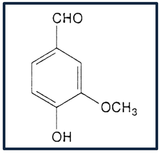
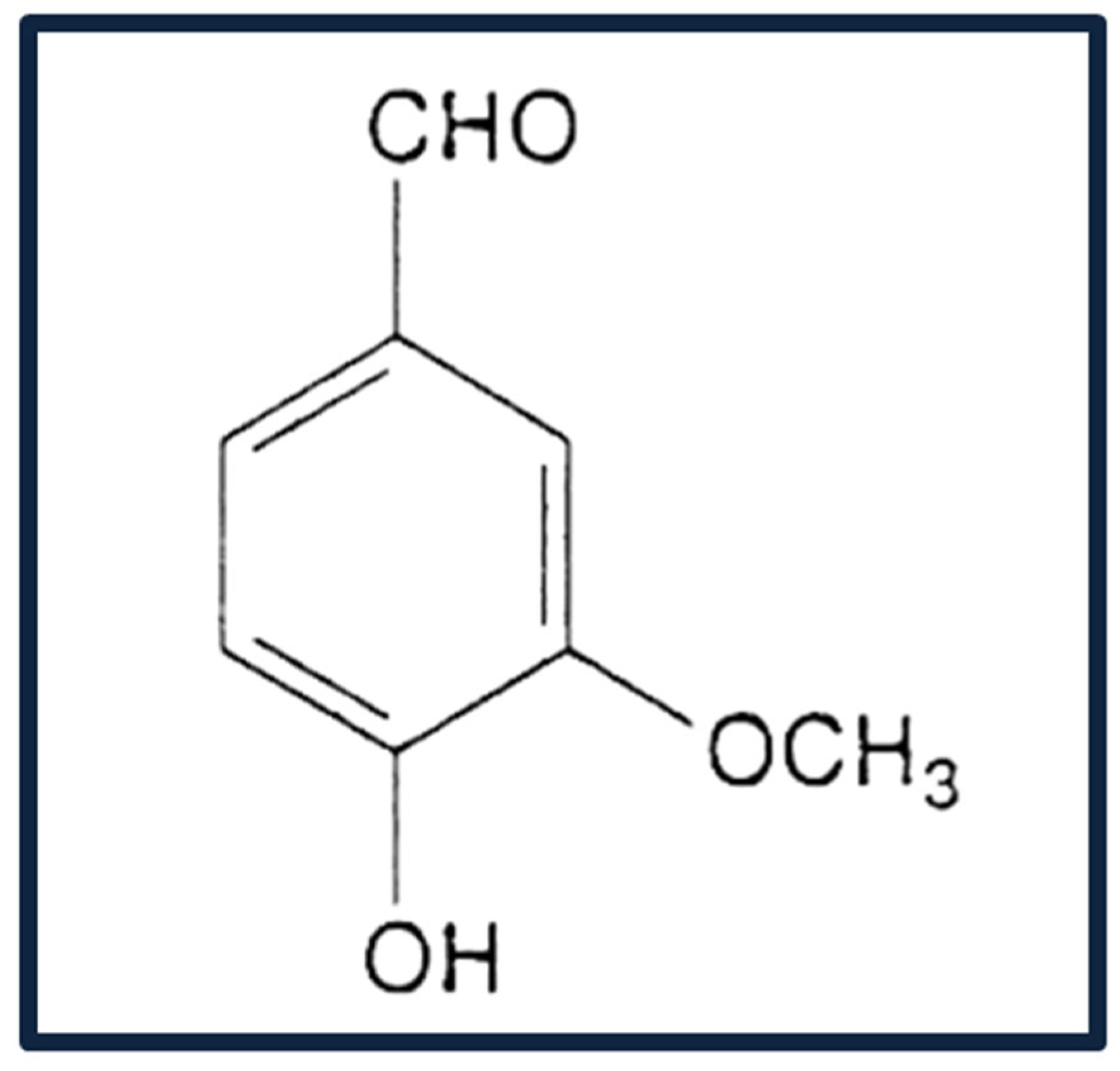

Disclaimer/Publisher’s Note: The statements, opinions and data contained in all publications are solely those of the individual author(s) and contributor(s) and not of MDPI and/or the editor(s). MDPI and/or the editor(s) disclaim responsibility for any injury to people or property resulting from any ideas, methods, instructions or products referred to in the content. |
© 2026 by the authors. Published by MDPI on behalf of the Romanian Society of Preventive Medicine. Licensee MDPI, Basel, Switzerland. This article is an open access article distributed under the terms and conditions of the Creative Commons Attribution (CC BY) license.