Review

Biopolymer-Based Adhesives for Biomedical and Industrial Use: Recent Advances, Challenges and Future Directions

by
Sumit Suryakant Kolte
,
Siddhi Sunil
,
Atharva Harinath Shastri
,
Vinayak Vijayan
and
Lihua Lou
*
NanoBio Mechanics and Manufacturing Laboratory (NBM2), Department of Mechanical Engineering, Clemson University, Clemson, SC 29634, USA
*
Author to whom correspondence should be addressed.
These authors contributed equally to this manuscript.
Adhesives 2026, 2(1), 3; https://doi.org/10.3390/adhesives2010003
Submission received: 13 November 2025 / Revised: 7 December 2025 / Accepted: 15 January 2026 / Published: 2 February 2026

Abstract

Biopolymer adhesives are moving toward frontline use in medicine and manufacturing as the limitations in some petrochemical systems, including cytotoxicity, challenges in wet adhesion for specific families of synthetic resins and formaldehyde emissions associated with amino-formaldehyde materials are becoming increasingly difficult to accept. This review integrates mechanisms, material classes and quantitative performance across biopolymer-based adhesives. We focus on architectures that combine permanent covalent anchoring with reversible, energy-dissipating bonds and on how functional group density, crosslink density, microstructure and additives act as design knobs for wet performance, durability and degradation. Across biomedical applications, chitosan, alginate, gelatin and related hydrogels achieve wet lap-shear strengths on the order of tens of kilopascals, cut liver-bleeding times by roughly half, provide strong antibacterial activity and close diabetic wounds by about 92 percent by day 14. Thermoresponsive alginate–gelatin sealants exceed clinically relevant burst pressures and microneedle patches withstand more than 120 mmHg while sealing arteries in under a minute. In industrial settings, dialdehyde-based starch resins deliver 0.83 to 1.05 MPa dry shear and maintain strength after water immersion while meeting stringent emission classes, and silane-modified nanocellulose in urea–formaldehyde markedly reduces free formaldehyde without sacrificing the internal bond. We conclude by identifying priorities for standardized wet testing, and lifetime matching of strength and degradation that can support large-scale clinical and industrial translation.

1. Introduction

The evolution of adhesive technology reflects a gradual shift from petrochemical convenience toward biological and environmentally aligned performance. While early biopolymer-based adhesives were motivated in part by the need to reduce dependence on petroleum resources and support more sustainable resources [1,2], their renewed development has also been driven by limitations in certain synthetic systems. Historically dominant systems offered a low cost and rapid setting but they can exhibit volatile organic emissions, formaldehyde release in amino-formaldehyde resins and brittle behavior in humid or biological environments [3,4,5]. At the same time, modern applications in surgery, dentistry, packaging and engineered wood increasingly demand strong adhesion under wet conditions, biocompatibility or recyclability, and end-of-life strategies that limit human and ecological toxicity [3,4,6,7]. In its broadest sense, an adhesive is a material that joins surfaces and develops sufficient internal strength, or cohesion, to transmit the load. Traditional families that meet this definition include reactive thermosets, hot-melts, pressure-sensitive adhesives and sealants [8]. Many of the most widely used products are based on cyanoacrylates, polyethylene glycol systems and amino-formaldehyde resins [3,4]. These synthetic platforms deliver high initial strength and a fast cure, and in several medical and industrial contexts, they remain dominantly important [3,4,5]. However, persistent limitations include poor or unreliable wet adhesion, cytotoxic or inflammatory degradation products, swelling or loss of integrity in physiological media, and formaldehyde or volatile organic compound emissions that drive regulatory and indoor air concerns [3,4,5,9].
Biopolymer-based adhesives have emerged in response to these challenges. They are built from polysaccharides such as starch, cellulose, chitosan and alginate, and from proteins such as gelatin and collagen, and they are often augmented by functional groups, including catechol and dynamic covalent bonds [10,11,12,13,14,15]. These matrices offer robust bonding, intrinsic biocompatibility and degradability, and in many cases, antimicrobial or antioxidant activity. Their development traces a trajectory from early protein and starch glues to contemporary catechol-modified and dynamically crosslinked hydrogels for tissue sealing and emission-controlling binders for engineered wood [4,6,10,11,12,13,14,15]. As a result, biopolymer adhesives have progressed from green alternatives to high-performance options in settings where conventional synthetics often fail. Existing reviews highlight several gaps that motivate the present review. Wet-condition tests are not yet standardized, which limits the comparison of lap-shear, peel and burst data across studies [7,16]. Strength and degradation profiles are not always matched to clinical or service lifetimes, and many promising laboratory systems rely on crosslinkers or additives that are difficult to scale under the current regulatory frameworks [3,4,6,7,16,17]. Life-cycle assessments and explicit regulatory roadmaps remain incomplete for most bio-based formulations [3,4]. In line with this focus, we primarily concentrate on natural polysaccharide and protein-based biopolymers that are engineered for adhesion in biomedical and industrial settings. Biobased thermoplastic polyesters such as PLA (polylactic acid) and other bulk bioplastics are discussed only where they directly function as adhesive matrices, as their literature is dominated by structural applications rather than interfacial adhesion [18,19].
Accordingly, this review outlines the adhesion principles that govern bonding, surveys the major biopolymer classes and their sources, and summarizes the chemical and physical strategies used to tune their properties. In addition, it compiles quantitative benchmarks for mechanical performance, adhesion tests, biodegradation and cytocompatibility, and discusses biomedical and industrial use-cases ranging from wound closure and dental repair to wood panels and packaging. The concluding sections identify crosslinking strategies, stimuli-responsive designs, low-toxicity chemistries and testing frameworks that are likely to enable the translation of biopolymer adhesives into routine clinical practice and large-scale industrial deployment.

2. Adhesion Principles of Biopolymer-Based Adhesives

In this review, we present the adhesion principles within an integrated framework to support later discussions, rather than providing an exhaustive examination of each principle.

2.1. Mechanisms of Adhesion

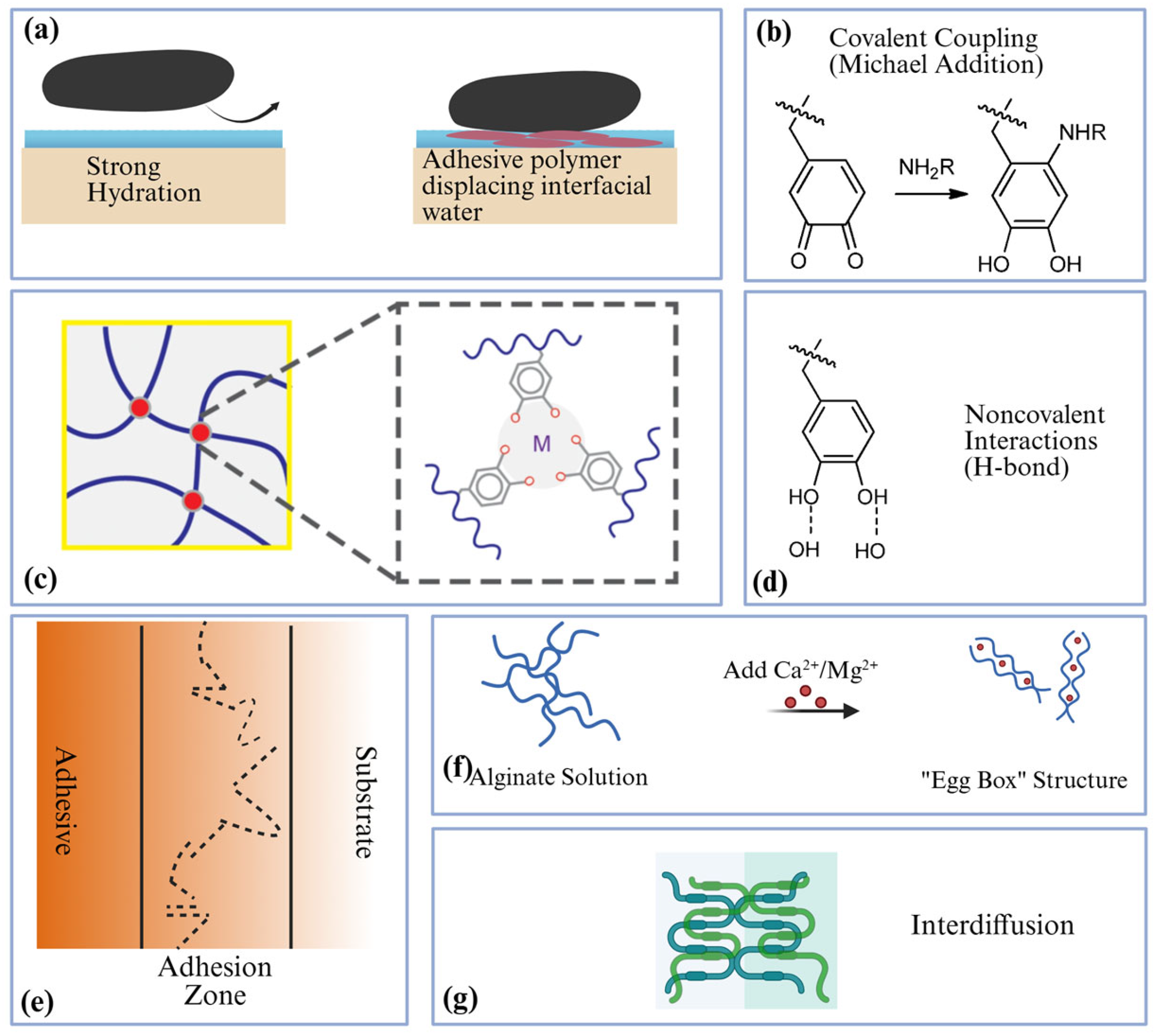
Adhesion arises from complementary chemical and physical mechanisms that are shared across synthetic and renewable adhesives (Figure 1a–g). The examples highlighted in this section are primarily based on biopolymers, but the underlying principles, like chemical bonding, physical interlocking, and polymer chain mobility, are general to all classes of adhesives. Chemical anchoring relies on several classes of interactions. Catechol groups oxidize to quinones that form covalent bonds with tissue nucleophiles, while Fe3+-catechol coordination creates reversible sacrificial bonds that contribute to toughness and self-healing [11,13,15]. Aldehyde-based polysaccharides and proteins react with amines through imine (Schiff-base) formation, and dense hydrogen bonding together with π π stacking further increases interfacial energy [11,13,15]. These features are illustrated in Figure 1b–d.
Physical strategies amplify and organize these chemistries at the interface. Microneedles fabricated from GelMA (gelatin methacrylate) generate mechanical interlocking [20], and tannic-acid coatings add polyphenol-mediated hydrogen bonding and π π stacking [10]. Suction-cup microtextures generate negative pressure on wet skin [21], whereas electrospun and cryogel matrices increase the real contact area and manage exudate to stabilize the interface [6,22,23,24]. These concepts are illustrated in Figure 1a,c,e. Another general mechanism of adhesion is interdiffusion, in which polymer chains from the adhesive and substrate penetrate across the interface to form entanglements [25]. This mechanism is observed in hydrogel–tissue interfaces [26] where chain mobility allows for interlocking (Figure 1g). Analogous principles extend to engineered wood and other structural materials. Esterification between citric acid and hydroxyl groups during hot pressing [27], aldehyde-amine and acetal formation in vanillin–chitosan systems [4], and silane–compatibilized nanocellulose in urea–formaldehyde resins [28] broaden the range of chemistries beyond petrochemical adhesives and, at the same time, reduce emissions. Figure 1a provides a simplified view of ionic crosslinking functional groups that are relevant to these systems.
Figure 1. Overview of key adhesion mechanisms in biopolymer-based adhesives, showing (a) interfacial dehydration and wetting control, (b) covalent coupling, (c) metal coordination, (d) noncovalent interactions, (e) mechanical interlocking, (f) ionic crosslinking, and (g) interdiffusion. (b,d) were reproduced from [29] under the Creative Commons Attribution (CC-BY) license, Copyright 2017, Journal of the American Chemical Society. (c) was reproduced from [30] under the Creative Commons Attribution (CC-BY) license, Copyright 2013, Scientific Reports (created with BioRender.com).
Adhesives 02 00003 g001

2.2. Functional Requirements for Biomedical and Industrial Adhesives

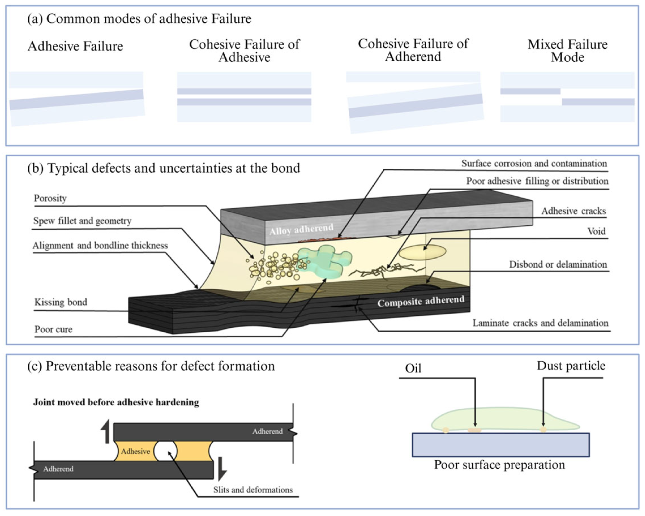
The functional requirements of bioadhesives are shaped by the ways joints can fail and by the defects that arise during fabrication, as illustrated in the common failure modes and bond defects in Figure 2a–c. Clinical use demands strong wet adhesion, reliable hemostasis on moving and bleeding tissue, cytocompatibility, controlled degradation, and increasingly therapeutic complementary effects. Recent hydrogels meet these criteria quantitatively. PVA (polyvinyl alcohol) and chitosan reinforced with bimetal–organic frameworks (MOFs) reduce rat liver-bleeding time by 20.1%, and achieve more than 99% antibacterial activity against Escherichia coli and Staphylococcus aureus, especially with high AgCu@MoF [31]. In diabetic rats, A bilirubin, β -cyclodextrin adhesive showed significantly higher wound closure (65%) when compared to an untreated group (18.7%), and couples sealing with antioxidant enhanced healing [24]. Microneedle patches with polyphenol interfaces withstand burst pressures above 120 mmHg and seal tissue, which is a clinically relevant threshold for suture-less hemorrhage control [20]. Thermoresponsive alginate and gelatin systems strengthened by catechol-Fe coordination and imine chemistry provide wet adhesion as high as 40 kPa, endure significant compressive strain with full recovery, exceed 360 mmHg burst pressure on the ex vivo intestine, and remain repositionable intraoperatively [13]. In the oral cavity, a GelMA/Gel-Phe (gelatin methacrylate/gelatin–phenylalanine derivative) adhesive cures within 60 s, with a compressive modulus ranging from 20 to 60 kPa depending on the curing time, and sustains a burst pressure of 11.3 ± 2.5 kPa, which is sufficient to withstand blood pressure during homeostasis [32].
Industrial applications impose a different but equally stringent set of requirements. Adhesives must maintain their internal bond and bending strength while limiting emissions and moisture sensitivity, and failure modes analogous to those in Figure 2a–c are often driven by processing defects. Oxidized starch formulation with a silane-based crosslinker achieves 7.88 MPa dry shear strength and 4.09 MPa wet shear strength, and Dialdehyde starch (DAS) is reported to have similar strength with E0-class emissions (very low formaldehyde emission classes) [34]. APTES ((3-Aminopropyl) triethoxysilane) nanocellulose reduces free formaldehyde by roughly 39% while maintaining internal bond strengths above 0.29 MPa [28]. Together, these examples show that biopolymer-based systems can meet or exceed conventional performance benchmarks in both biomedical and industrial settings.

2.3. Properties Influencing Adhesive Performance

Structure and property relationships provide practical levers for meeting the functional targets outlined above and for avoiding the defect-based failures depicted in Figure 2b,c. The density of functional groups—for example, oxidation in DAS or catechol substitutions per glucose unit—balances reactivity with viscosity and processability [11,34,35,36]. Crosslink density, which is set by the photo-initiator dose and UV (ultraviolet) exposure time in GelMA, by Fe3+ coordination in catechol systems, and by genipin or EDC/NHS (1-Ethyl-3-(3-dimethylaminopropyl) carbodiimide/N-Hydroxysuccinimide) chemistry in chitosan or gelatin networks, trades stiffness against conformability and atraumatic removal, while the molecular weight of Jeffamine in CMCS (carboxymethyl chitosan) platforms tunes both the modulus and swelling [6,13,15,16,37,38]. Architecture further modulates performance. Electrospun fiber diameters near 200 to 350 nm optimize porosity and swelling [39]; microneedle height, base width, and spacing control insertion mechanics and holding force [20]; suction cup diameter dictates pull-off forces on wet tissue [21]; and 1 µ m pores and high swelling enable rapid exudate uptake with an increase in adhesion when EDC/NHS bridges are introduced (Heart—670.9 ± 50.1 J/m2, Intestine—607.6 ± 30. J/m2) [40]. Additives modify both function and mechanics. MOFs raise release antibacterial ions [31]: Ag (silver) or ZnO (zinc oxide) nanoparticles kill more than 90–99% of bacteria [41], bilirubin complexes and tea polyphenols scavenge reactive oxygen species with more than 95% DPPH (2,2-Diphenyl-1-picrylhydrazyl) clearance [24,42,43,44,45], and conversion nanoparticles can enable near-infrared activated curing of adhesives via internal generation of visible emissions, improving the curing speed and tissue bonding compared to non-light-activated gauze. For example, one NIR-curable (near-infrared-curable) adhesive achieved significantly faster hemostasis than standard gauze in an in vivo model [46].

3. Categories of Biopolymer-Based Adhesives

3.1. Classification of Biopolymers

Biopolymer adhesives are primarily built from polysaccharides and proteins, which provide the bulk network upon which more specialized chemistries are layered. Polysaccharides such as starch, cellulose, chitosan and alginate supply abundant hydroxyl or amine groups and are readily available from agricultural or marine feedstocks [10,11,12,13,14,34]. Proteins such as gelatin and collagen contribute peptide backbones and bioactive functional groups that support cell attachment and remodeling [10,11,12,13,14,15,32]. In practice, these matrices are rarely used alone. They are often hybridized with polyphenols, including catechols and tannins, or with inorganic reinforcements in order to tune wet adhesion, toughness, and barrier properties [10,11,12,13,14,15]. Different sectors draw on different members of this palette. Starch and soy derivatives dominate paper converting and some wood products, where they can be crosslinked or blended with synthetic resins to reach the viscosity and tack needed on high-speed lines [34]. Chitosan, alginate, gelatin and collagen are more common in biomedical sealants and dressings because they are inherently biocompatible and degradable [10,11,12,13,14,32]. Recombinant collagen reduces the risk of pathogen transmission and batch variability that accompany animal-derived sources, and GelMA provides a photocrosslinkable collagen derivative that retains cell adhesion while supporting rapid in situ gelation [11,13,15,32]. Composite and hybrid systems illustrate the modularity of this biopolymer toolbox. Examples include chitosan–tannic and acid–shellac blends for packaging, polysaccharide and polyphenol microneedle platforms for transdermal sealing, and catechol-modified alginate or gelatin networks that couple bioactivity with strong wet adhesion [10,13,20,24].

3.2. Sources and Extraction Methods

The sourcing and processing of these biopolymers strongly influence both performance and sustainability. Shellac is refined from the resinous secretion of Kerria lacca and supplied as purified lac, which can be dissolved or reprecipitated into films and coatings [10]. Chitosan is obtained by alkaline deacetylation of chitin that is harvested from crustacean shells [10,11,12,13,14]. The degree of deacetylation directly governs density and intermolecular interactions, which collectively determines gelation and ultimately adhesive strength [47]. Alginate is extracted from brown seaweeds and separated into mannuronic-and guluronic-acid-rich fractions that differ in gelation behavior, which allows for tailoring of mechanical properties and ion responsiveness, making the mannuronic and guluronic ratio a key determinant of ion-based crosslinking and adhesion performance [10,11,12,13,14]. Gelatin is produced by the partial hydrolysis of collagen, and further methacrylation yields GelMA, a photocrosslinkable derivative that can be cast as prepolymers and cured in situ [11,13,15]. The extent of collagen hydrolysis affects the molecular weight distribution, viscosity, and the cohesive strength in the resulting adhesive [48]. Recombinant collagen routes avoid animal sourcing entirely and provide biosynthetic protein with controlled sequence and purity, which is valuable for clinical translation [11,13,32].

3.3. Property Modification Strategies

Once a base polysaccharide or protein is selected, chemical and physical modification strategies provide finer control over adhesion, mechanics and functionality. Polysaccharides are commonly grafted with dopamine or other phenolic groups to introduce catechols that support wet adhesion and metal coordination [10,11,12,13,14,15]. The oxidation of backbones, such as dextran, generates aldehydes that participate in Schiff-base chemistry with tissue or polymer amines, enabling curing at room temperature [11,13,15]. In industrial formulations, silane-based nanocellulose is dispersed into urea–formaldehyde matrices. The silane treatment enhances the compatibility between hydrophilic fibrils and the resin phase, contributing to emission control by scavenging free formaldehyde [27,28]. Chemically, catechol grafting is now a standard route to impart wet adhesion, underwater bonding and self-healing, because catechol units support both covalent reactions and reversible coordination with metals [10,11,13,15]. Oxidized polysaccharide–amine condensation, for example, oxidized dextran or starch reacting with gelatin or chitosan, enables curing at room or body temperature and can be adjusted to match the desired degradation window [11,13,15]. Vanillin-mediated Schiff-base crosslinking offers a low-toxicity alternative for chitosan networks and has been used to replace harsher aldehydes in industrial contexts [4]. Photopolymerization of GelMA and related methacrylated systems provides rapid in situ gelation with spatial control, which is advantageous for minimally invasive delivery and patterned adhesion [13,15,43]. Physical modifications complement these chemistries by introducing fillers and functional agents that alter the mechanical response and add new capabilities. Metal–organic frameworks and inorganic nanoparticles, such as Ag or ZnO, increase the modulus and deliver sustained antibacterial activity [31,41,49], while black-phosphorus or MoS2 nanosheets add photothermal responsiveness for heating or ablation [49,50]. Cyclodextrin complexes help solubilize and stabilize labile drugs such as bilirubin and integrate them into adhesive matrices without sacrificing cohesion [24,51]. In engineered wood, citric acid serves as a formaldehyde-free curing agent that esterifies hydroxyl groups during hot pressing [27]; lignosulfonates act as formaldehyde scavengers and binders with UF (urea–formaldehyde) resins [5]; and APTES functionalized nanocellulose improves stress transfer across the bond line and reduces emissions [28]. Together, these chemical and physical strategies define a flexible design space in which adhesion strength, degradation behavior and added functions can be tuned to match biomedical or industrial requirements.

4. Properties of Biopolymer-Based Adhesives

4.1. Mechanical Properties

Mechanical performance underpins both the handling and long-term function of biopolymer adhesives. For soft-tissue applications, moduli in the range of tens to hundreds of kilopascals are generally required, allowing the adhesive to deform with the surrounding tissue instead of concentrating stress. Alginate–gelatin systems exemplify this behavior. They tolerate high compressive strains with full recovery, which allows them to accommodate organ motion without permanent set [13]. Catechol–alginate and Fe3+ coordination networks are stiffer and show tensile elongations, which reflect a balance between covalent crosslinks and reversible metal coordination [13]. GelMA and Gel-Phe oral adhesives are designed to match the modulus of gingiva, reaching values near 60 kPa [32].
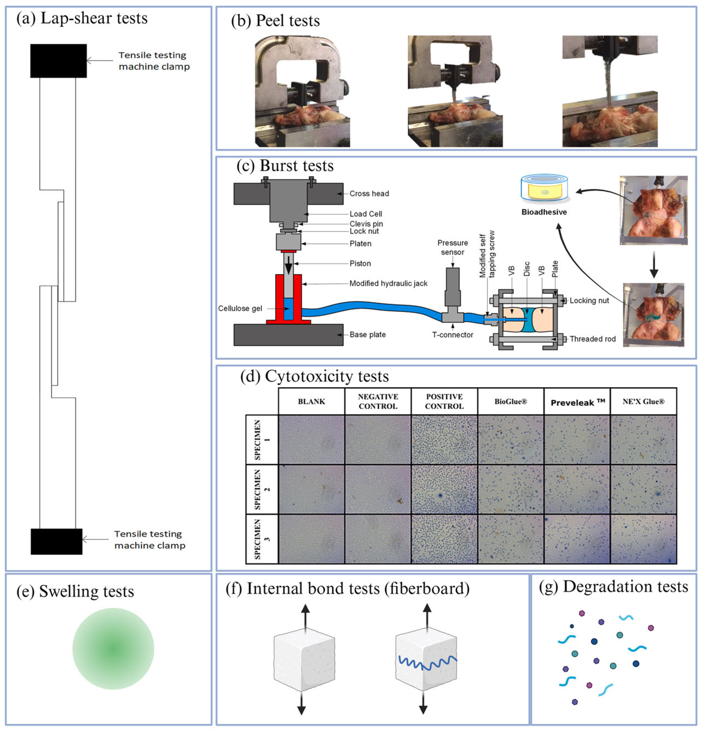
Adhesive strength is typically quantified through lap-shear tests of the type shown in Figure 3a. Chitosan-based hydrogels show good reversible adhesion properties, reaching lap-shear strengths of 24.31 ± 0.55 kPa on porcine skin [52]. This number is already comparable to or higher than several clinically used sealants. In industrial contexts, the mechanical targets shift to megapascal-scale shear. Dialdehyde-starch-modified resins achieved 1.19 to 1.36 MPa wet shear, which is sufficient to satisfy medium-density fiberboard specifications [4,34,53]. Packaging adhesives based on chitosan, tannic acid and shellac deliver tensile strength increases of 23 to 30%, relative to commercial references, which highlights that bio-based systems can also improve performance in load-bearing consumer applications [10].

4.2. Adhesion Tests

A variety of mechanical tests are used to probe adhesion under conditions that mimic use. Representative fixtures are summarized in Figure 3a–c,f. Wet lap-shear measurements on tissue or tissue analogs are the main metrics for soft tissue adhesives. They quantify the shear stress required to slide two adherends apart, and they are especially sensitive to surface preparation and interfacial chemistry. Peel and tack tests, illustrated in Figure 3b, complement lap-shear by emphasizing crack propagation and interfacial toughness rather than bulk deformation. These measurements are critical for skin contact adhesives and dressings, where gentle removal is as important as secure attachment. Burst tests assess the ability of an adhesive to seal defects in hollow organs against physiological pressures. In these experiments, a pressurized fluid is applied to a sealed perforation until failure, as shown schematically in Figure 3c. Thermoresponsive alginate–gelatin adhesives reinforced with catechol chemistry reach wet adhesion values of 40 kPa and withstand more than 360 mmHg in ex vivo intestinal burst tests, while remaining repositionable during surgery [13]. Microneedle patches with polyphenol-rich interfaces achieve around 480 mmHg and establish hemostatic seals, which is a clinically meaningful benchmark [20]. In dynamic crosslinked injectable mussel-inspired hydrogels, a higher degree of dopamine substitution caused a greater increase in adhesion strength, reaching values up to 21.41 kPa, which is around four times higher than in commercial fibrin glue [15].
In engineered wood and other fiberboard products, adhesion is characterized by internal bond and panel shear tests, rather than tissue assays. The EN 319 internal bond test, visually represented in Figure 3f, pulls a small block of board perpendicular to its plane and reports the stress at failure. Dialdehyde starch formulations and salinized nanocellulose UF systems maintain or exceed the standard thresholds in these tests while substantially lowering formaldehyde emissions [28,34,53]. These examples illustrate how test choice is tightly coupled to the targeted application and how biopolymer adhesives can satisfy very different mechanical and regulatory requirements.

4.3. Biodegradation and Cytocompatibility Assessment

Because bioadhesives often act as temporary scaffolds or sealants, their degradation and cytocompatibility profiles are as important as their mechanical properties. In vitro cytotoxicity is typically assessed by exposing fibroblasts, keratinocytes or other relevant cell types to extracts or direct contact, as illustrated in the micrographs in Figure 3d. For well-designed systems, cell viability routinely exceeds 85%, indicating that unreacted monomers and degradation products are not strongly harmful [56,57]. In vivo studies demonstrate that many formulations degrade on time scales that match the rate of tissue repair. The β -cyclodextrin-bilirubin hydrogel supports a 65% closure of diabetic mouse wounds, with high cell viability in the wound bed, suggesting that the adhesive provides both mechanical support and antioxidant protection during the critical early phase of healing [24]. Alginate–gelatin systems resorb completely in roughly three weeks, which aligns with their role as temporary sealants on soft organs [13]. Collagen–chitosan scaffolds degrade over about twenty-one days while maintaining more than 90% cell viability and promoting angiogenic repair in wound models [14,58,59]. Catechol-functional cellulose adhesives maintain fibroblast viability at approximately 96%, making them suitable for wound care and dressings [11]. Degradation tests, such as those schematized in Figure 3g, in conjunction with swelling assays, as shown in Figure 3e, provide a quantitative picture of how the network evolves in physiological environments.

4.4. Thermal and Chemical Stability Testing

Thermal and chemical stability determine whether biopolymer adhesives can survive processing, sterilization and long-term service. Thermogravimetric analysis and differential scanning calorimetry are routinely used to identify degradation onset temperatures and glass transitions. Chitosan, tannin and shellac blends show improved thermal stability compared with the individual components, which is important for packaging applications that experience elevated temperatures during sealing or storage [10]. Cellulose and nanocellulose exhibit degradation onset at temperatures near 280 °C, providing a comfortable margin for typical processing conditions [60]. Dialdehyde-starch-modified resins retain integrity during hot pressing of wood composites and reach E0 or E1 emission classes (very low or low formaldehyde emission classes) when combined with urea–formaldehyde resins [4,34]. Chemical stability is evaluated by immersing samples in relevant media, such as buffered saline, enzymatic solutions, or saliva, and tracking changes in mass and mechanical performance. Alginate–gelatin networks remain stable at a physiological pH over their intended service time and then gradually erode as ionic crosslinks exchange [13]. Janus oral hydrogel showed no degradation after exposure to protease-rich saliva, which is essential for dental and gingival applications [61]. In engineered wood, lignosulfonate binders delay the thermal decomposition of UF resins and trap formaldehyde [62,63]. However, the degree of substitution must be controlled because excessive lignosulfonate can increase moisture uptake and reduce long-term bond durability [5].

4.5. Incorporation of Agents Achieving Functional Properties

A distinctive advantage of biopolymer matrices is their ability to host functional agents without completely sacrificing adhesive performance. Metal–organic frameworks embedded in PVA or chitosan hydrogels deliver a sustained release of Zn2+ or Cu2+ ions while simultaneously providing structural support, which enhances both antibacterial activity and mechanical robustness [31]. Bilirubin encapsulated in β -cyclodextrin complexes can be dispersed uniformly within the bio-adhesive networks. This architecture provides antioxidant protection and a controlled release while maintaining strong adhesion [24]. Tea polyphenols and catechins introduce additional radical-scavenging and antimicrobial effects, and they can also participate in hydrogen bonding and π π interactions that reinforce the network [42,49]. In more structural contexts, small-molecule additives play a similar dual role. Vanillin acts as a benign crosslinker for chitosan, forming Schiff-base linkages that create robust networks with lower cytotoxicity than those formed with glutaraldehyde [4]. Related phenolic crosslinkers have been explored in both biomedical and wood-adhesive formulations, where they help to couple mechanical performance with improved safety profiles [10,42,43]. Together, these examples demonstrate that functionalization with bioactive or reinforcing agents can be designed to complement, rather than undermine, the primary adhesive task.

4.6. Self-Healing and Stimuli-Based Properties

Dynamic chemistries within biopolymer networks can endow adhesives with self-healing and stimulus-responsive behavior. In alginate–dopamine hydrogels, Fe3+-catechol coordination enables autonomous self-healing [64]. Thermogelling platforms that combine physical gelation with dynamic covalent bonds show immediate recovery of the storage modulus after large strain cycles, which allows them to withstand repetitive deformations without fatigue [6]. External stimuli can also be harnessed. Up conversion nanoparticle-doped systems respond to near-infrared light, which passes more deeply through tissue than ultraviolet light. When these particles convert NIR photons into higher energy emissions that trigger crosslinking, hemostasis times drop to around thirty-one seconds, as opposed to minutes for gauze [50,65]. In industrial settings, soy protein-based microcapsule concepts that release healing agents upon crack formation restore bond strength after damage, suggesting that self-repair concepts can also be translated beyond biomedical use [66,67].

5. Biomedical Applications for Biopolymer-Based Adhesives

5.1. Wound Closure and Tissue Sealing

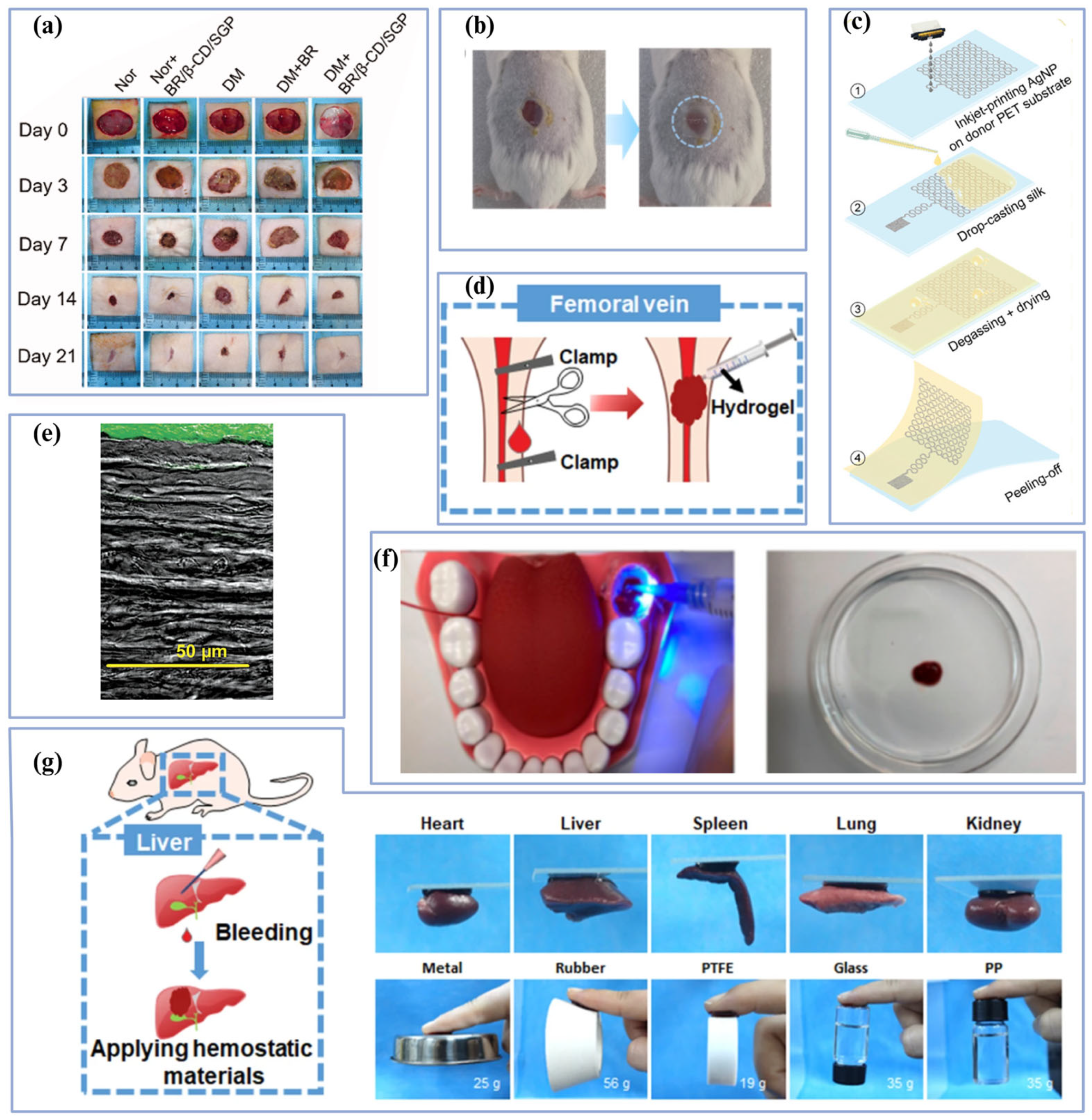
Across skin and organs, biopolymer adhesives now achieve sealing and healing outcomes that are competitive with, and, in some cases, superior to, sutures and staples. Representative wound models and organ sealing scenarios are shown in Figure 4a,b,d,g. Microneedle patches provide an immediate combination of mechanical interlocking and polyphenol-mediated bonding. These devices withstand burst pressures up to 480 mmHg and close tissue wounds [20].
Thermoresponsive alginate–gelatin adhesives reinforced with catechol chemistry further extend this capability. They can be repositioned during surgery. At 25 °C, the hydrogel can be easily peeled off, yet after curing at higher temperatures, it sustains burst pressures above 360 mmHg on ex vivo intestines and promotes faster wound closure with reduced scarring when compared with sutures or fibrin glues [13]. Such systems illustrate how dynamic covalent and coordination bonds can coexist with physical gelation to provide both handling flexibility and durable sealing. Bioadhesives can simultaneously serve as hemostats and therapeutic depots. PVA and chitosan hydrogels reinforced with metal–organic frameworks provide wet lap-shear strengths above 40 kPa on skin and significantly reduce bleeding in liver injury models [31]. Beyond its role in a reinforcing matrix, PVA itself has also been formulated into hydrogel adhesives (often combined with chitosan or alginate), where reversible hydrogen bonding produce wet adhesion in the range of 20 to 40 kPa [70]. Hyaluronic acid (HA)-based hydrogels represent another important family of biopolymer adhesives. HA modified using catechol, aldehyde, and photocrosslinking have been developed as injectable tissue glues with strong adhesion [71,72]. HA formulations have shown a burst pressure that exceeds those of conventional sealants, while remaining degradable and biocompatible [73]. Bilirubin/ β -cyclodextrin formulations achieve 65% closure, demonstrating that mechanical support and antioxidant-driven tissue repair can be integrated in a single platform [24]. Another practical route is collagen–chitosan scaffolds. Studies show that they accelerate re-epithelialization and stimulate neovascularization, and in full-thickness wound models, they can approach complete closure within about two weeks [59,74]. Together, these examples show that biopolymer adhesives can be tailored for acute hemostasis and long-term wound remodeling.

5.2. Dental and Orthopedic Applications

Dental and orthopedic environments present a different set of constraints, including constant exposure to saliva and repetitive mechanical loading. The applications shown in Figure 4f highlight how these challenges are being addressed. In the oral cavity, a GelMA and Gel-Phe system cures within 60 s under light exposure, matches gingival tissue mechanics and maintains adhesive strength in saliva [51]. In orthopedic contexts, higher modules and stable wet adhesion are required. Catechol-reinforced hydrogel networks achieve a mechanical strength of 0.2 MPa while maintaining robust wet adhesion, making them promising for cartilage interfaces where synovial fluid and compliance mismatch render conventional sealants less effective. For example, a catechol–chitosan–PEG dynamic hydrogel shows a compressive modulus of around 195 kPa and strong tissue adhesion [75], and a catechol-derivative adhesive achieves around 153 kPa lap-shear on wet porcine skin with high interfacial toughness [76]. At the tooth–restoration interface, performance can be boosted by pairing protease inhibitors with remineralizing fillers, matrix-metalloproteinase inhibitors such as chlorhexidine curb collagenolysis in the hybrid layer and improve aged resin and dentin bond strength [77], while calcium phosphate releasing additives promote apatite deposition, help stabilize bonds over months, and may lower the risk of secondary caries [78].

5.3. Drug Delivery

Many modern bioadhesives are designed to function as both mechanical sealants and local drug delivery depots, as illustrated by the wound and vascular applications in Figure 4a,d,g. Bioadhesives are also used to coat damaged cells for imaging, as shown in Figure 4e. Bilirubin/ β -cyclodextrin bioadhesives, for example, maintain controlled release of antioxidant bilirubin for about seven days while simultaneously providing strong tissue adhesion [24]. This sustained release reduces oxidative stress in the wound bed, contributing to improved healing outcomes in diabetic models. Thermogelling and near-infrared-triggered adhesives extend this concept by combining structural support with antimicrobial therapy. Systems that deliver antibiotics can reduce bacterial burden at the wound during the same period in which they provide sealing [79,80]. Beyond surface applications, biodegradable occluder platforms whose surfaces are modified with bioadhesive chemistries regulate the local immune response and accelerate endothelialization without compromising mechanical integrity [12]. These examples show that the adhesive interface can be engineered to stabilize implants and, at the same time, guide tissue remodeling and drug release.

5.4. Bioelectronics

Biopolymer adhesives are also emerging as critical components in soft bioelectronics. The devices and tissue interfaces in Figure 4c illustrate this direction. Additively manufactured bioadhesives that embed conductive nanoparticles or polymeric conductors can serve as soft, conformal substrates for biosensing and localized therapy [38]. Shear-thinning bioinks formulated from alginate, gelatin or chitosan print through narrow nozzles adhere to wet tissue or device surfaces and then cure on demand, enabling patterning of electrodes and interconnects in austere or resource-limited settings [38]. Because these matrices are inherently biocompatible and often biodegradable, they can provide temporary yet stable electrical interfaces that couple to tissue without the mechanical mismatch associated with rigid electronics.

6. Industrial Applications for Biopolymer-Based Adhesives

6.1. Wood and Paper Industry

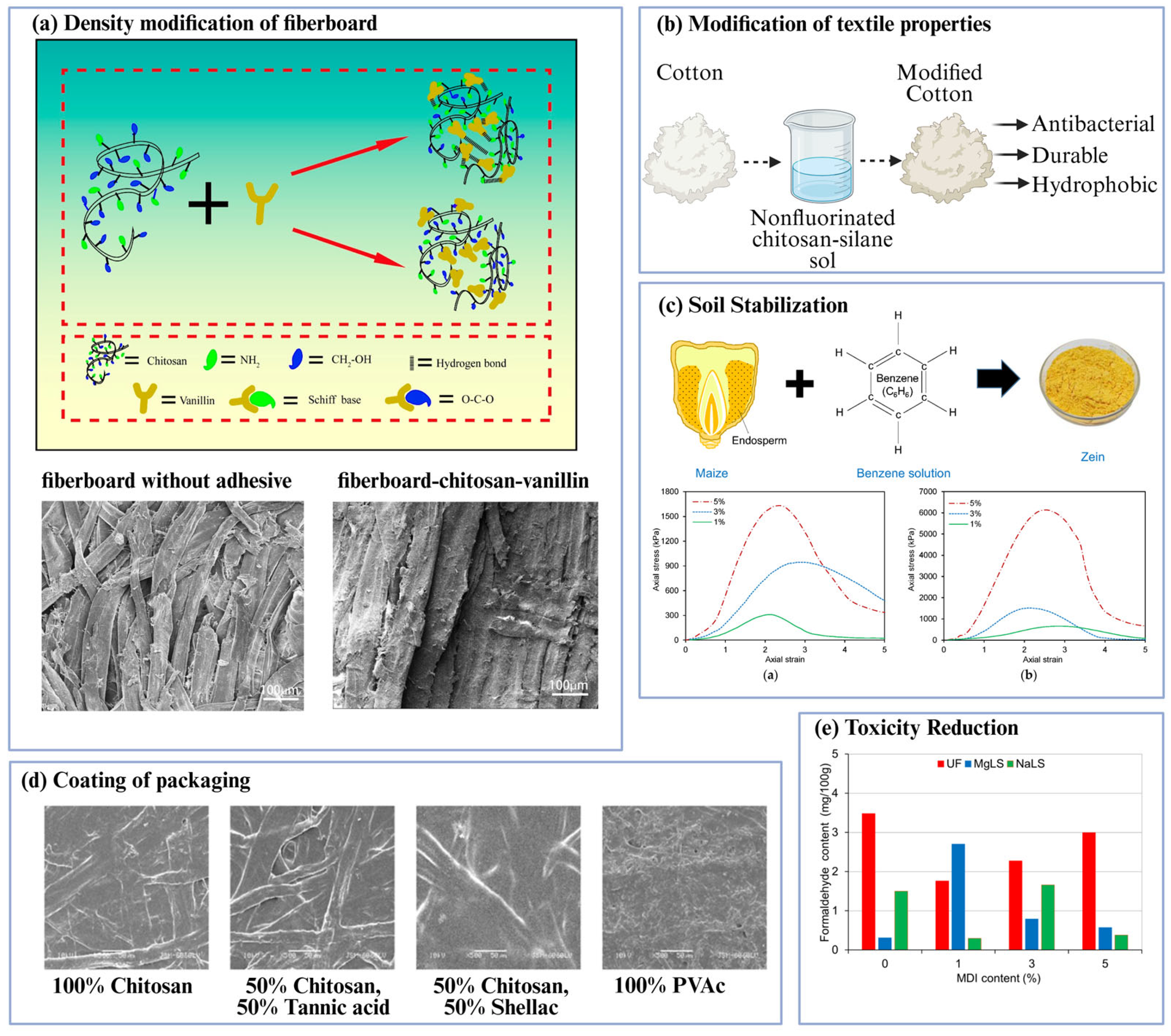
The wood and paper sectors have been early adopters of biopolymer-based adhesives because they face strong regulatory pressure to reduce formaldehyde emissions while preserving panel strength and durability. Figure 5a,e illustrate how these systems can both reinforce fiberboard and lower toxicity. In emission-controlled urea–formaldehyde resins, lignosulfonates act as binders and formaldehyde scavengers so that panels reach E0 or E1 emission classes while maintaining an acceptable internal bond strength [5]. Silane-modified nanocellulose disperses into UF matrices and improves compatibility between the hydrophilic fibrils and the resin phase. This approach maintains the internal bond and bending strength while contributing to reductions in free formaldehyde [28]. Formaldehyde-free systems based on citric acid curing offer an alternative route. Citric acid esterifies wood hydroxyl groups under hot pressing and yields molded products that no longer rely on formaldehyde chemistry [27]. Dialdehyde-starch-modified resins reach roughly MPa-level lap-shear strengths in plywood under both dry and 24 h cold water conditions (0.8 to 1.1 MPa dry and 0.6 to 1.0 MPa wet are typical in DAS systems) [53], and those magnitudes meet or exceed MDF performance benchmarks when compared to the EN 622-5 internal bond requirements. When substitution level, crosslink density and moisture management are carefully tuned, these formulations can reduce free formaldehyde by up to roughly 90% compared with conventional UF systems without compromising mechanical performance [4,5,27,28,34]. While most biopolymer-based adhesives are replacements for UF panels, it is useful to benchmark them against other adhesive families used in wood engineering. Commercial polyurethane wood adhesives typically provide dry lap-shear strength on the order of 7 to 25 Mpa, together with excellent water resistance and durability [81], and structural epoxy formulations can reach similar or higher shear strengths on wood and other substrates [82].

6.2. Packaging and the Food Industry

Biopolymer adhesives are also attractive for packaging, where direct or indirect food contact drives demand for safer chemicals. The coating micrographs in Figure 5d show how chitosan-based blends modify the surface morphology compared with conventional polyvinyl acetate films. Chitosan, tannic acid and shellac form hybrid networks in which chitosan provides film formation and cationic charge, tannic acid supplies additional hydrogen bonding and antimicrobial activity, and shellac contributes hydrophobicity and barrier properties [10,85]. These blends exhibit higher viscosity and stronger adhesion than neat chitosan. In tensile testing, they deliver gains of 23 to 30%, relative to commercial references, and reach peel strengths of 7 N to 9.5 N while improving water resistance [10]. Because they rely on renewable components and avoid toxic crosslinkers, such systems offer promising routes for paper and cardboard seams, inner coatings and other packaging applications that require both mechanical robustness and benign composition.

6.3. Textile and Processing in Other Industries

Beyond wood and packaging, biopolymer adhesives are starting to impact textiles, civil engineering and energy technologies. Figure 5b highlights a non-fluorinated chitosan–silane sol that is used to modify cotton. The resulting fabrics become more hydrophobic, durable and antibacterial, yet remain breathable and free from polyfluoroalkyl substances [83]. These coatings rely on quaternary chitosan–silane networks formed via a sol–gel process, which creates strong interfacial bonding with the cotton substrate and maintains performance over repeated washing and wettability cycles, although standardized test methods for such soft goods are still emerging [83]. In structural composites, bamboo-derived lignin–cellulose biocomposites and pectin-residue nanofibrils blended with polymeric methylene diphenyl diisocyanate (pMDI) demonstrate that bio-based binders can be integrated into high-temperature and high-load processes. These materials improve flame retardancy and dimensional stability while reducing reliance on petrochemical binders and halogenated flame retardants [5,86,87]. More broadly, lignin has gained increasing attention as a renewable adhesive precursor, where chemical modification enables its inclusion into phenol–formaldehyde or polyurethane networks, providing reduced petrochemical content while maintaining mechanical strength and resistance to moisture [88,89]. Binders that crosslink in situ and adhere under wet conditions are being explored for dust suppression and soil stabilization, as shown in Figure 5c [84]. Their degradability and low toxicity echo the advantages that are already exploited in biomedical applications, yet systematic performance baselines and long-term field data are still limited [84]. Printed hydrogels that encapsulate conductive nanoparticles extend these ideas into soft energy and sensing interfaces. When formulated from alginate, GelMA or chitosan bioinks, these matrices can be extruded or inkjet-printed onto complex three-dimensional geometries, where they cure on demand and maintain intimate contact with substrates [38]. Such systems can be utilized in biomedical and industrial applications, as the same printable, multifunctional adhesives can serve as tissue-conformal electrodes, corrosion monitoring coatings, or flexible interconnects in soft robotics.

7. Challenges and Future Perspective

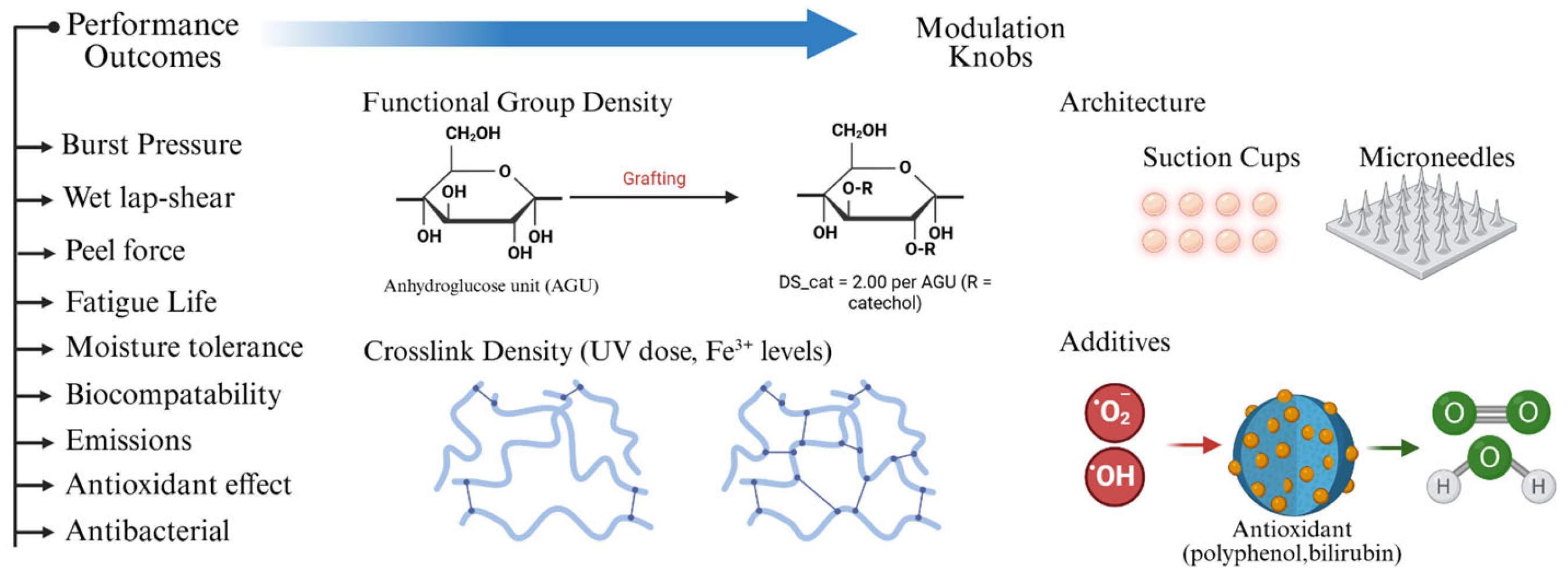
Life-cycle assessment and regulatory alignment are now as important to adhesive selection as modulus or lap-shear strength. Beyond local performance, biopolymer systems will increasingly be judged on their carbon footprint, end-of-life fate and worker exposure profiles [3,5]. Moisture sensitivity and limited durability persist when substitution ratios and crosslink density are not optimized for the service environment. These issues are particularly evident in lignosulfonate-modified UF panels, where excessive substitution can compromise the bond stability under humid conditions [5,90]. Dynamic bioadhesive networks can also soften or creep under repeated loading if the distribution of bond lifetimes is not matched to the time scale of use [15,50]. These observations underscore the need for standardized, application-relevant test batteries. For biomedical systems, this means wet lap-shear, peel and burst assays performed under physiologic media and loading, whereas for wood and composite panels, EN-series methods combined with accelerated humidity cycling and temperature excursions should become routine [3,4,5]. Future progress will depend on a small number of design knobs that translate the molecular structure into measurable performance outcomes. This relationship is summarized in Figure 6, which links functional group density, crosslink density, architecture and additives to outcomes such as burst pressure, wet lap-shear strength, peel force, fatigue life, moisture tolerance, biocompatibility, emissions, antioxidant effect and antibacterial activity. Representative quantitative benchmarks for both biomedical and industrial systems are compiled in Table 1 and highlight the range of performance already achieved by hydrogels, the bilirubin/ β -cyclodextrin bio-adhesive, microneedle patches, thermoresponsive alginate–gelatin sealants, mussel-inspired dynamic covalent networks and advanced wood and packaging adhesives.
Three strategic directions appear to be particularly promising for transitioning biopolymer adhesives from laboratory demonstrations to large-scale clinical and industrial applications. The first is multimodal crosslinking that combines permanent covalent bonds with reversible sacrificial interactions. Catechol-based systems that pair oxidation-driven covalent tethering with Fe3+ coordination and oxidized polysaccharide networks that mix imine bonds with secondary hydrogen bonding illustrate how such combinations can preserve wet strength while still allowing for controlled debonding or self-healing [15,91]. The second direction is the use of benign stimuli to trigger curing or softening on demand. Near-infrared light, pH, and temperature have all been used to activate crosslinking or to switch mechanical properties, as in NIR-responsive hemostatic platforms and thermogelling matrices that are set at body temperature [50,92]. The third direction is the systematic development of low-toxicity chemistries that remain compatible with existing regulatory pathways and manufacturing supply chains. Vanillin-crosslinked chitosan, oxidized dextran–gelatin adhesives, citric-acid esterification in wood products and silane–nanocellulose compatibilization of UF resins demonstrate that legacy aldehydes and isocyanates can be replaced while retaining competitive mechanical performance and meeting stringent emission limits [4,27,28,43]. If these design levers are coupled with transparent life-cycle data and harmonized testing standards, biopolymer-based adhesives are well-positioned to move from niche applications to mainstream use across medicine, packaging, construction and soft electronics [3,5,6,7,91].
One limitation of this review is that we present the adhesion principles within an integrated framework, rather than providing an exhaustive examination of each principle. Another key limitation of this review is its simultaneous coverage of biomedical and industrial adhesives, rather than a focused examination of a single application area.
Table 1. Representative quantitative benchmarks for both biomedical and industrial systems.
Adhesive System Application and Test ContextKey Quantitative Performance Reference
PVA, chitosan and bimetal organic framework hydrogelWound closure and liver hemostasis. Rat liver bleeding model.Reduces rat liver bleeding time by 20.1% shorter vs. control. Antibacterial activity greater than 99% vs. E. coli and S. aureus[31]
Bilirubin, β-cyclodextrin hydrogelDiabetic wound closure. Full-thickness wounds in diabetic mice.Significantly higher wound closure (65%) when compared to an untreated group (18.7%). Couples sealing with antioxidant-enhanced healing[24]
Microneedle patch Burst test for tissue sealing.Burst pressure is around 480 mmHg in Carbopol/chitosan.[20]
Thermoresponsive alginate–gelatin, catechol-Fe coordination, imine chemistryGastrointestinal and intestinal sealing. Ex vivo intestine.Wet adhesion is as high as 40 kPa. Endure significant compressive strain with full recovery. Exceed 360 mmHg burst pressure on ex vivo intestine.[13]
Mussel-inspired, dynamic crosslinked bioadhesivesWet lap-shear and strain tests.Wet lap-shear 21.4 kPa. Rapid self-healing and modulus recovery after strain cycles. The maximum strain the hydrogels could bear without system damage was 600%.[15]
GelMA, Gel-Phe dental adhesiveOral, gingival hemostasis and repair. Burst-pressure test.Cures within 60 s. Compressive modules between 20 and 60 kPa, based on the curing time, sustained a burst pressure of 11.3 ± 2.5 kPa.[32]
Cryogel architectures with EDC/NHS bridgingSwelling and adhesion tests.Pore size of 50 to 120 µm. Swelling around 900% in phosphate-buffered saline (PBS). Introduction of EDC/NHS bridges yields around an increase of 3 times in underwater adhesion vs. unmodified cryogels.[6]
Catechol-alginate, Fe3+ hydrogelsSelf-healing tests.Recovered to the original state immediately, once the strain was reduced to 1% after being subjected to a high strain (up to 400%).[13]
Thermogelling or NIR-activated hemostatic platformsHemostatic gels with light or thermal triggers.Hemostasis time shortened from 100 s with gauze to 31 s with adhesive.[50]
General clinical outcome benchmarks (biopolymer sealants)Wound models.Wound closure is commonly around 80 to 90% by days 10 to 14 in challenging models when adhesive is combined with bioactivity. [93]
Oxidized starch with silane crosslinkers, Dialdehyde-starch-modified UF resinParticleboard, shear tests and EN standards.Dry shear around 7.88 MPa. Wet shear around 4.09 MPa. E0-class emissions.[34]
APTES nanocellulose in UFParticleboard and strawboard. Internal bond (EN 319).Free formaldehyde was reduced by around 39%. Internal bond (IB) maintained greater than 0.29 Mpa.[28]
Lignosulfonate UF Engineered wood panels.Formaldehyde emissions reduced by up to 91%. pMDI blends reach super-E0 levels. Mechanical properties are maintained when crosslinking and substitution ratios are optimized.[5]
Chitosan, tannic acid, shellac blendsPaper and cardboard packaging. Tensile and peel tests.Tensile strength increased by 30% compared to the commercial reference for chitosan–tannic acid. Increased by 23% for shellac–chitosan. Peel strength 7 N to 9.5 N. Improved water resistance from shellac domains.[10]
General industrial benchmarks for bio-based wood adhesivesParticleboard, EN 319 internal bond, EN 317 water soakMeet or exceed IB and bend requirements while cutting emissions to E0 or E1 and sometimes super-E0. Water immersion shear retention is maintained with proper crosslinking.[4,27,28,34,41]

8. Conclusions

Cumulative evidence suggests that biopolymer-based adhesives can deliver strong wet adhesion with clinically and industrially relevant performance, while also offering functions that conventional synthetic systems rarely provide. These materials combine sealing with antibacterial, antioxidant and angiogenic activity, enabling repositionable curing on tissue, and scavenge formaldehyde or other volatile components in wood composites. They seal wounds in less than a minute, achieve more than 90% wound closure by days 10 to 14 in difficult-to-heal models, match gingival mechanics while maintaining adhesion in saliva, and meet engineered-wood strength targets while reaching E0 or E1 emission classes and reducing free formaldehyde by up to roughly 91%. The near-term agenda is to consolidate these gains into robust practice. Standardized wet-condition tests are necessary to enable the comparison of lap-shear, peel, burst, and fatigue data across laboratories and to translate these results into regulatory language. Degradation must be matched to the intended lifetime, using adhesives that either detach atraumatically or resorb in synchrony with tissue repair or the panel’s service life. At the same time, low-toxicity crosslinkers, bio-derived monomers, and renewable feedstocks, such as oxidized polysaccharides, vanillin, citric acid and lignosulfonate, need to be scaled and integrated into existing manufacturing lines. With these elements in place, biopolymer adhesives are poised to become default choices wherever wet adhesion, safety and end-of-life impact are not negotiable, enabling product classes that range from saliva-stable oral hemostats to E0-grade structural panels.

Author Contributions

Conceptualization, L.L.; methodology, L.L. and V.V.; validation, L.L. and V.V.; formal analysis, S.S., A.H.S. and S.S.K.; investigation, S.S., A.H.S. and S.S.K.; data curation, S.S., A.H.S. and S.S.K.; writing—original draft preparation, L.L., V.V., S.S., A.H.S. and S.S.K.; writing—review and editing, L.L., V.V., S.S., A.H.S. and S.S.K.; visualization, L.L., V.V., S.S., A.H.S. and S.S.K.; supervision, L.L. All authors have read and agreed to the published version of the manuscript.

Funding

This research received no external funding.

Data Availability Statement

All data discussed in this review are derived from previously published studies. No new data were generated.

Acknowledgments

L.L. would like to acknowledge startup funds and support from the Creative Inquiry project at Clemson University.

Conflicts of Interest

The authors declare no conflicts of interest.

Abbreviations

The following abbreviations are used in this manuscript:
AgSilver (Argentum)
APTES(3-Aminopropyl)triethoxysilane
CMCSCarboxymethyl chitosan
DASDialdehyde starch
DPPH2,2-Diphenyl-1-picrylhydrazyl
E0, E1Very low, low formaldehyde emission classes
EDC1-Ethyl-3-(3-dimethylaminopropyl)carbodiimide
GelMAGelatin methacrylate
Gel-PheGelatin–phenylalanine derivative
MOFMetal–organic framework
NHSN-Hydroxysuccinimide
NIRNear-infrared
PBSPhosphate-buffered saline
PLAPolylactic acid
PVAPolyvinyl alcohol
UFUrea–formaldehyde
UVUltraviolet
ZnOZinc oxide
pMDIPolymeric methylene diphenyl diisocyanate

References

  1. Alexandra Heinrich, L. Future opportunities for bio-based adhesives–advantages beyond renewability. Green Chem. 2019, 21, 1866–1888. [Google Scholar] [CrossRef]
  2. Díaz López, F.J.; Montalvo, C. A comprehensive review of the evolving and cumulative nature of eco-innovation in the chemical industry. J. Clean. Prod. 2015, 102, 30–43. [Google Scholar] [CrossRef]
  3. Yadav, D.; Giri, P.; Das, C. Polymer-based biomaterials and their applications in tissue adhesives. J. Adhes. Sci. Technol. 2024, 38, 2019–2046. [Google Scholar] [CrossRef]
  4. Cao, Y.; Qin, C.; Zhao, Z.; Wang, Z.; Jin, C. Preparation and Properties of Medium-Density Fiberboards Bonded with Vanillin Crosslinked Chitosan. Polymers 2023, 15, 2509. [Google Scholar] [CrossRef] [PubMed]
  5. Bekhta, P.; Noshchenko, G.; Réh, R.; Kristak, L.; Sedliačik, J.; Antov, P.; Mirski, R.; Savov, V. Properties of Eco-Friendly Particleboards Bonded with Lignosulfonate-Urea-Formaldehyde Adhesives and pMDI as a Crosslinker. Materials 2021, 14, 4875. [Google Scholar] [CrossRef]
  6. Liu, C.; Liu, J.; Wu, M.; Ni, H.; Feng, J.; Zhao, L.; Zhang, J. Cryogel wound dressings based on natural polysaccharides perfectly adhere to irregular wounds for rapid haemostasis and easy disassembly. Wound Repair. Regen. 2024, 32, 393–406. [Google Scholar] [CrossRef]
  7. Fronza, B.M.; Braga, R.R.; Cadenaro, M. Dental Adhesives—Surface Modifications of Dentin Structure for Stable Bonding. Dent. Clin. N. Am. 2022, 66, 503–515. [Google Scholar] [CrossRef]
  8. Marques, A.C.; Mocanu, A.; Tomić, N.Z.; Balos, S.; Stammen, E.; Lundevall, A.; Abrahami, S.T.; Günther, R.; de Kok, J.M.M.; Teixeira de Freitas, S. Review on Adhesives and Surface Treatments for Structural Applications: Recent Developments on Sustainability and Implementation for Metal and Composite Substrates. Materials 2020, 13, 5590. [Google Scholar] [CrossRef]
  9. Slezak, P.; Klang, A.; Ferguson, J.; Monforte, X.; Schmidt, P.; Bauder, B.; Url, A.; Osuchowski, M.; Redl, H.; Spazierer, D.; et al. Tissue reactions to polyethylene glycol and glutaraldehyde-based surgical sealants in a rabbit aorta model. J. Biomater. Appl. 2020, 34, 1330–1340. [Google Scholar] [CrossRef]
  10. Vrabič-Brodnjak, U. Bio-Based Adhesives Formulated from Tannic Acid, Chitosan, and Shellac for Packaging Materials. Polymers 2023, 15, 1302. [Google Scholar] [CrossRef]
  11. Tang, Z.; Bian, S.; Lin, Z.; Xiao, H.; Zhang, M.; Liu, K.; Li, X.; Du, B.; Huang, L.; Chen, L.; et al. Biocompatible Catechol-Functionalized Cellulose-Based Adhesives with Strong Water Resistance. Macro Mater. Eng. 2021, 306, 2100232. [Google Scholar] [CrossRef]
  12. Kong, P.; Liu, X.; Li, Z.; Wang, J.; Gao, R.; Feng, S.; Li, H.; Zhang, F.; Feng, Z.; Huang, P.; et al. Biodegradable Cardiac Occluder with Surface Modification by Gelatin–Peptide Conjugate to Promote Endogenous Tissue Regeneration. Adv. Sci. 2024, 11, 2305967. [Google Scholar] [CrossRef] [PubMed]
  13. Liang, Y.; Xu, H.; Li, Z.; Zhangji, A.; Guo, B. Bioinspired Injectable Self-Healing Hydrogel Sealant with Fault-Tolerant and Repeated Thermo-Responsive Adhesion for Sutureless Post-Wound-Closure and Wound Healing. Nano-Micro Lett. 2022, 14, 185. [Google Scholar] [CrossRef] [PubMed]
  14. Kang, D.; Wang, W.; Li, Y.; Ma, Y.; Huang, Y.; Wang, J. Biological Macromolecule Hydrogel Based on Recombinant Type I Collagen/Chitosan Scaffold to Accelerate Full-Thickness Healing of Skin Wounds. Polymers 2023, 15, 3919. [Google Scholar] [CrossRef]
  15. Wang, R.; Liu, L.; He, X.; Xia, Z.; Zhao, Z.; Xi, Z.; Yu, J.; Wang, J. Dynamic Crosslinked Injectable Mussel-Inspired Hydrogels with Adhesive, Self-Healing, and Biodegradation Properties. Polymers 2023, 15, 1876. [Google Scholar] [CrossRef]
  16. Baran, E.T. Functionalized Carboxymethyl Chitosan Derivatives in Wound Healing. In Multifaceted Carboxymethyl Chitosan Derivatives: Properties and Biomedical Applications; Jayakumar, R., Ed.; Advances in Polymer Science; Springer Nature: Cham, Switzerland, 2023; Volume 292, pp. 281–311. ISBN 978-3-031-44099-1. [Google Scholar]
  17. Radwan-Pragłowska, J.; Janus, Ł.; Piątkowski, M.; Sierakowska, A.; Galek, T.; Szajna, E.; Bogdał, D.; Tupaj, M. Fungal Chitosan-Derived Biomaterials Modified with Kalanchoe pinnata as Potential Hemostatic Agents—Development and Characterization. Polymers 2021, 13, 1300. [Google Scholar] [CrossRef]
  18. Saini, T.; Meena, J.; Verma, V.; Saini, S.; Malik, R. Polyvinyl Alcohol: Recent Advances and Applications in Sustainable Materials. Polym. -Plast. Technol. Mater. 2025, 64, 794–825. [Google Scholar] [CrossRef]
  19. Lee, D.H.; Song, Y.H.; Ahn, H.J.; Lee, J.; Woo, H.C.; Lee, D.H.; Song, Y.H.; Ahn, H.J.; Lee, J.; Woo, H.C. Fabrication and Characterization of Biopolymers Using Polyvinyl Alcohol and Cardanol-Based Polyols. Molecules 2024, 29, 4807. [Google Scholar] [CrossRef]
  20. Freundlich, E.; Shimony, N.; Gross, A.; Mizrahi, B. Bioadhesive microneedle patches for tissue sealing. Bioeng. Transl. Med. 2024, 9, e10578. [Google Scholar] [CrossRef]
  21. Huang, R.; Zhang, X.; Li, W.; Shang, L.; Wang, H.; Zhao, Y. Suction Cups-Inspired Adhesive Patch with Tailorable Patterns for Versatile Wound Healing. Adv. Sci. 2021, 8, 2100201. [Google Scholar] [CrossRef]
  22. Matei, E.; Gaidau, C.; Râpă, M.; Constantinescu, R.; Savin, S.; Berechet, M.D.; Predescu, A.M.; Berbecaru, A.C.; Coman, G.; Predescu, C. Sustainable Rabbit Skin Glue to Produce Bioactive Nanofibers for Nonactive Wound Dressings. Materials 2020, 13, 5388. [Google Scholar] [CrossRef] [PubMed]
  23. Sosiati, H.; Rahman, A.; Sahputra, K.K.; Ibnul Fatoni, M.F. Effects of Aloe Vera/Chitosan Ratios on the Tensile Properties of Aloe Vera/Chitosan/Polyvinyl Alcohol Nanofibrous Membranes for Wound Care Dressing. Makara J. Sci. 2025, 29, 10. [Google Scholar] [CrossRef]
  24. Yao, Q.; Shi, Y.; Xia, X.; Tang, Y.; Jiang, X.; Zheng, Y.-W.; Zhang, H.; Chen, R.; Kou, L. Bioadhesive hydrogel comprising bilirubin/β-cyclodextrin inclusion complexes promote diabetic wound healing. Pharm. Biol. 2021, 59, 1137–1147. [Google Scholar] [CrossRef] [PubMed]
  25. Jabbari, E.; Peppas, N.A. Polymer-Polymer Interdiffusion and Adhesion. J. Macromol. Sci. Part C 1994, 34, 205–241. [Google Scholar] [CrossRef]
  26. Xie, W.; Tian, Z.; Xue, R.; Duan, X.; Yuan, Z.; Wan, Z.; Su, X.; Feng, Y.; Jiang, Y.; Wang, H.; et al. Cascaded diffusion-driven self-reinforced adhesive hydrogels for hydrophobic tissue closure. Compos. Part B Eng. 2025, 303, 112580. [Google Scholar] [CrossRef]
  27. Ando, D.; Umemura, K. Bond Structures between Wood Components and Citric Acid in Wood-Based Molding. Polymers 2020, 13, 58. [Google Scholar] [CrossRef]
  28. Kawalerczyk, J.; Walkiewicz, J.; Dziurka, D.; Mirski, R.; Brózdowski, J. APTES-Modified Nanocellulose as the Formaldehyde Scavenger for UF Adhesive-Bonded Particleboard and Strawboard. Polymers 2022, 14, 5037. [Google Scholar] [CrossRef]
  29. Ahn, B.K. Perspectives on Mussel-Inspired Wet Adhesion. J. Am. Chem. Soc. 2017, 139, 10166–10171. [Google Scholar] [CrossRef]
  30. Xu, Z. Mechanics of metal-catecholate complexes: The roles of coordination state and metal types. Sci. Rep. 2013, 3, 2914. [Google Scholar] [CrossRef]
  31. Zhang, N.; Zhang, X.; Zhu, Y.; Wang, D.; Li, R.; Li, S.; Meng, R.; Liu, Z.; Chen, D. Bimetal–Organic Framework-Loaded PVA/Chitosan Composite Hydrogel with Interfacial Antibacterial and Adhesive Hemostatic Features for Wound Dressings. Polymers 2023, 15, 4362. [Google Scholar] [CrossRef]
  32. Chang, W.-C.; Tai, A.-Z.; Tsai, N.-Y.; Li, Y.-C.E. An Injectable Hybrid Gelatin Methacryloyl (GelMA)/Phenyl Isothiocyanate-Modified Gelatin (Gel-Phe) Bioadhesive for Oral/Dental Hemostasis Applications. Polymers 2021, 13, 2386. [Google Scholar] [CrossRef] [PubMed]
  33. Omairey, S.; Jayasree, N.; Kazilas, M. Defects and uncertainties of adhesively bonded composite joints. SN Appl. Sci. 2021, 3, 769. [Google Scholar] [CrossRef]
  34. Neitzel, N.; Hosseinpourpia, R.; Adamopoulos, S. A dialdehyde starch-based adhesive for medium-density fiberboards. BioResources 2023, 18, 2155–2171. [Google Scholar] [CrossRef]
  35. Xie, C.; Yang, R.; Wan, X.; Li, H.; Ge, L.; Li, X.; Zhao, G. A Novel Nanofiber Hydrogel Adhesive Based on Carboxymethyl Cellulose Modified by Adenine and Thymine. Polymers 2024, 16, 1008. [Google Scholar] [CrossRef] [PubMed]
  36. Kim, J.; Lee, C.; Ryu, J.H. Adhesive Catechol-Conjugated Hyaluronic Acid for Biomedical Applications: A Mini Review. Appl. Sci. 2021, 11, 21. [Google Scholar] [CrossRef]
  37. Ebhodaghe, S.O. A short review on chitosan and gelatin-based hydrogel composite polymers for wound healing. J. Biomater. Sci. Polym. Ed. 2022, 33, 1595–1622. [Google Scholar] [CrossRef]
  38. Barnhill, J.; Gaston, J.D.; Deffenbaugh, P.I.; Wagner, L.; Liacouras, P.C.; Ho, V.B. Additive Manufacturing for Fabrication of Point-of-Care Therapies in Austere Environments. Mil. Med. 2023, 188, e1847–e1853. [Google Scholar] [CrossRef]
  39. Sharma, R.; Garg, T.; Goyal, A.K.; Rath, G. Development, optimization and evaluation of polymeric electrospun nanofiber: A tool for local delivery of fluconazole for management of vaginal candidiasis. Artif. Cells Nanomed. Biotechnol. 2016, 44, 524–531. [Google Scholar] [CrossRef]
  40. Xue, Y.-T.; Chen, M.-Y.; Cao, J.-S.; Wang, L.; Hu, J.-H.; Li, S.-Y.; Shen, J.-L.; Li, X.-G.; Zhang, K.-H.; Hao, S.-Q.; et al. Adhesive cryogel particles for bridging confined and irregular tissue defects. Mil. Med. Res. 2023, 10, 15. [Google Scholar] [CrossRef]
  41. Jarensungnen, C.; Jetsrisuparb, K.; Phanthanawiboon, S.; Theerakulpisut, S.; Hiziroglu, S.; Knijnenburg, J.T.N.; Okhawilai, M.; Kasemsiri, P. Development of eco-friendly antifungal and antibacterial adhesive derived from modified cassava starch waste/polyvinyl alcohol containing green synthesized nano-silver. Sci. Rep. 2023, 13, 13355. [Google Scholar] [CrossRef]
  42. Wu, Z.; Li, Z.; Li, Y.; Wang, H.; Yue, J.; Xing, T. Biomimetic Design of Underwater Adhesives Based on Tea Polyphenol-Modified Gelatin. Biomimetics 2025, 10, 149. [Google Scholar] [CrossRef]
  43. Yin, Y.; Xu, Q.; Wei, X.; Ma, Q.; Li, D.; Zhao, J. Rosmarinic Acid-Grafted Dextran/Gelatin Hydrogel as a Wound Dressing with Improved Properties: Strong Tissue Adhesion, Antibacterial, Antioxidant and Anti-Inflammatory. Molecules 2023, 28, 4034. [Google Scholar] [CrossRef] [PubMed]
  44. Dohi, K.; Satoh, K.; Ohtaki, H.; Shioda, S.; Miyake, Y.; Shindo, M.; Aruga, T. Elevated Plasma Levels of Bilirubin in Patients with Neurotrauma Reflect its Pathophysiological Role in Free Radical Scavenging. In Vivo 2005, 19, 855–860. [Google Scholar] [PubMed]
  45. Vinci, G.; D’Ascenzo, F.; Maddaloni, L.; Prencipe, S.A.; Tiradritti, M. The Influence of Green and Black Tea Infusion Parameters on Total Polyphenol Content and Antioxidant Activity by ABTS and DPPH Assays. Beverages 2022, 8, 18. [Google Scholar] [CrossRef]
  46. Jiang, M.; Liu, X.; Chen, Z.; Li, J.; Liu, S.; Li, S. Near-Infrared-Detached Adhesion Enabled by Upconverting Nanoparticles. iScience 2020, 23, 100832. [Google Scholar] [CrossRef]
  47. Li, K.; Liu, X.; Jiang, F.; Zhang, B.; Qiao, D.; Xie, F. In the process of polysaccharide gel formation: A review of the role of competitive relationship between water and alcohol molecules. Int. J. Biol. Macromol. 2024, 281, 136398. [Google Scholar] [CrossRef]
  48. Al Hajj, W.; Salla, M.; Krayem, M.; Khaled, S.; Hassan, H.F.; El Khatib, S. Hydrolyzed collagen: Exploring its applications in the food and beverage industries and assessing its impact on human health—A comprehensive review. Heliyon 2024, 10, e36433. [Google Scholar] [CrossRef]
  49. Wang, C.; Zhang, X.; Fan, Y.; Yu, S.; Liu, M.; Feng, L.; Sun, Q.; Pan, P. Principles and Design of Bionic Hydrogel Adhesives for Skin Wound Treatment. Polymers 2024, 16, 1937. [Google Scholar] [CrossRef]
  50. Zhang, H.; Zhang, J.; Peng, X.; Li, Z.; Bai, W.; Wang, T.; Gu, Z.; Li, Y. Smart Internal Bio-Glues. Adv. Sci. 2022, 9, 2203587. [Google Scholar] [CrossRef]
  51. Moon, Y.J.; Yoon, S.-J.; Koo, J.-H.; Yoon, Y.; Byun, H.J.; Kim, H.S.; Khang, G.; Chun, H.J.; Yang, D.H. β-Cyclodextrin/Triclosan Complex-Grafted Methacrylated Glycol Chitosan Hydorgel by Photocrosslinking via Visible Light Irradiation for a Tissue Bio-Adhesive. Int. J. Mol. Sci. 2021, 22, 700. [Google Scholar] [CrossRef]
  52. Song, Y.; Liu, C.; Xu, X.; Ren, L.; Zhou, X.; Xu, H.; Zhao, L.; Xin, J.; Wang, S.; Wang, Z. Chitosan-based multifunctional hydrogel with bio-adhesion and antioxidant properties for efficient wound hemostasis. Colloids Surf. B Biointerfaces 2024, 234, 113697. [Google Scholar] [CrossRef] [PubMed]
  53. Luo, J.; Zhang, J.; Gao, Q.; Mao, A.; Li, J. Toughening and Enhancing Melamine–Urea–Formaldehyde Resin Properties via in situ Polymerization of Dialdehyde Starch and Microphase Separation. Polymers 2019, 11, 1167. [Google Scholar] [CrossRef] [PubMed]
  54. Szymanski, L.; Gołaszewska, K.; Małkowska, J.; Gołębiewska, M.; Kaczyńska, J.; Gromadka, B.; Matak, D. Safety and performance of surgical adhesives. PLoS ONE 2022, 17, e0271531. [Google Scholar] [CrossRef] [PubMed]
  55. Caldeira, J.; Celiz, A.; Newell, N. A biomechanical testing method to assess tissue adhesives for annulus closure. J. Mech. Behav. Biomed. Mater. 2022, 129, 105150. [Google Scholar] [CrossRef]
  56. López-García, J.; Lehocký, M.; Humpolíček, P.; Sáha, P. HaCaT Keratinocytes Response on Antimicrobial Atelocollagen Substrates: Extent of Cytotoxicity, Cell Viability and Proliferation. J. Funct. Biomater. 2014, 5, 43–57. [Google Scholar] [CrossRef]
  57. Kummala, R.; Soto Véliz, D.; Fang, Z.; Xu, W.; Abitbol, T.; Xu, C.; Toivakka, M. Human Dermal Fibroblast Viability and Adhesion on Cellulose Nanomaterial Coatings: Influence of Surface Characteristics. Biomacromolecules 2020, 21, 1560–1567. [Google Scholar] [CrossRef]
  58. Wang, L.; Rao, R.R.; Stegemann, J.P. Delivery of Mesenchymal Stem Cells in Chitosan/Collagen Microbeads for Orthopaedic Tissue Repair. Cells Tissues Organs 2013, 197, 333–343. [Google Scholar] [CrossRef]
  59. Vojtová, L.; Pavliňáková, V.; Muchová, J.; Kacvinská, K.; Brtníková, J.; Knoz, M.; Lipový, B.; Faldyna, M.; Göpfert, E.; Holoubek, J.; et al. Healing and Angiogenic Properties of Collagen/Chitosan Scaffolds Enriched with Hyperstable FGF2-STAB® Protein: In Vitro, Ex Ovo and In Vivo Comprehensive Evaluation. Biomedicines 2021, 9, 590. [Google Scholar] [CrossRef]
  60. Shaikh, H.M.; Anis, A.; Poulose, A.M.; Al-Zahrani, S.M.; Madhar, N.A.; Alhamidi, A.; Alam, M.A. Isolation and Characterization of Alpha and Nanocrystalline Cellulose from Date Palm (Phoenix dactylifera L.) Trunk Mesh. Polymers 2021, 13, 1893. [Google Scholar] [CrossRef]
  61. Ju, Y.; Ma, C.; Ding, L.; Shi, M.; Wang, X.; Wu, D.; Wu, Q.; Qin, X.; Wang, Q. Surface enzyme-polymerization endows Janus hydrogel tough adhesion and regenerative repair in penetrating orocutaneous fistulas. Nat. Commun. 2024, 15, 10903. [Google Scholar] [CrossRef]
  62. Antov, P.; Savov, V.; Trichkov, N.; Krišťák, Ľ.; Réh, R.; Papadopoulos, A.N.; Taghiyari, H.R.; Pizzi, A.; Kunecová, D.; Pachikova, M. Properties of High-Density Fiberboard Bonded with Urea–Formaldehyde Resin and Ammonium Lignosulfonate as a Bio-Based Additive. Polymers 2021, 13, 2775. [Google Scholar] [CrossRef]
  63. Du, S.; Huang, N.; Zhang, Y.; Cai, C.; Zhu, X.; Peng, X.; He, L.; Xiao, H.; Chen, Y. Thermal stability and thermal degradation kinetics of urea-formaldehyde resin modified by sodium lignosulfonate. Wood Mater. Sci. Eng. 2024, 1–8. [Google Scholar] [CrossRef]
  64. Alegre-Requena, J.V.; Häring, M.; Herrera, R.P.; Díaz, D.D. Regulatory parameters of self-healing alginate hydrogel networks prepared via mussel-inspired dynamic chemistry. New J. Chem. 2016, 40, 8493–8501. [Google Scholar] [CrossRef]
  65. Zhang, B.; Wang, M.; Tian, H.; Cai, H.; Wu, S.; Jiao, S.; Zhao, J.; Li, Y.; Zhou, H.; Guo, W.; et al. Functional hemostatic hydrogels: Design based on procoagulant principles. J. Mater. Chem. B 2024, 12, 1706–1729. [Google Scholar] [CrossRef] [PubMed]
  66. Huang, Y.; Tu, Y.; Zhu, Z.; Zhang, J.; Li, F.; Chen, N. Self-repairing wood-based composites enabled by pressure-sensitive soy-protein-based adhesive containing microcapsules. Results Eng. 2025, 25, 104433. [Google Scholar] [CrossRef]
  67. Yong, V.; Netravali, A.N. Self-healing efficiency of cinnamaldehyde-crosslinked soy protein resins with elongated soy protein microcapsules. J. Appl. Polym. Sci. 2024, 141, e55324. [Google Scholar] [CrossRef]
  68. Mirbakht, S.S.; Golparvar, A.; Umar, M.; Kuzubasoglu, B.A.; Irani, F.S.; Yapici, M.K. Highly Self-Adhesive and Biodegradable Silk Bioelectronics for All-In-One Imperceptible Long-Term Electrophysiological Biosignals Monitoring. Adv. Sci. 2025, 12, 2405988. [Google Scholar] [CrossRef]
  69. Thierry, B.; Winnik, F.M.; Merhi, Y.; Tabrizian, M. Nanocoatings onto Arteries via Layer-by-Layer Deposition:  Toward the in Vivo Repair of Damaged Blood Vessels. J. Am. Chem. Soc. 2003, 125, 7494–7495. [Google Scholar] [CrossRef]
  70. Fang, Y.; Wang, L.; Zheng, X.; Ni, P.; Xu, Z.; Wang, Z.; Weng, Y.; Chen, Q.; Liu, H. Blood-triggered self-sealing and tissue adhesive hemostatic nanofabric. Nat. Commun. 2025, 16, 4910. [Google Scholar] [CrossRef]
  71. Wang, D.; Xu, P.; Wang, S.; Li, W.; Liu, W. Rapidly curable hyaluronic acid-catechol hydrogels inspired by scallops as tissue adhesives for hemostasis and wound healing. Eur. Polym. J. 2020, 134, 109763. [Google Scholar] [CrossRef]
  72. Milne, C.; Song, R.; Johnson, M.; Zhao, C.; Santoro Ferrer, F.; A, S.; Lyu, J.; Wang, W. Dual-Modified Hyaluronic Acid for Tunable Double Cross-Linked Hydrogel Adhesives. Biomacromolecules 2024, 25, 2645–2655. [Google Scholar] [CrossRef]
  73. Hu, X.; Grinstaff, M.W.; Hu, X.; Grinstaff, M.W. Advances in Hydrogel Adhesives for Gastrointestinal Wound Closure and Repair. Gels 2023, 9, 282. [Google Scholar] [CrossRef] [PubMed]
  74. Rodrigues, J.P.; da Costa Silva, J.R.; Ferreira, B.A.; Veloso, L.I.; Quirino, L.S.; Rosa, R.R.; Barbosa, M.C.; Rodrigues, C.M.; Gaspari, P.B.F.; Beletti, M.E.; et al. Development of collagenous scaffolds for wound healing: Characterization and in vivo analysis. J. Mater. Sci. Mater. Med. 2024, 35, 12. [Google Scholar] [CrossRef] [PubMed]
  75. Li, M.; Li, F.; Xu, J.; Zhu, L.; Xiang, J.; Zhu, C.; Dai, Z.; Tang, S.; Ouyang, F.; Yu, J.; et al. Bioadhesive chitosan hydrogel with dynamic covalent bonds and sustained kartogenin release for endogenous cartilage regeneration. Front. Bioeng. Biotechnol. 2025, 13, 1606726. [Google Scholar] [CrossRef] [PubMed]
  76. Chen, Y.; Ni, P.; Xu, R.; Wang, X.; Fu, C.; Wan, K.; Fang, Y.; Liu, H.; Weng, Y. Tough and On-Demand Detachable Wet Tissue Adhesive Hydrogel Made from Catechol Derivatives with a Long Aliphatic Side Chain. Adv. Healthc. Mater. 2023, 12, e2301913. [Google Scholar] [CrossRef]
  77. Kiuru, O.; Sinervo, J.; Vähänikkilä, H.; Anttonen, V.; Tjäderhane, L. MMP Inhibitors and Dentin Bonding: Systematic Review and Meta-Analysis. Int. J. Dent. 2021, 2021, 9949699. [Google Scholar] [CrossRef]
  78. Pratap Valluri, B.P.; Kanumuri, M.V.; Sajjan, G.; Rajulapati, K.S.; Varma Penmatsa, V.K.; Babu Mavidi, J. Effect of nano-hydroxyapatite incorporation on the immediate and long-term bond stability of a one-step self-etch adhesive. J. Dent. Mater. Tech. 2025, 14, 57–63. [Google Scholar] [CrossRef]
  79. Ruemke, S.; Rubalskii, E.; Salmoukas, C.; Hermes, K.; Natanov, R.; Kaufeld, T.; Gryshkov, O.; Mutsenko, V.; Rubalsky, M.; Burgwitz, K.; et al. Combination of Bacteriophages and Antibiotics for Prevention of Vascular Graft Infections—An In Vitro Study. Pharmaceuticals 2023, 16, 744. [Google Scholar] [CrossRef]
  80. Li, N.; Li, C.; Li, D.; Abbas, A.; Chen, X.; Ai, X.; Zhang, W.; Shu, G.; Lin, J.; Li, H.; et al. Combination of Slightly Acidic Electrolyzed Water and Hydrogel to Enhance Stability, Increase Antibacterial Efficacy, and Promote Infectious Wound Healing. Int. J. Mol. Sci. 2025, 26, 5908. [Google Scholar] [CrossRef]
  81. Demiral, M. Strength in Adhesion: A Multi-Mechanics Review Covering Tensile, Shear, Fracture, Fatigue, Creep, and Impact Behavior of Polymer Bonding in Composites. Polymers 2025, 17, 2600. [Google Scholar] [CrossRef]
  82. Maurya, D.K.; Madhusudhanan, U.; Upadhyay, C.S. Pull-off adhesion strength of bonded wood in longitudinal and transverse direction. Next Mater. 2025, 9, 101138. [Google Scholar] [CrossRef]
  83. Mondal, M.I.H.; Ahmed, F.; Rahman, M.H. Fabrication of Bio-Based Composite Materials for Antimicrobial Cotton Fabric with Microbial Anti-Adhesive Activity. Biopolymers 2025, 116, e23635. [Google Scholar] [CrossRef] [PubMed]
  84. Babatunde, Q.O.; Byun, Y.H. Soil Stabilization Using Zein Biopolymer. Sustainability 2023, 15, 2075. [Google Scholar] [CrossRef]
  85. Muñoz-Tebar, N.; Pérez-Álvarez, J.A.; Fernández-López, J.; Viuda-Martos, M. Chitosan Edible Films and Coatings with Added Bioactive Compounds: Antibacterial and Antioxidant Properties and Their Application to Food Products: A Review. Polymers 2023, 15, 396. [Google Scholar] [CrossRef] [PubMed]
  86. Ma, Q.; Zheng, G.; Jiang, J.; Fan, W.; Ge, S. Recycling of Waste Bamboo Biomass and Papermaking Waste Liquid to Synthesize Sodium Lignosulfonate/Chitosan Glue-Free Biocomposite. Molecules 2023, 28, 6058. [Google Scholar] [CrossRef]
  87. Hernandez, J.A.; Soni, B.; Iglesias, M.C.; Vega Erramuspe, I.B.; Frazier, C.E.; Peresin, M.S. Soybean hull pectin and nanocellulose: Tack properties in aqueous pMDI dispersions. J. Mater. Sci. 2022, 57, 5022–5035. [Google Scholar] [CrossRef]
  88. Paez, J.; Fatehi, P. Incorporation of lignin into adhesives: A review. Green Chem. 2025, 27, 12499–12537. [Google Scholar] [CrossRef]
  89. Oladzadabbasabadi, N.; Dheyab, M.A.; Tavassoli, M.; Naebe, M.; Jafarzadeh, S.; Ghasemlou, M.; Ivanova, E.P.; Adhikari, B. Leveraging lignin as mussel-bioinspired adhesives and fillers for sustainable food-packaging applications: A review. Int. J. Biol. Macromol. 2025, 318, 145029. [Google Scholar] [CrossRef]
  90. Ji, X.; Guo, M.; Zhu, L.; Du, W.; Wang, H. Synthesis Mechanism of an Environment-Friendly Sodium Lignosulfonate/Chitosan Medium-Density Fiberboard Adhesive and Response of Bonding Performance to Synthesis Mechanism. Materials 2020, 13, 5697. [Google Scholar] [CrossRef]
  91. Conejo-Cuevas, G.; Ruiz-Rubio, L.; Sáez-Martínez, V.; Pérez-González, R.; Gartziandia, O.; Huguet-Casquero, A.; Pérez-Álvarez, L. Spontaneous Gelation of Adhesive Catechol Modified Hyaluronic Acid and Chitosan. Polymers 2022, 14, 1209. [Google Scholar] [CrossRef]
  92. Chen, I.-C.; Su, C.-Y.; Chen, P.-Y.; Hoang, T.C.; Tsou, Y.-S.; Fang, H.-W. Investigation and Characterization of Factors Affecting Rheological Properties of Poloxamer-Based Thermo-Sensitive Hydrogel. Polymers 2022, 14, 5353. [Google Scholar] [CrossRef]
  93. Zhu, J.; Zhou, H.; Gerhard, E.M.; Zhang, S.; Parra Rodríguez, F.I.; Pan, T.; Yang, H.; Lin, Y.; Yang, J.; Cheng, H. Smart bioadhesives for wound healing and closure. Bioact. Mater. 2023, 19, 360–375. [Google Scholar] [CrossRef]
Figure 2. (a) Common modes of adhesive failure, (b) typical defects and uncertainties at the bond line, and (c) preventable reasons for defect formation. (b,c) were reproduced from [33] under the Creative Commons Attribution (CC-BY) license, Copyright 2021, SN Applied Sciences (created with BioRender.com).
Adhesives 02 00003 g002
Figure 3. Representative tests used to characterize biopolymer-based adhesives: (a) lap-shear, (b) peel, (c) burst, (d) cytotoxicity, (e) swelling, (f) internal bond testing of fiberboard, and (g) degradation assays. (a,d) were reproduced from [54] under the Creative Commons Attribution (CC-BY) license, Copyright 2022, PLOS one. (b,c) were reproduced from [55] under the Creative Commons Attribution (CC-BY) license, Copyright 2022, Journal of Mechanical Behavior of Biomedical Materials (created with BioRender.com).
Adhesives 02 00003 g003
Figure 4. Representative biomedical applications of biopolymer-based adhesives, including (a) wound healing in diabetic models, (b) hydrogel patches for surface wounds, (c) bioelectronics for recording EMG and ECG, (d) hemostasis in femoral vein, (e) targeted damage coating, (f) dental hemostasis and sealing, and (g) hemostatic performance across soft organs. (a) was reproduced from [24] under the Creative Commons Attribution (CC-BY) license, Copyright 2021, Pharmaceutical Biology. (b) was reproduced from [31] under the Creative Commons Attribution (CC-BY) license, Copyright 2023, Polymers. (c) was reproduced from [68] under the Creative Commons Attribution (CC-BY) license, Copyright 2025, Advanced Sciences. (d,g) were reproduced from [13] under the Creative Commons Attribution (CC-BY) license, Copyright 2022, Nano-Micro Letters. (e) was reproduced with permission from [69], Copyright 2003, Journal of American Chemical Society. (f) was reproduced from [32] under the Creative Commons Attribution (CC-BY) license, Copyright 2021, Polymers (created with BioRender.com).
Adhesives 02 00003 g004
Figure 5. Representative industrial applications of biopolymer-based adhesives, including (a) density modification of fiberboard via chitosan–vanillin networks, (b) chitosan–silane finishes for functional textiles [83], (c) binders for soil stabilization, (d) coatings for packaging and (e) lignosulfonate systems that reduce formaldehyde emissions in wood panels. (a) was reproduced from [4] under the Creative Commons Attribution (CC-BY) license, Copyright 2023, Polymers. (c) was reproduced from [84] under the Creative Commons Attribution (CC-BY) license, Copyright 2023, Sustainability. (d) was reproduced from [10] under the Creative Commons Attribution (CC-BY) license, Copyright 2023, Polymers. (e) was reproduced from [5] under the Creative Commons Attribution (CC-BY) license, Copyright 2021, Materials (created with BioRender.com).
Adhesives 02 00003 g005
Figure 6. Conceptual framework linking performance outcomes to modulation knobs in biopolymer-based adhesives (created with BioRender.com).
Adhesives 02 00003 g006
Disclaimer/Publisher’s Note: The statements, opinions and data contained in all publications are solely those of the individual author(s) and contributor(s) and not of MDPI and/or the editor(s). MDPI and/or the editor(s) disclaim responsibility for any injury to people or property resulting from any ideas, methods, instructions or products referred to in the content.