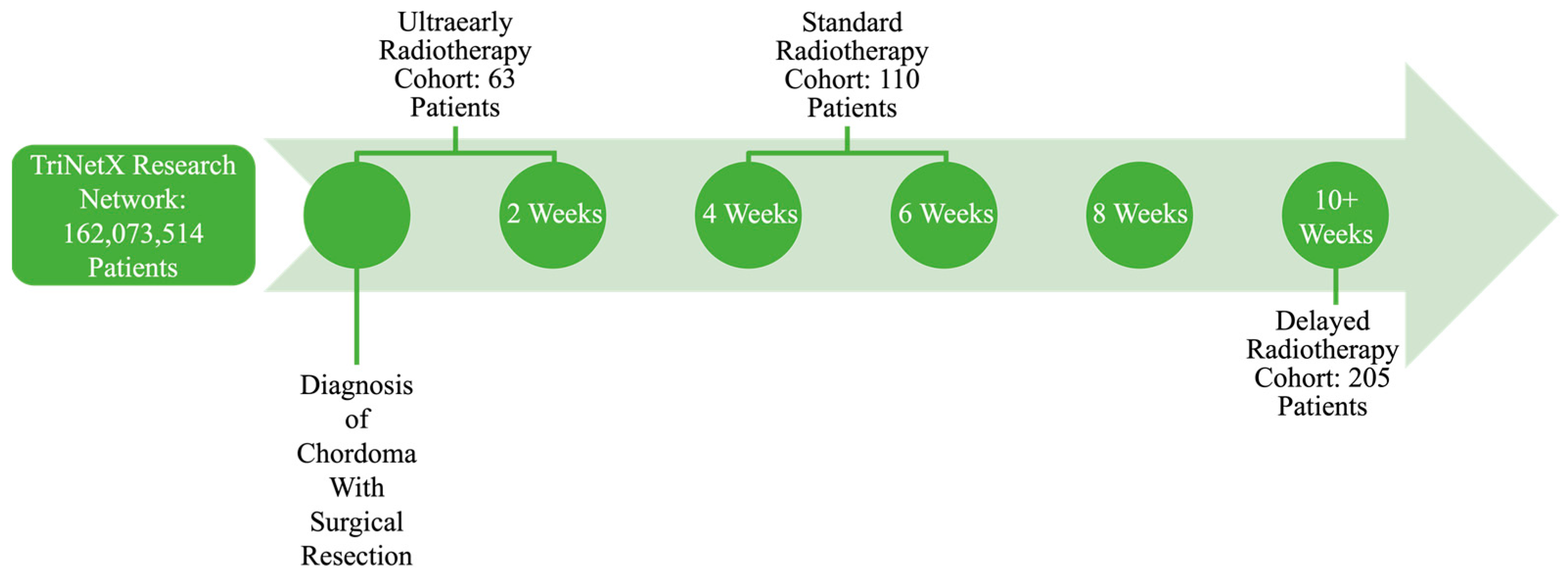
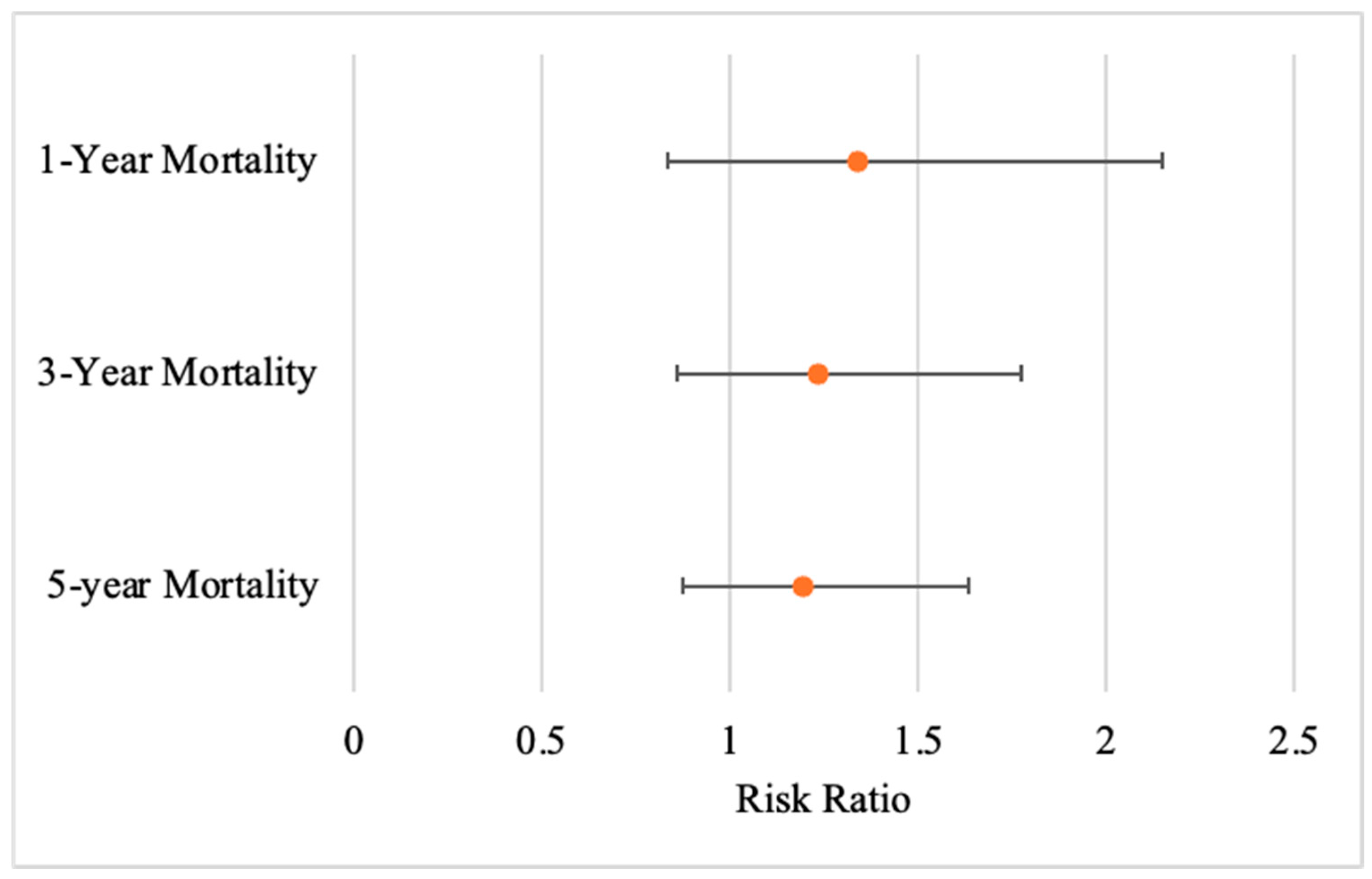
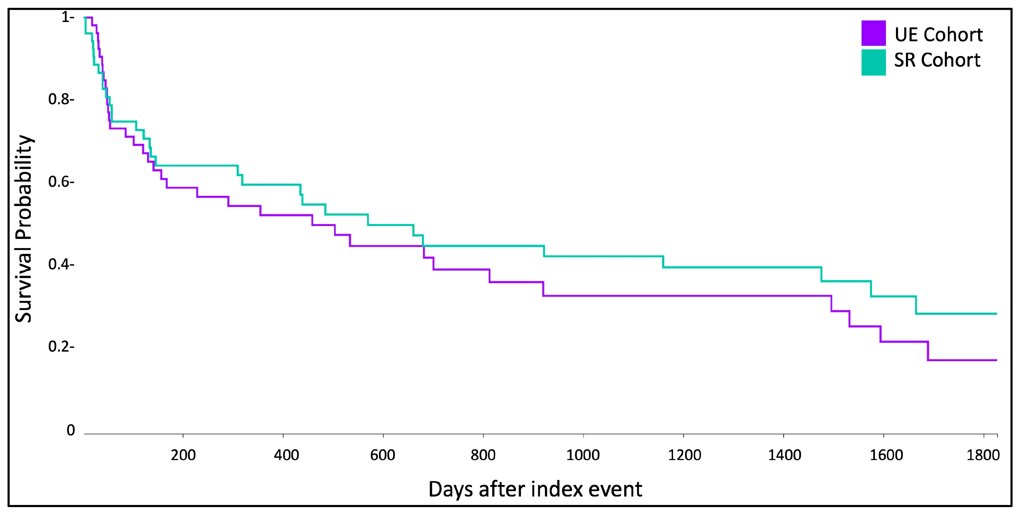
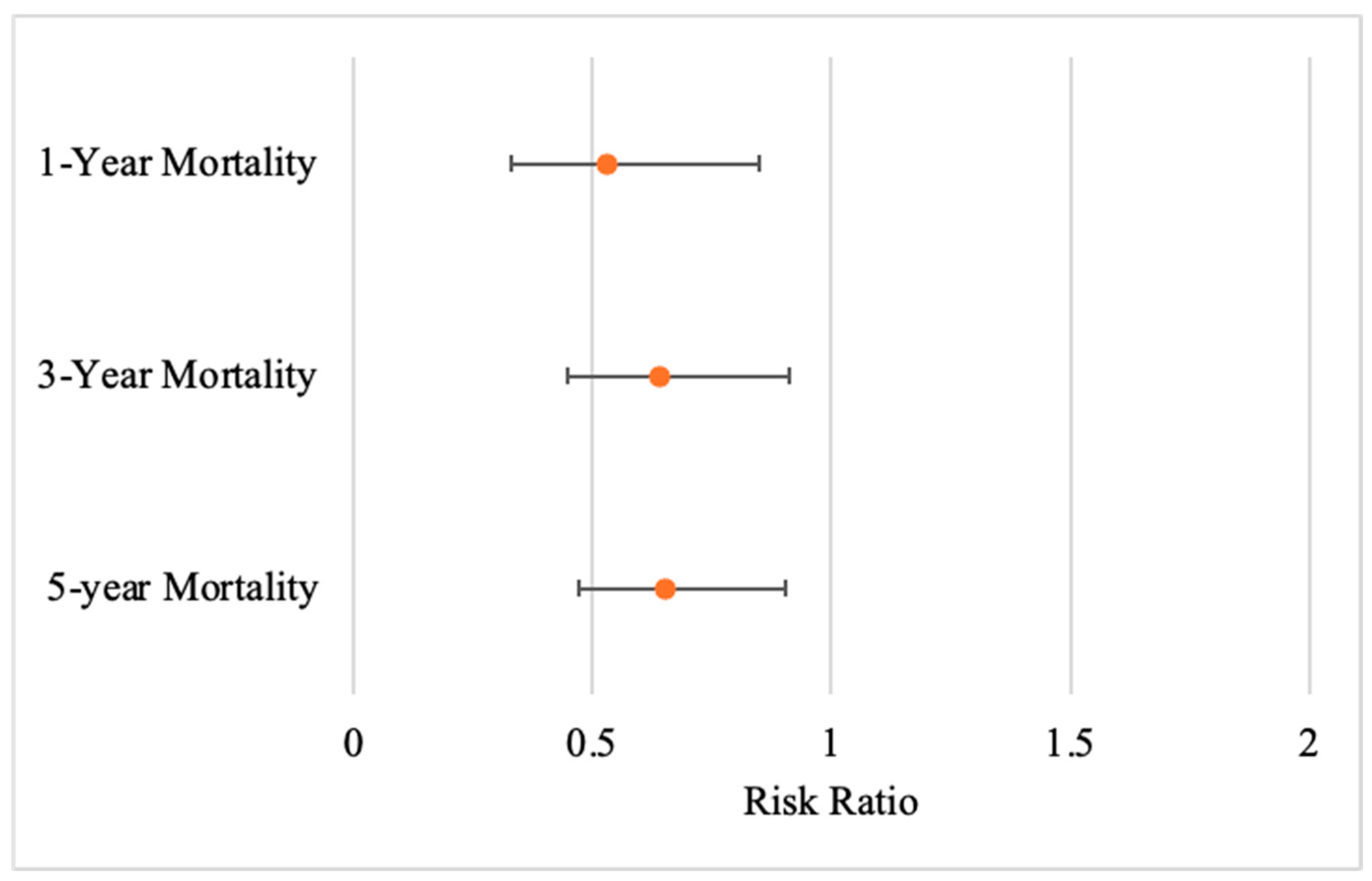
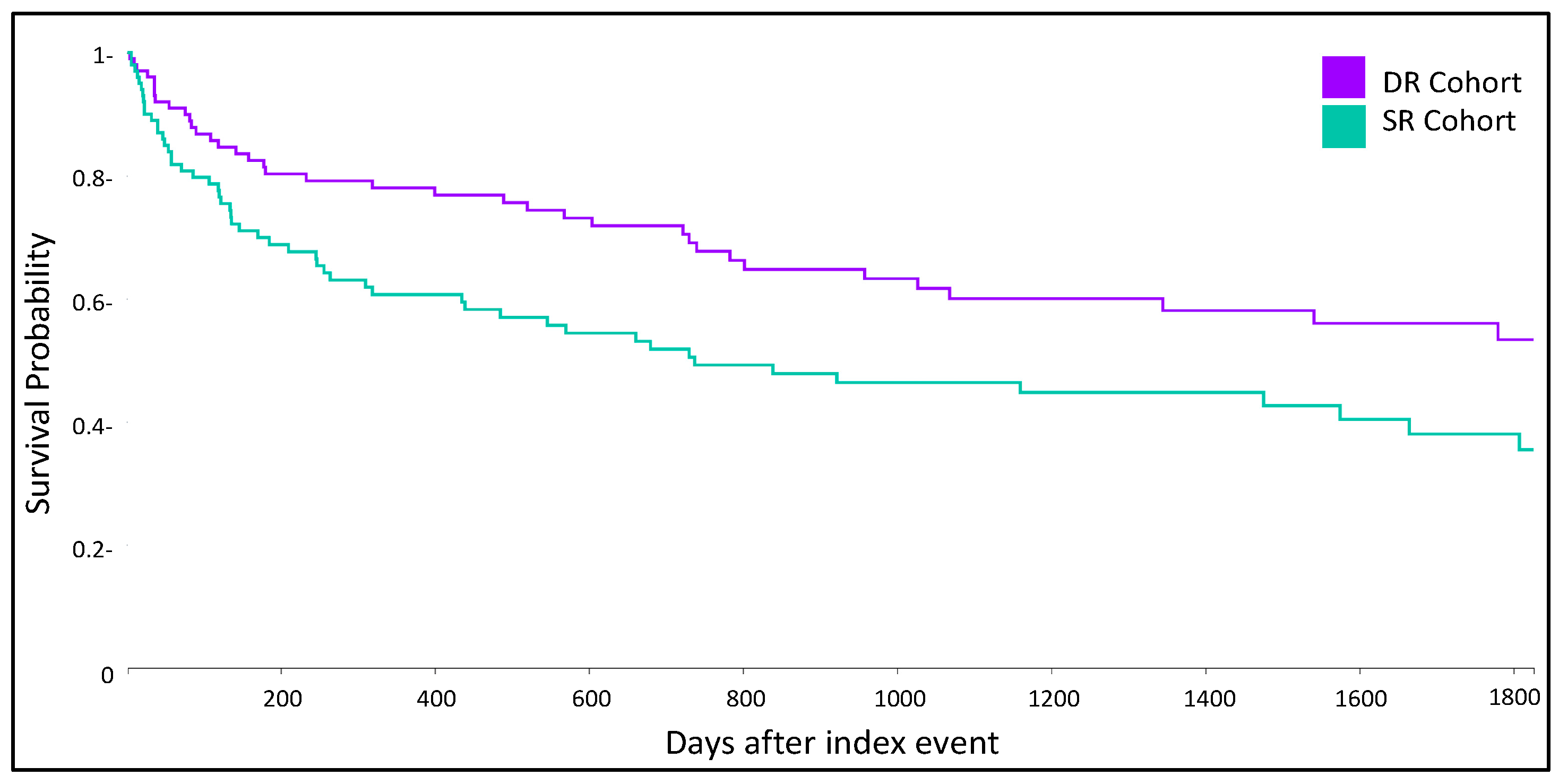
Our findings add to the limited evidence on the optimal timing of adjuvant radiotherapy in chordoma management. We observed no significant differences in 1, 3, or 5-year survival between ultraearly (<2 weeks) and standard (4–6 weeks) radiotherapy after surgery. However, we did see significantly decreased 1, 3 and 5-year mortality in patients with delayed (≥10 weeks) radiotherapy. Contrary to assumptions of the benign nature of delayed treatment for slow-growing chordoma, our results suggest a notable long-term survival benefit.
4.1. Impact of Radiotherapy Timing on Outcomes
In this multi-institutional cohort, delayed radiotherapy timing did impact 1, 3 and 5-year survival. Collectively, these findings suggest that, within the range of intervals examined in this study, moderate delays in adjuvant radiotherapy are associated with improved survival in chordoma patients treated with surgery and postoperative radiotherapy. These findings differ from established patterns in malignancies, particularly head and neck cancers and soft tissue sarcomas, where postponing postoperative radiotherapy is consistently linked to poorer survival or local control, and where several meta-analyses and large cohorts have emphasized detriment when adjuvant radiation starts beyond ~6 weeks after surgery [
15,
16,
17].
Various explanations may support the divergence seen here. First, the pathophysiology of chordoma differs from epithelial and mesenchymal tumors. Chordomas are slow growing, locally aggressive neoplasms with prolonged natural histories and late recurrences. The indolent kinetics and dose–response profile seemingly suggest a wider timing window without compromising long-term disease control [
21,
22]. Moreover, chordoma radiotherapy commonly uses high biological doses using proton or carbon-ion modalities [
12,
19]. Although our dataset did not capture modality or dose, the use of specialized radiotherapy for chordoma may play a role in treatment delays and long-term outcomes [
23,
24,
25,
26,
27].
Existing chordoma-specific guidance reinforces the importance of adjuvant radiotherapy but generally does not prescribe a rigid numeric cutoff for timing [
28,
29]. Consensus best-practice recommendations and National Comprehensive Cancer Network bone cancer guidelines emphasize wide or maximally safe resection followed by high-dose radiation, ideally delivered in the postoperative setting, and many institutional series describe typical practice patterns in which adjuvant proton or high-dose photon therapy is initiated within the first 2–3 months after surgery [
28,
30]. In several spine and skull base chordoma cohorts, adjuvant treatment is commonly started within approximately 90 days, consistent with broader oncology conventions that operationally define “adjuvant” radiotherapy as treatment delivered within about three months of surgery [
31,
32,
33,
34]. Importantly, our delayed radiotherapy group largely falls within this clinically accepted adjuvant window.
From a practical standpoint our data shows that the initiation of adjuvant radiotherapy at later postoperative intervals was associated with significantly lower mortality compared with standard timing. Our results comparing UE to SR also suggest that within a window extending to roughly 10 weeks after surgery, modest variation in start time is unlikely to produce large differences in overall survival. However, this association should not be interpreted as evidence that intentionally postponing radiotherapy is therapeutically advantageous. Rather, it suggests that moderate, clinically driven delays within the adjuvant period may not only be safe but potentially beneficial. In real-world decision-making, a reasonable approach remains to initiate adjuvant radiotherapy once the wound is adequately healed, systemic comorbidities are optimized, and a definitive treatment planning has been completed, ideally commencing treatment within the first 2–3 months postoperatively. For patients who experience unavoidable delays in initiating radiotherapy, our findings provide some reassurance that survival may not be compromised, and may even appear improved, by starting radiotherapy closer to the 10-week mark. However, our study was not designed to evaluate outcomes beyond this timeframe, and we cannot exclude the possibility that much longer delays would adversely affect local control or survival.
Accordingly, our results are supportive of a pragmatic time window rather than a strict deadline, challenging the notion of expediting radiotherapy administration post-surgical resection and suggesting that a modest delay in initiation is safe in the chordoma population. When feasible, adjuvant radiotherapy for chordoma should be initiated as wound and patient factors allow, generally closer to 10 weeks after surgery. Within this interval, clinical judgment can reasonably prioritize wound healing, referral to high-volume centers, and access to advanced radiotherapy techniques without strong evidence that modest shifts in start date will meaningfully worsen survival. Future studies incorporating margin status, volumetrics, modality/dose, and centralized outcome adjudication will be required to determine whether a narrower “optimal” interval exists and to clarify whether very late initiation beyond the adjuvant window is associated with worse long-term outcomes.
4.2. Stratification of Radiotherapy Timing Impact by Tumor Anatomical Location
Because tumor location is clinically relevant in chordoma, we explored whether the association between radiotherapy timing and survival differed between skull base and spinal disease [
35]. Within our TriNetX cohort, however, the vast majority of patients carried diagnostic codes corresponding to vertebral column, sacral, or pelvic chordoma. Of the 378 patients included in the primary analysis, 360 (95%) met criteria for spinal chordoma, whereas only a small minority had isolated skull base involvement. The resulting skull base subgroup was too small to support meaningful comparison across the three radiotherapy timing windows, limiting our ability to perform the stratified analysis for this anatomical site. This constraint is consistent with the broader literature, in which most large series either focus predominantly on skull base disease or aggregate skull base and spinal cases together [
36,
37].
Given the predominance of spinal tumors in our combined cohort, we conducted a dedicated subgroup analysis restricted to patients with spinal chordoma. In this spinal-only cohort, the pattern of results closely mirrored the overall findings. UE again showed no statistically significant difference in 1, 3, or 5-year mortality compared with standard timing. In contrast, DR was associated with a robust and statistically significant reduction in mortality relative to standard timing at 1, 3, and 5-years with all confidence intervals excluding one. These spinal-specific results confirm that the survival advantage associated with delayed radiotherapy in our primary analysis is not an artifact of mixing disparate anatomical locations, but is driven largely by patients with mobile spine and sacral chordomas.
Anatomical location nonetheless remains an important biological and therapeutic modifier [
38]. Prior comparative series have demonstrated differences in presentation, resectability, and long-term outcomes between skull base and spinal chordomas, with skull-based tumors often constrained by proximity to critical neurovascular structures, and mobile spine or sacral tumors more frequently amenable to wider en bloc resections at the cost of substantial morbidity [
39,
40]. Recent national database and multi-center analysis further suggests that adjuvant proton therapy may offer a particular survival benefit for spinal chordoma compared with photon radiotherapy, underscoring that treatment modality and center volume strongly influence outcomes in this subgroup [
41,
42].
In light of these considerations, our stratified analysis supports two key inferences. First, for spinal chordoma specifically, delayed initiation of adjuvant radiotherapy within the adjuvant window is consistently associated with improved survival over standard timing, reinforcing the main conclusion of our study. Second, because skull-based cases were too few for adequately powered comparisons, our findings should not be generalized uncritically to skull base chordoma, where surgical constraints, dosimetric limitations, and a long-standing emphasis on early high-dose proton therapy may yield a different optimal timing paradigm. Future collaborative efforts that deliberately oversample skull base chordoma and collect detailed, location-specific surgical and radiotherapy data will be required to define whether the survival benefit associated with delayed radiotherapy and spinal disease also applies to skull-based tumors, or whether timing should be tailored more explicitly to the anatomical site.
4.4. Limitations
Several other limitations must be acknowledged. First, this study was retrospective and relied on administrative coding, which introduces the potential for misclassification and unmeasured confounding [
45]. Second, although propensity score matching was applied, important clinical details such as the extent of resection, tumor size, histologic subtype, radiation modality, and the anatomic location of where radiation was applied were unavailable in TriNetX and may have influenced outcomes [
4,
6]. Third, secondary outcome events were rare, preventing meaningful evaluation of postoperative morbidity. Finally, this analysis was restricted to survival at one, three, and five years; data on local recurrence, progression-free survival, or functional outcomes were not available, limiting the interpretation of the oncologic implications of treatment timing [
9].
Despite these limitations, this study has notable strengths. The use of a large, multi-institutional dataset allows for a broader representation of chordoma patients than is typically possible in single-institution series. Furthermore, rigorous statistical methods, including propensity score matching, strengthen the validity of the comparisons made across treatment groups.



 ,
,