Article

The Impact of Radiotherapy Timing on Postoperative Outcomes in Chordoma Patients—A TriNetX Study

1
Department of Neurosurgery, Drexel University College of Medicine, Philadelphia, PA 19104, USA
2
Department of Neurosurgery, Noorda College of Osteopathic Medicine, Provo, UT 84606, USA
3
Global Neurosciences Institute, Atlantic City, NJ 08401, USA
4
Department of Neurosurgery, University of Michigan Medical School, Ann Arbor, MI 48109, USA
*
Author to whom correspondence should be addressed.
Complications 2026, 3(1), 5; https://doi.org/10.3390/complications3010005
Submission received: 21 October 2025 / Revised: 4 January 2026 / Accepted: 4 February 2026 / Published: 10 February 2026

Abstract

Introduction: Chordomas are rare, locally aggressive tumors of the spine and skull base typically managed with maximal surgical resection followed by adjuvant radiotherapy. Although postoperative radiotherapy improves local control, the optimal interval for initiation remains uncertain, as early delivery may exacerbate wound-related complications while delayed initiation may allow tumor progression. Methods: We performed a retrospective cohort analysis using the multi-center, national TriNetX Research Network. Adults with histologically confirmed skull base and/or spinal chordoma who underwent surgical resection followed by radiotherapy were stratified into ultraearly (≤2 weeks), standard (4–6 weeks), or delayed (≥10 weeks) radiotherapy initiation groups. Propensity score matching was used to adjust for demographic and clinical covariates. The primary outcome was all-cause mortality at 1, 3, and 5-years. Secondary outcomes included wound dehiscence, surgical site infection, and neurologic complications. Results: A total of 378 patients met the inclusion criteria. Ultraearly radiotherapy was not associated with significant differences in mortality at 1 year (RR 1.338; 95% CI 0.833–2.15; p = 0.22), 3 years (RR 1.233; 95% CI 0.858–1.772; p = 0.25), or 5 years (RR 1.196; 95% CI 0.876–1.633; p = 0.25) compared with standard timing. Delayed radiotherapy, however, demonstrated significantly reduced mortality at 1 year (RR: 0.53; 95% CI: 0.331–0.851; p = 0.01), 3 years (RR 0.641; 95% CI 0.449–0.914; p = 0.01), and 5 years (RR 0.654; 95% CI 0.473–0.905; p = 0.01) compared with standard timing. Event counts for secondary outcomes were insufficient for robust statistical comparison. Conclusions: Radiotherapy timing following surgical resection of chordoma did not impact short-term survival, but delayed radiotherapy significantly decreased 1, 3 and 5-year mortality. Rare secondary complications were seen. These findings suggest that the delayed initiation of radiotherapy may be helpful for patients with chordoma, supporting the need for prospective, long-term studies to clarify the balance between oncologic efficacy and perioperative morbidity.

1. Introduction

Chordomas are rare, slow-growing neoplasms that arise from remnants of the embryonic notochord and most commonly arise along the axial skeleton, particularly at the skull base and sacrum [1]. Despite their indolent histology, chordomas are clinically challenging because of their locally aggressive growth, high recurrence rates, and proximity to critical neurovascular structures [2,3]. Surgical resection remains the cornerstone of management; however, achieving complete resection is often limited by anatomic constraints and the risk of significant morbidity [4,5,6]. As a result, adjuvant radiotherapy has emerged as an essential component of therapy, with several series demonstrating improved local control and increased progression-free survival when high-dose radiotherapy is administered following resection [7,8].
Although the benefit of postoperative radiotherapy is well recognized, the optimal timing of treatment initiation remains uncertain. Early radiotherapy is thought to provide the theoretical advantage of eradicating microscopic residual disease before tumor repopulation can occur, potentially improving disease control [9,10]. However, initiating radiotherapy soon after surgery may interfere with wound healing, increase the risk of infection, or exacerbate radiation-induced tissue injury [11,12,13]. Conversely, postponing radiotherapy might decrease perioperative complications and facilitate sufficient recovery. Delayed therapy beyond 6–8 weeks may lead to early local recurrence control which has been seen in various soft tissue sarcomas and head and neck cancers [14,15,16,17]. These competing considerations have contributed to heterogenous practice patterns, with some institutions favoring prompt initiation and others adopting more cautious, delayed approaches to balance oncologic efficacy against surgical morbidity [18,19]. The unique biology of chordoma, characterized by slow growth, late recurrence, and dependence on escalated-dose photon or particle therapy, may confer unique treatment considerations compared with fast growing tumors [20].
To address this knowledge gap, we conducted a retrospective cohort analysis using the TriNetX Research Network to examine whether the timing of postoperative radiotherapy is associated with differences in survival and complication rates among chordoma patients undergoing surgical resection. By stratifying patients into ultraearly (UE), standard (SR), and delayed radiotherapy (DR) initiation groups and applying propensity score matching to adjust for confounders, this study provides one of the largest comparative assessments of radiotherapy timing in this disease.

2. Materials and Methods

2.1. Data Source

The multi-center, national TriNetX Research Network database was accessed on 4 December 2025 through a retrospective query of the Research dataset. This platform aggregates de-identified clinical data, such as demographics, diagnoses, procedures, and medication prescriptions, from 111 healthcare organizations and over 160 million patients. The database was queried without any specific time-period restriction. Institutional Board review exemption was approved through the Drexel University College of Medicine (Philadelphia, PA, USA).

2.2. Cohort Selection

All patients ≥ 18 years of age diagnosed with chordoma who underwent surgical resection were included in this retrospective cohort analysis. Cohorts included both cranial (skull base) and spinal chordomas to capture the full anatomical spectrum of disease. Patients were identified in the TriNetX Research Network using the International Classification of Diseases, Tenth Revision, Clinical Modification (ICD-10-CM) codes and Current Procedural Terminology (CPT) codes. Eligible diagnoses included malignant neoplasm of the skull and face bones (ICD-10-CM C41.0), malignant neoplasm of the vertebral column (ICD-10-CM 41.2), and malignant neoplasm of pelvic bones, sacrum, and coccyx (ICD-10-CM C41.4). Patients with secondary malignant neoplasm of bone and bone marrow (ICD-10-CM C79.5) were excluded. In addition to the overall cohort, subgroups were curated for cranial and spinal chordomas separately. For these subgroups, patients were stratified based on the presence of the relevant ICD-10-CM codes (C41.0 for cranial chordoma; C41.2 or C41.4 for spinal chordoma), and the ICD-10 indicators corresponding to the alternate chordoma location were added to each subgroup’s exclusion criteria to ensure anatomical specificity.
Procedural eligibility required evidence of surgical resection of the vertebral column, identified by one or more of the following CPT codes: partial excision of posterior vertebral component (e.g., spinous process, lamina, or facet) for intrinsic bony lesion, single vertebral segment, cervical (CPT 22100), thoracic (CPT 22101), lumbar (CPT 22102), or each additional segment (CPT 22103); laminectomy for biopsy/excision of intraspinal neoplasm, extradural, cervical (CPT 63275), thoracic (CPT 63276), lumbar (CPT 63277), or sacral (CPT 63278); arthrodesis, posterior, for spinal deformity, with or without cast, up to 6 vertebral segments (CPT 22800), 7 to 12 segments (CPT 22802), or 13 or more segments (CPT 22804); and arthrodesis, anterior, for spinal deformity, with or without cast, 2 to 3 segments (CPT 22808), 4 to 7 segments (CPT 22810), or 8 or more segments (CPT 22812).
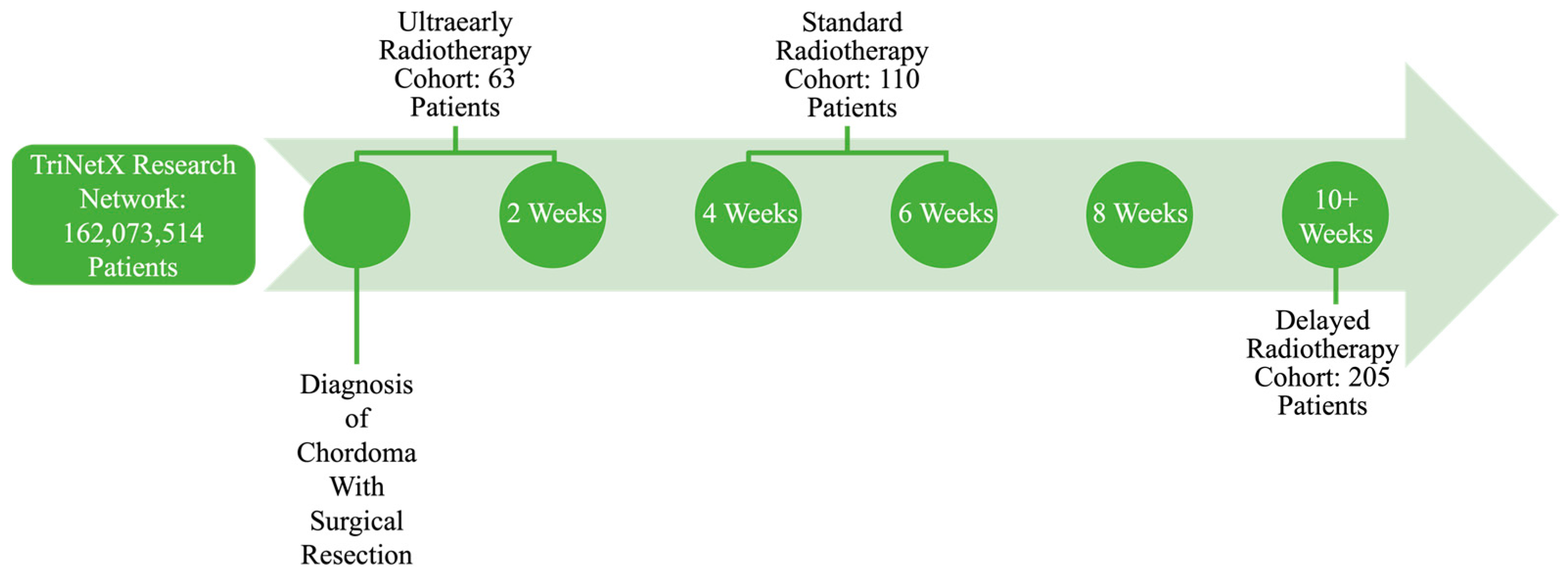
Patients were then stratified by timing of postoperative radiotherapy using a curated radiation exposure code in TriNetX (TNX Curated 1001). Ultraearly radiotherapy was defined as initiation within 2 weeks of surgery, standard timing as between 4 and 6 weeks, and delayed timing as ≥10 weeks following resection (Figure 1).

2.3. Variables

All variables used for propensity score matching were measured at the index event (diagnosis of chordoma with surgical resection and initiation of radiotherapy) and any time before. Demographic covariates included age at index, sex (male or female), and race/ethnicity categories of Hispanic or Latino, White, Black or African American, and Asian. Clinical covariates included heart failure (ICD-10-CM I50), type 2 diabetes mellitus (ICD-10-CM E11), chronic kidney disease (ICD-10-CM N18), and ischemic heart disease (ICD-10-CM I25). Treatment-related covariates included prior chemotherapy exposure (TNX curated variable: Chemotherapy Lines of Treatment [TNX Curated 10301]) and prior encounters for antineoplastic radiation therapy (ICD-10-CM Z51.0). However, in the comparison between ultraearly radiotherapy and delayed radiotherapy with standard timing, no patients in either cohort had prior chemotherapy or radiation therapy; therefore, these variables were retained in the matching model but omitted from the baseline characteristics table for that subgroup. In the delayed versus standard comparison prior treatment variables were included and displayed as applicable.

2.4. Outcome Measures

The primary outcome was all-cause mortality at 1, 3, and 5-years following surgical resection and adjuvant radiotherapy for chordoma. Secondary outcomes included postoperative wound dehiscence (ICD-10-CM T81.3), surgical site infections (ICD-10-CM T81.4, T81.41, T81.42), and neurologic complications (ICD-10-CM G95, G96).

2.5. Statistical Analysis

Descriptive statistics were used to summarize baseline demographic and clinical characteristics across radiotherapy timing groups. Risk ratios (RRs) with 95% confidence intervals (CIs), as well as Kaplan–Meier (KM) survival curves were calculated using the TriNetX analytical platform (https://trinetx.com/) to compare outcomes between ultraearly (≤2 weeks), standard (4–6 weeks), and delayed (≥10 weeks) radiotherapy cohorts. The cohort receiving standard radiotherapy timing served as the reference group for RR calculation. Propensity score matching was performed using the greedy nearest neighbor algorithm at a 1:1 ratio, based on the variables defined in Section 2.3. These covariates were selected as potential confounders that could influence both treatment assignment and clinical outcomes. Mortality metrics, including median survival time and mortality rates for the chosen time-points, were calculated using WebPlotDigitizer v5 with the KM curves as the inputted material.

3. Results

3.1. Ultraearly vs. Standard Radiotherapy Timing in Spinal and Cranial Chordoma

A total of 63 patients received ultraearly radiotherapy within 2 weeks after surgery for chordoma and 110 patients received radiotherapy between 4 and 6 weeks after surgery. Following propensity score matching, baseline demographic and clinical characteristics were similar between the groups for comparison of outcomes at 1, 3, and 5-years post-surgery with adjuvant radiotherapy (Table 1).
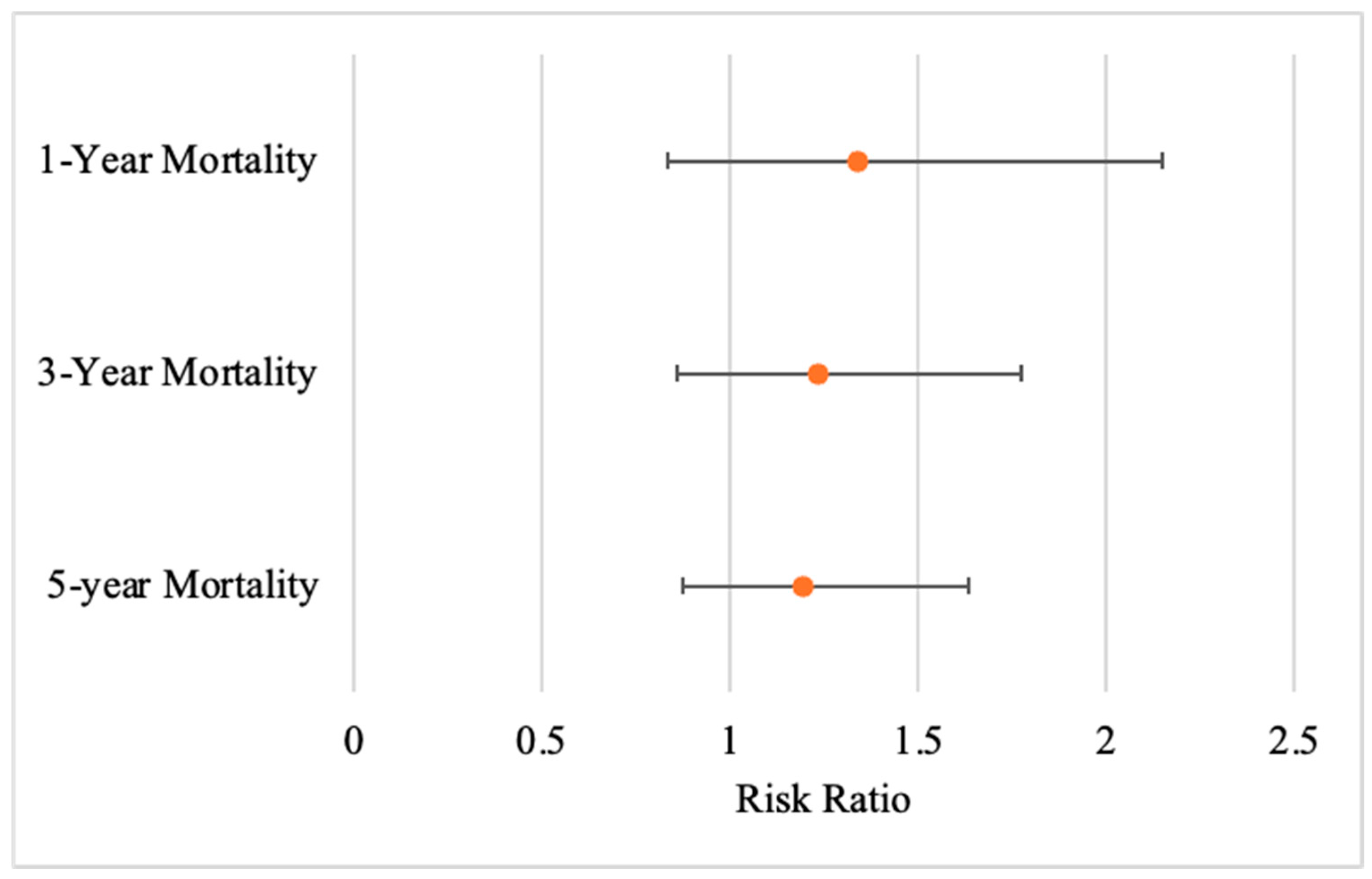
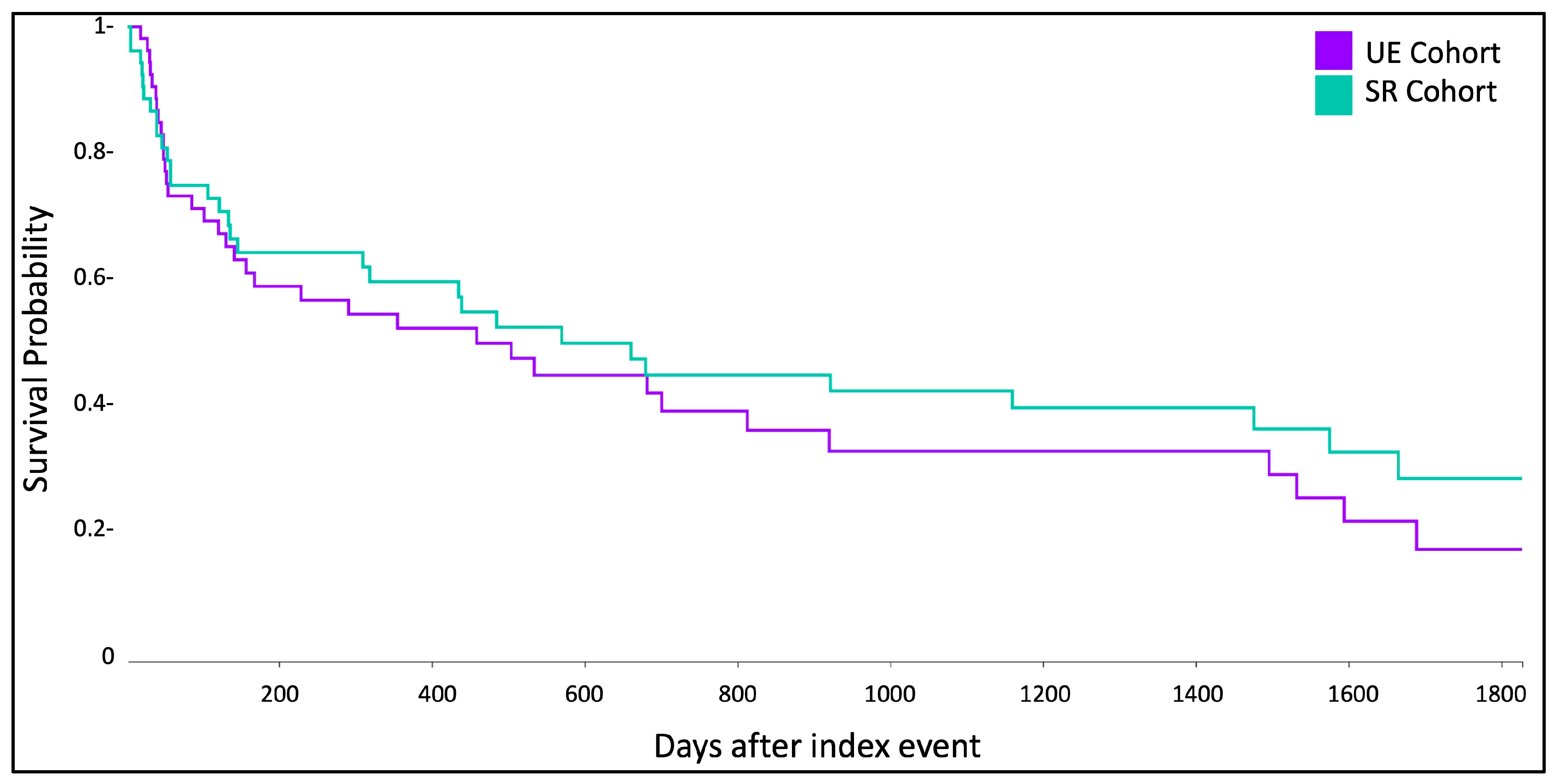
Patients who received ultraearly radiotherapy (≤2 weeks) demonstrated no significant difference in mortality compared to those who underwent standard timing (4–6 weeks) (Figure 2 and Figure 3).
Table 1. Patient demographics for outcomes of ultraearly (≤2 Weeks) vs. standard (4–6 Weeks) radiotherapy timing cohorts.
Cohort Demographics
1, 3, and 5-Year Outcomes
CharacteristicsPropensity Score Matched Sample No. (%)
UESRSMD
n = 55n = 55
Age at Index (years)55.659.20.20
Gender
Female20 (36.36%)17 (30.91%)0.12
Male35 (63.64%)38 (69.09%)0.12
Race
White39 (70.91%)38 (69.09%)0.040
Hispanic or Latino10 (18.18%)10 (18.18%)0
Black or African American10 (18.18%)11 (20.00%)0.046
Asian10 (18.18%)10 (18.18%)0
Chronic Medical Conditions
Heart Failure10 (18.18%)10 (18.18%)0
Type 2 Diabetes Mellitus10 (18.18%)10 (18.18%)0
Chronic Kidney Disease10 (18.18%)10 (18.18%)0
Chronic Ischemic Heart Disease10 (18.18%)10 (18.18%)0
Figure 2. Mortality risk for UE cohort compared to SR cohort. Dots represent the RR and lines represent the upper and lower bounds of the 95% CI.
Complications 03 00005 g002
Figure 3. 5-year KM curves for UE cohort compared to SR cohort.
Complications 03 00005 g003
Specifically, ultraearly radiotherapy was not associated with altered risk of one-year mortality (RR: 1.338; 95% CI: 0.833–2.15; p = 0.22), three-year mortality (RR: 1.233; 95% CI: 0.858–1.772; p = 0.25), or five-year mortality (RR: 1.196; 95% CI: 0.876–1.633; p = 0.25) (Table 2). Moreover, median survival rates and mortality rates were comparable between cohorts (Table 3).

Subgroup Analysis of Ultraearly vs. Standard Radiotherapy Timing in Spinal Chordoma

A total of 62 spinal chordoma cases received ultraearly radiotherapy, and 106 patients received radiotherapy between 4 and 6 weeks after surgery. Following propensity score matching, baseline demographic and clinical characteristics were similar between the groups for comparison of outcomes at 1, 3, and 5-years post-surgery with adjuvant radiotherapy (Table 4).
The patients with spinal chordoma who received ultraearly radiotherapy (≤2 weeks) demonstrated no significant difference in mortality compared to those who underwent standard timing (4–6 weeks) (Table 5).

3.2. Delayed vs. Standard Radiotherapy Timing Cohorts in Spinal and Cranial Chordoma

A total of 205 patients received delayed radiotherapy at least 10 weeks after surgery and 110 patients received standard radiotherapy between 4 and 6 weeks after surgery. Following propensity score matching, baseline demographic and clinical characteristics were well-balanced between the groups for comparison of outcomes at 1, 3, and 5-years post-surgery with adjuvant radiotherapy (Table 6).
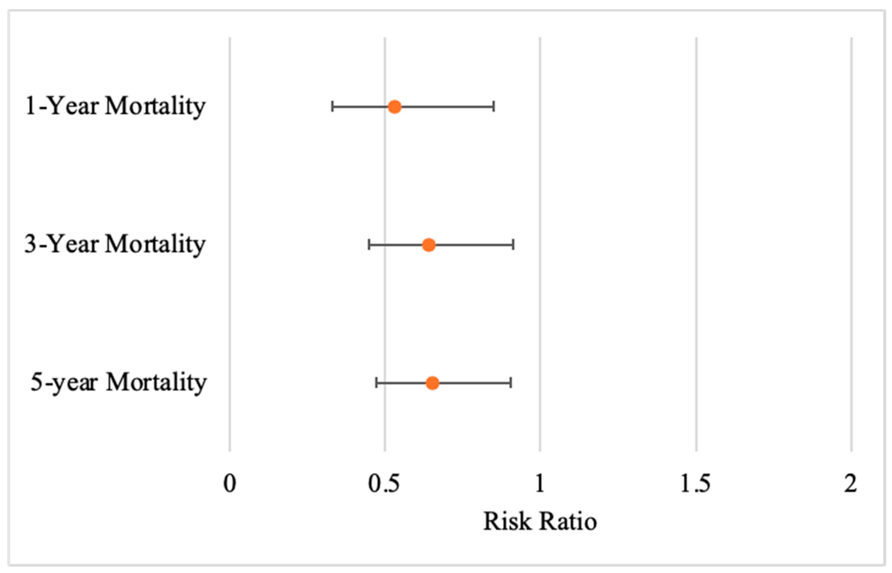
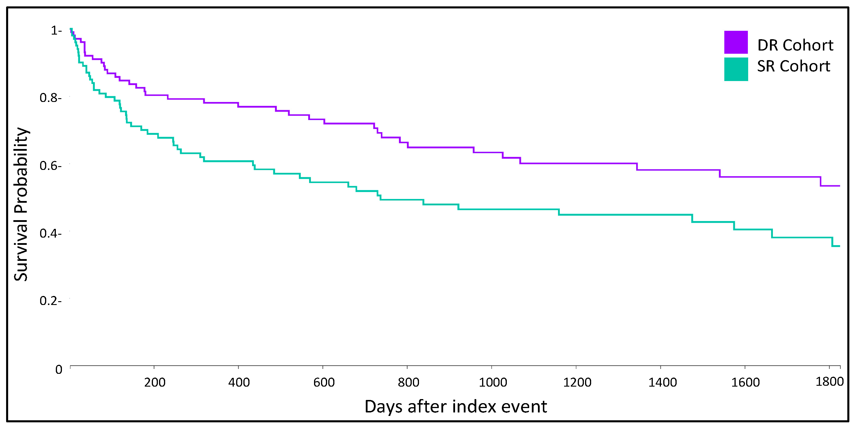
Patients who received delayed radiotherapy (≥10 weeks) demonstrated a significant difference in mortality compared to those who underwent standard timing (4–6 weeks) at 1, 3, and 5-years post-procedure (Figure 4 and Figure 5).
Specifically, delayed radiotherapy was associated with a significantly reduced risk of one-year mortality (RR: 0.53; 95% CI: 0.331–0.851; p = 0.01), three-year mortality (RR: 0.641; 95% CI: 0.449–0.914; p = 0.01), and five-year mortality (RR: 0.654; 95% CI: 0.473–0.905; p = 0.01) (Table 7). Moreover, median survival rates and mortality rates were noticeably different between cohorts (Table 8).

Subgroup Analysis of Delayed vs. Standard Radiotherapy Timing in Spinal Chordoma

A total of 192 spinal chordoma cases received delayed radiotherapy, and 106 patients received radiotherapy between 4 and 6 weeks after surgery. Following propensity score matching, baseline demographic and clinical characteristics were similar between the groups for comparison of outcomes at 1, 3, and 5-years post-surgery with adjuvant radiotherapy (Table 9).
The patients with spinal chordoma who received delayed radiotherapy (≥10 weeks) demonstrated significant differences in mortality compared to those who underwent standard timing (4–6 weeks) (Table 10).

4. Discussion

Our findings add to the limited evidence on the optimal timing of adjuvant radiotherapy in chordoma management. We observed no significant differences in 1, 3, or 5-year survival between ultraearly (<2 weeks) and standard (4–6 weeks) radiotherapy after surgery. However, we did see significantly decreased 1, 3 and 5-year mortality in patients with delayed (≥10 weeks) radiotherapy. Contrary to assumptions of the benign nature of delayed treatment for slow-growing chordoma, our results suggest a notable long-term survival benefit.

4.1. Impact of Radiotherapy Timing on Outcomes

In this multi-institutional cohort, delayed radiotherapy timing did impact 1, 3 and 5-year survival. Collectively, these findings suggest that, within the range of intervals examined in this study, moderate delays in adjuvant radiotherapy are associated with improved survival in chordoma patients treated with surgery and postoperative radiotherapy. These findings differ from established patterns in malignancies, particularly head and neck cancers and soft tissue sarcomas, where postponing postoperative radiotherapy is consistently linked to poorer survival or local control, and where several meta-analyses and large cohorts have emphasized detriment when adjuvant radiation starts beyond ~6 weeks after surgery [15,16,17].
Various explanations may support the divergence seen here. First, the pathophysiology of chordoma differs from epithelial and mesenchymal tumors. Chordomas are slow growing, locally aggressive neoplasms with prolonged natural histories and late recurrences. The indolent kinetics and dose–response profile seemingly suggest a wider timing window without compromising long-term disease control [21,22]. Moreover, chordoma radiotherapy commonly uses high biological doses using proton or carbon-ion modalities [12,19]. Although our dataset did not capture modality or dose, the use of specialized radiotherapy for chordoma may play a role in treatment delays and long-term outcomes [23,24,25,26,27].
Existing chordoma-specific guidance reinforces the importance of adjuvant radiotherapy but generally does not prescribe a rigid numeric cutoff for timing [28,29]. Consensus best-practice recommendations and National Comprehensive Cancer Network bone cancer guidelines emphasize wide or maximally safe resection followed by high-dose radiation, ideally delivered in the postoperative setting, and many institutional series describe typical practice patterns in which adjuvant proton or high-dose photon therapy is initiated within the first 2–3 months after surgery [28,30]. In several spine and skull base chordoma cohorts, adjuvant treatment is commonly started within approximately 90 days, consistent with broader oncology conventions that operationally define “adjuvant” radiotherapy as treatment delivered within about three months of surgery [31,32,33,34]. Importantly, our delayed radiotherapy group largely falls within this clinically accepted adjuvant window.
From a practical standpoint our data shows that the initiation of adjuvant radiotherapy at later postoperative intervals was associated with significantly lower mortality compared with standard timing. Our results comparing UE to SR also suggest that within a window extending to roughly 10 weeks after surgery, modest variation in start time is unlikely to produce large differences in overall survival. However, this association should not be interpreted as evidence that intentionally postponing radiotherapy is therapeutically advantageous. Rather, it suggests that moderate, clinically driven delays within the adjuvant period may not only be safe but potentially beneficial. In real-world decision-making, a reasonable approach remains to initiate adjuvant radiotherapy once the wound is adequately healed, systemic comorbidities are optimized, and a definitive treatment planning has been completed, ideally commencing treatment within the first 2–3 months postoperatively. For patients who experience unavoidable delays in initiating radiotherapy, our findings provide some reassurance that survival may not be compromised, and may even appear improved, by starting radiotherapy closer to the 10-week mark. However, our study was not designed to evaluate outcomes beyond this timeframe, and we cannot exclude the possibility that much longer delays would adversely affect local control or survival.
Accordingly, our results are supportive of a pragmatic time window rather than a strict deadline, challenging the notion of expediting radiotherapy administration post-surgical resection and suggesting that a modest delay in initiation is safe in the chordoma population. When feasible, adjuvant radiotherapy for chordoma should be initiated as wound and patient factors allow, generally closer to 10 weeks after surgery. Within this interval, clinical judgment can reasonably prioritize wound healing, referral to high-volume centers, and access to advanced radiotherapy techniques without strong evidence that modest shifts in start date will meaningfully worsen survival. Future studies incorporating margin status, volumetrics, modality/dose, and centralized outcome adjudication will be required to determine whether a narrower “optimal” interval exists and to clarify whether very late initiation beyond the adjuvant window is associated with worse long-term outcomes.

4.2. Stratification of Radiotherapy Timing Impact by Tumor Anatomical Location

Because tumor location is clinically relevant in chordoma, we explored whether the association between radiotherapy timing and survival differed between skull base and spinal disease [35]. Within our TriNetX cohort, however, the vast majority of patients carried diagnostic codes corresponding to vertebral column, sacral, or pelvic chordoma. Of the 378 patients included in the primary analysis, 360 (95%) met criteria for spinal chordoma, whereas only a small minority had isolated skull base involvement. The resulting skull base subgroup was too small to support meaningful comparison across the three radiotherapy timing windows, limiting our ability to perform the stratified analysis for this anatomical site. This constraint is consistent with the broader literature, in which most large series either focus predominantly on skull base disease or aggregate skull base and spinal cases together [36,37].
Given the predominance of spinal tumors in our combined cohort, we conducted a dedicated subgroup analysis restricted to patients with spinal chordoma. In this spinal-only cohort, the pattern of results closely mirrored the overall findings. UE again showed no statistically significant difference in 1, 3, or 5-year mortality compared with standard timing. In contrast, DR was associated with a robust and statistically significant reduction in mortality relative to standard timing at 1, 3, and 5-years with all confidence intervals excluding one. These spinal-specific results confirm that the survival advantage associated with delayed radiotherapy in our primary analysis is not an artifact of mixing disparate anatomical locations, but is driven largely by patients with mobile spine and sacral chordomas.
Anatomical location nonetheless remains an important biological and therapeutic modifier [38]. Prior comparative series have demonstrated differences in presentation, resectability, and long-term outcomes between skull base and spinal chordomas, with skull-based tumors often constrained by proximity to critical neurovascular structures, and mobile spine or sacral tumors more frequently amenable to wider en bloc resections at the cost of substantial morbidity [39,40]. Recent national database and multi-center analysis further suggests that adjuvant proton therapy may offer a particular survival benefit for spinal chordoma compared with photon radiotherapy, underscoring that treatment modality and center volume strongly influence outcomes in this subgroup [41,42].
In light of these considerations, our stratified analysis supports two key inferences. First, for spinal chordoma specifically, delayed initiation of adjuvant radiotherapy within the adjuvant window is consistently associated with improved survival over standard timing, reinforcing the main conclusion of our study. Second, because skull-based cases were too few for adequately powered comparisons, our findings should not be generalized uncritically to skull base chordoma, where surgical constraints, dosimetric limitations, and a long-standing emphasis on early high-dose proton therapy may yield a different optimal timing paradigm. Future collaborative efforts that deliberately oversample skull base chordoma and collect detailed, location-specific surgical and radiotherapy data will be required to define whether the survival benefit associated with delayed radiotherapy and spinal disease also applies to skull-based tumors, or whether timing should be tailored more explicitly to the anatomical site.

4.3. Secondary Outcomes and Limitations of Event Capture

Secondary outcomes, including postoperative wound dehiscence, surgical site infections, and neurologic complications, were not identified in a significant quantity among the ultraearly, standard and delayed radiotherapy treatment groups. Unfortunately, given the low event counts for these outcomes, robust statistical comparisons across radiotherapy timing groups was not possible. This limitation underscores one of the inherent challenges of working with rare diseases such as chordoma: despite leveraging a large, federated network like TriNetX, the absolute number of patients experiencing specific complications remained too small to draw conclusions. Prior studies do suggest that early radiotherapy after surgery can increase wound complication risk [43,44]. Future studies incorporating larger pooled datasets, prospective registries, or collaborative international consortia may be required to capture sufficient events for rigorous complication analyses.

4.4. Limitations

Several other limitations must be acknowledged. First, this study was retrospective and relied on administrative coding, which introduces the potential for misclassification and unmeasured confounding [45]. Second, although propensity score matching was applied, important clinical details such as the extent of resection, tumor size, histologic subtype, radiation modality, and the anatomic location of where radiation was applied were unavailable in TriNetX and may have influenced outcomes [4,6]. Third, secondary outcome events were rare, preventing meaningful evaluation of postoperative morbidity. Finally, this analysis was restricted to survival at one, three, and five years; data on local recurrence, progression-free survival, or functional outcomes were not available, limiting the interpretation of the oncologic implications of treatment timing [9].
Despite these limitations, this study has notable strengths. The use of a large, multi-institutional dataset allows for a broader representation of chordoma patients than is typically possible in single-institution series. Furthermore, rigorous statistical methods, including propensity score matching, strengthen the validity of the comparisons made across treatment groups.


5. Conclusions

In this multi-institutional retrospective cohort study, delayed initiation was significantly associated with decreased mortality at 1, 3 and 5 years compared to standard postoperative timing. Rather than indicating harm, our results suggest that a modest delay in radiotherapy initiation may be clinically acceptable, and potentially beneficial, in the chordoma population, especially when required to facilitate wound healing, multidisciplinary coordination, or access to specialized high-dose radiotherapy techniques. While residual confounding cannot be fully excluded, the consistency and magnitude of the observed survival benefit underscore the need to reconsider rigid expectations for early radiotherapy initiation in chordoma. Prospective, multi-institutional registries with detailed surgical, radiotherapeutic, and tumor-specific data will be crucial to validate these findings, refine the optimal time window, and better inform evidence-based clinical practice guidelines.

Author Contributions

Conceptualization, K.S., S.T.R., R.R., and M.K.; data curation, K.S.; formal analysis, K.S., S.T.R., M.C., C.R., and M.K.; investigation, K.S. and S.T.R.; methodology, K.S., S.T.R., R.R., and M.K.; project administration, K.S. and M.K.; supervision, R.R. and M.K.; validation, K.S., S.T.R., M.C., C.R., R.R., and M.K.; visualization, K.S., S.T.R., M.C., C.R., R.R., and M.K.; writing—original draft, K.S. and M.K.; writing—review and editing, K.S., S.T.R., M.C., C.R., R.R., and M.K. All authors have read and agreed to the published version of the manuscript.

Funding

This research received no external funding.

Institutional Review Board Statement

Ethical review and approval were waived for this study because any data displayed on the TriNetX Platform in aggregate form, or any patient-level data provided in a dataset generated by the TriNetX Platform, only contains de-identified data as per the de-identification standard defined in Section §164.514(a) of the HIPAA Privacy Rule. This formal determination by a qualified expert, refreshed in December 2020, supersedes the need for TriNetX’s previous waiver from the Western Institutional Review Board (IRB). This means that analysis on the TriNetX Platform or offline analysis of downloaded datasets is not considered “human subjects research” and is exempt from IRB review. Furthermore, TriNetX analytics only display data in the form of aggregated and statistical summaries of de-identified information. Therefore, no PHI or personal data is made available to users on the platform.

Informed Consent Statement

This retrospective study is exempt from informed consent. The data reviewed is a secondary analysis of existing data, does not involve intervention or interaction with human subjects, and is de-identified per the de-identification standard defined in Section §164.514(a) of the HIPAA Privacy Rule. The process by which the data is de-identified is attested to through a formal determination by a qualified expert as defined in Section §164.514(b)(1) of the HIPAA Privacy Rule. This formal determination by a qualified expert refreshed on December 2020.

Data Availability Statement

Restrictions apply to the availability of these data. Data were obtained from TriNetX, LLC and are available from the authors with the permission of TriNetX, LLC.

Acknowledgments

The authors would like to acknowledge Eric Young, for their analytical support on this project.

Conflicts of Interest

Michael Karsy receives funding from JNJ/Ethicon, he is a consulting physician for Leica Biosystems, and receives royalties from Thieme Medical Publishing. The funding sponsors had no role in the design of the study; in the collection, analyses, or interpretation of data; in the writing of the manuscript; or in the decision to publish the results. Michael Karsy is not employed by any of the aforementioned companies. The remaining authors declare that the research was conducted without any commercial or financial relationships that could be construed as a potential conflict of interest.

Abbreviations

The following abbreviations are used in this manuscript:
RRRelative Risk
CIConfidence interval
KMKaplan–Meier
ICD-10-CMInternational Classification of Diseases, Tenth Revision, Clinical Modification
CPTCurrent Procedural Terminology
UEUltraearly Radiotherapy
SRStandard Radiotherapy
DRDelayed Radiotherapy

References

  1. Kaloostian, P.E.; Zadnik, P.L.; Etame, A.B.; Vrionis, F.D.; Gokaslan, Z.L.; Sciubba, D.M. Surgical Management of Primary and Metastatic Spinal Tumors. Cancer Control 2014, 21, 133–139. [Google Scholar] [CrossRef] [Scilit]
  2. Sciubba, D.M.; Petteys, R.J.; Dekutoski, M.B.; Fisher, C.G.; Fehlings, M.G.; Ondra, S.L.; Rhines, L.D.; Gokaslan, Z.L. Diagnosis and management of metastatic spine disease: A review. J. Neurosurg. Spine 2010, 13, 94–108. [Google Scholar] [CrossRef] [Scilit]
  3. Laufer, I.; Rubin, D.G.; Lis, E.; Cox, B.W.; Stubblefield, M.D.; Yamada, Y.; Bilsky, M.H. The NOMS Framework: Approach to the Treatment of Spinal Metastatic Tumors. Oncologist 2013, 18, 744–751. [Google Scholar] [CrossRef] [Scilit] [PubMed]
  4. Yahanda, A.T.; Buchowski, J.M.; Wegner, A.M. Treatment, complications, and outcomes of metastatic disease of the spine: From Patchell to PROMIS. Ann. Transl. Med. 2019, 7, 216. [Google Scholar] [CrossRef] [Scilit]
  5. Newman, W.C.; Patel, A.; Goldberg, J.L.; Bilsky, M.H. The importance of multidisciplinary care for spine metastases: Initial tumor management. Neuro Oncol. Pract. 2020, 7, i25–i32. [Google Scholar] [CrossRef] [Scilit] [PubMed]
  6. Chanbour, H.; Chen, J.W.; Gangavarapu, L.S.; Bendfeldt, G.A.; LaBarge, M.E.; Ahmed, M.; Roth, S.G.; Chotai, S.; Luo, L.Y.; Abtahi, A.M.; et al. Unplanned Readmission is Associated with Decreased Overall Survival and Performance after Metastatic Spine Surgery. Spine 2023, 48, 653–663. [Google Scholar] [CrossRef] [Scilit]
  7. Kumar, N.; Madhu, S.; Bohra, H.; Pandita, N.; Wang, S.S.Y.; Lopez, K.G.; Tan, J.H.; Vellayappan, B.A. Is there an optimal timing between radiotherapy and surgery to reduce wound complications in metastatic spine disease? A systematic review. Eur. Spine J. 2020, 29, 3080–3115. [Google Scholar] [CrossRef] [Scilit]
  8. Azad, T.D.; Varshneya, K.; Herrick, D.B.; Pendharkar, A.V.; Ho, A.L.; Stienen, M.; Zygourakis, C.; Bagshaw, H.P.; Veeravagu, A.; Ratliff, J.K.; et al. Timing of Adjuvant Radiation Therapy and Risk of Wound-Related Complications Among Patients with Spinal Metastatic Disease. Glob. Spine J. 2021, 11, 44–49. [Google Scholar] [CrossRef] [Scilit]
  9. Ahluwalia, R.; Chanbour, H.; Zeoli, T.; Abtahi, A.M.; Stephens, B.F.; Zuckerman, S.L. Does Timing of Radiation Therapy Impact Wound Healing in Patients Undergoing Metastatic Spine Surgery? Diagnostics 2024, 14, 1059. [Google Scholar] [CrossRef] [Scilit] [PubMed]
  10. Cooper, J.S.; Pajak, T.F.; Forastiere, A.A.; Jacobs, J.; Campbell, B.H.; Saxman, S.B.; Kish, J.A.; Kim, H.E.; Cmelak, A.J.; Rotman, M.; et al. Postoperative Concurrent Radiotherapy and Chemotherapy for High-Risk Squamous-Cell Carcinoma of the Head and Neck. N. Engl. J. Med. 2004, 350, 1937–1944. [Google Scholar] [CrossRef] [Scilit]
  11. Yolcu, Y.U.; Zreik, J.; Wahood, W.; Bhatti, A.U.R.; Bydon, M.; Houdek, M.T.; Rose, P.S.; Mahajan, A.; Petersen, I.A.; Haddock, M.G.; et al. Comparison of Oncologic Outcomes and Treatment-Related Toxicity of Carbon Ion Radiotherapy and En Bloc Resection for Sacral Chordoma. JAMA Netw. Open 2022, 5, e2141927. [Google Scholar] [CrossRef] [Scilit]
  12. Hug, E.B.; Loredo, L.N.; Slater, J.D.; Devries, A.; Grove, R.I.; Schaefer, R.A.; Rosenberg, A.E.; Slater, J.M. Proton radiation therapy for chordomas and chondrosarcomas of the skull base. J. Neurosurg. 1999, 91, 432–439. [Google Scholar] [CrossRef] [Scilit]
  13. Borkowska, A.M.; Pieńkowski, A.; Chmiel, P.; Skóra, T.; Michalik, R.; Rutkowski, P.; Spałek, M.J. Long-Term Outcomes of Patients Diagnosed with Sacral Chordoma in a Retrospective Multicenter Study. Cancer Control 2025, 32, 10732748251323730. [Google Scholar] [CrossRef] [Scilit]
  14. O'Sullivan, B.; Davis, A.M.; Turcotte, R.; Bell, R.; Catton, C.; Chabot, P.; Wunder, J.; Kandel, R.; Goddard, K.; Sadura, A.; et al. Preoperative versus postoperative radiotherapy in soft-tissue sarcoma of the limbs: A randomised trial. Lancet 2002, 359, 2235–2241. [Google Scholar] [CrossRef] [Scilit] [PubMed]
  15. Machtay, M.; Moughan, J.; Trotti, A.; Garden, A.S.; Weber, R.S.; Cooper, J.S.; Forastiere, A.; Ang, K.K. Factors Associated with Severe Late Toxicity After Concurrent Chemoradiation for Locally Advanced Head and Neck Cancer: An RTOG Analysis. J. Clin. Oncol. 2008, 26, 3582–3589. [Google Scholar] [CrossRef] [Scilit] [PubMed]
  16. Vivek, N.; Sharma, R.; Prasad, K.; Mannion, K.; Sinard, R.J.; Langerman, A.; Rosenthal, E.; Rohde, S.; Whitaker, R.; Lockney, N.; et al. Impact of Postoperative Radiation Therapy Delay and Treatment Facility Location on Survival in Head and Neck Cancer Patients. Head Neck 2025, 47, 1807–1815. [Google Scholar] [CrossRef] [Scilit]
  17. Schwartz, D.L.; Einck, J.; Hunt, K.; Bruckner, J.; Conrad, E.; Koh, W.-J.; Laramore, G.E. The effect of delayed postoperative irradiation on local control of soft tissue sarcomas of the extremity and torso. Int. J. Radiat. Oncol. Biol. Phys. 2002, 52, 1352–1359. [Google Scholar] [CrossRef] [Scilit]
  18. Fourquet, J.; Sunyach, M.-P.; Vilotte, F.; Le Péchoux, C.; Ranchère-Vince, D.; Bonvalot, S.; Coindre, J.-M.; Terrier, P.; Meeus, P.; Helfre, S.; et al. Time interval between surgery and start of adjuvant radiotherapy in patients with soft tissue sarcoma: A retrospective analysis of 1131 cases from the French Sarcoma Group. Radiother. Oncol. 2016, 120, 156–162. [Google Scholar] [CrossRef] [Scilit] [PubMed]
  19. Takagi, M.; Demizu, Y.; Nagano, F.; Terashima, K.; Fujii, O.; Jin, D.; Mima, M.; Niwa, Y.; Katsui, K.; Suga, M.; et al. Treatment outcomes of proton or carbon ion therapy for skull base chordoma: A retrospective study. Radiat. Oncol. 2018, 13, 232–239. [Google Scholar] [CrossRef] [Scilit]
  20. Walcott, B.P.; Nahed, B.V.; Mohyeldin, A.; Coumans, J.-V.; Kahle, K.T.; Ferreira, M.J. Chordoma: Current concepts, management, and future directions. Lancet Oncol. 2012, 13, e69–e76. [Google Scholar] [CrossRef] [Scilit]
  21. Ghaith, A.K.; Akinduro, O.O.; Alexander, A.Y.; Goyal, A.; Bon-Nieves, A.; de Macedo Filho, L.; Otamendi-Lopez, A.; Nathani, K.R.; Abode-Iyamah, K.; Jentoft, M.E.; et al. Immunohistochemical markers predicting long-term recurrence following clival and spinal chordoma resection: A multicenter study. Neurosurg. Focus 2023, 54, E15. [Google Scholar] [CrossRef] [Scilit]
  22. Barber, S.M.; Sadrameli, S.S.; Lee, J.J.; Fridley, J.S.; Teh, B.S.; Oyelese, A.A.; Telfeian, A.E.; Gokaslan, Z.L. Chordoma—Current Understanding and Modern Treatment Paradigms. J. Clin. Med. 2021, 10, 1054. [Google Scholar] [CrossRef] [Scilit]
  23. Munzenrider, J.E.; Liebsch, N.J. Proton therapy for tumors of the skull base. Strahlenther. Onkol. 1999, 175, 57–63. [Google Scholar] [CrossRef] [Scilit]
  24. Uhl, M.; Mattke, M.; Welzel, T.; Roeder, F.; Oelmann, J.; Habl, G.; Jensen, A.; Ellerbrock, M.; Jäkel, O.; Haberer, T.; et al. Highly effective treatment of skull base chordoma with carbon ion irradiation using a raster scan technique in 155 patients: First long-term results. Cancer 2014, 120, 3410–3417. [Google Scholar] [CrossRef] [Scilit]
  25. Weber, D.C.; Malyapa, R.; Albertini, F.; Bolsi, A.; Kliebsch, U.; Walser, M.; Pica, A.; Combescure, C.; Lomax, A.J.; Schneider, R. Long term outcomes of patients with skull-base low-grade chondrosarcoma and chordoma patients treated with pencil beam scanning proton therapy. Radiother. Oncol. 2016, 120, 169–174. [Google Scholar] [CrossRef] [Scilit] [PubMed]
  26. Fung, V.; Calugaru, V.; Bolle, S.; Mammar, H.; Alapetite, C.; Maingon, P.; De Marzi, L.; Froelich, S.; Habrand, J.-L.; Dendale, R.; et al. Proton beam therapy for skull base chordomas in 106 patients: A dose adaptive radiation protocol. Radiother. Oncol. 2018, 128, 198–202. [Google Scholar] [CrossRef] [Scilit] [PubMed]
  27. Koto, M.; Ikawa, H.; Kaneko, T.; Hagiwara, Y.; Hayashi, K.; Tsuji, H. Long-term outcomes of skull base chordoma treated with high-dose carbon-ion radiotherapy. Head Neck 2020, 42, 2607–2613. [Google Scholar] [CrossRef] [Scilit] [PubMed]
  28. Biermann, J.S.; Chow, W.; Reed, D.R.; Lucas, D.; Adkins, D.R.; Agulnik, M.; Benjamin, R.S.; Brigman, B.; Budd, G.T.; Curry, W.T.; et al. NCCN Guidelines Insights: Bone Cancer, Version 2.2017. J. Natl. Compr. Cancer Netw. 2017, 15, 155–167. [Google Scholar] [CrossRef] [Scilit]
  29. Palavani, L.B.; Borges, P.; Andreão, F.F.; Borges, J.; Batista, S.; Brenner, L.B.O.; Ferreira, M.Y.; Reis, P.C.A.; Pontes, J.; Negri, H.; et al. Optimizing radiotherapy strategies for skull base chordoma: A comprehensive meta-analysis and systematic review of treatment modalities and outcomes. Neurosurg. Focus 2024, 56, E11. [Google Scholar] [CrossRef] [Scilit]
  30. Palm, R.F.; Oliver, D.E.; Yang, G.Q.; Abuodeh, Y.; Naghavi, A.O.; Johnstone, P.A.S. The role of dose escalation and proton therapy in perioperative or definitive treatment of chondrosarcoma and chordoma: An analysis of the National Cancer Data Base. Cancer 2019, 125, 642–651. [Google Scholar] [CrossRef] [Scilit]
  31. Holliday, E.B.; Mitra, H.S.; Somerson, J.S.; Rhines, L.D.; Mahajan, A.; Brown, P.D.; Grosshans, D.R. Postoperative Proton Therapy for Chordomas and Chondrosarcomas of the Spine: Adjuvant Versus Salvage Radiation Therapy. Spine 2015, 40, 544–549. [Google Scholar] [CrossRef] [Scilit]
  32. Wang, L.; Tian, K.; Ma, J.; Wang, K.; Jia, G.; Wu, Z.; Zhang, L.; Zhang, J. Effect comparisons among treatment measures on progression-free survival in patients with skull base chordomas: A retrospective study of 234 post-surgical cases. Acta Neurochir. 2017, 159, 1803–1813. [Google Scholar] [CrossRef] [Scilit]
  33. Dial, B.L.; Kerr, D.L.; Lazarides, A.L.; Catanzano, A.A.; Green, C.L.; Risoli, T.; Blazer, D.G.; Goodwin, R.C.; Brigman, B.E.; Eward, W.C.; et al. The Role of Radiotherapy for Chordoma Patients Managed with Surgery: Analysis of the National Cancer Database. Spine 2020, 45, E742–E751. [Google Scholar] [CrossRef] [Scilit]
  34. Nunna, R.; Patel, S.; Karuparti, S.; Ortiz-Torres, M.; Ryoo, J.; Ansari, D.; Carr, S.; Mehta, A.I. Chordoma of the Skull Base: A National Cancer Database Analysis of Current Practice Patterns and Outcomes. World Neurosurg. 2022, 168, e260–e268. [Google Scholar] [CrossRef] [Scilit]
  35. Zhou, Y.; Hu, B.; Wu, Z.; Cheng, H.; Dai, M.; Zhang, B. The clinical outcomes for chordomas in the cranial base and spine: A single center experience. Medicine 2019, 98, e15980. [Google Scholar] [CrossRef] [Scilit]
  36. Zhang, G.-J.; Cui, Y.-S.; Li, H. Survival and treatment of cranial and spinal chordomas: A population-based study. Neurosurg. Rev. 2022, 45, 637–647. [Google Scholar] [CrossRef] [Scilit]
  37. Fiore, G.; Porto, E.; Bertani, G.A.; Marcus, H.J.; Saladino, A.; Pradilla, G.; DiMeco, F.; Locatelli, M. The burden of skull base chordomas: Insights from a meta-analysis of observational studies. Neurosurg. Focus 2024, 56, E13. [Google Scholar] [CrossRef] [Scilit]
  38. Zuckerman, S.L.; Bilsky, M.H.; Laufer, I. Chordomas of the Skull Base, Mobile Spine, and Sacrum: An Epidemiologic Investigation of Presentation, Treatment, and Survival. World Neurosurg. 2018, 113, e618–e627. [Google Scholar] [CrossRef] [Scilit] [PubMed]
  39. Sanusi, O.; Arnaout, O.; Rahme, R.J.; Horbinski, C.; Chandler, J.P. Surgical Resection and Adjuvant Radiation Therapy in the Treatment of Skull Base Chordomas. World Neurosurg. 2018, 115, e13–e21. [Google Scholar] [CrossRef] [Scilit] [PubMed]
  40. Baig Mirza, A.; Bartram, J.; Okasha, M.; Al Banna, Q.; Vastani, A.; Maratos, E.; Gullan, R.; Thomas, N. Surgical Management of Spinal Chordoma: A Systematic Review and Single-Center Experience. World Neurosurg. 2021, 156, e111–e129. [Google Scholar] [CrossRef] [Scilit] [PubMed]
  41. El-Hajj, V.G.; Ghaith, A.K.; Hoang, H.; Nguyen, R.H.; Al-Saidi, N.N.; Graepel, S.P.; Atallah, E.; Elmi-Terander, A.; Lehrer, E.J.; Brown, P.D.; et al. Impact of proton versus photon adjuvant radiotherapy on overall survival in the management of skull base and spinal chordomas: A National Cancer Database analysis. J. Neurosurg. 2025, 142, 239–247. [Google Scholar] [CrossRef] [Scilit]
  42. Ghaith, A.K.; Yang, X.; Khalilullah, T.; Tang, L.; Radwan, H.; Weinberg, J.; Khalifeh, J.; Ahmed, A.K.; Azad, T.; Foster, C.; et al. Impact of proton versus photon radiotherapy on overall survival in the management of spinal chordoma and mortality risk prediction: A nationwide analysis. J. Neuro Oncol. 2025, 175, 405–417. [Google Scholar] [CrossRef] [Scilit]
  43. Kovach, S.J.; Levin, W.P.; Shabason, J.E.; Guttmann, D.M.; LeBrun, D.G.; Weber, K.L.; Ferguson, P.C. Predictors of Wound Complications following Radiation and Surgical Resection of Soft Tissue Sarcomas. Sarcoma 2017, 2017, 5465130. [Google Scholar] [CrossRef] [Scilit] [PubMed]
  44. Jarvers, J.-S.; Lange, M.; Schiemann, S.; Pfränger, J.; Heyde, C.-E.; Osterhoff, G. Risk factors for wound-related complications after surgical stabilization of spinal metastases with a special focus on the effect of postoperative radiation therapy. BMC Surg. 2021, 21, 423–426. [Google Scholar] [CrossRef] [Scilit] [PubMed]
  45. Quan, H.; Sundararajan, V.; Halfon, P.; Fong, A.; Burnand, B.; Luthi, J.-C.; Saunders, L.D.; Beck, C.A.; Feasby, T.E.; Ghali, W.A. Coding Algorithms for Defining Comorbidities in ICD-9-CM and ICD-10 Administrative Data. Med. Care 2005, 43, 1130–1139. [Google Scholar] [CrossRef] [Scilit] [PubMed]
Figure 1. Outline of patient selection criteria from TriNetX.
Complications 03 00005 g001
Figure 4. Mortality risk for DR cohort compared to standard radiotherapy timing. Dots represent the RR and lines represent the upper and lower bounds of the 95% CI.
Complications 03 00005 g004
Figure 5. 5-year KM curves for DR cohort compared to standard radiotherapy timing.
Complications 03 00005 g005
Table 2. Risk ratios for ultraearly (≤2 weeks) vs. standard (4–6 weeks) radiotherapy timing cohorts.
Mortality Risk
OutcomesRisk RatioLower BoundUpper Boundp-Value
1-year Mortality1.3380.8332.150.22
3-year Mortality1.2330.8581.7720.25
5-year Mortality1.1960.8761.6330.25
Table 3. Mortality metrics for ultraearly (≤2 weeks) vs. standard (4–6 weeks) radiotherapy timing cohorts.
Mortality Metrics
UESR
Median Survival Time502 days593 days
1-Year Survival52.6%62.6%
3-Year Survival34.2%44.0%
5-Year Survival19.1%29.6%
Table 4. Patient demographics for outcomes of ultraearly (≤2 weeks) vs. standard (4–6 weeks) radiotherapy timing in spinal chordoma.
Cohort Demographics
1, 3, and 5-Year Outcomes
CharacteristicsPropensity Score Matched Sample No. (%)
UESRSMD
n = 54n = 54
Age at Index (years)54.855.50.035
Gender
Female19 (35.19%)18 (33.33%)0.039
Male35 (64.82%)36 (66.67%)0.039
Race
White37 (68.52%)37 (68.52%)0
Hispanic or Latino10 (18.52%)10 (18.52%)0
Black or African American10 (18.52%)12 (22.22%)0.092
Asian10 (18.52%)10 (18.52%)0
Chronic Medical Conditions
Heart Failure10 (18.52%)10 (18.52%)0
Type 2 Diabetes Mellitus10 (18.52%)10 (18.52%)0
Chronic Kidney Disease10 (18.52%)10 (18.52%)0
Chronic Ischemic Heart Disease10 (18.52%)10 (18.52%)0
Table 5. Risk ratios for ultraearly (≤2 weeks) vs. standard (4–6 weeks) radiotherapy timing cohorts in spinal chordoma.
Mortality Risk
OutcomesRisk RatioLower BoundUpper Boundp-Value
1-year Mortality1.1780.7461.8590.48
3-year Mortality1.1270.7941.5990.50
5-year Mortality1.1080.8211.4960.50
Table 6. Patient demographics for outcomes of delayed (≥10 weeks) vs. standard (4–6 weeks) radiotherapy timing cohorts.
Cohort Demographics
1, 3, and 5-Year Outcomes
CharacteristicsPropensity Score Matched Sample No. (%)
DRSRSMD
n = 107n = 107
Age at Index (years)52.354.40.12
Gender
Female38 (35.51%)42 (39.25%)0.077
Male69 (64.49%)65 (60.75%)0.077
Race
White75 (70.09%)79 (73.83%)0.083
Hispanic or Latino10 (9.35%)10 (9.35%)0
Black or African American19 (17.78%)17 (15.89%)0.050
Asian10 (9.35%)10 (9.35%)0
Chronic Medical Conditions
Heart Failure10 (9.35%)10 (9.35%)0
Type 2 Diabetes Mellitus14 (13.08%)16 (14.95%)0.054
Chronic Kidney Disease10 (9.35%)13 (12.15%)0.091
Chronic Ischemic Heart Disease14 (13.08%)14 (13.08%)0
Prior Treatments
Chemotherapy Exposure10 (9.35%)10 (9.35%)0
Prior Radiation Encounter10 (9.35%)10 (9.35%)0
Table 7. Risk ratios for delayed (≥10 weeks) vs. standard (4–6 weeks) radiotherapy timing cohorts.
Mortality Risk
OutcomesRisk RatioLower BoundUpper Boundp-Value
1-year Mortality0.530.3310.8510.01
3-year Mortality0.6410.4490.9140.01
5-year Mortality0.6540.4730.9050.01
Table 8. Mortality metrics for delayed (≥10 weeks) vs. standard (4–6 weeks) radiotherapy timing cohorts.
Mortality Metrics
DRSR
Median Survival Time>1825 days593 days
1-Year Survival80.1%62.6%
3-Year Survival64.3%44.0%
5-Year Survival55.0%29.6%
Table 9. Patient demographics for outcomes of delayed (≥10 weeks) vs. standard (4–6 weeks) radiotherapy timing in spinal chordoma.
Cohort Demographics
1, 3, and 5-Year Outcomes
CharacteristicsPropensity Score Matched Sample No. (%)
DRSRSMD
n = 102n = 102
Age at Index (years)54.854.70.0006
Gender
Female42 (41.18%)39 (38.24%)0.060
Male60 (58.82%)63 (61.76%)0.060
Race
White75 (73.53%)75 (73.53%)0
Hispanic or Latino10 (9.80%)10 (9.80%)0
Black or African American14 (13.73%)17 (16.67%)0.082
Asian10 (9.80%)10 (9.80%)0
Chronic Medical Conditions
Heart Failure10 (9.80%)10 (9.80%)0
Type 2 Diabetes Mellitus15 (14.71%)16 (15.69%)0.027
Chronic Kidney Disease10 (9.80%)12 (11.77%)0.063
Chronic Ischemic Heart Disease14 (13.73%)15 (14.71%)0
Prior Treatments
Chemotherapy Exposure10 (9.80%)10 (9.80%)0
Prior Radiation Encounter10 (9.80%)10 (9.80%)0
Table 10. Risk ratios for delayed radiotherapy (≥10 weeks) vs. standard (4–6 weeks) radiotherapy timing cohorts in spinal chordoma.
Mortality Risk
OutcomesRisk RatioLower BoundUpper Boundp-Value
1-year Mortality0.5560.3520.8810.01
3-year Mortality0.6940.4930.9780.03
5-year Mortality0.6840.4990.9390.02
Disclaimer/Publisher’s Note: The statements, opinions and data contained in all publications are solely those of the individual author(s) and contributor(s) and not of MDPI and/or the editor(s). MDPI and/or the editor(s) disclaim responsibility for any injury to people or property resulting from any ideas, methods, instructions or products referred to in the content.