Review

Kinase Chemical Probes and Beyond

by
Claudia Tredup
1,2,* and
Susanne Müller
1,2,*
1
Institute of Pharmaceutical Chemistry, Goethe University Frankfurt, Max-von-Laue-Str. 9, 60438 Frankfurt am Main, Germany
2
Structural Genomics Consortium (SGC), Buchmann Institute for Molecular Life Sciences (BMLS), Max-von-Laue-Str. 15, 60438 Frankfurt am Main, Germany
*
Authors to whom correspondence should be addressed.
Kinases Phosphatases 2026, 4(1), 5; https://doi.org/10.3390/kinasesphosphatases4010005
Submission received: 29 January 2026 / Revised: 26 February 2026 / Accepted: 26 February 2026 / Published: 2 March 2026

Abstract

Kinases are signaling molecules that are central to all aspects of life. Consequently, their dysregulation is implicated in numerous diseases, making kinases one of the most successful family of drug targets. However, due to the conserved catalytic domain of kinases, these drugs are frequently not selective for a specific target, and selective inhibitors—‘chemical probes’—are therefore necessary to understand the role of specific proteins or isoforms. Through the SGC chemical probe program, selective kinase inhibitors have been made available, focusing on understudied kinases. Here, we discuss recent examples of this effort and showcase how selectivity for these probes has been achieved using different approaches.

1. Introduction

Kinases have long been recognized as critical signaling molecules and drug targets. Indeed, 2025 marked a milestone for the field, recording the highest number of FDA approvals for kinase inhibitors to date, reaching an overall number of 100 FDA approvals of a small-molecule protein kinase inhibitor with Novartis Pharmaceuticals Corporation’s BTK inhibitor remibrutinib (Rhapsido) (Hanover, NJ, USA) [1]. While the majority of kinase drugs have been approved for use in oncology, there is growing interest in their use for other indications, particularly in the treatment of immunological diseases [1]. Given the central role of kinases in regulating critical cellular processes including cell proliferation, subcellular translocation, inflammation, neurodegeneration, and metabolism, it is anticipated that further kinase drugs for protein kinases, as well as the less-studied lipid kinases, will be developed.
Initially, the development of protein kinase inhibitors targeting the ATP site was considered to be extremely challenging due to the conserved ATP binding pocket and the high concentration of ATP within the cell. This was a particularly challenging task, due to the size of the family. The kinome contains 518 protein kinases, which can be divided into eight major groups, including tyrosine kinases, as well as approximately 60 atypical protein kinases, such as lipid kinases, which are responsible for the phosphorylation of lipid molecules [2]. Kinase targeting drugs are focused on a relatively small subset of the kinome and address mostly members in five of the eight groups (TK, TKL, STE, CMGC, and AGC) and one atypical protein kinase group (PIKK) as well as one family in the lipid kinases (PI3K; phosphatidylinositol 4,5-bisphosphate 3-kinase) [1,3]. Besides drugs, a plethora of kinase inhibitors have been developed, covering a majority of about 68% of kinases with at least one small molecule binder. Nevertheless, a third of the kinome remains, so far, unliganded and poorly studied [4].
Based on their binding modes, kinase inhibitors can been categorized into different types: type-I inhibitors bind in and around the ATP binding pocket of a kinase in its active state; type-II inhibitors target the ATP pocket of an inactive kinase by protruding into an adjacent binding pocket such as created by the outward position of the DFG motif; type-III inhibitors bind outside of the ATP site in a ATP non-competitive way; and type-IV inhibitors target kinases outside the catalytic domain. Type-III and type-IV (allosteric) inhibitors are generally considered to be selective as the binding site is usually unique to a certain kinase, but exceptions to this rule can be found, for example based on the availability of specific amino acids in the binding site, which may allow deep penetration in the so-called back-pocket of a kinase or allowing covalent targeting via a suitable amino acid, frequently a cysteine [5,6,7]. In order to selectively target a specific kinase, different strategies have been employed, which often can be applied to a small group of kinases, as reviewed in [7,8,9]. These include, besides the already-mentioned allosteric inhibitors and covalent approaches, conformational rigidity, interaction with gatekeeper residues, bivalent inhibitors linking ATP-competitive moieties to a second group, substrate targeting, and degrader strategies [10]. Despite these different approaches, the generation of selective kinase inhibitors remains challenging, and multiple machine learning and prediction programs have been developed in an attempt to tackle the issue of kinase inhibitor selectivity, e.g., [11,12].
Although kinase drugs are widely used by the biological research community as tools to understand a kinase’s function, these molecules are often not selective for a given target but optimized for effectiveness of treatment. For example, Sunitinib, Nintendanib, and other FDA-approved inhibitors for the extensively liganded VEGFR are all promiscuous molecules, targeting additional members of the receptor tyrosine kinase group [3]. Therefore, mechanistic tools with defined potency and selectivity are warranted to understand the biological function of a specific kinase or closely related kinase family members, such as, for example, LIMK1 and LIMK2. As a result, in 2015, the Structural Genomics Consortium (SGC) launched its kinase chemical probe program with the goal to generate chemical probes for protein and lipid kinases and make these chemical probes freely available to the scientific community https://www.thesgc.org/chemical-probes (accessed on 29 January 2026). Criteria for chemical probes were defined according to general guidelines for chemical probes as described on the Chemical Probes Portal (Portal, https://www.chemicalprobes.org/) (accessed on 29 January 2026) with regards to potency, selectivity, and target engagement or modulation in cells. As commercial kinases panels covering a large part of the kinome are readily available, all kinase probes are extensively profiled. A comprehensive data package is evaluated by an external chemical probe review committee of experts in chemical biology. Building on this effort, the Donated Chemical Probe Program (https://www.sgc-ffm.uni-frankfurt.de/) (accessed on 29 January 2026) provides a framework for both industry and academic groups to contribute kinase (and other) chemical probes and make them openly available via the SGC [13]. By combining rigorous profiling standards with open access, the program expands the repertoire of available chemical probes across the kinome. To date, the set contains 59 kinase chemical probes for 48 kinases. Here, we provide an update on the recent progress of kinase chemical probes made available via the SGC and highlight some molecules covering understudied targets.

2. Introducing Chemical Probes

High-quality chemical probes are used to reliably interrogate kinase activity in cells and model systems. To ensure that conclusions from biological experiments using kinase inhibitors can be drawn with confidence, the Chemical Probes Portal has established stringent criteria for chemical probes, distinct from tool compounds or clinical drug candidates. An essential requirement for a kinase chemical probe is high biochemical potency below 100 nM for the target, demonstrated through enzymatic or biophysical assays and measured under conditions relevant to the catalytic mechanism of the kinase target. Given the high similarity of ATP binding sites across the kinome, it is crucial to assess selectivity across a large panel of kinases, as a lack of adequate selectivity is one of the most common reasons kinase inhibitors fail to qualify as chemical probes. A selectivity of larger than 30-fold within the family is considered necessary. Commercially available kinase panels provide the opportunity to screen inhibitors against panels of around 400 wild-type kinases, in biochemical assays, enabling broad selectivity profiling across the kinome. Achieving isoform selectivity within highly conserved kinase families remains a challenge. In some kinase families, cysteine residues are present in only one isoform, providing a powerful strategy for isoform-selective targeting using covalent inhibitors [6,7]. For covalent kinase chemical probes, clear evidence of irreversible target engagement is required, and the underlying mechanism should be characterized. Best practice includes quantitative characterization of the kinetic parameters such as the rate of inactivation (kinact) and the affinity of the initial non-covalent complex (Ki), as well as kinact/Ki as a key measure of potency and efficiency of covalent labelling [14].
An alternative strategy to overcome the selectivity issue within this highly conserved protein family is targeted protein degradation using proteolysis targeting chimeras (PROTACs) or other degradation-based modalities [14,15]. For these modalities, additional direct evidence of binding to both the target and the E3-ligase is required. For both covalent and degrader kinase probes, robust demonstration of proteome-wide selectivity is essential to ensure specificity in a biological system.
As in vitro potency alone is insufficient, chemical probes must show cellular target engagement at concentrations compatible with selective activity, typically below 1 µM. To profile the engagement of kinase inhibitors in a live-cell context, beyond the low-throughput immunoblot analysis of substrate phosphorylation, NanoBRET kinase assays are now available across nearly 200 kinases [16].
Finally, a critical element of chemical probe criteria is the availability of a negative control compound. This is typically a close structural analogue of the probe but lacks significant activity against the target kinase. A window of 100-fold selectivity relative to the active compound is considered adequate [17]. The control compound helps confirm that the phenotype observed with the chemical probe compound is specifically driven by the targeted protein and not due to nonspecific interactions with an off-target. However, the generation and selection of negative controls is not always straightforward, as compound modifications affecting binding to the target may also affect binding to a potential off-target. A comparative study by Lee and Schapira (2021) [18] examined the selectivity profiles of four unrelated chemical probes and their corresponding negative controls. The authors demonstrated that negative controls, which differ from the probe by as little as a single heavy atom, can exhibit strikingly different off-target profiles. Specifically, they could show that such chemical modifications render the negative control also inactive against up to 80% of known off-targets, thus greatly diminishing the usefulness of such a compound. Poorly chosen negative controls may therefore lead to misinterpretation of the results. To improve the robustness of experimental findings, it is therefore encouraged to use, if available, two orthogonal chemical probes that target the same protein, but are structurally unrelated, along with matched negative controls for each probe in the biological assay to target validation [17,18,19,20,21]. By adopting these best practices and online resources for chemical probes, researchers can ensure the robustness of their generated data.
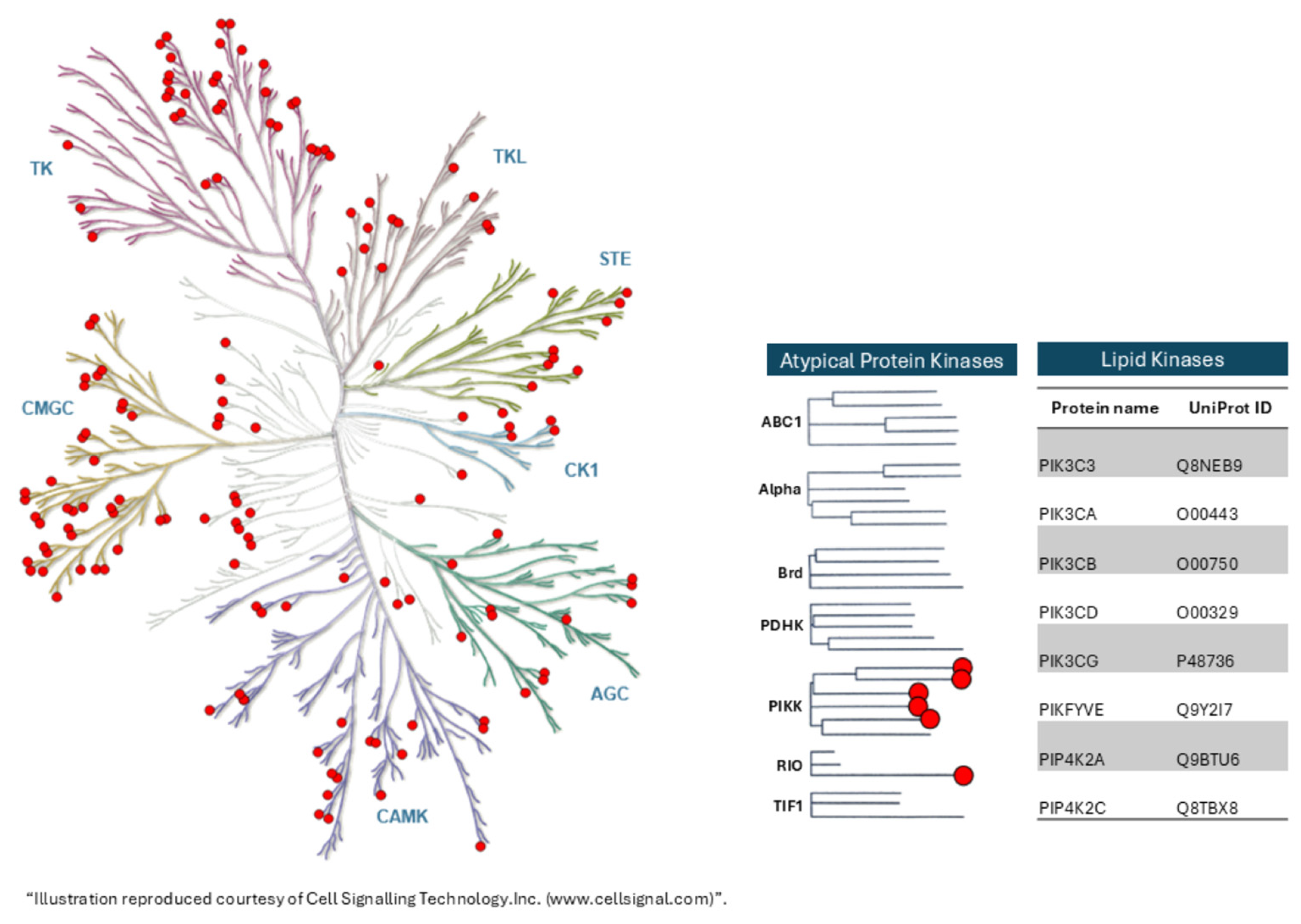
The Chemical Probes Portal is a free, open online resource which evaluates small-molecule reagents for their suitability as chemical probes via a large group (>260) of recognized chemical probe experts, the Scientific Expert Review Panel, or SERP [22,23]. Molecules valued with a three- or four-star rating (out of four) in cells are recommended for use as chemical probes, with a three-star rating indicating that it is the ‘best available probe for this target’ or a ‘high-quality probe that is a useful orthogonal tool’ and a four-star rating indicating that it is ‘recommended as a probe for this target’. Currently, the Portal features 201 kinase probes that have been rated 3 stars or above covering 169 targets (Figure 1) of which 13 are covalent inhibitors, indicating that selective kinase targeting can be achieved with increasing success. Multiple probes are available for several kinases, acting, for example, as degraders or inhibitors for the same target, thus enabling assessment of catalytic versus scaffolding effects when used in phenotypic assays.
While the majority of efforts to develop inhibitors continue to focus on already-well-known kinases, the SGC chemical probe program aims to enable the yet understudied, or so-called “dark”, kinases [24]. A list of understudied kinases that have been selected as part of the complementary NIH’s ‘Illuminating The Druggable Genome’ project has recently been published [25], and further information can be found at the publicly available Dark Kinase Knowledgebase (https://darkkinome.org/) (accessed on 29 January 2026). Following the stringent kinase chemical probe criteria mentioned above, the SGC program employs a rigorous evaluation process, which involves two independent expert review committees. Inhibitors that meet chemical probe criteria are approved and subsequently made openly available to the global research community without any restriction on use, ensuring broad access to high-quality, well-characterized chemical tools (https://www.thesgc.org/chemical-probes) (accessed on 29 January 2026). This approach facilitates more rapid characterization and validation of underexplored kinase targets and contributes to a broader understanding of kinase biology.
Building on this framework, the Donated Chemical Probes (DCP) initiative provides a complementary mechanism through which researchers from both industry and academia can contribute highly selective kinase probes for use by the community (https://www.sgc-ffm.uni-frankfurt.de/) (accessed on 29 January 2026) [13]. A hallmark of both programs is the availability of comprehensive profiling data provided for each probe, including off-target activities and recommended conditions for use as well as the physical probe and control compounds. Together these initiatives enable researchers worldwide to investigate kinase function using standardized chemical probes, thereby accelerating discovery and improving reproducibility across studies. Some highlights of kinase chemical probes are described below in further detail (Table 1).

3. Targeting Dark Kinases

The small death-associated protein kinase family (DAPK family) comprises five members (DAPK1, DAPK2, DAPK3, DRAK1, and DRAK2) and belongs to the calcium/calmodulin-dependent serine/threonine kinases [26]. Both DRAK1 (STK17A) and DRAK2 (STK17B) are highly understudied members of this family, but chemical probes are now available to study their respective functions. CK156 is a highly selective and potent (IC50 of 49 nM in a radiometric assay and a KD of 21 nM determined by ITC) macrocyclic pyrazolo [1,5-a]pyrimidine-based compound, which inhibits STK17A in a type I modus [27]. A cellular NanoBRET assay revealed a potent IC50 of 181 nM on DRAK1. The compound fulfilled all probe criteria and was tested in a cellular model of glioblastoma. Overexpression of DRAK1 has been reported in multiple gliomas, and expression of DRAK1 was found to be necessary for glial neoplasia [28]. However, the results obtained using the CK156 chemical probe did not reflect the effects observed in genetic knockdown experiments in glioblastoma, indicating the importance of this kinase’s scaffolding functions [27]. Nevertheless, this kinase remains an attractive target in several cancers, and other modalities, such as degrader molecules, could unlock its therapeutic potential [29]. SGC-STK17B-1 is a thieno [3,2-d] pyrimidine-based ATP-competitive inhibitor that binds to DRAK2 in a unique P-loop conformation [30]. This highly selective compound specifically binds to this kinase with an IC50 of 190 nM in a cellular NanoBRET assay with excellent selectivity over DRAK1 (>10 µM) as well as Aurora kinase B (AURKB), a potential off-target identified in the selectivity screen [30]. Subsequent experiments using this chemical probe revealed a critical role of DRAK2 in suppression of ferroptosis in multiple myeloma. Treatment of multiple myeloma cells for 3 days with high doses of SGC-STK17B-1 negatively affected proliferation of multiple myeloma cells and induced lipid peroxidation as well as increased intracellular labile iron levels. Because of the limited aqueous solubility and stability in liver microsomes, the compound was further optimized, resulting in compound AP-024, which was suitable for in vivo experiments. This improved compound-induced ferroptosis and significantly reduced tumor growth in multiple myeloma xenograft mouse models. Using the compound, the authors further showed that two regulators of iron uptake and transport are direct targets of STK17B, namely the iron-responsive element binding protein 2 (IREB2) and heat shock protein family B member 1 (HSPB1) [31].

4. Using Different Targeting Mechanisms to Study Isoform Selectivity of LIM Kinases

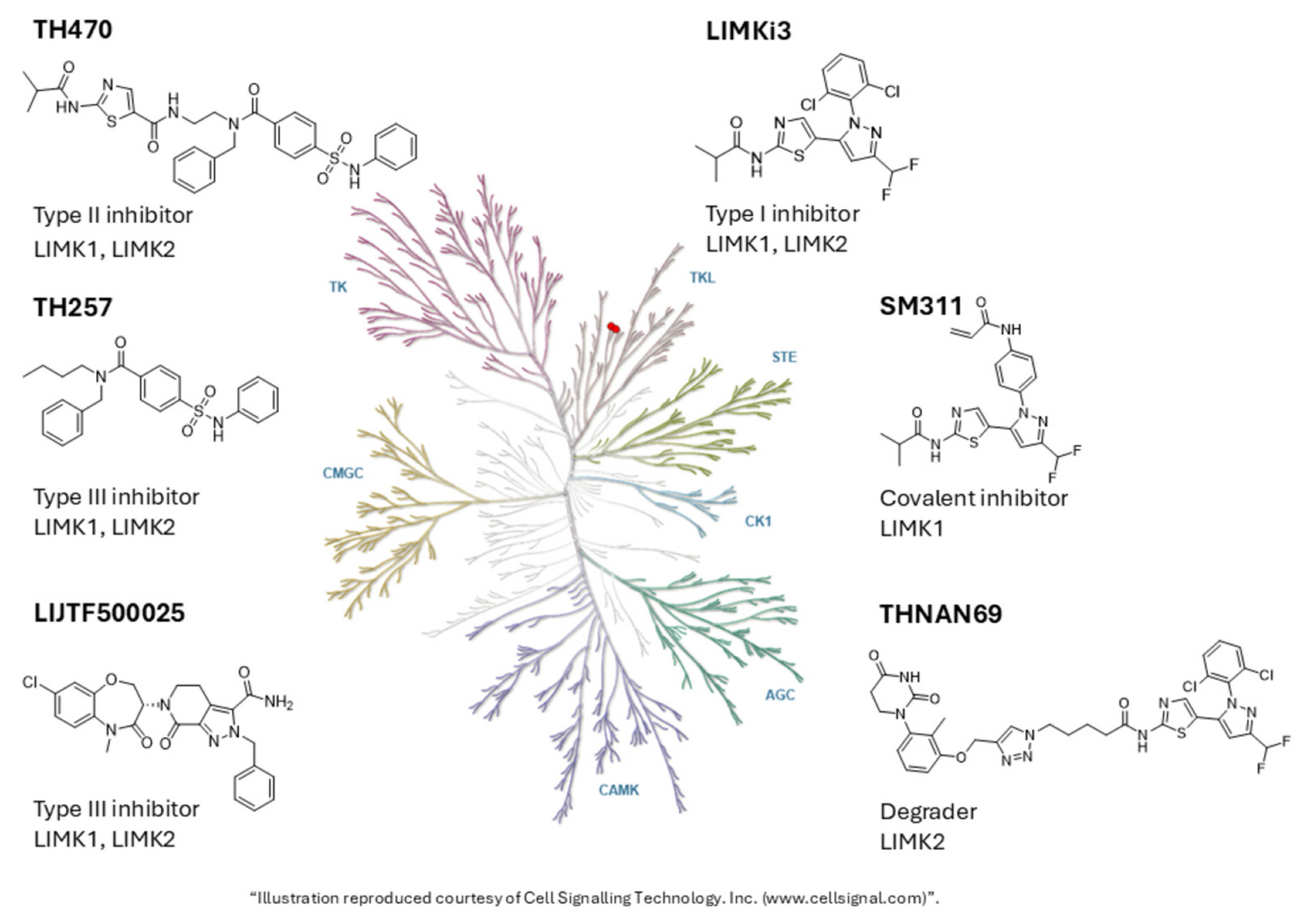
LIM kinases (LIMKs) 1 and 2 are structurally conserved cytoplasmic tyrosine-like kinases which recognize serine/threonine as well as tyrosine residues. Human LIMK1 and LIMK2 show an overall structural conservation with >70% sequence similarity within the kinase domain. Their expression differs, with LIMK1 being predominantly expressed in the brain as well as to a smaller extent in heart and skeletal muscle, while LIMK2 is more widely expressed [32,33]. Both kinases play a pivotal role in various cellular processes, including cell morphology, motility, and apoptosis, by regulating the actin cytoskeleton and other relevant proteins, such as cofilin, which are essential for these processes. Cofilin phosphorylation plays also a role in Alzheimer’s disease and Fragile-X syndrome, making these kinases attractive targets for these diseases in addition to others such as heart disease and cancer. Several dual inhibitors for LIMK1 and LIMK2 have been developed [34,35,36], and a number of them have been approved as chemical probes: LIMKi3, a type I inhibitor, TH470, a type II inhibitor, and two type III inhibitors, TH257 and LIJTF500025 [37] (Figure 2, Table 1). All chemical probes are highly potent in RapidFire MS assay with IC50s below the criterion of 100 nM, and only small differences in potency between LIMK1 and LIMK2 are observed. Although LIJTF500025 demonstrated a somewhat higher potency in vitro for LIMK2, evaluation in the cellular NanoBRET assay did not reveal significant selectivity of LIMK2 over LIMK1. All chemical probes have excellent selectivity across the kinome, with RIPK1 as the only off-target for the allosteric chemical probe LIJTF500025 [34,36]. Interestingly, comparative studies of quantitative phospho-proteomics revealed differential effects of the diverse inhibitor types. Whereas the type I inhibitor LIMKi3 and the type II inhibitor TH470 regulated phosphorylation sites in a similar manner, the type III inhibitor TH257 affected less phosphorylation sites which, moreover, differed from those affected by the type I and type II inhibitors. In addition to effects on the actin cytoskeleton or microtubules, a strong link to phosphorylation changes in proteins involved in RNA processing and RNA binding function as well as mRNA processing has been observed [36]. Conversely, the type I inhibitor LIMKi3 showed only a small effect in a neurite outgrowth model of the Fragile-X chromosome, whereas the type II inhibitor TH470 and the allosteric inhibitor TH257 strongly inhibited neurite outgrowth, suggesting that allosteric inhibitors might be more effective in modulating neuronal phenotypes mediated by the deregulation of LIMKs [34,36]. Thus, the availability of chemical probes belonging to different kinase inhibitor types enabled the study of the effect of different kinase conformations.
More recently, through different targeting strategies, it has been possible to address inhibition of LIMK isoforms, selectively. This has been enabled by targeting a specific cysteine, C349, located in the glycine-rich loop region of LIMK1, which is not conserved in LIMK2 [38]. The covalent inhibitor, which is based on the dual inhibitor LIMKi3 linked to a weak covalent warhead, shows more than 100-fold selectivity of LIMK1 (IC50 of 45 nM) over LIMK2 (IC50 of 6.74 µM) in a cellular NanoBRET assay, while remaining highly selective across the kinome and the proteome as demonstrated in a chemoproteomics experiment (Figure 2, Table 1). The chemical probe, SM311, presented a two-step irreversible inactivation mode with kinact = 2.83 × 10⁻³ s−1 and kinact/Ki = 6.95 × 10⁴ M−1 s−1 with a Ki value of 40.71 nM [38]. A structurally related negative control retaining the covalent warhead, which is inactive on LIMK1 (IC50 over 25 µM in a cellular NanoBRET assay), is available. Significantly, SM311 exhibited inhibition of cofilin phosphorylation, albeit to a lesser extent than the dual inhibitor LIMKi3, in line with the notion that both LIMK isoforms contribute to phosphorylation of this substrate. Complementarily, THNAN69, a LIMK2 selective degrader, has been developed, completing the set of LIMK isoform-specific chemical probes [39] (Figure 2, Table 1). Also, the PROTAC is based on the dual LIMKi3 inhibitor linked to a CRBN binding handle. Proteomics analysis revealed specific LIMK2 degradation (DC50 = 1 nM, Dmax ~ 90% at 10 nM doses) while LIMK1 protein levels were not affected. The accompanying negative control, which does not bind to CRBN, does not show any LIMK2 degradation up to 10 µM in a cellular assay. Interestingly, phosphoproteomics revealed no downregulation of phosphorylated cofilins. Using different targeting strategies, not only have selective chemical probes from different chemical inhibitor types been generated for LIMKs but, also, subtype selectivity could be achieved, enabling scientists to differentially study the role of LIMK1 versus LIMK2.

5. Chemical Probes for Lipid Kinases

Lipid kinases catalyze the phosphorylation of hydrophobic or amphipathic small molecules using ATP as phosphate donor. Contrary to protein kinases, they are less well studied [40]. The 37 lipid kinases have been subdivided into different groups with the largest being the diacylglycerol kinases (DGKs). The best studied groups are the PI3 kinases for which inhibitors, as well as approved drugs and isoform-specific molecules have been generated (https://www.chemicalprobes.org/search?q=Pi3k) (accessed on 29 January 2026) [41]. Additionally, selective inhibitors for a small number of other lipid kinases have been generated. BMS (Cambridge, MA, USA) recently published a first-in-class dual inhibitor for diacylglycerol kinase α (DGKα) and DGKζ (BMS-986408). This low nanomolar inhibitor acts both as an inhibitor and a proteosome-dependent degrader of these lipid kinases; however, further experiments to understand the precise mechanism of degradation are still required. It is presently unknown if BMS-986408 functions as a molecular glue or if the natural turnover of these proteins is accelerated [42].
The lipid kinase Phosphoinositide Kinase, FYVE-Type Zinc Finger Containing (PIKFYve, PIP5K3) kinase is a ubiquitously expressed kinase that phosphorylates the substrate phosphatidylinositol-3-phosphate (PI(3)P) to yield phosphatidylinositol-3,5-bisphosphate (PI(3,5)P2) and phosphatidylinositol 5-phosphate (PI(5)P). The kinase plays a role in cellular homeostasis, membrane trafficking, endosomal transport, lysosomal function, and signaling of Toll like receptors, inhibiting the IL-12/23 response. Additionally, a role in modulating neurotransmitter release and receptor recycling, impacting synaptic transmission and neuronal plasticity, has been reported. Several potent and selective inhibitors for the lipid kinase PIKfyve have been described, sharing a morpholino-pyridine or morpholino-pyrimidine with the morpholine and forming a critical hydrogen bond with the hinge region of the kinase [43,44,45]. In order to generate a chemical probe from an alternative scaffold, a library of indolyl pyrimidinamines was synthesized to which low binding to PIKFYVE has been reported. Optimization of the initial compounds resulted in a highly potent and cell-active chemical probe (SGC-PIKFYVE-1) that inhibits PIKfyve with 6.91 nM affinity in an enzymatic in vitro assay and bound to the kinase with an IC50 of 4 nM in a cellular NanoBRET assay. Comprehensive evaluation of a kinome-wide binding assessment confirmed that this PIKfyve probe demonstrates excellent selectivity. The probe is accompanied by a negative control compound that does not inhibit PIKfyve and exhibits excellent selectivity [43]. The PIKfyve chemical probe was shown to disrupt multiple phases of the β-coronavirus lifecycle, specifically viral replication and viral entry, in line with data observed with other PIKfyve inhibitors, whereas the negative control compound did not have any effect [43]. Subsequent experiments using the chemical probe revealed a role of PIKfyve in inhibition of Nav1.7 and Nav1.8 channels, without affecting voltage-gated calcium or potassium currents in sensory neurons. Moreover, intraperitoneal injection of SGC-PIKFYVE-1 produced an antinociceptive effect in a neuropathic pain model although the precise molecular mechanism remains to be explored [46]. Additional optimization of the chemical probes resulted in a compound with improved pharmacokinetic profiles and selectivity suitable for in vivo experiments [47].
Several well-characterized molecules are also available for inhibiting or degrading phosphatidylinositol-5-phosphate 4-kinases (PIP4Ks) as described in [48,49,50,51] and https://www.chemicalprobes.org/search?q=PIP4, accessed on 29 January 2026.
The PIP4K family, comprising PIP4K2A, PIP4K2B, and PIP4K2C, is family of type II lipid kinases that catalyze the conversion of phosphatidylinositol-5-phosphate (PI5P) to phosphatidylinositol 4,5-bisphosphate (PI(4,5)P2). PI(4,5)P2-mediated signaling regulates key cellular processes, including proliferation, survival, glucose uptake, and cytoskeletal organization, which have been reported to be dysregulated in cancer. Accordingly, aberrant PIP4K activity has been implicated in several malignancies, including breast cancer, acute leukemia, glioblastoma, and soft tissue sarcomas [52,53,54].
In recent years PIP4Ks have emerged as potential therapeutic targets, particularly in tumors harboring p53 mutations or depletions [55]. Genetic depletion of PIP4K2A and PIP4K2B selectively suppresses tumor growth in p53-deficient models both in vitro and in vivo. Mechanistically, loss of PIP4K2A/B leads to increased AKT phosphorylation, elevated oxygen consumption, and accumulation of reactive oxygen species (ROS), resulting in metabolic stress and the induction of senescence [55].
Pharmacological inhibition of PIP4K2A has been similarly associated with AKT hyperactivation, which enhances oxidative stress by increasing mitochondrial respiration and suppressing ROS scavengers downstream of FoxO-mediated transcription. Under conditions of elevated basal oxidative stress, as observed in p53-deficient tumors, this AKT-driven ROS accumulation sensitizes cells to senescence and apoptosis. Moreover, PIP4K2A knockdown in p53-mutant THP-1 acute myeloid leukemia cells impairs proliferation, induces G1 cell cycle arrest and apoptosis, and prevents leukemia development in xenograft models, accompanied by activation of AKT and mTOR signaling [56]. Interestingly, pharmacologic inhibition of PIP4K2A with the potent and highly selective PIP4K2A chemical probe BAY-091 did not recapitulate the hypothesized antiproliferative effects in p53-deficient tumor cells, indicating a more complex role for PIP4K2A signaling [57]. BAY-091 inhibits PIP4K2A at a single-digit nanomolar IC50 in vitro and is selective for PIP4K2 in a comprehensive kinase panel of 373 kinases. In a cellular context, target engagement has been proven by an in-cell thermal melt assay at 1 µM. When tested in THP-1 cells at concentration of up to 50 μM, BAY-091 did not increase the levels of p-AKT (Ser 473 and Thr308). In line with these findings, no consistent effect on mitochondrial ROS levels in THP-1 cells were observed after treatment with the PIP4K2A chemical probe. Together, these findings underscore the critical importance of well-characterized, potent, and selective chemical probes in distinguishing on-target pharmacological effects from genetic or compensatory phenomena.
Table 1. SGC and DCP kinase chemical probes mentioned in this manuscript.
Chemical Probe Kinase (Uniprot-ID)Chemical Probe StructureNegative ControlNegative Control Structure
CK156
[27]
STK17A
(Q9UEE5)
Kinasesphosphatases 04 00005 i001CKJB71Kinasesphosphatases 04 00005 i002
SGC-
STK17B-1
[30]
STK17B
(O94768)
Kinasesphosphatases 04 00005 i003SGC-
STK17B-1N
Kinasesphosphatases 04 00005 i004
LIMKi3
[36]
LIMK1, LIMK2
(P53667, P53671)
Kinasesphosphatases 04 00005 i005TH-263Kinasesphosphatases 04 00005 i006
TH470
[36]
LIMK1, LIMK2
(P53667, P53671)
Kinasesphosphatases 04 00005 i007TH-263Kinasesphosphatases 04 00005 i008
TH257
[36]
LIMK1, LIMK2 (P53667, P53671)Kinasesphosphatases 04 00005 i009TH-263Kinasesphosphatases 04 00005 i010
LIJTF500025
[37]
LIMK1, LIMK2 (P53667, P53671)Kinasesphosphatases 04 00005 i011LIJTF500120Kinasesphosphatases 04 00005 i012
SM311
[38]
LIMK1
(P53667)
Kinasesphosphatases 04 00005 i013SM311-NCKinasesphosphatases 04 00005 i014
THNAN69
[39]
LIMK2
(P53671)
Kinasesphosphatases 04 00005 i015THNAN69-NCKinasesphosphatases 04 00005 i016
BAY-091
[57]
PIP4K2A
(P48426)
Kinasesphosphatases 04 00005 i017BAY-0361Kinasesphosphatases 04 00005 i018
SGC-PIKFYVE-1
[43]
PIKfyve
(Q9Y2I7)
Kinasesphosphatases 04 00005 i019SGC-PIKFYVE-1NKinasesphosphatases 04 00005 i020

6. Discussion and Perspective

Despite initial challenges in overcoming kinase catalytic site conservation, strategies have been developed to achieve selective inhibition of specific kinases and even distinguishing between different isoforms. Cellular selectivity panels, taking into account the Km of ATP in cells, complement the available in vitro panels, providing excellent tools to assess the selectivity of a compound in a relevant system. Moreover, negative control compounds as well as orthogonal chemical probes available for many kinases help to verify that biological phenotypes are due to inhibition of the intended target. Additionally, genetic knockdowns can be employed to ensure that a compound only binds to the target that have been identified in in vitro profiling assays, and the phenotype is due to inhibition of the intended protein. Accordingly, adding a compound to cells where the proposed target protein has been knocked down will reveal whether the compound’s effect depends on that target. For kinases, re-expression of wild-type but not kinase-dead kinase in the knockout cells should restore sensitivity to the inhibitor. Thus, a suite of tools is available to ensure the selectivity of a chemical probe for important targets [58].
However, given the size of the kinome, generating chemical probes with appropriate controls for every kinase remains a formidable task. To address this issue, kinase chemogenomic sets have been assembled by Wells et al. and other scientists as part of the recently finished Innovative Medicine Initiative funded program EUbOPEN [20,59,60]. A chemogenomic library comprises chemogenomic compounds that fulfil similar potency criteria as chemical probes and are well validated but bind to more than one target. For kinase chemogenomic compounds, a maximum of 10 other kinases functioning as off-targets is acceptable. Ideally, different chemotypes or kinase inhibitor types are available for each target with complementary selectivity profiles https://www.eubopen.org/chemogenomics/chemogenomics-criteria, (accessed on 29 January 2026). Thus, using a set of well-characterized compounds with overlapping target profiles enables identification of the target responsible for a specific phenotype. Additionally, machine learning and AI approaches, based on the plethora of structural and biochemical and cellular data available for kinases, are gaining traction and will increasingly facilitate more accurate chemical probe design, minimizing off-target effects and substantially reducing trial-and-error experimentation. Ultimately, the availability of kinase chemical probes will also aid the development of novel and selective kinase drugs, reducing side-effects and unwanted toxicities, and enabling targeted precision medicine.

Author Contributions

Conceptualization, writing—review and editing, C.T. and S.M. All authors have read and agreed to the published version of the manuscript.

Funding

We gratefully acknowledge support from the Structural Genomics Consortium (SGC), a registered charity (no: 1097737) that receives direct member funding from Amgen Inc., Janssen Pharmaceutica NV, and Bristol-Myers Squibb Company, as well as grant funding from the Innovative Health Initiative Joint Undertaking (IHI JU; LIGAND-AI, grant agreement No. 101252959). The JU receives support from the European Union’s Horizon Europe research and innovative programme, COCIR, EFPIA, EuropaBio, MedTech Europe, Vaccines Europe, Enamine, and The Hospital for Sick Children. Views and opinions expressed are, however, those of the authors only and do not necessarily reflect those of the aforementioned parties. Neither of the aforementioned parties can be held responsible for them. SM receives funding from the translational cancer program of the German Cancer Aid TACTIC 2500 (70115201).

Institutional Review Board Statement

Not applicable.

Informed Consent Statement

Not applicable.

Data Availability Statement

No new data was created or analyzed in this study. Data sharing is not applicable to this article.

Conflicts of Interest

The authors declare no conflicts of interest.

References

  1. Mullard, A. FDA approves 100th small-molecule kinase inhibitor. Nat. Rev. Drug Discov. 2025, 24, 891–895. [Google Scholar] [CrossRef] [Scilit]
  2. Manning, G.; Whyte, D.B.; Martinez, R.; Hunter, T.; Sudarsanam, S. The protein kinase complement of the human genome. Science 2002, 298, 1912–1934. [Google Scholar] [CrossRef] [Scilit] [PubMed]
  3. Attwood, M.M.; Fabbro, D.; Sokolov, A.V.; Knapp, S.; Schioth, H.B. Trends in kinase drug discovery: Targets, indications and inhibitor design. Nat. Rev. Drug Discov. 2021, 20, 839–861, Erratum in Nat. Rev. Drug Discov. 2021, 20, 798. https://doi.org/10.1038/s41573-021-00303-4. [Google Scholar] [CrossRef] [Scilit] [PubMed]
  4. Kwak, H.A.; Liu, L.; Tredup, C.; Rohm, S.; Prinos, P.; Bottcher, J.; Schapira, M. Chemical coverage of human biological pathways. Drug Discov. Today 2024, 29, 104144. [Google Scholar] [CrossRef] [Scilit] [PubMed]
  5. Muller, S.; Chaikuad, A.; Gray, N.S.; Knapp, S. The ins and outs of selective kinase inhibitor development. Nat. Chem. Biol. 2015, 11, 818–821. [Google Scholar] [CrossRef] [Scilit]
  6. Wang, G.; Seidler, N.J.; Rohm, S.; Pan, Y.; Liang, X.J.; Haarer, L.; Berger, B.T.; Sivashanmugam, S.A.; Wydra, V.R.; Forster, M.; et al. Probing the Protein Kinases’ Cysteinome by Covalent Fragments. Angew. Chem. Int. Ed. Engl. 2025, 64, e202419736. [Google Scholar] [CrossRef] [Scilit]
  7. Chaikuad, A.; Koch, P.; Laufer, S.A.; Knapp, S. The Cysteinome of Protein Kinases as a Target in Drug Development. Angew. Chem. Int. Ed. Engl. 2018, 57, 4372–4385. [Google Scholar] [CrossRef] [Scilit]
  8. Anderson, B.; Rosston, P.; Ong, H.W.; Hossain, M.A.; Davis-Gilbert, Z.W.; Drewry, D.H. How many kinases are druggable? A review of our current understanding. Biochem. J. 2023, 480, 1331–1363. [Google Scholar] [CrossRef] [Scilit]
  9. Fang, Z.; Grutter, C.; Rauh, D. Strategies for the selective regulation of kinases with allosteric modulators: Exploiting exclusive structural features. ACS Chem. Biol. 2013, 8, 58–70. [Google Scholar] [CrossRef] [Scilit]
  10. Kossakowski, K.; Cherniienko, A.; Zaprutko, L.; Pawelczyk, A. FDA-approved kinase inhibitors in PROTAC design, development and synthesis. J. Enzym. Inhib. Med. Chem. 2025, 40, 2542357. [Google Scholar] [CrossRef] [Scilit]
  11. Tian, Y.; Lu, R.; Gong, X.; Zhao, W.; Li, Y.; Wang, X.; Jia, X.; Li, Q.; Yang, Y.; Tong, H.H.Y.; et al. Enhancing kinase-inhibitor activity and selectivity prediction through contrastive learning. Nat. Commun. 2025, 16, 10860. [Google Scholar] [CrossRef] [Scilit] [PubMed]
  12. Zhang, M.; Liu, Y.; Jang, H.; Nussinov, R. Strategy toward Kinase-Selective Drug Discovery. J. Chem. Theory Comput. 2023, 19, 1615–1628. [Google Scholar] [CrossRef] [Scilit] [PubMed]
  13. Tredup, C.; Ndreshkjana, B.; Schneider, N.S.; Tjaden, A.; Kemas, A.M.; Youhanna, S.; Lauschke, V.M.; Berger, B.T.; Kramer, A.; Berger, L.M.; et al. Deep Annotation of Donated Chemical Probes (DCP) in Organotypic Human Liver Cultures and Patient-Derived Organoids from Tumor and Normal Colorectum. ACS Chem. Biol. 2023, 18, 822–836. [Google Scholar] [CrossRef] [Scilit] [PubMed]
  14. Hartung, I.V.; Rudolph, J.; Mader, M.M.; Mulder, M.P.C.; Workman, P. Expanding Chemical Probe Space: Quality Criteria for Covalent and Degrader Probes. J. Med. Chem. 2023, 66, 9297–9312. [Google Scholar] [CrossRef] [Scilit] [PubMed]
  15. Reinecke, M.; Brear, P.; Vornholz, L.; Berger, B.T.; Seefried, F.; Wilhelm, S.; Samaras, P.; Gyenis, L.; Litchfield, D.W.; Medard, G.; et al. Chemical proteomics reveals the target landscape of 1,000 kinase inhibitors. Nat. Chem. Biol. 2024, 20, 577–585. [Google Scholar] [CrossRef] [Scilit]
  16. Robers, M.B.; Wilkinson, J.M.; Vasta, J.D.; Berger, L.M.; Berger, B.T.; Knapp, S. Single tracer-based protocol for broad-spectrum kinase profiling in live cells with NanoBRET. STAR Protoc. 2021, 2, 100822. [Google Scholar] [CrossRef] [Scilit]
  17. Arrowsmith, C.H.; Audia, J.E.; Austin, C.; Baell, J.; Bennett, J.; Blagg, J.; Bountra, C.; Brennan, P.E.; Brown, P.J.; Bunnage, M.E.; et al. The promise and peril of chemical probes. Nat. Chem. Biol. 2015, 11, 536–541, Erratum in Nat. Chem. Biol. 2015, 11, 887. https://doi.org/10.1038/nchembio1115-887c. [Google Scholar] [CrossRef] [Scilit]
  18. Lee, J.; Schapira, M. The Promise and Peril of Chemical Probe Negative Controls. ACS Chem. Biol. 2021, 16, 579–585. [Google Scholar] [CrossRef] [Scilit]
  19. Sterling, J.; Baker, J.R.; McCluskey, A.; Munoz, L. Systematic literature review reveals suboptimal use of chemical probes in cell-based biomedical research. Nat. Commun. 2023, 14, 3228. [Google Scholar] [CrossRef] [Scilit]
  20. Tredup, C.; Ackloo, S.; Beck, H.; Brown, P.J.; Bullock, A.N.; Ciulli, A.; Dikic, I.; Edfeldt, K.; Edwards, A.M.; Elkins, J.M.; et al. Toward target 2035: EUbOPEN—A public-private partnership to enable & unlock biology in the open. RSC Med. Chem. 2025, 16, 457–464. [Google Scholar] [CrossRef] [Scilit]
  21. Blagg, J.; Workman, P. Choose and Use Your Chemical Probe Wisely to Explore Cancer Biology. Cancer Cell 2017, 32, 268–270, Erratum in Cancer Cell 2017, 32, 9–25. https://doi.org/10.1016/j.ccell.2017.06.005. [Google Scholar] [CrossRef] [Scilit] [PubMed]
  22. Antolin, A.A.; Sanfelice, D.; Crisp, A.; Villasclaras Fernandez, E.; Mica, I.L.; Chen, Y.; Collins, I.; Edwards, A.; Muller, S.; Al-Lazikani, B.; et al. The Chemical Probes Portal: An expert review-based public resource to empower chemical probe assessment, selection and use. Nucleic Acids Res. 2023, 51, D1492–D1502. [Google Scholar] [CrossRef] [Scilit] [PubMed]
  23. Sanfelice, D.; Antolin, A.A.; Crisp, A.; Chen, Y.; Bellenie, B.; Brennan, P.E.; Edwards, A.; Muller, S.; Al-Lazikani, B.; Workman, P. The Chemical Probes Portal—2024: Update on this public resource to support best-practice selection and use of small molecules in biomedical research. Nucleic Acids Res. 2025, 53, D1663–D1669. [Google Scholar] [CrossRef] [Scilit] [PubMed]
  24. Koch, S.; Bajorath, J. Identifying and evaluating understudied protein kinases using biological and chemical criteria. RSC Med. Chem. 2025, 16, 3386–3392. [Google Scholar] [CrossRef] [Scilit] [PubMed]
  25. Vella, V.; Giamas, G.; Ditsiou, A. Diving into the dark kinome: Lessons learned from LMTK3. Cancer Gene Ther. 2022, 29, 1077–1079. [Google Scholar] [CrossRef] [Scilit]
  26. Farag, A.K.; Roh, E.J. Death-associated protein kinase (DAPK) family modulators: Current and future therapeutic outcomes. Med. Res. Rev. 2019, 39, 349–385. [Google Scholar] [CrossRef] [Scilit]
  27. Kurz, C.G.; Preuss, F.; Tjaden, A.; Cusack, M.; Amrhein, J.A.; Chatterjee, D.; Mathea, S.; Berger, L.M.; Berger, B.T.; Kramer, A.; et al. Illuminating the Dark: Highly Selective Inhibition of Serine/Threonine Kinase 17A with Pyrazolo [1,5-a]pyrimidine-Based Macrocycles. J. Med. Chem. 2022, 65, 7799–7817. [Google Scholar] [CrossRef] [Scilit]
  28. Chen, A.S.; Wardwell-Ozgo, J.; Shah, N.N.; Wright, D.; Appin, C.L.; Vigneswaran, K.; Brat, D.J.; Kornblum, H.I.; Read, R.D. Drak/STK17A Drives Neoplastic Glial Proliferation through Modulation of MRLC Signaling. Cancer Res. 2019, 79, 1085–1097. [Google Scholar] [CrossRef] [Scilit]
  29. Ouaakki, H.; Bravo, E., Jr.; Taylor, J.; Feng, Y. Therapeutic potential of STK17A targeted therapies. Bioorg. Med. Chem. Lett. 2026, 132, 130506. [Google Scholar] [CrossRef] [Scilit]
  30. Picado, A.; Chaikuad, A.; Wells, C.I.; Shrestha, S.; Zuercher, W.J.; Pickett, J.E.; Kwarcinski, F.E.; Sinha, P.; de Silva, C.S.; Zutshi, R.; et al. A Chemical Probe for Dark Kinase STK17B Derives Its Potency and High Selectivity through a Unique P-Loop Conformation. J. Med. Chem. 2020, 63, 14626–14646. [Google Scholar] [CrossRef] [Scilit]
  31. Yan, Z.; Han, Z.; Wang, Y.; Beus, M.; Zhang, Y.; Picado, A.; Wells, C.I.; Wu, J.; Weidenhammer, L.B.; Pires, K.M.; et al. Targeting STK17B kinase activates ferroptosis and suppresses drug resistance in multiple myeloma. Blood 2026, 147, 48–60. [Google Scholar] [CrossRef] [Scilit] [PubMed]
  32. Lateef, O.M.; Foote, C.; Power, G.; Manrique-Acevedo, C.; Padilla, J.; Martinez-Lemus, L.A. LIM kinases in cardiovascular health and disease. Front. Physiol. 2024, 15, 1506356. [Google Scholar] [CrossRef] [Scilit] [PubMed]
  33. Manetti, F. LIM kinases are attractive targets with many macromolecular partners and only a few small molecule regulators. Med. Res. Rev. 2012, 32, 968–998. [Google Scholar] [CrossRef] [Scilit] [PubMed]
  34. Collins, R.; Lee, H.; Jones, D.H.; Elkins, J.M.; Gillespie, J.A.; Thomas, C.; Baldwin, A.G.; Jones, K.; Waters, L.; Paine, M.; et al. Comparative Analysis of Small-Molecule LIMK1/2 Inhibitors: Chemical Synthesis, Biochemistry, and Cellular Activity. J. Med. Chem. 2022, 65, 13705–13713. [Google Scholar] [CrossRef] [Scilit]
  35. Villalonga, E.; Mosrin, C.; Normand, T.; Girardin, C.; Serrano, A.; Zunar, B.; Doudeau, M.; Godin, F.; Benedetti, H.; Vallee, B. LIM Kinases, LIMK1 and LIMK2, Are Crucial Node Actors of the Cell Fate: Molecular to Pathological Features. Cells 2023, 12, 805. [Google Scholar] [CrossRef] [Scilit]
  36. Hanke, T.; Mathea, S.; Woortman, J.; Salah, E.; Berger, B.T.; Tumber, A.; Kashima, R.; Hata, A.; Kuster, B.; Muller, S.; et al. Development and Characterization of Type I, Type II, and Type III LIM-Kinase Chemical Probes. J. Med. Chem. 2022, 65, 13264–13287. [Google Scholar] [CrossRef] [Scilit]
  37. Mandel, S.; Hanke, T.; Mathea, S.; Chatterjee, D.; Saraswati, H.; Berger, B.T.; Schwalm, M.P.; Yamamoto, S.; Tawada, M.; Takagi, T.; et al. Repurposing of the RIPK1-Selective Benzo [1,4]oxazepin-4-one Scaffold for the Development of a Type III LIMK1/2 Inhibitor. ACS Chem. Biol. 2025, 20, 1087–1098. [Google Scholar] [CrossRef] [Scilit]
  38. Mandel, S.; Hanke, T.; Prendiville, N.; Baena-Nuevo, M.; Berger, L.M.; Farges, F.; Schwalm, M.P.; Berger, B.T.; Kraemer, A.; Elson, L.; et al. Covalent Targeting Leads to the Development of a LIMK1 Isoform-Selective Inhibitor. J. Med. Chem. 2025, 68, 15026–15049. [Google Scholar] [CrossRef] [Scilit]
  39. Azeez, R.A.K.; Saraswati, H.; Mosler, T.; Hanke, T.; Ho, H.; Neder, N.; Sivashanmugam, S.A.; Heinz, M.; Schwalm, M.P.; Giuliani, G.; et al. Available online: https://www.sgc-ffm.uni-frankfurt.de/#!specificprobeoverview/THNAN69 (accessed on 29 January 2026).
  40. Ware, T.B.; Hsu, K.-L. Advances in chemical proteomic evaluation of lipid kinases—DAG kinases as a case study. Curr. Opin. Chem. Biol. 2021, 65, 101–108. [Google Scholar] [CrossRef] [Scilit]
  41. Sirico, M.; D’Angelo, A.; Gianni, C.; Casadei, C.; Merloni, F.; De Giorgi, U. Current State and Future Challenges for PI3K Inhibitors in Cancer Therapy. Cancers 2023, 15, 703. [Google Scholar] [CrossRef] [Scilit]
  42. Wichroski, M.; Liu, S.Q.; Zasadil, L.M.; Benci, J.L.; Gedeon, P.C.; Condon, K.J.; Joshi, S.; Posy, S.; Carlson, P.; Maier, A.; et al. Discovery of BMS-986408, a First-In-Class Dual DGKalpha and DGKzeta Inhibitor that Unleashes PD-1 Checkpoint and CAR T-cell Immunotherapies. Cancer Immunol. Res. 2025, 13, 1342–1362. [Google Scholar] [CrossRef] [Scilit] [PubMed]
  43. Drewry, D.H.; Potjewyd, F.M.; Bayati, A.; Smith, J.L.; Dickmander, R.J.; Howell, S.; Taft-Benz, S.; Min, S.M.; Hossain, M.A.; Heise, M.; et al. Identification and Utilization of a Chemical Probe to Interrogate the Roles of PIKfyve in the Lifecycle of beta-Coronaviruses. J. Med. Chem. 2022, 65, 12860–12882. [Google Scholar] [CrossRef] [Scilit] [PubMed]
  44. Ikonomov, O.C.; Sbrissa, D.; Shisheva, A. Small molecule PIKfyve inhibitors as cancer therapeutics: Translational promises and limitations. Toxicol. Appl. Pharmacol. 2019, 383, 114771. [Google Scholar] [CrossRef] [Scilit] [PubMed]
  45. Rivero-Rios, P.; Weisman, L.S. Roles of PIKfyve in multiple cellular pathways. Curr. Opin. Cell Biol. 2022, 76, 102086. [Google Scholar] [CrossRef] [Scilit]
  46. Rodriguez-Palma, E.J.; Loya-Lopez, S.; Min, S.M.; Calderon-Rivera, A.; Gomez, K.; Khanna, R.; Axtman, A.D. Targeting Na(v)1.7 and Na(v)1.8 with a PIKfyve inhibitor to reverse inflammatory and neuropathic pain. Neurobiol. Pain 2025, 17, 100174. [Google Scholar] [CrossRef] [Scilit]
  47. Min, S.M.; Bashore, F.M.; Smith, J.L.; Havener, T.M.; Howell, S.; Li, H.; Counago, R.M.; Popov, K.I.; Axtman, A.D. Development of a Second-Generation, In Vivo Chemical Probe for PIKfyve. J. Med. Chem. 2025, 68, 3282–3308. [Google Scholar] [CrossRef] [Scilit]
  48. Manz, T.D.; Sivakumaren, S.C.; Ferguson, F.M.; Zhang, T.; Yasgar, A.; Seo, H.S.; Ficarro, S.B.; Card, J.D.; Shim, H.; Miduturu, C.V.; et al. Discovery and Structure-Activity Relationship Study of (Z)-5-Methylenethiazolidin-4-one Derivatives as Potent and Selective Pan-phosphatidylinositol 5-Phosphate 4-Kinase Inhibitors. J. Med. Chem. 2020, 63, 4880–4895. [Google Scholar] [CrossRef] [Scilit]
  49. Teng, M.; Jiang, J.; Wang, E.S.; Geng, Q.; Toenjes, S.T.; Donovan, K.A.; Mageed, N.; Yue, H.; Nowak, R.P.; Wang, J.; et al. Targeting the Dark Lipid Kinase PIP4K2C with a Potent and Selective Binder and Degrader. Angew. Chem. Int. Ed. Engl. 2023, 62, e202302364. [Google Scholar] [CrossRef] [Scilit]
  50. Rooney, T.P.C.; Aldred, G.G.; Boffey, H.K.; Willems, H.M.G.; Edwards, S.; Chawner, S.J.; Scott, D.E.; Green, C.; Winpenny, D.; Skidmore, J.; et al. The Identification of Potent, Selective, and Brain Penetrant PI5P4Kgamma Inhibitors as In Vivo-Ready Tool Molecules. J. Med. Chem. 2023, 66, 804–821. [Google Scholar] [CrossRef] [Scilit]
  51. Drewry, D.H.; Potjewyd, F.M.; Smith, J.L.; Howell, S.; Axtman, A.D. Identification of a chemical probe for lipid kinase phosphatidylinositol-5-phosphate 4-kinase gamma (PI5P4Kγ). Curr. Res. Chem. Biol. 2023, 3, 100036. [Google Scholar] [CrossRef] [Scilit]
  52. Lima, K.; Pereira-Martins, D.A.; de Miranda, L.B.L.; Coelho-Silva, J.L.; Leandro, G.D.S.; Weinhauser, I.; Cavaglieri, R.C.; Leal, A.M.; da Silva, W.F.; Lange, A.; et al. The PIP4K2 inhibitor THZ-P1-2 exhibits antileukemia activity by disruption of mitochondrial homeostasis and autophagy. Blood Cancer J. 2022, 12, 151. [Google Scholar] [CrossRef] [Scilit] [PubMed]
  53. Arora, G.K.; Palamiuc, L.; Emerling, B.M. Expanding role of PI5P4Ks in cancer: A promising druggable target. FEBS Lett. 2022, 596, 3–16. [Google Scholar] [CrossRef] [Scilit] [PubMed]
  54. Ravi, A.; Palamiuc, L.; Emerling, B.M. Crucial Players for Inter-Organelle Communication: PI5P4Ks and Their Lipid Product PI-4,5-P(2) Come to the Surface. Front. Cell Dev. Biol. 2021, 9, 791758. [Google Scholar] [CrossRef] [Scilit] [PubMed]
  55. Emerling, B.M.; Hurov, J.B.; Poulogiannis, G.; Tsukazawa, K.S.; Choo-Wing, R.; Wulf, G.M.; Bell, E.L.; Shim, H.S.; Lamia, K.A.; Rameh, L.E.; et al. Depletion of a putatively druggable class of phosphatidylinositol kinases inhibits growth of p53-null tumors. Cell 2013, 155, 844–857. [Google Scholar] [CrossRef] [Scilit] [PubMed]
  56. Gurpinar, E.; Vousden, K.H. Hitting cancers’ weak spots: Vulnerabilities imposed by p53 mutation. Trends Cell Biol. 2015, 25, 486–495. [Google Scholar] [CrossRef] [Scilit] [PubMed]
  57. Wortmann, L.; Brauer, N.; Holton, S.J.; Irlbacher, H.; Weiske, J.; Lechner, C.; Meier, R.; Karen, J.; Sioberg, C.B.; Putter, V.; et al. Discovery and Characterization of the Potent and Highly Selective 1,7-Naphthyridine-Based Inhibitors BAY-091 and BAY-297 of the Kinase PIP4K2A. J. Med. Chem. 2021, 64, 15883–15911. [Google Scholar] [CrossRef] [Scilit]
  58. Ortiz-Ruiz, M.J.; Popoola, O.; Mitsopoulos, K.; Te-Poele, R.; Samant, R.S.; Box, G.; Court, W.; Brandon, A.H.; Gowan, S.; Mallinger, A.; et al. Mediator Kinase Inhibitor Selectivity and Activity in Colorectal Cancer. ACS Chem. Biol. 2025, 20, 1792–1804. [Google Scholar] [CrossRef] [Scilit]
  59. Wells, C.I.; Al-Ali, H.; Andrews, D.M.; Asquith, C.R.M.; Axtman, A.D.; Dikic, I.; Ebner, D.; Ettmayer, P.; Fischer, C.; Frederiksen, M.; et al. The Kinase Chemogenomic Set (KCGS): An Open Science Resource for Kinase Vulnerability Identification. Int. J. Mol. Sci. 2021, 22, 566. [Google Scholar] [CrossRef] [Scilit]
  60. Muller, S.; Ackloo, S.; Al Chawaf, A.; Al-Lazikani, B.; Antolin, A.; Baell, J.B.; Beck, H.; Beedie, S.; Betz, U.A.K.; Bezerra, G.A.; et al. Target 2035—Update on the quest for a probe for every protein. RSC Med. Chem. 2022, 13, 13–21. [Google Scholar] [CrossRef] [Scilit]
Figure 1. Kinase targets covered by high-quality chemical probes. A total of 169 unique human kinase targets with available high-quality chemical probes are mapped onto the human kinome tree using KinHub and complemented by a table of lipid kinases. Kinases are highlighted by red circles for which at least one chemical probe rated three or four stars on the Chemical Probes Portal is available.
Kinasesphosphatases 04 00005 g001
Figure 2. Dual and isoform-selective LIMK1 and LIMK2 chemical probes. A set of well-characterized chemical probes targeting LIMK1 and LIMK2 is available, covering different inhibitor classes. These include type I (LIMKi3), type II (TH470), and type III inhibitors (TH257 and LIJTF500025), all of which are highly potent and display excellent kinome-wide selectivity. The covalent probe SM311 selectively targets LIMK1 by exploiting a non-conserved cysteine residue, while a LIMK2-selective PROTAC, THNAN69, enables targeted degradation of LIMK2. Highlighted by red dots are LMK1 and LIMK2, the only kinases that are bound by these chemical probes.
Kinasesphosphatases 04 00005 g002
Disclaimer/Publisher’s Note: The statements, opinions and data contained in all publications are solely those of the individual author(s) and contributor(s) and not of MDPI and/or the editor(s). MDPI and/or the editor(s) disclaim responsibility for any injury to people or property resulting from any ideas, methods, instructions or products referred to in the content.