Review

Overcoming Microenvironment-Driven Resistance to CAR-T Therapy in Multiple Myeloma

1
Department of Internal Medicine, Virginia Mason Franciscan Health–St. Joseph Medical Center, Tacoma, WA 98405, USA
2
Saint Louis University School of Medicine, St. Louis, MO 63104, USA
3
Touro College of Osteopathic Medicine, New York, NY 10027, USA
*
Author to whom correspondence should be addressed.
Lymphatics 2026, 4(1), 15; https://doi.org/10.3390/lymphatics4010015
Submission received: 30 January 2026 / Revised: 1 March 2026 / Accepted: 3 March 2026 / Published: 5 March 2026

Abstract

B cell maturation antigen (BCMA)-targeted chimeric antigen receptor T cell (CAR-T) therapy has transformed the treatment landscape for relapsed or refractory multiple myeloma (MM), with products such as idecabtagene vicleucel and ciltacabtagene autoleucel achieving high initial response rates, and in selected patient populations, durable treatment-free remission. However, a substantial proportion of patients still experience relapse, including antigen-positive progression, highlighting persistent limitations in long-term disease control across diverse clinical settings. An increasing body of evidence indicates that resistance to CAR-T therapy in MM is driven not only by tumor-intrinsic factors, but also by extrinsic pressures imposed by the bone marrow microenvironment (BMME). This review integrates current understanding of tumor-niche interactions that impair CAR-T persistence, trafficking, and effector function, including immunosuppressive cellular networks, inhibitory cytokine signaling, metabolic constraints, stromal adhesion, antigen modulation, and marrow remodeling. This review further examines emerging therapeutic strategies and next-generation CAR-T platforms.

1. Introduction

Multiple myeloma (MM) is a difficult-to-control hematologic disease driven by uncontrolled expansion of malignant plasma cells and the uncontrolled production of monoclonal antibodies. It is characterized by widespread immune dysfunction and disruption of the bone marrow microenvironment (BMME), leading to bone reabsorption, lytic lesions, and myelosuppression. These processes drive morbidity and mortality through skeletal fragility, increased calcium reabsorption with hypercalcemia, kidney failure, and immunosuppression with opportunistic infections. Despite advances in treatment, relapses and development of treatment resistance have made lasting remission a challenge. Chimeric antigen receptor T cell (CAR-T) therapy is a rapidly growing area of study in autoimmune and oncologic diseases and has shown promise for directed therapies against new therapeutic targets, including investigations of MM and targets within the BMME to address mechanisms of treatment resistance.
Many trials have investigated CAR-T therapies in MM, with over 70 trials spanning diverse targets and strategies. Trials such as KarMMa, CARTITUDE-1, and CARTITUDE-4 were groundbreaking in demonstrating CAR-T efficacy against malignant plasma cells in MM. KarMMa evaluated idecabtagene vicleucel, a B cell maturation antigen (BCMA)-directed CAR-T, in triple-therapy-resistant MM refractory to immunomodulatory therapy, proteasome inhibitors, and anti-CD38 antibodies. KarMMa demonstrated efficacy in aggressive treatment-resistant disease, but control was not definitive; the average response duration was 10.7 months, and progression-free survival was 8.8 months after a single dose. Cytokine release syndrome (CRS) and transient, but clinically significant cytopenias with related complications occurred in almost all the treated patients, though mortality attributed to idecabtagene vicleucel was small. KarMMa also demonstrated effective T cell expansion and durability, but persistence did not guarantee durable disease control, as reflected by the response duration and progression-free survival (Table 1) [1,2,3,4].
The CARTITUDE trials evaluated the safety and use of ciltacabtagene autoleucel in relapsed or refractory multiple myeloma. Ciltacabtagene also targets BCMAs and demonstrated significant efficacy in resistant MM in CARTITUDE-1, with an overall response rate of 97.9% and a median time to best response of 2.6 months [5]. Compared with KarMMa, CARTITUDE-1 reported higher response rates and improved duration of this response and progression-free survival. These outcomes occurred despite a lack of detectable CAR-T cells in most patients by 6 months, suggesting durability of the clinical effect without CAR-T survival. Hematologic adverse events occurred in over 90% of the patients, with most recovering within 1–2 months (Table 1) [6,7,8,9].
Table 1. The anti-BCMA CAR-T trial outcomes. This table outlines some of the characteristics and results of landmark trials regarding the safety, efficacy, and adverse events regarding the use of anti-BCMA CAR-T therapy in the treatment of MM.
TrialProductPopulationORR, %≥3. Median PFS, moMedian DoR, moCRS ≥ G3, %ICANS ≥ G3, %
KarMMa-1 [6] Idecabtagene vicleucelTriple therapy RRMM 73338.810.753
KarMMa-2 [2,3,4] Idecabtagene vicleucelHR NDMM87.177.411.415.72.70
KarMMa-3 [1] Idecabtagene vicleucelTriple therapy RRMM 714413.84697
CARTITUDE-1 [5,9,10,11]Ciltacabtagene autoleucelTriple therapy RRMM 9782.534.933.949
CARTITUDE-2 [7,8] Ciltacabtagene autoleucelTriple therapy RRMM 95909.111.5100
CARTITUDE-4 [12] Ciltacabtagene autoleucelLenalidomide RRMM84.673.1not reachedNot reached1.10.1
A key barrier is durability of remission, limited by waning CAR-T persistence and CAR-T exhaustion with reduced effectiveness. While prolonged antigen exposure commonly drives T cell exhaustion through nuclear factor of activated T cells (NFAT) and inhibitory immune checkpoint pathways, extrinsic BMME factors such as transforming growth factor beta (TGF-β) and galactin-9 (GAL9) can push T cell populations toward exhaustion. Post-infusion proliferation of an inhibitory T cell lineage (Tregs) against CAR-T cells has been noted and is suspected to contribute to exhaustion and relapse [13]. MM cell lines have also been observed to shed BCMA into the BMME as a soluble form that can bind CAR-T cells and reduce binding affinity to membrane-bound BCMA, facilitating antigen-positive escape [14].
Although the treatment options for multiple myeloma are far more extensive than they have historically been, no therapies consistently induce complete remission due to multiple mechanisms, often centered on the BMME. CAR-T therapy has been a revolutionary addition to MM treatment, but is still imperfect, and we must overcome barriers to reverse resistant disease. As identified above, impaired CAR-T durability in MM most often reflects intrinsic and extrinsic factors that reduce proliferation and cause exhaustion of chimeric T cells. This review further explores the extrinsic BMME factors that drive these barriers and summarizes the current efforts to bypass them. This review also highlights the need for biomarker monitoring to recognize therapy failure earlier and identify foci of resistance that can more effectively guide salvage therapy.

2. Barriers of the MM Bone Marrow Niche

2.1. Cell-Mediated Immunosuppression

As a malignant plasma cell, MM is uniquely able to insert itself into a protective BM niche consisting of multiple cell lines and pathways. The BM niche is comprised of hematopoietic stem cells, mesenchymal stem cells, vascular endothelial cells, osteoblasts, and BM stromal cells, which maintain an environment conducive to appropriate proliferation and growth. Within this microenvironment, MM cells leverage Tregs, myeloid-derived suppressor cells (MDSCs), macrophages, monocytes, and natural killer (NK) cells to promote tumor survival, often by blunting immune responses. These cells also generate a cytokine milieu shaped by MM cells to reinforce immunosuppression and protect tumor cells [15,16,17].
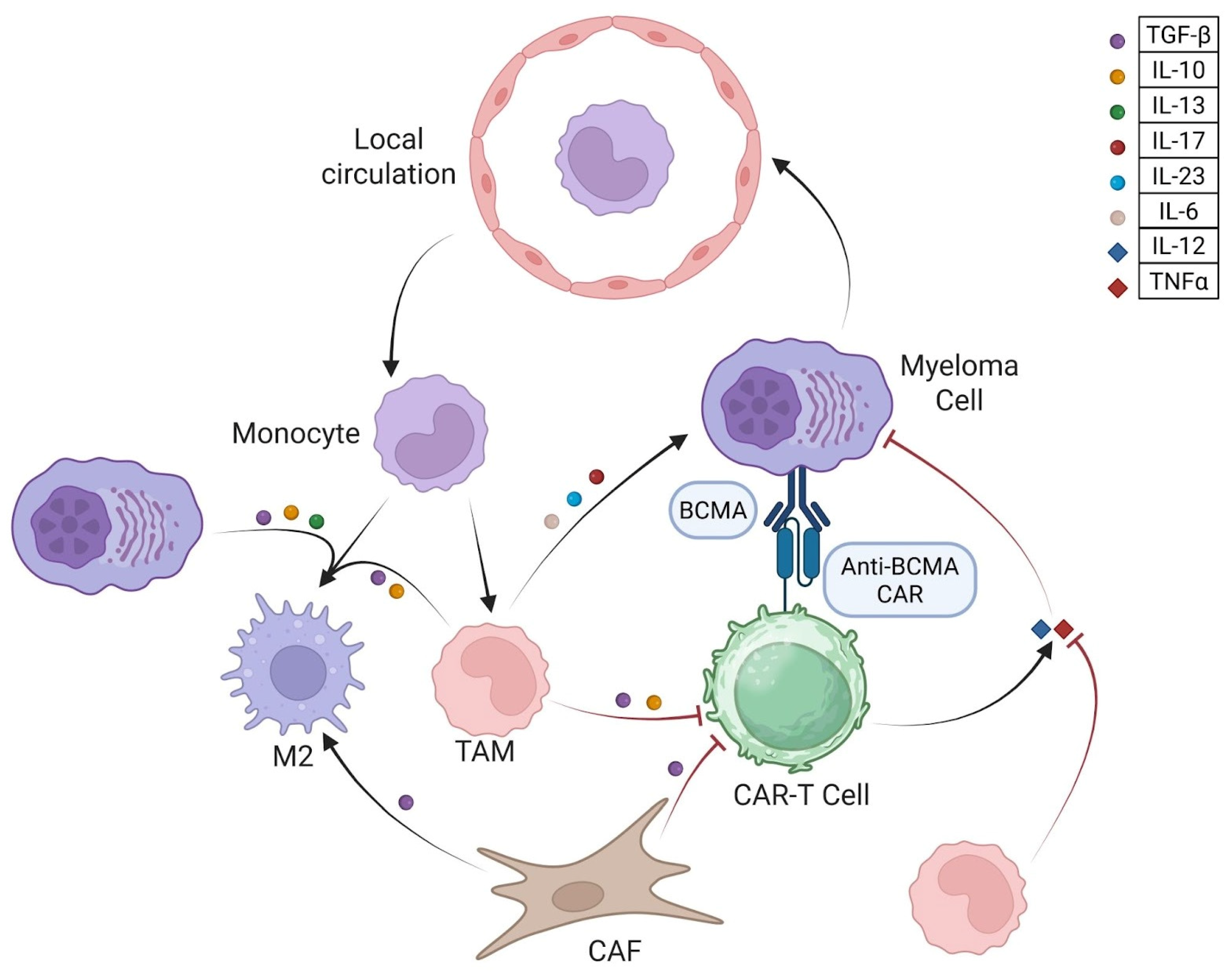
Within the tumor microenvironment (TME), macrophages regulate innate and adaptive immunity, and their polarization state shapes antitumor immune responses with a spectrum spanning from pro-inflammatory, tumoricidal type 1 macrophages (M1) phenotypes to immunoregulatory, tumor-supportive M2 phenotypes. In MM, the BMME favors M2-like polarization through soluble mediators, including transforming growth factor-β (TGF-β), interleukin (IL)-10, and IL-13, along with tumor-derived metabolic and mechanical cues (Figure 1) [18]. Myeloma cells recruit circulating monocytes and reprogram tissue-resident macrophages into tumor-associated macrophages (TAMs), which dominate the marrow niche and reinforce immune dysfunction. TAMs sustain immunosuppression through inhibitory cytokines such as TGF-β and IL-10, while also producing pro-tumor inflammatory mediators, including IL-17 and IL-23, promoting genomic instability, impairing immune surveillance, and accelerating functional exhaustion of effector lymphocytes (Figure 1). TAMs expand in tumor niches and support functions overlapping with type 2 macrophages (M2s), including anti-inflammatory IL-10 signaling, propagation of M2 differentiation, production of IL-6 (critical to MM development) and TGF-β, and inhibition of IL-12 and tumor necrosis factor alpha (TNFα) signaling (Figure 1) [15]. Collectively, macrophage and monocyte-driven signaling establishes a profoundly immunosuppressive marrow environment that limits CAR-T persistence and durability of response. In MM patients treated with CAR-T therapy, increased monocyte numbers and increased TGF-β activity were associated with increased markers of T cell exhaustion and decreased proliferation, which may reduce persistence of the chimeric T cells [19].
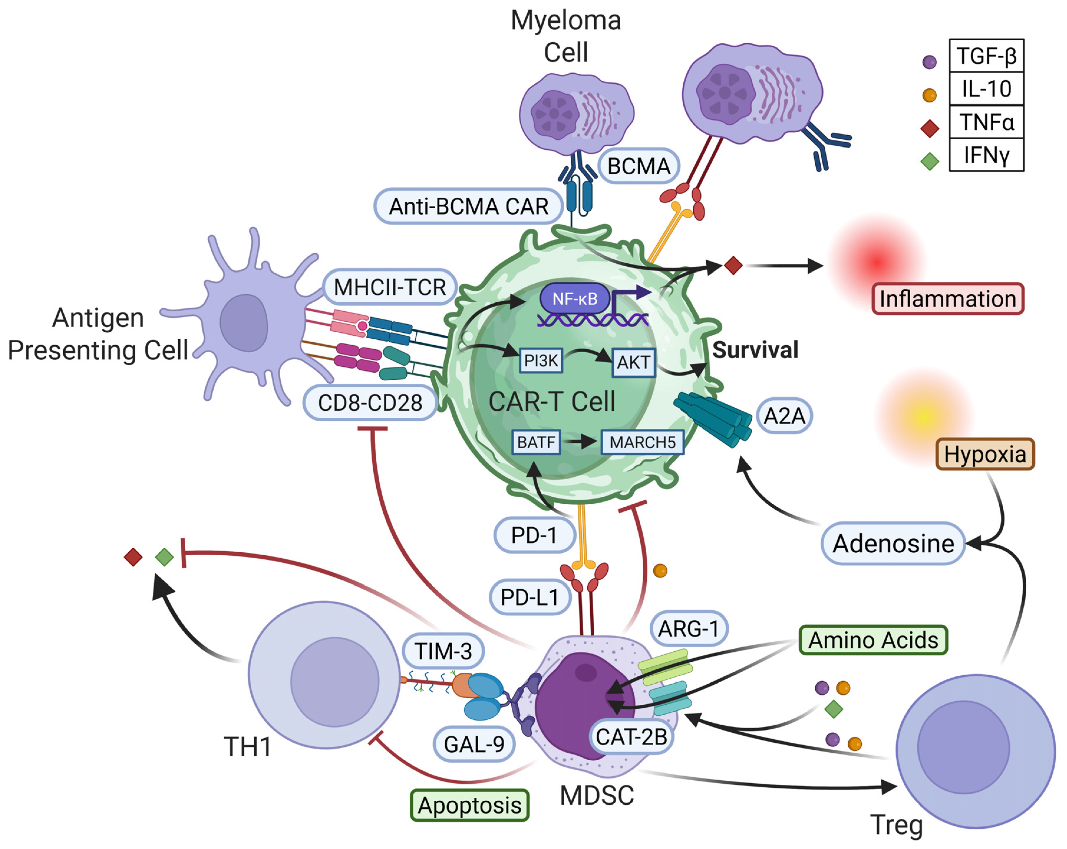
MDSCs are myeloid progenitor cells that are particularly contributory in malignancy. In tumor niches, MDSCs often show increased programmed death ligand (PD-L)1 expression, in part as a response to exaggerated hypoxia. PD-L1 binding to programmed cell death protein 1 (PD-1) on T cells is associated with MDSC-derived IL-10 and suppression of effector T cell activity, including CAR-T cells (Figure 2) [20]. Mechanistically, PD-1 acts as a co-inhibitor that interferes with T cell receptor (TCR) activation and CD28 signaling, preventing effective phosphorylation of downstream pathways such as phosphoinositide 3-kinase (PI3K) and protein kinase C theta (PKCθ), suppressing nuclear factor kappa B (NF-κB), decreasing TNFα production, and reducing cytotoxic activity [21,22,23]. PD-1 activation also induces basic leucine zipper ATF-like transcription factor (BATF), a transcription factor for membrane-associated ring-CH-type finger 5 (MARCH5), decreasing the T cell receptor responses to cytokines, including IL-2, and promoting receptor degradation [24]. PD-1 binding can additionally impair glucose and amino acid processing by T cells, resulting in impaired metabolism and function (Figure 2) [25]. GAL-9 is another factor found on MDSCs that interact with T cell immunoglobulin and mucin domain 3 (TIM-3) on CAR-T cells, decreasing interferon gamma (IFN-γ) production and inducing T cell death (T-helper type 1 (TH1) cells specifically) (Figure 2) [26]. This loop causes expansion of the MDSCs with further T cell disruption and immunosuppression.
Beyond ligand/receptor signaling, MDSCs suppress T cells through metabolic restriction. Cytokines, including TGF-β, IL-4, IL-10, and IFN-γ, increase the quantities of cationic amino acid transporter 2B (CAT-2B) and arginase-1 (ARG-1) on MDSCs, driving L-arginine uptake from the microenvironment and stunting T cell proliferation (Figure 2). MDSCs can also deplete intracellular L-arginine within T cells via methylglyoxal transfer, impairing proteins incorporating L-arginine and crippling T cell function. MDSCs further deplete tryptophan and cystine required by T cells [27]. Adenosine plays a large role in these interactions. MDSCs and hypoxic environments cause increased production of adenosine compounds that degrade into adenosine in the microenvironment. This available adenosine then binds to MDSCs, stromal cells, and T cells, causing increased immunosuppression through MDSC activity and inhibition of T cell function (Figure 2) [28]. MDSCs also generate reactive oxygen species (ROS) and nitric oxygen (NO), which can impede proliferation and promote apoptosis in cytotoxic T cells and CAR-T cells. In parallel, MDSCs promote differentiation of Tregs, often through TGF-β signaling and ROS production [28].
Closely related to MDSCs and TAMs are cancer-associated fibroblasts (CAFs). CAFs are a stromal subtype that produce fibroblast activating protein (FAP) and secrete IL-6 and TGF-β (Figure 1). In MM, the number of CAFs is increased, and they promote tumor growth, while also suppress CAR-T proliferation and degranulation through inhibitory cytokine pathways [29].
Tregs represent a significant immunosuppressive pathway that is reinforced by the cell populations above and promotes further Treg differentiation. This is mediated by IL-10 production to suppress effector T cell function and TGF-β to both suppress effector cells and promote differentiation toward Tregs. PD-1/PD-L1 interactions on T cells, influenced by MDSCs, also shift T cell populations toward Tregs. Once differentiated, Tregs participate in adenosine generation and suppression of effector T cell activity (Figure 2) [30].
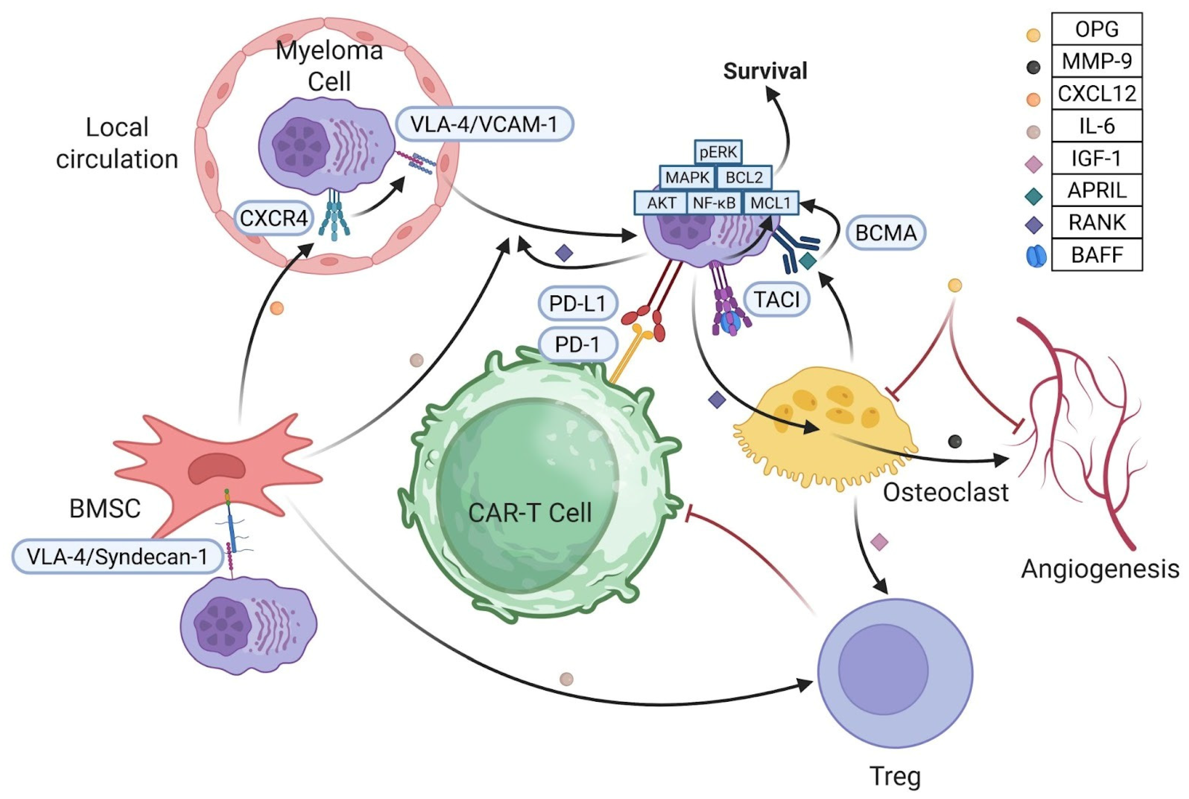
Some other supporting cells, including osteoclasts, produce cytokines that promote MM survival and immunosuppression. A proliferation-inducing ligand (APRIL) produced by osteoclasts binds BCMA on MM cells and increases PD-L1 expression, causing PD-1/PD-L1-mediated suppression of T cells and a loop in which activated T cells drive osteoclast activity and additional APRIL production [31,32]. APRIL binding BCMA also promotes MM survival and growth through the protein kinase RNA-like endoplasmic reticulum kinase (pERK1/2), mitogen-activated protein kinase (MAPK), protein kinase B (AKT), NF-κB, myeloid cell leukemia 1 (MCL1) and B cell lymphoma 2 (BCL2) pathways (Figure 3) [33]. B cell activating factor (BAFF), produced by multiple cell types, including macrophages and some MM cells, binds BCMA and transmembrane activator and cancer-associated-macrophage-like (CAML) interactor (TACI) on MM cells to promote anti-apoptotic pathways, including BCL2 and MCL1, reinforcing APRIL function to bind BCMA and TACI (Figure 3) [31,33,34]. Osteoclast-derived insulin-like growth factor-1 (IGF-1) promotes MM growth through MAPK, AKT, and NF-κB signaling similar to APRIL [34,35]. IGF-1 can also strengthen immunosuppression by promoting Treg proliferation (Figure 3) [31,35]. While APRIL, BAFF, and IGF-1 do not often directly contribute to CAR-T therapy failure, they emit strong survival signals (in part via BCMA binding) with some capacity to induce Treg predominance. A reciprocal and self-amplifying feedback loop exists between malignant plasma cells and osteoclasts. Myeloma cells induce osteoclast differentiation and activation, while hyperactivated osteoclasts enhance tumor progression, promote angiogenesis, and suppress immune surveillance. The emerging evidence demonstrates activated osteoclast upregulation of PD-1/PD-L1 signaling, diminishing T cell proliferation and cytotoxic activity and attenuating antitumor immune responses [15,36].
Vascular remodeling further defines the myeloma marrow ecosystem and contributes to immune exclusion. Crosstalk between osteoclasts and myeloma cells increases the number of angiogenic mediators, such as vascular endothelial growth factor (VEGF) and osteopontin, driving endothelial expansion and reinforcing osteoclastogenesis. Receptor activator of nuclear factor kappa-B ligand (RANKL)-driven osteoclast activation promotes angiogenesis through osteoclast-derived matrix metalloproteinase-9 (MMP-9), while osteoprotegerin inhibits both osteoclast formation and neovascular development, positioning the RANKL/osteoprotegerin (OPG) axis as a central regulator of marrow remodeling (Figure 3) [15,37,38]. Collectively, this osteoclast–vasculature–myeloma triad establishes a remodeled marrow niche that protects malignant cells, suppresses immune effector function, and limits effective CAR-T engagement within the tumor-infiltrated BM.

2.2. Stromal and Endothelial Adhesion Networks

Stromal features of the BMME contribute to disease progression drug resistance across different disease states and may contribute to suppression of CAR-T activity. A major mechanism is adhesion networks between MM cells, integrins, and fibronectin that confer resistance to apoptosis and are observed in drug resistant MM as cell adhesion-mediated drug resistance (CAM-DR) [39]. Adhesion to the ECM also induces growth-stimulating and immunosuppressive cytokine production from other BM cell lines. This resistance to apoptosis may extend to CAR-T therapy, representing an additional barrier to CAR-T durability in MM. While these interactions have not often been studied directly in relation to CAR-T therapy in MM, their anti-apoptotic effects are in direct competition with the cytotoxic effects of CAR-T therapy [31,40].
Stromal and endothelial changes may contribute to CAR-T failure through mechanisms that intersect with cell-mediated suppression. The PD-L1 and GAL9 pathways expand MDSCs, while sustaining immunosuppressive function, and MDSC-derived ROS further increases stromal hostility [41]. Stromal C-X-C motif chemokine ligand (CXCL)12 contributes to MM protection within the niche. Bone marrow stromal cells (BMSCs) are a primary producer of CXCL12, which acts as a chemoattractant for CXCL12–C-X-C motif chemokine receptor (CXCR)4 expressed on hematopoietic stem cells, including MM B cells (Figure 3) [38]. This chemotaxis is aided by integrins such as very late antigen-4 (VLA-4) and vascular cell adhesion molecule 1 (VCAM-1), promoting MM adhesion and migration into BM; CXCR4 signaling also enhances homing and cell survival through IL-6 and VEGF activity (Figure 3) [37,39]. Osteoclast activity through osteopontin (OPN) production and RANK signaling has also been associated with CXCL12/CXCR4 in MM [42]. While CXCL12/CXCR4 does not directly prevent T cell migration into the MM niche, it preferentially homes MM cells and macrophages that are differentiated to suppress T cell activity [38].
Within the BM, MM cells bind BMSCs, as well as bind fibronectin via syndecan-1 and VLA-4, increasing structural coherence and inducing IL-6 production by BMSCs. IL-6 promotes MM survival and Treg differentiation and is a key driver of CAM-DR (Figure 3). While these integrins do not directly inhibit CAR-T cells in MM, T cell activity has been suppressed in high-density collagen environments in breast cancer. This correlates with variable regions of T cell infiltration within the MM niche, though this appears to be significantly mediated by dendritic cells [43,44]. These BMSC-dependent stromal networks are key drivers of CAM-DR, and the activity of BMSCs and their anti-apoptotic effects on MM cells have been linked to resistance to CAR-T cell activity, strengthening the connection between these survival mechanisms and treatment failure [45]. While future research will need to further elucidate the roles CAM-DR, fibronectin, and adhesion networks play in CAR-T therapy failure in MM, these competing survival and anti-apoptotic pathways are in direct opposition to CAR-T cell function and must be overcome when significant enough to influence treatment success.
The BMME in MM also develops physical and metabolic constraints that limit CAR-T function. MM marrow is more hypoxic than unaffected marrow due to proliferation; hypoxia acidifies the microenvironment and increases genetic instability, promoting MM progression. Hypoxia increases adenosine, which binds the adenosine A2A (A2A) receptors on T cells to suppress proliferation and tumor cytotoxicity, and also impacts macrophages and other immune lineages to further induce immunosuppression (Figure 2) [46,47].
One additional mechanism of escape is BCMA shedding into the microenvironment, mediated by γ-secretase cleavage of membrane-bound BCMA, which reduces the effectiveness of BCMA-targeted CAR-T therapy [48]. These barriers share overlapping pathways, but also distinct contributions to CAR-T limitation in MM; optimizing CAR-T will require addressing each barrier through CAR-T modification and/or adjunct therapies targeting these inhibitory mechanisms.

3. Therapeutic Strategies for Overcoming BMME Barriers

3.1. Antigen Density and Shedding

BCMA remains the primary antigen target for the majority of the current FDA-approved CAR-T cell therapies. Numerous studies have demonstrated that antigen density is a critical determinant of treatment response, as insufficient target expression limits CAR-T cell activation, persistence, and cytotoxic efficacy [14,49]. One potential approach to addressing low-level BCMA membrane expression involves targeting the γ-secretase complex. Inhibition of γ-secretase has been shown to increase surface BCMA density, potentially enhancing CAR-T cell engagement and therapeutic efficacy [48,50].
An alternative strategy to overcome resistance associated with low-level BCMA expression is the development of dual-targeted CAR-T cell therapies. These approaches aim to reduce antigen escape by simultaneously targeting multiple surface antigens. One promising target is G protein-coupled receptor class C group 5 member D (GPRC5D), which is regulated independently of BCMA and has been shown to retain expression even after BCMA-directed therapy failure [51]. Although the precise biological function of GPRC5D is not understood, its persistent expression following BCMA escape suggests potential utility as either a dual-target or sequential immunotherapy option after relapse. However, clinical studies evaluating GPRC5D-targeted approaches have produced variable outcomes, with some demonstrating limited or inconsistent benefits [51].
There are many other emerging targets of interest that are being studied for the use of multiple myeloma treatment and this remains an area of interest. The other potential therapeutic targets that still need further research include semaphorin 4A (SEMA4A), Fc receptor-homolog 5 (FcRH5), signaling lymphocytic activation molecule family member 7 (SLAMF7), CD229, and integrin β7 [52,53] (Table 2).

3.2. Trafficking and Retention

The CXCL12/CXCR4 receptor ligand axis is a key regulator of plasma cell trafficking, retention, and long-term survival within the BM [54]. The BM niche itself presents a significant barrier to effective cellular immunotherapy. To overcome niche constraints on CAR-T cell access, CAR-T cells engineered to coexpress chemokine receptors have been developed as homing strategies. CAR-T cells coexpressing CXCR4 leverage the same chemokine gradients used by myeloma cells, demonstrating enhanced trafficking to the BM, improved tumor engagement, and increased antitumor activity; early preclinical and clinical studies suggest deeper responses and reduced disease burden compared to conventional CAR-T cells lacking chemokine receptor coexpression [55,56].
Similar strategies employing alternative chemokine receptors, such as C-C motif chemokine receptor (CCR)2b and C-X-C motif chemokine receptor 1 (CX3CR1), have also demonstrated improved CAR-T cell migration and efficacy in solid and hematologic malignancies [57]. These approaches appear to function through analogous mechanisms, wherein engineered receptor expression enables CAR-T cells to respond to tumor-derived chemokine gradients, thereby enhancing access [58].
Beyond cellular engineering strategies, pharmacologic disruption of the CXCL12/CXCR4 axis has an established clinical precedent. Plerixafor, also known as AMD3100, is a small-molecule CXCR4 antagonist that competitively binds CXCR4 and prevents CXCL12-mediated signaling activation. Plerixafor has been FDA-approved since 2008 for hematopoietic stem cell mobilization in patients undergoing autologous transplantation for non-Hodgkin lymphoma and multiple myeloma [59,60,61,62,63]. Plerixafor was originally developed and evaluated as a potential antiviral agent for HIV due to CXCR4’s role as a viral coreceptor [64]. By competitively inhibiting CXCL12 binding to CXCR4, plerixafor disrupts retention signals within the BM, leading to rapid mobilization of hematopoietic stem and progenitor cells into the peripheral circulation [65]. While this mechanism is currently used for stem cell collection, its therapeutic application in multiple myeloma is of increasing interest. Transient disruption of the CXCL12/CXCR4 axis may dislodge malignant plasma cells from their protective niches, rendering them more susceptible to immune-mediated killing or cytotoxic therapies [66]. Beyond mobilization, plerixafor has demonstrated potential to sensitize tumor cells to chemotherapy and radiation. Preclinical studies further suggest that plerixafor may inhibit tumor progression and metastasis by disrupting CXCR4-mediated tumor–stroma interactions, although it has not received FDA approval for direct cancer treatment [59,60].
LY2510924 is a next-generation cyclic peptide CXCR4 antagonist designed to improve tissue penetration and pharmacokinetic stability. The preclinical and clinical data indicate that LY2510924 may suppress tumor progression, while increasing tumor sensitivity to chemotherapy and reducing systemic toxicities associated with broad CXCR4 inhibition. Its peptide-based structure supports targeted molecular engineering to improve stability, bioavailability, and pharmacokinetics. Administered as a daily subcutaneous injection, LY2510924 demonstrates rapid plasma absorption with peak concentrations reached within 1–2 h and an elimination half-life of approximately 10–12 h. Metabolism occurs predominantly via proteolysis with minimal renal excretion, supporting use across varying degrees of renal function. Clinical studies suggest that LY2510924 may enhance responses when combined with immune checkpoint inhibitors, chemotherapy, and potentially radiation therapy [59].
POL6326 is a peptide-based CXCR4 antagonist primarily studied for stem cell mobilization, offering an alternative to plerixafor with reliable efficacy and a favorable safety profile, though development has been limited by high manufacturing costs and a relatively short in vivo duration of action [59]. X4P-001 (mavorixafor/Xolremdi) is an orally bioavailable CXCR4 antagonist that competitively inhibits CXCL12 binding, remodeling the tumor immune microenvironment to suppress tumor growth, metastasis, and angiogenesis. Relative to older CXCR4 antagonists (e.g., plerixafor and LY2510924), mavorixafor exhibits enhanced receptor-binding affinity, an extended half-life, reduced immunogenicity, superior tissue penetration, and improved bioavailability; oral administration also does not cause injection site reactions, and early clinical studies (n = 25) reported a favorable safety profile at doses up to 50 mg/day without dose-limiting toxicities [59].
BKT-140 (BL-8040) is distinguished among the CXCR4 antagonists by its ability to induce rapid, selective apoptosis of leukemia and myeloma cells both in vitro and in vivo upon CXCR4 engagement. In contrast to plerixafor, which demonstrates biphasic effects on MM cell survival and proliferation, BKT-140 exhibits direct cytotoxic activity and synergizes with chemotherapeutic agents such as rapamycin to enhance MM cell death [32] (Table 2).

3.3. Immunosuppression

An approach to overcoming macrophage-driven suppression includes “armored” CAR-T cells engineered to secrete pro-inflammatory cytokines such as IL-12, IL-15, and IL-18, which can reprogram the marrow niche toward an immune-stimulatory state. These cytokines enhance T cell survival, promote TH1 polarization, and counteract M2 dominance within the TME.
Preclinical studies demonstrate that pharmacologic TGF-β blockade, alone or combined with stimulator of interferon genes (STING) agonists, can reprogram the myeloid compartment toward a pro-inflammatory state. STING ligands such as 5,6-dimethylxanthenone-4-acetic acid (DMXAA) and cyclic guanosine monophosphate-adenosine monophosphate (cGAMP) increase CXCL9 and CXCL10 expression, recruit CXCR3+ TH1 cells, expand M1s, and reduce M2 and MDSC signatures, collectively enhancing CAR-T persistence and function [15].
Early clinical translation of these strategies is encouraging. In a phase Ib study, the TGF-β receptor I inhibitor, vactosertib, combined with pomalidomide was well tolerated by patients with relapsed/refractory MM and demonstrated favorable progression-free outcomes. Treatment reduced PD-1 expression on CD8+ T cells, decreased PD-L1 and PD-L2 expression on CD138+ myeloma cells, and enhanced autologous T cell cytotoxicity, indicating partial reversal of niche-mediated immune dysfunction [67].
Inhibitory immune checkpoint signaling through the PD-1/PD-L1 axis represents an additional major contributor to CAR-T dysfunction in MM. Multiple engineering strategies have been developed to overcome this barrier, including CAR-T cells that secrete PD-1 or PD-L1-blocking single-chain variable fragments, express dominant-negative PD-1 receptors, or incorporate PD-1 chimeric switch receptors that convert inhibitory signals into activation. Gene-editing approaches such as CRISPR-mediated PD-1 knockout or shRNA-based suppression further protect CAR-T cells from exhaustion [15].
Despite these advances, PD-1/PD-L1 antibody monotherapy has demonstrated limited efficacy in relapsed/refractory MM, as evidenced by early clinical trials of nivolumab and pembrolizumab. In contrast, combination strategies show substantially greater activity. In MM patient-derived co-culture systems, pairing PD-1 or PD-L1 blockade with IMiDs enhances NK and T cell cytotoxicity, increases interferon-γ and granzyme B production, and reduces PD-1/PD-L1 expression on both effector and tumor cells [31,34].
Engineered CAR-T cells capable of secreting checkpoint-blocking antibodies further support this approach, demonstrating enhanced antitumor activity, increased endogenous T cell infiltration, and reduced M2 populations in multiple preclinical models. Early clinical testing of IFN-γ-induced anti-PD-1-secreting CAR-T cells has yielded partial responses with acceptable toxicity profiles, supporting further development of this platform [68].
IMiDs remain central partners in combination strategies designed to overcome immune suppression. By binding cereblon and redirecting the E3 ubiquitin ligase complex to degrade ikaros zinc finger (IKZF)1 and IKZF3, IMiDs exert both direct antimyeloma effects and potent immune-stimulatory activity. Although lenalidomide can enhance wingless (Wnt)/β-catenin signaling in plasma cells, it also augments T cell activation and cytokine production [34,53].
Importantly, IMiDs synergize with checkpoint inhibition. In MM patient-derived co-culture models, combining lenalidomide with PD-1 or PD-L1 blockade enhances cytotoxicity by NK and T cells, increases interferon-γ and granzyme B secretion, and decreases checkpoint expression on effector and tumor cells. Lenalidomide and pomalidomide also enhance immune activation by stimulating CD4+ T cell IL-2 secretion, promoting NK cell activation, and augmenting T cell proliferation, cytokine production, and TH1 polarization. Combination strategies incorporating IMiDs with monoclonal antibodies such as daratumumab (anti-CD38) and elotuzumab (anti-SLAMF7), checkpoint inhibitors, or bispecific T cell engagers have demonstrated synergistic effects mediated by enhanced T and NK cell-dependent cytotoxicity and are being explored alongside CAR T cell therapy to mitigate resistance, reduce relapse, and improve CAR T cell persistence [69]. These findings highlight IMiDs as critical immune modulators capable of enhancing CAR-T functional capacity within an immunosuppressive niche [31].
Despite major therapeutic advances, immune checkpoint inhibitors alone have shown limited benefit due to immune-related toxicities. Their optimal role likely lies in rational combination strategies with IMiDs or engineered CAR-T constructs where synergistic immune activation can overcome microenvironmental resistance [13,34] (Table 2).

3.4. Adhesion and Extracellular Matrix Barriers

Effective CAR-T cytotoxicity requires a coordinated sequence of events, including chemokine-guided trafficking, endothelial transmigration, active infiltration through extracellular matrix (ECM) components, and formation of stable immunologic synapses with target cells. Tumors disrupt each of these steps through vascular remodeling, abnormal chemokine gradients, and ECM thickening, as discussed previously. Hypoxia, nutrient deprivation, and acidic conditions within the TME further compromise CAR-T survival and persistence [15].
One strategy to overcome ECM density involves engineering CAR-T cells to express or secrete ECM-modifying enzymes. Heparanase is particularly relevant, as in vitro expanded T cells lose endogenous heparanase expression due to tumor protein p53 (TP53)-mediated repression of the heparanase (HPSE) promoter, limiting their ability to degrade heparan sulfate proteoglycans. Restoring heparanase expression enhances CAR-T infiltration and antitumor activity in neuroblastoma xenograft models. Similarly, glypican-3-targeted CAR-T cells coexpressing IL-7 and PH20 hyaluronidase demonstrate improved trafficking and infiltration in hepatocellular carcinoma models, supporting dual-function constructs that combine cytokine support with enzymatic ECM remodeling [15,40].
Collectively, these findings establish a strong rationale for engineering CAR-T cells with heparanase, hyaluronidase, or integrin-modulating elements to facilitate deeper penetration into stroma-rich tumor regions. Direct enzymatic degradation of ECM components represents an additional approach to improving CAR-T infiltration. Mesothelin-targeted CAR-T cells engineered to secrete a modified human hyaluronidase (sPH20-IgG2) exhibit superior infiltration and antitumor activity in gastric cancer xenograft models. Clinical studies of pegylated recombinant human hyaluronidase (PEGPH20) have shown mixed results in pancreatic adenocarcinoma, underscoring both the promise and complexity of stromal remodeling strategies [15] (Table 2).
Other ECM-targeting enzymes, including collagenases such as MMP-8, remain largely preclinical, while strategies involving bacterial collagenase are limited by immunogenicity and are not suitable for systemic CAR-T therapy [15]. Additional approaches to overcoming ECM-mediated exclusion have been proposed, including direct intratumoral delivery of ECM-degrading agents and localized stromal modulation strategies. While methods such as bacterial collagenase injection can transiently reduce matrix density and improve immune cell penetration, their clinical utility is limited by significant immunogenicity and lack of specificity, rendering them unsuitable for systemic CAR-T-based therapies. These approaches nonetheless underscore the importance of ECM remodeling as a determinant of effective immune infiltration and highlight the need for safer, more targeted strategies [40].
Preclinical targeting of VLA-4 adhesion molecules reduces the tumor burden, and clinically relevant agents such as natalizumab disrupt myeloma–stromal interactions. More broadly, cell adhesion molecules, including CD38, SLAMF7, and integrins, contribute to MM progression, angiogenesis, and drug resistance. Therapeutics targeting these pathways establish a rationale for combination strategies aimed at weakening stromal protection and enhancing CAR-T cell access [70]. Together, these strategies aim to reduce ECM density, enhance CAR-T motility, and promote stable tumor engagement. By facilitating deeper infiltration into stroma-rich tumor regions and improving access to malignant cells, ECM-targeted interventions represent critical adjuncts to adoptive cellular immunotherapy and provide strong rationale for engineering combined approaches in CAR-T design.

3.5. Marrow Remodeling

Osteoclast-driven bone resorption and concomitant marrow vascular remodeling constitute central features of the myeloma-supportive niche and pose significant barriers to effective CAR-T cell trafficking, persistence, and cytotoxic function. Beyond mediating pathological bone destruction, osteoclasts generate chemokine-enriched microdomains that attract malignant plasma cells and promote their survival. Therapeutic strategies aimed at suppressing osteoclast activity represent a rational approach to destabilizing the myeloma niche and indirectly enhancing CAR-T cell efficacy. Immunomodulatory drugs, including lenalidomide and pomalidomide, inhibit osteoclast differentiation through downregulation of purine-rich box (PU.)1 and pERK signaling pathways and normalize the RANKL/OPG ratio, thereby reducing the osteoclastogenic signaling driven by myeloma cells.
Bisphosphonates selectively accumulate at sites of active remodeling, inducing osteoclast apoptosis, inhibiting osteoclast maturation, and preserving osteoblast viability to reduce pathological bone resorption. Denosumab, a monoclonal antibody targeting RANKL, prevents RANKL–RANK interactions on osteoclast precursors and increases trabecular and cortical bone masses; a phase III trial demonstrated non-inferiority versus zoledronic acid in preventing skeletal-related events in newly diagnosed myeloma, expanding the options for patients with renal dysfunction or bisphosphonate intolerance [31].
Emerging bone-anabolic strategies further complement the anti-resorptive approaches. Targeting the Wnt signaling pathway, a key regulator of osteoblast differentiation, has demonstrated efficacy in preclinical myeloma models. Inhibition of soluble Wnt antagonists, such as dickkopf-1 (DKK1) and sclerostin, combined with activation of low-density lipoprotein receptor-related protein 6 (LRP6), prevents bone loss and markedly increases the trabecular bone volume in murine systems, including 5TGM1-bearing models. Importantly, combined anti-LRP6/DKK1 strategies enhance bone mass by reducing osteoclast number and surface area without promoting tumor growth, supporting their potential role as niche-modifying adjuncts to cellular immunotherapy [71].
Given the niche dependence on angiogenesis, anti-vascular therapies may further diminish stromal support and improve immune accessibility. Sorafenib, targeting rapidly accelerated fibrosarcoma (RAF) and VEGFR-2, exhibits antimyeloma activity in vitro and retains cytotoxic efficacy despite stromal interactions or exposure to pro-survival cytokines (IL-6, VEGF, and IGF), with mechanisms including downregulation of MCL-1 and suppression of signal transducer and activator of transcription 3 (STAT3) and MEK/extracellular signal-regulated kinase (ERK) signaling; synergy has been demonstrated with mammalian target of rapamycin (mTOR) inhibitors, proteasome inhibitors, and corticosteroids, and sorafenib also inhibits angiogenesis [72]. VEGF-targeting agents such as bevacizumab, erlotinib, and apatinib are widely used in solid tumors, but have had limited application in myeloma due to toxicity and resistance. Angiopoietin-2 (Ang2) is a mediator of neovascular remodeling and resistance to VEGF-directed therapies; Ang2 inhibition, particularly combined with chemotherapy, radiotherapy, or immunotherapy, has demonstrated enhanced antitumor responses and may be a promising adjunct in myeloma [73].
Restoring osteoblast activity is another anabolic strategy to reverse myeloma-induced skeletal deterioration and disrupt niche-mediated tumor support. Carfilzomib promotes osteoblast differentiation from mesenchymal stromal cells and suppresses osteoclast differentiation through inhibition of RANKL-induced NF-κB signaling and downregulation of alpha(v)beta(3) (αVβ3) integrin expression; these effects have been validated in healthy and myeloma-bearing murine models [74]. Proteasome inhibitors as a class, particularly bortezomib, similarly inhibit RANKL-dependent osteoclastogenesis, while promoting osteoblast differentiation and osteocyte survival, with clinical studies showing increased bone formation markers, enhanced bone healing, and improved bone mineral density in myeloma patients receiving proteasome inhibitor-based therapy [74]. Additional strategies targeting the osteoclast–myeloma axis, including CCR2 blockade or inhibition of osteoclast-derived growth factors such as IGF-1, offer mechanistically grounded approaches to weakening marrow niche protection and enhancing responsiveness to immunotherapy [42] (Table 2).

4. Optimization, Safety, and Future Developments

4.1. Safety, Regulatory, and Biomarker Integration

Despite the clinical efficacy of CAR-T cell therapy in MM, treatment is frequently accompanied by distinct toxicities that require careful safety monitoring; we require measures to regulate treatment-induced toxicity, including engineered CAR-T modification, regulatory oversight, and biomarker-informed management.
Among acute CAR-T cell-intrinsic toxicities, CRS remains the most common and clinically significant adverse event, typically arising within hours or days after infusion and ranging from fever and hypotension to severe hypoxia, capillary leak, and life-threatening multiorgan dysfunction (grade 1–4) [75]. In a cohort of 142 patients with relapsed or refractory acute lymphoblastic leukemia, lymphoma, or MM who received lymphodepleting chemotherapy, followed by CAR-T cell infusion, CRS occurred in 82%, 90%, and 90% of these patients, respectively, with a fever observed at a median of 8.5 days post-infusion [76]. In MM, a high BM tumor burden has been identified as an independent risk factor for CRS severity; prophylactic strategies, including premedication with tocilizumab prior to CAR T cell or bispecific antibody administration, are under investigation, and management remains severity-guided with supportive care and escalation to IL-6 receptor blockade (tocilizumab) with or without corticosteroids for severe or life-threatening cases [75].
Immune effector cell-associated neurotoxicity syndrome (ICANS) is another major acute toxicity that often occurs concurrently with or after CRS and includes manifestations such as confusion, headache, tremor, ataxia, aphasia, seizures, coma, and cerebral edema [9]. In the CARTITUDE-1 trial, approximately 5% of patients treated with ciltacabtagene autoleucel experienced movement and neurocognitive treatment-emergent adverse events, and implementation of mitigation strategies including enhanced bridging therapy to reduce baseline tumor burden, early and aggressive treatment of CRS and ICANS, structured handwriting assessments for early detection, and neurotoxicity monitoring beyond 100 days reduced the incidence from 5% to less than 1% [9].
Hematologic toxicities are common following CAR-T cell therapy. Acute cytopenias occur early due to lymphodepleting chemotherapy and baseline marrow involvement, often affecting multiple lineages and requiring transfusions, growth factor support, and infection prophylaxis. Prolonged cytopenias persisting beyond 90 days are of particular concern, have been associated with inferior outcomes, and may reflect complex inflammatory- and immune-mediated mechanisms [77]. Delayed BM-targeted toxicities further contribute; BCMA targeting on malignant plasma cells and normal B cells can result in prolonged plasma cell aplasia, global immune suppression, and sustained marrow inflammation, with humoral immune reconstitution delayed for over a year and vaccine responsiveness diminished, frequently necessitating immunoglobulin replacement therapy [78]. Grade 3 or higher cytopenias lasting more than one month have been reported in 20–40% of patients, with cytopenias beyond 90 days in approximately 33% of evaluable individuals; prolonged neutropenia, anemia, and thrombocytopenia occur at rates of approximately 20%, 7%, and 10%, respectively [77].
Endothelial dysfunction has emerged as a unifying mechanism across early and delayed CAR-T cell-associated toxicities. CAR-T cell-driven inflammation can damage the vascular endothelium and tumor stroma, contributing to CRS severity, thrombotic microangiopathy, and end-organ dysfunction [37]. Several early hematopoietic cell transplantation (HCT)-related complications, including sinusoidal obstruction syndrome, engraftment syndrome, capillary leak syndrome, transplant-associated thrombotic microangiopathy, acute graft-versus-host disease-like manifestations, and vascular idiopathic pneumonia syndrome, similarly share endothelial injury as a central feature [79]. Delayed cytopenias may present as intermittent, continuous, or de novo late-onset events from weeks to months after apparent recovery and are strongly linked to persistent inflammation (elevated IL-6 and TNF-α), compounded by prior lymphodepletion; these cytopenias increase risks of infection, bleeding, and transfusion dependence, and in severe or refractory cases, reinfusion of banked autologous stem cells has facilitated hematologic recovery [80].
Additionally, CAR-T therapy, such as ciltacabtagene autoleucel, is associated with a very significant increase in the risk of life-threatening infection. It is also associated with rare side effects such as Parkinsonism, Guillain–Barre syndrome, secondary hematological malignancies, and immune effector cell-associated enterocolitis (IEC-EC), a lymphocytic infiltration in the GI tract that can cause fatal or significant long-term gastrointestinal symptoms.
To reduce toxicity while preserving antitumor efficacy, multiple engineering approaches have been incorporated into next-generation CAR platforms. Suicide switches enable rapid elimination of CAR-T cells during uncontrollable toxicity, including herpes simplex virus thymidine kinase (HSV-TK), inducible caspase 9 (iCasp9), and antibody-mediated depletion, though these irreversibly terminate therapy and limit broader applicability [81]. Logic-gated CAR circuits offer more selective control through Boolean logic (“AND,” “OR,” “NOT,” and “IF–THEN”) to constrain activation to defined antigen combinations or microenvironmental conditions, thereby limiting the off-tumor effects; computational modeling has supported rational circuit design to localize activity with greater precision [81,82].
Transient messenger ribonucleic acid (mRNA)-based CAR-T cell platforms provide an additional safety strategy by enabling temporary CAR expression without permanent genomic modification. In contrast to viral vector-based approaches with sustained expression and expansion, mRNA CAR expression degrades within 7–10 days, enabling controlled, titratable dosing to balance efficacy with toxicity mitigation; this non-viral strategy offers improved safety, reduced CRS risk, and potential for cost-effective, off-the-shelf products. The preclinical data and early clinical results from the Descartes-08 trial (NCT03448978) in relapsed/refractory MM demonstrated durable responses with a favorable therapeutic index, supporting subsequent trials of optimized constructs such as Descartes-11 in newly diagnosed MM with residual disease after induction therapy [83,84]. Regulated cytokine-release strategies similarly aim to address CRS by modulating CAR-T cell inflammatory signaling; while cytokine blockade (e.g., anakinra and tocilizumab) can manage severe CRS and ICANS, excessive suppression may attenuate efficacy, underscoring the need for balanced immune modulation [85,86].
From a regulatory perspective, close monitoring of hematopoietic recovery after CAR-T cell therapy is essential. Although cytopenias are common, recent studies indicate that more than 80% of patients recover adequate hemoglobin, platelet, and absolute neutrophil counts by three months, with gradual normalization of approximately 15% by three months and 60% by nine months [87]. Regulatory considerations also include documenting off-target stromal and endothelial effects, as shared antigen expression (e.g., BCMA, CD19, mucin 1 (MUC1), and CD38) on BM stromal or endothelial cells may contribute to tissue injury beyond CRS-mediated mechanisms [87].
Biomarker integration is increasingly central for predicting response, monitoring toxicity, and clarifying relapse mechanisms in MM CAR-T cell therapy. Peripheral blood markers such as the absolute lymphocyte count at day +14 correlate with progression-free survival, soluble BCMA provides a dynamic serum marker of tumor burden and relapse, and cytokine profiling (IL-6, IFN-γ, and TNF-α) supports early CRS and neurotoxicity detection and grading. Flow cytometry remains essential for assessing residual disease and CAR persistence, with peak CAR expansion typically around day 7 post-infusion. Serial marrow aspirates, complemented by approaches such as spatial transcriptomics, further enable characterization of the microenvironment and identification of stromal-driven mechanisms of CAR dysfunction and resistance [14,49].

4.2. Strategies to Broaden and Optimize CAR-T Use

Optimization of CAR design remains a major focus for improving durability and safety. Ongoing research is underway to develop strategies to broaden and optimize CAR-T use through earlier deployment of this therapy, ongoing engineering efforts for safety and efficacy, and expanding accessibility.
An increasing body of evidence supports deploying CAR-T cell therapy earlier in the MM treatment course, including as consolidation after first relapse or autologous transplantation in high-risk populations. The CARTITUDE-4 phase 3 study demonstrated ciltacabtagene autoleucel significantly prolonged progression-free survival compared to standard therapies in patients with from one to three prior lines of treatment. Notably, among standard-risk patients who received ciltacabtagene autoleucel as early as the second line of therapy, a remarkable 80% remained progression-free and off treatment at 30 months.
Earlier line use leverages superior T cell fitness before extensive treatment-related exhaustion, improving expansion, persistence, and measurable residual disease clearance; these advantages are reinforced by lower tumor burden at earlier stages and translate into more durable responses than later-line settings. Patients with high-risk cytogenetics (e.g., t(4;14) and del(17p)) and those who relapse within 12 months of autologous transplantation may be especially appropriate candidates, given associations with immune-response-free marrow phenotypes and poor outcomes with conventional therapies [88,89,90].
Broader implementation also depends on scalable manufacturing and real-time safety monitoring. Automated closed-system platforms (e.g., CliniMACS Prodigy and Lonza Cocoon) are reducing variability and supporting decentralized production, while Gracell Biologics boasts a 1–3 day turnaround time with their FasT-CAR platform. In parallel, allogeneic off-the-shelf products and non-viral gene transfer systems (e.g., Sleeping Beauty and CRISPR-Cas9) may improve accessibility and reduce costs. Real-time biomarker monitoring remains central for toxicity management, with inflammatory cytokines (IL-6, IL-8, and IFN-γ) and endothelial activation markers (Ang2 and von Willebrand factor) serving as early indicators of severe CRS and neurotoxicity. Integration of high-throughput sequencing for minimal residual disease assessment and non-invasive imaging to track CAR biodistribution further supports individualized strategies to improve safety, scalability, and overall therapeutic impact in MM [50,91,92,93,94].
Next-generation constructs increasingly incorporate 4-1BB (CD137) costimulatory domains that favor long-lived stem cell memory differentiation and mitochondrial fitness, whereas CD28-based designs drive rapid effector differentiation with shorter persistence. Manufacturing strategies that incorporate homeostatic cytokines such as interleukin-15 help preserve less-differentiated phenotypes by limiting mTOR complex 1 (C1) activity and reducing exhaustion marker expression. Additional engineering approaches aim to mitigate CRS through tuning CAR signaling strength via hinge and transmembrane modifications or by incorporating inducible safety switches such as iCasp9. Clinically, early IL-6 receptor blockade with tocilizumab has been effective for CRS management without compromising long-term outcomes and supports movement toward outpatient-compatible CAR-T cell delivery [95,96,97,98].

4.3. New Developments in T Cell Redirection and CAR-T Therapy

Emerging monoclonal antibody therapies are integrating a bispecific antigen T cell redirection (TCR) strategy which competes in the space of CAR-T therapies. These new agents, including Teclistamab, Talquetamab, and Elranatamab, use a dual-binding action toward CD3 antigens on T cells and BCMA or GPRC5D antigens on MM cells to serve as a bridge connecting native T cells with MM cells to increase proximity and enhance T cell activation. These off-the-shelf therapies were studied in heavily treated RRMM, with ORR as high as 73% and sCR + CR as high as 32% and with significantly reduced incidences of grade III CRS and ICANS. During the 2025 ASH Annual Meeting, the preliminary findings from the MagnetisMM-30 trial (n-22) showed Elranatamab plus iberdomide was announced to have a 95.5% ORR in patients with BCMA-naive RRMM, while demonstrating no incidence of grade ≥3 CRS or ICANS. The toxicity profile of erlanatamab plus iberdomide demonstrated TEAEs occurred in all the patients, and 86.4% of cases were grade 3 or 4 in severity, mostly consisting of cytopenias and two occurrences (9%) of grade 3 infections [99]. This class of drugs is promising on its own and have the capacity to be studied as an adjunct to multiple modes of therapies in the future, including CAR-T.
Looking ahead in CAR-T therapy, there is considerable anticipation regarding anitocabtagene autoleucel, which is expected to have fewer side effects, while maintaining or exceeding the efficacy of ciltacabtagene autoleucel. Anitocabtagene is an anti-BCMA CAR-T cell therapy that utilizes a synthetic D-domain binder. The phase 1 data showed significant response rates with an ORR of 100% and a CRR of 76%. In addition to this phenomenal response, the safety profile showed remarkable safety profiles with no CRS of grade ≥3 and only one patient with grade 3 ICANS [100,101].
Furthermore, Gc012F/AzD0120, a BCMA/CD19 dual-targeted CAR-T therapy holds promise for improved efficacy and safety, but is still in the early stages of clinical trials. The existing data has shown a similarly astounding 100% overall response and a 62.5% rate of complete responses or better when used as a first-line therapy for elderly patients >70 with an Eastern Cooperative Oncology Group Performance Status (ECOG) < 3 and newly diagnosed MM. The safety profile demonstrated great tolerability with a few grade ≥ 3 CRS patients and only with patients with up to grade 2 ICANS. Though no large population study data is available currently, the study introduces prospects for CAR-T treatment for elderly patients who may have otherwise been excluded [102]. Similarly, in patients with heavily treated RRMM, preliminary results from the DURGA-1 trial (n-25) showed an ORR of 100% and a CR of 30–40% (dose-dependant), with an incredible safety profile with grade ≥3 AEs mostly involving cytopenias, no cases of grade ≥ 3 CRS, and no cases of ICANS, related non-ICANS neurotoxicity, IEC-colitis, or secondary primary malignancies reported.
As these therapies are refined in targeting and safety, future directions in myeloma treatment may encompass allogeneic CAR-T cell therapies and in vivo CAR-T cell generation, which could further enhance the treatment options and curative outcomes (Table 3).

5. Conclusions

BCMA-directed CAR T Cell therapy has dramatically altered the course of multiple myeloma for those who previously had limited options. Despite these advances, CAR T therapy has a way to go as relapse still remains a significant challenge. This review highlights that CAR T failure is due to multiple factors as discussed, including intrinsic T cell limitations (exhaustion, impaired persistence, and differentiation state), along with extrinsic factors (BM niche, antigen density, immunosuppression, adhesion and extracellular network matrix barriers, etc.).
The emerging strategies are aimed at overcoming these barriers. This review demonstrates that to overcome these challenges will require a multifaceted approach that will need to be individualized to the patient. Advances in CAR-T design (dual-target constructs, synthetic binders, transient MRNA platforms, and logic gated/armored CAR T designs) have been shown to improve resilience, reduce toxicity, and allow for remodeling of the tumor niche. Niche disruptive strategies targeting the CXCL12/CXCR4 axis enhance tumor accessibility and homing to the BM. Additional immunotherapies reprogram the microenvironment by means of cytokine secreting CAR-T cells, checkpoint inhibitors, and TGF-B or STING pathway modulation.
Equally significant is the integration of biomarker-guided monitoring to predict responses, detect early relapses, and manage toxicity. Soluble BCMA, cytokine profiling, immune phenotyping, and spatial analyses of the marrow microenvironment provide insight that can inform patient selection, timing of the therapy, and combination strategies. As CAR T therapy is started earlier in the disease course, the potential for long-term disease control becomes more realistic.
In summary, CAR-T therapy represents a groundbreaking, but still evolving modality for multiple myeloma treatment. Future progress will depend on a multifaceted approach in cellular engineering, microenvironmental modulation, safety optimization, and accessibility. By directly addressing the biological complexity of the disease process, next-generation CAR-T strategies have the potential to allow for remission and redefine long-term outcomes for patients with this historically incurable disease.

Author Contributions

Conceptualization, G.S., R.K., and K.H.; writing—G.S., R.K., K.H., N.A., H.K. (Heather Kissel) and H.K. (Helen Khuu); visualization, K.H. created in https://BioRender.com. All authors have read and agreed to the published version of the manuscript.

Funding

This research received no external funding.

Institutional Review Board Statement

Not applicable.

Informed Consent Statement

Not applicable.

Data Availability Statement

No new data were created or analyzed in this study. Data sharing is not applicable to this article.

Acknowledgments

We thank Tony Blau of the University of Washington for his expert opinion regarding the future of CAR-T therapy in multiple myeloma, which helped guide our discussion on new and developing treatments. During the preparation of this manuscript/study, the authors used ChatGPT 5.2 Thinking for the purposes of manuscript revision. The authors have reviewed and edited the output and take full responsibility for the content of this publication. Biorender was used in the creation of all figures in this manuscript.

Conflicts of Interest

The authors declare no conflicts of interest.

Abbreviations

The following abbreviations are used in this manuscript:
A2AAdenosine A2A
AKTProtein kinase B
Ang2Angiopoietin-2
APRILA proliferation-inducing ligand
ARG-1Arginase-1
αVβ3Alpha(v)beta(3)
BAFFB cell activating factor
BATFBasic leucine zipper ATF-like transcription factor
BCL2B cell lymphoma 2
BCMAB cell maturation antigen
BMBone marrow
BMMEBone marrow microenvironment
BMSCBone marrow stromal cell
C1Complex 1
CAFCancer associated fibroblasts
CAM-DRCell adhesion mediated drug resistance
CAMLCancer-associated-macrophage-like
CAR-TChimeric antigen receptor T cell
CAT-2BCationic amino acid transporter 2B
CCRC-C motif chemokine receptor
cGAMPCyclic guanosine monophosphate-adenosine monophosphate
CRComplete response
CRSCytokine release syndrome
CX3CR1C-X-3C motif chemokine receptor 1
CXCLC-X-C motif chemokine ligand
CXCRC-X-C motif chemokine receptor
DKK1Dickkopf-1
DMXAA5,6-dimethylxanthenone-4-acetic acid
DoRDuration of response
ECMExtracellular matrix
ECOGEastern Cooperative Oncology Group Performance Status
ERKExtracellular signal-regulated kinase
FAPFibroblast activating protein
FcRH5Fc receptor-homolog 5
GAL9Galectin-9
GPRC5DG protein-coupled receptor class C group 5 member D
HCTHematopoietic cell transplantation
HPSEHeparanase
HRHigh risk
HSV-TKHerpes simplex virus thymidine kinase
ICANSImmune effector cell-associated neurotoxicity syndrome
iCasp9Inducible caspase 9
IEC-ECImmune Effector Cell-associated Enterocolitis
IFN-γInterferon gamma
IGF-1Insulin-like growth factor-1
IgG2Immunoglobulin G2
IKZFIkaros zinc finger
IL-*Interleukin
IMiDImmunomodulatory drug
LRP6Low-density lipoprotein receptor-related protein 6
M1Type 1 macrophage
M2Type 2 macrophage
MAPK/MEKMitogen-activated protein kinase
MARCH5Membrane-associated ring-CH-type finger 5
MCL1Myeloid cell leukemia 1
MDSCMyeloid derived suppressor cells
MMMultiple Myeloma
MMP-9Matrix metalloproteinase-9
MOAMechanism of action
mRNAMessenger ribonucleic acid
mTORMammalian target of rapamycin
MUC1Mucin 1
NDMMNewly diagnosed multiple myeloma
NF-κBNuclear factor kappa B
NFATNuclear factor of activated T cells
NKNatural killer
NONitric oxide
OPGOsteoprotegerin
ORROverall response rate
PD-1Programmed cell death protein 1
PD-LProgrammed death ligand
PEGPH20Pegylated recombinant human hyaluronidase
pERKProtein kinase RNA-like endoplasmic reticulum kinase
PFSProgression free survival
PI3KPhosphoinositide 3-kinase
PKCθProtein kinase C theta
PU.Purine-rich box
RAFRapidly accelerated fibrosarcoma
RANKLReceptor activator of nuclear factor kappa-B ligand
ROSReactive oxygen species
RRMMRelapsed and refractor multiple myeloma
SEMA4ASemaphorin 4A
SLAMF7Signaling lymphatic activation molecule family member 7
STAT3Signal transducer and activator of transcription 3
STINGStimulator of interferon genes
TACItransmembrane activator and CAML interactor
TAMTumor associated macrophages
TCRT cell receptor
TGF-βTransforming growth factor beta
TH1T-helper type 1
TIM-3T cell immunoglobulin and mucin domain 3
TMETumor microenvironment
TNFαTumor necrosis factor alpha
TP53Tumor protein p53
TregRegulatory T cell
VCAM-1Vascular cell adhesion molecule 1
VEGFVascular endothelial growth factor
VLA-4Very late antigen-4
WntWingless

References

  1. Rodriguez-Otero, P.; Ailawadhi, S.; Arnulf, B.; Patel, K.; Cavo, M.; Nooka, A.K.; Manier, S.; Callander, N.; Costa, L.J.; Vij, R.; et al. Ide-cel or Standard Regimens in Relapsed and Refractory Multiple Myeloma. N. Engl. J. Med. 2023, 388, 1002–1014. [Google Scholar] [CrossRef] [Scilit] [PubMed]
  2. Basudhar, D.; Descalzi-Montoya, D.; Tang, H.; Wang, M.; Wu, F.; Bergsagel, P.L.; Leleu, X.; Hosseyni, D.; Chaudhry, M.; Martin, N.; et al. Cross-Cohort Correlative Analysis of Clinically High-Risk Patients (pts) Treated with Idecabtagene Vicleucel (ide-cel) in KarMMa-2: Association between Progression-Free Survival (PFS) and Tumor Burden and Immune Status at Apheresis. Blood 2024, 144, 3382. [Google Scholar] [CrossRef] [Scilit]
  3. Dhodapkar, M.V.; Alsina, M.; Berdeja, J.G.; Patel, K.K.; Richard, S.; Vij, R.; Leleu, X.; Egan, D.N.; Bergsagel, P.L.; Reshef, R.; et al. Efficacy and Safety of Idecabtagene Vicleucel (ide-cel) in Patients with Clinical High-Risk Newly Diagnosed Multiple Myeloma (NDMM) with an Inadequate Response to Frontline Autologous Stem Cell Transplantation (ASCT): KarMMa-2 Cohort 2c Extended Follow-up. Blood 2023, 142, 2101. [Google Scholar] [CrossRef] [Scilit]
  4. Paul, B.; Dhodapkar, M.V.; Alsina, M.; Berdeja, J.G.; Richard, S.; Vij, R.; Leleu, X.; Egan, D.N.; Bergsagel, P.L.; Reshef, R.; et al. Idecabtagene Vicleucel (Ide-cel) in Patients (Pts) with Newly Diagnosed Multiple Myeloma (NDMM) with an Inadequate Response to Front-Line Autologous Stem Cell Transplantation (ASCT): KarMMa-2 Cohort 2c Extended Follow-up. Blood 2024, 144, 3388. [Google Scholar] [CrossRef] [Scilit]
  5. Jagannath, S.; Martin, T.G.; Lin, Y.; Cohen, A.D.; Raje, N.; Htut, M.; Deol, A.; Agha, M.; Berdeja, J.G.; Lesokhin, A.M.; et al. Long-Term (≥5-Year) Remission and Survival After Treatment with Ciltacabtagene Autoleucel in CARTITUDE-1 Patients with Relapsed/Refractory Multiple Myeloma. J. Clin. Oncol. 2025, 43, 2766–2771. [Google Scholar] [CrossRef] [Scilit]
  6. Munshi, N.C.; Anderson, L.D.; Shah, N.; Madduri, D.; Berdeja, J.; Lonial, S.; Raje, N.; Lin, Y.; Siegel, D.; Oriol, A.; et al. Idecabtagene Vicleucel in Relapsed and Refractory Multiple Myeloma. N. Engl. J. Med. 2021, 384, 705–716. [Google Scholar] [CrossRef] [Scilit]
  7. Hillengass, J.; Cohen, A.D.; Delforge, M.; Einsele, H.; Goldschmidt, H.; Weisel, K.; Raab, M.-S.; Scheid, C.; Schecter, J.M.; de Braganca, K.C.; et al. MM-183 CARTITUDE-2 Cohort A: Updated Clinical Data and Biological Correlative Analyses of Ciltacabtagene Autoleucel (cilta-cel) in Lenalidomide-Refractory Patients with Progressive Multiple Myeloma (MM) After 1–3 Prior Lines of Therapy (LOT). Clin. Lymphoma Myeloma Leuk. 2022, 22, S411. [Google Scholar] [CrossRef] [Scilit]
  8. Cohen, A.D.; Mateos, M.V.; Cohen, Y.C.; Rodriguez-Otero, P.; Paiva, B.; van de Donk, N.; Martin, T.; Suvannasankha, A.; De Braganca, K.C.; Corsale, C.; et al. Efficacy and safety of cilta-cel in patients with progressive multiple myeloma after exposure to other BCMA-targeting agents. Blood 2023, 141, 219–230. [Google Scholar] [CrossRef] [Scilit]
  9. Cohen, A.D.; Parekh, S.; Santomasso, B.D.; Gállego Pérez-Larraya, J.; van de Donk, N.; Arnulf, B.; Mateos, M.V.; Lendvai, N.; Jackson, C.C.; De Braganca, K.C.; et al. Incidence and management of CAR-T neurotoxicity in patients with multiple myeloma treated with ciltacabtagene autoleucel in CARTITUDE studies. Blood Cancer J. 2022, 12, 32. [Google Scholar] [CrossRef] [Scilit] [PubMed]
  10. Berdeja, J.G.; Madduri, D.; Usmani, S.Z.; Jakubowiak, A.; Agha, M.; Cohen, A.D.; Stewart, A.K.; Hari, P.; Htut, M.; Lesokhin, A.; et al. Ciltacabtagene autoleucel, a B-cell maturation antigen-directed chimeric antigen receptor T-cell therapy in patients with relapsed or refractory multiple myeloma (CARTITUDE-1): A phase 1b/2 open-label study. Lancet 2021, 398, 314–324, Erratum in Lancet 2021, 398, 1216. [Google Scholar] [CrossRef] [Scilit]
  11. Munshi, N.; Martin, T.; Usmani, S.Z.; Berdeja, J.; Jakubowiak, A.; Agha, M.; Cohen, A.D.; Deol, A.; Htut, M.; Lesokhin, A.; et al. S202: Cartitude-1 final results: Phase 1b/2 study of ciltacabtagene autoleucel in heavily pretreated patients with relapsed/refractory multiple myeloma. HemaSphere 2023, 7, e6102468. [Google Scholar] [CrossRef] [Scilit]
  12. San-Miguel, J.; Dhakal, B.; Yong, K.; Spencer, A.; Anguille, S.; Mateos, M.-V.; Larrea, C.F.D.; Martínez-López, J.; Moreau, P.; Touzeau, C.; et al. Cilta-cel or Standard Care in Lenalidomide-Refractory Multiple Myeloma. N. Engl. J. Med. 2023, 389, 335–347. [Google Scholar] [CrossRef] [Scilit]
  13. Liu, Z.; Xu, X.; Liu, H.; Zhao, X.; Yang, C.; Fu, R. Immune checkpoint inhibitors for multiple myeloma immunotherapy. Exp. Hematol. Oncol. 2023, 12, 99. [Google Scholar] [CrossRef] [Scilit]
  14. Shen, Y.; Liu, J.; Wang, B.; Zhang, Y.; Xu, Y.; Wang, X.; Jia, Y.; Meng, X.; Wang, X.; Fan, X.; et al. Serum soluble BCMA can be used to monitor relapse of multiple myeloma patients after chimeric antigen receptor T-cell immunotherapy. Curr. Res. Transl. Med. 2023, 71, 103378. [Google Scholar] [CrossRef] [Scilit]
  15. Andrea, A.E.; Chiron, A.; Mallah, S.; Bessoles, S.; Sarrabayrouse, G.; Hacein-Bey-Abina, S. Advances in CAR-T Cell Genetic Engineering Strategies to Overcome Hurdles in Solid Tumors Treatment. Front. Immunol. 2022, 13, 830292. [Google Scholar] [CrossRef] [Scilit]
  16. Li, K.; Shi, H.; Zhang, B.; Ou, X.; Ma, Q.; Chen, Y.; Shu, P.; Li, D.; Wang, Y. Myeloid-derived suppressor cells as immunosuppressive regulators and therapeutic targets in cancer. Signal Transduct. Target. Ther. 2021, 6, 362. [Google Scholar] [CrossRef] [Scilit] [PubMed]
  17. Ho, M.; Xiao, A.; Yi, D.; Zanwar, S.; Bianchi, G. Treating Multiple Myeloma in the Context of the Bone Marrow Microenvironment. Curr. Oncol. 2022, 29, 8975–9005. [Google Scholar] [CrossRef] [Scilit] [PubMed]
  18. Beider, K.; Bitner, H.; Leiba, M.; Gutwein, O.; Koren-Michowitz, M.; Ostrovsky, O.; Abraham, M.; Wald, H.; Galun, E.; Peled, A.; et al. Multiple myeloma cells recruit tumor-supportive macrophages through the CXCR4/CXCL12 axis and promote their polarization toward the M2 phenotype. Oncotarget 2014, 5, 11283. [Google Scholar] [CrossRef] [Scilit] [PubMed]
  19. Ledergor, G.; Fan, Z.; Wu, K.; McCarthy, E.; Hyrenius-Wittsten, A.; Starzinski, A.; Chang, H.; Bridge, M.; Kwek, S.; Cheung, A.; et al. CD4+ CAR T-cell exhaustion associated with early relapse of multiple myeloma after BCMA CAR T-cell therapy. Blood Adv. 2024, 8, 3562–3575. [Google Scholar] [CrossRef] [Scilit]
  20. Abdalsalam, N.M.F.; Ibrahim, A.; Saliu, M.A.; Liu, T.M.; Wan, X.; Yan, D. MDSC: A new potential breakthrough in CAR-T therapy for solid tumors. Cell Commun. Signal 2024, 22, 612. [Google Scholar] [CrossRef] [Scilit]
  21. Hui, E.; Cheung, J.; Zhu, J.; Su, X.; Taylor, M.J.; Wallweber, H.A.; Sasmal, D.K.; Huang, J.; Kim, J.M.; Mellman, I.; et al. T cell costimulatory receptor CD28 is a primary target for PD-1-mediated inhibition. Science 2017, 355, 1428–1433. [Google Scholar] [CrossRef] [Scilit]
  22. Chmiest, D.; Podavini, S.; Ioannidou, K.; Vallois, D.; Décaillet, C.; Gonzalez, M.; Quadroni, M.; Blackney, K.; Schairer, R.; de Leval, L.; et al. PD1 inhibits PKCθ-dependent phosphorylation of cytoskeleton-related proteins and immune synapse formation. Blood Adv. 2024, 8, 2908–2923. [Google Scholar] [CrossRef] [Scilit]
  23. Daniels, M.A.; Teixeiro, E. The NF-κB signaling network in the life of T cells. Front. Immunol. 2025, 16, 1559494. [Google Scholar] [CrossRef] [Scilit]
  24. Liu, R.; Li, H.F.; Li, S. PD-1-mediated inhibition of T cell activation: Mechanisms and strategies for cancer combination immunotherapy. Cell Insight 2024, 3, 100146. [Google Scholar] [CrossRef] [Scilit] [PubMed]
  25. Patsoukis, N.; Bardhan, K.; Chatterjee, P.; Sari, D.; Liu, B.; Bell, L.N.; Karoly, E.D.; Freeman, G.J.; Petkova, V.; Seth, P.; et al. PD-1 alters T-cell metabolic reprogramming by inhibiting glycolysis and promoting lipolysis and fatty acid oxidation. Nat. Commun. 2015, 6, 6692. [Google Scholar] [CrossRef] [Scilit]
  26. Dardalhon, V.; Anderson, A.C.; Karman, J.; Apetoh, L.; Chandwaskar, R.; Lee, D.H.; Cornejo, M.; Nishi, N.; Yamauchi, A.; Quintana, F.J.; et al. Tim-3/galectin-9 pathway: Regulation of Th1 immunity through promotion of CD11b+Ly-6G+ myeloid cells. J. Immunol. 2010, 185, 1383–1392. [Google Scholar] [CrossRef] [Scilit]
  27. Yang, Y.; Li, C.; Liu, T.; Dai, X.; Bazhin, A.V. Myeloid-Derived Suppressor Cells in Tumors: From Mechanisms to Antigen Specificity and Microenvironmental Regulation. Front. Immunol. 2020, 11, 1371. [Google Scholar] [CrossRef] [Scilit] [PubMed]
  28. Li, N.; Tang, N.; Cheng, C.; Hu, T.; Wei, X.; Han, W.; Wang, H. Improving the anti-solid tumor efficacy of CAR-T cells by inhibiting adenosine signaling pathway. Oncoimmunology 2020, 9, 1824643. [Google Scholar] [CrossRef] [Scilit]
  29. Sakemura, R.; Hefazi, M.; Siegler, E.L.; Cox, M.J.; Larson, D.P.; Hansen, M.J.; Manriquez Roman, C.; Schick, K.J.; Can, I.; Tapper, E.E.; et al. Targeting cancer-associated fibroblasts in the bone marrow prevents resistance to CART-cell therapy in multiple myeloma. Blood 2022, 139, 3708–3721. [Google Scholar] [CrossRef] [Scilit]
  30. Malek, E.; de Lima, M.; Letterio, J.J.; Kim, B.G.; Finke, J.H.; Driscoll, J.J.; Giralt, S.A. Myeloid-derived suppressor cells: The green light for myeloma immune escape. Blood Rev. 2016, 30, 341–348. [Google Scholar] [CrossRef] [Scilit]
  31. Tai, Y.T.; Cho, S.F.; Anderson, K.C. Osteoclast Immunosuppressive Effects in Multiple Myeloma: Role of Programmed Cell Death Ligand 1. Front. Immunol. 2018, 9, 1822. [Google Scholar] [CrossRef] [Scilit] [PubMed]
  32. Kim, H.Y.; Hwang, J.Y.; Kim, S.W.; Lee, H.J.; Yun, H.J.; Kim, S.; Jo, D.Y. The CXCR4 Antagonist AMD3100 Has Dual Effects on Survival and Proliferation of Myeloma Cells In Vitro. Cancer Res. Treat. 2010, 42, 225–234. [Google Scholar] [CrossRef] [Scilit]
  33. Tai, Y.-T.; Acharya, C.; An, G.; Moschetta, M.; Zhong, M.Y.; Feng, X.; Cea, M.; Cagnetta, A.; Wen, K.; van Eenennaam, H.; et al. APRIL and BCMA promote human multiple myeloma growth and immunosuppression in the bone marrow microenvironment. Blood 2016, 127, 3225–3236. [Google Scholar] [CrossRef] [Scilit] [PubMed]
  34. Lu, Q.; Yang, D.; Li, H.; Niu, T.; Tong, A. Multiple myeloma: Signaling pathways and targeted therapy. Mol. Biomed. 2024, 5, 25. [Google Scholar] [CrossRef] [Scilit] [PubMed]
  35. Mitsiades, C.S.; Mitsiades, N.; Poulaki, V.; Schlossman, R.; Akiyama, M.; Chauhan, D.; Hideshima, T.; Treon, S.P.; Munshi, N.C.; Richardson, P.G.; et al. Activation of NF-κB and upregulation of intracellular anti-apoptotic proteins via the IGF-1/Akt signaling in human multiple myeloma cells: Therapeutic implications. Oncogene 2002, 21, 5673–5683. [Google Scholar] [CrossRef] [Scilit]
  36. Tai, Y.T.; Lin, L.; Xing, L.; Cho, S.F.; Yu, T.; Acharya, C.; Wen, K.; Hsieh, P.A.; Dulos, J.; van Elsas, A.; et al. APRIL signaling via TACI mediates immunosuppression by T regulatory cells in multiple myeloma: Therapeutic implications. Leukemia 2019, 33, 426–438. [Google Scholar] [CrossRef] [Scilit]
  37. Hong, F.; Shi, M.; Cao, J.; Wang, Y.; Gong, Y.; Gao, H.; Li, Z.; Zheng, J.; Zeng, L.; He, A.; et al. Predictive role of endothelial cell activation in cytokine release syndrome after chimeric antigen receptor T cell therapy for acute lymphoblastic leukaemia. J. Cell Mol. Med. 2021, 25, 11063–11074. [Google Scholar] [CrossRef] [Scilit]
  38. Ullah, T.R. The role of CXCR4 in multiple myeloma: Cells’ journey from bone marrow to beyond. J. Bone Oncol. 2019, 17, 100253. [Google Scholar] [CrossRef] [Scilit]
  39. Damiano, J.S.; Cress, A.E.; Hazlehurst, L.A.; Shtil, A.A.; Dalton, W.S. Cell adhesion mediated drug resistance (CAM-DR): Role of integrins and resistance to apoptosis in human myeloma cell lines. Blood 1999, 93, 1658–1667. [Google Scholar] [CrossRef] [Scilit]
  40. Klabukov, I.; Kabakov, A.E.; Yakimova, A.; Baranovskii, D.; Sosin, D.; Atiakshin, D.; Ignatyuk, M.; Yatsenko, E.; Rybachuk, V.; Evstratova, E.; et al. Tumor-Associated Extracellular Matrix Obstacles for CAR-T Cell Therapy: Approaches to Overcoming. Curr. Oncol. 2025, 32, 79. [Google Scholar] [CrossRef] [Scilit]
  41. Lak, S.; Janelle, V.; Djedid, A.; Boudreau, G.; Brasey, A.; Lisi, V.; Smaani, A.; Carli, C.; Busque, L.; Lavallée, V.-P.; et al. Combined PD-L1 and TIM3 blockade improves expansion of fit human CD8+ antigen-specific T cells for adoptive immunotherapy. Mol. Ther. Methods Clin. Dev. 2022, 27, 230–245. [Google Scholar] [CrossRef] [Scilit]
  42. Moreaux, J.; Hose, D.; Kassambara, A.; Reme, T.; Moine, P.; Requirand, G.; Goldschmidt, H.; Klein, B. Osteoclast-gene expression profiling reveals osteoclast-derived CCR2 chemokines promoting myeloma cell migration. Blood 2011, 117, 1280–1290. [Google Scholar] [CrossRef] [Scilit]
  43. Kuczek, D.E.; Larsen, A.M.H.; Thorseth, M.L.; Carretta, M.; Kalvisa, A.; Siersbæk, M.S.; Simões, A.M.C.; Roslind, A.; Engelholm, L.H.; Noessner, E.; et al. Collagen density regulates the activity of tumor-infiltrating T cells. J. Immunother. Cancer 2019, 7, 68. [Google Scholar] [CrossRef] [Scilit] [PubMed]
  44. Robinson, M.H.; Villa, N.Y.; Jaye, D.L.; Nooka, A.K.; Duffy, A.; McCachren, S.S.; Manalo, J.; Switchenko, J.M.; Barnes, S.; Potdar, S.; et al. Regulation of antigen-specific T cell infiltration and spatial architecture in multiple myeloma and premalignancy. J. Clin. Investig. 2023, 133, e167629. [Google Scholar] [CrossRef] [Scilit]
  45. Holthof, L.C.; van der Schans, J.J.; Katsarou, A.; Poels, R.; Gelderloos, A.T.; Drent, E.; van Hal-van Veen, S.E.; Li, F.; Zweegman, S.; van de Donk, N.; et al. Bone Marrow Mesenchymal Stromal Cells Can Render Multiple Myeloma Cells Resistant to Cytotoxic Machinery of CAR T Cells through Inhibition of Apoptosis. Clin. Cancer Res. 2021, 27, 3793–3803. [Google Scholar] [CrossRef] [Scilit]
  46. Steingold, J.M.; Hatfield, S.M. Targeting Hypoxia-A2A Adenosinergic Immunosuppression of Antitumor T Cells During Cancer Immunotherapy. Front. Immunol. 2020, 11, 570041. [Google Scholar] [CrossRef] [Scilit]
  47. Gastelum, G.; Veena, M.; Lyons, K.; Lamb, C.; Jacobs, N.; Yamada, A.; Baibussinov, A.; Sarafyan, M.; Shamis, R.; Kraut, J.; et al. Can Targeting Hypoxia-Mediated Acidification of the Bone Marrow Microenvironment Kill Myeloma Tumor Cells? Front. Oncol. 2021, 11, 703878. [Google Scholar] [CrossRef] [Scilit]
  48. Pont, M.J.; Hill, T.; Cole, G.O.; Abbott, J.J.; Kelliher, J.; Salter, A.I.; Hudecek, M.; Comstock, M.L.; Rajan, A.; Patel, B.K.R.; et al. γ-Secretase inhibition increases efficacy of BCMA-specific chimeric antigen receptor T cells in multiple myeloma. Blood 2019, 134, 1585–1597. [Google Scholar] [CrossRef] [Scilit]
  49. García-Calderón, C.B.; Sierro-Martínez, B.; García-Guerrero, E.; Sanoja-Flores, L.; Muñoz-García, R.; Ruiz-Maldonado, V.; Jimenez-Leon, M.R.; Delgado-Serrano, J.; Molinos-Quintana, Á.; Guijarro-Albaladejo, B.; et al. Monitoring of kinetics and exhaustion markers of circulating CAR-T cells as early predictive factors in patients with B-cell malignancies. Front. Immunol. 2023, 14, 1152498. [Google Scholar] [CrossRef] [Scilit] [PubMed]
  50. Ali, A.; DiPersio, J.F. ReCARving the future: Bridging CAR T-cell therapy gaps with synthetic biology, engineering, and economic insights. Front. Immunol. 2024, 15, 1432799. [Google Scholar] [CrossRef] [Scilit] [PubMed]
  51. Yang, X.; Wang, F.; Yuan, X.; Yang, B.; Chen, J.; Cheng, J.; Liu, G.; Tang, D.; Xu, X.; Wang, S.; et al. Efficacy and safety of chimeric antigen receptor T cells targeting BCMA and GPRC5D in relapsed or refractory multiple myeloma. Front. Immunol. 2024, 15, 1466443. [Google Scholar] [CrossRef] [Scilit]
  52. Nasiri, F.; Asaadi, Y.; Mirzadeh, F.; Abdolahi, S.; Molaei, S.; Gavgani, S.P.; Rahbarizadeh, F. Updates on CAR T cell therapy in multiple myeloma. Biomark. Res. 2024, 12, 102. [Google Scholar] [CrossRef] [Scilit]
  53. Abe, Y.; Ishida, T. Immunomodulatory drugs in the treatment of multiple myeloma. Jpn. J. Clin. Oncol. 2019, 49, 695–702. [Google Scholar] [CrossRef] [Scilit]
  54. Burger, J.A.; Kipps, T.J. CXCR4: A key receptor in the crosstalk between tumor cells and their microenvironment. Blood 2006, 107, 1761–1767. [Google Scholar] [CrossRef] [Scilit]
  55. Kim, M.Y. CXCR4 to improve both T cell homing and function. Blood 2023, 141, 2546–2547. [Google Scholar] [CrossRef] [Scilit]
  56. Moles, M.W.; Erdlei, H.; Menzel, L.; Massaro, M.; Fiori, A.; Bunse, M.; Schrimpf, M.; Gerlach, K.; Gudipati, V.; Reiser, J.; et al. CXCR4 has a dual role in improving the efficacy of BCMA-redirected CAR-NK cells in multiple myeloma. Front. Immunol. 2024, 15, 1383136. [Google Scholar] [CrossRef] [Scilit]
  57. Pandey, M. Targeting CXCL12/CXCR4 Axis in Multiple Myeloma. J. Hematol. Thromboembolic Dis. 2014, 2, 1000159. [Google Scholar] [CrossRef]
  58. Whilding, L.M.; Halim, L.; Draper, B.; Parente-Pereira, A.C.; Zabinski, T.; Davies, D.M.; Maher, J. CAR T-Cells Targeting the Integrin αvβ6 and Co-Expressing the Chemokine Receptor CXCR2 Demonstrate Enhanced Homing and Efficacy against Several Solid Malignancies. Cancers 2019, 11, 674. [Google Scholar] [CrossRef] [Scilit] [PubMed]
  59. Rajendran, A.; Elumalai, V.; Balasubramaniyam, S.; Elumalai, K. C-X-C chemokine receptor type 4 (CXCR4) antagonism in precision oncology: Clinical applications and future directions. Cancer Pathog. Ther. 2025, 3, E30–E66. [Google Scholar] [CrossRef] [Scilit] [PubMed]
  60. Azab, A.K.; Runnels, J.M.; Pitsillides, C.; Moreau, A.-S.; Azab, F.; Leleu, X.; Jia, X.; Wright, R.; Ospina, B.; Carlson, A.L.; et al. CXCR4 inhibitor AMD3100 disrupts the interaction of multiple myeloma cells with the bone marrow microenvironment and enhances their sensitivity to therapy. Blood 2009, 113, 4341–4351, Erratum in Blood 2024, 143, 2673. [Google Scholar] [CrossRef] [Scilit]
  61. Brave, M.; Farrell, A.; Ching Lin, S.; Ocheltree, T.; Pope Miksinski, S.; Lee, S.-L.; Saber, H.; Fourie, J.; Tornoe, C.; Booth, B.; et al. FDA Review Summary: Mozobil in Combination with Granulocyte Colony-Stimulating Factor to Mobilize Hematopoietic Stem Cells to the Peripheral Blood for Collection and Subsequent Autologous Transplantation. Oncology 2010, 78, 282–288. [Google Scholar] [CrossRef] [Scilit]
  62. Rueda, A.; Serna, N.; Mangues, R.; Villaverde, A.; Unzueta, U. Targeting the chemokine receptor CXCR4 for cancer therapies. Biomark. Res. 2025, 13, 68. [Google Scholar] [CrossRef] [Scilit]
  63. Steinberg, M.; Silva, M. Plerixafor: A chemokine receptor-4 antagonist for mobilization of hematopoietic stem cells for transplantation after high-dose chemotherapy for non-hodgkin’s lymphoma or multiple myeloma. Clin. Ther. 2010, 32, 821–843. [Google Scholar] [CrossRef] [Scilit]
  64. De Clercq, E. Mozobil® (Plerixafor, AMD3100), 10 years after its approval by the US Food and Drug Administration. Antivir. Chem. Chemother. 2019, 27, 2040206619829382. [Google Scholar] [CrossRef] [Scilit]
  65. Fricker, S.P. Physiology and pharmacology of plerixafor. Transfus. Med. Hemother 2013, 40, 237–245. [Google Scholar] [CrossRef] [Scilit]
  66. Ren, Z.; Lantermans, H.; Kuil, A.; Kraan, W.; Arenzana-Seisdedos, F.; Kersten, M.J.; Spaargaren, M.; Pals, S.T. The CXCL12gamma chemokine immobilized by heparan sulfate on stromal niche cells controls adhesion and mediates drug resistance in multiple myeloma. J. Hematol. Oncol. 2021, 14, 11, Erratum in J. Hematol. Oncol. 2021, 14, 29. [Google Scholar] [CrossRef] [Scilit]
  67. Malek, E.; Rana, P.S.; Swamydas, M.; Daunov, M.; Miyagi, M.; Murphy, E.; Ignatz-Hoover, J.J.; Metheny, L.; Seong Jin, K.; Driscoll, J.J. Vactosertib, a novel TGF-β1 type I receptor kinase inhibitor, improves T-cell fitness: A single-arm, phase 1b trial in relapsed/refractory multiple myeloma. Res. Sq. 2023, 3, 3112163. [Google Scholar] [CrossRef] [Scilit]
  68. Yang, D.D.; Macmorland, W.; Arnold, J.N. Current strategies for armoring chimeric antigen receptor T-cells to overcome barriers of the solid tumor microenvironment. Front. Immunol. 2025, 16, 1643941. [Google Scholar] [CrossRef] [Scilit] [PubMed]
  69. D’Souza, C.; Prince, H.M.; Neeson, P.J. Understanding the Role of T-Cells in the Antimyeloma Effect of Immunomodulatory Drugs. Front. Immunol. 2021, 12, 632399. [Google Scholar] [CrossRef] [Scilit] [PubMed]
  70. Bou Zerdan, M.; Nasr, L.; Kassab, J.; Saba, L.; Ghossein, M.; Yaghi, M.; Dominguez, B.; Chaulagain, C.P. Adhesion molecules in multiple myeloma oncogenesis and targeted therapy. Int. J. Hematol. Oncol. 2022, 11, Ijh39. [Google Scholar] [CrossRef] [Scilit]
  71. Simic, M.K.; Mohanty, S.T.; Xiao, Y.; Cheng, T.L.; Taylor, V.E.; Charlat, O.; Croucher, P.I.; McDonald, M.M. Multi-Targeting DKK1 and LRP6 Prevents Bone Loss and Improves Fracture Resistance in Multiple Myeloma. J. Bone Min. Res. 2023, 38, 814–828. [Google Scholar] [CrossRef] [Scilit]
  72. Ramakrishnan, V.; Timm, M.; Haug, J.L.; Kimlinger, T.K.; Wellik, L.E.; Witzig, T.E.; Rajkumar, S.V.; Adjei, A.A.; Kumar, S. Sorafenib, a dual Raf kinase/vascular endothelial growth factor receptor inhibitor has significant anti-myeloma activity and synergizes with common anti-myeloma drugs. Oncogene 2010, 29, 1190–1202. [Google Scholar] [CrossRef] [Scilit] [PubMed]
  73. Liu, N.; Liu, M.; Fu, S.; Wang, J.; Tang, H.; Isah, A.D.; Chen, D.; Wang, X. Ang2-Targeted Combination Therapy for Cancer Treatment. Front. Immunol. 2022, 13, 949553. [Google Scholar] [CrossRef] [Scilit]
  74. Accardi, F.; Toscani, D.; Bolzoni, M.; Dalla Palma, B.; Aversa, F.; Giuliani, N. Mechanism of Action of Bortezomib and the New Proteasome Inhibitors on Myeloma Cells and the Bone Microenvironment: Impact on Myeloma-Induced Alterations of Bone Remodeling. Biomed. Res. Int. 2015, 2015, 172458. [Google Scholar] [CrossRef] [Scilit]
  75. What Is Cytokine Release Syndrome (CRS)? International Myeloma Foundation. 2024. Available online: https://www.myeloma.org/managing-complications-side-effects/cytokine-release-syndrome-crs (accessed on 25 January 2026).
  76. Yan, Z.; Zhang, H.; Cao, J.; Zhang, C.; Liu, H.; Huang, H.; Cheng, H.; Qiao, J.; Wang, Y.; Wang, Y.; et al. Characteristics and Risk Factors of Cytokine Release Syndrome in Chimeric Antigen Receptor T Cell Treatment. Front. Immunol. 2021, 12, 611366. [Google Scholar] [CrossRef] [Scilit] [PubMed]
  77. Jain, T.; Knezevic, A.; Pennisi, M.; Chen, Y.; Ruiz, J.D.; Purdon, T.J.; Devlin, S.M.; Smith, M.; Shah, G.L.; Halton, E.; et al. Hematopoietic recovery in patients receiving chimeric antigen receptor T-cell therapy for hematologic malignancies. Blood Adv. 2020, 4, 3776–3787. [Google Scholar] [CrossRef] [Scilit]
  78. Sharma, N.; Reagan, P.M.; Liesveld, J.L. Cytopenia after CAR-T Cell Therapy-A Brief Review of a Complex Problem. Cancers 2022, 14, 1501. [Google Scholar] [CrossRef] [Scilit]
  79. Moreno-Castaño, A.B.; Iraola, G.; Martínez-Cibrián, N.; Albiol, N.; Prats, D.M.; Martinez-Sanchez, J.; Castro, P.; Diaz-Ricart, M. Endothelial dysfunction and hemostatic imbalance in CAR T-cell-associated toxicities: Pathophysiological insights and the role of circulating biomarkers. Front. Immunol. 2025, 16, 1699894. [Google Scholar] [CrossRef] [Scilit]
  80. Jain, T.; Olson, T.S.; Locke, F.L. How I treat cytopenias after CAR T-cell therapy. Blood 2023, 141, 2460–2469. [Google Scholar] [CrossRef] [Scilit]
  81. Charrot, S.; Hallam, S. CAR-T Cells: Future Perspectives. Hemasphere 2019, 3, e188. [Google Scholar] [CrossRef] [Scilit] [PubMed]
  82. Zhang, J.; Du, B.; Liu, M. Let’s turn the CAR-T cells ON and OFF precisely. Cancer Cell 2022, 40, 1264–1266. [Google Scholar] [CrossRef] [Scilit]
  83. Kitte, R.; Serfling, R.; Blache, U.; Seitz, C.; Schrader, S.; Köhl, U.; Fricke, S.; Bär, C.; Tretbar, U.S. Optimal Chimeric Antigen Receptor (CAR)-mRNA for Transient CAR T Cell Generation. Int. J. Mol. Sci. 2025, 26, 965. [Google Scholar] [CrossRef] [Scilit]
  84. Lin, L.; Cho, S.-F.; Xing, L.; Wen, K.; Li, Y.; Yu, T.; Hsieh, P.A.; Chen, H.; Kurtoglu, M.; Zhang, Y.; et al. Preclinical evaluation of CD8+ anti-BCMA mRNA CAR T cells for treatment of multiple myeloma. Leukemia 2021, 35, 752–763. [Google Scholar] [CrossRef] [Scilit]
  85. Rodríguez-Lobato, L.G.; Ganzetti, M.; Fernández de Larrea, C.; Hudecek, M.; Einsele, H.; Danhof, S. CAR T-Cells in Multiple Myeloma: State of the Art and Future Directions. Front. Oncol. 2020, 10, 1243. [Google Scholar] [CrossRef] [Scilit]
  86. Thakar, M.S.; Kearl, T.J.; Malarkannan, S. Controlling Cytokine Release Syndrome to Harness the Full Potential of CAR-Based Cellular Therapy. Front. Oncol. 2019, 9, 1529. [Google Scholar] [CrossRef] [Scilit]
  87. Zhang, Y.; Li, Y.; Cao, W.; Wang, F.; Xie, X.; Li, Y.; Wang, X.; Guo, R.; Jiang, Z.; Guo, R. Single-Cell Analysis of Target Antigens of CAR-T Reveals a Potential Landscape of “On-Target, Off-Tumor Toxicity”. Front. Immunol. 2021, 12, 799206. [Google Scholar] [CrossRef] [Scilit]
  88. Dhakal, B. Point-Counterpoint Chimeric Antigen Receptor T-cell Therapy for First Relapse in Multiple Myeloma: A Promising Strategy. Hematologist 2024, 21. [Google Scholar] [CrossRef] [Scilit]
  89. Gagelmann, N.; Ayuk, F.A.; Klyuchnikov, E.; Wolschke, C.; Berger, S.C.; Kröger, N. Impact of high-risk disease on the efficacy of chimeric antigen receptor T-cell therapy for multiple myeloma: A meta-analysis of 723 patients. Haematologica 2023, 108, 2799–2802. [Google Scholar] [CrossRef] [Scilit] [PubMed]
  90. Herzberg, C.; Schiele, P.; Walter, A.L.; Hamm, F.; Obermayer, B.; Kath, J.; Busch, D.; Stroux, A.; Frick, M.; Penack, O.; et al. Prior chemotherapy deteriorates T-cell quality for CAR T-cell therapy in B-cell non-Hodgkin’s lymphoma. J. Immunother. Cancer 2025, 13, e010709, Erratum in J. Immunother. Cancer 2026, 13, e010709corr1. [Google Scholar] [CrossRef] [Scilit] [PubMed]
  91. Abdo, L.; Batista-Silva, L.R.; Bonamino, M.H. Cost-effective strategies for CAR-T cell therapy manufacturing. Mol. Ther. Oncol. 2025, 33, 200980. [Google Scholar] [CrossRef] [Scilit]
  92. Abou-El-Enein, M.; Elsallab, M.; Feldman, S.A.; Fesnak, A.D.; Heslop, H.E.; Marks, P.; Till, B.G.; Bauer, G.; Savoldo, B. Scalable Manufacturing of CAR T cells for Cancer Immunotherapy. Blood Cancer Discov. 2021, 2, 408–422. [Google Scholar] [CrossRef] [Scilit]
  93. Wang, J.; Caimi, P.F. CAR assembly line: Taking CAR T-cell manufacturing to the next level. Best. Pract. Res. Clin. Haematol. 2024, 37, 101595. [Google Scholar] [CrossRef] [Scilit]
  94. Zou, B.; Song, Y.; Li, N.; Fan, Z.; Li, J.; Peng, Y.; Wei, W.; Zhang, Y.; Su, Y.; Meng, X.; et al. Biomarkers for predicting efficacy of chimeric antigen receptor T cell therapy and their detection methods. iLABMED 2024, 2, 14–26. [Google Scholar] [CrossRef] [Scilit]
  95. Alizadeh, D.; Wong, R.A.; Yang, X.; Wang, D.; Pecoraro, J.R.; Kuo, C.F.; Aguilar, B.; Qi, Y.; Ann, D.K.; Starr, R.; et al. IL15 Enhances CAR-T Cell Antitumor Activity by Reducing mTORC1 Activity and Preserving Their Stem Cell Memory Phenotype. Cancer Immunol. Res. 2019, 7, 759–772. [Google Scholar] [CrossRef] [Scilit]
  96. Huang, M.; Deng, J.; Gao, L.; Zhou, J. Innovative strategies to advance CAR T cell therapy for solid tumors. Am. J. Cancer Res. 2020, 10, 1979–1992. [Google Scholar]
  97. López-Cantillo, G.; Urueña, C.; Camacho, B.A.; Ramírez-Segura, C. CAR-T Cell Performance: How to Improve Their Persistence? Front. Immunol. 2022, 13, 878209. [Google Scholar] [CrossRef] [Scilit]
  98. Philipson, B.I.; O’Connor, R.S.; May, M.J.; June, C.H.; Albelda, S.M.; Milone, M.C. 4-1BB costimulation promotes CAR T cell survival through noncanonical NF-κB signaling. Sci. Signal 2020, 13, eaay8248. [Google Scholar] [CrossRef] [Scilit]
  99. Rosa, K. Elranatamab Plus Iberdomide Shows Early Efficacy in R/R Multiple Myeloma. OncLive. 2025. Available online: https://www.onclive.com/view/elranatamab-plus-iberdomide-shows-early-efficacy-in-r-r-multiple-myeloma (accessed on 29 January 2026).
  100. Martin, T.; Raje, N.S.; Miguel, J.S.; Patel, K.; McLoughlin, L.; Lui, C.; Jackson, C.C.; Heery, C.R.; van de Donk, N.W.C.J.; Berdeja, J.G.; et al. MM-382 iMMagine-3: A Phase 3, Randomized Study to Compare the Efficacy and Safety of Anitocabtagene Autoleucel (Anito-Cel) with Standard of Care in Patients with Relapsed/Refractory Multiple Myeloma (RRMM). Clin. Lymphoma Myeloma Leuk. 2024, 24, S554–S555. [Google Scholar] [CrossRef] [Scilit]
  101. Bishop, M.R.; Rosenblatt, J.; Dhakal, B.; Raje, N.; Cook, D.; Gaballa, M.R.; Emmanuel-Alejandro, E.; Nissen, D.; Banerjee, K.; Chan, R.J.; et al. Phase 1 Study of Anitocabtagene Autoleucel for the Treatment of Patients with Relapsed and/or Refractory Multiple Myeloma (RRMM): Efficacy and Safety with 34-Month Median Follow-up. Blood 2024, 144, 4825. [Google Scholar] [CrossRef] [Scilit]
  102. Feng, J.; Cao, H.; Zhang, M.; Feng, X.; Fu, S.; Wei, G.; Hong, R.; Shen, L.; Lin, J.; Liu, Z.; et al. One-year follow-up of CD19/BCMA dual-targeting fastcar-T GC012F (AZD0120) therapy in patients with refractory systemic lupus erythematosus. Blood 2025, 146, 2384. [Google Scholar] [CrossRef] [Scilit]
  103. Kite Announces New Data for Pivotal iMMagine-1 Study at ASH 2025, Highlighting Anito-cel’s Opportunity in Relapsed or Refractory Multiple Myeloma. Gilead. 2025. Available online: https://www.gilead.com/news/news-details/2025/kite-announces-new-data-for-pivotal-immagine-1-study-at-ash-2025-highlighting-anito-cels-opportunity-in-relapsed-or-refractory-multiple-myeloma (accessed on 29 January 2026).
  104. Richard, S.; Gaballa, M.; Gregory, T.; Chhabra, S.; Anderson, L.; Costa, L.; Costello, C.; Goldsmith, S.; Hansen, D.; Rajeeve, S.; et al. Safety and efficacy of AZD0120, a BCMA/CD19 dual-targeting CAR T-cell therapy, in relapsed/refractory multiple myeloma: Preliminary Results from the DURGA-1 Phase 1b/2 study. Blood 2025, 146, 269. [Google Scholar] [CrossRef] [Scilit]
  105. Ryan, C. BCMA/CD19-Directed CAR T-Cell Therapy Yields 100% ORR in Newly Diagnosed Myeloma. OncLive. 2025. Available online: https://www.onclive.com/view/bcma-cd19-directed-car-t-cell-therapy-yields-100-orr-in-newly-diagnosed-myeloma (accessed on 29 January 2026).
  106. Moreau, P.; Garfall, A.L.; van de Donk, N.W.C.J.; Nahi, H.; San-Miguel, J.F.; Oriol, A.; Nooka, A.K.; Martin, T.; Rosinol, L.; Chari, A.; et al. Teclistamab in Relapsed or Refractory Multiple Myeloma. N. Engl. J. Med. 2022, 387, 495–505. [Google Scholar] [CrossRef] [Scilit] [PubMed]
  107. Chari, A.; Minnema, M.C.; Berdeja, J.G.; Oriol, A.; van de Donk, N.; Rodríguez-Otero, P.; Askari, E.; Mateos, M.V.; Costa, L.J.; Caers, J.; et al. Talquetamab, a T-Cell-Redirecting GPRC5D Bispecific Antibody for Multiple Myeloma. N. Engl. J. Med. 2022, 387, 2232–2244. [Google Scholar] [CrossRef] [Scilit] [PubMed]
  108. TALVEY—MonumenTAL-1 (MMY1001) Study. J&J Medical Connect. 2025. Available online: https://www.jnjmedicalconnect.com/products/talvey/medical-content/talvey-monumental1-mmy1001-study#sup-2 (accessed on 29 January 2026).
  109. Bahlis, N.J.; Costello, C.L.; Raje, N.S.; Levy, M.Y.; Dholaria, B.; Solh, M.; Tomasson, M.H.; Damore, M.A.; Jiang, S.; Basu, C.; et al. Elranatamab in relapsed or refractory multiple myeloma: The MagnetisMM-1 phase 1 trial. Nat. Med. 2023, 29, 2570–2576. [Google Scholar] [CrossRef] [Scilit] [PubMed]
Figure 1. Cell-mediated signaling pathways interfering with CAR-T cell efficacy against MM cells. CAR-T cells can be limited by multiple pathways from multiple cell lines within the TME and within BM. MM cells recruit monocytes from peripheral circulation into the TME, and then promote differentiation to M2 cells through production of TGF-β, IL-10, and IL-13. Monocytes in the TME can also differentiate to TAMs which also produce TGF-β and IL-10. This same TGF-β and IL-10, as well as TGF-β from CAFs, suppress CAR-T cell function. TAMs have an additional function by producing IL-6, IL-23, and IL-17, while promoting MM cell survival and function and inhibiting IL-12 and TNFα produced by CAR-T cells, which would otherwise inhibit MM cell function (red arrows).
Lymphatics 04 00015 g001
Figure 2. Various anti-CAR-T immunosuppressive pathways within the MM tumor microenvironment. MDSCs bind to CAR-T cells through PD-1/PD-L1 interactions, causing activation of BATF and production of MARCH5 within the T cells. In the MDSCs, IL-10 is produced and inhibits CAR-T cell function. MDSCs also inhibit CD8/CD28 interactions between CAR-T cells and antigen presenting cells to impede CAR-T cell function and survival, though this is not the primary anti-tumor pathway for anti-BCMA CAR-T cell activity. MDSCs influence Treg differentiation, leading to IL-10, TGF-β, and adenosine production by Tregs. IL-10 and TGF-β, in addition to circulating INF-γ, increase the ARG-1 and CAT-2B expression levels of MDSCs, leading to consumption of amino acids, preventing metabolism in CAR-T cells. Adenosine, both from Tregs and from hypoxia, binds to the A2A receptors on CAR-T cells and impedes function. MDSCs bind T cells through GAL-9/TIM-3, leading to apoptosis, and have a particular affinity for TH1 cells. PD-1/PD-L1 binding also occurs between CAR-T cells and myeloma cells, leading to CAR-T cell suppression.
Lymphatics 04 00015 g002
Figure 3. Osteoclast and BMSC effects on MM and CAR-T cells in the bone marrow microenvironment. Osteoclasts secrete APRIL, which bind to BCMA on MM cells, increasing PD-L1 expression on MM cells. Binding BCMA also reinforces multiple pathways, including pERK, MAPK, BCL2, AKT, NF-κB, and MCL1, which have various effects on MM cells, ultimately leading to cell growth, expansion, and survival. MM cells also produce BAFF that binds to TACI to similarly stimulate BCL2 and MCL1 survival pathways. MM cell activation releases RANK, causing osteoclast production of MMP-9 encouraging new angiogenesis. Osteoclast-driven production of IGF-1 promotes Treg differentiation and direct CAR-T cell suppression. BMSCs can bind MM cells through VLA-4/syndecan-1 interactions, causing release of CXCL12 and IL-6. CXCL12 is a chemoattractant for CXCR4 on circulating MM cells, the binding of which stimulates VLA-4/VCAM-1 binding for migration into the bone marrow ME. IL-6 from BMSCs and RANK from MM cells further promote this migration and homing of circulating MM cells into the bone marrow ME. IL-6 from BMSCs also promotes Treg differentiation.
Lymphatics 04 00015 g003
Table 2. Comparison of CAR-T therapy outcomes and adverse events. Summary table of BMME barriers to CAR-T therapy, strategies to overcome them, and example therapies utilizing these strategies.
Resistance MechanismsStrategyKey Examples
Antigen DensityUse non-BCMA targets or dual-targeting therapiesAnti-GPRC5D targeting therapy (RD118)
Antigen SheddingTarget γ-secretase inhibition to increase BCMA expressionγ-secretase inhibitors (Nirogacestat)
Cell TraffickingInhibition of the CXCL12/CXCR4 axisPlerixafor, LY2510924, POL6326, BKT-140
ImmunosuppressionStimulation of pro-inflammatory signalingTGF-β receptor 1 inhibition (Vactosertib)
PD-1/PD-L1 inhibition (Nivolumab)
Immunomodulatory drugs (Lenalidomide)
Adhesion NetworksProduction of heparanase, collaginase, or hyaluronidase to break down ECM, targeting of VLA-4Pegylated recombinant human hyaluronidase (PEGPH20)
Bone Marrow RemodelingPrevention of osteoclast activity, angiogenesis, and stimulation of osteoblast activityOsteoclast suppression (Bortezomib, Bisphosphonates, Denosumab)
Anti-VEGFR-2 (Sorafenib)
Anti-VEGF (Bevacizumab)
Osteoblast stimulator (Carfilzomib)
Table 3. Comparison of CAR-T therapy outcomes and adverse events. Results from original phase 1/2 trials demonstrate efficacy and adverse effects of CAR-T and bispecific T cell engager(BiTE) therapies in RRMM evaluating MOA mechanism of action and rates of ORR objective response, ≥ CR-complete remission + strict complete remission, grade ≥ 3 cytopenias, CRS-cytokine release syndrome, ICANS-immune effector cell-associated neurotoxicity syndrome, and grade ≥ 3 infections. ‡ Pooled data from three arms of MonumenTal-1 trial including two T cell redirection-naive dose dependent arms, and 3rd arm including patients exposed to prior T cell redirection therapy showing minimal variability between arms.
TherapyMOAORR, %≥CR, %Cytopenias Grade ≥ 3, %CRS, %ICANS, %Infections
Grade ≥ 3, %
Anitocabtagene autoleucel [101,103]CAR-T
BCMA
10079Neutropenia 71 Total95Total 189
Anemia26Grade I 47Grade I 8
Thrombocytopenia 28Grade II 45Grade II 5
Grade III 3Grade III 3
GC012F/
AZD0120 [104,105]
CAR-T
BCMA/CD19
10030–40Neutropenia52 Total 64Total 0 20
Anemia0Grade I 75
Thrombocytopenia 0 Grade II 0
Grade III 0
Teclistamab [106]Bispecific Ab
BCMA/CD3
6339Neutropenia 64 Total 72Grade I/II 345
Anemia 37Grade I 50
Thrombocytopenia 21Grade II 21
Grade III <1
Talquetamab [107,108]Bispecific Ab
GPRC5D/CD3
71 ‡38 ‡Neutropenia 30 ‡Total 76 ‡Total 9 ‡21 ‡
Anemia 29Grade I 57Grade I 3
Thrombocytopenia 21Grade II 17Grade II 4
Grade III 1Grade III/IV 2
Elranatamab [109]Bispecific Ab
BCMA/CD3
6438Neutropenia 51Total 66.7Total327
Anemia 49Grade I 33
Thrombocytopenia 31Grade II 33Grade III/IV <1
Grade III/IV 0
Disclaimer/Publisher’s Note: The statements, opinions and data contained in all publications are solely those of the individual author(s) and contributor(s) and not of MDPI and/or the editor(s). MDPI and/or the editor(s) disclaim responsibility for any injury to people or property resulting from any ideas, methods, instructions or products referred to in the content.