
Systematic Review

Genetic Contributions to Breast Cancer-Related Lymphedema—Does Subclinical Disease Increase Risk? A Systematic Review

1
Hiram C. Polk Department of Surgery, Division of Plastic Surgery, University of Louisville Medical Center, 530 S Jackson St., Louisville, KY 40202, USA
2
School of Medicine, University of Louisville, Louisville, KY 40202, USA
*
Author to whom correspondence should be addressed.
Lymphatics 2026, 4(1), 10; https://doi.org/10.3390/lymphatics4010010
Submission received: 11 December 2025 / Revised: 5 January 2026 / Accepted: 5 February 2026 / Published: 9 February 2026

Abstract

Breast cancer-related lymphedema (BCRL) is a chronic and debilitating complication of breast cancer treatment, commonly associated with mastectomy, axillary lymph node dissection, and adjuvant radiation therapy. Though demographic and treatment-related risk factors for BCRL are well documented, emerging evidence suggests that certain genetic polymorphisms may predispose some patients to developing the condition. This review aims to summarize the current research regarding the genetic variants implicated in the development and severity of BCRL. Several candidate genes related to lymphangiogenesis, inflammation, immune cell activation, and lymphatic contractility have been identified. Unfortunately, the existing literature remains limited by the small number of manuscripts, modest sample sizes, and heterogeneous methodologies of available studies. However, further research may shed light on screening options and lead to more personalized treatment strategies to mitigate the incidence and severity of secondary lymphedema.

1. Introduction

Breast cancer-related lymphedema (BCRL) is a feared complication of comprehensive breast cancer care. Damaged lymphatic channels allow lymph to pool in the soft tissue, which causes tissue fibrosis in a vicious cycle that leads to progressive swelling, resultant worsening of fibrosis, pain, and disability in the affected limb. While extrinsic intervention and demographic data including axillary dissection, age greater than sixty years, axillary radiation, and obesity have been found to increase risk of BCRL [1], intrinsic genetic predisposition is less commonly described. Identifying candidate genetic profiles that predispose patients to lymphedema could allow for risk stratification and guide patient selection for prophylactic microsurgical and clinical intervention. The present review seeks to report current candidate genetic factors that may contribute to the development of BCRL.

2. Materials and Methods

2.1. Eligibility Criteria

Human and animal studies that included genetic analysis to determine an association of the expression of a protein or gene with the development of BCRL were selected for inclusion. Studies unavailable in English, reviews, conference abstracts, and book chapters were excluded.

2.2. Search Strategy

A computerized search was conducted on 18 August 2025, by the first and second authors independently from 1975 to August 2025 using PubMed, Embase, CINAHL, CENTRAL, and MedLine. The following MeSH terms were used: genetic predisposition, genetic variant, polymorphism, mutation, SNP, germline susceptibility, inherited risk, genomic risk, lymphedema, cancer-associated lymphedema, secondary lymphedema, breast cancer-related lymphedema, and breast cancer-associated lymphedema.

2.3. Study Review and Data Collection

Two researchers independently searched and filtered studies based on titles and abstracts using the inclusion and exclusion criteria described above. Full-text articles were then reviewed for relevance and content. Articles were excluded if the study design did not meet the above criteria. If no agreement could be reached, a third senior author designated whether the article should be included or excluded. The Preferred Reporting Items for Systematic Reviews and Meta-Analyses (PRISMA) 2020 statement was the basis of our organization [2].

3. Results

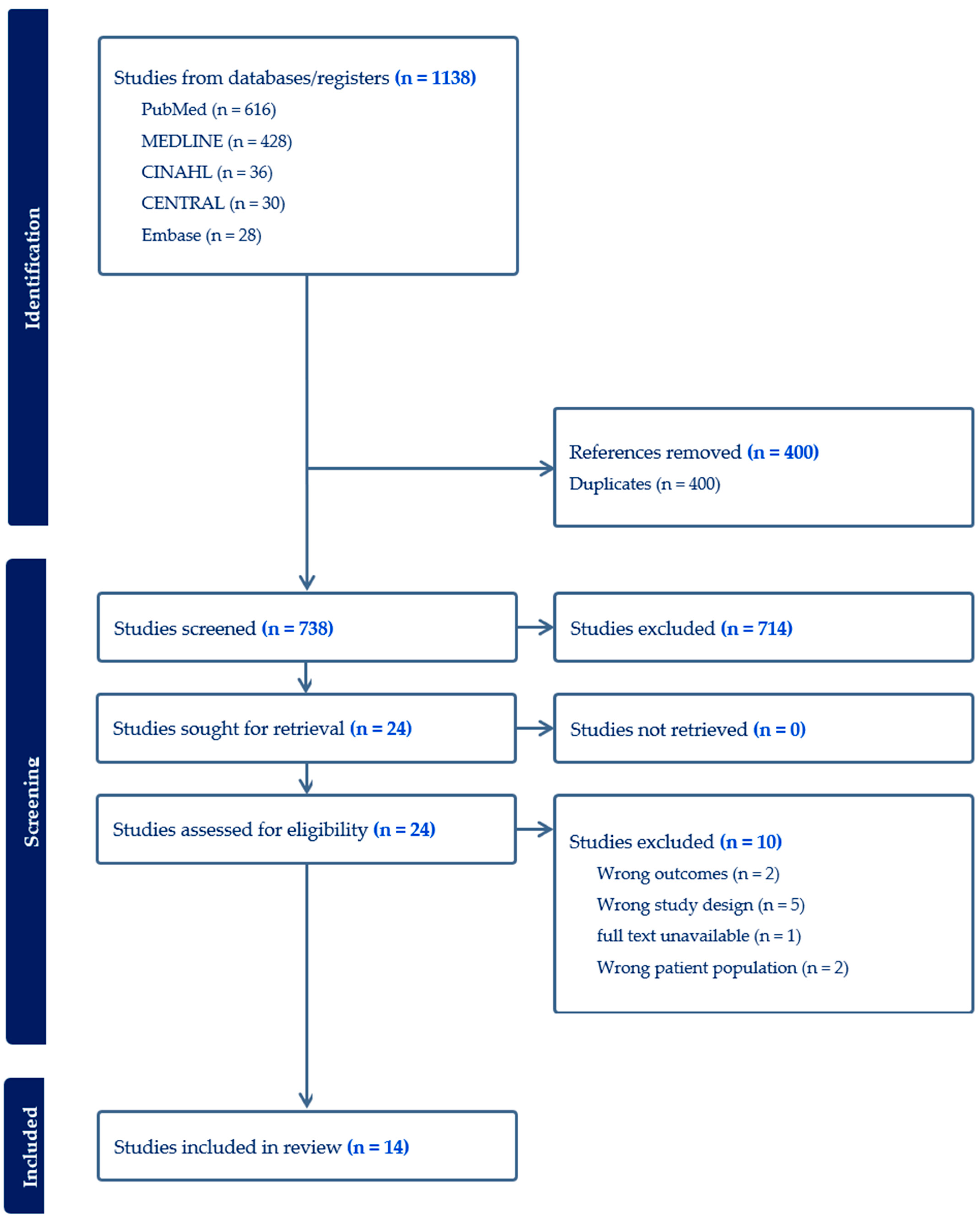
Our initial search across all databases returned 1138 sources. Four hundred of these were duplicates, leaving a total of 738 titles and abstracts to screen. A total of 24 studies were assessed in their entirety, of which 10 did not meet the final inclusion criteria. Five studies were excluded for having outcomes that did not align with genetic risks for BCRL. Two were excluded because their study outcomes did not report genetic profiles. Two additional studies were excluded because they did not investigate genetic predisposition to lymphedema in breast cancer care. One study was excluded because a full text was not available. After completion of screening and full-text review, 14 articles were included [3,4,5,6,7,8,9,10,11,12,13,14,15,16] (see Figure 1). Of these 14 articles, 9 were human studies, and the remaining 5 utilized animal models. Our search identified a body of research working to isolate target genes that may have the potential to stratify baseline genetic risk for BCRL. Results suggest that direct genetic inquiry in this area is relatively new, with included study publication dates ranging from 2008 to 2025.
A total of 29 unique genetic factors were identified. In total, 7 were associated with lymphatic structure and development, 14 with inflammation and immunological response to injury, 3 with immune activation and adhesion, and 1 cluster of genes associated with lymphatic function and contractility (see Table 1). To support transparent evaluation of the evidence base, the key methodological characteristics of the included studies are summarized in Table 2 to allow the readers to assess the relative strength and comparability of each investigation.

4. Discussion

4.1. Genes Impacting Lymphatic Structure and Development

Connexins are a class of proteins that play a role in lymphangiogenesis embryologically and coordinate migration and elongation of lymphatic endothelial cells to form intraluminal valve leaflets, which prevent backflow and stasis of lymphatic fluid [17]. A 2025 investigation into lymphatic-specific connexins in a mouse model (Cx37, Cx47, Cx43, and Cx45) found that Cx37 and Cx45 haploinsufficiency had the greatest impact on the development of subclinical embryologically flawed lymphatic vessels with significantly higher backflow and worse competence compared to wild type [15]. Two previous investigations found significantly increased baseline risk of BCRL in patients with Cx47 polymorphisms and suggest that possible inherited abnormalities in lymphatic structure and function may predispose patients to secondary lymphedema [14,16]. Finegold’s 2012 investigation did not completely control for environmental factors and found higher rates of connexin mutation in the cohort affected by BCRL, but this group was significantly older, had significantly more lymph nodes removed at their index procedure, and had higher rates of post-operative radiation [16]. In 2018, Hadizadeh et al. undertook a cohort study that controlled for these and presented two cohorts with no significant underlying demographic or treatment differences and still found a statistically significant relationship between connexin 37 mutation and BCRL [14].
Vascular endothelial growth factor (VEGF) is a primary growth factor in lymphangiogenesis in infancy and adulthood. It is a chemotactic agent that attracts monocytes and macrophages, which release additional growth factors and participate in the formation of lymphatic channels [18]. Mutations in VEGF and its downstream pathways were identified in three studies, with two finding significant associations with the development of BCRL [3,8]. Fu et al. in 2016 did not detect a statistically significant increase in risk of developing BCRL based on VEGF mutations in their cohort; however, this study had a small total prospective cohort of 140 patients with 16% developing lymphedema [11].
SRY-box transcription factor 18 (SOX18) is another crucial embryologic growth factor that plays a role in the differentiation of lymphatic endothelial precursor cells and has been implicated in driving lymphatic metastatic spread in some solid tumors [19,20]. Newman et al. did not detect a significant association between SOX18 polymorphisms and BCRL [3]. Likewise, the group also investigated lymphatic vessel endothelial hyaluronan receptor 1 (LYVE1) and neuropilin-2, which are also associated with malignant lymphangiogenesis, and did not find a significant association between genetic isoforms and the development of BCRL [3,21,22].
Forkhead Box C2 (FOXC2) is upregulated during lymphatic development and interacts with PROX1 to form lymphatic and venous valve structures embryologically and maintain them throughout development and adult life [23]. Newman et al.’s 2012 study did not detect a significant increase in risk of BCRL among isoforms of FOXC2; however, Miaskowski et al. did detect a small increase in risk in a larger sample, but noted that functional studies of the SNP haplotypes they identified would be needed to show a true association [3,8]. Clinical studies have shown that PROX1 has minimal evidence of clustering and did not significantly impact the risk of development of BCRL in a human cohort [3]. While no clinical association in human cohorts was found in the present review, PROX1-haploinsufficient mice did demonstrate leakier lymphatics, increased local inflammation, and worse pumping mechanics following lymphatic insult [10].
Adrenomedullin (AM) is a peptide hormone in the same family as calcitonin gene-related peptide (CGRP) that contributes to vasodilatory responses and lymphatic formation through a downstream cyclic adenosine monophosphate pathway [24]. A 2013 animal study by Nikitenko et al. found that deficiency of functional AM, even in the presence of healthy receptors, led to increased hind-limb circumference in an adult mouse model of lymphatic injury [7]. AM polymorphisms were not found to differ significantly between patients who developed BCRL and those who did not in Newman et al.’s 2012 study [3].
Finegold et al. noted that Hepatocyte Growth Factor (HGF) and its high-affinity receptor Mesenchymal Epithelial Transitional Tyrosine Kinase (MET) had a similar frequency of mutation in affected individuals as several other implicated genes in their 2008 study. However, no statistical investigation was undertaken, and no other identified studies investigated this relationship [6].
Lymphatic extracellular matrices have also been implicated in disease development in BCRL. GATA-binding transcription factor (GATA2) heterozygous-deficient mice demonstrated more tightly packed collagen around damaged lymph vessels in a 2024 mouse model investigation [12]. It was noted that aberrant fibroblast collagen deposition created a dense environment that hindered lymphatic healing [12]. Additionally, reintroduction of wild-type soluble collagen allowed for healthy recanalization and suggests that stiffness of extracellular scaffolding around healing lymphatics could contribute to disease development and progression [12].

4.2. Inflammatory and Immunological Genes

A number of interleukin gene variants were evaluated in the human studies included in this review. Polymorphisms in pro-inflammatory interleukins IL-1 [4,11], IL-6 [4,11], and IL-13 [11] were associated with increased risk of BCRL. No associations were found for CXCL8 (formerly IL-8) [4,11] or IL17A [4]. Anti-inflammatory interleukin associations were also evaluated for IL-4 [5] and IL-10 [5], with studies identifying variants of each that were significantly associated with increased risk of development of BCRL. The studies investigating these inflammatory mediators did not consistently identify risks associated with polymorphisms in these genes, with some cohorts detecting no risk in gene variants that others did. Inflammatory pathways undoubtedly contribute to lymphedema, but these results do not identify a particular aberration that leads to BCRL but rather strengthen evidence that inflammation plays a critical role in the development and morbidity of lymphedema.
RAR Related Orphan Receptor C (RORC) has been associated with lymphoid organ development in animal studies and plays a role in activation of IL-2 transcription [25]. Single-nucleotide polymorphisms of this gene were found to be significantly associated with lymphedema development in a single study. The authors hypothesized that the implicated SNPs have altered transcription factor binding sites, which may affect the activity of the gene product and could alter inflammatory activity at sites of injury in affected patients [3].
Nuclear factor kappa beta (NFKB) is a transcriptional regulator involved in angiogenesis, apoptosis, cell proliferation, and inflammation. Leung et al. reported a single, rare allele of the gene that was associated with a 3.1-fold increased risk of development of BCRL [5]. These findings were supported by additional clinical demonstrations of increased risk of development of BCRL based on NFKB polymorphisms [4].
Interferon Gamma (IFNG) is a non-specific excitatory cytokine that acts as an early reactant in infection and the inflammatory cascade. Both ligand and receptor genes for INFG were investigated by Miaskowski et al. in 2024, with no association found between genetic polymorphism and development of BCRL [4]. This group and Fu et al.’s 2016 study also tested associations between tumor necrosis factor (TNF) polymorphisms, another pro-inflammatory mediator, and likewise found no association linking it to BCRL [11].
Toll-like receptors (TLRs) are proteins that participate in the innate immune response and activate inflammatory and immunogenic signals when they bind to pathogen-associated molecular patterns that are foreign to an organism. A 2011 investigation showed that mice with TLR knockouts for TLR-2, TLR-4, or TLR-9 all demonstrated significantly increased tail circumference after tail lymph node excision compared to wild-type mice. The authors propose that decreased macrophage and local inflammatory responses to injury decrease the animals’ ability to induce lymphangiogenesis [13]. They also found that TLR deficiency was associated with increased concentrations of tightly packed type I collagen in the extracellular matrix, which may impede lymphatic growth and organization [13].

4.3. Immune Cell Activation and Adhesion Genes

In Miaskowski et al.’s 2013 polygenic investigation, they included three genes that participate in inflammatory activation, adhesion, and migration and found that lymphocyte cytosolic protein 2 (LCP2), spleen tyrosine kinase (SYK), and vascular cell adhesion molecule-1 (VCAM-1) polymorphisms were all significant risk modifiers for the development of BCRL [8]. The pathophysiology of lymphedema can be traced in part to dysfunction of immune cell adhesion to lymphatic endothelium, thereby compromising clearance of protein-rich interstitial fluid and microenvironmental debris. Upregulation of VCAM1 typically mediates monocyte and lymphocyte adherence to endothelium. Polymorphic variations to this transmembrane sialoglycoprotein may attenuate or exaggerate the adhesion mechanism. Miaskowski and colleagues showed that variation in SNP rs3176861 of the VCAM1 gene was shown to be significantly associated with the modulation of lymphedema [8]. Likewise, a regression analysis by Miaskowski et al. showed that carrying two copies of the for SYK rs158689 allele was associated with nearly a 3.5-fold increase in the odds of developing lymphedema [8]. They also found that a single nucleotide polymorphism in SYK was able to explain up to ~3% of the variance in lymphedema risk [8]. This may be due to the central role of SYK in the activation and integrin signaling of B-cells, T-cells, and myeloid cells. Genetic perturbations in SYK may result in a milieu that impedes lymphangiogenesis and lymphatic remodeling due to a shift in the balance between adaptive immune resolution and chronic low-grade inflammation.
The group’s regression analysis for LCP2 rs315721 was also able to explain 18.9% of the variance for the odds of developing lymphedema [8]. LCP2 contributes to lymphatic vessel development by modulating hematopoietic signaling pathways that control the separation of the blood and lymphatic networks. In this pathway, SYK interacts as an upstream kinase for the downstream adaptor molecule LCP2. Together, these molecules comprise a portion of the core regulatory axis essential for maintaining vascular segregation. A single genetic variation in either gene could disrupt the coordinated signaling between SYK and LCP2, leading to a potential alteration within the cascade that ensures proper vascular compartmentalization.
Miaskowski et al. provide compelling data to support the notion that the risk for developing lymphedema can be attributed in part to polymorphisms in adhesion and immune-signaling genes. These findings suggest that altered endothelial-immune cell interaction may impair lymphatic recovery or promote maladaptive remodeling in the post-operative setting of breast cancer treatment, thus prompting further consideration of immune activation and adhesion genes as potential modulators and therapeutic targets in treatment strategies aimed at lymphedema prevention and management.

4.4. Function and Contractility

Lymphatic pump activity is largely driven by the potassium gradient across lymphatic smooth muscle cell membranes. The movement of K+ channels across the cell membrane maintains the resting membrane potential, allowing action potentials to fire and trigger smooth muscle contraction and subsequent lymphatic flow [26].
The mechanism by which K+ channel dysfunction leads to the development of BCRL likely begins with impaired K+ channel activity leading to contractile dysfunction and subsequent loss of pulsatile lymphatic flow in patients with loss-of-function K+ channel variants. This results in endothelial barrier dysfunction mediated by myosin light chain phosphorylation, promoting the accumulation of protein-rich interstitial fluid that creates a pro-inflammatory microenvironment [27,28]. Combined with mechanical stress from edema, this triggers inflammatory pathways that potentiate profibrogenic mechanisms within fibroblasts, causing increased collagen deposition and progressive fibrosis of lymphatic channels [28]. Collectively, this establishes a positive feedback loop in which progressive fibrosis further impairs lymphatic flow and perpetuates disease progression [28].
A single investigation by Smoot et al. in 2017 demonstrated that polymorphisms in potassium channels that play a major role in lymphatic contractility increased the risk of developing BCRL [9]. KCNA1 encodes for components of the K+ voltage-gated channel, which is responsible for the hyperpolarization of the cell membrane and the return of the membrane to its resting state. Two genes, KCNJ3 and KCNJ6, encode for components of the G-protein inwardly rectifying potassium channels, which work to stabilize the cell’s resting potential by promoting an influx of potassium current into the cell. KCNK3 encodes part of the two-pore K+ channel, which establishes potassium leak currents necessary for the maintenance of the membrane’s resting potential. Polymorphisms in these genes may therefore enhance or disrupt lymphatic smooth muscle contraction and lead to altered lymphatic drainage. The study identified six such polymorphisms across these four genes that were found to be significantly associated with BCRL: KCNA1 rs4766311, KCNK3 rs1662988, KCNJ3 rs1037091, KCNJ6 rs2211845, KCNJ6 rs991985, and KCNJ6 rs2836019. Of these, all were associated with a decreased likelihood of developing secondary lymphedema except for KCNJ6 rs2211845 and KCNJ6 rs991985, which were associated with increased odds [9].

4.5. Limitations

The present study sought to amalgamate the current state of the literature regarding genetic predisposition to BCRL. The heterogeneity of findings, numerous genes implicated in disease, and differences in study design unfortunately preclude meta-analytics at this time, which would significantly strengthen any recommendations that could be made based on the presented findings.
Measurement of lymphedema and diagnostic criteria to identify patients and disease differed among the presented studies. Hind limb swelling and lymphatic uptake of Evans blue were utilized by Nikitenko et al. and Watanabe-Asaka et al. [7,12]. Zampell’s work created a lymphedematous state using a tail injury model of lymphedema, which is commonly employed in the literature [13,29]. Hespe utilized lymphoscintigraphy in the included experimental animals to diagnose lymphedema [10]. Davis et al.’s investigation into valve function in connexin-mutated mice directly measured valve backflow and leaflet gap dimensions at the bench to determine the effects of the included genes on valve dynamics [15]. Bioimpedance resistance ratios were used by Smoot et al., Leung et al., and in both reports by Miaskowski et al. [4,5,8,9]. Measurement of arm circumference via tape measure or via infrared perometry was described as well [3,11,30]. Physician diagnosis and International Society of Lymphology criteria were utilized in some cohorts as well [6,14,16]. Inconsistent diagnostic criteria may impact probands included in analytics and could affect reported results.

5. Conclusions

BCRL is a complex, likely polygenic disease process. The present study suggests that a number of subclinical phenotypic manifestations of subtle genetic mutations may predispose patients to the development of BCRL. Our investigation was limited by a relatively small number of manuscripts that met strict inclusion criteria, but it is strengthened by our standardized search and screening approach. Inflammatory mediators were commonly implicated but may represent nonspecific contributions to the development of lymphedema. Subclinical lymphatic dysfunction caused by intrinsic genetic alteration in genes associated with the structure and development of lymphatics leverages promising potential preoperative screening tools for patients who require mastectomy for breast cancer or prophylaxis. A prospective clinical trial collecting genetic data from patients prior to operative intervention to evaluate their genotype and monitor the development of BCRL, based on the presented results, could begin to shape effective genetic screening. Such risk stratification may directly inform prophylactic surgical and perioperative risk-reducing strategies for secondary lymphedema. Individuals with high-risk genotypes could be considered for adjunct surgical procedures such as prophylactic lymphaticovenular anastomosis or the lymphatic microsurgical preventive healing approach (LYMPHA). These patients may also benefit from enrollment in structured prospective surveillance programs, early intervention compression protocols, and immediate post-operative physiotherapy or manual lymphatic drainage to support lymphatic flow and shoulder mobility. As high-risk patients become identifiable prior to surgery, future research can help determine which interventions most effectively preserve function and optimize quality of life after mastectomy.

Author Contributions

Conceptualization, A.J.J., Q.L.C., C.H.C.; Methodology, A.J.J., Q.L.C.,C.H.C.; Software, A.J.J., B.J.P., C.L., R.S.; Validation, A.J.J., B.J.P., C.L.; Validation, A.J.J., R.S., B.J.P.; Resources, A.J.J.; Data Curation, C.L., A.J.J., Q.L.C., C.H.C., B.J.P.; writing—original draft preparation, A.J.J., Q.L.C., C.H.C., B.J.P., C.L., R.S.; writing review and editing, A.J.J., Q.L.C., C.H.C., B.J.P., C.L., R.S.; visualization, A.J.J.; supervision, A.J.J., R.S.; project administration, A.J.J. All authors have read and agreed to the published version of the manuscript.

Funding

This research received no external funding.

Conflicts of Interest

The authors declare no conflicts of interest.

References

  1. Sakorafas, G.H.; Peros, G.; Cataliotti, L.; Vlastos, G. Lymphedema following axillary lymph node dissection for breast cancer. Surg. Oncol. 2006, 15, 153–165. [Google Scholar] [CrossRef] [Scilit]
  2. Page, M.J.; McKenzie, J.E.; Bossuyt, P.M.; Boutron, I.; Hoffmann, T.C.; Mulrow, C.D.; Shamseer, L.; Tetzlaff, J.M.; Akl, E.A.; Brennan, S.E.; et al. The PRISMA 2020 statement: An updated guideline for reporting systematic reviews. Rev. Esp. Cardiol. (Engl. Ed.) 2021, 74, 790–799. [Google Scholar] [CrossRef]
  3. Newman, B.; Lose, F.; Kedda, M.A.; Francois, M.; Ferguson, K.; Janda, M.; Yates, P.; Spurdle, A.B.; Hayes, S.C. Possible genetic predisposition to lymphedema after breast cancer. Lymphat. Res. Biol. 2012, 10, 2–13. [Google Scholar] [CrossRef] [Scilit]
  4. Miaskowski, C.; Conley, Y.P.; Cooper, B.A.; Paul, S.M.; Smoot, B.J.; Hammer, M.J.; Fu, M.; Levine, J.D. Identification of a Higher Risk Lymphedema Phenotype and Associations with Cytokine Gene Polymorphisms. J. Pain. Symptom Manag. 2024, 67, 375–383 e373. [Google Scholar] [CrossRef] [Scilit]
  5. Leung, G.; Baggott, C.; West, C.; Elboim, C.; Paul, S.M.; Cooper, B.A.; Abrams, G.; Dhruva, A.; Schmidt, B.L.; Kober, K.; et al. Cytokine candidate genes predict the development of secondary lymphedema following breast cancer surgery. Lymphat. Res. Biol. 2014, 12, 10–22. [Google Scholar] [CrossRef] [Scilit] [PubMed]
  6. Finegold, D.N.; Schacht, V.; Kimak, M.A.; Lawrence, E.C.; Foeldi, E.; Karlsson, J.M.; Baty, C.J.; Ferrell, R.E. HGF and MET mutations in primary and secondary lymphedema. Lymphat. Res. Biol. 2008, 6, 65–68. [Google Scholar] [CrossRef] [Scilit]
  7. Nikitenko, L.L.; Shimosawa, T.; Henderson, S.; Makinen, T.; Shimosawa, H.; Qureshi, U.; Pedley, R.B.; Rees, M.C.; Fujita, T.; Boshoff, C. Adrenomedullin haploinsufficiency predisposes to secondary lymphedema. J. Investig. Dermatol. 2013, 133, 1768–1776. [Google Scholar] [CrossRef] [Scilit] [PubMed]
  8. Miaskowski, C.; Dodd, M.; Paul, S.M.; West, C.; Hamolsky, D.; Abrams, G.; Cooper, B.A.; Elboim, C.; Neuhaus, J.; Schmidt, B.L.; et al. Lymphatic and angiogenic candidate genes predict the development of secondary lymphedema following breast cancer surgery. PLoS ONE 2013, 8, e60164. [Google Scholar] [CrossRef] [Scilit]
  9. Smoot, B.; Kober, K.M.; Paul, S.M.; Levine, J.D.; Abrams, G.; Mastick, J.; Topp, K.; Conley, Y.P.; Miaskowski, C.A. Potassium Channel Candidate Genes Predict the Development of Secondary Lymphedema Following Breast Cancer Surgery. Nurs. Res. 2017, 66, 85–94. [Google Scholar] [CrossRef] [Scilit] [PubMed]
  10. Hespe, G.E.; Ly, C.L.; Kataru, R.P.; Mehrara, B.J. Baseline Lymphatic Dysfunction Amplifies the Negative Effects of Lymphatic Injury. Plast. Reconstr. Surg. 2019, 143, 77e–87e. [Google Scholar] [CrossRef] [Scilit]
  11. Fu, M.R.; Conley, Y.P.; Axelrod, D.; Guth, A.A.; Yu, G.; Fletcher, J.; Zagzag, D. Precision assessment of heterogeneity of lymphedema phenotype, genotypes and risk prediction. Breast 2016, 29, 231–240. [Google Scholar] [CrossRef] [Scilit]
  12. Watanabe-Asaka, T.; Hayashi, M.; Harada, T.; Uemura, S.; Takai, J.; Nakamura, Y.; Moriguchi, T.; Kawai, Y. Perturbed collagen metabolism underlies lymphatic recanalization failure in Gata2 heterozygous deficient mice. J. Biochem. 2024, 175, 551–560. [Google Scholar] [CrossRef] [Scilit]
  13. Zampell, J.C.; Elhadad, S.; Avraham, T.; Weitman, E.; Aschen, S.; Yan, A.; Mehrara, B.J. Toll-like receptor deficiency worsens inflammation and lymphedema after lymphatic injury. Am. J. Physiol. Cell Physiol. 2012, 302, C709–C719. [Google Scholar] [CrossRef] [Scilit]
  14. Hadizadeh, M.; Mohaddes Ardebili, S.M.; Salehi, M.; Young, C.; Mokarian, F.; McClellan, J.; Xu, Q.; Kazemi, M.; Moazam, E.; Mahaki, B.; et al. GJA4/Connexin 37 Mutations Correlate with Secondary Lymphedema Following Surgery in Breast Cancer Patients. Biomedicines 2018, 6, 23. [Google Scholar] [CrossRef] [Scilit] [PubMed]
  15. Davis, M.J.; Castorena-Gonzalez, J.A.; Li, M.; Simon, A.M.; Srinivasan, R.S. Hierarchical Requirement for Endothelial Cell Connexins Cx37, Cx47, Cx43, and Cx45 in Lymphatic Valve Function. Function 2025, 6, zqaf034. [Google Scholar] [CrossRef] [Scilit]
  16. Finegold, D.N.; Baty, C.J.; Knickelbein, K.Z.; Perschke, S.; Noon, S.E.; Campbell, D.; Karlsson, J.M.; Huang, D.; Kimak, M.A.; Lawrence, E.C.; et al. Connexin 47 mutations increase risk for secondary lymphedema following breast cancer treatment. Clin. Cancer Res. 2012, 18, 2382–2390. [Google Scholar] [CrossRef] [Scilit] [PubMed]
  17. Sabine, A.; Agalarov, Y.; Maby-El Hajjami, H.; Jaquet, M.; Hagerling, R.; Pollmann, C.; Bebber, D.; Pfenniger, A.; Miura, N.; Dormond, O.; et al. Mechanotransduction, PROX1, and FOXC2 cooperate to control connexin37 and calcineurin during lymphatic-valve formation. Dev. Cell 2012, 22, 430–445. [Google Scholar] [CrossRef] [Scilit]
  18. Cursiefen, C.; Chen, L.; Borges, L.P.; Jackson, D.; Cao, J.; Radziejewski, C.; D’Amore, P.A.; Dana, M.R.; Wiegand, S.J.; Streilein, J.W. VEGF-A stimulates lymphangiogenesis and hemangiogenesis in inflammatory neovascularization via macrophage recruitment. J. Clin. Investig. 2004, 113, 1040–1050. [Google Scholar] [CrossRef] [PubMed]
  19. Francois, M.; Caprini, A.; Hosking, B.; Orsenigo, F.; Wilhelm, D.; Browne, C.; Paavonen, K.; Karnezis, T.; Shayan, R.; Downes, M.; et al. Sox18 induces development of the lymphatic vasculature in mice. Nature 2008, 456, 643–647. [Google Scholar] [CrossRef] [Scilit]
  20. Duong, T.; Proulx, S.T.; Luciani, P.; Leroux, J.C.; Detmar, M.; Koopman, P.; Francois, M. Genetic ablation of SOX18 function suppresses tumor lymphangiogenesis and metastasis of melanoma in mice. Cancer Res. 2012, 72, 3105–3114. [Google Scholar] [CrossRef] [Scilit]
  21. Jackson, D.G.; Prevo, R.; Clasper, S.; Banerji, S. LYVE-1, the lymphatic system and tumor lymphangiogenesis. Trends Immunol. 2001, 22, 317–321. [Google Scholar] [CrossRef] [Scilit] [PubMed]
  22. Jubb, A.M.; Sa, S.M.; Ratti, N.; Strickland, L.A.; Schmidt, M.; Callahan, C.A.; Koeppen, H. Neuropilin-2 expression in cancer. Histopathology 2012, 61, 340–349. [Google Scholar] [CrossRef] [Scilit]
  23. Norrmen, C.; Ivanov, K.I.; Cheng, J.; Zangger, N.; Delorenzi, M.; Jaquet, M.; Miura, N.; Puolakkainen, P.; Horsley, V.; Hu, J.; et al. FOXC2 controls formation and maturation of lymphatic collecting vessels through cooperation with NFATc1. J. Cell Biol. 2009, 185, 439–457. [Google Scholar] [CrossRef] [Scilit]
  24. Jin, D.; Harada, K.; Ohnishi, S.; Yamahara, K.; Kangawa, K.; Nagaya, N. Adrenomedullin induces lymphangiogenesis and ameliorates secondary lymphoedema. Cardiovasc. Res. 2008, 80, 339–345. [Google Scholar] [CrossRef] [Scilit]
  25. Kurebayashi, S.; Ueda, E.; Sakaue, M.; Patel, D.D.; Medvedev, A.; Zhang, F.; Jetten, A.M. Retinoid-related orphan receptor gamma (RORgamma) is essential for lymphoid organogenesis and controls apoptosis during thymopoiesis. Proc. Natl. Acad. Sci. USA 2000, 97, 10132–10137. [Google Scholar] [CrossRef] [Scilit] [PubMed]
  26. Telinius, N.; Kim, S.; Pilegaard, H.; Pahle, E.; Nielsen, J.; Hjortdal, V.; Aalkjaer, C.; Boedtkjer, D.B. The contribution of K(+) channels to human thoracic duct contractility. Am. J. Physiol. Heart Circ. Physiol. 2014, 307, H33–H43. [Google Scholar] [CrossRef] [Scilit]
  27. Hall, J.D.; Farzaneh, S.; Babakhani Galangashi, R.; Pujari, A.; Sweet, D.T.; Kahn, M.L.; Jimenez, J.M. Lymphoedema conditions disrupt endothelial barrier function in vitro. J. R. Soc. Interface 2022, 19, 20220223. [Google Scholar] [CrossRef] [Scilit] [PubMed]
  28. Brakenhielm, E.; Gonzalez, A.; Diez, J. Role of Cardiac Lymphatics in Myocardial Edema and Fibrosis: JACC Review Topic of the Week. J. Am. Coll. Cardiol. 2020, 76, 735–744. [Google Scholar] [CrossRef] [Scilit]
  29. Hassanein, A.H.; Sinha, M.; Neumann, C.R.; Mohan, G.; Khan, I.; Sen, C.K. A Murine Tail Lymphedema Model. J. Vis. Exp. 2021, 168, e61848. [Google Scholar] [CrossRef] [Scilit]
  30. Fu, M.R.; Axelrod, D.; Guth, A.A.; Scagliola, J.; Rampertaap, K.; El-Shammaa, N.; Qiu, J.M.; McTernan, M.L.; Frye, L.; Park, C.S.; et al. A Web- and Mobile-Based Intervention for Women Treated for Breast Cancer to Manage Chronic Pain and Symptoms Related to Lymphedema: Results of a Randomized Clinical Trial. JMIR Cancer 2022, 8, e29485. [Google Scholar] [CrossRef] [Scilit]
Figure 1. PRISMA diagram. Shows the PRISMA-organized results of the literature review.
Lymphatics 04 00010 g001
Table 1. Relevant genes and their functional classifications.
GeneFunctionReference
Connexin IsoformsLymphatic Structure and DevelopmentDavis et al. 2025 [15], Hadizadeh et al. 2018 [14], Finegold et al. 2012 [16]
Vascular Endothelial Growth Factor (VEGF)Lymphatic Structure and DevelopmentNewman et al. 2012 [3], Miaskowski et al. 2013 [8], Fu et al. 2016 [11]
Forkhead Box Protein C2 (FOXC2)Lymphatic Structure and DevelopmentNewman et al. 2012 [3], Miaskowski et al. 2013 [8]
Prospero Homeobox 1 (PROX1)Lymphatic Structure and DevelopmentNewman et al. 2012 [3], Hespe et al. 2019 [10]
SYR-Box Transcription Factor (SOX18)Lymphatic Structure and DevelopmentNewman et al. 2012 [3]
Lymphatic Vessel Endothelial Hyaluronan Receptor 1 (LYVE1)Lymphatic Structure and DevelopmentNewman et al. 2012 [3]
Adrenomedullin (ADM)Lymphatic Structure and DevelopmentNewman et al. 2012 [3], Nikitenko et al. 2013 [7]
Hepatocyte Growth Factor/Mesenchymal Epithelial Transitional Tyrosine Kinase (HGF/MET)Lymphatic Structure and DevelopmentFinegold et al. 2008 [6]
Neuropilin-2Lymphatic Structure and DevelopmentMiaskowski et al. 2013 [8]
GATA Binding Protein 1 (GATA2)Lymphatic Structure and DevelopmentWatanabe-Asaka 2024 [12]
IL4Inflammation and Immune ResponseMiaskowski et al. 2024 [4], Fu et al. 2016 [11], Leung et al. 2014 [5]
IL10Inflammation and Immune ResponseMiaskowski et al. 2024 [4], Leung et al. 2014 [5], Fu et al. 2016 [11]
IL2Inflammation and Immune ResponseMiaskowski et al. 2024 [4]
IL17AInflammation and Immune ResponseMiaskowski et al. 2024 [4]
IL1Inflammation and Immune ResponseMiaskowski et al. 2024 [4], Fu et al. 2016 [11]
IL6Inflammation and Immune ResponseMiaskowski et al. 2024 [4], Fu et al. 2016 [11]
IL13Inflammation and Immune ResponseMiaskowski et al. 2024 [4], Fu et al. 2016 [11]
C-X-C Motif Chemokine Ligand 8 (CXCL8 (Formerly IL-8))Inflammation and Immune ResponseMiaskowski et al. 2024 [4], Fu et al. 2016 [11]
RAR Related Orphan Receptor C (RORC)Inflammation and Immune ResponseNewman et al. 2012 [3]
Interferon Gamma (IFNG)Inflammation and Immune ResponseMiaskowski et al. 2024 [4]
IFNG Receptor 1Inflammation and Immune ResponseMiaskowski et al. 2024 [4]
Tumor Necrosis Factor (TNF)Inflammation and Immune ResponseMiaskowski et al. 2024 [4], Fu et al. 2016 [11]
Nuclear Factor Kappa Beta (NFKB)Inflammation and Immune ResponseMiaskowski et al. 2024 [4], Leung et al. 2014 [5]
Toll-Like Receptor (TLR)Inflammation and Immune ResponseZampell et al. 2012 [13]
Lymphocyte Cytosolic Protein 2 (LCP2)Immune Cell Activation and AdhesionMiaskowski et al. 2013 [8]
Spleen Tyrosine Kinase (SYK)Immune Cell Activation and AdhesionMiaskowski et al. 2013 [8]
Vascular Cell Adhesion Molecule 1 (VCAM-1)Immune Cell Activation and AdhesionMiaskowski et al. 2013 [8]
Voltage-Gated Potassium ChannelsFunction and ContractilitySmoot et al. 2017 [9]
Table 2. Summary of key methodological features of the included manuscripts.
SourceGene Variants InvestigatedSample SizeStudy TypeClinical Outcomes
Newman et al. 2012 [3]SOX18, VEGFC, VEGFD, VEGFR2, VEGFR3, RORC, FOXC2, LYVE1, ADM, PROX1120Case-control Polymorphisms in VEGFR2, VEGFR3, and RORC found to be associated with lymphedema
Miaskowski et al. 2024 [4]CXCL8, IFNG, IFNG1, IFNGR1, IL1R1, IL1R2, IL2, IL4, IL6, IL17A, IL1B, IL10, IL13, IL17A, NFKB1, NFKB2, TNFSF155Observational (cross-sectional and longitudinal)Polymorphisms in IL1B and IL6 found to be associated with increased lymphedema severity
Leung et al. 2014 [5]IFNG1, IFNGR1, IL1B, IL1R1, IL1R2, IL2, IL4, IL6, IL8, IL10, IL13, IL17A, NFKB1, NFKB2, TNFA542Observational (cross-sectional and longitudinal)Polymorphisms in IL4, IL10, and NFKB2 associated with inflammatory responses in the development of lymphedema
Finegold et al. 2008 [6]HGF, MET239Case–controlMutations in HGF and MET found to contribute to lymphedema
Nikitenko et al. 2013 [7]ADMNot specifiedIn vivo experimental studyReduced adrenomedullin levels increase susceptibility to secondary lymphedema
Miaskowski et al. 2013 [8]ANGPT2, FOXC2, HGF, LCP2, LYVE1, MET, NRP2, PROX1, RORC, SOX17, SYK, VCAM1, VEGFB, VEGFC, VEGFD, VEGFR2, VEGFR3543Observational (cross-sectional and longitudinal)Polymorphisms in LCP2, NRP2, SYK, VCAM1, and three haplotypes (FOXC2 A03, NRP2 F03, VEGFC B03) associated with lymphangiogensis and angiogenesis
Smoot et al. 2017 [9]KCNA1, KCND2, KCNS1, KCNJ3, KCNJ5, KCNJ6, KCNJ9, KCNK2, KCNK3, KCNK9542Observational (cross-sectional and longitudinal)Polymorphisms in KCNA1, KCNJ3, and KCNJ6, and KCNK3 associated with lymphedema
Hespe et al. 2019 [10] Prox1Not specifiedIn vivo experimental studySubclinical lymphatic dysfunction intensifies the effects of lymphatic injury
Fu et al. 2016 [11]VEGF-C, VEGF-D, IL1-a, IL-4, IL6, IL8, IL10, IL13, TNF-a140Prospective cohort (genetic association)Polymorphisms in IL4 contribute to impaired limb mobility, polymorphisms in IL6 and IL4 contribute to fluid accumulation, and polymorphisms in VEGF-C and IL-13 contribute to patient discomfort
Watanabe-Asaka et al. 2024 [12]GATA2Not specifiedIn vivo experimental studyPlacement of soluble type I collagen significantly improved lymphatic recanalization in GATA2 heterozygous-deficient mice, suggesting a potential therapeutic strategy for GATA2-related lymphedema
Zampell et al. 2012 [13]TLR2, TLR4, TLR9, VEGF-A, VEGF-C, LYVE-1Not specifiedIn vivo experimental studyLoss of TLR signaling (TLR2, TLR4, TLR9) in mice impairs lymphatic repair and lymphangiogenesis
Hadizadeh et al. 2018 [14]GJA4102Retrospective case–controlPolymorphisms in GJA4 associated with an increased risk of secondary lymphedema
Davis et al. 2025 [15]GJA4, GJA1, GJC1, GJC2Not specifiedIn vivo experimental study4 connexin isoforms (Cx37, Cx47, Cx43, Cx45) contribute to lymphatic valve function, and polymorphisms in GJA4 predispose to lymphedema
Finegold et al. 2012 [16] GJC2, FOXC2, HGF, MET, FLT4188Case-control study with in vitro functional validationPolymorphisms in GJC2 implicated in impaired lymphatic gap junctions
Disclaimer/Publisher’s Note: The statements, opinions and data contained in all publications are solely those of the individual author(s) and contributor(s) and not of MDPI and/or the editor(s). MDPI and/or the editor(s) disclaim responsibility for any injury to people or property resulting from any ideas, methods, instructions or products referred to in the content.