Review

Natural Antihypertensive Drug Leads from Northeast India’s Biodiversity Hotspots: Molecular Targets, Signaling Pathways, and Safety Profiles: A Review


1
Department of Botany, Cotton University, Guwahati 781001, Assam, India
2
Department of Zoology, Cell & Biochemical Technology Laboratory, Cotton University, Guwahati 781001, Assam, India
3
Department of Molecular Biology and Biotechnology, Cotton University, Guwahati 781001, Assam, India
4
Department of Environmental Biology and Wildlife Sciences, Cotton University, Guwahati 781001, Assam, India
5
Programme of Biotechnology, Faculty of Science, Assam Down Town University, Panikhaiti, Guwahati 781026, Assam, India
*
Author to whom correspondence should be addressed.
Drugs Drug Candidates 2026, 5(3), 43; https://doi.org/10.3390/ddc5030043
Submission received: 14 May 2026 / Revised: 14 July 2026 / Accepted: 20 July 2026 / Published: 27 July 2026
(This article belongs to the Section Drug Candidates from Natural Sources)

Abstract

Hypertension is a major global health concern affecting over one billion people worldwide and is a leading risk factor for cardiovascular diseases, stroke, and kidney failure. Plant-derived bioactive compounds provide promising natural strategies for hypertension management by improving endothelial function, promoting vasorelaxation and diuresis, reducing oxidative stress, and modulating key molecular pathways. This review focuses on some important traditional medicinal plants from Northeast India with potential antihypertensive activity, summarizing their phytochemical constituents, pharmacological evidence, and proposed mechanisms of action. Northeast India lies within the Eastern Himalayas and Indo-Burma biodiversity hotspots and is recognized for its rich plant diversity. We conducted a systematic literature search using PubMed, Google Scholar, ScienceDirect, and Scopus following PRISMA guidelines. We further evaluated identified compounds for ADMET using open-source computational tools. Ten medicinal plants: Clerodendrum colebrookeanum, Moringa oleifera, Garcinia dulcis, Centella asiatica, Terminalia bellirica, Hibiscus sabdariffa, Citrus limon, Passiflora edulis, Garcinia cowa, and Solanum torvum yielded 22 bioactive compounds with notable antihypertensive activity. These compounds exhibited antioxidant, anti-inflammatory, and vasorelaxant effects via nitric oxide synthase activation, MAPK/NF-κB inhibition, ACE inhibition, calcium channel modulation, and reactive oxygen species scavenging. ADMET profiling indicated favorable pharmacokinetics, with vitexin, isovitexin, isoquercetin, and nobiletin showing low predicted toxicity and high stability.

1. Introduction

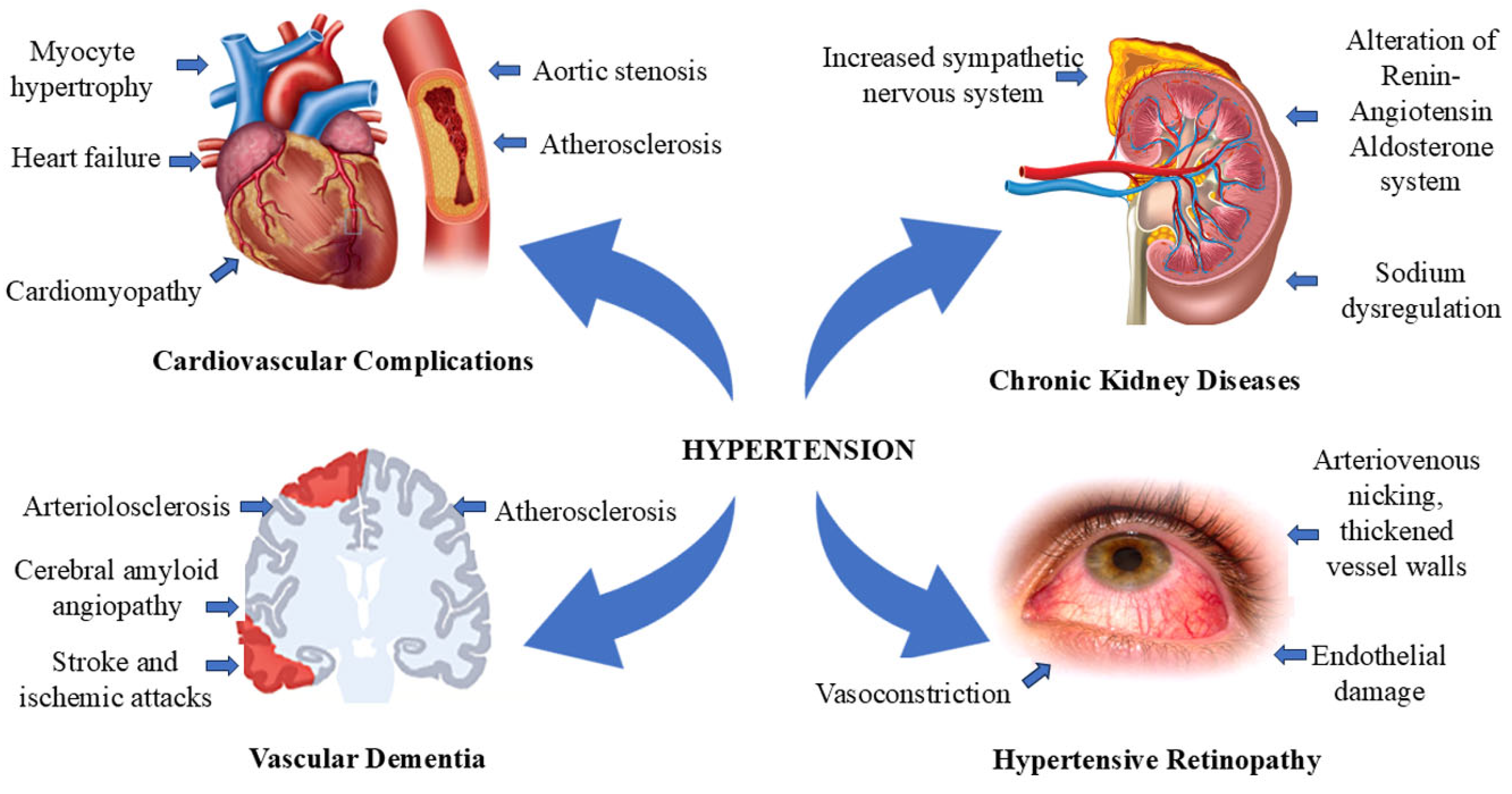
Hypertension, commonly known as high blood pressure, refers to abnormally elevated arterial pressure. A normal blood pressure reading is defined as a systolic blood pressure less than 120 mmHg and a diastolic blood pressure less than 80 mmHg. Hypertension is typically diagnosed when systolic pressure is 140 mmHg or higher and/or the diastolic pressure is 90 mmHg or higher. According to a 2010 study, the global prevalence of hypertension is increasing, primarily due to the aging population, with an estimated 31.1% of the global adult population (1.39 billion people) affected by hypertension [1,2]. According to the American Heart Association (AHA) hypertension guidelines, systolic and diastolic blood pressure levels are classified into four categories: (1) normal blood pressure: systolic BP less than 120 mmHg and diastolic BP less than 80 mmHg; (2) elevated blood pressure: systolic BP between 120 and 129 mmHg and diastolic BP below 80 mmHg; (3) stage 1 hypertension: systolic BP between 130 and 139 mmHg or diastolic BP between 80 and 89 mmHg; (4) stage 2 hypertension: systolic BP of 140 mmHg or higher or diastolic BP of 90 mmHg or higher [3]. Several factors play key roles in the development of hypertension, including obesity, insulin resistance, high alcohol intake, high salt intake, aging, a sedentary lifestyle, stress, low potassium intake, and low calcium intake [4,5]. Hypertension is strongly associated with the development of major cardiovascular and renal conditions, including peripheral artery disease, chronic kidney disease, myocardial infarction, atrial fibrillation, heart failure, stroke, coronary artery disease, and cognitive impairment (Figure 1). It is one of the leading contributors to global mortality and disability [1,6].
The global prevalence of hypertension among adults remained relatively stable between 1990 and 2019. During this period, the prevalence declined in high-income countries, from 38% in 1990 to 32% in 2019, and in the WHO European Region, from 45% to 37%. However, these reductions were offset by slight to moderate increases in other regions, including the WHO Western Pacific Region (including countries such as Australia, New Zealand, China, South Korea, the Philippines, Malaysia, and Japan), where prevalence increased from 24% to 28%, and the WHO South-East Asia Region (including India, Nepal, Indonesia, and Thailand), where it rose from 29% to 32%. During this period, the number of hypertensive adults in the Western Pacific more than doubled (from 144 million to 346 million), while Europe and the Americas experienced a 41% increase, and Southeast Asia and the Western Pacific combined saw a dramatic 144% increase in the number of hypertensive individuals [4,7].
The data indicated that men had a slightly greater age-standardized incidence of hypertension (31.9%) than women did (30.1%). The prevalence was lower in high-income countries (28.5%) than in low-income countries (31.5%). Among men, South Asia reported the lowest prevalence (26.4%), whereas Eastern Europe and Central Asia had the highest (39.0%). For women, the lowest prevalence was observed in high-income countries (25.3%), whereas 36.3% was recorded in sub-Saharan Africa [1,8,9]. In a study conducted between August 2021 and August 2023 in the United States, the prevalence of adult hypertension was 47.7%. The rate of hypertension was higher in men (50.8%) than in women (44.6%) and increased with age, ranging from 23.4% for those aged 18–39 years, 52.5% for those aged 40–59 years, and 71.6% for those aged 60 and older [10].
Hypertension is a major public health concern in India, contributing to 57% of all stroke-related deaths and 24% of deaths from coronary heart disease (CHD) [11]. It significantly impacts the country’s cardiovascular health and places a burden on the healthcare system. Overall, hypertension affects approximately 33% of the urban population and 25% of the rural population. According to the 2019–2020 National Family Health Survey (NFHS-5), the prevalence of hypertension has risen to 24% in men and 21% in women, whereas it has increased to 19% and 17%, respectively, in the 2015–2016 survey [12,13]. In India, the prevalence of hypertension was highest in northeastern (NE) states, namely Sikkim (20.2%), Nagaland (17.6%), Assam (17.6%), Arunachal Pradesh (16.6%), and Tripura (15.4%), compared with other states and union territories, as per the National Family Health Survey (NFHS) 2015–2016 report [14]. According to the Indian Health Survey (2019–2020), the prevalence of hypertension in northeastern India is 27.6% in men and 22.3% in women. The different socioeconomic factors, along with diverse cultures and wide differences in dietary habits, are likely to affect the prevalence of hypertension in this region [15].
Various risk factors, including socioeconomic factors (such as low levels of education and high household economic status) and lifestyle behaviors (such as smoking, alcohol consumption, low physical activity, overweight/obesity, and dietary habits), significantly influence the development of hypertension [14,15,16,17,18,19,20]. Patients with hypertension may experience a range of symptoms, including chest pain, shortness of breath, palpitations, claudication, peripheral edema, headaches, blurred vision, nocturia, hematuria, and dizziness. In contrast, symptoms indicative of secondary hypertension includes muscle cramps, arrhythmias (associated with hypokalaemia or primary aldosteronism), excessive sweating, frequent headaches and palpitations (seen in pheochromocytoma), muscle weakness or tetany, snoring, and daytime sleepiness [20]. An effective development strategy for the primary healthcare system should focus on delivering accessible and affordable hypertension management [21]. The advancement of digital health technologies, including non-intrusive devices for ambulatory blood pressure measurement and reliable data storage systems, enables rapid analysis and enhances physician–patient interactions, leading to more accurate BP diagnosis and treatment. The use of existing antihypertensive drugs, along with both new and established free and fixed drug combinations, can be more effective in combating hypertension [22]. In developing nations, emerging risk factors for hypertension include low birth weight, air and noise pollution, and increased sugar consumption. Two major challenges in managing hypertension are accurate diagnosis and effective blood pressure control [23].
The major causes of this failure include a lack of blood pressure monitoring practices, poor patient adherence to long-term therapy, and therapeutic inertia [24]. This low adherence to antihypertensive treatment leads to resistance and increases the risk of cardiovascular morbidity and mortality [25,26]. Several side effects are associated with prolonged use of modern antihypertensive drugs, including dizziness, tiredness, headache, an upset stomach and swelling, as well as an altered risk of incident depression [27]. On the basis of clinical observations, angiotensin-converting enzyme inhibitors (ACEIs), which are first-line drugs for the long-term treatment of chronic hypertension, can cause a dry cough and are associated with a rare but potential risk of airway obstruction or exacerbation of bronchoconstriction in susceptible individuals [28,29,30]. Some studies have reported that angiotensin II receptor blockers (ARBs) are considered major precipitating factors for angioedema and should be prescribed with caution, especially in patients with cardiovascular risk factors, following ACE inhibitor-induced angioedema [31,32].
Northeast India is one of the country’s richest biodiversity hotspots, harboring nearly half of India’s plant diversity [33]. The region, comprising the eight states of Arunachal Pradesh, Assam, Manipur, Meghalaya, Mizoram, Nagaland, Sikkim, and Tripura, is also culturally diverse, home to over 200 ethnic groups with distinct languages, dialects, and sociocultural identities [34,35]. Given the rich heritage of herbal remedies, rural and indigenous communities in the region rely heavily on traditional medicine systems. Therefore, the ethnomedicinal potential of Northeast India should be prioritized, as it could lead to the discovery of more potent plant-derived bioactive compounds [36]. The main aim and objectives of the present study are to provide a systematic review highlighting the importance of potent traditional medicinal plants in Northeast India as alternative solutions for the management of hypertension. The present study focuses on the available evidence regarding the potential antihypertensive activity of crude extract/bioactive compounds derived from selected medicinal plants, and their proposed molecular mechanisms of action. In addition, an in silico ADMET study of identified compounds was included to provide preliminary insights into their pharmacokinetic properties and predicted safety profiles, thereby supporting their prioritization for further preclinical and clinical investigation.

2. Results and Discussion

2.1. Plants with Antihypertensive Activity

Medicinal and aromatic plants, particularly those with ethnopharmacological uses, have been utilized as a natural source of remedies and healthcare for millennia, with their bioactive molecules serving as integral components of nature that have supported the health of human societies throughout history [37,38]. The use of medicinal plants, a source of various phytochemical compounds known as secondary metabolites, is valuable for assessing the quality of therapeutic ingredients. Combinations of bioactive compounds in plant-based therapies offer protection from toxicity, enhance absorption, improve bioavailability and metabolism, and consequently reduce potential adverse effects [39,40]. Plant phenolic compounds play crucial roles in several mechanisms associated with the management of hypertension, such as reducing platelet aggregation, decreasing the oxidation of low-density lipoprotein (LDL), and increasing endothelial nitric oxide production [41]. Additionally, these plants contain various bioactive compounds that function as mediators of angiotensin-converting enzyme (ACE) inhibitors, diuretics, calcium channel blockers, β-adrenergic antagonists, and vasodilators, contributing to their therapeutic potential. Consequently, they have been widely used to manage conditions such as congestive heart failure, systolic hypertension, angina pectoris, atherosclerosis, venous insufficiency, and cardiac arrhythmia [42,43].
In this study, 22 ethnomedicinal plants traditionally used by various ethnic groups in the region for managing high blood pressure and associated cardiovascular complications were considered for their potential antihypertensive properties [44,45,46,47,48,49,50,51,52,53,54]. However, owing to the limited availability of in vitro and in vivo studies, as well as insufficient data on the specific bioactive compounds responsible for antihypertensive effects, only 10 species were selected for detailed analysis. The selection was based on the presence of recent phytochemical and pharmacological evidence supporting their antihypertensive potential (Table 1).

2.1.1. Clerodendrum colebrookeanum Walp. (Family: Lamiaceae)

Clerodendrum colebrookeanum Walp., commonly known as ‘Nafafu’, is widely used to treat hypertension among the different communities of hills and plains in the northeastern region of India. Tender leaves of the plants were found to be of common use in the treatment of the disease in the form of boiling in water as soup or as a decoction of tender leaves or leaf twigs with a piece of garlic prepared and taken with rice [55]. The major bioactive compounds, acteoside (verbascoside) and osmanthuside, belong to a group of phenylpropanoid glycosides and exhibit antihypertensive activity.
Lokesh and Amitsankar [56] investigated in vitro Rho-kinase (ROCK-II), phosphodiesterase-5 (PDE-5), angiotensin-converting enzyme (ACE), and in vivo fructose-induced hypertension in a rat model and isolated frog hearts, as well as ex vivo muscarinic action in isolated rat ileum. The 100 μg/mL test samples demonstrated calcium antagonism in the rat ileum, whereas the 50 μg/mL and 75 μg/mL treatments inhibited ROCK-II and PDE-5, respectively, with the ethyl acetate fraction causing a maximum inhibition of 68.62% for ROCK-II and 52.28% for PDE-5. However, the ACE inhibition assay did not significantly affect the samples, except for the ethyl acetate fraction (8.28%) and n-butanol (1.70%). The test samples also produced negative inotropic and chronotropic effects on isolated frog hearts and led to a significant reduction (p ˂ 0.001) in systolic blood pressure and heart rate in hypertensive rats compared with those in the control group [56].
Molecular docking and molecular dynamics simulation studies reported that the phytoconstituents of acteoside and osmanthuside of C. colebrookeanum interact with the active site of antihypertensive drug target proteins of Rho-associated coiled-coil protein kinase (ROCK-I and II) and phosphodiesterase 5 (PDE5) in a stable manner [57]. Acteoside is a phenylpropanoid glycoside that can abolish nitric oxide (NO) pathway inhibitors and inhibits angiotensin-converting enzyme (ACE) at a concentration of 30 μM and an IC50 value of 365 μM [58,59]. In lipopolysaccharide (LPS)-induced sepsis, acteoside increases mitochondrial biogenesis, restores sepsis-induced mitochondrial changes, and inhibits apoptosis in cardiomyocytes [60,61].
In addition to its antihypertensive effect, C. colebrookeanum shows potential as an anticoagulant and anti-thrombotic drug. The aqueous extract was fractionated, and the active anticoagulant fraction had an in vitro anticoagulant activity similar to warfarin and nattokinase and higher than heparin and clerofibrase. It did not inhibit thrombin or factor Xa directly, but its anticoagulant effect was mainly attributed to the fibrinogenolytic activity and partial inhibition of platelet aggregation. The isolated fraction and clerofibrase exhibited strong thrombolytic properties in vitro and were able to inhibit platelet aggregation in the presence of collagen and ADP without causing any cytotoxicity. In vivo, the active fraction was found to produce plasma defibrinogenating and anticoagulant effects at 12.5–50 mg/kg in mice in κ-carrageenan-induced thrombus formation, which could lead to therapeutic potential in cardiovascular disease management [62].
Acteoside has been identified as having poor oral bioavailability by pharmacokinetic studies. In rats, acteoside had plasma half-lives of about 5 and 28 min after intravenous and oral administration, respectively, and a plasma protein-binding ratio of 75.5% after intravenous administration, with an oral bioavailability of 0.12 ± 0.04% at a dose of 100 mg/kg [63]. In agreement with this observation, in vitro experiments were found to have 50.1 ± 3.04% bio accessibility after simulated gastrointestinal digestion and 0.461–0.698% transport across Caco-2 cell monolayers. The low apparent permeability coefficient (Papp = 4.75 × 10⁻⁷ cm/s) indicates poor intestinal absorption, and P-glycoprotein-mediated efflux plays a role in its low oral bioavailability [64]. Toxicological evaluation also showed a maximum tolerable human dose of 0.443 mg/kg/day, and the oral acute (LD50) and chronic toxicity values for rats were 2.527 mol/kg and 3.783 mg/kg, respectively [65].

2.1.2. Moringa oleifera Lam. (Family: Moringaceae)

In Assamese, Moringa oleifera Lam., commonly known as the “drumstick tree” or “horseradish tree,” is called Sajina. It is originally from the sub-Himalayan region of the northwest region of India and has spread around South America, Africa, Arabia, Southeast Asia, the Pacific Islands, and the Caribbean. The leaves and seeds have several medicinal properties such as antihypertensive, antimicrobial, anti-inflammatory, antidiabetic, antioxidant, and anticarcinogenic. In the North Eastern region, especially in Assam, the leaves are consumed as a vegetable to treat hypertension. The paste prepared from leaves is also used in the center of the head to help alleviate the symptoms of high blood pressure [66,67].
The aqueous leaf extract of M. oleifera showed dose-dependent vasorelaxation in isolated mesenteric arterial beds of L-NAME-induced hypertensive rats, with maximum relaxation seen at 3 mg. There is reduced vasorelaxant response to KCl-induced contractions versus methoxamine-induced contractions, implying the role of the endothelium-derived hyperpolarizing factor (EDHF) pathway. Furthermore, the extract blocked the extracellular Ca2+ influx mediated by voltage-operated and receptor-operated Ca2+ channels (VOCCs and ROCCs) and reduced the intracellular Ca2+ release through IP3-sensitive channels at 1 and 3 mg/mL [68]. The extract is also rich in antioxidant phytochemicals such as catechin (767.72 μg/g), isoquercetin (811.37 μg/g), quercetin (204.66 μg/g), gallic acid (212.67 μg/g) and tannic acid (632.84 μg/g). The extract was found to inhibit vascular superoxide anion production and oxidative stress in L-NAME-induced hypertensive rats at dose levels of 30 and 60 mg/kg [69].
The antihypertensive effect of M. oleifera can be attributed to several bioactive peptides that have been isolated from it. Leu-Gly-Phe-Phe (LGF) and Gly-Leu-Phe-Phe (GLEF) peptides from leaves were found to inhibit both angiotensin-converting enzyme (ACE) and renin. When administered to spontaneously hypertensive rats, LGF and GLEF lowered the systolic blood pressure by 19.4 and 18.2 mmHg, respectively, and the diastolic blood pressure by 12.0 and 13.8 mmHg, respectively, with ACE IC50 values of 0.29 ± 0.13 and 1.88 ± 0.08 mM, respectively, and renin IC50 values of 0.31 ± 0.04 and 2.80 ± 0.08 mM, respectively [70]. The results from the in silico analyses showed that other ACE inhibitory peptides, such as IPPAYSK, ILVDR, FFFPK, and LLDPR, with IC50 values of 116.06, 81.25, 510.67, and 277.16 μM, respectively, were identified from the leaves. IWHHTFYNELR is a peptide derived from the seed that showed the highest ACE inhibition with an IC50 value of 0.697 μM [71,72].
The ethyl acetate extract of M. oleifera, which is rich in quercetin-3-O-glucoside, showed higher ACE inhibitory activity than the methanolic extract. ACE inhibition was 56.37%, 59.16%, and 75.74% at concentrations of 7, 15, and 28 μg/mL, respectively, and systolic blood pressure was reduced to 87.71 ± 0.96 mmHg in L-NAME-induced hypertensive mice [73]. Other phytochemicals play a role in cardiovascular protection. Indole alkaloid N, α-L-rhamnopyranosyl vincosamide has been shown to be a cardioprotectant against isoproterenol-induced cardiac toxicity in rats. Oral administration (40 mg/kg for 7 days) improved the antioxidant capacities, decreased lipid peroxidation, and decreased the levels of serum cardiac enzymes such as troponin-T, creatine kinase-MB, lactate dehydrogenase, and glutamate pyruvate transaminase [74]. Similarly, ethanolic extract of the leaves of the plant, which contains niaziridin and niazirin, was seen to lower the pressure in the pulmonary artery of monocrotaline-treated rats. The antihypertensive effect was accompanied by increased antioxidant activity of the superoxide dismutase (SOD) enzyme, indicating that antioxidant activity plays a role in the therapeutic action of this compound [75].
In a pharmacokinetic study of quercetin-3-O-glucoside, a single oral dose of 325 μmol resulted in a mean plasma quercetin level of 5.0 ± 1.0 μmol/L with peak levels observed 18.5 ± 0.8 min after. The elimination half-life of 18.5 ± 0.8 h and the urinary excretion of only 3.0 ± 0.3% of the administered dose as quercetin aglycone or conjugates after 24 h indicate a high systemic bioavailability of quercetin [76]. A toxicological study showed that quercetin from the onion skin has an oral LD50 of 3807 mg/kg in mice. A dosage of more than 1000 mg/kg, however, resulted in damage to liver and kidney tubules and elevated serum AST, ALT, and creatinine levels [77]. Niazirin also had good pharmacokinetic characteristics. Following oral administration at doses of 5, 20, and 40 mg/kg, peak plasma concentrations (Cmax) reached 380.66 ± 51.33, 1203.17 ± 226.15, and 2579.63 ± 584.81 μg/L, respectively, with corresponding half-lives of 3.36 ± 1.75, 3.15 ± 1.11, and 2.74 ± 0.54 h. Intravenous administration produced a Cmax of 3459.20 ± 996.55 μg/L and a shorter half-life of 0.59 ± 0.12 h. Oral bioavailability ranged from 46.78% to 52.61% [78]. In addition, the in silico ADMET analyses suggested that the LD50 was between 500 and 5000 mg/kg, corresponding to a relatively low acute toxicity [79].

2.1.3. Garcinia dulcis (Roxb.) Kurz (Family: Clusiaceae)

Garcinia dulcis (Roxb.) Kurz is a tropical fruit tree indigenous to Southeast Asia, with a rich history of use in traditional medicine for various ailments. Different parts of the plant are rich in bioactive compounds, primarily xanthones and flavonoids, which possess significant pharmacological properties, including antiatherosclerosis, antibacterial, anticancer, antihypertensive, and antimalarial effects [80]. Camboginol, an isoprenylated benzophenone extracted from the fresh, ripe fruit of G. dulcis, significantly reduced the heart rate (HR), mean arterial blood pressure (MABP), diastolic blood pressure (DBP), and systolic blood pressure (SBP) in both normotensive and two-kidney, one-clip (2K1C) hypertensive rats, with pD2 values of 9.67 ± 0.19 and 8.01 ± 0.66, respectively. A previous study suggested that camboginol exerts its antihypertensive effects by promoting the opening of ATP-sensitive potassium channels and activating endothelial nitric oxide synthase (eNOS) and the prostacyclin signaling pathway [81]. Another bioactive compound morelloflavone, which is a biflavonoid from fruits of G. dulcis, can have significant diuretic and hypotensive effects by the lowering mean arterial pressure, increasing the renal blood flow, restoring blunt baroreflex sensitivity and increasing the urine flow rate compared with the vehicle control (138 ± 6 vs. 152 ± 1 mmHg, 3.44 ± 0.49 vs. 2.29 ± 0.25 mL/min/g KW and 42.0 ± 9.4 vs. 18.2 ± 3.9 μL/min/g KW, p < 0.05) by triggering the nitric oxide signaling pathway [82]. Thongsepee et al. (2020) reported that camboginol and morelloflavone-rich flower extracts had diuretic and hypotensive effects without liver toxicity in acute and sub-chronic treatment in male Wistar rats [83].
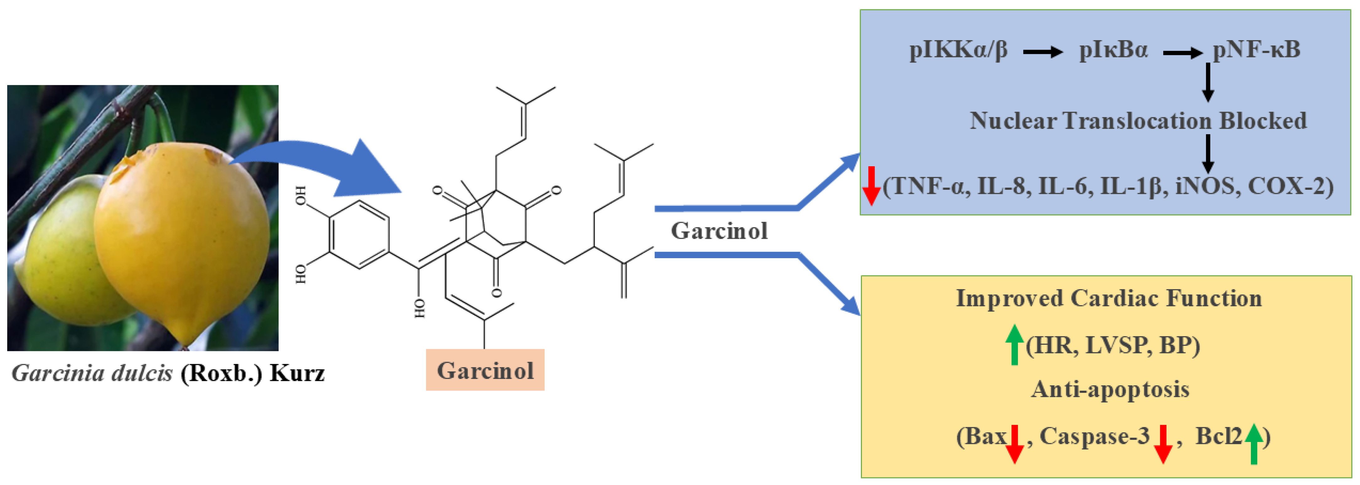
Garcinol, a bioactive benzophenone-derived compound, exhibited significant anti-inflammatory activity in LPS-activated THP-1 and RAW 264.7 macrophage cells. The IC50 values of garcinol were 67.86 ± 1.25 μM for RAW 264.7 cells and 78.45 ± 2.13 μM for THP-1 cells after 24 h of incubation at concentrations ranging from 10 to 100 μM. Coincubation with garcinol at concentrations of 20 and 30 μM significantly suppressed the elevated mRNA and protein expression levels of proinflammatory mediators, including inducible nitric oxide synthase (iNOS) and cyclooxygenase-2 (COX-2), as well as proinflammatory cytokines such as TNF-α, IL-8, IL-6, and IL-1β (Figure 2). In addition, coincubation with garcinol significantly modulated the nuclear factor kappa B (NF-κB) signaling pathway in both cell lines by inhibiting the LPS-induced increases in phosphorylated NF-κB (pNF-κB), IκBα (pIκBα), and IKKα/β (pIKKα/β) in a dose-dependent manner. This inhibition reduced the production of proinflammatory molecules by decreasing the nuclear translocation of pNF-κB from the cytosol [84]. Additionally, at a dose of 100 mg/kg, garcinol significantly decreased the maximum rate of left ventricular pressure development (LV-dp/dt_max) and increased the heart rate (HR), left ventricular systolic pressure (LVSP), systolic blood pressure, and diastolic blood pressure. In rats with isoproterenol-induced heart failure and in cardiac H9C2 cells, garcinol treatment reduced the mRNA and protein expression levels of cleaved caspase-3, total caspase-3, and Bax, while increasing Bcl-2 levels in heart tissue, indicating its antiapoptotic effect [85].
The pharmacokinetic analysis of garcinol in Sprague-Dawley rats revealed maximum plasma concentrations of 2317.69 ± 180.44 and 3446.14 ± 190.12 ng/mL after oral doses of 22.5 mg/kg and 45 mg/kg, respectively, and 11749.75 ± 300.00 ng/mL after intravenous administration. The half-lives of garcinol at these respective doses and administration routes were 25.43 ± 1.24, 24.46 ± 3.52, and 11.94 ± 5.29 h. The study also reported that bioavailability was greater in the 45 mg/kg group (35.72 ± 0.97%) than in the 22.5 mg/kg group (26.64 ± 0.23%) [86]. A study revealed that 40% garcinol had no adverse effects at a single dose of 2000 mg/kg. The study also revealed that repeated doses of 100 mg/kg/day over 28 and 90 days had no adverse effects on reproductive organs [87].

2.1.4. Centella asiatica (L.) Urb. (Family: Apiaceae)

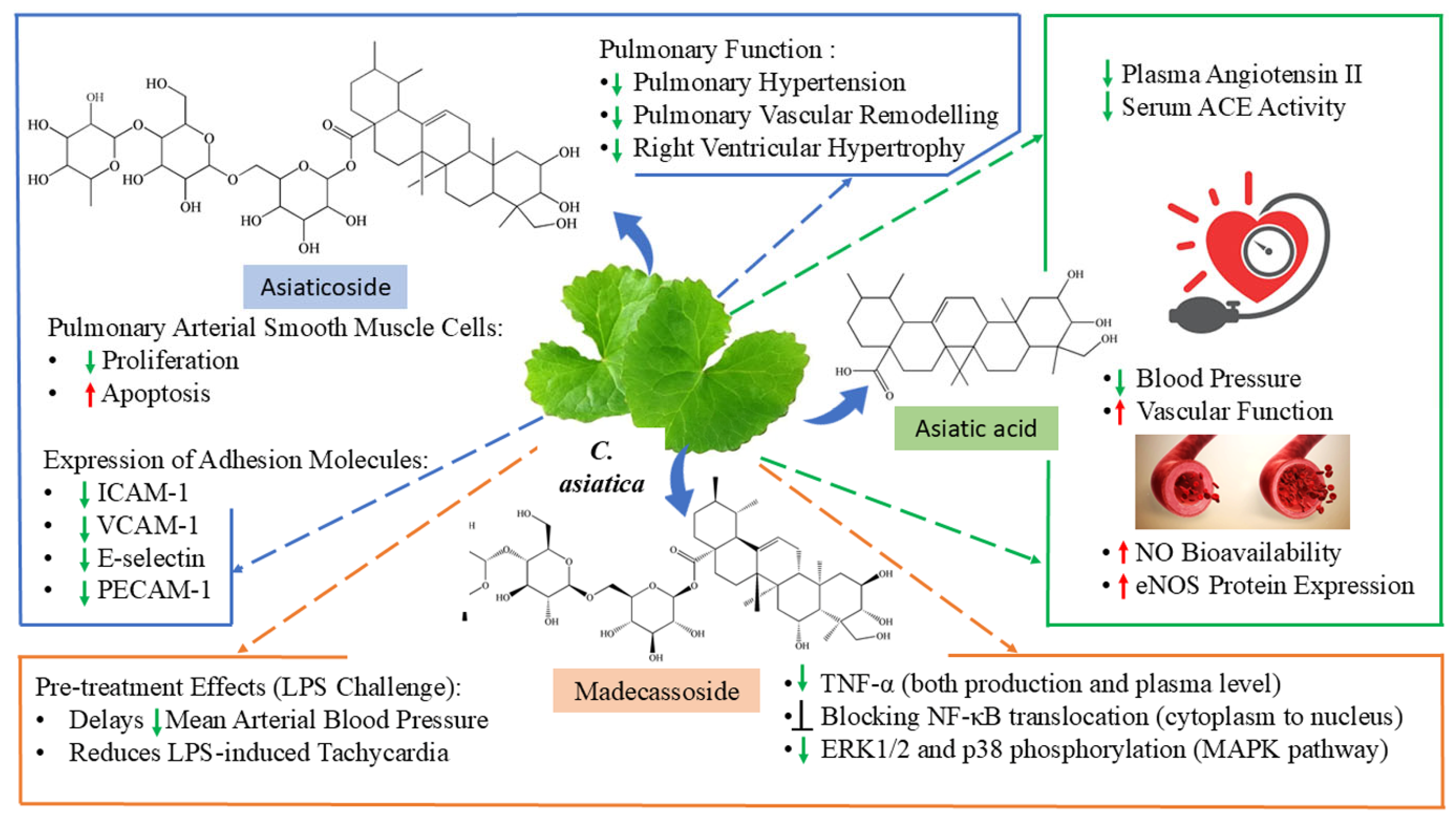
Centella asiatica L., commonly known as mandukparni or Indian pennywort, is a significant medicinal herb that has traditionally been utilized in Indian Ayurvedic medicine for millennia. Despite numerous studies over the past few decades investigating its biologically active components and mechanism of action, the plant is recommended for treating various skin conditions such as eczema and psoriasis, as well as for diarrhea, fever, wound healing, epilepsy, gastric ulcers, inflammation, and venous hypertension [88]. Asiaticoside, a saponin from C. asiatica, attenuated pulmonary hypertension, pulmonary vascular remodeling, and right ventricular hypertrophy in hypoxia-induced pulmonary hypertension rats by restraining excessive TGF-β1/Smad2/3 signaling and inhibiting the proliferation of pulmonary arterial smooth muscle cells [89]. In human umbilical vein endothelial cells (HUVECs) with oxidized low-density lipoprotein (oxLDL)-induced inflammation, asiaticoside at concentrations of 10–30 μM significantly modulated endothelial hyperpermeability and the levels of key inflammatory markers. Specifically, asiaticoside altered the levels of adenosine triphosphate (ATP) to 90% (p < 0.005), intercellular adhesion molecule-1 (ICAM-1) to 80% (p < 0.05), vascular cell adhesion molecule-1 (VCAM-1) to 105% (p < 0.01), E-selectin to 65% (p < 0.005), platelet endothelial cell adhesion molecule-1 (PECAM-1) to 70% (p < 0.05), and endothelial hyperpermeability to 105% (p < 0.01) [90].
Renovascular disease results from gradual blockage of the renal arteries, leading to various symptoms, including renovascular hypertension and ischemic nephropathy. Renovascular hypertension (RVH) is a prevalent cause of secondary hypertension and has been extensively researched as the classic example of angiotensin-dependent hypertension [91]. Asiatic acid found in C. asiatica (30 mg/kg/day) has been shown to inhibit the renin–angiotensin–aldosterone system (RAAS), as evidenced by reduced plasma angiotensin II levels and decreased serum angiotensin-converting enzyme (ACE) activity, while also restoring the AngII–AT1R–gp91phox–NF-κB signaling pathway [92]. In L-NAME-induced hypertensive rats, asiatic acid lowered blood pressure and improved vascular function, likely by increasing nitric oxide (NO) bioavailability through the upregulation of endothelial nitric oxide synthase (eNOS) expression and by reducing inflammation and oxidative stress [93,94]. Furthermore, asiatic acid supplementation improved vascular function, suppressed RAAS activation, and alleviated metabolic and cardiovascular complications in a rat model of metabolic syndrome induced by a high-carbohydrate, high-fat (HCHF) diet [92]. Madecassoside, a triterpenoid bioactive compound derived from Centella asiatica, inhibited the LPS-induced production of TNF-α in neonatal rat cardiomyocytes in a concentration-dependent manner [95]. At a dose of 20 mg/kg, madecassoside significantly reduced the LPS-induced increase in plasma TNF-α levels in rats by preventing the nuclear translocation of NF-κB and inhibiting the phosphorylation of p38 and ERK1/2 (Figure 3). Furthermore, pretreatment with madecassoside attenuated LPS-induced tachycardia and delayed the decline in mean arterial blood pressure [96].
In a pharmacokinetic study of madecassoside, asiaticoside, and asiatic acid in humans, participants were given oral capsules (250 mg) containing these bioactive compounds. Both madecassoside and asiaticoside reached peak plasma concentrations (9.57 ± 3.34 and 5.18 ± 3.43, respectively) within 1 h after dosing, whereas asiatic acid reached its peak (79.76 ± 19.28) between 1 and 2 h post-treatment. During the study, the plasma concentrations increased significantly with increasing dose; however, the half-lives of asiaticoside (10.37 ± 8.84), madecassoside (8.06 ± 5.62), and asiatic acid (9.86 ± 9.77) did not significantly change. The study also revealed that madecassoside and asiaticoside exhibited bioavailabilities of 27.99 ± 23.88% and 17.09 ± 13.52%, respectively, compared with 0.74 ± 0.25% for asiatic acid [97]. The LD50 of Centella asiatica was greater than 2000 mg/kg body weight in rats. No acute (single dose of 2000 mg/kg) or sub-chronic toxicity (90-day repeated dose of 1000 mg/kg) was observed [98].

2.1.5. Terminalia bellirica (Gaertn.) Roxb. (Family: Combretaceae)

Terminalia bellirica (Gaertn.) Roxb, which is distributed across Southeast Asian countries, is traditionally utilized in Indian Ayurvedic medicine to treat diabetes, rheumatism, and hypertension [99]. The major polyphenolic compound gallic acid and crude extract of T. bellirica inhibited the inflammatory response and reactive oxygen species production by suppressing the MAPK/NF-κB pathway and activating the Akt/AMPK/Nrf2 pathway in LPS-stimulated RAW264.7 macrophages. However, both gallic acid and crude extract enhanced antioxidant enzyme expression and improved acute kidney injury in an LPS-shocked mouse model [100]. The tannin-rich crude extract of T. bellirica reduced pulmonary arterial pressure, slowed pulmonary arterial remodeling, increased GSH-Px and SOD activity, decreased malondialdehyde (MDA) levels (p < 0.05), and reduced Bax expression in the lung tissues of high-altitude pulmonary hypertension (HAPH) rats. Conversely, the expression levels of Bcl-2, Nrf2, and HO-1 were elevated (p < 0.05). In cell experiments, TTR reduced H2O2-induced apoptosis and ROS production in PAECs (p < 0.05), decreased Bax expression, and increased Bcl-2, Nrf2, and HO-1 expression (p < 0.05) [101]. The development of atherosclerosis is closely linked to the inflammatory response initiated by macrophages. Within atherosclerotic plaques, macrophages secrete reactive oxygen species (ROS), matrix metalloproteinases (MMPs), and proinflammatory cytokines [102]. A previous study demonstrated that crude extract of Terminalia bellirica (TBE) significantly downregulated the mRNA expression of lectin-like oxidized LDL receptor-1 (LOX-1), interleukin-1 (IL-1), and tumor necrosis factor-alpha (TNF-α). Additionally, TBE reduced intracellular ROS production and MMP-9 secretion in THP-1 macrophages [103]. The aqueous-methanolic extract of T. bellirica has also been shown to lower blood pressure, potentially by inhibiting calcium release from intracellular stores and blocking calcium influx through membrane calcium channels [104]. The aqueous and ethyl acetate fractions have anticholinergic, Ca2+ antagonist, and antispasmodic effects [105].
The pharmacokinetic study of gallic acid from a Triphala formulation containing T. bellirica revealed that the plasma concentration was nearly doubled in the group receiving the 4000 mg dose compared with the 2000 mg dose group. The Cmax values were 70.81 ng/mL (range: 46.26–90.88) and 41.84 ng/mL (range: 32.29–59.54), respectively, in healthy Thai subjects (16 males and 16 females) aged 20–45 years, with body mass indices (BMIs) between 20 and 25 kg/m2. Following the oral administration of Triphala, gallic acid was rapidly absorbed, reaching a median tmax of 1 h. The study also revealed that the terminal elimination half-life was 1 h for both the 2000 mg and the 4000 mg doses [106]. Owing to its rapid absorption and metabolism, gallic acid has low bioavailability [107]. The LD50 of gallic acid was determined to be greater than 2000 mg/kg in albino mice. A high dose of gallic acid (900 mg/kg/day) administered over 28 days did not result in any significant changes in morphological or behavioral parameters [108].

2.1.6. Hibiscus sabdariffa L. (Malvaceae)

Hibiscus sabdariffa L., commonly known as roselle, which is also referred to as red sorrel, originates from India and Malaysia. This tropical plant is widely grown in tropical and subtropical areas around the globe, such as Central and West Africa and Southeast Asia. Scientific research has revealed that H. sabdariffa has various pharmacological benefits, including antihypertensive, antihyperlipidemic, anti-inflammatory, antimicrobial, diuretic, uricosuric, and anemia-treating properties [109]. The anthocyanin- and hibiscus acid-rich aqueous extract of H. sabdariffa inhibited ACE and had a vasodilatory effect [110]. A study revealed that, out of 121 participants in pilot interventions with high blood pressure (BP) (≥140/90 mmHg), 61.8% of participants achieved the recommended blood pressure target of <140/90 mmHg [111]. In total, 500 mg capsules of H. sabdariffa at a concentration of 6 mg/g of anthocyanins, once daily with meals for a duration of four weeks, significantly reduced systolic blood pressure (p = 0.049) and triglyceride levels (0.044) in a randomized controlled trial involving 45 participants [112]. Additionally, in a randomized, double-blind, placebo-controlled study among 80 participants, 175 mg of H. sabdariffa with a total combination dosage of 500 mg over a 12-week period resulted in a progressive reduction in systolic blood pressure [113].
The vascular effects of hibiscus acid from Hibiscus sabdariffa on rat aortic rings were investigated in vitro via myography techniques. Hibiscus acid induced concentration-dependent relaxation of the rat aorta precontracted with either phenylephrine (3 μM) or KCl (60 mM), independent of the endothelium. The concentration required to achieve 50% relaxation (IC50) in aortas precontracted with phenylephrine (PE) was 0.09 ± 0.01 mg/mL [114]. A previous study reported that hibiscus acid exhibits antihypertensive activity by inhibiting the enzymes α-amylase and α-glucosidase, thereby reducing the absorption of sugars and starches and contributing to weight loss. As a result, hibiscus acid may help prevent metabolic syndromes such as dyslipidemia, arterial hypertension, atherosclerosis, acute myocardial infarction, diabetes, and chronic kidney disease [115]. The antihypertensive effect of H. sabdariffa is achieved mainly through mechanisms such as diuresis, vasodilation, regulation of calcium influx, the inhibition of ACE, and the blockade of AT1 receptors [116].
The bioavailability and pharmacokinetics of hibiscus acid, the predominant organic acid in Hibiscus sabdariffa beverages, were evaluated in a human clinical study involving 12 volunteers. Hibiscus acid was rapidly absorbed, with a maximum plasma concentration (Cmax) of 28.5 µM observed as early as 0.5 h post-intake. Notably, a biphasic absorption pattern was recorded, with additional peaks at 3 and 5 h, indicating potential enterohepatic recirculation. The time to reach maximum concentration (Tmax) for hibiscus acid was approximately 2.5–2.8 h, which aligns with typical intestinal absorption kinetics. In terms of excretion, hibiscus acid was predominantly eliminated in urine within the 0–6 h window, with a peak occurring between 3 and 6 h. Additionally, its metabolite, hibiscus acid dimethyl ether, showed sustained plasma levels and delayed urinary excretion, peaking at 12 h post-consumption. These findings suggest high bioavailability of hibiscus acid and its derivatives, with implications for prolonged physiological activity and potential health benefits such as anti-inflammatory effects [117].

2.1.7. Citrus limon (L.) Osbeck (Family: Rutaceae)

Citrus limon L., which is commonly known as lemon, belongs to the Rutaceae family. Fruit juice (lemon juice) has traditionally been used as a remedy for scurvy, high blood pressure, the common cold, cough, and irregular menstruation. The major bioactive compounds of lemon include flavonoids, phenolic acids, monoterpenoids, coumarins, carboxylic acids, amino acids, and vitamins [118]. A previous study reported that lemon juice can significantly inhibit the activity of angiotensin-1 converting enzyme (ACE) and hypocholesterolemia in a high-cholesterol diet-fed rat model in a dose-dependent manner. Owing to the high phenolic content (64.5 mg/L) in the lemon juice, the inhibition of ACE activity results in antioxidant scavenging activities and the inhibition of Fe2+ and sodium nitroprusside-induced lipid peroxidation in the livers of the rats in vitro. In an in vivo experiment, the administration of juice significantly reduced total cholesterol in plasma, triglyceride, and LDL-cholesterol levels and increased plasma HDL-cholesterol levels in a high-cholesterol diet-treated rat model [119].
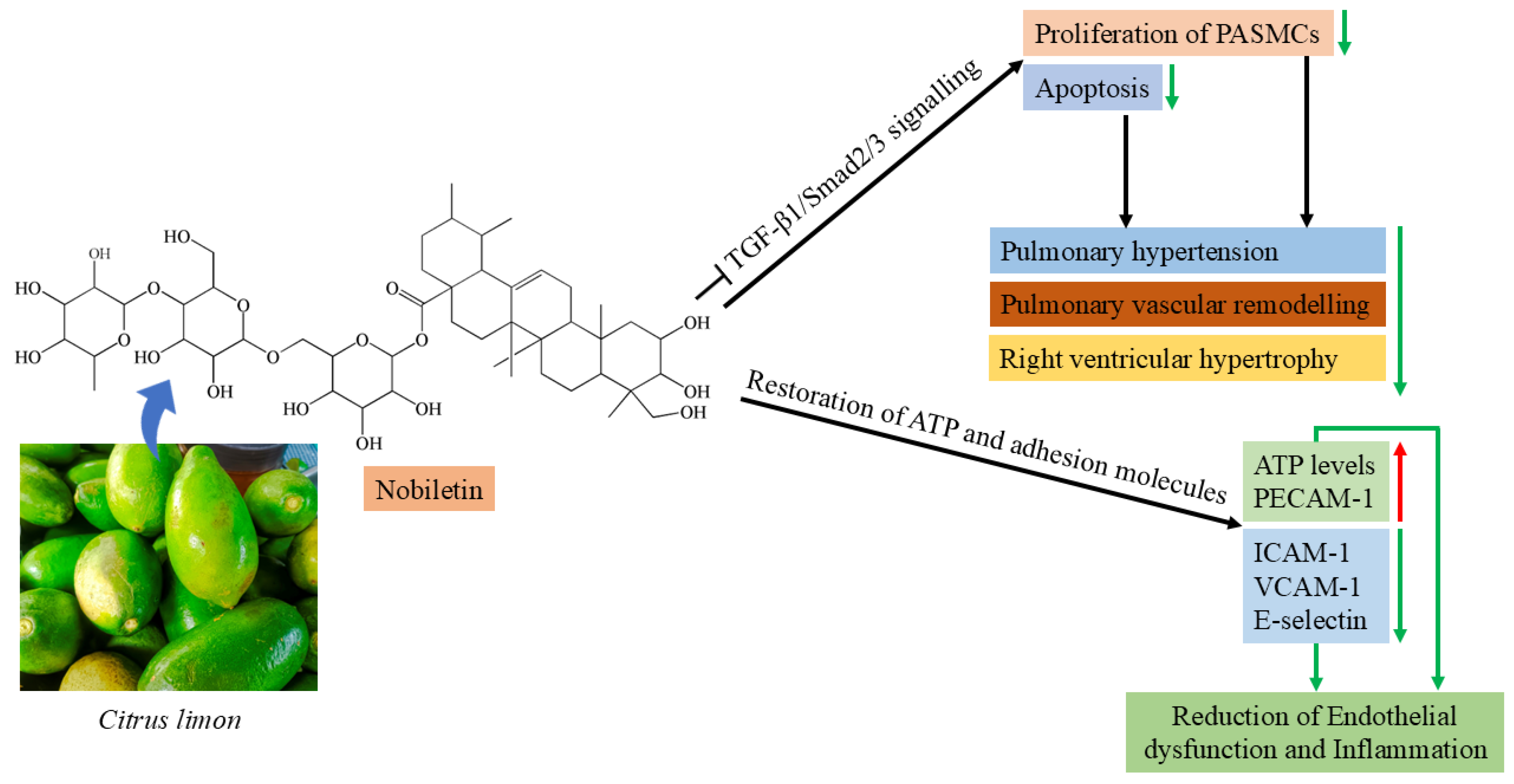
Citric acid is the major ingredient in lemon juice; it can significantly relax the thoracic aorta of spontaneously hypertensive rats and cause vasodilation at a concentration of 40. 1 μg/mL (0.21 mmol/L) [120]. Nobiletin, a polymethoxylated flavone obtained from immature citrus peels, induces the phosphorylation of vasodilator–stimulator phosphoprotein (VASP) in human platelet cells via a noncyclic nucleotide-related mechanism (Figure 4) [121]. Studies have shown that nobiletin lowers blood pressure and improves the contractile response to sympathetic nerve stimulation in the mesenteric vascular beds of male Sprague-Dawley rats treated with L-NAME. Nobiletin restored plasma NOx levels and increased the protein expression of endothelial nitric oxide synthase (eNOS), nuclear factor erythroid 2-related factor 2 (Nrf2), and heme oxygenase-1 (HO-1), while significantly reducing the expression of oxidative stress markers (p < 0.05). Additionally, nobiletin attenuated the thickening of the aortic wall, cross-sectional area, vascular smooth muscle cell proliferation, and collagen deposition, as well as glomerular extracellular matrix accumulation and renal fibrosis. These effects were achieved by downregulating the expression of matrix metalloproteinases (MMP-2 and MMP-9) and transforming growth factor (TGF) proteins and by upregulating adiponectin receptor 1 in models involving L-NAME and high-fat diet-induced vascular injury [122,123]. A previous study revealed that nobiletin effectively decreased the ventricular systolic pressure and attenuated right ventricular hypertrophy and medial wall thickening in monocrotaline-treated rats. By inhibiting Src/STAT3 activation and pulmonary artery smooth muscle cell proliferation through the downregulation of Pim1 and NFATc2, nobiletin has been shown to have protective effects on pulmonary arterial hypertension in rats (Figure 4) [124,125].
Hesperidin and G-hesperidin, two important bioflavonoids found in citrus fruits, exhibit antihypertensive effects primarily by upregulating the vascular nitric oxide synthase (NOS) pathway. The beneficial impact of these compounds on hypertension is attributed to increased nitric oxide (NO) bioavailability and protection of endothelial function from reactive oxygen species (ROS) through antioxidant mechanisms [126,127]. Additionally, hesperidin significantly reduces blood pressure in spontaneously hypertensive rats by suppressing the renin–angiotensin system (RAS) cascade and decreasing oxidative stress [128].
The pharmacokinetics of citrate, following intravenous administration, were measured in a study aimed at predicting citrate accumulation in humans to assess its therapeutic use. In this study, 12 healthy volunteers received trisodium citrate at a dose of 3.7 mmol/L of plasma flow. Blood samples were collected before infusion, throughout 120 min of infusion, and up to 120 min post-infusion. The plasma citrate levels were quantified via HPLC. The maximum plasma citrate concentration was 0.56 ± 0.45 mmol/L, with a total administered dose of 57.1 ± 10.5 mmol. The total body clearance rate was 686.64 ± 353.60 L/min [129]. Citric acid also plays a critical role in enhancing the bioavailability of certain nutrients and medications by adjusting the pH of formulations. For example, its acidic properties aid in stabilizing and improving the palatability of oral medications, thereby facilitating better absorption and patient compliance [130]. Another compound, nobiletin, which has maximum concentrations in both the plasma and brain, was observed 1 h after a single oral dose of 50 mg/kg. The peak concentrations were 1.78 μg/mL in the plasma and 4.20 μg/mL in the brain. The area under the concentration–time curve (AUC(0–t)) was 7.49 μg·h/mL in the plasma and 20.66 μg·h/mL in the brain. The mean elimination half-lives (t½) in the plasma and brain are 1.80 h and 11.42 h, respectively [131]. A study reported that the absolute bioavailability of nobiletin was 22.37% ± 4.52 in lean rats after oral administration. The gut microbiota present in rats plays an important role in nobiletin metabolism [132]. An LD50 study revealed the safety of nobiletin at 5000 mg/kg [133]. In another study, hesperidin exhibited different pharmacokinetic rates in the different parts of the intestine after oral administration of 709 μmol/kg in mice [134]. The rates of bioavailability in the duodenum, jejunum, cecum, and colon were reported to be 0.14%, 0.49%, 2.12%, and 1.06%, respectively. Owing to its low solubility in water, the gut microbiota in the gastrointestinal tract helps in absorption, biotransformation, and rapid excretion [135]. Hesperidin showed a median lethal dose (LD50) of 4837.5 mg/kg, and few observed adverse effects in a subchronic toxicity study at 1000 mg/kg in Sprague-Dawley rats [136].

2.1.8. Passiflora edulis Sims (Family: Passifloraceae)

Passion fruit (Passiflora edulis), which is commonly found in tropical and subtropical regions, has numerous health benefits, including antitumour, antihypertensive, antioxidant, antidiabetic, and hypolipidemic properties [137]. At a dose of 50 mg/kg per day, purple passion fruit peel (PFP) extracts significantly reduced systolic blood pressure by 12.3 mmHg (p < 0.1) and decreased serum nitric oxide levels by 65% (p < 0.05) in spontaneously hypertensive rats. Furthermore, a 4-week randomized, double-blind, placebo-controlled trial was conducted in hypertensive human subjects to evaluate the effects of 400 mg of PFP extract daily compared with a placebo. The trial’s primary outcome was measured through blood pressure readings. Compared with the placebo group, participants who received the PFP extract experienced significant reductions in both systolic and diastolic blood pressures of 30.9 ± 6.3 mmHg and 24.6 ± 3.3 mmHg, respectively (p < 0.001). No adverse effects were reported by the participants [138]. High-performance liquid chromatography (HPLC) analysis of the peel extract confirmed the presence of cyanidin 3-O-glucoside, quercetin 3-O-glucoside, and edulilic acid, a novel cyclic acid glucoside unique to P. edulis [139]. Sukketsiri et al. (2023) investigated the antioxidant, anti-inflammatory, antilipid, and vascular relaxant activities of seeds and fruit extracts from P. edulis [140]. Both extracts exhibited antioxidant activity, inhibited pancreatic lipase and cholesterol esterase, reduced nitric oxide levels during LPS-induced inflammation in RAW264.7 cells, and promoted ex vivo vasorelaxation in rat aortic rings. These effects were attributed to the high content of piceatannol and polyphenolic stilbenes in the seed extract, as well as β-carotene and γ-tocopherol in the fruit extract. The long-term administration of piceatannol increased endothelial nitric oxide synthase (eNOS) protein levels and eNOS mRNA expression in a dose-dependent manner. Additionally, piceatannol enhanced the phosphorylation of eNOS [141].
The pharmacokinetic study of the primary antihypertensive compound piceatannol revealed that the plasma AUC, urine t1/2, CL, and Vd were 8.48 ± 2.48 mg·h/mL, 19.88 ± 5.66 h, 2.13 ± 0.92 L/h/kg, and 10.76 ± 2.88 L/kg (mean ± SEM), respectively, following a single intravenous dose of 10 mg/kg piceatannol in male Sprague-Dawley rats [142]. A previous study revealed that piceatannol, a natural analog of resveratrol, has an oral bioavailability of over 1%, indicating that it is more stable than resveratrol [143].

2.1.9. Garcinia cowa Roxb. ex Choisy (Family: Clusiaceae)

Garcinia cowa is an evergreen medium-sized tree, commonly known as cowa fruit/kau thekera, which is widely distributed throughout the Southeast Asian region. Various plant parts, i.e., leaves, fruit, bark, latex and roots, have been used in traditional medicine for the treatment of various diseases, such as fever, indigestion and blood circulation [144]. Recent studies have shown that the leaf of G. cowa caused significant relaxation of the aortic rings of rats in a concentration-dependent manner. The vasorelaxant effect of G. cowa leaf extract primarily involves the generation of nitric oxide and prostanoids, as well as the activation of ATP-sensitive potassium channels. The phytochemical investigation of the tested crude extract confirmed the presence of kaempferol, vitexin, and isovitexin, which are possible bioactive compounds for vasorelaxant effects [145]. Previous studies confirmed that the compound kaempferol induces vessel relaxation through the activation of the NO–cGMP–PKG signaling pathway, enhancement of bradykinin-induced relaxation, and stimulation of large-conductance Ca2+-activated potassium channels [146]. In contrast, two additional compounds, vitexin and isovitexin, were found to reduce phorbol ester-induced vascular contraction primarily by inhibiting MEK activity and reducing ERK1/2 phosphorylation. This mechanism also leads to the activation of small-conductance Ca2+-activated potassium (SKCa) channels and the upregulation of Kir6.1 ATP-sensitive K+ channels [147,148].
In LPS-induced RAW264.7 macrophages, phloroglucinol benzophenones (garciniacowones F, H, and I) and xanthones (cowanol, mangostanin, and cratoxylone) isolated from the leaves of Garcinia cowa had inhibitory effects on nitric oxide (NO) production, with IC50 values ranging from 5.4 to 18.6 μM, respectively. Nitric oxide is a highly reactive molecule produced in the body through both enzymatic and nonenzymatic pathways. It is acknowledged as a proinflammatory mediator that participates in numerous biological functions and may be pivotal in inflammatory conditions. Many diseases, such as neurodegenerative and cardiovascular disorders, cancer, obesity, and diabetes, are closely linked to inflammation [149].
Pharmacokinetic studies of vitexin were conducted following intravenous and oral administration to mice at doses of 10 mg/kg and 30 mg/kg, respectively. After administration, vitexin was rapidly and widely distributed throughout the body, reaching peak concentrations at approximately 19.14 ± 1.07 min. It was quickly eliminated from the plasma, with a half-life (t1/2) of 29.11 ± 2.31 min, a clearance rate (CL) of 0.89 ± 0.06 L·kg/min, and an oral bioavailability of 3.91% [150]. The plasma concentration of the compound isovitexin sharply decreased from 22.5 μg/mL to 2.77 μg/mL at 1 h after intravenous tail administration at a dose of 2 mg/kg in rats. A pharmacokinetic study reported that isovitexin had a half-life t1/2 of 1.05 ± 0.325 h, with a mean residual time (MRT) of 1.229 ± 0.429 h, and the area under the curve (AUC) was 11.39 ± 5.05 μg/mL/h [151]. In an in vitro study, vitexin showed cytotoxicity (IC50) against breast cancer cell lines at a 12 mg/kg concentration [152]. In in vivo, up to 2 g/kg of both vitexin and isovitexin did not result in any cytotoxicity in normoglycemic mice or diabetic rats after up to 14 days of monitoring [153]. Following intravenous administration of kaempferol at doses of 10 and 25 mg/kg, the plasma concentration–time profiles indicated a large volume of distribution (8–12 L/kg) and high clearance (~3 L/h/kg), with a terminal half-life of approximately 3–4 h. When kaempferol is administered orally, it shows relatively rapid absorption, reaching a peak plasma concentration (tmax) within approximately 1–2 h. However, its bioavailability (F) remains low at approximately 2%, likely due to extensive first-pass metabolism involving glucuronidation and other pathways in the gut and liver [154].

2.1.10. Solanum torvum Sw (L.) (Family: Solanaceae)

Solanum torvum is commonly known as Turkey berry and is native to Asia, Africa, the West Indies, and South America. The fruits are edible and are consumed for medicinal purposes to treat hypertension [155]. The ethanolic fruit extract of Solanum torvum significantly reduced the systolic blood pressure and the serum glucose, cholesterol, and triglyceride levels. It also ameliorated vascular reactivity to catecholamines and corrected fructose-induced metabolic disturbances. In male albino rats, administration of the extract caused a rightward shift in the cumulative concentration–response curve (CCRC) of angiotensin II (Ang II) in isolated strips of the ascending colon, indicating a reduced contractile response [156]. The aqueous and methanolic extracts of S. torvum significantly inhibited platelet aggregation and reduced arterial blood pressure in Wistar rats. Aqueous extracts have been shown to induce antiplatelet aggregation via thrombin and adenosine diphosphate at a concentration of 2 mg/mL via intravenous injection. The methanolic extracts reduced the heart rate at all doses, whereas the aqueous extract only reduced the heart rate at 5 mg/kg. Aqueous extracts reduced blood pressure at doses of 1 and 2 mg/kg without affecting heart rate [157]. Similarly, chronic oral administration of aqueous extracts of Solanum torvum fruits significantly reduced arterial hypertension and cardiac hypertrophy in L-NAME-treated rats by increasing urine volume and sodium excretion. In vitro, the extract induced strong, partially endothelium-dependent contraction of the aortic ring [158]. Additionally, the methanolic extract of S. torvum fruits exhibited angiotensin-converting enzyme (ACE) inhibitory activity, with an IC50 value of 1.2 mg/mL. Bioassay-guided fractionation confirmed the presence of dihydroxycyclopentyl-3-(3,4-dihydroxyphenyl) acrylate, a methyl salicylate glycoside, which had an IC50 of 778 μg/mL. In addition, the presence of torvumoside and lariciresinol-4, 4-O-β-D- diglucoside potentially contributes to ACE inhibitory activity through their antioxidant activity [159].

2.2. In Silico ADME/T Profiling of Target Plant Bioactive Compounds

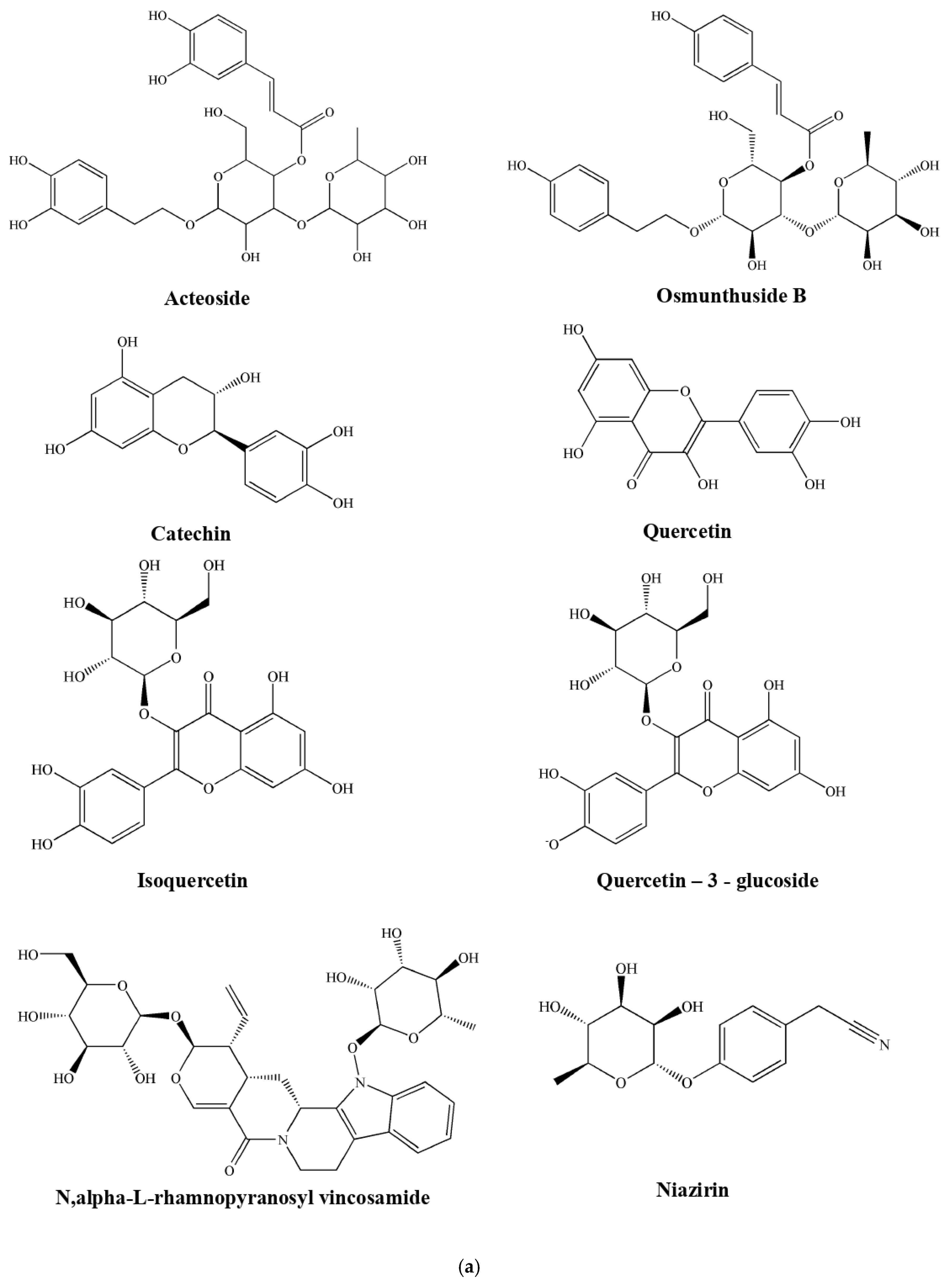
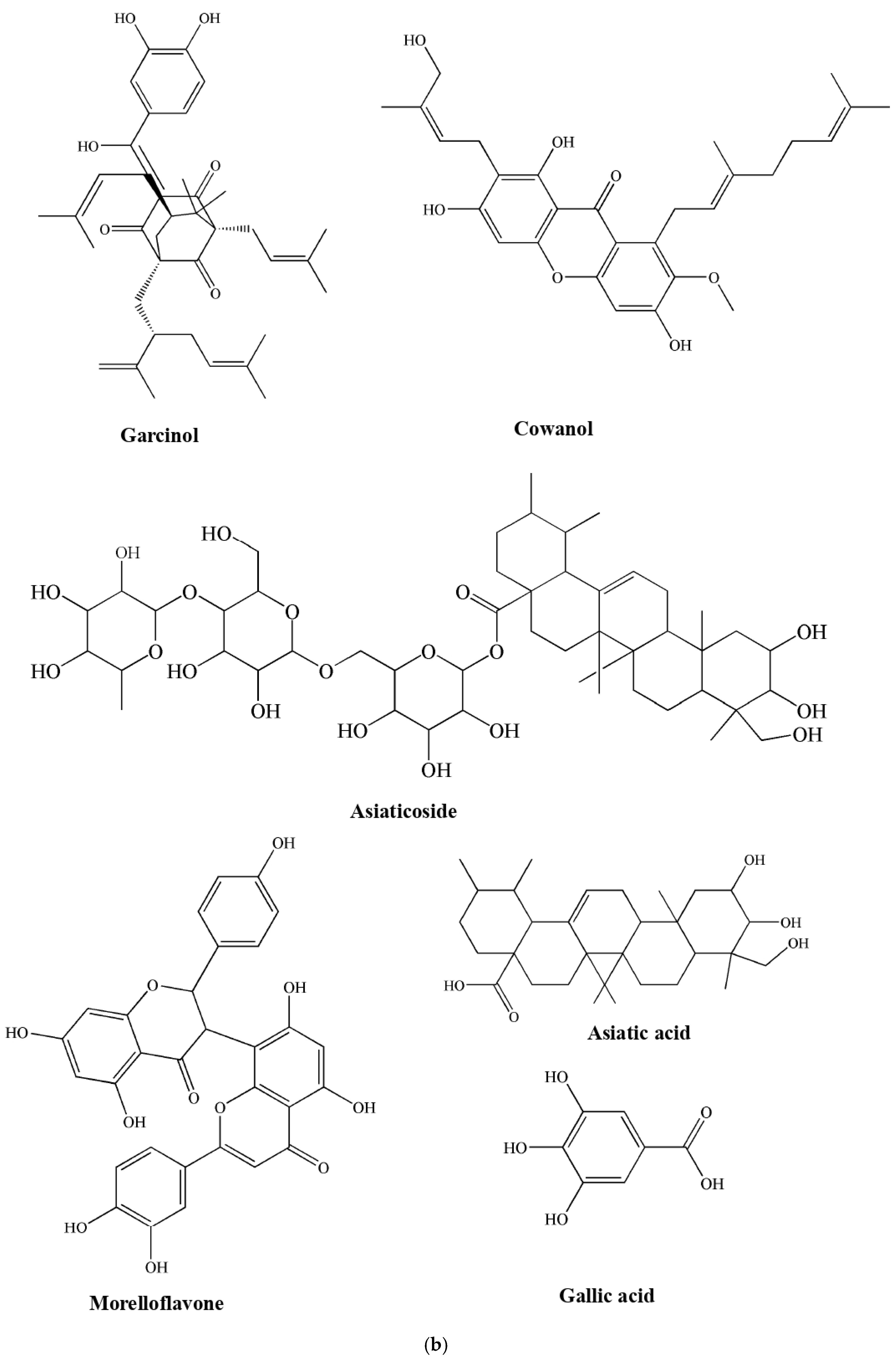
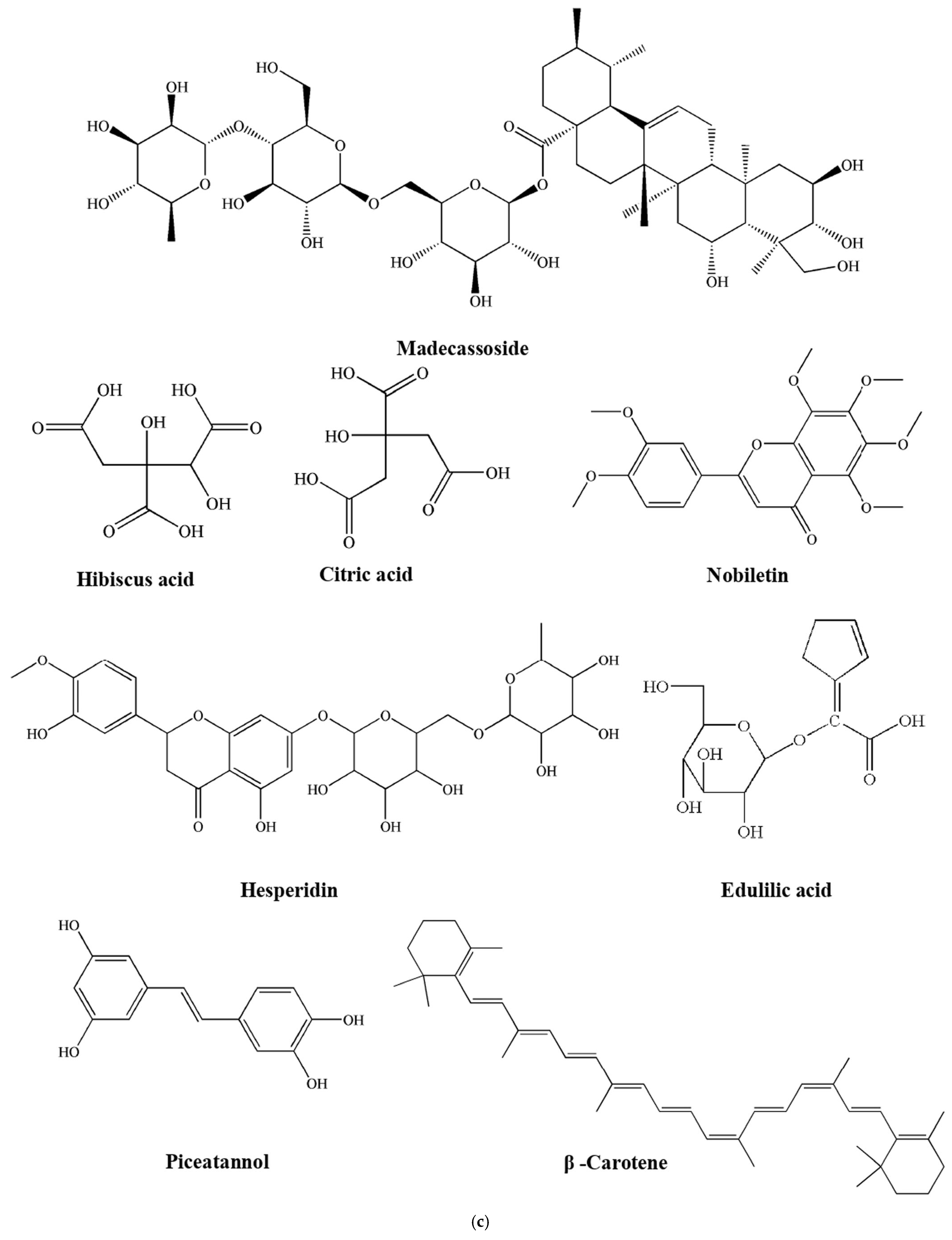
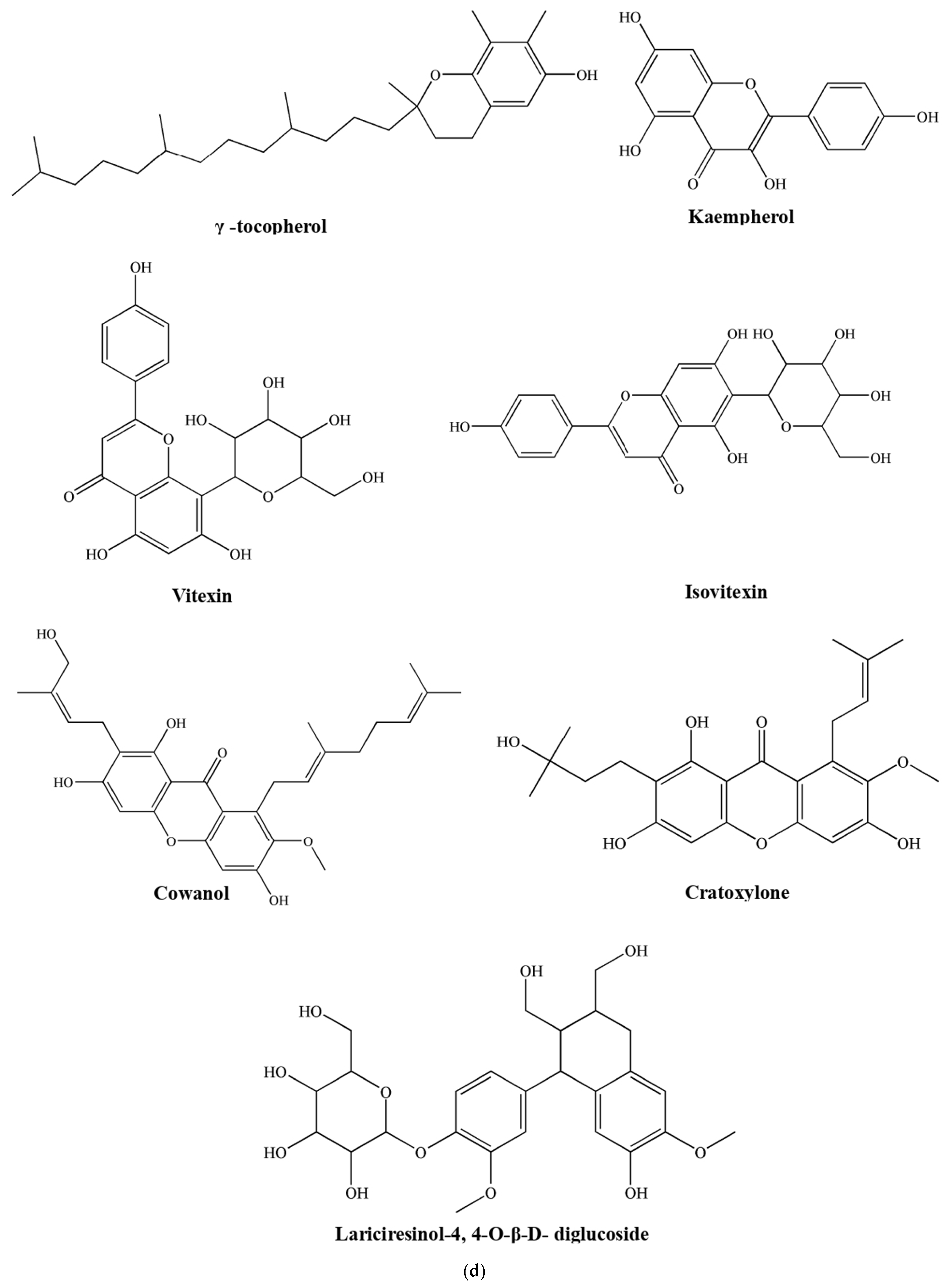
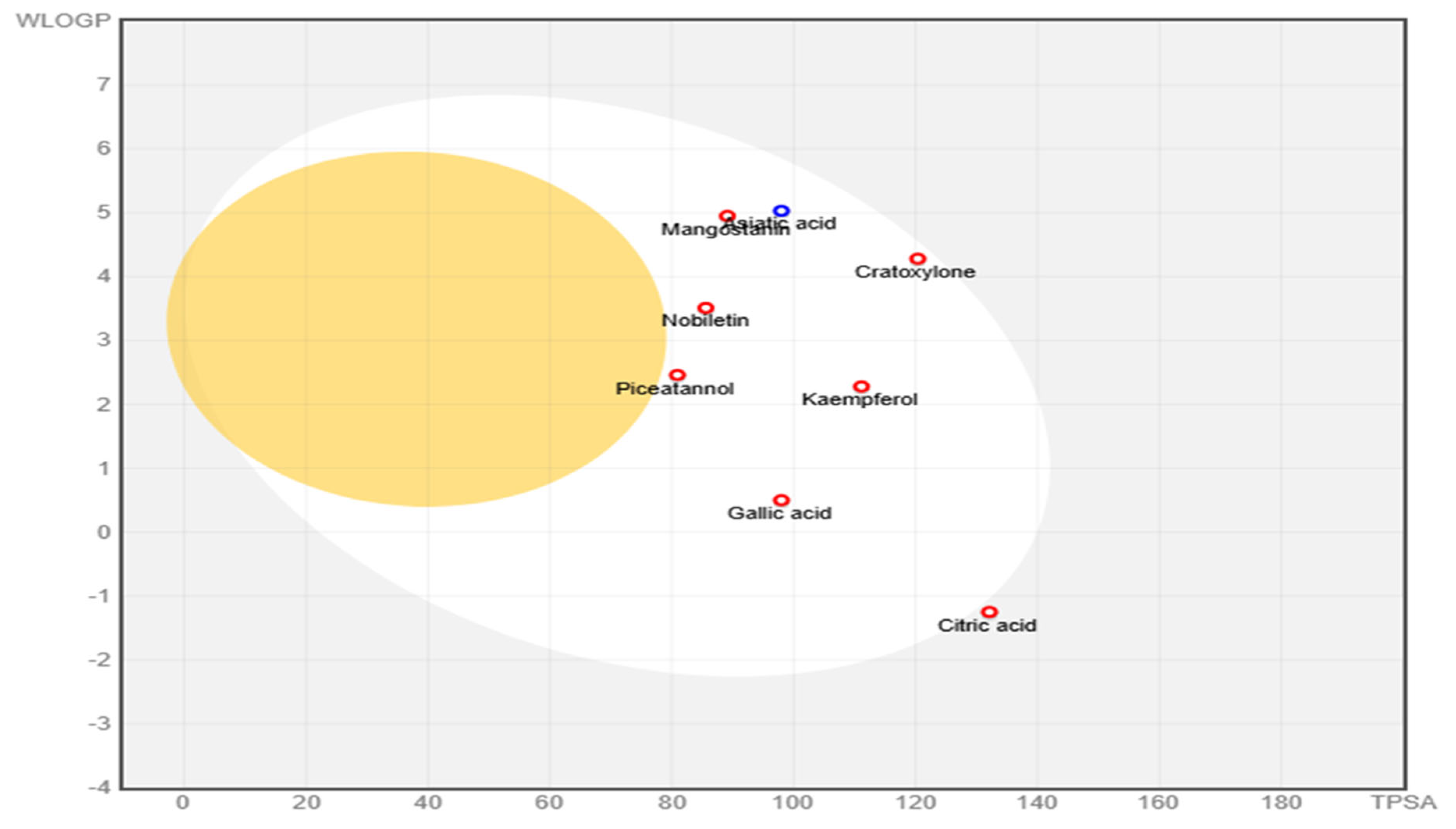
This analysis examines the absorption, distribution, metabolism, excretion, and toxicity (ADMET) characteristics and drug-likeness of compounds sourced from plants (Figure 5a–d) with antihypertensive effects (Supplementary Tables S1–S6). The physicochemical assessment indicated that most compounds are within the acceptable drug-like range, characterized by a molecular weight under 500 Da, a topological polar surface area below 140 Å2, and favorable hydrogen bonding potential. Notably, compounds such as kaempferol, cratoxylone, piceatannol, and niazirin exhibit optimal physicochemical properties, including low rotatable bond counts and balanced hydrophilic–lipophilic profiles, suggesting good oral bioavailability (Figure 6).
Lipophilicity analysis, which is based on consensus LogP values, revealed that the majority of these compounds fall within the ideal range of 1–3, with kaempferol (1.58), niazirin (0.32), and piceatannol (2.14) demonstrating a favorable lipophilic balance that minimizes the risk of poor membrane permeability or excessive tissue accumulation. Furthermore, water solubility assessments revealed that compounds such as niazirin, citric acid, hibiscus acid, and osmanthuside are highly soluble, which is beneficial for formulation and oral delivery, whereas garcinol and morelloflavone present solubility challenges that could hinder bioavailability and require advanced formulation techniques. Pharmacokinetic evaluations suggest that several compounds, including kaempferol, cratoxylone, piceatannol, gallic acid, and niazirin, exhibit high gastrointestinal absorption and are not substrates for P-glycoprotein, increasing their suitability for oral administration. Importantly, none of the compounds are predicted to penetrate the blood–brain barrier, a desirable feature of antihypertensive agents, as central nervous system access is generally not preferred for this class of drugs. Kaempferol and nobiletin exhibited inhibition of CYP450 enzymes, particularly CYP3A4 and CYP1A2, which raises concerns about potential drug–drug interactions in combination therapies. Most candidates demonstrated favorable drug-likeness, adhering to established criteria such as the Lipinski, Ghose, Veber, Egan, and Muegge rules, indicating strong oral bioavailability potential. Notably, kaempferol, cratoxylone, niazirin, piceatannol, and nobiletin did not violate these filters and achieved bioavailability scores of 0 or 1, reinforcing their classification as highly drug-like. In contrast, larger polyphenolic compounds such as acteoside, hesperidin, and cyanidin-3-O-glucoside exhibited multiple violations and lower bioavailability scores, suggesting limited oral drug potential in their current forms. The majority of the top-performing compounds presented minimal medicinal chemistry alerts, such as PAINS and Brenk alerts, indicating a low risk of nonspecific binding or false positives in biological assays. The synthetic accessibility scores were generally favorable, ranging from 0 to 2, although more complex molecules such as garcinol received a higher score of 3, indicating challenges in synthesis or scalability. Overall, kaempferol, cratoxylone, piceatannol, niazirin, and nobiletin have emerged as promising candidates for further research because of their balanced ADMET profiles, minimal rule violations, high predicted oral bioavailability, and advantageous medicinal chemistry properties. These findings support their advancement into in vitro and in vivo studies targeting critical hypertensive mechanisms, such as angiotensin-converting enzyme (ACE) inhibition, endothelin antagonism, or calcium channel modulation, while also considering formulation enhancement strategies such as nanocarriers or prodrug approaches for compounds with poor solubility yet promising pharmacodynamic profiles.
A thorough in silico toxicity (T) assessment of 22 bioactive compounds derived from plants, known for their antihypertensive and antihyperglycemic effects, was conducted via the ProTox-III platform, which yielded significant insights into their therapeutic potential. The evaluation covered various toxicity endpoints, including organ-specific toxicity, ecotoxicity, blood–brain barrier permeability, receptor interactions, activation of stress response pathways, molecular initiating events, and interactions with cytochrome P450 enzymes. Vitexin, isovitexin, isoquercetin, and nobiletin consistently exhibited low toxicity across key parameters, including organ toxicity, mutagenicity, and immunotoxicity. Hibiscus acid, madecassoside, and asiaticoside had high predictive probabilities (over 0.90) for hepatotoxicity and neurotoxicity, raising concerns about the risks of prolonged systemic exposure (Supplementary Table S7). Additionally, garcinol and morelloflavone exhibited cardiotoxicity predictions above 0.85, which is particularly alarming for substances aimed at cardiovascular treatment. From a genotoxicity standpoint, gallic acid, hesperidin, and acteoside presented high mutagenicity scores near 0.98, warranting further investigation through in vitro and in vivo assays. The high cytotoxicity predictions for kaempferol, piceatannol, and morelloflavone underscore the need for careful assessment of therapeutic windows and dosage. Notably, hesperidin, osmanthuside, and acteoside had high immunotoxicity, suggesting potential adverse effects on immune function. Receptor-binding simulations indicated strong affinities (scores >0.90) for nuclear hormone receptors, including PPAR-γ, estrogen, androgen, and aryl hydrocarbon receptors, suggesting possible endocrine-disrupting effects alongside their therapeutic applications. Furthermore, stress response pathways such as Nrf2/ARE, MMP, HSE, and p53 pathways were activated by many compounds, indicating both antioxidant capabilities and the risk of mitochondrial dysfunction, particularly with hibiscus acid and piceatannol, which are predicted to disrupt the mitochondrial membrane potential.
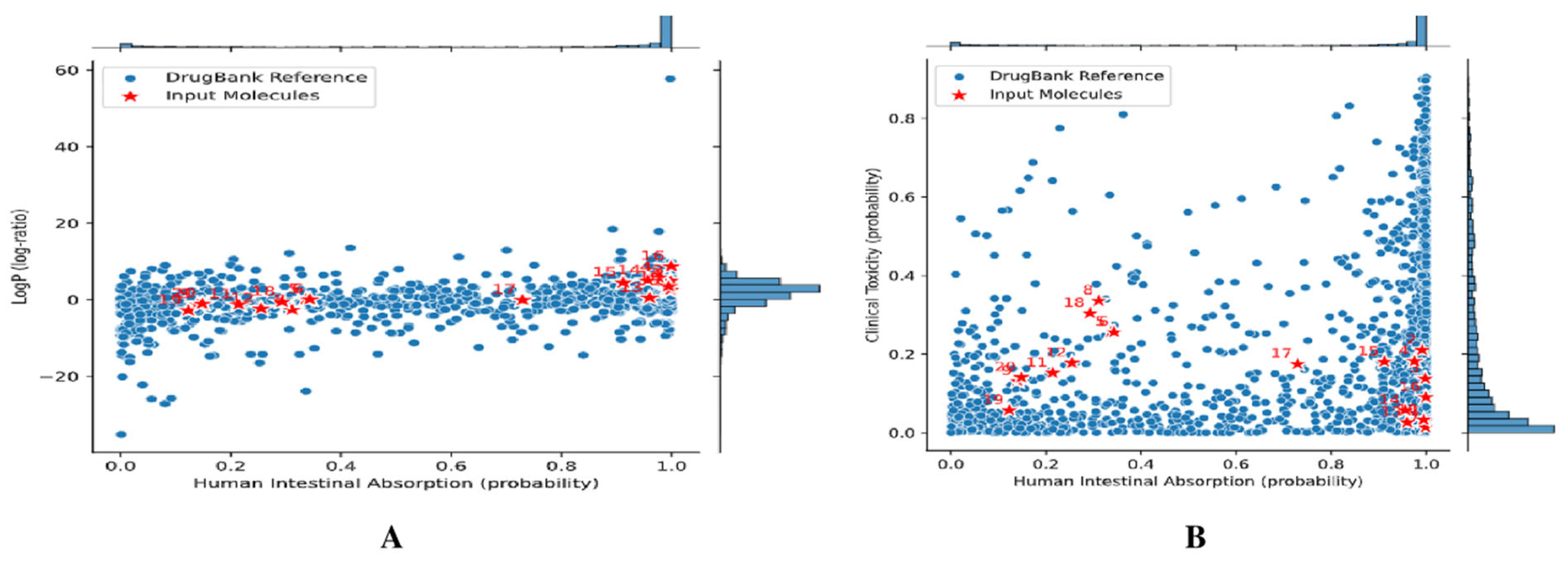
The ADME/T profiling of these compounds has highlighted both significant advantages and certain obstacles regarding their potential as lead molecules for antihypertensive activity. A comparison with approved drugs from DrugBank, which focus on essential parameters such as intestinal absorption and calculated lipophilicity (ClogP), indicated a considerable overlap in chemical space (Figure 7), underscoring the novel and drug-like characteristics of these natural compounds. Nevertheless, some candidates showed deviations in specific ADMET parameters, including solubility, permeability, or metabolic stability. To address these issues, employing a bioisosteric modification strategy could prove beneficial in optimizing their pharmacokinetic and safety profiles. This targeted approach aims to increase the affinity for the intended targets while reducing undesirable traits, thereby increasing the chances of successful lead development.

2.3. Prospects and Challenges of the Use of Plant Bioactive Compounds in Modern Drug Development

Chemical compounds derived from medicinal plants play crucial roles in treating various health conditions, often resulting in fewer side effects than synthetic drugs do. These plant-based remedies also offer affordable primary healthcare solutions, particularly in regions lacking adequate conventional medical infrastructure, where cultural beliefs and traditional practices guide treatment choices [160]. The presence of multiple phytochemicals in a single plant can lead to synergistic therapeutic effects, benefiting the management of both metabolic and infectious diseases[161]. Emerging biotechnological tools, such as tissue culture techniques, present promising avenues for the large-scale cultivation of potent and endangered medicinal plants [162]. However, the effective isolation and purification of key bioactive compounds remain significant challenges. Employing advanced extraction methods alongside modern analytical technologies is essential for the efficient and accurate recovery of natural products from plants.
In this review, we identified a total of 22 significant bioactive compounds (Figure 5a–d) with antihypertensive activity from 10 medicinal plants. However, we were unable to elucidate the chemical structures of certain compounds due to the lack of available information in chemical databases such as PubChem. These include two compounds, namely (E)-2,3-dihydroxycyclopentyl-3-(3′,4′-dihydroxyphenyl) acrylate and torvumoside [methyl salicylate 2-O-(2′-O-β-apiofuranosyl, 6′-O-β-xylopyranosyl)-β-glucopyranoside) from Solanum torvum, niaziridin from Moringa oleifera, edulilic acid from Passiflora edulis, and three compounds, namely garciniacowones F, H, and I from Garcinia cowa. Moreover, before clinical application, it is important to assess the safety, efficacy, and toxicity of these bioactive compounds to understand their pharmacokinetic behavior in biological systems. As illustrated in Table 2 of this review, many plant-derived compounds presented low IC50 values, indicating strong therapeutic potential while exhibiting high LD50 values (from reported preclinical/clinical literature), suggesting low toxicity, particularly in the context of antihypertensive effects. Nonetheless, a common limitation of these compounds is their poor bioavailability, which often hinders effective site-specific action. To overcome this, nanotechnology-based approaches, such as nanoparticle formulations, may offer a promising strategy to increase bioavailability and ensure targeted therapeutic delivery [163].
In addition, 12 other plant species, Elsholtzia communis (Collett & Hemsl.) Dunn, Cissampelos pareira L., Tithonia diversifolia (Hemsl.) A. Gray, Fagopyrum esculentum Moench, Allium hookeri Thwaites, Blumea balsamifera (L.) DC., Sarcococca saligna (D. Don) Müll. Arg., Catharanthus roseus (L.) G. Don, Citrus maxima (Burm.) Merr., Mikania micrantha Kunth, Cuscuta cassytoides Nees ex Engelm., and Bidens pilosa L., have limited representation in the scientific literature, with only one to two studies available on their antihypertensive activity. To substantiate and establish their potential, there is an urgent need for comprehensive experimental studies and repeated scientific evaluations employing standardized in vitro and in vivo models, along with detailed elucidation of their mechanisms of action and identification of their respective bioactive compounds. Ethnobotany serves as a foundational approach in the discovery of modern drugs, as it is rooted in traditional knowledge and practices. Therefore, proper ethnobotanical documentation, followed by rigorous scientific validation, is crucial for identifying more potent and pharmacologically significant plant-derived compounds with antihypertensive potential from this region.
Table 1. Potent bioactive compounds from the selected plants with their mechanism of action and antihypertensive activity.
Sl. No.Scientific Name and FamilyParts UsedBioactive CompoundExperimental ModelMechanism of ActionActivityReferences
1.Clerodendrum colebrookeanum Walp. (Family: Lamiaceae)LeavesActeosideIn silico study Inhibit ROCK- I and II, PDE-5Antihypertensive[57]
Rat aortaNO/cyclic GMP overproductionAntihypertensive[58]
Rat PlasmaInhibit ACEAnti-inflammatory[59]
LPS-induced mice Increase NO production, restore sepsis-induced mitochondrial changes, inhibit apoptosis in cardiomyocytesAnti-inflammatory[60]
OsmanthusideIn silico studyInhibit ROCK-II, PDE-5Antihypertensive[57]
2. Moringa oleifera Lam. (Family: Moringaceae)Leaves, Catechin, isoquercetin, quercetin, gallic acidL-NAME-induced hypertensive ratSuppress vascular superoxide anion production and oxidative stress reductionAntihypertensive[69]
Quercetin- 3-O glucosideL-NAME-induced hypertensive ratInhibit ACE, systolic blood pressureAntihypertensive[73]
N, α-L-rhamnopyranosyl vincosamideIsoproterenol (ISO)-induced cardiac toxicity in ratsLowered cardiac markerAntioxidant[74]
Niaziridin and niazirinMonocrotaline-induced hypertensive ratReduced pulmonary arterial blood pressureAntioxidant[75]
LGF, GLEF Spontaneously hypertensive ratInhibit ACE and renin, reduced systolic and diastolic blood pressureAntihypertensive[70]
IPPAYSK, ILVDR, FFFPK, and LLDPRIn silico studyInhibit ACEAntihypertensive [72]
SeedIWHHTFYNELRIn silico studyInhibit ACEAntihypertensive[72]
3. Garcinia dulcis (Roxb.) Kurz (Family: Clusiaceae)FruitCamboginol2-kidney-1-clip hypertensive ratActivate nitric oxide synthase, prostacyclin signaling pathway, and ATP-activated potassium channelVasorelaxant[81]
Morelloflavone2-kidneys-1-clip hypertensive ratActivate nitric oxide signaling pathwayDiuretic, Vasorelaxant[82,83]
GarcinolLPS-activated THP-1 and Raw 264.7 macrophagesInhibit TNF-α, IL-8, IL-6, IL-1β, iNOS, COX-2, and NF-κBAnti-inflammatory[84]
Male albino Wistar ratsCaspase-3 and BaxAnti-inflammatory[85]
4.Centella asiatica (L.) Urb. (Family: Apiaceae)LeavesAsiaticosideHypoxia-induced pulmonary hypertension in rats Inhibit TGF-β1/Smad2/3 signaling pathwayAnti-inflammatory[89,90]
Human umbilical endothelial cellsReduce VCAM-1, PECAM-1, E-selectin levelsAnti-inflammatory
Asiatic acid2K-1C hypertensive ratsIncreased plasma angiotensin II, serum angiotensin ACE activity, restored Ang II-AT1R-gp91phox-NF-κB pathwayAnti-inflammatory [92]
L-NAME-induced hypertensive ratsIncrease NO bioavailability through upregulation of eNOS protein expressionVasorelaxant[93,94]
MadecassosideNeonatal rat cardiomyocytesInhibit TNF-α production, NF-κB, phosphorylation of ERK1/2 and p38Anti-inflammatory[96]
5.Terminalia bellirica (Gaertn.) Roxb. (Family: Combretaceae)FruitGallic acidLPS-induced RAW 264.7 macrophageInhibit MAPK/NF-κB pathway, activate Akt/AMPK/Nrf2 pathwayAnti-inflammatory and ROS scavenger[100]
6.Hibiscus sabdariffa L. (Malvaceae)CalyxHibiscus acidRat aortaInhibition of Ca2+ influx via voltage-dependent Ca2+ channelsVasorelaxant[114]
α-amylase inhibitory assayInhibit α-amylase/α-glucosidaseVasorelaxant[115]
7.Citrus limon (L.) Osbeck (Family: Rutaceae)FruitCitric acidSpontaneously hypertensive male ratsVascular muscle relaxantVasorelaxant[120]
NobiletinHuman platelet cellsActivate VSAP
Inhibit NOx and eNOS
Vasorelaxant[121]
L-NAME induced hypertensive ratsInhibit MMP-2 and MMP-9Vasorelaxant[122]
High-fat-diet fed rats Upregulation of AdipoR1, Suppression of TGF-β1 in kidneyVasorelaxant and renal alteration[123]
Monocrotaline-induced PAH ratsInhibit Src/STAT3Antihypertensive[124]
Monocrotaline-induced PAH ratsInhibit the phosphorylation level of PI3K/Akt/STAT3Anti-inflammatory[125]
Hesperidin,
G-Hesperidin
MCF-7 cellsUpregulation of vascular NO synthaseVasorelaxant, [126]
Stroke-prone spontaneously hypertensive ratsROS scavengerAntioxidant[127]
2K-1C hypertensive ratsDownregulation of renin-angiotensin system Antihypertensive[128]
8.Passiflora edulis Sims (Family: Passifloraceae)FruitEdulilic acidSpontaneously induced hypertensive rat Decrease systolic and diastolic blood pressure Antihypertensive[139]
Piceatannol, beta carotene, gamma tocopherolLPS-induced RAW 264.7 cells and rat aorta ROS scavenger,
Inhibit pancreatic lipase, cholesterol esterase
Antioxidant, Vasorelaxant[140]
9.Garcinia cowa Roxb. ex Choisy (Family: Clusiaceae)LeavesKaempferolRat aortaActivate NO–cGMP–PKG signaling pathwayVasorelaxant[146]
VitexinRat aortaInhibit MEK activity, reducing ERK1/2 phosphorylationVasorelaxant[147]
IsovitexinL-NAME-induced hypertensive ratStimulate NO release
Activate SKCa
Upregulate Kir 6.1 ATP-sensitive K+ channels
Vasorelaxant[148]
Garciniacowones F, H, I, Cowanol, Nagostanin and CratoxyloneLPS-induced RAW 264.7 cells Regulation of NO overproductionAnti-inflammatory[149]
10. Solanum torvum Sw (L.) (Family: Solanaceae)FruitDihydroxycyclopentyl-3-(3,4-dihydroxyphenyl) acrylate,
Torvumoside,
Lariciresinol-4,4-O-β-D- diglucoside
Bovine plasmaInhibit ACE activityAntihypertensive[159]
Table 2. Safety, efficacy, and toxicity profile of antihypertensive potential bioactive compounds.
Sl. No.Plant NameIsolated CompoundIC50BioavailabilityToxicity
(LD50)
Clinical Trial StatusReferences
1.Clerodendrum colebrookeanum WalpActeoside0.127 μM4% in beagle dogsat >5 g/kg in oral and IP administration in both rats and mice.Phase 2 and Phase 3
(ClinicalTrials.gov ID NCT02662283)
[164,165]
Osmanthuside23.14 ± 0.51 mg/mL0.17% according to Swiss ADMELD50 data not availableNA[166,167]
2.Moringa oleifera Lam.Catechin44.53 μMLess than 5% in ratsat 1084 mg/kg via IP administration in rats. Dyspnea-like symptoms occur.Phase 1
(ClinicalTrials.gov ID NCT03278925
[168,169]
Isoquercetin/Quercetin- 3-O glucoside79 μg mL-120% in humans>5 g/kg IP in mice.Phase 2
(ClinicalTrials.gov ID NCT04474626)
[76,168,170]
Quercetin4.48 μMLess than 10%484 µg/mL in ZebrafishPhase 2
(ClinicalTrials.gov ID NCT01708278)
[171,172,173]
N, α-L-rhamnopyranosyl vincosamide19.92 ± 1.19 g/mLNALeaf extract at 2000 mg/kg in rats.NA[74,174]
NiaziridinNANANANA
Niazirin18.55 uM46.78–52.61%3750 mg/kg in miceNA[78,175]
3.Garcinia dulcis (Roxb.) KurzCamboginol/Garcinol0.1 μMModerate oral bioavailability of around 26–36% in Sprague-Dawley rats.40% Garcinol has a low toxicity profile in rodentsNA[87,176,177]
Morelloflavone0.48 mM0.17 according to Swiss ADMENo toxicological reportNA[166,177]
4.Centella asiatica (L.) Urb.Asiaticoside300 μMLess than 1% in male Wistar rats.>2000 g/kg of leaf extract orally (No LD50 reported for the compound)Phase 2 and Phase 3
(ClinicalTrials.gov ID NCT06231212)
[178,179]
Asiatic acid60 µM16.25%Showed cardiotoxicity in zebrafishPhase 2 and Phase 3
(ClinicalTrials.gov ID NCT06231212)
[179,180,181]
Madecassoside26 µMLess than 1% in male Wistar ratNo toxicological reportPhase 2 and Phase 3
(ClinicalTrials.gov ID NCT06231212)
[178,182]
5. Terminalia bellirica (Gaertn.) RoxbGallic acid3.5 μg/mLLow bioavailability due to fast absorption and metabolism.4300 gm/kg (IP), 320 mg/kg (IV) in mouse; 5000 mg/kg (Oral) in rabbit Phase 1
(ClinicalTrials.gov ID NCT04970589)
[107,168]
6.Hibiscus sabdariffa LHibiscus acid37.15 μg/mL10% in oral administration in humans.>5000 mg/kg of calyx extract (IP) in mice. Above 500 mg/kg of H. sabdariffa extract alter the taste of rats. [117,183]
7.Citrus limon (L.) OsbeckCitric acid0.64 ± 0.04 μM/mL94.80% in rabbits5040 mg/kg (Oral) in Mouse and 3000 mg/kg (Oral) in rats. [184,185]
Nobiletin6.12 µM19.93 ± 3.93% and 46.20 ± 5.03% in oil suspension and emulsion, respectively.Data are not available [186,187,188]
Hesperdin16.08 mMLess than 20%4837.5 mg/kg (Oral) in rats. [136,189,190]
8.Passiflora edulis SimsPiceatannol60 µM17.70 ± 0.91% after oral administration217 mg/kg (IP) in mousePhase 1
(ClinicalTrials.gov ID NCT06127381)
[168,191,192]
Beta carotene7.0 μg/mL60.7%>10,000 mg/kg (Oral) in rats.Phase 4
(ClinicalTrials.gov ID NCT03005496)
[193,194]
9.Garcinia cowa
Roxb. ex Choisy
Kaempferol178 µM~2% in ratsNo toxic effect seen up to 2000 mg/kg/day in SD ratsPhase 1
(ClinicalTrials.gov ID NCT02191241)
[154,195,196]
Vitexin52.80 ± 1.65 μMVery low bioavailability1 mg/kg (IP) in mice showed LD25Phase 4
(ClinicalTrials.gov ID NCT01647984)
[168,197,198]
Isovitexin18.6 ± 1.3 μg/mL14.58% oral bioavailability in mouse2.56 mol/kgNA[199,200,201]
Cowanol13.4 μM-2.778 mol/kg in ratNA[149,202]
Garciniacowone F11.0 μMNANANA[149]
Garciniacowone H7.7 μMNANANA[149]
Garciniacowone I5.8 μMNANANA[149]
Mangostanin/9-Hydroxycalabaxanthone18.6 μMNANANA[149]
Cratoxylone5.4 μMNANANA[149]
10.Solanum torvum Sw (L.)(E)-2,3- dihydroxycyclopentyl-3-(3′,4′-dihydroxyphenyl) acrylate(778 8 g/mL) 2.7 mMNANANA[159]
Torvumoside (methyl salicylate 2-O-(2′O-β-apiofuranosyl, 6′-O-β-xylopyranosyl)-β-glucopyranoside)NANANANA
Lariciresinol-4, 4-O-β-D- diglucosideNANANANA

3. Materials and Methods

3.1. Systematic Review of Hypertension and Medicinal Plant-Related Therapeutic Evidence

In this systematic review, relevant research articles were identified through comprehensive searches in major scientific databases, including PubMed, Google Scholar, ScienceDirect, and Scopus via the ‘Preferred Reporting Items for Systematic Reviews and Meta-Analyses (PRISMA) 2020 [203]. The introduction provides a concise overview of hypertension, covering its definition, classification, global prevalence, causes, symptoms, and association with cardiovascular diseases. It also discusses current limitations in diagnostic approaches and underscores the potential of affordable, plant-based therapeutic strategies.
Studies evaluating the antihypertensive and hypotensive effects of selected medicinal plants were included on the basis of demonstrated mechanisms of action. These mechanisms include inhibition or reduction in angiotensin-converting enzyme (ACE) activity; elevation of nitric oxide (NO) levels; inhibition of NF-κB; calcium channel blocking or antagonistic activity; enhancement of flow-mediated dilation (FMD); reduction in proinflammatory cytokines such as TNF-α and IL-6; inhibition of vascular cell adhesion molecule-1 (VCAM-1); suppression of platelet-derived growth factor (PDGF) proliferation; inhibition of lipid peroxidation (LPS); promotion of urinary excretion of potassium (K+), sodium (Na+), and chloride (Cl) ions; inhibition of angiotensin II; activation of endothelial nitric oxide synthase (eNOS); and scavenging of reactive oxygen species (ROS). Collectively, these factors contribute to vasorelaxant, vasodilatory, anti-inflammatory, anti-proliferative, and antioxidant effects.
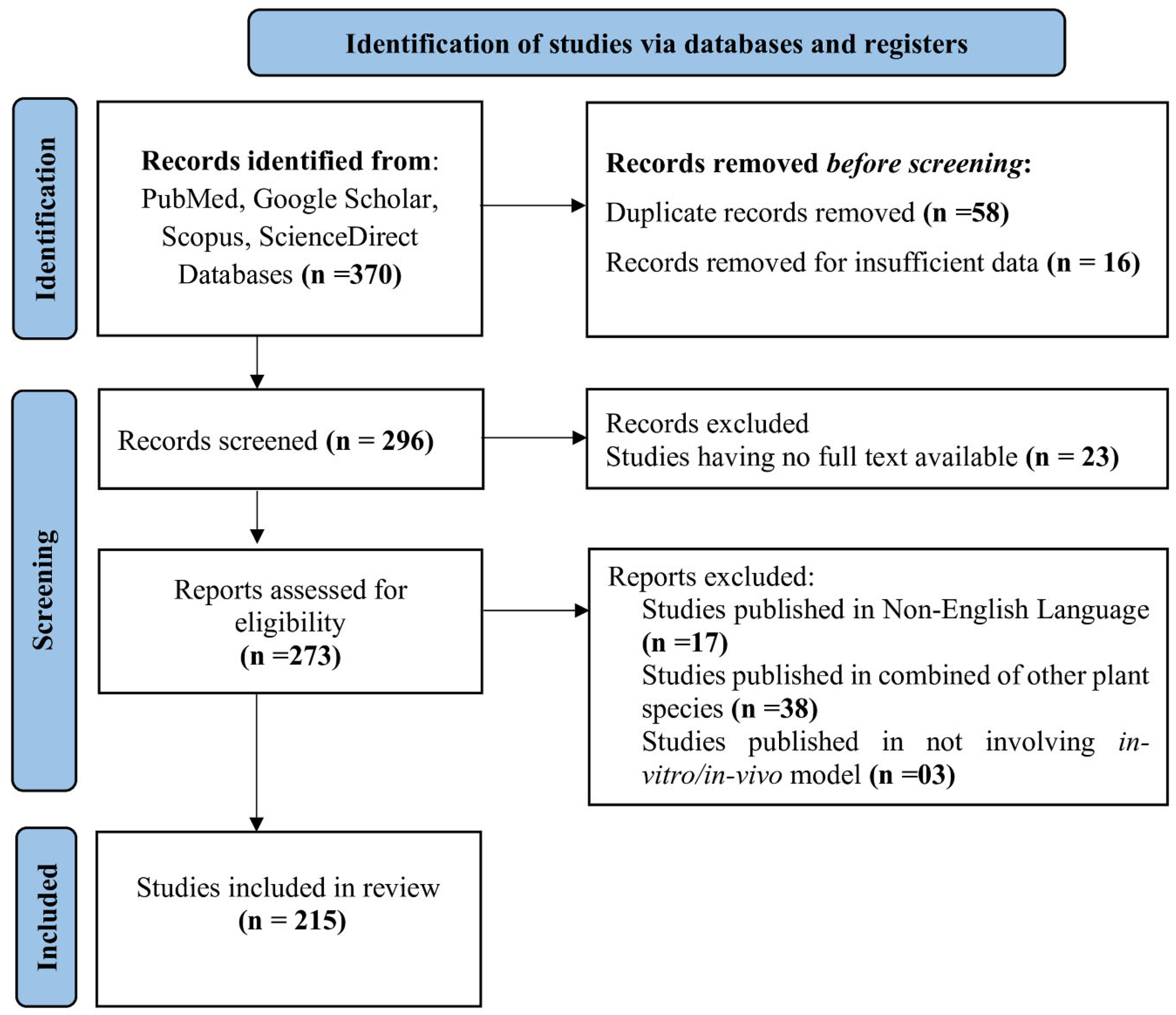
A total of 370 articles were initially retrieved. Following the removal of 58 duplicates, further exclusions were made: 16 articles due to insufficient data, 23 for lack of full-text access, 17 non-English publications, 38 studies involving plant combinations instead of single species, and 3 studies lacking in vitro or in vivo models. After applying these criteria, 215 studies were selected for inclusion (Figure 8).
Inclusion criteria are as follows:
  • The full texts of the articles were published in English.
  • Studies reporting antihypertensive activity via in vitro, in vivo, or both approaches via standardized protocols.
  • Studies have focused on single plant species, utilizing crude extracts, bioactive fractions, or isolated pure compounds. Pharmacokinetic studies of major compounds regardless of the source plant were also included to provide information on their absorption, distribution, metabolism, excretion, and bioavailability.
  • Peer-reviewed studies published up to 2024.
Exclusion criteria are as follows:
  • Articles available only as abstracts without full text.
  • Book chapters or conference proceedings not indexed in PubMed, Google Scholar, Scopus, or ScienceDirect.
  • Non-English language publications.
  • Studies lacking scientific rigor or proper methodology or not published in peer-reviewed and refereed journals.
Additional data sources are as follows:
The updated and verified names of the plant species mentioned in the study were obtained from the World [204]. Additional data, including IC50 values, LD50 values, and bioavailability information, were obtained from [166,168]. Clinical trial information related to the selected medicinal plants or their active constituents was extracted from [205]. The chemical structures of the bioactive compounds were illustrated via ChemDraw 8.0 software.

3.2. An In Silico Evaluation of Identified Plant-Based Compounds with Antihypertensive Activity

In recent years, computer-aided drug discovery (CADD) has played a pivotal role in advancing this research, primarily through ligand-based and structure-based approaches [206,207,208]. Emerging computational techniques such as molecular docking, network pharmacology, metabolomics, and molecular dynamics simulations have been increasingly employed to explore and predict the antihypertensive potential of plant-derived compounds [209,210,211]. These methodologies enable the identification of active phytoconstituents, their molecular targets, and mechanistic pathways, thereby accelerating the development of novel therapeutics for hypertension.
The assessment of the ADME (Absorption-A, Distribution-D, Metabolism-M, and Excretion-E) and toxicity (T) characteristics of compounds is essential in the early stages of drug discovery, as these characteristics influence the pharmacokinetic profile and safety of potential drug candidates [212]. This study conducted a thorough ADMET and toxicity evaluation of 22 plant-derived compounds known for their antihypertensive effects to determine their drug-likeness and appropriateness for initial drug development. Two compounds, madecassoside and asiaticoside, were omitted from further analysis because of their high molecular weights. ADMET profiling utilizes three sophisticated in silico tools: SwissADME, ProTox 3.0, and ADMET-AI. SwissADME (https://tox.charite.de/protox3/, accessed on 15 May 2025) was employed to predict essential physicochemical properties, including lipophilicity, through models such as iLOGP and XLOGP3, as well as pharmacokinetic parameters such as gastrointestinal absorption and blood–brain barrier permeability, while also assessing drug-likeness via established filters such as those of Lipinski and Ghose. Additionally, synthetic accessibility was evaluated, and any PAINS or Brenk alerts were identified to gauge medicinal chemistry viability [213]. ProTox 3.0, a cutting-edge toxicity prediction platform leveraging molecular similarity and machine learning, was utilized to analyze 61 toxicity endpoints, encompassing acute oral toxicity and organ toxicity [214]. Finally, ADMET-AI was used to compare the input compounds against the Drugbank library, i.e., ALogP and clinical toxicology versus human intestinal absorption [215].

4. Conclusions

Although synthetic antihypertensive drugs are essential for controlling high blood pressure and preventing complications such as heart attack and stroke, they may cause a variety of adverse effects. The use of traditional medicines provides several benefits over synthetic drugs, such as fewer side effects, a holistic approach to health, cultural relevance, sustainability, cost–effectiveness, synergistic effects, personalized treatment, and a lower risk of resistance. These advantages make traditional medicine an important complement or alternative to synthetic drugs, especially in enhancing overall health and well-being. Ethnomedicinal plants with antihypertensive properties present a valuable opportunity for developing new drugs that are natural and potentially safer than synthetic medications. Leveraging the wide range of bioactive compounds and traditional wisdom linked to these plants, researchers can create innovative and efficient treatments for hypertension, thereby addressing a significant global health concern. Establishing the safety, efficacy, and bioavailability of plant-derived bioactive compounds through rigorous preclinical and clinical studies is essential for the development of standardized and effective plant-based therapeutics. Such evidence would strengthen the scientific basis for the potential integration of traditional medicinal knowledge into modern healthcare and facilitate the development of novel antihypertensive drugs.

5. Limitations of the Study

The ADMET and toxicity profiles for 22 bioactive compounds identified in this review were obtained solely from in silico platforms (SwissADME, ProTox 3.0 and ADMET-AI). These predictions are based on similarity algorithms and machine learning models and not experiential evidence and should be validated via in vitro and in vivo studies. Due to the lack of structural data in public chemical databases, the chemical structures of a few compounds in this study, including a dihydroxycyclopentyl acrylate derivative and torvumoside (Solanum torvum), niaziridin (Moringa oleifera), edulilic acid (Passiflora edulis), and garcinia cowones F, H and I (Garcinia cowa), could not be completely resolved, restricting their computational evaluation.
However, several promising compounds have poor oral bioavailability, such as asiaticoside (<1% in rats) and kaempferol (~2% in rats), which could be limited in their therapeutic potential unless their bioavailability is enhanced by formulation approaches like drug delivery systems based on the use of nanoparticles. The in silico toxicity screening also predicted safety concerns for a few compounds, such as hepatotoxicity and neurotoxicity (hibiscus acid, madecassoside and asiaticoside), cardiotoxicity (garcinol and morelloflavone), mutagenicity (gallic acid, hesperidin, and acteoside), and possible endocrine-disrupting activity due to the interaction with nuclear hormone receptors. While these predictions do not provide evidence of toxicity, they do indicate the importance of achieving thorough experimental safety testing.
No new experimental or clinical data were generated, and this is a literature review and analysis. Standardized in vitro and in vivo studies are needed to confirm the proposed antihypertensive mechanisms, detailed pharmacokinetic and bioavailability studies, dose-dependent effects on toxicological studies, and formulation optimization for bioavailability in future studies. But if the preclinical evidence is significant, well-designed clinical trials will be needed to prove the safety and effectiveness of these compounds in humans.

Supplementary Materials

The following supporting information can be downloaded at: https://www.mdpi.com/article/10.3390/ddc5030043/s1, Table S1: Physicochemical Property calculation; Table S2: Lipophilicity; Table S3: Water Solubility; Table S4: Pharmacokinetics; Table S5: Drug likeness; Table S6: Medicinal Chemistry; Table S7: Toxicity Prediction of Compounds with Hypertensive activity.

Author Contributions

P.B.: Writing—original draft, Conceptualization, Validation. S.H.: Writing—original draft. A.S.: Writing—original draft. H.U., M.C., R.B.: Writing—review and editing, Conceptualization, Validation, Supervision. D.G.: Writing—original draft, Data Curation, Software. A.K.V.: Writing—review and editing, Visualization, Funding acquisition, Data curation, Conceptualization, Supervision. All authors have read and agreed to the published version of the manuscript.

Funding

Department of Biotechnology (DBT), Government of India, DBT Builder Project (Grant No: BT/INF/22/SP45376/2022).

Institutional Review Board Statement

Not Applicable.

Data Availability Statement

Data related to the in silico study is included in the Supplementary File.

Acknowledgments

We are deeply grateful to the Department of Biotechnology (DBT), Government of India, for their generous financial support provided through the DBT Builder project (Grant No: BT/INF/22/SP45376/2022). This funding has played a vital role in facilitating the successful completion of our research. The authors acknowledge the use of OpenAI’s language model for minor language editing, specifically grammar corrections and readability enhancements, during the preparation of this manuscript. All scientific content, experimental design, data analysis, interpretation, and conclusions represent the original work of the authors.

Conflicts of Interest

The authors affirm that they have no known financial or personal conflicts of interest that could have influenced the work presented in this paper.

Abbreviations

ACEAngiotensin-converting enzyme
ACEIsAngiotensin-converting enzyme inhibitors
ADMETAbsorption, distribution, metabolism, excretion and toxicity
AHAAmerican Heart Association
ALTAlanine aminotransferase
ASTAspartate aminotransferase
AUCArea under the curve
BBBBlood–brain barrier
BMIBody mass index
BPBlood pressure
CADDComputer-aided drug discovery
CCRCCumulative concentration-response curve
CHDCoronary heart disease
COX-2Cyclooxygenase-2
DBPDiastolic blood pressure
EDHFEndothelium-derived hyperpolarizing factor
eNOSEndothelial nitric oxide synthase
FMDFlow-mediated dilation
HAPHHigh-altitude pulmonary hypertension
HCHFHigh-carbohydrate, high-fat
HO-1Heme oxygenase-1
HRHeart rate
HUVECsHuman umbilical vein endothelial cells
ICAM-1Intercellular adhesion molecule-1
IL-1Interleukin-1
iNOSInducible nitric oxide synthase
LDLLow-density lipoprotein
LOX-1Lectin-like oxidized LDL receptor-1
LPSLipopolysaccharide
MABPMean arterial blood pressure
MDAMalondialdehyde
MMPsMatrix metalloproteinases
NF- κBNuclear factor kappa B
NFHSNational Family Health Survey
NONitric oxide
NOSNitric oxide synthase
Nrf2Nuclear factor erythroid 2
oxLDLOxidized low-density lipoprotein
PDE-5Phosphodiesterase-5
PDGFPlatelet-derived growth factor
PECAM-1Platelet endothelial cell adhesion molecule-1
RAASRenin–angiotensin–aldosterone system
RASRenin-angiotensin system
ROCK-IIRho-kinase-II
ROSReactive oxygen species
RVHRenovascular hypertension
SBPSystolic blood pressure
SKCaSmall-conductance Ca2+-activated potassium channel
SODSuperoxide dismutase
TGFTransforming growth factor
TNF-αTumor necrosis factor-alpha
VASPVasodilator-stimulated phosphoprotein
VCAM-1Vascular cell adhesion molecule-1
VOOCVoltage-operated calcium channels
WHOWorld Health Organization

References

  1. Mills, K.T.; Stefanescu, A.; He, J. The global epidemiology of hypertension. Nat. Rev. Nephrol. 2020, 16, 223–237. [Google Scholar] [CrossRef] [Scilit] [PubMed]
  2. Singh, S.; Shankar, R.; Singh, G.P. Prevalence and associated risk factors of hypertension: A cross-sectional study in urban Varanasi. Int. J. Hypertens. 2017, 2017, 5491838. [Google Scholar] [CrossRef] [Scilit] [PubMed]
  3. Carey, R.M.; Whelton, P.K. Prevention, detection, evaluation, and management of high blood pressure in adults: Synopsis of the 2017 ACC/AHA hypertension guideline. Ann. Intern. Med. 2018, 168, 351–358. [Google Scholar] [CrossRef] [Scilit] [PubMed]
  4. World Health Organization. First WHO Report Details Devastating Impact of Hypertension and Ways to Stop It. 2023. Available online: https://www.who.int/news/item/19-09-2023-first-who-report-details-devastating-impact-of-hypertension-and-ways-to-stop-it (accessed on 22 April 2025).
  5. Charchar, F.J.; Presetes, P.R.; Mills, C.; Ching, S.M.; Neupane, D.; Marques, F.Z.; Sharman, J.E.; Vogt, L.; Burrell, L.M.; Korostovtseva, L.; et al. Lifestyle management of hypertension: International Society of Hypertension position paper endorsed by the World Hypertension League and European Society of Hypertension. J. Hypertens. 2024, 42, 23–49. [Google Scholar] [CrossRef] [Scilit] [PubMed]
  6. Oparil, S.; Acelajado, M.C.; Bakris, G.L.; Berlowitz, D.R.; Cífková, R.; Dominiczak, A.F.; Grassi, G.; Jordan, J.; Poulter, N.R.; Rodgers, A.; et al. Hypertension. Nat. Rev. Dis. Prim. 2018, 4, 18014. [Google Scholar] [CrossRef] [Scilit] [PubMed]
  7. Kario, K.; Okura, A.; Hoshide, S.; Mogi, M. The WHO global report 2023 on hypertension: Warning the emerging hypertension burden and its treatment strategy. Hypertens. Res. 2024, 47, 1099–1102. [Google Scholar] [CrossRef] [Scilit] [PubMed]
  8. Mills, K.T.; Bundy, J.D.; Kelly, T.N.; Reed, J.E.; Kearney, P.M.; Reynolds, K.; Chen, J.; He, J. Global disparities of hypertension prevalence and control: A systematic analysis of population-based studies from 90 countries. Circulation 2016, 134, 441–450. [Google Scholar] [CrossRef] [Scilit] [PubMed]
  9. Ibrahim, M.M.; Damasceno, A. Hypertension in developing countries. Lancet 2012, 380, 611–619. [Google Scholar] [CrossRef] [Scilit] [PubMed]
  10. Fryar, C.D.; Kit, B.; Carroll, M.D.; Afful, J. Hypertension prevalence, awareness, treatment, and control among adults aged 18 and older: United States, August 2021–August 2023. In National Health Statistics Reports; NCHS: Hyattsville, MD, USA, 2024. [Google Scholar]
  11. Tripathy, J.P.; Thakur, J.S.; Jeet, G.; Chawla, S.; Jain, S. Alarmingly high prevalence of hypertension and pre-hypertension in North India-results from a large cross-sectional STEPS survey. PLoS ONE 2017, 12, e0188619. [Google Scholar] [CrossRef] [Scilit] [PubMed]
  12. Gupta, R. Trends in hypertension epidemiology in India. J. Hum. Hypertens. 2004, 18, 73–78. [Google Scholar] [CrossRef] [Scilit] [PubMed]
  13. Ministry of Health and Family Welfare. National Family Health Survey (NFHS-5), 2019–21, Government of India, New Delhi. 2021. Available online: https://web.archive.org/web/20260405075224/https://mohfw.gov.in/sites/default/files/NFHS-5_Phase-II_0.pdf (accessed on 22 April 2025).
  14. Ghosh, S.; Kumar, M. Prevalence and risk factors of hypertension among persons aged 15–49 in India. BMJ Open 2019, 9, e029714. [Google Scholar] [CrossRef] [Scilit] [PubMed]
  15. Marbaniang, S.P.; Chungkham, H.S.; Lhungdim, H. A structured additive modeling of diabetes and hypertension in Northeast India. PLoS ONE 2022, 17, e0262560. [Google Scholar] [CrossRef] [Scilit] [PubMed]
  16. Li, G.; Wang, H.; Wang, K.; Wang, W.; Dong, F.; Qian, Y.; Gong, H.; Hui, C.; Xu, G.; Li, Y.; et al. Association between smoking and blood pressure in men: A cross-sectional study. BMC Public Health 2017, 17, 797. [Google Scholar] [CrossRef] [Scilit] [PubMed]
  17. Marbaniang, S.P.; Lhungdim, H.; Chauhan, S.; Srivastava, S. Interaction of multiple risk factors and population attributable fraction for type 2 diabetes and hypertension among adults aged 15–49 years in Northeast India. Diabetes Metab. Syndr. Clin. Res. Rev. 2021, 15, 102227. [Google Scholar] [CrossRef] [Scilit] [PubMed]
  18. Meshram, I.; Boiroju, N.K.; Longvah, T. Prevalence of overweight/obesity, hypertension and its associated factors among women from Northeast India. Indian Heart J. 2022, 74, 56–62. [Google Scholar] [CrossRef] [Scilit] [PubMed]
  19. Sobngwi, E.; Mbanya, J.C.; Unwin, N.C.; Kengne, A.P.; Fezeu, L.; Minkoulou, E.M.; Aspray, T.J.; Alberti, K.G.M.M. Physical activity and its relationship with obesity, hypertension and diabetes in urban and rural Cameroon. Int. J. Obes. 2002, 26, 1009–1016. [Google Scholar] [CrossRef] [Scilit] [PubMed]
  20. Unger, T.; Borghi, C.; Charchar, F.; Khan, N.A.; Poulter, N.R.; Prabhakaran, D.; Ramirez, A.; Schlaich, M.; Stergiou, G.S.; Tomaszewski, M.; et al. 2020 International Society of Hypertension global hypertension practice guidelines. Hypertension 2020, 75, 1334–1357. [Google Scholar] [CrossRef] [Scilit] [PubMed]
  21. Constant, A.F.; Geladari, E.V.; Geladari, C.V. The economic burden of hypertension. In Hypertension and Cardiovascular Disease; Andreadis, E., Ed.; Springer: Cham, Switzerland, 2016; pp. 351–359. [Google Scholar] [CrossRef] [Scilit]
  22. Verdecchia, P.; Cavallini, C.; Angeli, F. Advances in the treatment strategies in hypertension: Present and future. J. Cardiovasc. Dev. Dis. 2022, 9, 72. [Google Scholar] [CrossRef] [Scilit] [PubMed]
  23. Ibrahim, M.M. Hypertension in developing countries: A major challenge for the future. Curr. Hypertens. Rep. 2018, 20, 38. [Google Scholar] [CrossRef] [Scilit] [PubMed]
  24. Pathak, A.; Poulter, N.R.; Kavanagh, M.; Kreutz, R.; Burnier, M. Improving the management of hypertension by tackling awareness, adherence, and clinical inertia: A symposium report. Am. J. Cardiovasc. Drugs 2022, 22, 251–261. [Google Scholar] [CrossRef] [Scilit] [PubMed]
  25. Parati, G.; Lombardi, C.; Pengo, M.; Bilo, G.; Ochoa, J.E. Current challenges for hypertension management: From better hypertension diagnosis to improved patients’ adherence and blood pressure control. Int. J. Cardiol. 2021, 331, 262–269. [Google Scholar] [CrossRef] [Scilit] [PubMed]
  26. Elahi, A.; Ali, A.A.; Khan, A.H.; Samad, Z.; Shahab, H.; Aziz, N.; Almas, A. Challenges of managing hypertension in Pakistan—A review. Clin. Hypertens. 2023, 29, 17. [Google Scholar] [CrossRef] [Scilit] [PubMed]
  27. Kessing, L.V.; Rytgaard, H.C.; Ekstrøm, C.T.; Torp-Pedersen, C.; Berk, M.; Gerds, T.A. Antihypertensive drugs and risk of depression: A nationwide population-based study. Hypertension 2020, 76, 1263–1279. [Google Scholar] [CrossRef] [Scilit] [PubMed]
  28. Sun, W.; Zhang, H.; Guo, J.; Zhang, X.; Zhang, L.; Li, C.; Zhang, L. Comparison of the efficacy and safety of different ACE inhibitors in patients with chronic heart failure: A PRISMA-compliant network meta-analysis. Medicine 2016, 95, e2554. [Google Scholar] [CrossRef] [Scilit] [PubMed]
  29. Yılmaz, I.; Türk, M.; Ketencioğlu, B.B.; Çetinkaya, Z.; Tutar, N.; Oymak, F.S.; Gülmez, I.; Adkinson, N.F. The presence of underlying asthma should be investigated in patients diagnosed with ACE inhibitor induced cough. Clin. Respir. J. 2020, 14, 382–388. [Google Scholar] [CrossRef] [Scilit] [PubMed]
  30. Wu, Z.; Yao, T.; Wang, Z.; Liu, B.; Wu, N.; Lu, M.; Shen, N. Association between angiotensin-converting enzyme inhibitors and the risk of lung cancer: A systematic review and meta-analysis. Br. J. Cancer 2023, 128, 168–176. [Google Scholar] [CrossRef] [Scilit] [PubMed]
  31. Alhowary, A.; Odat, H.; Alali, O.; Al-Omari, A. Intraoperative angioedema induced by angiotensin II receptor blocker: A case report. Patient Saf. Surg. 2018, 12, 27. [Google Scholar] [CrossRef] [Scilit] [PubMed]
  32. Rasmussen, E.R.; Pottegård, A.; Bygum, A.; von Buchwald, C.; Homøe, P.; Hallas, J. Angiotensin II receptor blockers are safe in patients with prior angioedema related to angiotensin-converting enzyme inhibitors—A nationwide registry-based cohort study. J. Intern. Med. 2019, 285, 553–561. [Google Scholar] [CrossRef] [Scilit] [PubMed]
  33. Roy, A.; Das, S.K.; Tripathi, A.; Singh, N.U.; Barman, H. Biodiversity in North East India and Their Conservation. Progress. Agric. 2015, 15, 182–189. [Google Scholar] [CrossRef] [Scilit]
  34. Mao, A.; Hynniewta, T.M.; Sanjappa, M. Plant wealth of Northeast India with reference to ethnobotany. Indian J. Tradit. Knowl. 2009, 8, 96–103. [Google Scholar]
  35. Ministry of Home Affairs, Government of India, North East Division. Available online: https://www.mha.gov.in/en/commoncontent/north-east-division (accessed on 29 August 2025).
  36. Dutta, B.K.; Dutta, P.K. Potential of ethnobotanical studies in North East India: An overview. Indian J. Tradit. Knowl. 2005, 4, 7–14. [Google Scholar]
  37. Chaachouay, N.; Zidane, L. Plant-derived natural products: A source for drug discovery and development. Drugs Drug Candidates 2024, 3, 184–207. [Google Scholar] [CrossRef] [Scilit]
  38. Theodoridis, S.; Drakou, E.G.; Hickler, T.; Thines, M.; Nogues-Bravo, D. Evaluating natural medicinal resources and their exposure to global change. Lancet Planet. Health 2023, 7, e155–e163. [Google Scholar] [CrossRef] [Scilit] [PubMed]
  39. Pant, P.; Pandey, S.; Dall’Acqua, S. The influence of environmental conditions on secondary metabolites in medicinal plants: A literature review. Chem. Biodivers. 2021, 18, e2100345. [Google Scholar] [CrossRef] [Scilit] [PubMed]
  40. Vaou, N.; Stavropoulou, E.; Voidarou, C.; Tsakris, Z.; Rozos, G.; Tsigalou, C.; Bezirtzoglou, E. Interactions between medical plant-derived bioactive compounds: Focus on antimicrobial combination effects. Antibiotics 2022, 11, 1014. [Google Scholar] [CrossRef] [Scilit] [PubMed]
  41. de Figueiredo, E.A.; Alves, N.F.B.; Monteiro, M.M.d.O.; Cavalcanti, C.d.O.; da Silva, T.M.S.; da Silva, T.M.G.; Braga, V.d.A.; Oliveira, E.d.J. Antioxidant and antihypertensive effects of a chemically defined fraction of syrah red wine on spontaneously hypertensive rats. Nutrients 2017, 9, 574. [Google Scholar] [CrossRef] [Scilit] [PubMed]
  42. Kamyab, R.; Namdar, H.; Torbati, M.; Ghojazadeh, M.; Araj-Khodaei, M.; Fazljou, S.M.B. Medicinal plants in the treatment of hypertension: A review. Adv. Pharm. Bull. 2021, 11, 601–617. [Google Scholar] [CrossRef] [Scilit] [PubMed]
  43. Rastogi, S.; Pandey, M.M.; Rawat, A. Traditional herbs: A remedy for cardiovascular disorders. Phytomedicine 2016, 23, 1082–1089. [Google Scholar] [CrossRef] [Scilit] [PubMed]
  44. Basak, S.; Sarma, G.C.; Rangan, L. Ethnomedical uses of Zingiberaceous plants of Northeast India. J. Ethnopharmacol. 2010, 132, 286–296. [Google Scholar] [CrossRef] [Scilit] [PubMed]
  45. Bhattacharjya, D.K.; Akhtar, J.; Deka, P.; Bharadwaj, A. An ethnobotanical survey on phytomedicines based on traditional knowledge in the Barpeta district, Assam, India. J. Ayurveda Integr. Med. 2023, 14, 100763. [Google Scholar] [CrossRef] [Scilit] [PubMed]
  46. Langshiang, A.S.; Debnath, A.; Bhattacharjee, A.; Paul, C.; Debnath, B. Traditional healing practices of Pnar and War communities in West Jaintia Hills district of Meghalaya, Northeast India. Indian J. Tradit. Knowl. 2020, 19, 776–787. [Google Scholar] [CrossRef] [Scilit]
  47. Jain, A.; Sundriyal, M.; Roshnibala, S.; Kotoky, R.; Kanjilal, P.; Singh, H.; Sundriyal, R. Dietary use and conservation concern of edible wetland plants at Indo-Burma hotspot: A case study from Northeast India. J. Ethnobiol. Ethnomed. 2011, 7, 29. [Google Scholar] [CrossRef] [Scilit] [PubMed]
  48. Kichu, M.; Malewska, T.; Akter, K.; Imchen, I.; Harrington, D.; Kohen, J.; Vemulpad, S.R.; Jamie, J.F. An ethnobotanical study of medicinal plants of Chungtia village, Nagaland, India. J. Ethnopharmacol. 2015, 166, 5–17. [Google Scholar] [CrossRef] [Scilit] [PubMed]
  49. Laldingliani, T.B.C.; Thangjam, N.M.; Zomuanawma, R.; Bawitlung, L.; Pal, A.; Kumar, A. Ethnomedicinal study of medicinal plants used by Mizo tribes in Champhai district of Mizoram, India. J. Ethnobiol. Ethnomed. 2022, 18, 22. [Google Scholar] [CrossRef] [Scilit] [PubMed]
  50. Lokendrajit, N.; Devi, S.I.; Swapana, N.; Singh, C. Ethno-medicinal plants of Manipur used in the treatment of blood pressure. NeBio-Int. J. Environ. Biodivers. 2012, 3, 39–41. [Google Scholar]
  51. Ozukum, A.; Changkija, S.; Tripathi, S.K. Ethnobotanical studies on the Khiamniungan tribe in Tuensang district of Nagaland, Northeast India: Ethnomedicinal plants. Pleione 2019, 13, 70–81. [Google Scholar] [CrossRef] [Scilit]
  52. Ralte, L.; Sailo, H.; Singh, Y.T. Ethnobotanical study of medicinal plants used by the indigenous community of the western region of Mizoram, India. J. Ethnobiol. Ethnomed. 2024, 20, 2. [Google Scholar] [CrossRef] [Scilit] [PubMed]
  53. Sen, S.; Chakraborty, R.; De, B.; Devanna, N. An ethnobotanical survey of medicinal plants used by ethnic people in West and South district of Tripura, India. J. For. Res. 2011, 22, 417–426. [Google Scholar] [CrossRef] [Scilit]
  54. Tangjang, S.; Namsa, N.D.; Aran, C.; Litin, A. An ethnobotanical survey of medicinal plants in the Eastern Himalayan zone of Arunachal Pradesh, India. J. Ethnopharmacol. 2011, 134, 18–25. [Google Scholar] [CrossRef] [Scilit] [PubMed]
  55. Nath, S.C.; Bordoloi, D.N. Clerodendrum colebrookianum, a folk remedy for the treatment of hypertension in northeastern India. Pharm. Biol. 1991, 29, 127–130. [Google Scholar] [CrossRef] [Scilit]
  56. Lokesh, D.; Amitsankar, D. Evaluation of mechanism for antihypertensive action of Clerodendrum colebrookianum Walp.; Used by folklore healers in north-east India. J. Ethnopharmacol. 2012, 143, 207–212. [Google Scholar] [CrossRef] [Scilit] [PubMed]
  57. Arya, H.; Syed, S.B.; Singh, S.S.; Ampasala, D.R.; Coumar, M.S. In silico investigations of chemical constituents of Clerodendrum colebrookianum in the anti-hypertensive drug targets: ROCK, ACE, and PDE5. Interdiscip. Sci. Comput. Life Sci. 2018, 10, 792–804. [Google Scholar] [CrossRef] [Scilit] [PubMed]
  58. Lau, C.-W.; Chen, Z.-Y.; Wong, C.-M.; Yao, X.; He, Z.; Xu, H.; Huang, Y. Attenuated endothelium-mediated relaxation by acteoside in rat aorta: Role of endothelial [Ca2+] i and nitric oxide/cyclic GMP pathway. Life Sci. 2004, 75, 1149–1157. [Google Scholar] [CrossRef] [Scilit] [PubMed]
  59. Oh, H.; Kang, D.; Kwon, T.; Jang, K.K.; Chai, K.; Yun, Y.; Chung, H.; Lee, H. Four glycosides from the leaves of Abeliophyllum distichum with inhibitory effects on angiotensin converting enzyme. Phyther. Res. 2003, 17, 811–813. [Google Scholar] [CrossRef] [Scilit] [PubMed]
  60. Zhu, X.; Sun, M.; Guo, H.; Lu, G.; Gu, J.; Zhang, L.; Shi, L.; Gao, J.; Zhang, D.; Wang, W.; et al. Verbascoside protects from LPS-induced septic cardiomyopathy via alleviating cardiac inflammation, oxidative stress and regulating mitochondrial dynamics. Ecotoxicol. Environ. Saf. 2022, 233, 113327. [Google Scholar] [CrossRef] [Scilit] [PubMed]
  61. Xiao, Y.; Ren, Q.; Wu, L. The pharmacokinetic property and pharmacological activity of acteoside: A review. Biomed. Pharmacother. 2022, 153, 113296. [Google Scholar] [CrossRef] [Scilit] [PubMed]
  62. Gogoi, D.; Ramani, S.; Bhartari, S.; Chattopadhyay, P.; Mukherjee, A.K. Characterization of active anticoagulant fraction and a fibrin(ogen)olytic serine protease from leaves of Clerodendrum colebrookianum, a traditional ethno-medicinal plant used to reduce hypertension. J. Ethnopharmacol. 2019, 243, 112099. [Google Scholar] [CrossRef] [Scilit] [PubMed]
  63. Wu, Y.; Lin, L.; Sung, J.; Tsai, T.H. Determination of acteoside in Cistanche deserticola and Boschniakia rossica and its pharmacokinetics in freely-moving rats using LC–MS/MS. J. Chromatogr. B 2006, 844, 89–95. [Google Scholar] [CrossRef] [Scilit] [PubMed]
  64. Zhou, F.; Huang, W.; Li, M.; Zhong, Y.; Wang, M.; Lu, B. Bioaccessibility and absorption mechanism of phenylethanoid glycosides using simulated digestion/Caco-2 intestinal cell models. J. Agric. Food Chem. 2018, 66, 4630–4637. [Google Scholar] [CrossRef] [Scilit] [PubMed]
  65. Kallingal, A.; Kundil, V.T.; Ayyolath, A.; Karlapudi, A.P.; Joseph, T.M.; E., J.V. Molecular modeling study of tectoquinone and acteoside from Tectona grandis Linn: A new SARS-CoV-2 main protease inhibitor against COVID-19. J. Biomol. Struct. Dyn. 2022, 40, 1764–1775. [Google Scholar] [CrossRef] [Scilit] [PubMed]
  66. Adefegha, S.A.; Oboh, G.; Iyoha, A.E.; Oyagbemi, A.A. Comparative effects of horseradish (Moringa oleifera) leaves and seeds on blood pressure and crucial enzymes relevant to hypertension in rat. PharmaNutrition 2019, 9, 100152. [Google Scholar] [CrossRef] [Scilit]
  67. Singha, R.; Singha, S. Moringa (Moringa oleifera): Sustainable farming for North East India’s circular economy. Think. India J. 2020, 22, 56–64. [Google Scholar]
  68. Aekthammarat, D.; Pannangpetch, P.; Tangsucharit, P. Moringa oleifera leaf extract induces vasorelaxation via endothelium-dependent hyperpolarization and calcium channel blockade in mesenteric arterial beds isolated from L-NAME hypertensive rats. Clin. Exp. Hypertens. 2020, 42, 490–501. [Google Scholar] [CrossRef] [Scilit] [PubMed]
  69. Aekthammarat, D.; Pannangpetch, P.; Tangsucharit, P. Moringa oleifera leaf extract lowers high blood pressure by alleviating vascular dysfunction and decreasing oxidative stress in L-NAME hypertensive rats. Phytomedicine 2019, 54, 9–16. [Google Scholar] [CrossRef] [Scilit] [PubMed]
  70. Ma, K.; Wang, Y.; Wang, M.; Wang, Z.; Wang, X.; Ju, X.; He, R. Antihypertensive activity of the ACE–renin inhibitory peptide derived from Moringa oleifera protein. Food Funct. 2021, 12, 8994–9006. [Google Scholar] [CrossRef] [Scilit] [PubMed]
  71. Chen, L.; Cheng, F.; Chen, H.; Shu, G. Preparation and identification of novel angiotensin-I-converting enzyme inhibitory peptides from Moringa oleifera leaf. LWT 2024, 205, 116472. [Google Scholar] [CrossRef] [Scilit]
  72. Liu, W.; Wei, G.; He, R.; Tian, Y.; Huang, A.; Wang, X. Health-promoting activities of Moringa oleifera Lam. seeds protein hydrolysates and its ultra-filtered peptide fractions. Food Chem. Adv. 2023, 2, 100324. [Google Scholar] [CrossRef] [Scilit]
  73. Acuram, L.K.; Hernandez, C.L.C. Anti-hypertensive effect of Moringa oleifera Lam. Cogent Biol. 2019, 5, 1596526. [Google Scholar] [CrossRef] [Scilit]
  74. Panda, S.; Kar, A.; Sharma, P.; Sharma, A. Cardioprotective potential of N, α-l-rhamnopyranosyl vincosamide, an indole alkaloid, isolated from the leaves of Moringa oleifera in isoproterenol induced cardiotoxic rats: In vivo and in vitro studies. Bioorganic Med. Chem. Lett. 2013, 23, 959–962. [Google Scholar] [CrossRef] [Scilit] [PubMed]
  75. Chen, K.H.; Chen, Y.J.; Yang, C.H.; Liu, K.W.; Chang, J.L.; Pan, S.F.; Pan, S.F.; Lin, T.B.; Chen, M.J. Attenuation of the extract from Moringa oleifera on monocrotaline-induced pulmonary hypertension in rats. Chin. J. Physiol. 2012, 55, 22–30. [Google Scholar] [CrossRef] [Scilit] [PubMed]
  76. Olthof, M.R.; Hollman, P.C.H.; Vree, T.B.; Katan, M.B. Bioavailabilities of quercetin-3-glucoside and quercetin-4′-glucoside do not differ in humans. J. Nutr. 2000, 130, 1200–1203. [Google Scholar] [CrossRef] [Scilit] [PubMed]
  77. Dibal, N.I.; Garba, S.H.; Jacks, T.W. Acute toxicity of quercetin from onion skin in mice. Pharm. Biomed. Res. 2020, 7, 269–276. [Google Scholar] [CrossRef] [Scilit]
  78. Wang, F.; Yang, G.; Zhou, Y.; Song, H.; Xiong, L.; Wang, L.; Shen, X. Pharmacokinetics of niazirin from Moringa oleifera Lam in rats by UPLC-MS/MS: Absolute bioavailability and dose proportionality. eFood 2022, 3, e39. [Google Scholar] [CrossRef] [Scilit]
  79. Oyedara, O.O.; Agbedahunsi, J.M.; Adeyemi, F.M.; Juárez-Saldivar, A.; Fadare, O.A.; Adetunji, C.O.; Rivera, G. Computational screening of phytochemicals from three medicinal plants as inhibitors of transmembrane protease serine 2 implicated in SARS-CoV-2 infection. Phytomed. Plus 2021, 1, 100135. [Google Scholar] [CrossRef] [Scilit] [PubMed]
  80. Khamthong, N.; Hutadilok-Towatana, N. Phytoconstituents and biological activities of Garcinia dulcis (Clusiaceae): A review. Nat. Product. Commun. 2017, 12, 453–460. [Google Scholar] [CrossRef] [Scilit]
  81. Thongsepee, N.; Mahabusarakam, W.; Thong-asa, W.; Hiranyachattada, S. Vasorelaxant mechanisms of camboginol from Garcinia dulcis in normotensive and 2-kidneys-1-clip hypertensive rat. Songklanakarin J. Sci. Technol. 2018, 40, 1248–1258. [Google Scholar] [CrossRef]
  82. Thongsepee, N.; Mahabusarakam, W.; Hiranyachattada, S. Diuretic and hypotensive effect of morelloflavone from Garcinia dulcis in two-kidneys-one-clip (2K1C) hypertensive rat. Sains Malays. 2017, 46, 1479–1490. [Google Scholar] [CrossRef] [Scilit]
  83. Thongsepee, N.; Srisawata, U.; Mahabussarakam, W.; Ekarattanawong, S.; Suttirak, N.; Hiranyachattada, S. Effects of oral administration of Garcinia dulcis flower extract on arterial blood pressure and renal excretory functions in rats. ScienceAsia 2020, 46, 671–678. [Google Scholar] [CrossRef] [Scilit]
  84. Chantree, P.; Martviset, P.; Thongsepee, N.; Sangpairoj, K.; Sornchuer, P. Anti-inflammatory effect of garcinol extracted from Garcinia dulcis via modulating NF-κB signaling pathway. Nutrients 2023, 15, 575. [Google Scholar] [CrossRef] [Scilit] [PubMed]
  85. Li, M.; Li, X.; Yang, L. Cardioprotective effects of garcinol following myocardial infarction in rats with isoproterenol-induced heart failure. AMB Express 2020, 10, 137. [Google Scholar] [CrossRef] [Scilit] [PubMed]
  86. Gajula, S.N.R.; Talari, S.; Chilvery, S.; Godugu, C.; Sonti, R. A unique in vivo pharmacokinetic profile, in vitro metabolic stability and hepatic first-pass metabolism of garcinol, a promising novel anticancer phytoconstituent, by liquid chromatography–mass spectrometry. RPS Pharm. Pharmacol. Rep. 2023, 2, rqad017. [Google Scholar] [CrossRef] [Scilit]
  87. Majeed, M.; Bani, S.; Bhat, B.; Pandey, A.; Mundkur, L.; Neupane, P. Safety profile of 40% garcinol from Garcinia indica in experimental rodents. Toxicol. Rep. 2018, 5, 750–758. [Google Scholar] [CrossRef] [Scilit] [PubMed]
  88. Gohil, K.J.; Patel, J.A.; Gajjar, A.K. Pharmacological review on Centella asiatica: A potential herbal cure-all. Indian J. Pharmacol. 2010, 72, 546–551. [Google Scholar] [CrossRef] [PubMed]
  89. Wang, X.-B.; Wang, W.; Zhu, X.-C.; Ye, W.-J.; Cai, H.; Wu, P.-L.; Huang, X.-Y.; Wang, L.-X. The potential of asiaticoside for TGF-β1/Smad signaling inhibition in prevention and progression of hypoxia-induced pulmonary hypertension. Life Sci. 2015, 137, 56–64. [Google Scholar] [CrossRef] [Scilit] [PubMed]
  90. Jing, L.; Haitao, W.; Qiong, W.; Fu, Z.; Nan, Z.; Xuezheng, Z. Anti-inflammatory effect of asiaticoside on human umbilical vein endothelial cells induced by ox-LDL. Cytotechnology 2018, 70, 855–864. [Google Scholar] [CrossRef] [Scilit] [PubMed]
  91. Herrmann, S.M.; Textor, S.C. Current Concepts in the Treatment of Renovascular Hypertension. Am. J. Hypertens. 2017, 31, 139–149. [Google Scholar] [CrossRef] [Scilit] [PubMed]
  92. Maneesai, P.; Bunbupha, S.; Kukongviriyapan, U.; Senggunprai, L.; Kukongviriyapan, V.; Prachaney, P.; Pakdeechote, P. Effect of asiatic acid on the Ang II–AT1R–NADPH oxidase–NF-κB pathway in renovascular hypertensive rats. Naunyn-Schmiedeberg’s Arch. Pharmacol. 2017, 390, 1073–1083. [Google Scholar] [CrossRef] [Scilit] [PubMed]
  93. Bunbupha, S.; Pakdeechote, P.; Kukongviriyapan, U.; Prachaney, P.; Kukongviriyapan, V. Asiatic acid reduces blood pressure by enhancing nitric oxide bioavailability with modulation of eNOS and p47phox expression in L-NAME-induced hypertensive rats. Phyther. Res. 2014, 28, 1506–1512. [Google Scholar] [CrossRef] [Scilit] [PubMed]
  94. Bunbupha, S.; Prachaney, P.; Kukongviriyapan, U.; Kukongviriyapan, V.; Welbat, J.U.; Pakdeechote, P. Asiatic acid alleviates cardiovascular remodelling in rats with L-NAME-induced hypertension. Clin. Exp. Pharmacol. Physiol. 2015, 42, 1189–1197. [Google Scholar] [CrossRef] [Scilit] [PubMed]
  95. Razali, N.N.M.; Ng, C.T.; Fong, L.Y. Cardiovascular protective effects of Centella asiatica and its triterpenes: A review. Planta Medica 2019, 85, 1203–1215. [Google Scholar] [CrossRef] [Scilit] [PubMed]
  96. Cao, W.; Li, X.-Q.; Zhang, X.-N.; Hou, Y.; Zeng, A.-G.; Xie, Y.-H.; Wang, S.-W. Madecassoside suppresses LPS-induced TNF-α production in cardiomyocytes through inhibition of ERK, p38, and NF-κB activity. Int. Immunopharmacol. 2010, 10, 723–729. [Google Scholar] [CrossRef] [Scilit] [PubMed]
  97. Songvut, P.; Chariyavilaskul, P.; Khemawoot, P.; Tansawat, R. Pharmacokinetics and metabolomics investigation of an orally modified formula of standardized Centella asiatica extract in healthy volunteers. Sci. Rep. 2021, 11, 6850. [Google Scholar] [CrossRef] [Scilit] [PubMed]
  98. Deshpande, P.O.; Mohan, V.; Thakurdesai, P. Preclinical safety assessment of standardized extract of Centella asiatica (L.) Urban leaves. Toxicol. Int. 2015, 22, 10–20. [Google Scholar] [CrossRef] [Scilit] [PubMed]
  99. Kasabri, V.; Flatt, P.R.; Abdel-Wahab, Y.H. A Terminalia bellirica stimulates the secretion and action of insulin and inhibits starch digestion and protein glycation in vitro. Br. J. Nutr. 2009, 103, 212–217. [Google Scholar] [CrossRef] [Scilit] [PubMed]
  100. Tanaka, M.; Kishimoto, Y.; Sasaki, M.; Sato, A.; Kamiya, T.; Kondo, K.; Iida, K. Terminalia bellirica extract and gallic acid attenuate LPS-induced inflammation and oxidative stress via MAPK/NF-κB and Akt/AMPK/Nrf2 pathways. Oxidative Med. Cell. Longev. 2018, 2018, 9364364. [Google Scholar] [CrossRef] [Scilit] [PubMed]
  101. Aimaier, S.; Tao, Y.; Lei, F.; Yupeng, Z.; Wenhui, S.; Aikemu, A.; Maimaitiyiming, D. Protective effects of Terminalia bellirica tannin-induced Nrf2/HO-1 signaling pathway in rats with high-altitude pulmonary hypertension. BMC Complement. Med. Ther. 2023, 23, 150. [Google Scholar] [CrossRef] [Scilit] [PubMed]
  102. Libby, P.; Ridker, P.M.; Maseri, A. Inflammation and atherosclerosis. Circulation 2002, 105, 1135–1143. [Google Scholar] [CrossRef] [Scilit] [PubMed]
  103. Tanaka, M.; Kishimoto, Y.; Saita, E.; Suzuki-Sugihara, N.; Kamiya, T.; Taguchi, C.; Iida, K.; Kondo, K. Terminalia bellirica extract inhibits low-density lipoprotein oxidation and macrophage inflammatory response in vitro. Antioxidants 2016, 5, 20. [Google Scholar] [CrossRef] [Scilit] [PubMed]
  104. Khan, A.U.; Gilani, A.H. Pharmacodynamic evaluation of Terminalia bellerica for its antihypertensive effect. J. Food Drug Anal. 2008, 16, 8. [Google Scholar] [CrossRef] [Scilit]
  105. Gilani, A.H.; Khan, A.-U.; Ali, T.; Ajmal, S. Mechanisms underlying the antispasmodic and bronchodilatory properties of Terminalia bellerica fruit. J. Ethnopharmacol. 2008, 116, 528–538. [Google Scholar] [CrossRef] [Scilit] [PubMed]
  106. Jumpa-ngern, P.; Kietinun, S.; Sriyakul, K.; Tungsukruthai, P.; Mahavorasirikul, W.; Na-Bangchang, K. Pharmacokinetics of gallic acid following single-dose administration of Triphala formulation in healthy Thai subjects. Sci. Technol. Asia 2022, 27, 57–66. [Google Scholar]
  107. Xiang, Z.; Guan, H.; Zhao, X.; Xie, Q.; Xie, Z.; Cai, F.; Dang, R.; Li, M.; Wang, C. Dietary gallic acid as an antioxidant: A review of its food industry applications, health benefits, bioavailability, nano-delivery systems, and drug interactions. Food Res. Int. 2024, 180, 114068. [Google Scholar] [CrossRef] [Scilit] [PubMed]
  108. Variya, B.C.; Bakrania, A.K.; Madan, P.; Patel, S.S. Acute and 28-days repeated dose sub-acute toxicity study of gallic acid in albino mice. Regul. Toxicol. Pharmacol. 2019, 101, 71–78. [Google Scholar] [CrossRef] [Scilit] [PubMed]
  109. Almajid, A.; Bazroon, A.; AlAhmed, A.; Bakhurji, O. Exploring the health benefits and therapeutic potential of Roselle (Hibiscus sabdariffa) in human studies: A comprehensive review. Cureus 2023, 15, e49309. [Google Scholar] [CrossRef] [Scilit] [PubMed]
  110. Sanou, A.; Konaté, K.; Belemnaba, L.; Sama, H.; Kaboré, K.; Dakuyo, R.; Nitiéma, M.; Dicko, M.H. In vivo diuretic activity and anti-hypertensive potential of Hibiscus sabdariffa extract by inhibition of angiotensin-converting enzyme and hypertension precursor enzymes. Foods 2024, 13, 534. [Google Scholar] [CrossRef] [Scilit] [PubMed]
  111. Al-Anbaki, M.; Cavin, A.-L.; Nogueira, R.C.; Taslimi, J.; Ali, H.; Najem, M.; Mahmood, M.S.; Khaleel, I.A.; Mohammed, A.S.; Hasan, H.R.; et al. Hibiscus sabdariffa, a treatment for uncontrolled hypertension: Pilot comparative intervention. Plants 2021, 10, 1018. [Google Scholar] [CrossRef] [Scilit] [PubMed]
  112. Asgary, S.; Soltani, R.; Zolghadr, M.; Keshvari, M.; Sarrafzadegan, N. Evaluation of the effects of roselle (Hibiscus sabdariffa L.) on oxidative stress and serum levels of lipids, insulin and hs-CRP in adult patients with metabolic syndrome. J. Complement. Integr. Med. 2016, 13, 175–182. [Google Scholar] [CrossRef] [Scilit] [PubMed]
  113. Marhuenda, J.; Pérez-Piñero, S.; Arcusa, R.; Victoria-Montesinos, D.; Cánovas, F.; Sánchez-Macarro, M.; García-Muñoz, A.M.; Querol-Calderón, M.; López-Román, F.J. A randomized, double-blind, placebo-controlled trial to determine the effectiveness of a polyphenolic extract (Hibiscus sabdariffa and Lippia citriodora) for reducing blood pressure in prehypertensive and type 1 hypertensive subjects. Molecules 2021, 26, 1783. [Google Scholar] [CrossRef] [Scilit] [PubMed]
  114. Zheoat, A.M.; Gray, A.I.; Igoli, J.O.; Ferro, V.A.; Drummond, R.M. Hibiscus acid from Hibiscus sabdariffa (Malvaceae) has a vasorelaxant effect on the rat aorta. Fitoterapia 2019, 134, 5–13. [Google Scholar] [CrossRef] [Scilit] [PubMed]
  115. Hansawasdi, C.; Kawabata, J.; Kasai, T. A-Amylase inhibitors from roselle (Hibiscus sabdariffa Linn.) tea. Biosci. Biotechnol. Biochem. 2000, 64, 1041–1043. [Google Scholar] [CrossRef] [Scilit] [PubMed]
  116. Nwachukwu, D.; Aneke, E.; Nwachukwu, N.; Obika, L.; Nwagha, U.; Eze, A. Effect of Hibiscus sabdariffa on blood pressure and electrolyte profile of mild to moderate hypertensive Nigerians: A comparative study with hydrochlorothiazide. Niger. J. Clin. Pract. 2015, 18, 762–770. [Google Scholar] [CrossRef] [Scilit] [PubMed]
  117. Arce-Reynoso, A.; Mateos, R.; Mendivil, E.J.; Zamora-Gasga, V.M.; Sáyago-Ayerdi, S. Bioavailability of bioactive compounds in Hibiscus sabdariffa beverage as a potential anti-inflammatory. Food Res. Int. 2023, 174, 113581. [Google Scholar] [CrossRef] [Scilit] [PubMed]
  118. Klimek-Szczykutowicz, M.; Szopa, A.; Ekiert, H. Citrus limon (Lemon) phenomenon—A review of the chemistry, pharmacological properties, applications in the modern pharmaceutical, food, and cosmetics industries, and biotechnological studies. Plants 2020, 9, 119. [Google Scholar] [CrossRef] [Scilit] [PubMed]
  119. Oboh, G.; Bello, F.O.; Ademosun, A.O.; Akinyemi, A.J.; Adewuni, T.M. Antioxidant, hypolipidemic, and anti-angiotensin-1-converting enzyme properties of lemon (Citrus limon) and lime (Citrus aurantifolia) juices. Comp. Clin. Pathol. 2015, 24, 1395–1406. [Google Scholar] [CrossRef] [Scilit]
  120. Nakamura, K.; Suzuki, Y.; Goto, K.; Yamaguchi, S.; Hiramitsu, M. Antihypertensive and vasorelaxant effects of citric acid and lemon juice in spontaneously hypertensive rats: In vivo and ex vivo studies. Nutrients 2023, 15, 3849. [Google Scholar] [CrossRef] [Scilit] [PubMed]
  121. Jayakumar, T.; Lin, K.-C.; Lu, W.-J.; Lin, C.-Y.; Pitchairaj, G.; Li, J.-Y.; Sheu, J.-R. Nobiletin, a citrus flavonoid, activates vasodilator-stimulated phosphoprotein in human platelets through non-cyclic nucleotide-related mechanisms. Int. J. Mol. Med. 2017, 39, 174–182. [Google Scholar] [CrossRef] [Scilit] [PubMed]
  122. Potue, P.; Wunpathe, C.; Maneesai, P.; Kukongviriyapan, U.; Prachaney, P.; Pakdeechote, P. Nobiletin alleviates vascular alterations through modulation of Nrf-2/HO-1 and MMP pathways in L-NAME-induced hypertensive rats. Food Funct. 2019, 10, 1880–1892. [Google Scholar] [CrossRef] [Scilit] [PubMed]
  123. Bunbupha, S.; Apaijit, K.; Maneesai, P.; Prasarttong, P.; Pakdeechote, P. Nobiletin ameliorates high-fat diet-induced vascular and renal changes by reducing inflammation with modulating AdipoR1 and TGF-β1 expression in rats. Life Sci. 2020, 260, 118398. [Google Scholar] [CrossRef] [Scilit] [PubMed]
  124. Cheng, X.; Li, Q.; Liu, J.; Wu, G.; Wang, T. Nobiletin protects against monocrotaline-induced pulmonary arterial hypertension in rats by regulating Src/STAT3 signaling pathway. Int. J. Clin. Exp. Med. 2017, 10, 10342–10350. [Google Scholar]
  125. Yin, Q.; Wang, S.; Yang, J.; Fan, C.; Yu, Y.; Li, J.; Mei, F.; Zhang, S.; Xi, R.; Zhang, X. Nobiletin attenuates monocrotaline-induced pulmonary arterial hypertension through PI3K/Akt/STAT3 pathway. J. Pharm. Pharmacol. 2023, 75, 1100–1110. [Google Scholar] [CrossRef] [Scilit] [PubMed]
  126. Liu, L.; Xu, D.-M.; Cheng, Y.-Y. Distinct effects of naringenin and hesperetin on nitric oxide production from endothelial cells. J. Agric. Food Chem. 2008, 56, 824–829. [Google Scholar] [CrossRef] [Scilit] [PubMed]
  127. Ikemura, M.; Sasaki, Y.; Giddings, J.C.; Yamamoto, J. Preventive effects of hesperidin, glucosyl hesperidin and naringin on hypertension and cerebral thrombosis in stroke-prone spontaneously hypertensive rats. Phyther. Res. 2012, 26, 1272–1277. [Google Scholar] [CrossRef] [Scilit] [PubMed]
  128. Wunpathe, C.; Potue, P.; Maneesai, P.; Bunbupha, S.; Prachaney, P.; Kukongviriyapan, U.; Kukongviriyapan, V.; Pakdeechote, P. Hesperidin suppresses renin-angiotensin system mediated NOX2 over-expression and sympathoexcitation in 2K-1C hypertensive rats. Am. J. Chin. Med. 2018, 46, 751–767. [Google Scholar] [CrossRef] [Scilit] [PubMed]
  129. Zheng, Y.; Xu, Z.; Zhu, Q.; Liu, J.; Qian, J.; You, H.; Gu, Y.; Hao, C.; Jiao, Z.; Ding, F. Citrate pharmacokinetics in critically ill patients with acute kidney injury. PLoS ONE 2013, 8, e65992. [Google Scholar] [CrossRef] [Scilit] [PubMed]
  130. Lambros, M.; Tran, T.; Fei, Q.; Nicolaou, M. Citric acid: A multifunctional pharmaceutical excipient. Pharmaceutics 2022, 14, 972. [Google Scholar] [CrossRef] [Scilit] [PubMed]
  131. Singh, S.P.; Wahajuddin; Tewari, D.; Patel, K.; Jain, G.K. Permeability determination and pharmacokinetic study of nobiletin in rat plasma and brain by validated high-performance liquid chromatography method. Fitoterapia 2011, 82, 1206–1214. [Google Scholar] [CrossRef] [Scilit] [PubMed]
  132. Zhang, M.; Xin, Y.; Feng, K.; Yin, B.; Kan, Q.; Xiao, J.; Cao, Y.; Ho, C.-T.; Huang, Q. Comparative analyses of bioavailability, biotransformation, and excretion of nobiletin in lean and obese rats. J. Agric. Food Chem. 2020, 68, 10709–10718. [Google Scholar] [CrossRef] [Scilit] [PubMed]
  133. Deng, M.-F.; Yan, Y.-Z.; Zhu, S.-S.; Zhou, K.; Tan, S.-J.; Zeng, P. A serum pharmacochemistry and network pharmacology-based approach to study the anti-depressant effect of Chaihu-Shugan San. Comb. Chem. High Throughput Screen. 2025, 28, 533–550. [Google Scholar] [CrossRef] [Scilit] [PubMed]
  134. Guo, X.; Li, K.; Guo, A.; Li, E. Intestinal absorption and distribution of naringin, hesperidin, and their metabolites in mice. J. Funct. Foods 2020, 74, 104158. [Google Scholar] [CrossRef] [Scilit]
  135. Pyrzynska, K. Hesperidin: A review on extraction methods, stability and biological activities. Nutrients 2022, 14, 2387. [Google Scholar] [CrossRef] [Scilit] [PubMed]
  136. Li, Y.; Kandhare, A.D.; Mukherjee, A.A.; Bodhankar, S.L. Acute and sub-chronic oral toxicity studies of hesperidin isolated from orange peel extract in Sprague Dawley rats. Regul. Toxicol. Pharmacol. 2019, 105, 77–85. [Google Scholar] [CrossRef] [Scilit] [PubMed]
  137. He, X.; Luan, F.; Yang, Y.; Wang, Z.; Zhao, Z.; Fang, J.; Wang, M.; Zuo, M.; Li, Y. Passiflora edulis: An insight into current researches on phytochemistry and pharmacology. Front. Pharmacol. 2020, 11, 617. [Google Scholar] [CrossRef] [Scilit] [PubMed]
  138. Zibadi, S.; Tehrani, P.M.; Moriguchi, S.; Lu, Y.; Foo, L.Y.; Faridhosseini, R.; Watson, R.R. Antihypertensive effects of purple passion fruit peel extract in rats and humans. FASEB J. 2006, 20, A576. [Google Scholar] [CrossRef] [Scilit]
  139. Zibadi, S.; Farid, R.; Moriguchi, S.; Lu, Y.; Foo, L.Y.; Tehrani, P.M.; Ulreich, J.B.; Watson, R.R. Oral administration of purple passion fruit peel extract attenuates blood pressure in female spontaneously hypertensive rats and humans. Nutr. Res. 2007, 27, 408–416. [Google Scholar] [CrossRef] [Scilit]
  140. Sukketsiri, W.; Daodee, S.; Parhira, S.; Malakul, W.; Tunsophon, S.; Sutthiwong, N.; Tanasawet, S.; Chonpathompikunlert, P. Chemical characterization of Passiflora edulis extracts and their in vitro antioxidant, anti-inflammatory, anti-lipid activities, and ex-vivo vasodilation effect. J. King Saud. Univ.–Sci. 2023, 35, 102431. [Google Scholar] [CrossRef] [Scilit]
  141. Kinoshita, Y.; Kawakami, S.; Yanae, K.; Sano, S.; Uchida, H.; Inagaki, H.; Ito, T. Effect of long-term piceatannol treatment on eNOS levels in cultured endothelial cells. Biochem. Biophys. Res. Commun. 2013, 430, 1164–1168. [Google Scholar] [CrossRef] [Scilit] [PubMed]
  142. Roupe, K.A.; Yáñez, J.A.; Teng, X.W.; Davies, N.M. Pharmacokinetics of selected stilbenes: Rhapontigenin, piceatannol and pinosylvin in rats. J. Pharm. Pharmacol. 2006, 58, 1443–1450. [Google Scholar] [CrossRef] [Scilit] [PubMed]
  143. Hu, W.-H.; Dai, D.K.; Zheng, B.Z.-Y.; Duan, R.; Dong, T.T.-X.; Qin, Q.-W.; Tsim, K.W.-K. Piceatannol, a natural analog of resveratrol, exerts anti-angiogenic efficiencies by blockage of vascular endothelial growth factor binding to its receptor. Molecules 2020, 25, 3769. [Google Scholar] [CrossRef] [Scilit] [PubMed]
  144. Baruah, S.; Barman, P.; Basumatary, S.; Bhuyan, B. Diversity and ethnobotany of genus Garcinia L. (Clusiaceae) in Assam, eastern Himalaya. Ethnobot. Res. Appl. 2021, 21, 1–14. [Google Scholar] [CrossRef] [Scilit]
  145. Yorsin, S.; Sriwiriyajan, S.; Chongsa, W. Vasorelaxing effect of Garcinia cowa leaf extract in rat thoracic aorta and its underlying mechanisms. J. Tradit. Complement. Med. 2023, 13, 219–225. [Google Scholar] [CrossRef] [Scilit] [PubMed]
  146. Tettey, C.O.; Yang, I.-J.; Shin, H.-M. Vasodilatory effect of kaempferol-7-O-α-L-rhamnopyranoside via NO–cGMP–PKG signaling. Arch. Biochem. Biophys. 2019, 667, 1–5. [Google Scholar] [CrossRef] [Scilit] [PubMed]
  147. Je, H.G.; Hong, S.M.; Je, H.D.; Sohn, U.D.; Choi, Y.S.; Seo, S.Y.; Min, Y.S.; Chung, S.J.; Shin, Y.K.; Lee, T.J.; et al. The inhibitory effect of vitexin on the agonist-induced regulation of vascular contractility. Pharmazie 2014, 69, 224–228. [Google Scholar] [CrossRef] [Scilit]
  148. Tirloni, C.A.S.; Palozi, R.A.C.; Schaedler, M.I.; Guarnier, L.P.; Silva, A.O.; Marques, M.A.; Gasparotto, F.M.; Lourenço, E.L.B.; de Souza, L.M.; Junior, A.G. Influence of Luehea divaricata Mart. extracts on peripheral vascular resistance and the role of nitric oxide and both Ca2+-sensitive and Kir6.1 ATP-sensitive K+ channels in the vasodilatory effects of isovitexin on isolated perfused mesenteric beds. Phytomedicine 2019, 56, 74–82. [Google Scholar] [CrossRef] [Scilit] [PubMed]
  149. Raksat, A.; Phukhatmuen, P.; Yang, J.; Maneerat, W.; Charoensup, R.; Andersen, R.J.; Wang, Y.A.; Pyne, S.G.; Laphookhieo, S. Phloroglucinol benzophenones and xanthones from the leaves of Garcinia cowa and their nitric oxide production and α-glucosidase inhibitory activities. J. Nat. Prod. 2019, 83, 164–168. [Google Scholar] [CrossRef] [Scilit] [PubMed]
  150. Wang, Y.J.; Qu, G.L.; Zhang, W.J.; Xue, H.F.; Chen, Y.H.; Yin, J.J.; Lu, D.R.; Ying, X.X. Pharmacokinetics, tissue distribution and excretion of vitexin in mice. Lat. Am. J. Pharm. 2012, 31, 844–851. [Google Scholar]
  151. Li, Y.; Zhang, Y.; Yang, T.; Li, H.; Guo, J.; Zhao, Q.; Xie, J. Pharmacokinetics and tissue distribution study of isovitexin in rats by HPLC–MS/MS. J. Chromatogr. B 2015, 991, 13–20. [Google Scholar] [CrossRef] [Scilit] [PubMed]
  152. Ghazy, E.; Taghi, H.S. The autophagy-inducing mechanisms of vitexin, cinobufacini, and Physalis alkekengi hydroalcoholic extract against breast cancer in vitro and in vivo. J. Gastrointest. Cancer 2022, 53, 592–596. [Google Scholar] [CrossRef] [Scilit] [PubMed]
  153. Choo, C.; Sulong, N.; Man, F.; Wong, T.W. Vitexin and isovitexin from the leaves of Ficus deltoidea with in vivo α-glucosidase inhibition. J. Ethnopharmacol. 2012, 142, 776–781. [Google Scholar] [CrossRef] [Scilit] [PubMed]
  154. Barve, A.; Chen, C.; Hebbar, V.; Desiderio, J.; Saw, C.L.; Kong, A.N. Metabolism, oral bioavailability and pharmacokinetics of chemopreventive kaempferol in rats. Biopharm. Drug Dispos. 2009, 30, 356–365. [Google Scholar] [CrossRef] [Scilit] [PubMed]
  155. Ismail, A.; Anuar, T.A.F.T. Solanum torvum for hypertension: A systematic review. Res. J. Pharm. 2023, 10, 75–84. [Google Scholar] [CrossRef]
  156. Mohan, M.; Jaiswal, B.S.; Kasture, S. Effect of Solanum torvum on blood pressure and metabolic alterations in fructose hypertensive rats. J. Ethnopharmacol. 2009, 126, 86–89. [Google Scholar] [CrossRef] [Scilit] [PubMed]
  157. Nguelefack, T.B.; Mekhfi, H.; Dimo, T.; Afkir, S.; Nguelefack-Mbuyo, E.P.; Legssyer, A.; Ziyyat, A. Cardiovascular and anti-platelet aggregation activities of extracts from Solanum torvum fruits in rat. J. Complement. Integr. Med. 2008, 5, 1–12. [Google Scholar] [CrossRef] [Scilit]
  158. Nguelefack, T.; Mekhfi, H.; Dongmo, A.; Dimo, T.; Watcho, P.; Zoheir, J.; Legssyer, A.; Kamanyi, A.; Ziyyat, A. Hypertensive effects of oral administration of the aqueous extract of Solanum torvum fruits in L-NAME treated rats. J. Ethnopharmacol. 2009, 124, 592–599. [Google Scholar] [CrossRef] [Scilit] [PubMed]
  159. Simaratanamongkol, A.; Umehara, K.; Niki, H.; Noguchi, H.; Panichayupakaranant, P. Angiotensin-converting enzyme inhibitory activity of Solanum torvum and isolation of a novel methyl salicylate glycoside. J. Funct. Foods 2014, 11, 557–562. [Google Scholar] [CrossRef] [Scilit]
  160. Nisar, B.; Sultan, A.; Rubab, S.L. Comparison of medicinally important natural products versus synthetic drugs-a short commentary. Nat. Product. Chem. Res. 2017, 6, 308. [Google Scholar] [CrossRef]
  161. Efferth, T.; Koch, E. Complex Interactions between phytochemicals. The multi-target therapeutic concept of phytotherapy. Curr. Drug Targets 2011, 12, 122–132. [Google Scholar] [CrossRef] [Scilit] [PubMed]
  162. Zhou, L.G.; Wu, J.Y. Development and application of medicinal plant tissue cultures for production of drugs and herbal medicinals in China. Nat. Prod. Rep. 2006, 23, 789–810. [Google Scholar] [CrossRef] [Scilit] [PubMed]
  163. Yang, B.; Dong, Y.; Wang, F.; Zhang, Y. Nanoformulations to enhance the bioavailability and physiological functions of polyphenols. Molecules 2020, 25, 4613. [Google Scholar] [CrossRef] [Scilit] [PubMed]
  164. Budzianowska, A.; Totoń, E.; Romaniuk-Drapała, A.; Kikowska, M.; Budzianowski, J. Cytotoxic effect of phenylethanoid glycosides isolated from Plantago lanceolata L. Life 2023, 13, 556. [Google Scholar] [CrossRef] [Scilit] [PubMed]
  165. Zhang, W.; Huo, S.-X.; Wen, Y.-L.; Xing, H.; Zhang, Q.; Li, N.; Zhao, D.; Sun, X.-L.; Xu, J.; Yan, M.; et al. Pharmacokinetics of acteoside following single dose intragastric and intravenous administrations in dogs. Chin. J. Nat. Med. 2015, 13, 634–640. [Google Scholar] [CrossRef] [Scilit] [PubMed]
  166. IMPPAT: Indian Medicinal Plants, Phytochemistry and Therapeutics Database, Institute of Mathematical Sciences, Chennai. Available online: https://cb.imsc.res.in/imppat/ (accessed on 18 January 2025).
  167. Wu, X.; Feng, Y.; Lu, Y.; Li, Y.; Fan, L.; Liu, L.; Wu, K.; Wang, X.; Zhang, B.; He, Z. Effect of phenolic hydroxyl groups on inhibitory activities of phenylpropanoid glycosides against lipase. J. Funct. Foods 2017, 38, 510–518. [Google Scholar] [CrossRef] [Scilit]
  168. PubChem. National Center for Biotechnology Information. Available online: https://pubchem.ncbi.nlm.nih.gov (accessed on 15 January 2025).
  169. Catterall, F.; King, L.J.; Clifford, M.N.; Ioannides, C. Bioavailability of dietary doses of 3H-labelled tea antioxidants (+)-catechin and (−)-epicatechin in rat. Xenobiotica 2003, 33, 743–753. [Google Scholar] [CrossRef] [Scilit] [PubMed]
  170. Maiyo, F.C.; Moodley, R.; Singh, M. Cytotoxicity, antioxidant and apoptosis studies of quercetin-3-O-glucoside and benzyl isothiocyanate derivatives from Moringa oleifera. Anti-Cancer Agents Med. Chem. 2016, 16, 648–656. [Google Scholar] [CrossRef] [Scilit] [PubMed]
  171. Harishkumar, R.; Reddy, L.P.K.; Karadkar, S.H.; Al Murad, M.; Karthik, S.S.; Manigandan, S.; Selvaraj, C.I.; Christopher, J.G. Toxicity and selective biochemical assessment of quercetin, gallic acid, and curcumin in zebrafish. Biol. Pharm. Bull. 2019, 42, 1969–1976. [Google Scholar] [CrossRef] [Scilit] [PubMed]
  172. Kandemir, K.; Tomas, M.; McClements, D.J.; Capanoglu, E. Recent advances on the improvement of quercetin bioavailability. Trends Food Sci. Technol. 2022, 119, 192–200. [Google Scholar] [CrossRef] [Scilit]
  173. Liu, X.; Raghuvanshi, R.; Ceylan, F.D.; Bolling, B.W. Quercetin and its metabolites inhibit recombinant human angiotensin-converting enzyme 2 (ACE2) activity. J. Agric. Food Chem. 2020, 68, 13982–13989. [Google Scholar] [CrossRef] [Scilit] [PubMed]
  174. Alia, F.; Putri, M.; Anggraeni, N.; A Syamsunarno, M.R.A. The potency of Moringa oleifera Lam. as protective agent in cardiac damage and vascular dysfunction. Front. Pharmacol. 2022, 12, 724439. [Google Scholar] [CrossRef] [Scilit] [PubMed]
  175. Sultan, R.; Ahmed, A.; Wei, L.; Saeed, H.; Islam, M.; Ishaq, M. The anticancer potential of chemical constituents of Moringa oleifera targeting CDK-2 inhibition. BMC Complement. Med. Ther. 2023, 23, 396. [Google Scholar] [CrossRef] [Scilit] [PubMed]
  176. Koeberle, A.; Northoff, H.; Werz, O. Identification of 5-lipoxygenase and microsomal prostaglandin E2 synthase-1 as functional targets of the anti-inflammatory and anti-carcinogenic garcinol. Biochem. Pharmacol. 2009, 77, 1513–1521. [Google Scholar] [CrossRef] [Scilit] [PubMed]
  177. Pereañez, J.A.; Patiño, A.C.; Núñez, V.; Osorio, E. The biflavonoid morelloflavone inhibits enzymatic and biological activities of snake venom phospholipase A2. Chem.-Biol. Interact. 2014, 220, 94–101. [Google Scholar] [CrossRef] [Scilit] [PubMed]
  178. Anukunwithaya, T.; Tantisira, M.H.; Tantisira, B.; Khemawoot, P. Pharmacokinetics of a standardized extract of Centella asiatica ECa 233 in rats. Planta Medica 2017, 83, 710–717. [Google Scholar] [CrossRef] [Scilit] [PubMed]
  179. Zulkipli, N.N.; Zakaria, R.; Long, I.; Abdullah, S.F.; Muhammad, E.F.; Wahab, H.A.; Sasongko, T.H. In silico analyses and cytotoxicity study of asiaticoside and asiatic acid as potential mTOR inhibitors. Molecules 2020, 25, 3991. [Google Scholar] [CrossRef] [Scilit] [PubMed]
  180. Guo, Q.; Li, Q.; Liang, W.; Zhang, Y.; Jiang, C.; Zhang, Y.; Tan, J.; Zhao, H. Asiatic acid and madecassic acid cause cardiotoxicity via inflammation and excessive ROS production in zebrafish. J. Appl. Toxicol. 2024, 44, 1028–1039. [Google Scholar] [CrossRef] [Scilit] [PubMed]
  181. Yuan, Y.; Zhang, H.; Sun, F.; Sun, S.; Zhu, Z.; Chai, Y. Biopharmaceutical and pharmacokinetic characterization of asiatic acid in Centella asiatica. J. Ethnopharmacol. 2015, 163, 31–38. [Google Scholar] [CrossRef] [Scilit] [PubMed]
  182. Rani, K.; Tyagi, M.; Mazumder, M.; Singh, A.; Shanmugam, A.; Dalal, K.; Pillai, M.; Samudrala, G.; Kumar, S.; Srinivasan, A. Accelerated identification of serine racemase inhibitor from Centella asiatica. Sci. Rep. 2020, 10, 4640. [Google Scholar] [CrossRef] [Scilit] [PubMed]
  183. Ali, B.H.; Al Wabel, N.; Blunden, G. Phytochemical, pharmacological and toxicological aspects of Hibiscus sabdariffa L.: A review. Phyther. Res. 2005, 19, 369–375. [Google Scholar] [CrossRef] [Scilit] [PubMed]
  184. Formagio, A.S.N.; Ramos, D.D.; Vieira, M.C.; Ramalho, S.R.; Silva, M.M.; Zárate, N.A.H.; Foglio, M.A.; Carvalho, J.E. Phenolic compounds of Hibiscus sabdariffa and influence of organic residues on antioxidant and antitumoral properties. Braz. J. Biol. 2015, 75, 69–76. [Google Scholar] [CrossRef] [Scilit] [PubMed]
  185. Onyenekwe, P.C.; Ajani, E.O.; Ameh, D.A.; Gamaniel, K.S. Antihypertensive effect of roselle (Hibiscus sabdariffa) calyx infusion in spontaneously hypertensive rats and a comparison of its toxicity with that in Wistar rats. Cell Biochem. Funct. 1999, 17, 199–206. [Google Scholar]
  186. Gauniya, A.; Das, S.; Mallick, S.; Basu, S.P. Comparative bioavailability studies of citric acid and malonic acid-based aspirin effervescent tablets. J. Pharm. Bioallied Sci. 2010, 2, 118–120. [Google Scholar] [CrossRef] [Scilit] [PubMed]
  187. Hartwig, A. Citric acid and its alkali metal salt. MAK Collect. Occup. Health Saf. 2002, 3, 1833–1846. [Google Scholar] [CrossRef] [Scilit]
  188. Noh, Y.-H.; Lee, D.-B.; Lee, Y.-W.; Pyo, Y.-H. In vitro inhibitory effects of organic acids identified in commercial vinegars on α-amylase and α-glucosidase. Prev. Nutr. Food Sci. 2020, 25, 319–326. [Google Scholar] [CrossRef] [Scilit] [PubMed]
  189. Jiang, H.; Chen, H.; Jin, C.; Mo, J.; Wang, H. Nobiletin flavone inhibits growth and metastasis of human pancreatic cancer cells. J. BUON 2020, 25, 1070–1075. [Google Scholar] [PubMed]
  190. Donia, T.; Dabbour, N.M.; Loutfy, S.A. Hesperidin: Advances on resources, biosynthesis pathway, bioavailability, bioactivity, and pharmacology. In Handbook of Dietary Flavonoids; Xiao, J., Ed.; Springer: Cham, Switzerland, 2023; pp. 1–55. [Google Scholar] [CrossRef] [Scilit]
  191. Dai, Y.; Zhang, M.; Wang, S.; Tang, Y.; Lee, C.Y. Biotransformation of piceatannol: Promises to human health. Mol. Nutr. Food Res. 2020, 64, 1900905. [Google Scholar] [CrossRef] [Scilit] [PubMed]
  192. Ayan, I.Ç.; Güçlü, E.; Vural, H.; Dursun, H.G. Piceatannol induces apoptotic cell death in pancreatic cancer cells. Mol. Biol. Rep. 2022, 49, 11947–11957. [Google Scholar] [CrossRef] [Scilit] [PubMed]
  193. Burton, G.W.; Mogg, T.J.; Stupak, J.; Stark, F.C.; Twine, S.M.; Li, J. Safety and uptake of fully oxidized β-carotene. Food Chem. Toxicol. 2022, 168, 113387. [Google Scholar] [CrossRef] [Scilit] [PubMed]
  194. Rohmah, M.; Rahmadi, A.; Raharjo, S. Bioaccessibility and antioxidant activity of β-carotene-loaded nanostructured lipid carriers. Heliyon 2022, 8, e08955. [Google Scholar] [CrossRef] [Scilit] [PubMed]
  195. Guerrero, L.; Castillo, J.; Quiñones, M.; Garcia-Vallvé, S.; Arola, L. Inhibition of angiotensin-converting enzyme activity by flavonoids. PLoS ONE 2012, 7, e49493. [Google Scholar] [CrossRef] [Scilit] [PubMed]
  196. Kimoto, H.; Fujiwara, S.; Koyama, N.; Uesugi, T. Genotoxicity and subchronic toxicity of a kaempferol aglycone-rich product from horseradish leaves. Fundam. Toxicol. Sci. 2022, 9, 71–83. [Google Scholar] [CrossRef] [Scilit]
  197. Babaei, F.; Moafizad, A.; Darvishvand, Z.; Mirzababaei, M.; Hosseinzadeh, H.; Nassiri-Asl, M. Review of the effects of vitexin in oxidative stress-related diseases. Food Sci. Nutr. 2020, 8, 2569–2580. [Google Scholar] [CrossRef] [Scilit] [PubMed]
  198. Ni, M.; Hu, X.; Gong, D.; Zhang, G. Inhibitory mechanism of vitexin on α-glucosidase and its synergy with acarbose. Food Hydrocoll. 2020, 105, 105824. [Google Scholar] [CrossRef] [Scilit]
  199. Abu Bakar, A.R.; Abdalrahim, F.A.; Ismail, Z. Angiotensin converting enzyme inhibitory effect of standardised extracts from various varieties of Ficus deltoidea. Open Conf. Proceeding J. 2013, 4, 175. [Google Scholar] [CrossRef] [Scilit]
  200. Ferdausi, N.; Islam, S.; Rimti, F.; Quayum, S.; Arshad, E.; Ibnat, A.; Islam, T.; Arefin, A.; Ema, T.; Biswas, P.; et al. Point-specific interactions of isovitexin with the neighboring amino acid residues of the hACE2 receptor as a targeted therapeutic agent in suppressing the SARS-CoV-2 influx mechanism. J. Adv. Vet. Anim. Res. 2022, 9, 230–239. [Google Scholar] [CrossRef] [Scilit] [PubMed]
  201. Pal, S.; Sharma, S.; Porwal, K.; Riyazuddin, M.; Kulkarni, C.; Chattopadhyay, S.; Sanyal, S.; Gayen, J.R.; Chattopadhyay, N. Oral administration of isovitexin, a naturally occurring apigenin derivative showed osteoanabolic effect in ovariectomized mice: A comparative study with teriparatide. Calcif. Tissue Int. 2022, 111, 196–210. [Google Scholar] [CrossRef] [Scilit] [PubMed]
  202. Pyne, N.; Paul, S. Screening of medicinal plants unraveled the leishmanicidal credibility of Garcinia cowa; highlighting norcowanin, a novel anti-leishmanial phytochemical through in-silico study. J. Parasit. Dis. 2022, 46, 202–214. [Google Scholar] [CrossRef] [Scilit] [PubMed]
  203. Page, M.J.; McKenzie, J.E.; Bossuyt, P.M.; Boutron, I.; Hoffmann, T.C.; Mulrow, C.D.; Shamseer, L.; Tetzlaff, J.M.; Akl, E.A.; Brennan, S.E.; et al. The PRISMA 2020 statement: An updated guideline for reporting systematic reviews. BMJ 2021, 372, n71. [Google Scholar] [CrossRef] [Scilit] [PubMed]
  204. World Flora Online Plant List. Available online: https://www.wfoplantlist.org (accessed on 29 August 2025).
  205. ClinicalTrials.gov. U.S. National Library of Medicine. Available online: https://clinicaltrials.gov (accessed on 9 February 2025).
  206. Baruah, V.J.; Kalita, P.; Dey, A.; Talukdar, R.; Gogoi, D. Integrated computational approach toward discovery of multi-targeted natural products from Thumbai (Leucas aspera) for attuning NKT cells. J. Biomol. Struct. Dyn. 2022, 40, 2893–2907. [Google Scholar] [CrossRef] [Scilit] [PubMed]
  207. Gogoi, B.; Gogoi, D.; Gogoi, N.; Mahanta, S.; Buragohain, A.K. Network pharmacology based high throughput screening for identification of multi-targeted anti-diabetic compound from traditionally used plants. J. Biomol. Struct. Dyn. 2022, 40, 8004–8017. [Google Scholar] [CrossRef] [Scilit] [PubMed]
  208. Gogoi, B.; Gogoi, D.; Silla, Y.; Kakoti, B.B.; Bhau, B.S. Network pharmacology-based virtual screening of natural products from Clerodendrum species for identification of novel anti-cancer therapeutics. Mol. Biosyst. 2017, 13, 406–416. [Google Scholar] [CrossRef] [Scilit] [PubMed]
  209. Bhosle, S.; Bagali, S.; Parvatikar, P.P.; Das, K.K. Effect of bioactive compounds of Mucuna pruriens on proteins of Wnt/β-catenin pathway in pulmonary hypertension by in silico approach. Silico Pharmacol. 2024, 12, 110. [Google Scholar] [CrossRef] [Scilit] [PubMed]
  210. Gogoi, D.; Chaliha, A.K.; Sarma, D.; Kakoti, B.B.; Buragohain, A.K. Novel butyrylcholinesterase inhibitors through pharmacophore modeling, virtual screening and DFT-based approaches along-with design of bioisosterism-based analogues. Biomed. Pharmacother. 2017, 85, 646–657. [Google Scholar] [CrossRef] [Scilit] [PubMed]
  211. Gogoi, R.R.; Gogoi, D.; Bezbaruah, R.L. Virtual screening of compounds from Tabernaemontana divaricata for potential anti-bacterial activity. Bioinformation 2014, 10, 152–156. [Google Scholar] [CrossRef] [Scilit] [PubMed]
  212. Van De Waterbeemd, H.; Gifford, E. ADMET in silico modelling: Towards prediction paradise? Nat. Rev. Drug Discov. 2003, 2, 192–204. [Google Scholar] [CrossRef] [Scilit] [PubMed]
  213. Daina, A.; Michielin, O.; Zoete, V. SwissADME: A free web tool to evaluate pharmacokinetics, drug-likeness and medicinal chemistry friendliness of small molecules. Sci. Rep. 2017, 7, 42717. [Google Scholar] [CrossRef] [Scilit] [PubMed]
  214. Banerjee, P.; Kemmler, E.; Dunkel, M.; Preissner, R. ProTox 3.0: A webserver for the prediction of toxicity of chemicals. Nucleic Acids Res. 2024, 52, W513–W520. [Google Scholar] [CrossRef] [Scilit] [PubMed]
  215. Swanson, K.; Walther, P.; Leitz, J.; Mukherjee, S.; Wu, J.C.; Shivnaraine, R.V.; Zou, J. ADMET-AI: A machine learning ADMET platform for evaluation of large-scale chemical libraries. Bioinformatics 2024, 40, btae416. [Google Scholar] [CrossRef] [Scilit] [PubMed]
Figure 1. Pathophysiological and health-related consequences associated with hypertension.
Ddc 05 00043 g001
Figure 2. Garcinol, the major bioactive compound from G. dulcis, showed antihypertensive activities by suppressing proinflammatory mediators. (In the figure, red arrows indicate the downregulation of signaling molecules or pathways, and green arrows indicate upregulation of the respective molecules or pathways).
Ddc 05 00043 g002
Figure 3. Three major bioactive compounds from C. asiatica, namely Asiaticoside. Asiatic acid and Madecassoside showed antihypertensive activity by mitigating pulmonary dysfunction, followed by enhancing nitric oxide bioavailability, eNOS expression, vascular function, and reducing and blocking TNF-α, NF-κB, p38, and ERK1/2 pathways. (In the figure, green arrows indicate the downregulation of signaling molecules or pathways, red arrows indicate upregulation, and black inhibitory arrows indicate complete inhibition (blockade) of the respective molecules or pathways).
Ddc 05 00043 g003
Figure 4. Nobiletin, one of the prominent bioactive compounds from citrus peel, significantly reduced hypertension by blocking of TGF- β1/Smad2/3 signaling, as well as restoration of ATP and adhesion molecules. (In the figure, green arrows indicate downregulation and red arrows indicate upregulation of the respective molecules or signaling pathways).
Ddc 05 00043 g004
Figure 5. Chemical structures of bioactive compounds from the reported plant species having potent antihypertensive activities (ad).
Ddc 05 00043 g005aDdc 05 00043 g005bDdc 05 00043 g005cDdc 05 00043 g005d
Figure 6. SwissADME BOILED-Egg assessment of compounds. The white zone represents very probable HIA (GI) absorption, whereas the yellow zone shows very likely BBB permeability. The outer gray region consists of compounds that are expected to have poor absorption but no brain penetration.
Ddc 05 00043 g006
Figure 7. The comparison of the compound library (Input molecules, red star) with the Drugbank library, blue circle (A): ALogP vs. Human intestinal absorption (B): Clinical toxicology vs. Human intestinal absorption.
Ddc 05 00043 g007
Figure 8. PRISMA flow diagram for the selection process of studies included in this review.
Ddc 05 00043 g008
Disclaimer/Publisher’s Note: The statements, opinions and data contained in all publications are solely those of the individual author(s) and contributor(s) and not of MDPI and/or the editor(s). MDPI and/or the editor(s) disclaim responsibility for any injury to people or property resulting from any ideas, methods, instructions or products referred to in the content.