1. Introduction
Schizophrenia is a chronic, severe psychiatric disorder that typically requires long-term pharmacological treatment with antipsychotic medications [
1,
2]. However, their use is often limited by tolerability issues due to a wide range of adverse effects [
3]. These adverse effects vary significantly across different agents and span multiple clinical domains, with the most robust evidence supporting associations with endocrine and metabolic disturbances, movement-related side effects, and sedation or sleep-related problems [
4]. One of the most clinically significant of these is antipsychotic-induced hyperprolactinemia, which results from dopamine D2 receptor antagonism in the tuberoinfundibular pathway of the hypothalamus [
5,
6]. Among first-generation antipsychotics, phenothiazine derivatives such as perphenazine and fluphenazine and butyrophenone derivatives such as haloperidol exert substantial dopamine D2 receptor antagonism and may produce sustained prolactin elevation. However, prolactin-raising propensity is not restricted to first-generation agents; risperidone and paliperidone are second-generation antipsychotics with particularly prominent prolactin-elevating effects. Hyperprolactinemia has been reported in approximately 75% of patients receiving first-generation antipsychotics, while some second-generation agents, particularly risperidone, amisulpride, and paliperidone, may confer an even higher risk, affecting up to 80–90% of women. In contrast, other second-generation antipsychotics generally have lower or less sustained prolactin-raising effects, whereas aripiprazole, a partial D2 agonist, may reduce prolactin levels. Thus, prolactin elevation is better characterized by the pharmacological properties and D2 receptor effects of individual antipsychotics than by the conventional first- versus second-generation classification alone [
7,
8]. Dose–response analyses indicate that higher doses of aripiprazole are associated with lower prolactin levels, consistent with its partial dopamine agonist activity. In contrast, most other antipsychotics, including risperidone, paliperidone, olanzapine, and haloperidol, exhibit dose-dependent increases in prolactin, with levels rising progressively and, in some cases, reaching a plateau at higher doses. Other agents such as brexpiprazole, cariprazine, lumateperone, and quetiapine appear to have minimal effects on prolactin across commonly used dose ranges [
9]. Elevated prolactin levels can suppress the hypothalamic–pituitary–gonadal axis, resulting in hypogonadism and reduced sex-hormone production [
10]. Clinically, hyperprolactinemia may manifest as menstrual irregularities and sexual dysfunction [
11,
12] and, when persistent, may contribute to decreased BMD and increased risk of osteopenia, osteoporosis, and fracture [
13,
14]. Antipsychotic-induced hyperprolactinemia may impair bone mineralization through interconnected neuroendocrine and immune pathways. Dopamine D2-receptor blockade disrupts the tuberoinfundibular dopaminergic inhibition of prolactin secretion, leading to suppression of the hypothalamic GnRH–LH/FSH axis and consequent reductions in estrogen or testosterone [
15,
16]. Sex-hormone deficiency may subsequently promote immune-mediated osteoclastogenesis through increased IL-7-mediated T-cell activation and IFN-γ, TNF-α, and RANKL signaling, thereby enhancing bone resorption. In parallel, prolactin may directly affect bone remodeling by impairing osteoblast proliferation and mineralization, decreasing osteoprotegerin expression, and increasing RANKL expression, further shifting bone turnover toward resorption and ultimately contributing to reduced BMD [
15]. These adverse skeletal effects may be further compounded by modifiable risk factors that are common in patients with schizophrenia, including physical inactivity, inadequate dietary calcium intake, and vitamin D deficiency. Reduced physical activity, particularly in patients with negative or depressive symptoms, may also limit sunlight exposure and contribute to lower vitamin D levels, while disease-related disturbances in dietary intake may further exacerbate calcium and vitamin D deficiencies, collectively increasing susceptibility to reduced BMD [
13].
Although the pathophysiological link between hyperprolactinemia and bone loss is biologically plausible and supported by mechanistic studies, clinical data on the association between antipsychotic use, prolactin elevation, and bone health remains inconsistent [
15,
17]. Several cross-sectional and cohort studies have examined BMD in patients receiving prolactin-raising antipsychotics such as risperidone [
18,
19,
20], but findings have been heterogeneous [
17], For example, a cross-sectional study reported markedly higher rates of hyperprolactinemia in patients receiving prolactin-raising antipsychotics, yet found no significant differences in BMD compared with prolactin-sparing agents, suggesting a discordance between endocrine effects and skeletal outcomes [
19]. In contrast, a longitudinal study [
18] observed divergent trajectories over time, with BMD Z-scores increasing in patients receiving prolactin-sparing antipsychotics but remaining stable in those receiving prolactin-raising agents, although no significant between-group differences were detected at individual time points. Furthermore, a large population-based study demonstrated that antipsychotic use was associated with lower BMD at the spine and hip among women under 60 years of age, while no association was observed in older women or men [
21]. Previous studies are often limited by small sample sizes, methodological variability, and a lack of adequate control for confounding factors such as gender, age, BMI, and lifestyle behaviors [
22]. Moreover, most studies have reported bone density in absolute units (g/cm
2), with only half using age- or sex-adjusted T-scores or Z-scores, which are more clinically meaningful, particularly in younger or premenopausal populations [
17]. This lack of standardization in BMD reporting further complicates the interpretation and comparability of findings across studies.
Although prolactin-raising antipsychotics have been associated with impaired skeletal health, the independent contribution of circulating prolactin remains uncertain because BMD is simultaneously influenced by demographic, anthropometric, lifestyle, endocrine, and treatment-related factors. Previous studies have frequently compared prolactin-raising and prolactin-sparing antipsychotic regimens [
19,
23]; however, less is known about whether variation in serum prolactin concentration among patients already receiving prolactin-raising treatment is independently associated with BMD. Clarifying this relationship is particularly relevant in women because persistent hyperprolactinemia may disrupt gonadal function and estrogen exposure. The primary objective of this study was to examine the association between serum prolactin concentration and BMD Z-scores at the lumbar spine, femoral neck, and total hip among women with schizophrenia receiving prolactin-raising antipsychotics. The findings from this study may help inform clinical monitoring strategies and risk-reduction interventions in this vulnerable population.
2. Materials and Methods
2.1. Study Design and Setting
This cross-sectional observational study was designed to examine the association between serum prolactin concentration and BMD Z-scores among women with schizophrenia receiving prolactin-raising antipsychotic treatment. The study was conducted at a tertiary care psychiatric hospital in Thailand between 1 January 2021 and 31 July 2023.
2.2. Study Population
The inclusion criteria for this study were as follows: adult women aged ≥18 years with a diagnosis of schizophrenia who were receiving a prolactin-raising antipsychotic; had been receiving a stable dose of antipsychotic medication for a minimum of six months; had serum prolactin and DXA measurements and could provide written informed consent. Exclusion criteria included a current or past diagnosis of primary endocrine disorders such as hyperthyroidism or hyperparathyroidism; use of medications known to affect bone metabolism, including corticosteroids, anticonvulsants, or bisphosphonates; current pregnancy or lactation; and the presence of chronic medical conditions that could significantly impact bone health, such as chronic kidney disease or liver disease.
2.3. Sample Size Calculation
The required sample size was estimated for detecting a correlation between serum prolactin concentration and BMD. Assuming a moderate correlation of r = 0.30, a two-sided α of 0.05, and 80% statistical power, a minimum of 84 participants was required [
24]. The final analytical cohort comprised 68 women with complete data for the primary analysis. Therefore, the study may have limited statistical power to detect small-to-moderate associations, and effect estimates with 95% confidence intervals were emphasized when interpreting the findings.
2.4. Data Sources and Measurement
Data for this study were obtained from multiple sources, including structured patient interviews, clinical examinations, medical record reviews, and standardized laboratory and radiologic assessments. Demographic and clinical information were collected using a pre-validated case report form specifically developed for this study. Demographic variables included age (in years), smoking status (current smoker or non-smoker), caffeine use (yes or no) and alcohol use (yes or no). Clinical variables included duration of illness (in years), type of prolactin-raising antipsychotic medication, chlorpromazine (CPZ) equivalent dose [
25], used to compare the potency of different antipsychotic drugs, using 100 mg of oral chlorpromazine as the reference point, and duration of treatment with the current antipsychotic (in months). Information on cumulative exposure to concomitant psychotropic medications was not systematically available and therefore could not be incorporated into multivariable analyses. This potential source of residual confounding was considered in the interpretation of the findings.
Serum prolactin concentrations were measured using a chemiluminescent immunoassay (CLIA) on an automated immunoassay analyzer. Venous blood samples (3–5 mL) were collected between 08:00 and 10:00, approximately 2–3 h after awakening, to minimize the influence of diurnal variation. Hyperprolactinemia was defined as a serum prolactin concentration >20 ng/mL in men and >25 ng/mL in women. BMD was assessed using dual-energy X-ray absorptiometry (DXA) with a Lunar Prodigy Primo™ densitometer (GE Medical Systems, Madison, WI, USA). Measurements were obtained at the lumbar spine, femoral neck, and total hip. BMD was expressed as Z-scores, which compare an individual’s BMD with that expected for an age- and sex-matched reference population. To minimize measurement bias, laboratory personnel responsible for prolactin assays and technicians performing BMD assessments were blinded to participants’ clinical characteristics and antipsychotic treatment status during data collection and analysis. Uniform protocols were followed throughout all assessment procedures.
2.5. Statistical Analysis
Descriptive statistics were used to summarize the characteristics of the study population. Continuous variables were summarized as mean ± standard deviation when approximately normally distributed and as median with interquartile range (IQR) when distributions were skewed. Categorical variables were summarized as frequencies and percentages. Serum prolactin concentration demonstrated a right-skewed distribution and was therefore natural-log transformed for linear regression analyses.
Unadjusted associations between serum prolactin concentration and BMD Z-scores at the lumbar spine, femoral neck, and total hip were assessed using Spearman rank correlations. Univariable linear regression models with heteroskedasticity-robust standard errors were then used to estimate crude associations between natural-log-transformed serum prolactin concentration and BMD Z-score at each skeletal site.
Multivariable linear regression models were fitted separately for lumbar spine, femoral neck, and total hip BMD Z-scores. To limit model complexity in the modest sample, the same parsimonious set of clinically relevant covariates was specified a priori for each model: natural-log-transformed serum prolactin concentration, age, BMI, duration of prolactin-raising antipsychotic exposure, chlorpromazine-equivalent dose, and physical activity (≥1 session/week versus no physical activity). Covariates were selected based on clinical relevance rather than automated or significance-based variable-selection procedures. Heteroskedasticity-robust standard errors were used, and regression coefficients are reported with 95% confidence intervals.
Multicollinearity was assessed using variance inflation factors. Variance inflation factors were low (maximum VIF = 1.71; mean VIF = 1.34), indicating no evidence of problematic multicollinearity. Because risperidone accounted for the majority of antipsychotic exposure, a sensitivity analysis was performed by restricting the multivariable analyses to risperidone-treated participants while retaining the same model specification. All statistical analyses were performed using STATA version 17 and a two-tailed p-value of less than 0.05 was considered statistically significant.
2.6. Ethical Considerations
This study was approved by the Human Research Ethics Committee of the Department of Mental Health, Ministry of Public Health, Thailand, under approval number DMH.IRB.COA 012/2563 (Approval Date: 15 October 2020). Written informed consent was obtained from all participants in accordance with the protocol approved by the institutional ethics committee. Participants received information regarding the study before providing consent.
3. Results
3.1. Baseline Characteristics
A total of 68 women with schizophrenia receiving prolactin-raising antipsychotics were included in the analysis. The mean age was 30.13 ± 9.90 years and the mean BMI was 25.30 ± 6.23 kg/m2. Of the participants, 27 (39.71%) were classified as having obesity, 15 (22.06%) as overweight, 15 (22.06%) as normal weight, and 11 (16.18%) as underweight. Twenty-six participants (38.24%) reported physical activity of at least one session per week, whereas 42 (61.76%) reported no physical activity. Smoking and alcohol use were uncommon, reported by 3 (4.41%) and 5 (7.35%) participants, respectively.
Risperidone was the predominant prolactin-raising antipsychotic, used by 60 participants (88.24%), followed by perphenazine (4.41%), paliperidone (2.94%), and other first-generation antipsychotics (4.41%). The median chlorpromazine-equivalent dose was 150 mg/day (IQR, 50–211.11), and the median duration of prolactin-raising antipsychotic exposure was 3.51 years (IQR, 2.27–7.74). Serum prolactin concentrations were right-skewed, with a median concentration of 44.51 ng/mL (IQR, 16.36–78.37). Mean BMD Z-scores were −0.51 ± 1.50 at the lumbar spine, −0.99 ± 1.60 at the total hip, and −1.10 ± 1.57 at the femoral neck (
Table 1).
3.2. Unadjusted Associations Between Serum Prolactin and BMD Z-Scores
Serum prolactin concentration was not significantly correlated with BMD Z-scores at any of the three skeletal sites. Spearman rank correlations showed weak positive correlations at the lumbar spine (ρ = 0.147,
p = 0.232) and femoral neck (ρ = 0.141,
p = 0.252), and essentially no correlation at the total hip (ρ = 0.018,
p = 0.886) (
Table 2).
Consistent with the correlation analyses, univariable linear regression using natural-log-transformed serum prolactin showed no significant association with BMD Z-score at the lumbar spine (β = 0.269, 95% CI −0.147 to 0.684; p = 0.201), femoral neck (β = 0.263, 95% CI −0.123 to 0.649; p = 0.178), or total hip (β = −0.030, 95% CI −0.443 to 0.383; p = 0.885).
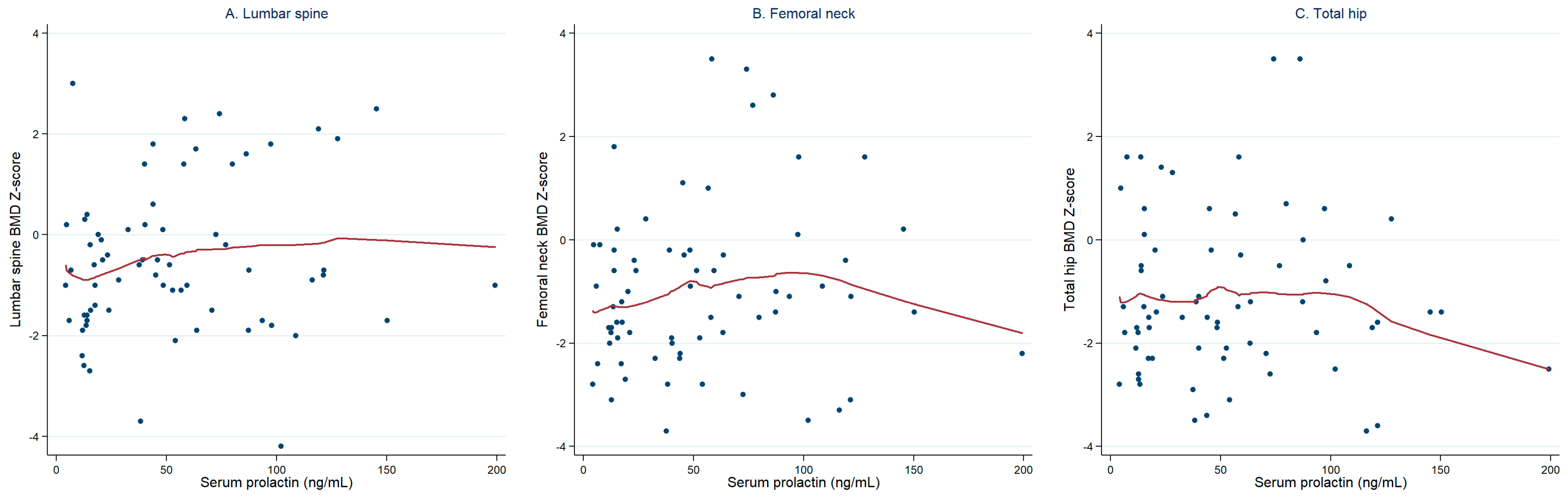
Figure 1 illustrates the relationships between serum prolactin concentrations and BMD Z-scores at the three skeletal sites. Visual inspection of the LOWESS-smoothed scatterplots showed no clear or consistent relationship between serum prolactin concentration and BMD Z-score at the lumbar spine, femoral neck, or total hip, consistent with the nonsignificant unadjusted associations presented in
Table 2.
3.3. Multivariable Associations with BMD Z-Scores
In multivariable linear regression analyses adjusted for age, BMI, duration of prolactin-raising antipsychotic exposure, chlorpromazine-equivalent dose, and physical activity, natural-log-transformed serum prolactin concentration was not significantly associated with BMD Z-score at the lumbar spine (β = 0.196, 95% CI −0.212 to 0.605;
p = 0.340), femoral neck (β = 0.157, 95% CI −0.142 to 0.456;
p = 0.298), or total hip (β = −0.224, 95% CI −0.587 to 0.139;
p = 0.221) (
Table 3).
Higher BMI was positively associated with BMD Z-scores at the femoral neck (β = 0.104, 95% CI 0.044–0.165; p = 0.001) and total hip (β = 0.099, 95% CI 0.039–0.160; p = 0.002), but not at the lumbar spine (p = 0.094). Physical activity was positively associated with BMD Z-scores at all three skeletal sites: lumbar spine (β = 0.870, 95% CI 0.112–1.627; p = 0.025), femoral neck (β = 1.515, 95% CI 0.731–2.299; p < 0.001), and total hip (β = 1.333, 95% CI 0.547–2.120; p = 0.001).
Longer duration of prolactin-raising antipsychotic exposure showed inverse but statistically nonsignificant associations with femoral neck (β = −0.066, 95% CI −0.131 to 0.0002; p = 0.051) and total hip Z-scores (β = −0.079, 95% CI −0.167 to 0.009; p = 0.076). Chlorpromazine-equivalent dose was positively associated with total hip Z-score (β = 0.0031, 95% CI 0.0004–0.0057; p = 0.023); however, corresponding associations were not observed at the lumbar spine or femoral neck.
The overall models were statistically significant for the lumbar spine (R2 = 0.201; p = 0.039), femoral neck (R2 = 0.351; p < 0.001), and total hip (R2 = 0.317; p = 0.001). The models accounted for 20.1%, 35.1%, and 31.7% of observed variance at the lumbar spine, femoral neck, and total hip, respectively (adjusted R2 approximately 0.12, 0.29, and 0.25).
3.4. Sensitivity Analysis Restricted to Risperidone-Treated Participants
Restricting the analysis to risperidone-treated participants (n = 60) did not materially alter the findings. Natural-log-transformed serum prolactin concentration remained unassociated with BMD Z-scores at the lumbar spine (adjusted β = 0.262, 95% CI −0.255 to 0.779; p = 0.314), femoral neck (adjusted β = 0.252, 95% CI −0.145 to 0.648; p = 0.209), and total hip (adjusted β = −0.077, 95% CI −0.521 to 0.367; p = 0.731). These findings were consistent with the primary analysis, suggesting that the principal results were not substantially influenced by heterogeneity in antipsychotic exposure.
4. Discussion
In this cross-sectional study of 68 women with schizophrenia receiving prolactin-raising antipsychotics, contemporaneous serum prolactin concentration was not independently associated with BMD Z-scores at the lumbar spine, femoral neck, or total hip after adjustment for age, BMI, duration of antipsychotic exposure, chlorpromazine-equivalent dose, and physical activity. The findings were consistent in the risperidone-only sensitivity analysis. In contrast, higher BMI was positively associated with femoral neck and total hip Z-scores, and physical activity was positively associated with BMD Z-scores at all three skeletal sites. Collectively, these findings support a multifactorial interpretation of skeletal health rather than a model in which a single contemporaneous prolactin measurement is the predominant determinant of BMD.
These findings should be interpreted within a multifactorial model of antipsychotic-associated bone health rather than as an isolated effect of serum prolactin. Prolactin-raising antipsychotics, particularly potent dopamine D2 receptor antagonists, may increase circulating prolactin through blockade of D2 receptors in the tuberoinfundibular pathway. Persistent hyperprolactinemia may subsequently suppress hypothalamic–pituitary–gonadal function and reduce gonadal steroid concentrations, potentially increasing bone resorption and reducing bone formation. At the same time, skeletal health in patients with schizophrenia is influenced by age, sex, nutritional status, body composition, physical activity, smoking, vitamin D and calcium status, and duration and intensity of antipsychotic exposure. Consequently, a single measurement of serum prolactin may capture only one component of a considerably more complex pathway leading to changes in bone mass.
Risperidone, a dopamine D2 receptor antagonist with a well-established propensity to elevate prolactin, constituted the predominant antipsychotic exposure in the present cohort [
26]. However, despite this pharmacological profile, serum prolactin concentration was not significantly associated with BMD Z-scores in either the primary analysis or the risperidone-restricted sensitivity analysis. This pharmacological mechanism is particularly relevant to the present cohort because risperidone constituted the predominant antipsychotic exposure. However, the absence of a significant relationship between serum prolactin and BMD contrasts with some previous studies that have reported a negative correlation, particularly in patients with prolonged exposure to prolactin-raising antipsychotics. For instance, a large-scale secondary analysis [
27] involving 402 patients found a negative correlation between serum prolactin levels and BMD in male patients treated with risperidone or conventional antipsychotics. That study further suggested that elevated prolactin may directly influence bone metabolism by increasing bone turnover and may also contribute to hypogonadism in men, leading to reduced testosterone levels and increased bone resorption. The difference from our findings may therefore reflect differences in sex distribution, age, cumulative antipsychotic exposure, endocrine status, and duration of hyperprolactinemia rather than a true absence of a biological relationship.
An isolated positive association between chlorpromazine-equivalent dose and total hip Z-score was observed in the primary analysis, whereas corresponding associations were not found at the lumbar spine or femoral neck. Moreover, this association was not reproduced in the sensitivity analysis restricted to risperidone-treated participants. Accordingly, this site-specific secondary finding should be interpreted cautiously and should not be considered evidence of a protective effect of higher antipsychotic dose. It may reflect residual confounding, differences in treatment selection, or chance in the context of multiple secondary comparisons.
The age and sex composition of our sample may be particularly important in explaining the observed findings. Previous studies have frequently evaluated older or postmenopausal women, who are inherently more susceptible to BMD decline due to age-related hormonal changes. For example, a large cross-sectional study in postmenopausal women with schizophrenia (mean age 60.4 years) reported a high prevalence of osteopenia or osteoporosis (66.2%) and identified multiple contributing factors, including age, treatment duration, BMI, and prolactin levels [
28]. Conversely, a population-based study reported significantly lower BMD among younger women (<60 years) using antipsychotics compared with non-users, whereas a similar association was not evident in older women or men [
21]. Together, these heterogeneous findings suggest that the relationship between antipsychotic exposure, prolactin, and bone may be modified by sex and reproductive or hormonal status. In our relatively young cohort of women, the skeletal consequences of hyperprolactinemia may not yet have accumulated sufficiently to produce detectable differences in DXA-derived BMD Z-scores.
Duration of exposure provides another plausible explanation. Although participants had established exposure to prolactin-raising antipsychotics, changes in BMD represent cumulative skeletal effects that may develop over substantially longer periods than biochemical hyperprolactinemia. Serum prolactin can increase soon after D2 receptor blockade, whereas measurable reductions in BMD may require prolonged endocrine disturbance and continued exposure to additional skeletal risk factors. This temporal discrepancy may partly explain why substantial prolactin elevation can coexist with an absence of a detectable cross-sectional prolactin–BMD relationship. The cross-sectional design also prevents determination of the duration of hyperprolactinemia or whether individual participants experienced persistently elevated prolactin concentrations throughout their antipsychotic exposure.
Body mass and metabolic factors may further modify this relationship. Greater body weight may exert a favorable mechanical-loading effect on bone, and adipose tissue may contribute to peripheral estrogen production. However, the relationship between obesity and skeletal health is complex and may be nonlinear. Both low and very high BMI have been associated with increased fracture risk through different mechanisms [
29]. Visceral adiposity, systemic inflammation, insulin resistance, poor nutritional quality, and reduced physical activity may counterbalance the mechanical benefits of greater body mass, while substantial weight loss may also contribute to bone loss [
30]. Thus, BMI should be considered a potential modifier or confounder of the prolactin–bone relationship rather than interpreted simply as a protective factor.
Physical activity showed the most consistent association with BMD across skeletal sites. Participants reporting at least one session of physical activity per week had higher adjusted Z-scores at the lumbar spine, femoral neck, and total hip. Although the cross-sectional design precludes causal inference and the measure did not capture exercise intensity or weight-bearing characteristics, this consistency supports consideration of physical activity as an important modifiable correlate of skeletal health in women receiving prolactin-raising antipsychotics.
Strengths of this study include the assessment of BMD at three clinically relevant skeletal sites using DXA and the use of multivariable analyses incorporating prespecified demographic, anthropometric, treatment-related, and lifestyle covariates. The use of BMD Z-scores was also appropriate for this relatively young female population because Z-scores provide age- and sex-referenced measures of bone density. In addition, we employed multivariable regression analyses to adjust for potential confounders enhancing the internal validity of our findings. The consistent positive association between higher BMI and BMD observed in our study is supported by evidence suggesting that mechanical loading and increased estrogen production from adipose tissue can enhance bone density. However, obesity’s effects on bone are complex and may follow a U-shaped relationship [
29], where both low and very high BMI increase fracture risk due to factors such as visceral fat, systemic inflammation, and metabolic disturbances [
31]. Antipsychotics, particularly risperidone, exert strong dopamine D2 receptor antagonism with relatively higher pituitary D2 receptor occupancy than central (striatal) binding, leading to disinhibition of prolactin secretion in the tuberoinfundibular pathway and subsequent hyperprolactinemia. This pharmacokinetic and receptor-binding profile, including limited blood–brain barrier penetration and prolonged exposure at the pituitary level [
26], explains its higher propensity to elevate prolactin compared with other antipsychotics and supports its classification in the present study. Simultaneously, they may influence hypothalamic appetite control, contributing to weight gain and increased BMI. These findings underscore the need to consider both BMI and metabolic health markers when evaluating skeletal risk in antipsychotic-treated patients.
Several limitations should be considered. First, the cross-sectional design precludes assessment of temporality or causality, and a single serum prolactin measurement may not adequately represent cumulative prolactin exposure, duration of hyperprolactinemia, or associated hypogonadism. Second, the final sample of 68 women was smaller than the originally calculated target, limiting statistical precision and the ability to detect small-to-moderate associations. Third, although a parsimonious covariate set was used to reduce model complexity, residual confounding remains possible [
32]. Gonadal hormones, serum 25-hydroxyvitamin D, detailed calcium homeostasis and intake, bone turnover markers, including procollagen type I N-propeptide (PINP) and β-isomerized C-terminal telopeptide of type I collagen (β-CTX-I) [
33], and cumulative concomitant medication exposure were not systematically available. Fourth, physical activity was assessed only as a dichotomous measure and did not capture exercise type, intensity, duration, or weight-bearing characteristics. Fifth, the cohort consisted exclusively of women and was predominantly treated with risperidone, limiting generalizability to men and to patients receiving other prolactin-raising antipsychotics. However, the risperidone-only sensitivity analysis produced findings consistent with the primary prolactin–BMD analysis. Finally, secondary associations involving BMI, physical activity, antipsychotic exposure duration, and chlorpromazine-equivalent dose should be interpreted cautiously because multiple associations were examined in an observational dataset.
Psychiatrists, primary care physicians, and other clinicians caring for women receiving prolactin-raising antipsychotics should consider bone health within a broader risk-assessment framework rather than relying on a single serum prolactin value. Persistent hyperprolactinemia accompanied by menstrual disturbance, reproductive endocrine symptoms, or other evidence of hypogonadism warrants clinical evaluation. Attention should also be given to modifiable skeletal risk factors, including physical inactivity, nutritional adequacy, smoking, alcohol use, and other medications that may affect bone. Antipsychotic treatment should continue to be individualized according to psychiatric efficacy and overall adverse-effect burden, with consideration of prolactin-related effects when clinically relevant.
In women with schizophrenia receiving prolactin-raising antipsychotics, contemporaneous serum prolactin concentration was not independently associated with lumbar spine, femoral neck, or total hip BMD Z-scores. This finding remained consistent when the analysis was restricted to risperidone-treated participants. Higher BMI and physical activity showed stronger associations with selected or multiple skeletal outcomes, supporting a multifactorial model of bone health. These findings should not be interpreted as excluding a potential skeletal effect of chronic hyperprolactinemia because a single cross-sectional prolactin measurement cannot characterize cumulative endocrine exposure or associated hypogonadism. Larger prospective studies incorporating serial prolactin measurements, gonadal hormones, nutritional measures, standardized bone turnover markers, and longitudinal DXA assessments are warranted.