Article

Anatomical Considerations for the Use of the Popliteal Vein as a Potential Alternative for Central Venous Cannulation

by
Aaron L. Graves
1,
Charles R. Marchese
1,
Bradley A. Creamer
2 and
Jennifer F. Dennis
3,*
1
College of Osteopathic Medicine, Kansas City University, 1750 Independence Avenue, Kansas City, MO 64113, USA
2
Department of Basic Sciences, Kansas City University, 2901 St. John’s Boulevard, Joplin, MO 64804, USA
3
Department of Academic Affairs, Kansas College of Osteopathic Medicine-Kansas Health Science University, Wichita, KS 67202, USA
*
Author to whom correspondence should be addressed.
Anatomia 2024, 3(3), 192-201; https://doi.org/10.3390/anatomia3030015
Submission received: 18 July 2024 / Revised: 14 August 2024 / Accepted: 19 August 2024 / Published: 22 August 2024

Abstract

:
Limited reports have evaluated the utility of the popliteal vein (PV) specific to cannulation. The objective of this study was to characterize the diameter and length of the PV to evaluate this area as a potential cannulation site. The popliteal region in 23 formalin-embalmed, prosected donors was dissected, and the PV was exposed from the adductor hiatus (AH) superiorly to the small saphenous vein (SSV) inferiorly. The diameter of the popliteal vein was measured at the AH, SSV, and half of the distance from the AH to the SSV (MID) using a brass plumb bob. The length of the PV was measured to the AH, SSV, MID, and femoral condyles (FCs). Overall, the mean diameters and mean lengths for the combined population were calculated, as well as individual limbs (right, left) and anatomical sex. Univariate analysis used to evaluate differences in mean diameter and length measurements based on anatomical sex revealed significant differences (p < 0.05) for both diameter and length at all the landmarks evaluated. Multivariate analysis of PV diameter at the AH and SSV landmarks was statistically significant (p < 0.05) when laterally and anatomical comparing sex. These data provide full characterization of the PV in support of its utility in vascular access.

1. Introduction

Central venous catheterization is a staple of emergency medicine, intensive care, and anesthesia. The complications from this procedure include hemothorax, pneumothorax, arterial cannulation, infection, thrombosis, and hematoma [1]. The preferred sites for cannulation are the internal jugular, subclavian, and femoral veins, each carrying their own approaches and potential complications. The use of central venous catheters is multipurposed; however, in the acutely ill patient, they serve as a reliable entry site for large amounts of fluid, blood products, and medications as they are better for long-term care when compared to peripheral IVs [2].
While not a primary site for cannulation, reports have noted using the popliteal vein for central venous access [3,4,5]. This typically occurs in situations where the patient is placed in a prone position such as emergency surgery or in the Intensive Care Unit when patients suffering from acute respiratory distress syndrome (ARDS) are flipped, which has been shown to improve V/Q function [6], improve PaO2/FiO2 ratio [7], and decrease mortality [7]. During the COVID-19 pandemic, the use of the prone position in patients with ARDS became a widely adapted therapeutic modality [6,7,8,9,10,11,12,13] and has been utilized with both intubated and non-intubated patients [11], leading to the development of new terminology: awake prone positioning (aPP) [11]. Retrospective cohort studies have demonstrated that the rate of utilizing the prone position has grown exponentially due to the pandemic, with rates of 70% or greater being reported [12,14,15], compared to rates of roughly 20% before the pandemic [16,17]. It has also been demonstrated that awake prone positioning was associated with reduced treatment failure and reduced need for intubation [13]. Limited reports have demonstrated the use of this approach for patients experiencing deep vein thrombosis [18], severe burns [19,20], or post-traumatic lung injury following blunt injury [21]. Although limited in number, successful cannulation of the PV in patients requiring prone positioning due to severe burns [22] and renal replacement therapy [23,24] have been reported.
Given the relevance and importance of establishing central venous access within the clinical management of various conditions, and the potential opportunity provided by the popliteal vein, it provokes an evaluation of the anatomical challenges, if any, that prevent the usage of this site for cannulation. Therefore, the objective of this study was to evaluate and characterize the anatomical variation in the popliteal vein, specifically as it relates to sex and laterality, with the end goal of determining its utility in catheterization.

2. Methods

2.1. Donor Population and Ethical Approval

Twenty-three prosected, formalin-embalmed donors from the Gift Body Program at Kansas City University (KCU) were evaluated in this study. Participation as a donor in the program is completely voluntary; donors with known blood-borne disease (i.e., Hepatitis) or excessively large body habitus are precluded from participation. All subjects gave their informed consent for inclusion before they participated in the study. The study was conducted in accordance with the Declaration of Helsinki, and the protocol was approved by the Institutional Biosafety Committee (1954741-1). Donors were embalmed with a proprietary blend of formalin-based embalming solution within 36 h of death. Fifteen males and eight females were included for a total of forty-six limbs. Medical history from the twenty-three donors was reviewed, and any significant history of surgical procedures in the region was noted. All limbs were evaluated physically to determine if there were any signs of surgical history in the area as this may not be included in the medical history provided. One donor had an absent popliteal vein unilaterally due to dissection error, and the limb was excluded from the study. After exclusion criteria were applied, 29 male limbs and 16 female limbs for a total of 45 limbs were included in the study.

2.2. Dissection and Measurement of the Popliteal Vein

With the donors in the prone position, the entirety of the popliteal vein from the adductor hiatus (AH, superior) to the branch point of the small saphenous vein (SSV, inferior) was dissected when viewed from the posterior aspect of the popliteal fossa. If the entirety of the popliteal vein was not visible within the superior/inferior parameters described above, further dissection occurred to gain appropriate visibility. Various landmarks of the region were then evaluated and visualized as these would be points from which measurement would be taken. If these landmarks were not explicitly visible in a consistent manner, further dissection occurred. The landmarks included were as follows: AH, SSV, half of the distance from AH to SSV (MID), and femoral condyles (FCs) (Figure 1).
The diameter of the popliteal vein was measured at the AH, MID, and SSV using a brass plumb bob (Figure 2A). Measurements of AH—SSV, MID-SSV, SSV-FCs, and AH—FCs were collected using a 150 mm electronic caliper (Mitutoyo, Takatsu-ku, Kawasaki, Japan). The methodology to include the use of the brass plumb bob was due to the understanding that the electronic caliper had the potential to alter the shape and measurements of the popliteal vein when clamped. The consistent expansion of the brass plumb bob was used to stretch each vessel to its max diameter (Figure 2B). The measurement was then taken directly distal to this point (Figure 2C). Taking the measurement using this protocol ensured that no manipulation of the vessel was caused by the caliper. All measurements and dissections were performed by the same investigator (AG) to reduce measurement error.

2.3. Statistical Analysis

Recorded vessel diameters and lengths were used to determine descriptive statistics (mean, standard deviation, confidence interval) for each group. Statistical differences between groups were determined by Levene’s statistical test for equality of variance, and Student’s t-test or the Mann–Whitney U (MWU) test was utilized for univariate analysis (males vs. females; left limb vs. right limb). If multivariate analysis was conducted (male left vs. male right vs. female left vs. female right), statistical comparisons were made by ANOVA. A p value of <0.05 was used to determine significance across all tests. All analysis was conducted using Jamovi Open-source Software (The jamovi project (2024). jamovi (Version 2.5) [Computer Software]), Sydney Australia, retrieved from https://www.jamovi.org (accessed 15 March 2024).

3. Results

3.1. Descriptive Statistics of the Popliteal Vein

The mean diameters of our entire sample population at the AH, MID, and SSV landmarks, as well as the mean length from the AH-SSV, AH-FCs, MID-SSV, and SSV-FCs are included in Table 1, respectively.

3.2. Laterality-Based Univariate Analysis

Diameter and length measurements were calculated for left and right limbs (Table 2). No statistical significance was found when comparing any diameter or length measurements between left and right limbs (Table 2).

3.3. Sex-Based Univariate Analysis

To determine any differences in the popliteal vein and anatomical sex, diameter and length measurements were compared between males and females (Table 3). The mean diameter at the AH, MID, and SSV was statistically larger in males than in females (p < 0.05) at all landmarks. For length measurements, values in female limbs were greater than those in male donors between all locations except SSV-FCs. In evaluating these differences statistically, only the AH-FCs approached significance, but they did not pass the threshold.

3.4. Multivariate Analysis of Anatomical Sex and Laterality

Diameter and length measurements were further compared between left and right limbs to evaluate differences across each limb and anatomical sex. Analysis of the differences in the diameter measurements is included in Table 4. When evaluating the diameter of the PV at the AH and SSV landmarks, the male left and right diameters were each larger than both the female left and right diameters, respectively (p < 0.05). In contrast, the PV diameter at the MID landmark of the left male limb was statistically larger than the female left only, while the male right limb was statistically larger than both the female left and female right. No significance difference was noted when comparing any male–male or female–female diameters at the AH, MID, or SSV. Finally, no statistically significant differences for length measurements were identified.

4. Discussion

These data provide a much-needed characterization of the PV in support of its available and optional utility for vascular access. The consistency of the diameter measurements in both male and female patients, from superior to inferior along the popliteal vein, indicates that canulation can occur anywhere, if inadvertent damage to other structures is avoided. Overall, the anatomical data described herein may be applied to various clinical settings and patient presentations requiring cannulation in prone positions.

4.1. Evaluating the PV for Cannulation

The diameter of the popliteal vein was a key element to explore and characterize so that this information could be compared to the known diameter of veins in the upper extremity where cannulation typically occurs [25,26,27,28]. Poiseuille’s law of laminar flow states that the flow rate of fluid is directly proportional to the radius of the tube it is running through and indirectly proportional to the length of the tube [29,30,31]. Simplified, the larger the IV catheter, the quicker that fluids can be administered to a patient. Hence, larger bore IV catheters directly correlate to the rapidness one can fluid-resuscitate a patient. An observational study using ultrasound in 176 participants reported that the maximum diameters of the basilic, brachial, and cephalic veins measured were 7.30 mm, 7.10 mm, and 6.10 mm, respectively [28]. The datum reported herein denote a larger mean diameter for the PV at all sites measured (AH, 7.69 mm; MID, 7.69 mm; SSV, 7.28 mm), as compared to the sites typically used in the upper extremity. Collectively, these results suggest that flow rate into the popliteal vein would not limit resuscitation based on diameter alone. Additionally, when combined with recent reports utilizing popliteal cannulation [22,23,24], this datum provide additional evidence to support consideration for popliteal cannulation for patients if prone positioning is warranted by their condition.
Using these data, a stepwise plan for cannulation in the popliteal vein was devised should the need arise (Figure 3). Ultrasound is recommended to best visualize where the sciatic nerve bifurcates to avoid potential injury. Once the sciatic bifurcation is identified, this location is marked as the superior most boundary. Ultrasound is used to locate the small saphenous vein off the popliteal vein, and its location serves as the inferior landmark. Because the change in diameter of the popliteal vein from the SSV to adductor hiatus is insignificant (Table 2), cannulation can occur anywhere between the SSV and the area where the sciatic nerve is not interfering with the track of the needle. This area is generally covered by a superficial layer of skin and fascia with limited overlap of the hamstring muscles. Following this straightforward evaluation will allow for the best outcome and the avoidance of injury to surrounding structures.

4.2. Clinical Application of Popliteal Vein Cannulation

The COVID-19 pandemic illustrated the need for novel ideas and techniques to be developed as new diseases and/or emergency responses arise. The need for central venous access in prone patients during the pandemic was high [32], but this prone positioning introduced challenges for the correct placement of catheters. Further, in patients suffering from major trauma, obtaining and maintaining patent vascular access is vital to ensuring proper management. The Advanced Trauma Life Support (ATLS) protocol dictates that two large bore peripheral IVs can be obtained as early as possible and that vascular access is maintained for the continued resuscitation and management of the patient [33]. However, in severe cases of traumatic injury (particularly large area burns [22], explosive injury, amputations) or disease [23,24], obtaining peripheral IVs and/or central lines may not be a viable option, and popliteal cannulation may be warranted. Our characterization of sex-based and laterality differences in the popliteal vein provides the important anatomical context necessary for clinicians to apply this information across a wide range of medical interventions, despite the historically atypical approach of popliteal cannulation.

4.3. Contraindications of Popliteal Vein Cannulation

A key consideration for placement in the popliteal vein would be the development of thrombosis. With indwelling central venous catheters, this may occur as a venous or mural thrombosis or a clot within the catheter itself [34,35]. These events for central lines may be as high as 18% [36], with symptomatic development occurring in 5% [37]. Studies have noted that lower-extremity (femoral) cannulation sites with central lines can have a higher risk of thrombosis compared to the subclavian approach [38]. It may stand to reason that the popliteal site will carry with it a similar risk, but additional data would need to be obtained. Current guidelines on thrombosis prophylaxis vary depending on the underlying disease process of the patient, with general agreement that prophylaxis is not needed for the central line itself but should rather be based off the diagnostic need for the underlying condition (i.e., malignancy, etc.) [39].
The insertion of any type of cannula introduces the risk of infection. Central venous catheters carry a heightened awareness of this risk due to the locality of the catheter tip near the heart and the increased risk of bacteremia and septicemia developing [40]. Depending on the site used, the risk of central-venous-catheter-bloodstream-related infections (CVCBRIs) can range from 5.3 to 8.75 per 1000 catheter days, resulting in a mortality as high as 25% [41,42]; however, this varies by facility and region of placement. While limited data exist for femoral vein catheters indicating that they held a higher risk of infection, the data appear to not be well founded [43]. Currently, no datum have been found on the relative risk of using the popliteal vein for CVC, supporting further investigation, such as through a randomized control trial.

4.4. Study Limitations

Select considerations should be noted in future evaluations of the PV as a cannulation site. Primarily, the measurements in this study were collected from embalmed donors. Additionally, the donor population was Caucasian with a mean age of 71.95 years, which limits the diversity of the sample, particularly when projecting generalizations from this research clinically across a wider variety of patients. Future studies using fresh donors, ultrasound, and/or medical imaging to measure the diameter of the popliteal vein would add to our understanding of PV cannulation.

5. Conclusions

While the popliteal vein will not become a primary area of central venous cannulation as compared to the upper extremity, these data, particularly the diameter measurements of the PV at various anatomical landmarks, support evaluation of its use as a site of entry when treating patients with specific conditions/presentations [22,23,24,44]. Given the increased availability of ultrasounds at the bedside or in the field, obtaining access via the popliteal vein would be an easily achieved feat by any physician trained in the central line procedure and could serve as a lifesaving modality when the time arises.

Author Contributions

Conceptualization, A.L.G. and J.F.D.; methodology, A.L.G., B.A.C., and J.F.D.; software, B.A.C.; formal analysis, A.L.G., C.R.M. and B.A.C.; resources, J.F.D.; data curation, A.L.G. and C.R.M.; writing—original draft preparation, A.L.G. and C.R.M.; writing—review and editing, A.L.G., C.R.M., B.A.C. and J.F.D.; supervision, J.F.D.; project administration, J.F.D. All authors have read and agreed to the published version of the manuscript.

Funding

This research received no external funding.

Institutional Review Board Statement

The study was conducted in accordance with the Declaration of Helsinki, and the protocol was approved by the Institutional Biosafety Committee (1954741-1).

Informed Consent Statement

All subjects gave their informed consent for inclusion before they participated in the study.

Data Availability Statement

The original contributions presented in the study are included in the article, further inquiries can be directed to the corresponding author.

Conflicts of Interest

The authors declare no conflicts of interest.

References

  1. Preventing Complications of Central Venous Catheterization|NEJM. Available online: https://www.nejm.org/doi/10.1056/NEJMra011883?url_ver=Z39.88-2003&rfr_id=ori:rid:crossref.org&rfr_dat=cr_pub%20%200www.ncbi.nlm.nih.gov (accessed on 26 March 2024).
  2. Fahy, B.; Sockrider, M. Central Venous Catheter. Am. J. Respir. Crit. Care Med. 2019, 199, P21–P22. [Google Scholar] [CrossRef] [PubMed]
  3. Cannulation of the Popliteal Vein as an Intraoperative Emergency Access in Prone Position: A Case Report—Tobias Kammerer, Tobias Brezina. 2022. Available online: https://journals.sagepub.com/doi/abs/10.1177/11297298211008091 (accessed on 26 March 2024).
  4. Yang, M.X.; Ng, P.K. Central Venous Catheter Insertion in the Prone Position-A Last Resort in Critically Ill COVID-19 Patients. J. Intensive Care Med. 2021, 36, 373–375. [Google Scholar] [CrossRef]
  5. Rabi Andaloussi, M.; Touab, R.; Samali, E.M.; Balkhi, H. The larger the better: Tourniquet-facilitated popliteal vein cannulation for vascular access in prone position. Anaesth. Crit. Care Pain. Med. 2021, 40, 100913. [Google Scholar] [CrossRef]
  6. Hadaya, J.; Benharash, P. Prone Positioning for Acute Respiratory Distress Syndrome (ARDS). JAMA 2020, 324, 1361. [Google Scholar] [CrossRef] [PubMed]
  7. Chua, E.X.; Zahir, S.M.I.S.M.; Ng, K.T.; Teoh, W.Y.; Hasan, M.S.; Ruslan, S.R.B.; Abosamak, M.F. Effect of prone versus supine position in COVID-19 patients: A systematic review and meta-analysis. J. Clin. Anesth. 2021, 74, 110406. [Google Scholar] [CrossRef]
  8. Guérin, C.; Albert, R.K.; Beitler, J.; Gattinoni, L.; Jaber, S.; Marini, J.J.; Munshi, L.; Papazian, L.; Pesenti, A.; Vieillard-Baron, A.; et al. Prone position in ARDS patients: Why, when, how and for whom. Intensive Care Med. 2020, 46, 2385–2396. [Google Scholar] [CrossRef]
  9. Parhar, K.K.S.; Zuege, D.J.; Shariff, K.; Knight, G.; Bagshaw, S.M. Prone positioning for ARDS patients-tips for preparation and use during the COVID-19 pandemic. Can. J. Anaesth. 2021, 68, 541–545. [Google Scholar] [CrossRef] [PubMed]
  10. Chua, E.X.; Wong, Z.Z.; Hasan, M.S.; Atan, R.; Yunos, N.A.M.; Yip, H.W.; Teoh, W.Y.; Ramli, M.A.S.; Ng, K.T. Prone ventilation in intubated COVID-19 patients: A systematic review and meta-analysis. Braz. J. Anesthesiol. 2022, 72, 780–789. [Google Scholar] [CrossRef]
  11. Kharat, A.; Simon, M.; Guérin, C. Prone position in COVID 19-associated acute respiratory failure. Curr. Opin. Crit. Care 2022, 28, 57–65. [Google Scholar] [CrossRef]
  12. Langer, T.; Brioni, M.; Guzzardella, A.; Carlesso, E.; Cabrini, L.; Castelli, G.; Dalla Corte, F.; De Robertis, E.; Favarato, M.; Forastieri, A.; et al. Prone position in intubated, mechanically ventilated patients with COVID-19: A multi-centric study of more than 1000 patients. Crit. Care 2021, 25, 128. [Google Scholar] [CrossRef]
  13. Ehrmann, S.; Li, J.; Ibarra-Estrada, M.; Perez, Y.; Pavlov, I.; McNicholas, B.; Roca, O.; Mirza, S.; Vines, D.; Garcia-Salcido, R.; et al. Awake prone positioning for COVID-19 acute hypoxaemic respiratory failure: A randomised, controlled, multinational, open-label meta-trial. Lancet Respir. Med. 2021, 9, 1387–1395. [Google Scholar] [CrossRef] [PubMed]
  14. Clinical characteristics and day-90 outcomes of 4244 critically ill adults with COVID-19: A prospective cohort study. Intensive Care Med. 2021, 47, 60–73. [CrossRef]
  15. Andrea, R.; Garcia-Alvarez, A.; Miró, J.; Poch Lopez de Briñas, E.J.; Martinez-Pastor, J.C.; Martinez-Palli, G.; Torres, A.; Guasch Casany, E.; Soy, D.; Urra, X.; et al. Clinical features, ventilatory management, and outcome of ARDS caused by COVID-19 are similar to other causes of ARDS. Intensive Care Med. 2020, 46, 2200–2211. [Google Scholar] [CrossRef]
  16. Guerin, C.; Beuret, P.; Constantin, J.M.; Bellani, G.; Garcia-Olivares, P.; Roca, O.; Meertens, J.H.; Maia, P.A.; Becher, T.; Peterson, J.; et al. A prospective international observational prevalence study on prone positioning of ARDS patients: The APRONET (ARDS Prone Position Network) study. Intensive Care Med. 2018, 44, 22–37. [Google Scholar] [CrossRef]
  17. Bellani, G.; Laffey, J.G.; Pham, T.; Fan, E.; Brochard, L.; Esteban, A.; Gattinoni, L.; Van Haren, F.; Larsson, A.; McAuley, D.F.; et al. Epidemiology, Patterns of Care, and Mortality for Patients With Acute Respiratory Distress Syndrome in Intensive Care Units in 50 Countries. JAMA 2016, 315, 788–800. [Google Scholar] [CrossRef] [PubMed]
  18. Koksoy, C.; Cetinkaya, O.A. Popliteal Access in the Supine Position for Endovenous Management of Deep Vein Thrombosis. EJVES Short Rep. 2019, 46, 5–8. [Google Scholar] [CrossRef]
  19. Hale, D.F.; Cannon, J.W.; Batchinsky, A.I.; Cancio, L.C.; Aden, J.K.; White, C.E.; Renz, E.M.; Blackbourne, L.H.; Chung, K.K. Prone positioning improves oxygenation in adult burn patients with severe acute respiratory distress syndrome. J. Trauma Acute Care Surg. 2012, 72, 1634–1639. [Google Scholar] [CrossRef] [PubMed]
  20. Oto, B.; Orosco, R.I.; Panter, E.; Velamuri, R.; Kar, A.R.; Caffrey, J. Prone Positioning of the Burn Patient With Acute Respiratory Distress Syndrome: A Review of the Evidence and Practical Considerations. J. Burn. Care Res. 2018, 39, 471–475. [Google Scholar] [CrossRef]
  21. Voggenreiter, G.; Neudeck, F.; Aufmkolk, M.; Fabinder, J.; Hirche, H.; Obertacke, U.; Schmit-Neuerburg, K.P. Intermittent prone positioning in the treatment of severe and moderate posttraumatic lung injury. Crit. Care Med. 1999, 27, 2375–2382. [Google Scholar] [CrossRef]
  22. Shi, L.; Chen, Q.; Yang, R.; Liao, L.; Zhang, J.; Liu, Y. Challenges of PICC placement via the popliteal vein under ultrasound guidance in a patient with severe burns: A case report. J. Vasc. Access, 2024; 11297298241245066, epub ahead of print. [Google Scholar] [CrossRef] [PubMed]
  23. Balakrishnan, N.; Beaini, H.; Carter, S.; Araj, F.G. Bedside popliteal vein cannulation for simultaneous plasmapheresis and renal replacement therapy in the prone position. J. Invasive Cardiol. 2024, 3. [Google Scholar] [CrossRef] [PubMed]
  24. Hamed, M.O.; Abdelmagid, M.; Ahmed, M. Popliteal venous access for renal replacement therapy in a critically ill patient with central access failure. BMJ Case Rep. 2024, 17, e258796. [Google Scholar] [CrossRef] [PubMed]
  25. Kim, J.T.; Kim, H.S.; Lim, Y.J.; Bahk, J.H.; Lee, K.H.; Kim, C.S.; Kim, S.D.; Jeon, Y. The influence of passive leg elevation on the cross-sectional area of the internal jugular vein and the subclavian vein in awake adults. Anaesth. Intensive Care 2008, 36, 65–68. [Google Scholar] [CrossRef] [PubMed]
  26. Clenaghan, S.; McLaughlin, R.E.; Martyn, C.; McGovern, S.; Bowra, J. Relationship between Trendelenburg tilt and internal jugular vein diameter. Emerg. Med. J. 2005, 22, 867–868. [Google Scholar] [CrossRef] [PubMed]
  27. Xie, S.; Yu, Q.; Li, T.; Xu, M.; Wu, J.; Li, Y. Comparison of the effect of different degrees of passive leg raising on the internal jugular vein cross-sectional area in patients before thoracic surgery. BMC Anesthesiol. 2019, 19, 78. [Google Scholar] [CrossRef]
  28. Sharp, R.; Cummings, M.; Childs, J.; Fielder, A.; Mikocka-Walus, A.; Grech, C.; Esterman, A. Measurement of Vein Diameter for Peripherally Inserted Central Catheter (PICC) Insertion: An Observational Study. J. Infus. Nurs. 2015, 38, 351–357. [Google Scholar] [CrossRef]
  29. Lake-Bakaar, G.; Ahmed, M.; Evenson, A.; Bonder, A.; Faintuch, S.; Sundaram, V. Hagen-Poiseuille’s law: The link between cirrhosis, liver stiffness, portal hypertension and hepatic decompensation. World J. Hepatol. 2015, 7, 28–32. [Google Scholar] [CrossRef]
  30. Berman, D.J.; Schiavi, A.; Frank, S.M.; Duarte, S.; Schwengel, D.A.; Miller, C.R. Factors that influence flow through intravascular catheters: The clinical relevance of Poiseuille’s law. Transfusion 2020, 60, 1410–1417. [Google Scholar] [CrossRef]
  31. Jayanthi, N.V.G.; Dabke, H.V. The effect of IV cannula length on the rate of infusion. Injury 2006, 37, 41–45. [Google Scholar] [CrossRef]
  32. Ilonzo, N.; Rao, A.; Soundararajan, K.; Vouyouka, A.; Han, D.; Tadros, R.; Kim, S.Y.; Love, B.; Ting, W.; Marin, M.; et al. The importance of a centralized line service during the COVID-19 pandemic. J. Vasc. Surg. 2020, 72, 403–404. [Google Scholar] [CrossRef]
  33. Verhoeff, K.; Saybel, R.; Mathura, P.; Tsang, B.; Fawcett, V.; Widder, S. Ensuring adequate vascular access in patients with major trauma: A quality improvement initiative. BMJ Open Qual. 2018, 7, e000090. [Google Scholar] [CrossRef]
  34. Citla Sridhar, D.; Abou-Ismail, M.Y.; Ahuja, S.P. Central venous catheter-related thrombosis in children and adults. Thromb. Res. 2020, 187, 103–112. [Google Scholar] [CrossRef]
  35. Gunawansa, N.; Sudusinghe, D.H.; Wijayaratne, D.R. Hemodialysis Catheter-Related Central Venous Thrombosis: Clinical Approach to Evaluation and Management. Ann. Vasc. Surg. 2018, 51, 298–305. [Google Scholar] [CrossRef]
  36. Verso, M.; Agnelli, G. Venous thromboembolism associated with long-term use of central venous catheters in cancer patients. J. Clin. Oncol. 2003, 21, 3665–3675. [Google Scholar] [CrossRef]
  37. Kamphuisen, P.W.; Lee, A.Y.Y. Catheter-related thrombosis: Lifeline or a pain in the neck? Hematol. Am. Soc. Hematol. Educ. Program 2012, 2012, 638–644. [Google Scholar] [CrossRef]
  38. Parienti, J.J.; Mongardon, N.; Mégarbane, B.; Mira, J.P.; Kalfon, P.; Gros, A.; Marqué, S.; Thuong, M.; Pottier, V.; Ramakers, M.; et al. Intravascular Complications of Central Venous Catheterization by Insertion Site. N. Engl. J. Med. 2015, 373, 1220–1229. [Google Scholar] [CrossRef] [PubMed]
  39. Wall, C.; Moore, J.; Thachil, J. Catheter-related thrombosis: A practical approach. J. Intensive Care Soc. 2016, 17, 160–167. [Google Scholar] [CrossRef] [PubMed]
  40. Gahlot, R.; Nigam, C.; Kumar, V.; Yadav, G.; Anupurba, S. Catheter-related bloodstream infections. Int. J. Crit. Illn. Inj. Sci. 2014, 4, 162–167. [Google Scholar] [CrossRef]
  41. The Risk of Bloodstream Infection in Adults with Different Intravascular Devices: A Systematic Review of 200 Published Prospective Studies. Available online: https://pubmed.ncbi.nlm.nih.gov/16970212/ (accessed on 26 March 2024).
  42. Soufir, L.; Timsit, J.F.; Mahe, C.; Carlet, J.; Regnier, B.; Chevret, S. Attributable morbidity and mortality of catheter-related septicemia in critically ill patients: A matched, risk-adjusted, cohort study. Infect. Control Hosp. Epidemiol. 1999, 20, 396–401. [Google Scholar] [CrossRef] [PubMed]
  43. The Risk of Catheter-Related Bloodstream Infection with Femoral Venous Catheters as Compared to Subclavian and Internal Jugular Venous Catheters: A Systematic Review of the Literature and Meta-Analysis. Available online: https://pubmed.ncbi.nlm.nih.gov/22809915/ (accessed on 26 March 2024).
  44. Woo, K.; Rigberg, D.; Lawrence, P.F. Safe Central Venous Access in an Overburdened Health System. JAMA 2021, 325, 299–300. [Google Scholar] [CrossRef]
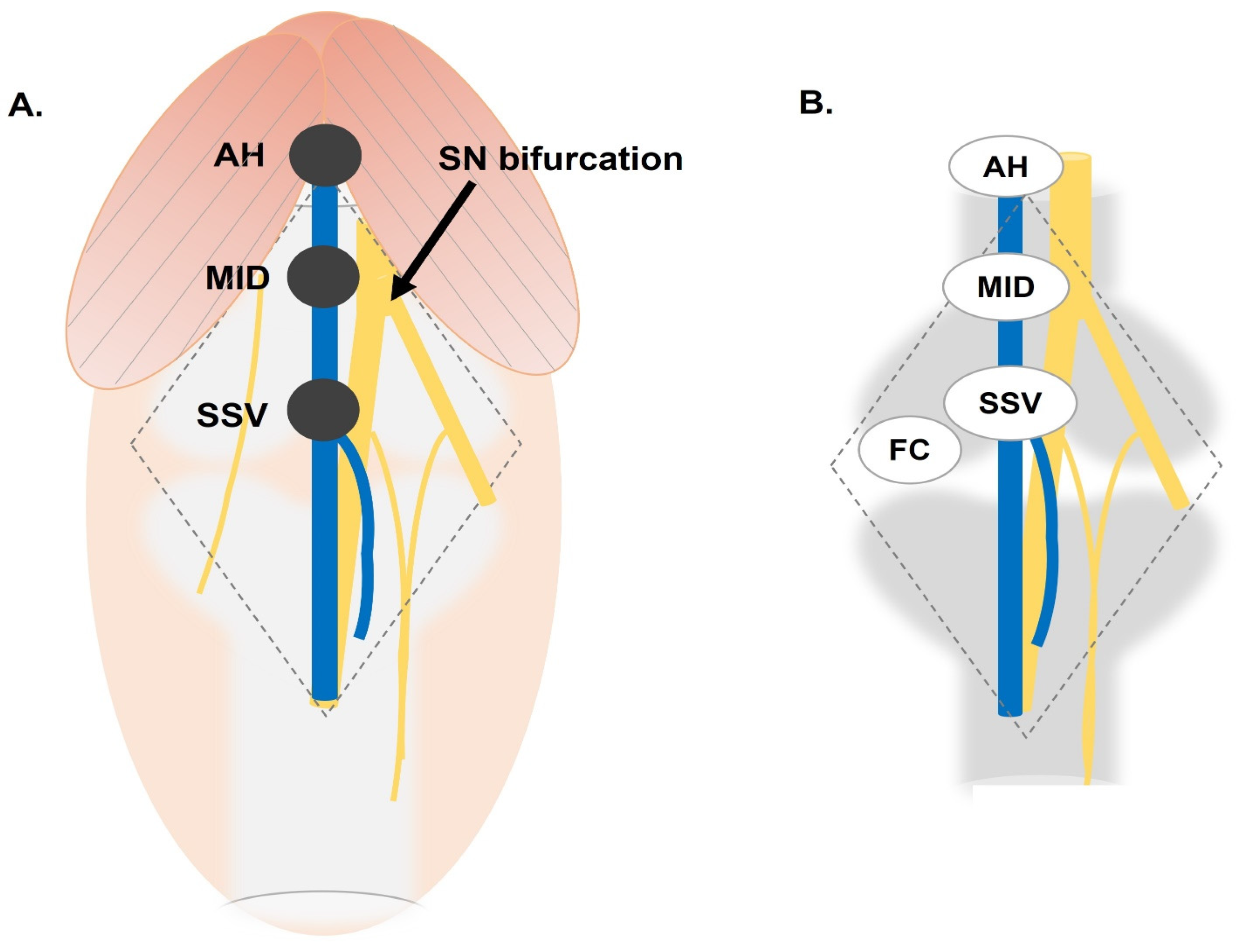
Figure 1. The popliteal vein regions and anatomical landmarks to evaluate prior to cannulation. (A) Graphic representation of the popliteal region and the anatomical landmarks utilized to characterize the popliteal vein (blue). (B) Graphic of anatomical landmarks adjacent to the sciatic nerve and its branches (yellow) and the bones of lower extremity (gray). AH, adductor hiatus; MID, half the distance between the AH and the small saphenous vein (SSV); FCs, femoral condyles.
Anatomia 03 00015 g001
Figure 2. Characterizing the popliteal vein. (A) Demonstration of popliteal vein (blue arrow) diameter measurement using a brass plumb bob (left). (B) The end of the tip of plumb bob was inserted into the patent end of the popliteal vein (blue arrow) to restore circular shape of the vein. The diameter measurement was recorded (C).
Anatomia 03 00015 g002
Figure 3. Flowchart for evaluating popliteal vein cannulation.
Anatomia 03 00015 g003
Table 1. Descriptive measurements of donor population.
LandmarkMeasurementMean (SD) (95% CI)
AHDiameter7.69 (1.37) (7.27–8.11)
MID7.69 (1.73) (7.17–8.21)
SSV7.28 (1.76) (6.75–7.81)
AH-SSVLength115.20 (35.39) (104.59–125.86)
AH-FCs145.99 (16.99) (140.83–151.15)
MID-SSV57.61 (17.70) (52.30–62.93)
SSV-FC32.41 (30.61) (23.21–41.60)
Combined N = 45 limbs, male N = 30 Limbs, female N = 15 Limbs; all measurements are in mm. AH, adductor hiatus; MID, 1/2 the distance between adductor hiatus (AH) and small saphenous vein (SSV); FCs, femoral condyles.
Table 2. Laterality-based analysis of diameter and length measurements.
LandmarkMeasurementLeft (SD) (95% CI)Right (SD) (95% CI)Testp Value
AHDiameter7.56 (1.35) (6.96–8.16)7.83 (1.40) (7.19–8.47)Student’s t0.53
MID7.59 (1.73) (6.82–8.35)7.79 (1.76) (7.03–8.56)Student’s t0.69
SSV7.27 (1.82) (6.46–8.07)7.30 (1.74) (6.55–8.05)Student’s t0.95
AH-SSVLength120.84 (32.80) (106.30–135.38)109.85 (37.64) (93.58–126.13)Student’s t0.30
AH-FCs146.56 (18.80) (138.22–154.89)145.45 (15.88) (138.58–152.32)Student’s t0.83
MID-SSV60.42 (16.40) (53.15–67.69)54.93 (18.82) (46.79–63.06)Student’s t0.30
SSV-FCs28.10 (21.40) (18.62–37.59)36.52 (37.42) (20.34–52.70)Mann–Whitney U0.56
Combined N = 45 limbs, left N = 22 limbs, right N = 23 limbs; all measurements are in mm. p value of (<0.05) was used to determine significance; AH, adductor hiatus; MID, 1/2 the distance between adductor hiatus (AH) and small saphenous vein (SSV). FCs, femoral condyles. Levene’s p value of (<0.05) was used to determine normality.
Table 3. Sex-based analysis of diameter and length measurements.
LandmarkMeasurementMales (SD) (95% CI)Females (SD) (95% CI)Testp Value
AHDiameter8.16 (1.33) (7.65–8.66)6.73 (0.87) (6.23–7.23)Student’s t<0.05
MID8.28 (1.60) (7.67–8.88)6.64 (1.46) (5.86–7.42)Student’s t<0.05
SSV7.94 (1.73) (7.29–8.60)6.09 (1.07) (5.52–6.65)MWU<0.05
AH-SSVLength107.91 (40.84) (92.38–123.45)128.48 (16.45) (119.71–137.24)MWU0.11
AH-FCs142.33 (18.10) (135.45–149.22)152.61 (13.47) (145.43–159.79)Student’s t0.05
MID-SSV53.96 (20.42) (46.19–61.72)64.24 (8.22) (59.86–68.62)MWU0.11
SSV-FC36.02 (36.79) (22.02–50.01)25.86 (12.25) (19.33–32.39)MWU0.72
Combined N = 45 limbs, male N = 30 limbs, female N = 15 limbs; all measurements are in mm. p value of (<0.05) was used to determine significance; AH, adductor hiatus; MID, 1/2 the distance between adductor hiatus (AH) and small saphenous vein (SSV). FCs, femoral condyles. Levene’s p value of (<0.05) was used to determine normality.
Table 4. Post hoc analysis of laterality vs. sex.
ANOVA
Laterality vs. SexMean DifferenceSEdftp
Diameter of PV at AHMale LeftMale Right−0.1670.45839.0−0.3640.718
Female Left1.4030.54639.02.5710.014
Female Right1.2580.60139.02.0940.043
Male RightFemale Left1.5690.53939.02.9110.006
Female Right1.4250.59539.02.3960.021
Female LeftFemale Right−0.1450.66539.0−0.2170.829
Diameter at MIDMale LeftMale Right−0.05200.58841.0−0.08850.930
Female Left1.82050.70141.02.59570.012
Female Right1.40430.70141.02.00220.052
Male RightFemale Left1.87260.69341.02.70290.010
Female Right1.45630.69341.02.10210.042
Female LeftFemale Right−0.41630.79141.0−0.52610.602
Diameter of PV at SSVMale LeftMale Right0.1040.58141.00.1780.859
Female Left2.0090.69341.02.8990.006
Female Right1.8080.69341.02.6090.013
Male RightFemale Left1.9060.68541.02.7830.008
Female Right1.7040.68541.02.4890.017
Female LeftFemale Right−0.2010.78241.0−0.2570.798
p value of (<0.05) was used to determine significance. Note: Comparisons are based on estimated marginal means.
Disclaimer/Publisher’s Note: The statements, opinions and data contained in all publications are solely those of the individual author(s) and contributor(s) and not of MDPI and/or the editor(s). MDPI and/or the editor(s) disclaim responsibility for any injury to people or property resulting from any ideas, methods, instructions or products referred to in the content.