Article

Beaton and Anson Type A Classification of the Sciatic Nerve and Piriformis Complex: Clinical Considerations for Sex and Laterality

1
College of Osteopathic Medicine, Kansas City University, 1750 Independence Avenue, Kansas City, MO 64113, USA
2
Department of Graduate Medical Education, Kansas City University, 1750 Independence Avenue, Kansas City, MO 64113, USA
3
Department of Basic Sciences, Kansas City University, 2901 St. John’s Boulevard, Joplin, MO 64804, USA
4
Department of Academic Affairs, Kansas Health Science University-Kansas College of Osteopathic Medicine, Wichita, KS 67202, USA
*
Author to whom correspondence should be addressed.
Anatomia 2024, 3(3), 182-191; https://doi.org/10.3390/anatomia3030014
Submission received: 23 July 2024 / Revised: 16 August 2024 / Accepted: 19 August 2024 / Published: 21 August 2024

Abstract

:
Variations of the sciatic nerve and piriformis muscle (SN-PM) relationship must be considered when discussing orthopedic procedures within the region as they may cause increased risk of SN injuries. Thirty-one formalin-embalmed, prosected donors were evaluated using the Beaton and Anson (B&A) classification system (1939). Major landmarks of the SN-PM relationship were identified, including the posterior superior iliac spine (PSIS), ischial tuberosity (IT), greater trochanter (GT), and the middle of the SN as it exits under the PM (S1). Distances measured included: PSIS-IT, PSIS-GT, IT-GT, PSIS-S1, IT-S1, GT-S1, S1-Q (distance of perpendicular line connecting S1 to PSIS-IT), and S1-R (distance of perpendicular line connecting S1 to PSIS-GT). Measurements from 49 lower extremities were evaluated using a two-tailed t-test to compare by sex and laterality; a one-tailed t-test was utilized to compare groups based on anatomical sex. Six donors displayed asymmetric B&A classifications, demonstrating gross anatomical differences within a single individual; however, no measurements were significant when comparing extremities. Seven measurements were statistically significant (p < 0.05) between sexes, indicating notable sex-based differences. These data highlight sex-based differences in the SN-PM relationship, as well as consistencies within measurements among extremities, which can be utilized by clinicians when treating male and female patients needing unilateral or bilateral orthopedic procedures or injections within the gluteal region.

1. Introduction

The sciatic nerve (SN), the largest nerve in the body, is composed of anterior and posterior divisions of spinal nerves L4-S2, and the posterior division of S3. Its distal branches, the common peroneal (CPN) and tibial nerves (TN), supply motor and sensory innervation to the majority of the thigh, leg, and foot [1,2]. Several anatomic variations of the relationships between the SN and the piriformis muscle (PM) exist as initially characterized by Beaton and Anson [3] and subsequent investigation has resulted in the current classification system comprising seven variants [2,4,5] (Figure 1). In this classification system, the relationships are given labels A–G and differ in the location the SN exits in relation to the PM and where the SN bifurcates into the CPN and TN. Type A is defined as ‘typical’ anatomy and has been reported as the most frequent variation encountered in patient populations. All other remaining types are designated as Non-Type A and include Types B–G. These Non-Type A variations, and the specific relationship between the SN and PM, have been speculated to play a role in piriformis syndrome, a leading cause of sciatica and sciatic-related nerve pain [1,6]. For example, Type B anatomy has been proposed to play a role in piriformis syndrome, as the CPN passes between two separated tendinous parts of the PM [7]. Therefore, SN variations should be considered when dealing intraoperatively with piriformis syndrome [8,9].
Due to the extent of lower extremity innervation supplied by the SN and its distal branches, proximal injury of the SN can dramatically impact quality of life. SN damage is a known risk during various orthopedic procedures within the gluteal region. The Southern/Moore approach for a posterior hip replacements [10,11], the Kocher–Langenbeck approach for posterior wall acetabular fracture repair, and gluteal muscular injections [12,13] have all been associated with SN damage and resulting SN palsy, specifically when anatomical variations are present [11,12,13].
Many studies [1,2,3,4,5,14,15,16] have attempted to further describe the prevalence and anatomical relationships of Non-Type A anatomical variations specific to other structures within the gluteal region, including the greater trochanter of the femur (GT), the ischial tuberosity (IT), and the posterior superior iliac spine (PSIS). Few studies have compared these relationships by sex [4,15], although a recent review has evaluated sex and laterality differences within the prevalence of Non-Type A variations [2]. Despite these reports, studies solely investigating this triangular relationship within the typical anatomy (Beaton and Anson Type A classification) have not been reported.
Non-Type A variations of the SN-PM relationship have a known risk of injury with a range of clinical approaches [10,12,13,17]; however, given the increased frequency of patients with the typical, Type A classification, further investigation of the most commonly encountered anatomy is warranted. Hence, the objective of this study was to further characterize the Type A classification and to complete a comparison of relationships in the SN-PM region based on anatomical sex and laterality. Collectively, these data augments current gaps in the literature specific to surgical populations involving both male and female patients, as well as individuals who require bilateral hip surgeries.

2. Material and Methods

2.1. Dissection of Cadaveric Donors

Thirty-one formalin-embalmed, prosected donors from the Gift Body Program at Kansas City University were evaluated in the study. All subjects gave their informed consent for inclusion before they participated in the study. The study was conducted in accordance with the Declaration of Helsinki, and the protocol was approved by the Institutional Biosafety Committee (1871804). This population consisted of 14 males (28 limbs) and 17 females (34 limbs). Medical history was reviewed and any significant history of surgical procedures in the region was noted. All limbs were classified as A or Non-A (B, C, D, E, F, or G classification) using the Beaton and Anson (B&A) classification [3] system (Figure 1). Donors with Type A classifications were included in the study for measurements and statistical analysis. Exclusion criteria for the distance measurements leg of this study included: significant medical history of procedures in the gluteal region, presence of suture material or staples, donors with Non-Type A classifications, and/or loss of key structures due to previous prosection. Prevalence counts of Non-Type A limbs were gathered for symmetrical comparisons, but no other data were gathered regarding these limbs. Thirteen limbs were excluded, and 49 limbs were utilized for statistical analysis.
Included donors were placed in the prone position. To aid evaluation of the SN-PM relationship, additional dissection was performed (CRM), if necessary, to expose the most prominent point of PSIS, the most lateral aspect of GT, and the most inferior aspect of IT, all of which were then utilized for measuring.

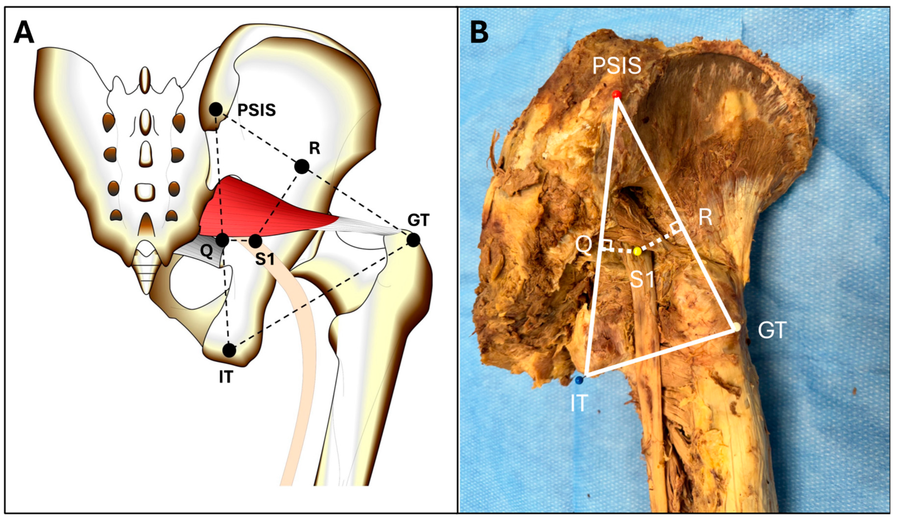
2.2. Evaluation of the Sciatic Nerve–Piriformis Relationship

To characterize the donor population and the B&A classifications present, a 150 mm electronic caliper (Mitutoyo, Takatsu-Ku, Kawasaki, Japan) was used to collect all measurements. Wan-ae-loh et al., (2020) previously defined the ‘anatomical structure (S1)’ as the midpoint of the SN immediately inferior to PM [4]. This definition of S1 was used to evaluate the relationship of the nerve compared to other landmarks. Similarly, to determine S1 in the present study, the width of the nerve was measured using the digital caliper, and the midpoint was marked with a T-pin. Distances between the aforementioned bony landmarks (PSIS-IT, IT-GT, PSIS-GT) were measured. Perpendicular lines connecting S1 to PSIS-IT and S1 to PSIS-GT were created. These intersection points were defined as Q and R, respectively and the distances S1-Q and S1-R were measured (Figure 2). All measurements were performed on all donors by the first author (CRM).

2.3. Statistical Analysis

Using the recorded measurements, descriptive and/or statistical analyses were completed between univariate groups (males vs. females; left limb vs. right limb) using Prism Statistical Software version 10.3.0 (GraphPad Software, Boston, MA, USA). A two-tailed t-test was utilized to compare groups based on anatomical sex or laterality (right and left limbs). A one-tailed t-test was utilized to compare groups based on anatomical sex. A p-value of <0.05 was used to determine significance. Statistical Power Analysis was performed to identify the effect size of the probability based on sample size using the Sample Size Calculator developed by Kane (Table S1, ClinCalc: https://clincalc.com/stats/samplesize.aspx. Updated 23 June 2024. Accessed on 14 August 2024).

3. Results

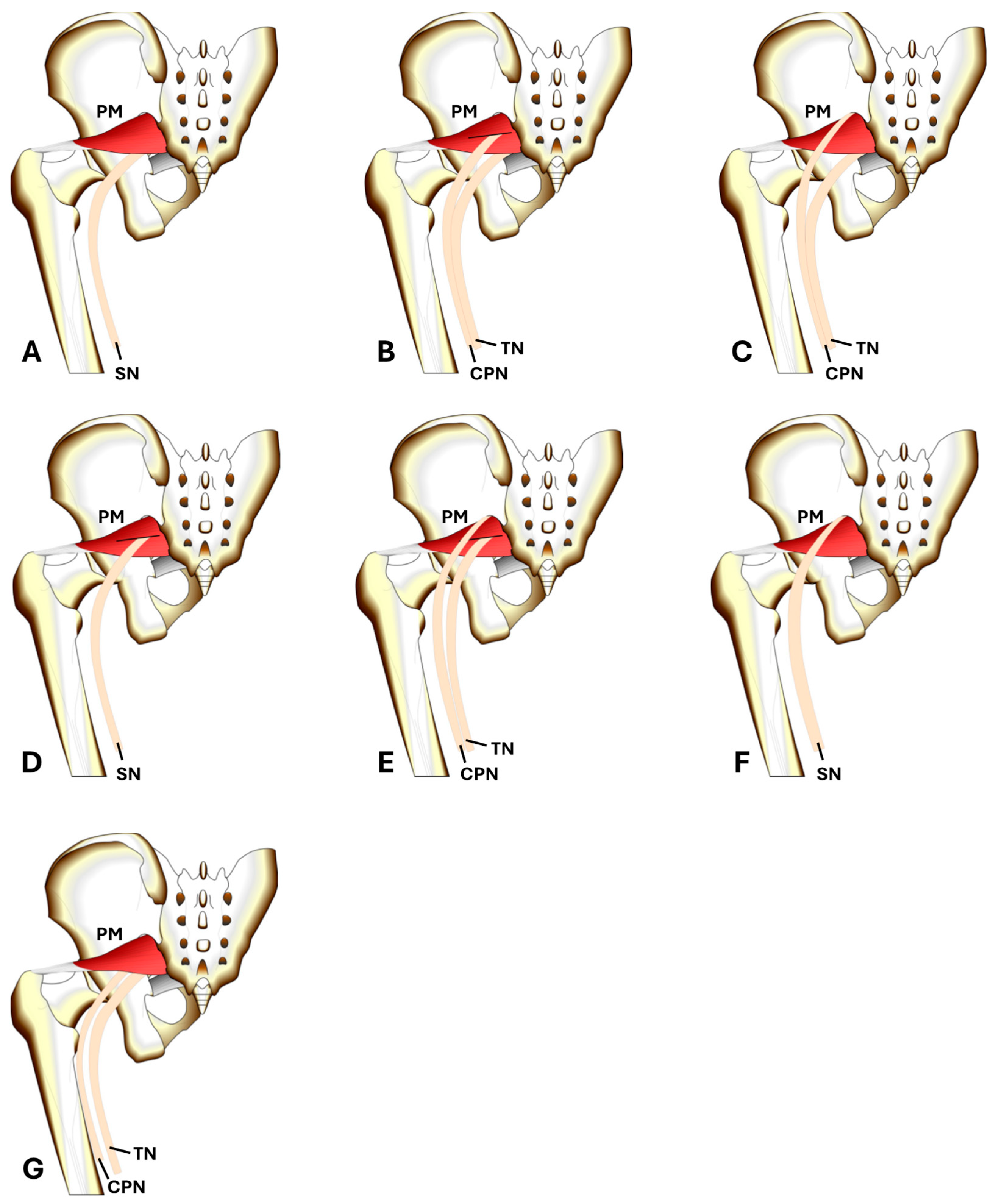
3.1. Beaton and Anson Classification of Donors

A total of 62 limbs were initially evaluated in the study. Of these 62 limbs, 56 were Type A classification and were then included in the next arm of the study. The remaining six limbs were Non-Type A classifications (Table 1). Bilateral symmetry was observed in 25 donors, all of which were relationship AA. Bilateral asymmetry occurred in six donors, and included AB, AC, AG, AG, AG, and AG relationships (Table 1).

3.2. Laterality Based Comparison of the SN-PM Region

The mean distances from PSIS-IT, PSIS-GT, IT-GT, PSIS-S1, IT-S1, GT-S1, S1-R, and S1-Q in left and right limbs are illustrated in Table 2. The distance from S1-R and S1-Q was larger in the right limbs compared to the left, but neither was statistically significant. No other patterns were appreciated, and no measurements were statistically significant when comparing left and right limbs (Table 2).

3.3. Sex-Based Comparison of the SN-PM Region

The mean distances from PSIS-IT, PSIS-GT, IT-GT, PSIS-S1, IT-S1, GT-S1, S1-R, and S1-Q are illustrated in Table 3. The triangular region between PSIS, IT, and GT had a significantly larger area in males when compared to females (p < 0.01) as determined by a two-tailed t-test (Table 3). A similar relationship was determined when evaluating the mean distance from S1 to the PSIS, IT, and GT, with the mean length being significantly larger in males compared to females (p < 0.01). The mean S1-R measurement was larger in males when compared to females (p < 0.01). By comparison, the mean S1-Q measurement was also larger in males when compared to females, but the difference was not statistically significant (p = 0.07) (Table 3). By comparison, when evaluating the measurements using a one-tailed t-test, all differences were significantly smaller in females as compared to male donors.

4. Discussion

The prevalence of Type A classifications as the most common anatomy present in patient populations has ranged in reports from 85.2% (95% CI: 78.4–87.0%) [16] to 90% (95% CI: 83–90%) [2]. Data from this study (90.32% Type A) are consistent with previous reports, suggesting the generalizability of data from this donor cohort to the general population. The significantly increased prevalence of the Type A classification supports the claim that most patients seen for hip-based procedures will most likely present with Type A anatomy. Other classification frequencies consistent with previously reported data include the Type C classification at 1.6%, which was previously noted with a frequency of approximately 2% [2,16]. This study did not have any occurrences of Types D–F, which are rare and reportedly make up less than 1% of the overall population [2]. Interestingly, this study’s Type B frequency of 1.61% (1/62) did not match well with previously reported values of 8% (95% CI: 5–10%) [2] and 9.8% (95% CI: 6.5–13.2%) [16]. Furthermore, the frequency of this study’s Type G variation was 6.55% (4/62), which is higher than the previously reported frequency of occurring in less than 1% of the population [2]. We propose this Type G variation may pose the largest risk in terms of inadequate anesthesia via sciatic nerve block, as the CPN and TN bifurcate before their exit from beneath the PM. Sciatic nerve blocks utilizing the transgluteal [18,19] and subgluteal [20,21] approaches may result in unequal distribution of anesthetic to both branches. This risk might be increased as compared to previous understanding due to inconsistent reports of the frequency of Type G anatomy within the population.
Although branching patterns and their frequency have been reported, there are also variations when comparing the classification of limbs bilaterally in the same donor. Symmetry, defined as having the same classification when comparing right and left limbs, existed in 80.62% of the donors (25/31) in the present study, lower in comparison to previously reported values [88% (95% CI: 81.7–93.2%)] [16]. Indeed, when discussing Type A anatomy, the present study shows that symmetrical relationships were only identified in donors displaying Type A variants and asymmetrical relationships always include a Type A variant (6/6). Interestingly, four of the six (66.6%) Non-Type A variants were in the right limb, while two of the six (33.3%) Non-Type A variants were in the left limb, suggesting that dominant limbs are more likely to display Non-Type A variations [22].
Though a decrease in symmetrical relationships was found in this study compared to prior studies, the consistency of measurements within the region when comparing right and left limbs aids clinicians in understanding similarities in procedures that are done on both hips within the same patient. Laterality is an important variable of note, as bilateral hip disease can occur in up to 42% of patients with osteoarthritis, the most common form of arthritis in the United States [23,24]. Furthermore, bilateral hip involvement frequently requires surgical intervention on both hips [24]. As the general population ages and obesity trends continue to rise, osteoarthritis will continue to increase, creating further instances in which bilateral hip replacements will be required [23].
Additionally, we found that sex-based differences within the SN-PM relationship exist, with all but one measurement being statistically significant in males vs. females when evaluated using a two-tailed t-test. Overall, the male-specific measurements were larger than those recorded in females. However, all measurements were statistically significant when analyzed using a one-tail t-test, indicating that female measurements were significantly smaller when comparing females to males. Thus, the more detailed and quantitative analysis evaluating sex-specific differences in the SN-PM relationship reported herein should benefit healthcare providers in the surgical treatment and rehabilitation of these patients. Indeed, it is understood that anatomical sex plays a role in the prevalence of osteoarthritis of the hip, with females at greater risk [25]. Any intervention specific to female patients, particularly when treating conditions where females have increased prevalence, the decreased SN-PM regional volume should be taken into consideration, especially when discussing specific treatments or surgical approaches.
Indeed, when evaluating total hip arthroplasties (THA), various approaches either directly or indirectly involve manipulation of the PM. When utilizing the minimally invasive Smith–Peterson (anterior) approach, the PM is often released from its insertion on the superior aspect of the GT to better allow for increased rotation of the femur to enhance exposure to the joint capsule [26]. Values that we have reported for the S1-GT distance demonstrate that males have more available length for PM release before encroaching on the SN as it comes under the PM, further supporting the need for a thorough evaluation of SN-PM landmarks based on anatomical sex.
When discussing the Southern and Moore approach for THA (posterior), the Watson–Jones approach for THA (anterolateral), or the Kocher and Langenbeck approach for posterior wall and/or posterior column acetabular fractures, our S1-GT values play a crucial role in locating the SN to allow for appropriate dissection and retractor placement when the nerve is not easily identified, or when it is not directly visible within the surgical field. In these approaches, aberrant retractor placement has been known to cause SN injury [27]. Understanding the values presented here can allow surgeons and junior residents to roughly locate where the SN may be in relation to other more easily visible structures.
Various studies have reported that the release of the PM during these posterior-based approaches (the Southern and Moore approach, the Kocher and Langenbeck approach), offers protection for the SN [28]. However, the findings in this study, as well as several others in the literature, confirm that the majority of patients will exhibit Type A anatomy with the SN presenting anterior to the PM. Reflecting the PM with this anatomical relationship present would not offer any protection to the SN. Results from this study show that the SN is closer to the GT as well as the line that connects GT/PSIS in females, demonstrating that females may be at increased risk of SN injury as a smaller proportion of PM needs to be reflected medially before exposing the SN.
Hip arthroscopy is a minimally invasive procedure that involves the placement of portals to allow for visualization and manipulation of internal structures with arthroscopes and other tools. The placement of these portal sites through the skin relies primarily on palpation of anatomical landmarks. The posterolateral approach, located inferomedial to the GT, is commonly used [29,30]. The palpation of these landmarks used in portal placement can be difficult depending on body habitus, which can increase risk of SN injury [31]. The present study shows that the location of the SN within females is closer to the GT than in males, demonstrating a smaller available window for portal placement, and subsequent increased risk of SN injury within females.
Sciatic nerve blocks are also a potential anesthetic option for lower extremity surgeries, including knee, foot, and ankle procedures. An anterior sciatic nerve block approach has been described and has been associated with worse outcomes when compared to the standard posterior sub gluteal approach [32]. The anterior approach has also been associated with decreased sensory block in the posterior femoral cutaneous nerve (PFCN) compared to the posterior approach [33]. Given that the majority of patients display Type A anatomy, in which the SN is anterior to the PM, the prevalence results of our study further support the use of a posterior approach, especially if Non-Type A anatomy is present, or if sensory block to the PFCN is necessary.

Limitations and Future Directions

This study is not without limitations. The measurements in this study were collected from formalin-embalmed Caucasian donors with a mean age of 71.95 years. This limited sample diversity should be considered when projecting generalizations from this research clinically across a wider varied group of patients. Further studies are needed to increase sample size within non-Caucasian populations to allow for better generalizability. Additionally, the Type A-anatomy does not allow for SN protection when the PM is reflected medially from its insertion point. However, the short external rotators do offer protection, as they are situated posterior to the SN. Further research needs to be done investigating if sex-based differences exist within the short external rotators to give clinicians adequate insight on SN protection with reflection of these muscles during posterior approaches to hip procedures.

5. Conclusions

Many sex-based differences within the SN and PM relationship exist, especially when Beaton and Anson Type A anatomy is compared. These differences offer clinicians important information on potential situations in which the SN may be at risk of iatrogenic injury, including various approaches for hip replacements, acetabular wall and acetabular column fracture fixations, arthroscopic portal placements, and sciatic nerve blocks. This study demonstrates that females may be at an increased risk in all situations mentioned above due to smaller available windows within approaches, and therefore smaller margins of error before the SN may be encountered with surgical instruments, retractors, or arthroscopic scopes.

Supplementary Materials

The following supporting information can be downloaded at: https://www.mdpi.com/article/10.3390/anatomia3030014/s1, Table S1: Statistical Power Analysis (SPA) of Laterality and Sex-Based Comparisons of the Sciatic Nerve-Piriformis Muscle Relationship.

Author Contributions

Conceptualization, C.R.M. and J.F.D.; Methodology, C.R.M., B.A.C. and J.F.D.; Software, C.R.M. and B.A.C.; Validation, C.R.M., B.J.P. and D.D.; Formal Analysis, C.R.M. and B.A.C.; Resources, J.F.D.; Data Curation, C.R.M. and A.L.G.; Writing—Original Draft Preparation, C.R.M. and B.J.P.; Writing—Review and Editing, C.R.M., B.J.P., A.L.G., D.D., B.A.C. and J.F.D.; Supervision, J.F.D.; Project Administration, J.F.D. All authors have read and agreed to the published version of the manuscript.

Funding

This project was partially supported by a Student Summer Research Fellowship (CRM) from Kansas City University.

Institutional Review Board Statement

The study was conducted in accordance with the Declaration of Helsinki, and the protocol was approved by the Institutional Biosafety Committee (1871804).

Informed Consent Statement

All subjects gave their informed consent for inclusion before they participated in the study.

Data Availability Statement

The original contributions presented in the study are included in the article/Supplementary Materials, further inquiries can be directed to the corresponding author/s.

Acknowledgments

The authors would like to acknowledge Anthony B. Olinger, for his expertise in creating Figure 1 and Figure 2A.

Conflicts of Interest

We have no conflicts of interest to report for this study.

References

  1. Berihu, B.A.; Debeb, Y.G. Anatomical variation in bifurcation and trifurcations of sciatic nerve and its clinical implications: In selected university in Ethiopia. BMC Res. Notes 2015, 8, 633. [Google Scholar] [CrossRef]
  2. Poutoglidou, F.; Piagkou, M.; Totlis, T.; Tzika, M.; Natsis, K. Sciatic Nerve Variants and the Piriformis Muscle: A Systematic Review and Meta-Analysis. Cureus 2020, 12, e11531. [Google Scholar] [CrossRef] [PubMed]
  3. Beaton, L.E.; Anson, B.J. The relation of the sciatic nerve and of its subdivisions to the piriformis muscle. Anat. Rec. 1937, 70, 1–5. [Google Scholar] [CrossRef]
  4. Wan-Ae-Loh, P.; Huanmanop, T.; Agthong, S.; Chentanez, V. Evaluation of the sciatic nerve location regarding its relationship to the piriformis muscle. Folia Morphol. 2020, 79, 681–689. [Google Scholar] [CrossRef]
  5. Jha, A.K.; Baral, P. Composite Anatomical Variations between the Sciatic Nerve and the Piriformis Muscle: A Nepalese Cadaveric Study. Case Rep. Neurol. Med. 2020, 2020, 7165818. [Google Scholar] [CrossRef] [PubMed]
  6. Han, S.K.; Kim, Y.S.; Kim, T.H.; Kang, S.H. Surgical Treatment of Piriformis Syndrome. Clin. Orthop. Surg. 2017, 9, 136. [Google Scholar] [CrossRef]
  7. Pećina, M. Contribution to the etiological explanation of the piriformis syndrome. Acta Anat. 1979, 105, 181–187. [Google Scholar] [PubMed]
  8. Fishman, L.M.; Dombi, G.W.; Michaelsen, C.; Ringel, S.; Rozbruch, J.; Rosner, B.; Weber, C. Piriformis syndrome: Diagnosis, treatment, and outcome—A 10-year study. Arch. Phys. Med. Rehabil. 2002, 83, 295–301. [Google Scholar] [CrossRef] [PubMed]
  9. Benzon, H.T.; Katz, J.A.; Benzon, H.A.; Iqbal, M.S. Piriformis Syndrome: Anatomic Considerations, a New Injection Technique, and a Review of the Literature. Anesthesiology 2003, 98, 1442–1448. [Google Scholar] [CrossRef]
  10. Moretti, V.M.; Post, Z.D. Surgical Approaches for Total Hip Arthroplasty. Indian J. Orthop. 2017, 51, 368–376. [Google Scholar] [CrossRef]
  11. Navarro, R.A.; Schmalzried, T.P.; Amstutz, H.C.; Dorey, F.J. Surgical approach and nerve palsy in total hip arthroplasty. J. Arthroplast. 1995, 10, 1–5. [Google Scholar] [CrossRef]
  12. Kim, H.J.; Park, S.H. Sciatic nerve injection injury. J. Int. Med. Res. 2014, 42, 887–897. [Google Scholar] [CrossRef]
  13. Kline, D.G.; Kim, D.; Midha, R.; Harsh, C.; Tiel, R. Management and results of sciatic nerve injuries: A 24-year experience. J. Neurosurg. 1998, 89, 13–23. [Google Scholar] [CrossRef]
  14. Güvençer, M.; Akyer, P.; Iyem, C.; Tetik, S.; Naderi, S. Anatomic considerations and the relationship between the piriformis muscle and the sciatic nerve. Surg. Radiol. Anat. 2008, 30, 467–474. [Google Scholar] [CrossRef] [PubMed]
  15. Haładaj, R.; Pingot, M.; Polguj, M.; Wysiadecki, G.; Topol, M. Anthropometric Study of the Piriformis Muscle and Sciatic Nerve: A Morphological Analysis in a Polish Population. Med. Sci. Monit. Int. Med. J. Exp. Clin. Res. 2015, 21, 3760–3768. [Google Scholar] [CrossRef]
  16. Tomaszewski, K.A.; Graves, M.J.; Henry, B.M.; Popieluszko, P.; Roy, J.; Pękala, P.A.; Hsieh, W.C.; Vikse, J.; Walocha, J.A. Surgical anatomy of the sciatic nerve: A meta-analysis. J. Orthop. Res. Off. Publ. Orthop. Res. Soc. 2016, 34, 1820–1827. [Google Scholar] [CrossRef]
  17. Gänsslen, A.; Grechenig, S.; Nerlich, M.; Müller, M. Standard Approaches to the Acetabulum Part 1: Kocher-Langenbeck Approach. Acta Chir. Orthop. Traumatol. Cech. 2016, 83, 141–146. [Google Scholar]
  18. Goldsmith, A.J.; Liteplo, A.; Hayes, B.D.; Duggan, N.; Huang, C.; Shokoohi, H. Ultrasound-guided transgluteal sciatic nerve analgesia for refractory back pain in the ED. Am. J. Emerg. Med. 2020, 38, 1792–1795. [Google Scholar] [CrossRef]
  19. Selame, L.A.; McFadden, K.; Duggan, N.M.; Goldsmith, A.J.; Shokoohi, H. Ultrasound-Guided Transgluteal Sciatic Nerve Block for Gluteal Procedural Analgesia. J. Emerg. Med. 2021, 60, 512–516. [Google Scholar] [CrossRef] [PubMed]
  20. Okutomi, Y.; Konishi, Y.; Kakinuma, A.; Sawamura, S. Preoperative Femoral Nerve Block and Postoperative Sciatic Nerve Block at the Subgluteal Space after Total Knee Arthroplasty: A Retrospective Cohort Study. Cureus 2023, 15, e50882. [Google Scholar] [CrossRef]
  21. Wiederhold, B.D.; Garmon, E.H.; Peterson, E.; Stevens, J.B.; O’Rourke, M.C. Nerve Block Anesthesia. In StatPearls; StatPearls Publishing: Treasure Island, FL, USA, 2024. Available online: http://www.ncbi.nlm.nih.gov/books/NBK431109/ (accessed on 7 May 2024).
  22. Teo, I.; Thompson, J.; Neo, Y.N.; Lundie, S.; Munnoch, D.A. Lower limb dominance and volume in healthy individuals. Lymphology 2017, 50, 197–202. [Google Scholar] [PubMed]
  23. Neogi, T.; Zhang, Y. Epidemiology of OA. Rheum. Dis. Clin. N. Am. 2013, 39, 1–19. [Google Scholar] [CrossRef]
  24. Stavrakis, A.I.; SooHoo, N.F.; Lieberman, J.R. Bilateral Total Hip Arthroplasty has Similar Complication Rates to Unilateral Total Hip Arthroplasty. J. Arthroplast. 2015, 30, 1211–1214. [Google Scholar] [CrossRef]
  25. Fu, M.; Zhou, H.; Li, Y.; Jin, H.; Liu, X. Global, regional, and national burdens of hip osteoarthritis from 1990 to 2019: Estimates from the 2019 Global Burden of Disease Study. Arthritis Res. Ther. 2022, 24, 8. [Google Scholar] [CrossRef] [PubMed]
  26. Bal, B.S.; Vallurupalli, S. Minimally invasive total hip arthroplasty with the anterior approach. Indian J. Orthop. 2008, 42, 301–308. [Google Scholar] [CrossRef] [PubMed]
  27. Prakash, S.; Rai, A.; Manhas, V.; Malhotra, R. Sciatic nerve palsy after direct anterior approach for total hip replacement. BMJ Case Rep. 2023, 16, e252818. [Google Scholar] [CrossRef] [PubMed]
  28. Chen, J.Y.; Sharma, I.; Sabbagh, R.S.; Narendran, N.B.; Everhart, J.S.; Slaven, J.E.; Archdeacon, M.T.; Sagi, H.C.; Mullis, B.H.; Natoli, R.M. Risk of Postoperative Sciatic Nerve Palsy after Posterior Acetabular Fracture Fixation: Does Patient Position Matter? J. Orthop. Trauma. 2023, 37, 64. [Google Scholar] [CrossRef]
  29. Stone, A.V.; Howse, E.A.; Mannava, S.; Miller, B.A.; Botros, D.; Stubbs, A.J. Basic Hip Arthroscopy: Diagnostic Hip Arthroscopy. Arthrosc. Tech. 2017, 6, e699–e704. [Google Scholar] [CrossRef]
  30. Martin, S. Portal Placement for Hip Arthroscopy. J. Med. Insight 2024, 30. [Google Scholar] [CrossRef]
  31. Kern, M.J.; Murray, R.S.; Sherman, T.I.; Postma, W.F. Incidence of Nerve Injury after Hip Arthroscopy. J. Am. Acad. Orthop. Surg. 2018, 26, 773. [Google Scholar] [CrossRef]
  32. Yektaş, A.; Balkan, B. Comparison of sciatic nerve block quality achieved using the anterior and posterior approaches: A randomised trial. BMC Anesthesiol. 2019, 19, 225. [Google Scholar] [CrossRef]
  33. Ota, J.; Sakura, S.; Hara, K.; Saito, Y. Ultrasound-Guided Anterior Approach to Sciatic Nerve Block: A Comparison with the Posterior Approach. Anesth. Analg. 2009, 108, 660. [Google Scholar] [CrossRef] [PubMed]
Figure 1. Beaton and Anson classification system of the sciatic nerve as depicted by A or Non-A (B, C, D, E, F, or G variations). (A) Type A variation, the SN exits medially under the PM; (B) the SN is pre-bifurcated with the CPN piercing the muscle belly of the PM; (C) the SN is pre-bifurcated with the CPN exiting above to the PM, and the TN exits medially underneath the PM; (D) Type D variation, the SN (in its entirety) pierces the PM as a single trunk; (E) the SN is pre-bifurcated with the CPN exiting above the PM, and the TN piercing the PM; (F) the SN (in its entirety) exits above the PM as a single trunk; (G) the SN is pre-bifurcated with the CPN and TN both exiting under the PM. SN, sciatic nerve; PM, piriformis muscle; CPN, common peroneal; TN, tibial nerve.
Anatomia 03 00014 g001
Figure 2. Measurement of the Sciatic Nerve and Piriformis Muscle relationship. (A) Pictorial illustration of measurement triangle. (B) Cadaveric image of measurement triangle. PSIS, posterior superior iliac spine; IT, ischial tuberosity; GT, greater trochanter; S1, middle of the SN as it exits under the PM; Q, perpendicular lines connecting S1 to PSIS-IT; R, perpendicular lines connecting and S1 to PSIS-GT.
Anatomia 03 00014 g002
Table 1. Beaton and Anton (B&A) classification of the donor population.
B&A ClassificationPrevalenceTotal LimbsMale LimbsFemale LimbsDonor Symmetry *
A90.3%56243225/31 (AA)
B1.6%1100/31 (BB)
C1.6%1100/31 (CC)
D00000/31 (DD)
E00000/31 (EE)
F00000/31 (FF)
G6.5%4220/31 (GG)
Total donors = 31, total limbs = 62, male donors = 14, male limbs = 28, female donors = 17, female limbs = 34. Mean age of the donor population was 71.95 years (male mean age = 77.89 years; female mean age = 77.35 years). Male donors had a mean height of 176.11 cm; female donors had a mean height of 162.11 cm. * All Non-Type A classifications were bilaterally asymmetric and occurred with a Type A classification of the contralateral limb of the same donor. Asymmetrical Non-Type classifications occurred as AB, AC, and AG (N = 4).
Table 2. Laterality-based comparisons of the sciatic nerve–piriformis muscle relationships.
MeasurementLeft, Mean (SD)Right, Mean (SD)Two-Tailed p-Value
PSIS-IT174.79 (12.22)175.57 (10.59)0.81
IT-GT104.62 (15.13)108.36 (12.74)0.35
GT-PSIS176.23 (11.30)169.87 (12.54)0.07
S1-PSIS89.90 (8.54)92.26 (7.63)0.32
S1-IT88.49 (9.18)89.56 (7.61)0.66
S1-GT97.78 (11.80)92.13 (12.27)0.11
S1-R31.33 (8.55)33.66 (6.59)0.29
S1-Q23.22 (5.89)24.26 (5.62)0.53
Combined N = 49. Left N = 25 limbs, right N = 24 limbs. All measurements in mm. All analyses performed utilized a two-tailed t-test. p-value of (<0.05) was used to determine significance. PSIS, posterior superior iliac spine; IT, ischial tuberosity; GT, greater trochanter of the femur; S1, middle of the sciatic nerve as it exits under piriformis muscle; S1-PSIS, middle of the sciatic nerve as it exits under piriformis muscle to the posterior superior iliac spine; S1-IT, middle of the sciatic nerve as it exits under piriformis muscle to the ischial tuberosity; S1-GT, middle of the sciatic nerve as it exits under piriformis muscle to the greater trochanter; S1-R, distance of perpendicular line connecting S1 to PSIS-GT; S1-Q, distance of perpendicular line connecting S1 to PSIS-IT.
Table 3. Sex-based comparisons of the sciatic nerve–piriformis muscle relationships.
MeasurementCombined, Mean (SD)Male,
Mean (SD)
Female,
Mean (SD)
Two-Tailed p-ValueOne-Tailed p-Value
PSIS-IT175.17 (11.34)183.86 (6.91)168.09 (9.10)<0.01<0.01
IT-GT106.45 (14.00)116.08 (10.36)98.60 (11.51)<0.01<0.01
GT-PSIS173.12 (12.23)180.80 (9.67)166.86 (10.48)<0.01<0.01
S1-PSIS91.06 (8.11)95.45 (8.31)87.47 (6.01)<0.01<0.01
S1-IT89.01 (8.38)94.54 (5.13)84.50 (7.83)<0.01<0.01
S1-GT95.01 (12.24)101.09 (10.28)90.07 (11.60)<0.01<0.01
S1-R32.47 (7.66)36.98 (6.41)28.79 (6.63)<0.01<0.01
S1-Q23.73 (5.72)25.36 (6.22)22.41 (5.01)0.07<0.01
Combined N = 49 limbs. Male N = 22 limbs, female N = 27 limbs. All measurements in mm. Analyses included a one-tailed and a two-tailed t-test to compare differences in anatomical sex. p-value of (<0.05) was used to determine significance. PSIS, posterior superior iliac spine; IT, ischial tuberosity; GT, greater trochanter of the femur; S1, middle of the sciatic nerve as it exits under piriformis muscle; S1-PSIS, middle of the sciatic nerve as it exits under piriformis muscle to the posterior superior iliac spine; S1-IT, middle of the sciatic nerve as it exits under piriformis muscle to the ischial tuberosity; S1-GT, middle of the sciatic nerve as it exits under piriformis muscle to the greater trochanter; S1-R, distance of perpendicular line connecting S1 to PSIS-GT; S1-Q, distance of perpendicular line connecting S1 to PSIS-IT.
Disclaimer/Publisher’s Note: The statements, opinions and data contained in all publications are solely those of the individual author(s) and contributor(s) and not of MDPI and/or the editor(s). MDPI and/or the editor(s) disclaim responsibility for any injury to people or property resulting from any ideas, methods, instructions or products referred to in the content.

