Detailed Morphology of the Incisive or Nasopalatine Canal
Abstract
:1. Introduction
2. Materials and Methods
3. Results
4. Discussion
5. Conclusions
Author Contributions
Funding
Institutional Review Board Statement
Informed Consent Statement
Data Availability Statement
Conflicts of Interest
References
- Al-Amery, S.M.; Nambiar, P.; Jamaludin, M.; John, J.; Ngeow, W.C. Cone beam computed tomography assessment of the maxillary incisive canal and foramen: Considerations of anatomical variations when placing immediate implants. PLoS ONE 2015, 10, e0117251. [Google Scholar] [CrossRef] [PubMed]
- Iamandoiu, A.V.; Ilie, O.C.; Jianu, A.M.; Rusu, M.C. The nasomaxillary or septo-premaxillary crest. Med. Evol. 2021, XXVII, 386–391. [Google Scholar]
- Song, W.C.; Jo, D.I.; Lee, J.Y.; Kim, J.N.; Hur, M.S.; Hu, K.S.; Kim, H.J.; Shin, C.; Koh, K.S. Microanatomy of the incisive canal using three-dimensional reconstruction of microCT images: An ex vivo study. Oral Surg. Oral Med. Oral Pathol. Oral Radiol. Endod. 2009, 108, 583–590. [Google Scholar] [CrossRef] [PubMed]
- Bahsi, I.; Orhan, M.; Kervancioglu, P. A sample of morphological eponym confusion: Foramina of Stenson/Stensen. Surg. Radiol. Anat. 2017, 39, 935–936. [Google Scholar] [CrossRef]
- Abrams, A.M.; Howell, F.V.; Bullock, W.K. Nasopalatine cysts. Oral Surg. Oral Med. Oral Pathol. 1963, 16, 306–332. [Google Scholar] [CrossRef]
- Liang, X.; Jacobs, R.; Martens, W.; Hu, Y.; Adriaensens, P.; Quirynen, M.; Lambrichts, I. Macro- and micro-anatomical, histological and computed tomography scan characterization of the nasopalatine canal. J. Clin. Periodontol. 2009, 36, 598–603. [Google Scholar] [CrossRef] [Green Version]
- Mraiwa, N.; Jacobs, R.; Van Cleynenbreugel, J.; Sanderink, G.; Schutyser, F.; Suetens, P.; van Steenberghe, D.; Quirynen, M. The nasopalatine canal revisited using 2D and 3D CT imaging. Dentomaxillofacial Radiol. 2004, 33, 396–402. [Google Scholar] [CrossRef]
- Rusu, M.C.; Sandulescu, M.; Bichir, C. Patterns of pneumatization of the tympanic plate. Surg. Radiol. Anat. 2020, 42, 347–353. [Google Scholar] [CrossRef]
- Bichir, C.; Rusu, M.C.; Vrapciu, A.D.; Maru, N. The temporomandibular joint: Pneumatic temporal cells open into the articular and extradural spaces. Folia Morphol. 2019, 78, 630–636. [Google Scholar] [CrossRef] [Green Version]
- Muresan, A.N.; Mogoanta, C.A.; Stanescu, R.; Rusu, M.C. The sinus septi nasi and other minor pneumatizations of the nasal septum. Rom. J. Morphol. Embryol. 2021, 62, 227–231. [Google Scholar] [CrossRef]
- Radlanski, R.J.; Emmerich, S.; Renz, H. Prenatal morphogenesis of the human incisive canal. Anat. Embryol. 2004, 208, 265–271. [Google Scholar] [CrossRef] [PubMed]
- Von Arx, T.; Lozanoff, S. Clinical Oral Anatomy: A Comprehensive Review for Dental Practitioners and Researchers; Springer: Cham, Switzerland, 2016; p. 572. [Google Scholar]
- Fernandez-Alonso, A.; Suarez-Quintanilla, J.A.; Muinelo-Lorenzo, J.; Bornstein, M.M.; Blanco-Carrion, A.; Suarez-Cunqueiro, M.M. Three-dimensional study of nasopalatine canal morphology: A descriptive retrospective analysis using cone-beam computed tomography. Surg. Radiol. Anat. 2014, 36, 895–905. [Google Scholar] [CrossRef] [PubMed]
- Acar, B.; Kamburoglu, K. Morphological and volumetric evaluation of the nasopalatinal canal in a Turkish population using cone-beam computed tomography. Surg. Radiol. Anat. 2015, 37, 259–265. [Google Scholar] [CrossRef] [PubMed]
- Neves, F.S.; Oliveira, L.K.; Ramos Mariz, A.C.; Crusoe-Rebello, I.; de Oliveira-Santos, C. Rare anatomical variation related to the nasopalatine canal. Surg. Radiol. Anat. 2013, 35, 853–855. [Google Scholar] [CrossRef]
- Bornstein, M.M.; Balsiger, R.; Sendi, P.; von Arx, T. Morphology of the nasopalatine canal and dental implant surgery: A radiographic analysis of 100 consecutive patients using limited cone-beam computed tomography. Clin. Oral Implant. Res. 2011, 22, 295–301. [Google Scholar] [CrossRef]
- Etoz, M.; Sisman, Y. Evaluation of the nasopalatine canal and variations with cone-beam computed tomography. Surg. Radiol. Anat. 2014, 36, 805–812. [Google Scholar] [CrossRef]
- Gorurgoz, C.; Oztas, B. Anatomic characteristics and dimensions of the nasopalatine canal: A radiographic study using cone-beam computed tomography. Folia Morphol. 2021, 80, 923–934. [Google Scholar] [CrossRef]
- Hakbilen, S.; Magat, G. Evaluation of anatomical and morphological characteristics of the nasopalatine canal in a Turkish population by cone beam computed tomography. Folia Morphol. 2018, 77, 527–535. [Google Scholar] [CrossRef] [Green Version]
- Jain, N.V.; Gharatkar, A.A.; Parekh, B.A.; Musani, S.I.; Shah, U.D. Three-Dimensional Analysis of the Anatomical Characteristics and Dimensions of the Nasopalatine Canal Using Cone Beam Computed Tomography. J. Maxillofac. Oral Surg. 2017, 16, 197–204. [Google Scholar] [CrossRef]
- Milanovic, P.; Selakovic, D.; Vasiljevic, M.; Jovicic, N.U.; Milovanovic, D.; Vasovic, M.; Rosic, G. Morphological Characteristics of the Nasopalatine Canal and the Relationship with the Anterior Maxillary Bone-A Cone Beam Computed Tomography Study. Diagnostics 2021, 11, 915. [Google Scholar] [CrossRef]
- Soumya, P.; Koppolu, P.; Pathakota, K.R.; Chappidi, V. Maxillary Incisive Canal Characteristics: A Radiographic Study Using Cone Beam Computerized Tomography. Radiol. Res. Pract. 2019, 2019, 6151253. [Google Scholar] [CrossRef] [PubMed] [Green Version]
- Suter, V.G.; Jacobs, R.; Brucker, M.R.; Furher, A.; Frank, J.; von Arx, T.; Bornstein, M.M. Evaluation of a possible association between a history of dentoalveolar injury and the shape and size of the nasopalatine canal. Clin. Oral Investig. 2016, 20, 553–561. [Google Scholar] [CrossRef] [PubMed]
- Tozum, T.F.; Guncu, G.N.; Yildirim, Y.D.; Yilmaz, H.G.; Galindo-Moreno, P.; Velasco-Torres, M.; Al-Hezaimi, K.; Al-Sadhan, R.; Karabulut, E.; Wang, H.L. Evaluation of maxillary incisive canal characteristics related to dental implant treatment with computerized tomography: A clinical multicenter study. J. Periodontol. 2012, 83, 337–343. [Google Scholar] [CrossRef] [PubMed]
- Thakur, A.R.; Burde, K.; Guttal, K.; Naikmasur, V.G. Anatomy and morphology of the nasopalatine canal using cone-beam computed tomography. Imaging Sci. Dent. 2013, 43, 273–281. [Google Scholar] [CrossRef] [Green Version]
- Sekerci, A.E.; Buyuk, S.K.; Cantekin, K. Cone-beam computed tomographic analysis of the morphological characterization of the nasopalatine canal in a pediatric population. Surg. Radiol. Anat. 2014, 36, 925–932. [Google Scholar] [CrossRef]
- Nasseh, I.; Aoun, G.; Sokhn, S. Assessment of the Nasopalatine Canal: An Anatomical Study. Acta Inform. Med. 2017, 25, 34–38. [Google Scholar] [CrossRef] [Green Version]


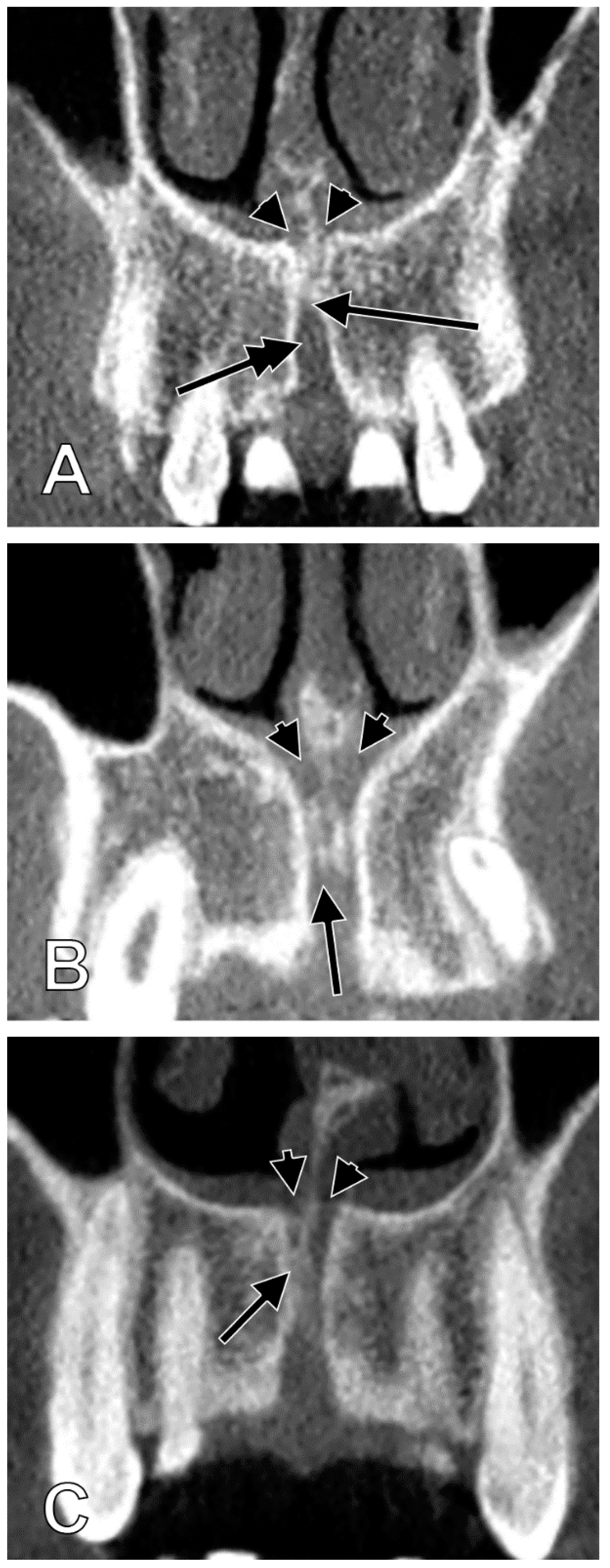
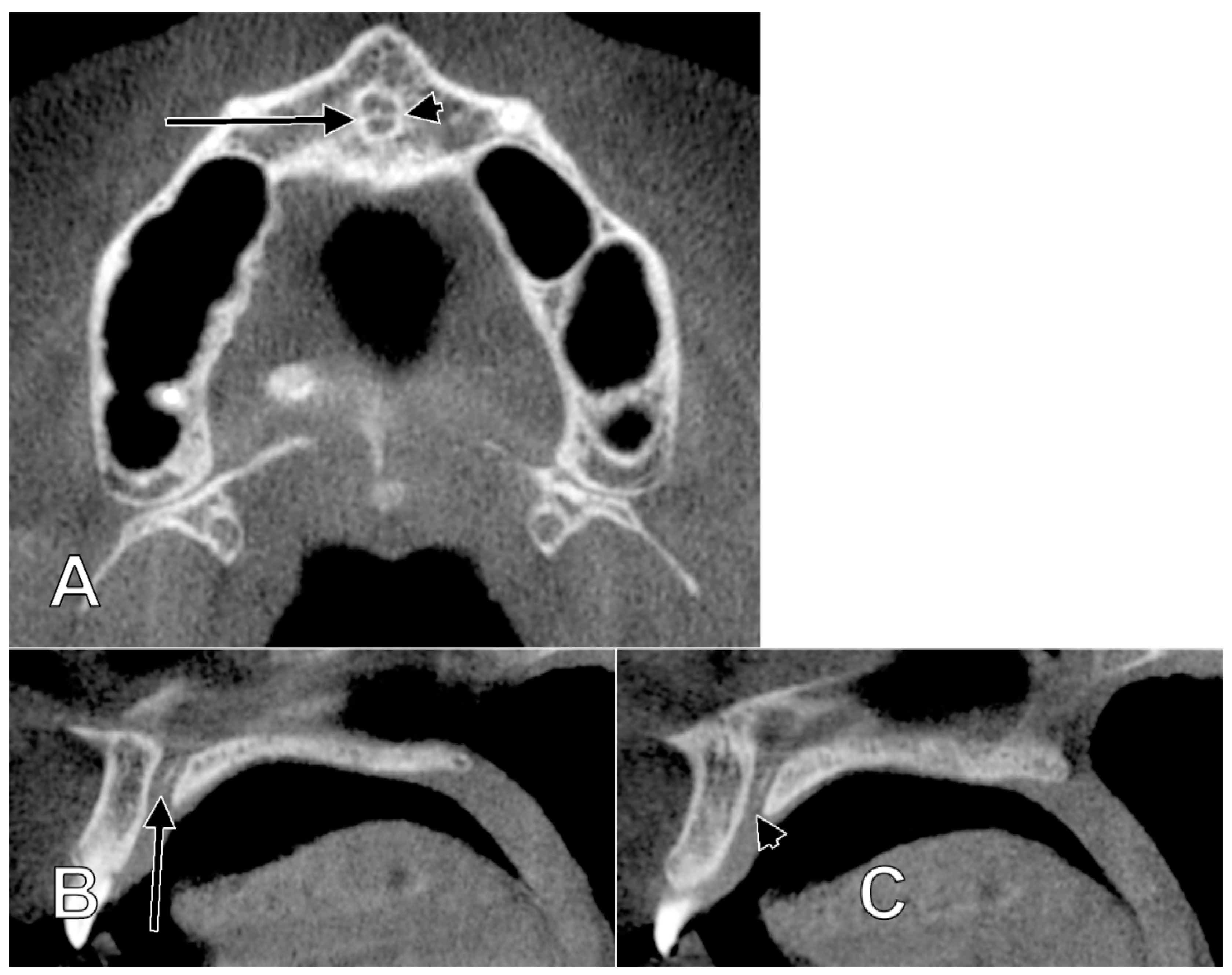
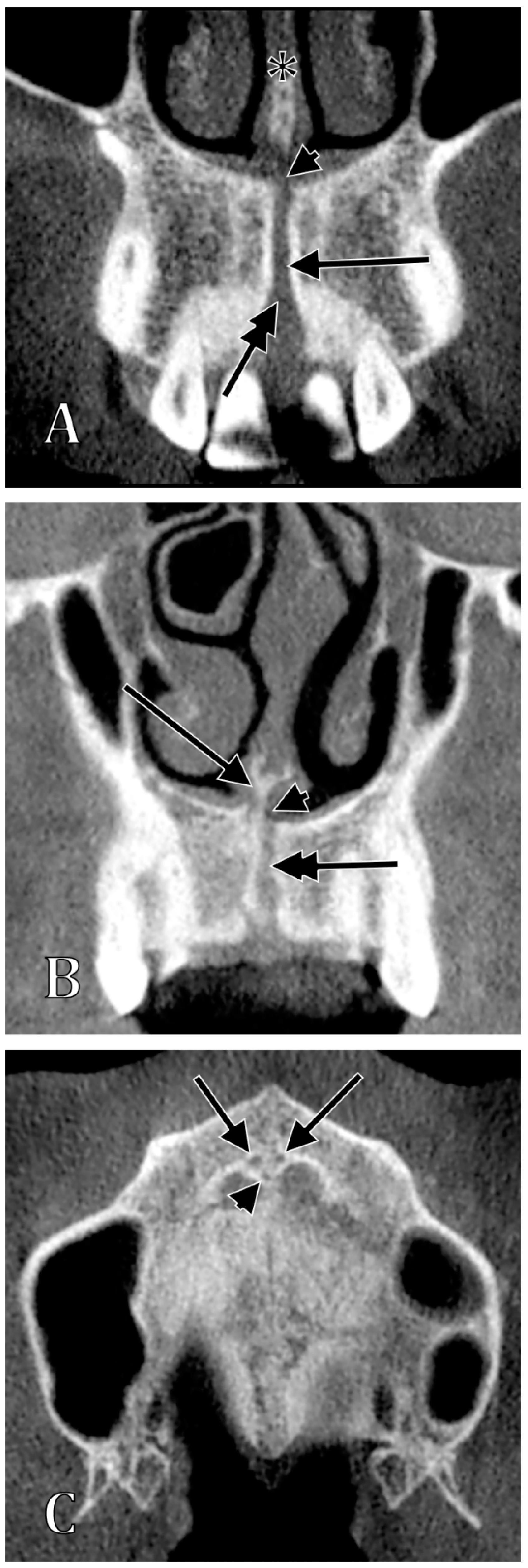
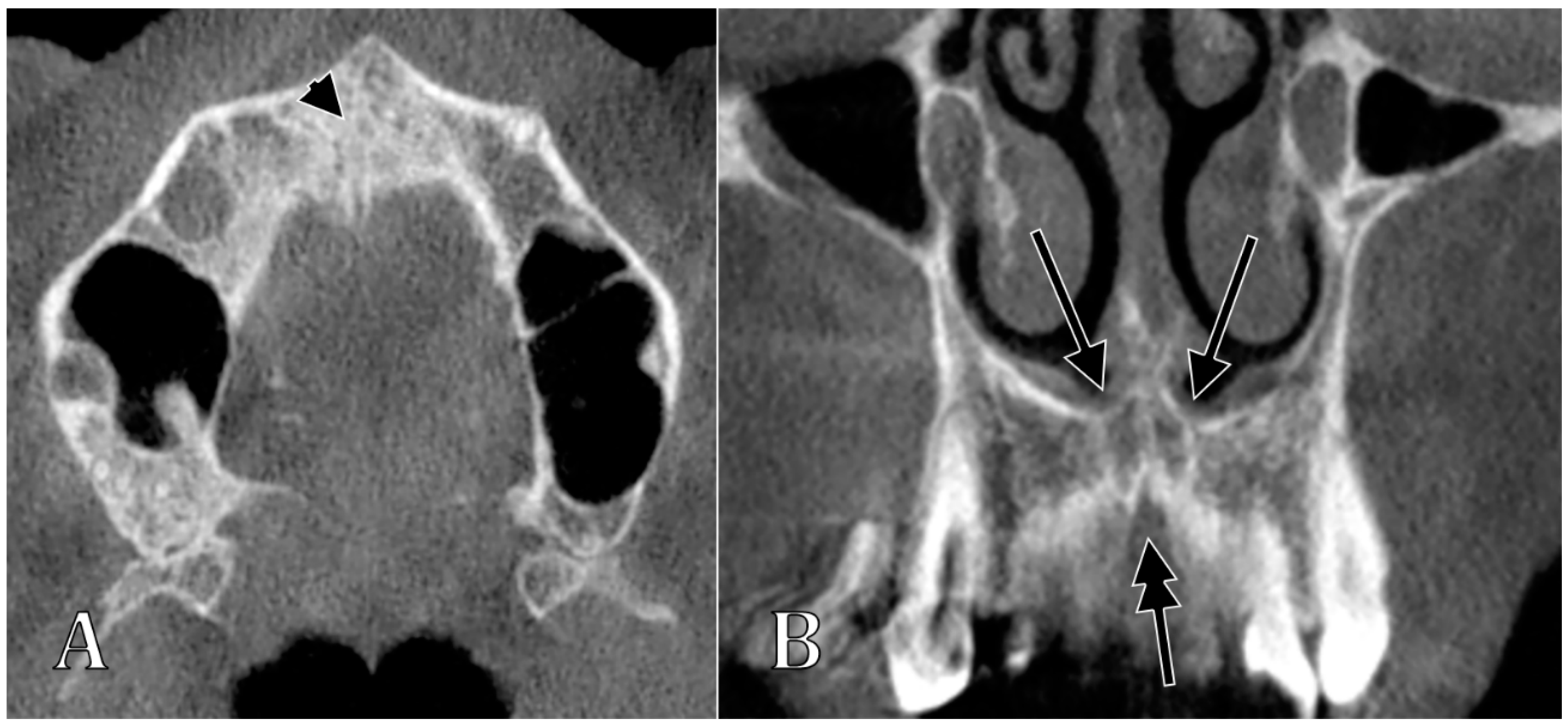
| Type of the NPC/IC | Characteristics of Types | Subtypes | Characteristics of Subtypes |
|---|---|---|---|
| I | NPC/IC present, 2 nasopalatine foramina | Ia | “Y”-shaped NPC/IC, with no secondary canaliculi |
| Ib | “Y”-shaped NPC/IC, with secondary canaliculi separated by a sagittal septum | ||
| Ic | “Y”-shaped NPC/IC, with unilateral secondary canaliculi separated by a coronal septum | ||
| Id | “Y”-shaped NPC/IC, with bilateral secondary canaliculi separated by a coronal septum | ||
| Ie | “Y”-shaped NPC/IC, with an added superiorly blind-ended median canal | ||
| If | parallel proper NPCs/ICs separated by septum | ||
| Ig | parallel proper NPCs/ICs unseparated by septum (NPC/IC unique, two nasopalatine foramina) | ||
| II | NPC/IC absent, 2 nasopalatine foramina | ||
| III | NPC/IC unique, 1 nasopalatine foramen | IIIa | unique median nasopalatine foramen, inferior to the nasomaxillary crest |
| IIIb | unique median nasopalatine foramen, on one side of the nasomaxillary crest | ||
| IV | NPC/IC present, 3 nasopalatine foramina, 1 median and 2 lateral | ||
| V | NPC/IC proper absent, absent nasopalatine foramina |
Publisher’s Note: MDPI stays neutral with regard to jurisdictional claims in published maps and institutional affiliations. |
© 2022 by the authors. Licensee MDPI, Basel, Switzerland. This article is an open access article distributed under the terms and conditions of the Creative Commons Attribution (CC BY) license (https://creativecommons.org/licenses/by/4.0/).
Share and Cite
Iamandoiu, A.V.; Mureşan, A.N.; Rusu, M.C. Detailed Morphology of the Incisive or Nasopalatine Canal. Anatomia 2022, 1, 75-85. https://doi.org/10.3390/anatomia1010008
Iamandoiu AV, Mureşan AN, Rusu MC. Detailed Morphology of the Incisive or Nasopalatine Canal. Anatomia. 2022; 1(1):75-85. https://doi.org/10.3390/anatomia1010008
Chicago/Turabian StyleIamandoiu, Andrei Valentin, Alexandru Nicolae Mureşan, and Mugurel Constantin Rusu. 2022. "Detailed Morphology of the Incisive or Nasopalatine Canal" Anatomia 1, no. 1: 75-85. https://doi.org/10.3390/anatomia1010008
APA StyleIamandoiu, A. V., Mureşan, A. N., & Rusu, M. C. (2022). Detailed Morphology of the Incisive or Nasopalatine Canal. Anatomia, 1(1), 75-85. https://doi.org/10.3390/anatomia1010008


