Article

Gamma Irradiation Enhances the In Vitro Biocontrol Potential of Trichoderma Species Against Major Rice Pathogens Rhizoctonia solani and Pyricularia oryzae

1
Hanoi Irradiation Center (HIC), km 12, 32 Road, Minh Khai, Bac Tu Liem, Hanoi 11900, Vietnam
2
Institute of Biology, Vietnam Academy of Science and Technology (VAST), 18-Hoang Quoc Viet, Cau Giay, Hanoi 10072, Vietnam
3
Faculty of Biotechnology, Graduate University of Science and Technology, Vietnam Academy of Science and Technology (VAST), 18-Hoang Quoc Viet, Cau Giay, Hanoi 10072, Vietnam
*
Authors to whom correspondence should be addressed.
Appl. Biosci. 2025, 4(3), 41; https://doi.org/10.3390/applbiosci4030041
Submission received: 7 June 2025 / Revised: 13 August 2025 / Accepted: 18 August 2025 / Published: 20 August 2025

Abstract

Improving the efficacy of microbial biocontrol agents is a pivotal strategy for sustainable management of rice blast and sheath blight caused by Pyricularia oryzae and Rhizoctonia solani, respectively, in Vietnam. In this study, Trichoderma sp. TVN-A0 and Trichoderma sp. TVN-H0 were irradiated by gamma to generate mutants for screening the enhanced antagonistic activity against P. oryzae and R. solani. The potential mutants were screened by antifungal metabolite production via the cellophane membrane assay (ICM), antagonistic performance through dual culture confrontation assays (IDC), volatile organic compound bioassays (IVOCs), and chitinase activity. As a result, among five potential mutants derived from each wild-type strain (AM1-AM5 and HM1-HM5), mutant AM2 originated from TVN-A0, and mutant HM2 derived from TVN-H0 demonstrated the highest inhibition rates and chitinase activities. The AM2 exhibited ICM of 96.71% against R. solani, 92.57% against P. oryzae, IDC of 87.76%, and IVOCs of 83.57%, while HM2 possessed ICM of 95.33% against R. solani, 85.28% against P. oryzae, IDC of 91.24%, and IVOCs of 79.33%. The genetic differences among mutants and their parents were investigated by RAPD. The non-GMO AM2 and HM2 mutants are promising candidates for biocontrol of the diseases caused by P. oryzae and R. solani in Vietnam.

1. Introduction

Rice (Oryza sativa) remains the world’s most vital staple crop, serving as the primary caloric source for over half of the global population and playing a central role in ensuring food security [1]. However, the interplay among climate change, intensified cropping systems, and the limited genetic diversity of modern rice varieties leads to a sharp increase in the incidence and severity of pests and diseases, thereby jeopardizing yield stability and threatening global supply chains [2,3]. Among the most pervasive and economically damaging rice diseases, blast—caused by Pyricularia oryzae—and sheath blight—caused by Rhizoctonia solani—represent major biotic constraints in leading rice-producing regions [4,5]. Asia, which accounts for over 90% of global rice production and includes top exporters such as Vietnam, is particularly susceptible to these fungal pathogens [6,7].
Conventional disease management in rice cultivation relies mainly on chemical fungicides. While these agrochemicals provide short-term efficacy, their excessive use has led to fungicide resistance, environmental degradation, and disruption of beneficial soil microbiota [8,9]. As a consequence, microbial biocontrol, particularly using Trichoderma spp., has garnered growing attention as an ecologically viable and environmentally sustainable alternative [10,11,12]. Trichoderma species exhibit multifaceted antagonistic mechanisms, including resource competition, mycoparasitism via hydrolytic enzyme production, and elicitation of systemic plant defenses [13,14]. Furthermore, their ability to enhance plant growth and resilience positions them as valuable components of climate-smart agricultural systems [15,16,17].
Although Trichoderma-based formulations have shown considerable potential, most commercially available products are developed from wild-type isolates [18,19]. Nevertheless, their efficacy in practical applications is often inconsistent, particularly underthe variable environmental conditions characteristic of tropical field settings such as those in Vietnam. To overcome this limitation, induced mutagenesis has emerged as a powerful approach to generate improved strains with superior and stable biocontrol efficacy. However, the successful adaptation of such strains to complex field environments remains a key challenge.
Ionizing radiation, especially gamma rays, offers a potent tool to artificially enhance microbial genetic diversity by inducing DNA damage, including both single- and double-strand breaks [20,21]. Unlike transgenic methods, gamma mutagenesis enables the development of enhanced microbial strains without the introduction of foreign DNA, thereby circumventing regulatory constraints associated with genetically modified organisms (GMOs). Previous studies have shown that gamma irradiation can significantly improve the biocontrol performance of Trichoderma spp. by stimulating the biosynthesis of hydrolytic enzymes and antifungal secondary metabolites [22]. Gamma-irradiated mutants have demonstrated broadened antagonistic spectra against key phytopathogens, including R. solani, Fusarium spp., Macrophominaphaseolina, Sclerotium cepivorum, and Sclerotium rolfsii [23,24,25].
In this study, we employed gamma irradiation to induce mutations in Trichoderma species—Trichoderma sp. TVN-A0 and Trichoderma sp. TVN-H0—with the specific aim of enhancing their antagonistic capacity against P. oryzae and R. solani, the causal agents of blast and sheath blight in rice. Mutants with improved biocontrol phenotypes were selected and comprehensively characterized. To our knowledge, this is the first report demonstrating the application of gamma-induced mutagenesis in Trichoderma spp. for the simultaneous control of two major rice diseases within tropical Vietnamese agroecosystems. The results provide a robust foundation for the development of next-generation Trichoderma-based biofungicides tailored to the specific challenges of rice production, while also contributing to broader efforts in climate-resilient, sustainable crop protection.

2. Materials and Methods

2.1. Materials

Trichoderma TVN-A0 (TVN-A0) was obtained from the Vietnam Type Culture Collection, Institute of Microbiology and Biotechnology, Vietnam National University, Hanoi. Trichoderma TVN-H0 (TVN-H0) was provided by the Department of Microbiology, Faculty of Biology, University of Science, Vietnam National University, Hanoi. Both strains are indigenous isolates that were previously characterized for their strong antagonistic activity against phytopathogenic fungi. Both strains were classified by internal transcribed spacer gene.
The fungal pathogens R. solani RHN1 (causal agent of sheath blight) and P. oryzae TB03 (causal agent of rice blast) were provided by the Department of Plant Pathology and Plant Immunology, Plant Protection Research Institute, Dong Ngac, Tu Liem, Hanoi, Vietnam. These strains were originally isolated in 2023 from rice fields in Thai Binh province, a major rice-growing region in the Red River Delta and represent predominant pathogenic variants in northern Vietnam.
Potato Dextrose Agar (PDA) medium was purchased from Sigma-Aldrich (USA). All other reagents were of analytical grade.

2.2. Methods

2.2.1. Classification of the Trichoderma Strains

The fungi TVN-H0 and TVN-A0 were cultured on PDA medium for 1–2 days at 25 °C. The mycelia cells were harvested and then rinsed in 1.5 mL of cold, sterile water. The cells were harvested by centrifugation at 13,000 rpm for 5 min afterwards suspended in 300 µL SCED (1 M sorbitol, 10 mM sodium citrate, 10 mM EDTA, and 10 mM dithiothreitol). To break the fungal cell walls, the samples were supplemented with 300 µL zymolyase (3 mg/mL) (Sigma-Aldrich, Saint Louis, MO, USA), mixed well, then incubated at 37 °C for 1 h. After adding 300 µL of SDS (sodium dodecyl sulfate) 1% and keeping it on ice for 5 min, 225 µL of potassium acetate 5 M were used to precipitate the protein fraction with gentle mixing. The supernatants were harvested by centrifugation at 13,000 rpm for 5 min. The phenol/chloroform/isoamyl alcohol (at a ratio of 24 v:24 v:1 v) was used to extract the small proteins from the DNA solution in the top phase. The genomic DNAs were precipitated by using two volumes of cold absolute ethanol, rinsed by two volumes of cold 70% ethanol, and then harvested by centrifugation at 13,000 rpm for 5 min. The genomic DNAs were dried by the SpeedVac system and then dissolved in 30 µL H2O containing RNAse (1 mg/mL) (Sigma-Aldrich) incubated at 37 °C for 30 min. The DNAs were dissolved in 30 µL TE buffer. The DNA concentration was measured by a NanoDrop NC-2000C Implen (Isogen, De Meern, The Netherlands) and stored at −20 °C. This protocol was used to extract genomic DNA from all mutants for RAPD analysis.
Primers ITS 1 and ITS 2 were used to amplify the internal transcribed spacer sequences from DNA genomes of TVN-H0 and TVN-A0 as the protocol described by Haque et al. [26]. The PCR products were checked by electrophoresis in 1% agarose gel using TAE buffer (40 mM Tris base, 20 mM acetic acid, 0.4 mM ethylenediaminetetraacetic acid) pH 8.3, purified by MEGAquick-spin™ Plus Total Fragment DNA Purification Kit (iNtRON Biotechnology, Seongnam-si, Gyeonggi-do, Republic of Korea) and sequenced by the ABI 3100 (Applied Biosystem, 850 Lincoln Centre Drive, Foster City, California). The DNA sequences were aligned into a reference database by BLASTN from GenBank (accessed on 5 August 2025) to search the homologous sequences. The ITS sequences of TVN-H0 and TVN-A0 strains and reference sequences from GenBank were used to construct a phylogeny by a maximum likelihood method with 1000 replications for each bootstrap value using MEGA5.

2.2.2. Gamma Irradiation and Spore Survivability

Spore suspensions of TVN-A0 and TVN-H0 were prepared as described by Darabzadeh et al. [27]. The suspensions at 108–109 CFU/mL were aliquoted into sterile 10 mL tubes and irradiated using a Co-60 gamma source at the Ha Noi Irradiation Center. Irradiation doses ranged from 0 to 1500 Gy, delivered at a rate of 0.23 Gy/s. This dose range was selected based on our previous study and preliminary trials indicating its effectiveness in generating mutations in filamentous fungi while maintaining spore viability [28]. Gafchromic dosimeters (for ≤1000 Gy) and B3 Dose Stix dosimeters (for >1000 Gy) were used to determine absorbed doses. Spore viability was assessed before and after irradiation by plating the treated suspensions on PDA, incubated at 25 ± 1 °C and a relative humidity of approximately 70% for 48 h. Colony-forming units (CFUs) were then enumerated. The surviving colonies after irradiation were individually cultured and subjected to screening for antagonistic activity against R. solani and P. oryzae.

2.2.3. Screening and Selection of Mutants

The mutants derived from the gamma-irradiated TVN-A0 and TVN-H0 strains were evaluated for antagonistic activity against R. solani and P. oryzae using the following three complementary in vitro assays:
  • Inhibition via cellophane membrane (ICM) was applied to assess the effect of non-volatile metabolites diffused through a sterile cellophane membrane on PDA medium as described by a previous study [29].
  • Inhibition via dual culture (IDC) was used to evaluate direct antagonism and spatial competition with R. solani on shared PDA plates.
  • Inhibition via volatile organic compounds (IVOCs) tested the effect of VOCs produced from the mutants against P. oryzae using a sealed sandwich plate system.
Inhibition rates were calculated using the following equation:
I (%) = (1 − DT/DC) × 100
where
I is the inhibition rate (%), designated as ICM, IDC, or IVOCs;
DT is the colony diameter (mm) in the treatment group;
DC is the colony diameter (mm) in the control group.
  • Cellophane Membrane Assay (ICM)
This assay followed a modified method from Liu et al. [29]. Trichoderma spores were inoculated onto sterile cellophane membranes (Bio-Rad) placed on PDA plates and incubated at 25 ± 1 °C with a relative humidity of approximately 70% for 48 h. After removing the membranes, mycelial plugs of R. solani (4 days old) or P. oryzae (7 days old) already prepared in PDA medium were placed at the center of the conditioned medium. Colony diameters were measured after 3 or 5 days, respectively, and inhibition (ICM, %) was calculated using Equation (1). Strains with high ICM values against both pathogens were advanced to the next assays.
The most promising mutants, identified based on their highest inhibition index (ICM), were further evaluated using two complementary assays: a dual culture assay for R. solani and a sandwich plate assay for P. oryzae. The choice of assay was based on the distinct growth characteristics of each pathogen. The dual culture assay was selected for R. solani due to its rapid growth, which enables direct observation of antagonistic interactions. In contrast, the sandwich plate assay was used for P. oryzae, which grows more slowly, thereby facilitating the assessment of Trichoderma-derived VOCs without physical contact.
  • Dual Culture Assay (IDC)
Plugs of Trichoderma mutants, wild-type strains, and R. solani were placed 50 mm apart on 90 mm PDA plates and incubated at 25 ± 1 °C with approximately 70% relative humidity for 3–5 days. The radial growth of R. solani was measured and compared to the control to determine IDC (%), following Equation (1).
  • Sandwich Plate Assay (IVOCs)
This assay followed a protocol from Alfiky [30] with modification. One PDA plate inoculated with Trichoderma and another cultured with P. oryzae were sealed in a face-to-face configuration, with the P. oryzae plate placed on top. The plates were sealed with three layers of Parafilm (PM-996, Bemis, Sheboygan Falls, WI, USA) and incubated at 25 ± 1 °C with approximately 70% relative humidity for 5–7 days. Colony growth was compared to a control plate paired with uninoculated PDA, and IVOCs (%) was calculated using Equation (1).

2.2.4. Chitinase Activity Assay

The mutants were cultured in a 50 mL chitinase-inducing medium prepared in 250 mL Erlenmeyer flasks. The medium contained (g/L): chitin (5.0), NaNO3 (2.0), KH2PO4 (1.0), MgSO4·7H2O (0.5), and KCl (0.5), adjusted to pH 5.0. The flasks were inoculated at 28 °C, 120 rpm for 14 days. After incubation, the cultures were centrifuged at 8000 rpm for 10 min to separate the cells. Chitinase activity was determined by measuring the release of N-acetyl-β-D-glucosamine in the supernatants at 540 nm as described by Mukhammadiev et al. [31].

2.2.5. Investigation of Genetic Changes by Randomly Amplified Polymorphic DNA (RAPD) Analysis

In order to investigate any changes occurred in the genomes of mutants compared to wild-type strains, the RAPD and electrophoresis were applied to observe the patterns of random amplified-genes.
The RAPD was conducted to amplify random DNA fragments from equal amounts of 10 ng genomic DNA using primers OPA-10, OPA-16, Eric1R, Eric2, BoxA1R [23], and M13 DNA, Cac5, Gtg5, and oligonucleotide GGCATCGGCC (designated as Crip1 in this study) as described in a previous study [32]. The PCR products were analyzed by electrophoresis in the 1.5% agarose gel using TAE buffer for running. To investigate the genetic differences between wild-type and the derivative mutants, the electrophoresis pictures were scanned by InfanView, and analyzed by ImageLab to obtain densitometric curves, DNA bands, and similarity indices. Levels of similarity of fingerprinting profiles between samples were calculated according to the Dice coefficient. The unweighted pair group method with arithmetic averages (UPGMA) was used to transform similarity coefficients into distance, subsequently creating a heatmap dendrogram [33]. An overview picture of the experimental design, providing a visual summary of the methodological framework applied in this study, presented in Figure 1.

2.2.6. Statistical Analysis

All data were analyzed using one-way analysis of variance (ANOVA). Means were compared by Duncan’s multiple range test at p < 0.05 using SPSS version 22.0. All experiments were performed in at least three biological replicates. Results are presented as mean ± standard deviation (SD) in tables and mean ± standard error (SE) in figures.

3. Results

3.1. Classification of Trichoderma Strains

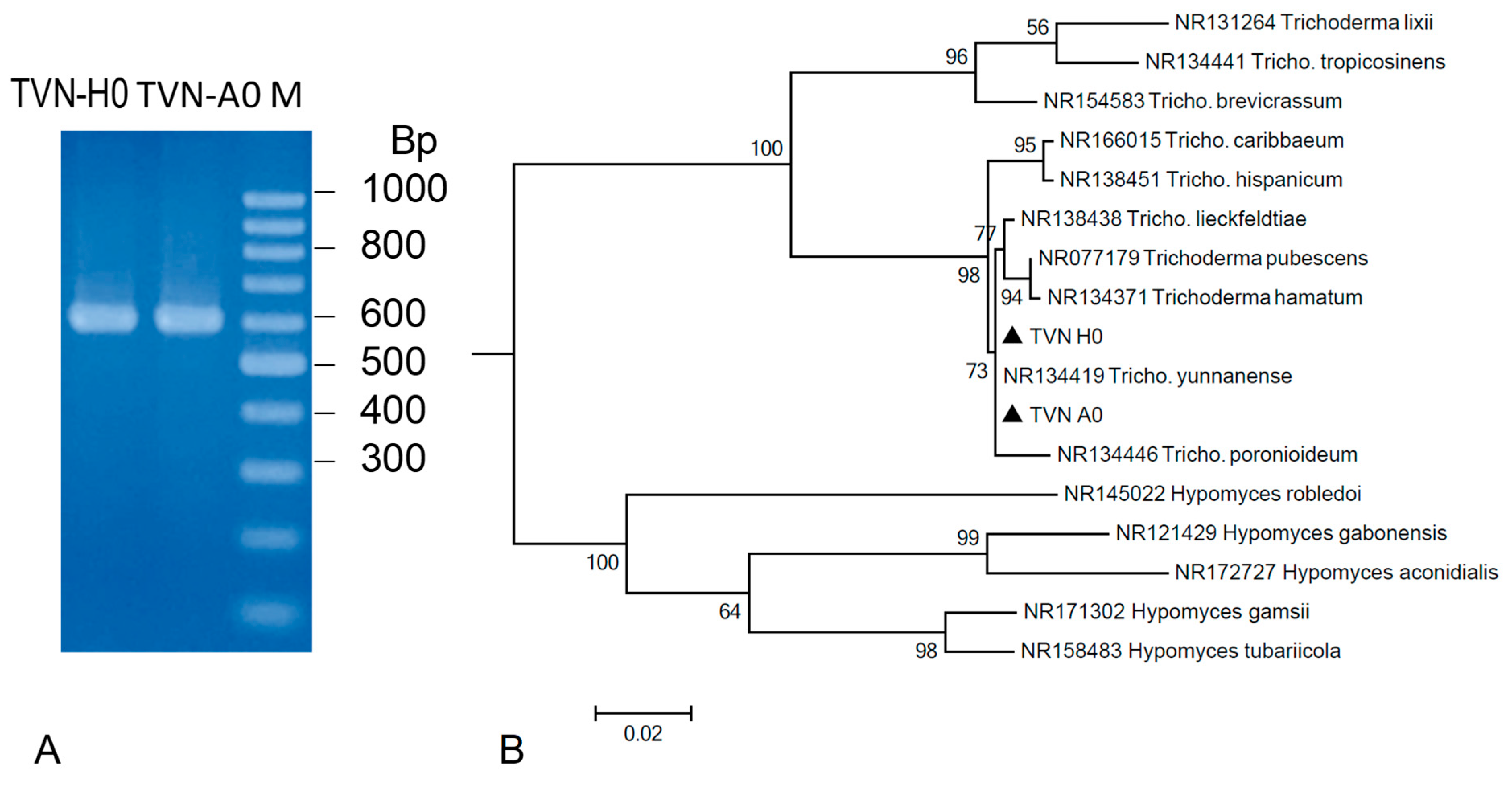
To confirm the classification of Trichoderma strains TVN-H0 and TVN-A0, the ITS regions (~600 bp) were amplified from their genomic DNA (Figure 2A) and subsequently sequenced. The ITS sequences of TVN-H0 (561 bp) and TVN-A0 (449 bp) showed the highest sequence indentities −99.27% and 99.78%, respectively—to the corresponding gene of Trichoderma yunnanense. Phylogenetic analysis revealed that both strains, TVN-H0 and TVN-A0, clustered within the Trichoderma genus and were closely related to T. yunnanense (Figure 2B). However, the ITS sequences alone did not provide enough genetic differentiation to classify these strains to species. Therefore, these strains were designated as Trichoderma sp.

3.2. Effects of Gamma Irradiation on Spore Survival Rate of Trichoderma

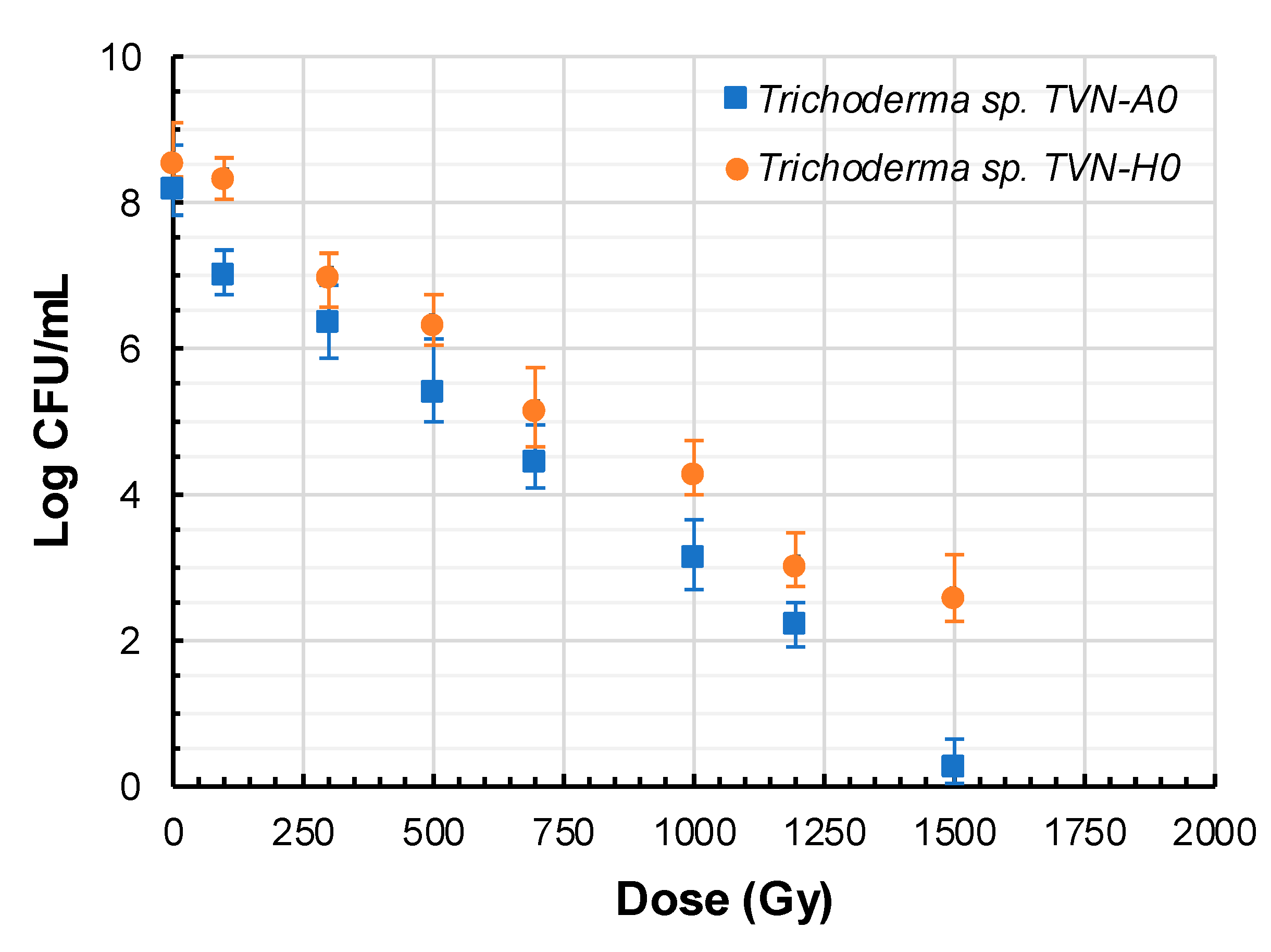
The correlation between the logarithm of viable spore counts (CFU/mL) and radiation dose (Figure 3) revealed a dose-dependent decline in spore survival, consistent with the general response of microorganisms to ionizing radiation. At doses ≤ 250 Gy, both strains retained relatively high viability (log CFU/mL ranging from 7.0 to 8.5). However, as the dose increased, the number of surviving spores declined sharply. Notably, TVN-H0 exhibited a slower rate of reduction in viability compared to TVN-A0, especially at doses ≥ 1000 Gy. At 1500 Gy, TVN-A0 was nearly completely inactivated, while a small proportion of viable spores of TVN-H0 still remained.

3.3. Potential Mutants with High Antagonistic Activity Against R. solani and P. oryzae

3.3.1. Antifungal Activity

Following gamma irradiation, hundreds of fungal colonies derived from the two parental Trichoderma strains were isolated and initially screened using the cellophane membrane assay. Selection was prioritized for colonies exhibiting altered morphology or enhanced growth rate to maximize the likelihood of identifying mutants with improved biocontrol potential.
During the evaluation process, rapid mycelial growth emerged as a notable trait among several promising isolates, highlighting their potential applicability in the development of biological formulations. A total of 391 individual colonies were recovered post-irradiation, including 238 from TVN-A0 and 153 from TVN-H0. All the isolates were cultured on PDA medium to assess radial growth. Among them, 52 isolates (13.30%) exhibited a higher growth rate than their respective wild-type strains after 48 h of incubation and were selected for further antifungal screening using the cellophane membrane method.
The obtained results (Table 1) showed that the top five mutants from each Trichoderma strain exhibited strong inhibitory effects against both pathogenic fungi, with statistically significant differences compared to the wild-type strains (p < 0.05).
The mutants AM1-AM5 of TVN-A0 and HM1-HM5 of TVN-H0 exhibited significantly higher inhibitory activity against R. solani and P. oryzae compared to their respective wild-type strains. Notably, several mutant lines showed superior antagonistic efficacy in specific experimental assays, indicating that gamma irradiation induced beneficial alterations enhancing their biocontrol potential.
For TVN-A0, mutant AM2 showed the highest levels of inhibition against R. solani and P. oryzae via metabolite-mediated mechanisms, with ICM reaching 96.71% and 92.57%, respectively. In the direct competition assay (IDC), this mutant AM2 also demonstrated strong inhibition of R. solani (IDC of 87.76%), second only to the mutant HM2 (IDC of 91.24%). Volatile organic compounds (VOCs) contributed significantly to antagonistic activity, with AM2 achieving the highest inhibition of P. oryzae in the VOC-based assay (IVOCs of 83.57%).
Regarding TVN-H0, mutant HM1 exhibited maximum inhibition against R. solani via metabolite secretion, with an ICM value of 100%, whereas HM2 was the most effective against P. oryzae, with an ICM of 85.28%. In the direct competition assay (IDC), HM2 displayed the highest level of inhibition against R. solani among all tested strains, with IDC reaching 91.24%. Notably, the mutant HM1 also exhibited strong VOC-mediated inhibition of P. oryzae (IVOCs of 78.42%).
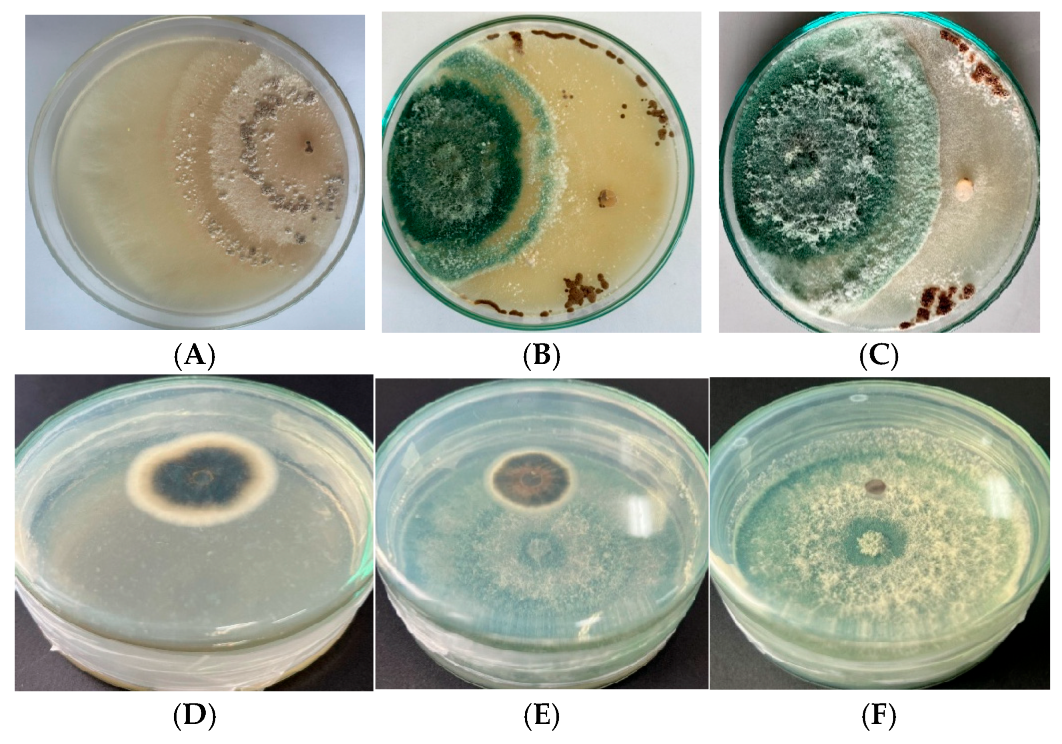
Overall, the mutants AM2 and HM2 emerged as the most promising candidates, demonstrating potent inhibitory effects against both R. solani and P. oryzae across all three antagonistic mechanisms. In particular, mutant AM2 exhibited broader and more consistent biocontrol potential than the other mutants evaluated. These findings are visually supported by dual culture and sandwich assays shown in Figure 4 and Figure 5, which illustrate the comparative antagonistic activity of AM2 and HM2, respectively, against the two pathogens.

3.3.2. Chitinase Activity

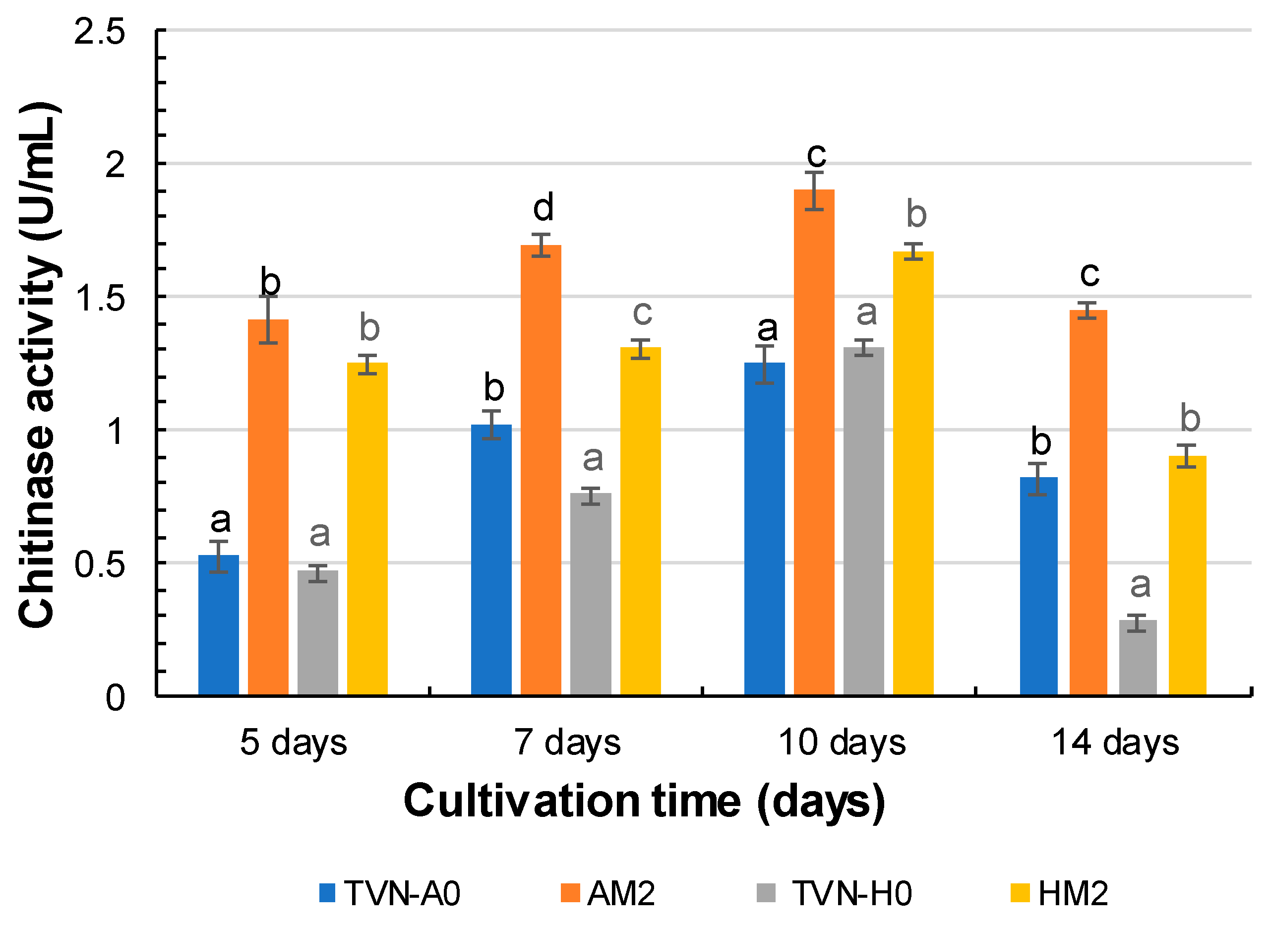
In this study, the chitinase activity of two promising mutants AM2 and HM2, was monitored and compared with their respective wild-type strains throughout the cultivation period (Figure 6).
The results revealed a progressive increase in chitinase activity for all strains from day 5 to day 10, followed by a decline on day 14. Notably, the mutant strains AM2 and HM2 exhibited significantly higher chitinase activity than their wild-type counterparts at all time points (p < 0.05). Specifically, AM2 reached peak activity on day 10 with 1.89 U/mL, representing a 1.52-fold increase compared to parental strain TVN-A0 (1.24 U/mL). Similarly, mutant HM2 reached 1.67 U/mL, 1.28 times higher than the wild-type TVN-H0 (1.30 U/mL).
After day 10, chitinase activity declined in all strains, with the most substantial reduction observed in wild-type strain TVN-H0, which dropped by 78.3% from 1.30 U/mL to 0.28 U/mL on day 14. In contrast, the mutant strains, AM2 and HM2, maintained relatively higher activity levels despite a slight decrease. Interestingly, TVN-A0 generally exhibited higher chitinase activity than TVN-H0 across most of the cultivation period, except on day 10, when TVN-H0 showed slightly greater activity. These findings suggest that gamma irradiation induced beneficial mutations, enhancing the ability of Trichoderma to produce chitinase. Furthermore, the data indicate that the optimal harvesting time for maximal chitinase yield falls within the 7–10-day incubation window, before activity begins to decline.

3.3.3. Investigation of the Genetic Changes by RAPD

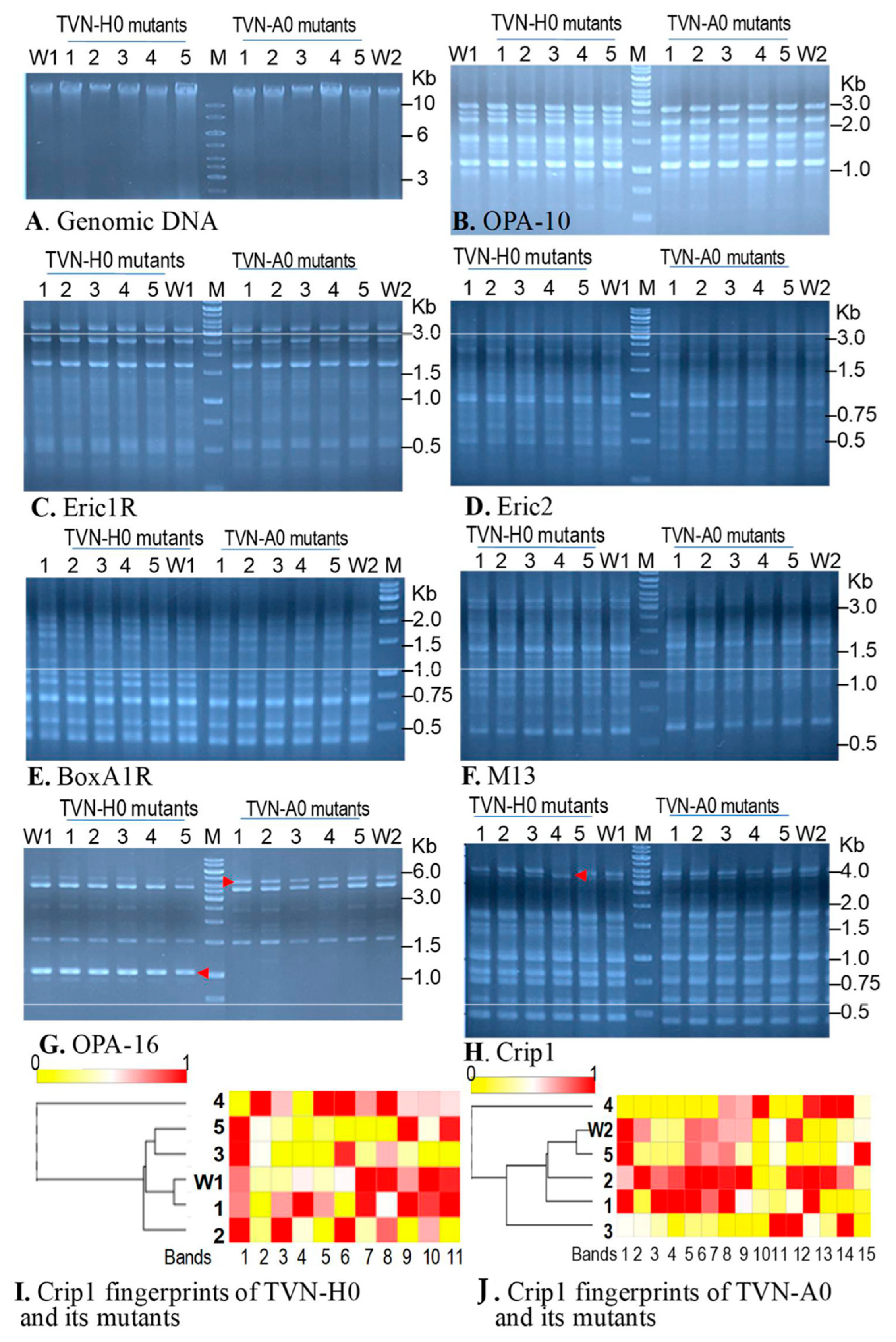
In this study, we use a total of nine primers, including OPA-10, OPA-16, Eric1R, Eric2, BoxA1R M13 DNA, Cac5, Gtg5, and Crip1, to investigate the genetic difference between wild-type strains and mutated strains. Among the primers, amplicons were successfully amplified by seven primers, OPA-10, OPA-16, Eric1R, Eric2, BoxA1R, M13 DNA, and Crip1 (Figure 7). However, the patterns of DNA fingerprints amplified by six primers from DNA genomes of the strains TVN-A0 and TVN-H0 were similar, excepting primer OPA-16. That means, of the nine investigated primers, the OPA-16 primer can help to distinguish genetic differences between the two parental strains (Figure 7G). By OPA-16 primer, a maximum of six DNA length polymorphisms were observed by the naked eye from the agarose gel, whereby each parent strain and its mutants contained three sharp and dominant bands (Figure 7G). Among the 3 bands, 2 bands showed similar sizes between two strains and their mutants; a band of ~1 kb was only amplified from TVN-H0 and its mutants (Figure 7G). Whereas the fragments of 4.5 kb were absolutely amplified from the genomes of TVN-A0 and all its mutants, but they were not prioritized for amplification from the genome of TVN-H0 and its mutants (Figure 7G).
Among the primers, we found that only Crip1 can assist in observing the genetic differentiation between parental strains and their mutants. In the case of Trichoderma sp. TVN-H0 and its mutants, by naked eyes, we found only mutant HM4 lost a DNA band of about 4 kb but improved the DNA band of 3.5 kb (Figure 7H). Withthe assistance of the ImageLab tool, a total of 11 DNA length polymorphisms amplified by Crip1 were detected in all Trichoderma sp. TVN-H0 and its mutants (Figure 7I). Based on the density of each band amplified from the same amount of DNA genomes, the heatmap and dendrogram showed that among five mutants, the mutant Trichoderma sp. HM1 has a Crip1 fingerprint similar to the one of the original strain, so it has been grouped with the wild-type strain in a cluster. Other mutants posed the genetic difference from their parental strain, wherein mutants HM3 and HM5 are in a cluster. The Crip1 fingerprint of HM2 is also different from the fingerprints of HM1-HM3 and HM5, while HM2 exhibited superior antifungal activities against both pathogens P. oryzae and R. solani. The mutant HM4 harbored genetic material profoundly different from other mutants and separated from a cluster including other mutants and the parental strain. Seeking the correlation between genetic changes and the phenotype of antifungal activities did not give any statistically significant difference; however, at least the differentiation was seen to be preliminary evidence for the genetic changes in bioactivity-improved mutants induced by the radiation.
Regarding Trichoderma sp. TVN-A0 and its irradiated mutants, with the assistance of the ImageLab tool, a total of 15 DNA length polymorphisms amplified by Crip1 were detected in all Trichoderma sp. TVN-A0 and its mutants (Figure 7J). The mutant Trichoderma sp. AM4 possessed the genetic difference from its parent to be separated into a group, while Trichoderma sp. TVN-A0 and other mutants grouped into a cluster in the dendrogram (Figure 7J). No identical fingerprints were recognized among five mutants and their original strain. This result indicates that all the mutants posed mutations in the genome to make the Crip1 fingerprint profiles different from the wild-type strain.

4. Discussion

The genus Trichoderma is widely recognized as a beneficial group of fungi in the development of sustainable agriculture, primarily due to its ability to produce antibiotics and lytic enzymes that degrade the cell walls of plant pathogens [34]. Numerous species within this genus have been reported to possess strong antagonistic potential and the ability to protect crops against a broad spectrum of phytopathogenic fungi [34], thereby highlighting the remarkable biological and functional diversity of Trichoderma. In this study, gamma irradiation was applied to induce genetic variability in two native strains of Trichoderma species—Trichoderma sp. TVN-A0 and Trichoderma sp. TVN-H0—with the aim of enhancing their biocontrol potential.
Gamma irradiation represents a potent tool for strain improvement by inducing beneficial genetic mutations that may enhance antifungal capabilities. Gamma irradiation causes single- and double-strand DNA breaks, leading to alterations in genomic structure and biological characteristics based on doses [35]. In this study, TVN-A0 was more sensitive to gamma radiation than TVN-H0; thus, when increasing the dose to 1500 Gy, the Log CFU/mL of survival spores of TVN-A0 was depleted (Figure 3), while the log CFU/g of live spore of TVN-H0 was higher than two. These differences may be attributed to multiple factors, including strain-specific characteristics, initial spore density, growth stage, culture medium composition, and intrinsic biological properties of each species. Moreover, variations in DNA repair systems and antioxidant defense mechanisms may also play crucial roles in the radiation resistance of Trichoderma strains.
The observed dose-dependent decline in spore viability is consistent with the mechanism of DNA strand breakage induced by gamma rays, which often leads to lethal mutations or loss of replication capacity. However, the radiation tolerance observed in TVN-A0 and TVN-H0 in this study was markedly higher compared to previously reported Trichoderma species. Specifically, Baharvand et al. reported that T. viride exhibited a survival rate of 9.7% at 400 Gy but was completely inactivated at 450 Gy [36]. Similarly, Soufi et al. found complete inactivation of T. aureoviride at comparable doses [25]. In contrast, El-Bialy et al. observed varying levels of radiation tolerance among Trichoderma strains in the following order: T. viride Td1 > T. koningii Tk1 > T. harzianum Tz1 > T. longibrachiatum Tl1 [37].
Although targeted mutagenesis techniques have become increasingly prevalent, gamma irradiation remains a powerful method for inducing random mutations and enhancing the biological characteristics of Trichoderma. Previous studies have demonstrated that this approach can significantly improve the antagonistic capacity of Trichoderma through multiple mechanisms, including the increased production of extracellular enzymes and antifungal secondary metabolites [22,24,36].
In the present study, after gamma irradiation, hundreds of colonies derived from the two original Trichoderma strains were isolated and screened using the cellophane membrane assay, and then we selected two mutants, AM2 and HM2, which exhibited significantly stronger antagonistic activity against both R. solani and P. oryzae compared to their respective wild-type strains (Table 1, Figure 4 and Figure 5). The mutant AM2 achieved the highest inhibition rates against R. solani (ICM: 96.71%) and P. oryzae (ICM: 92.57%), while HM2 also demonstrated strong inhibition (ICM: 95.33% for R. solani and 85.28% for P. oryzae). Moreover, both mutant strains exhibited significantly higher chitinase activity than their wild-type counterparts, wherein the activity of AM2 reached 1.89 U/mL (1.52-fold higher than the original strain TVN-A0) and the activity of HM2 reached 1.67 U/mL (1.28-fold higher than TVN-H0). Chitinase activity, which was significantly elevated in both AM2 and HM2 mutants, is widely recognized as a critical factor in the antagonistic potential of Trichoderma spp. This enzyme catalyzes the hydrolysis of chitin, the major structural component of fungal cell walls, thereby facilitating mycoparasitism and host lysis. Numerous studies have established a strong correlation between high chitinase activity and enhanced biocontrol efficacy [35,36], which supports the superior antifungal performance observed in these two mutant strains. In agreement with this study, in the study by Haggag et al., gamma irradiation at doses ranging from 200 to 500 Gy significantly enhanced the biocontrol activity of T. harzianum, T. viride, and T. koningii against Sclerotium cepivorum, the causal agent of white rot in onions [22]. The mutant strains not only exhibited improved growth and sporulation but also increased production of cell wall-degrading enzymes (chitinase, β-1,3-glucanase, cellulase) and antifungal secondary metabolites such as gliotoxin, trichodermin, and viridin. Similarly, Baharvand et al. applied gamma irradiation to T. viride spores and identified 250 Gy as the optimal dose for selecting strains with enhanced growth and antagonistic activity against Macrophominaphaseolina [36]. In another study, Wagh et al. reported that gamma doses from 0 to 50 krad generated mutant strains of T. viride with strong antagonism toward Sclerotium rolfsii, Rhizoctonia bataticola, and Fusarium oxysporum f. sp. ciceri, with maximum chitinase activity reaching 0.62 U/mg [38]. Ghasemi et al. also observed significantly elevated chitinase activity and inhibition of Rhizoctonia solani in mutant T. harzianum strains, with the T.h M8 mutant showing the highest chitinase activity (42.48 U/mg) [24].
An important observation in this study is that the ICM inhibition rates against R. solani consistently surpassed those against P. oryzae, possibly due to differential sensitivity of the pathogens to Trichoderma-secreted metabolites. This observation is supported by findings from Vinale et al., who reported that the efficacy of antifungal compounds produced by Trichoderma varies depending on the susceptibility of the fungal target [12]. Furthermore, P. oryzae may possess more advanced defense mechanisms, such as multidrug efflux pumps or membrane structural adaptations, which could reduce its sensitivity to Trichoderma-derived antifungal compounds [39,40].
In addition to the enhanced antagonistic effects and elevated chitinase activity, several mutant colonies displayed alterations in morphology and growth rate compared to the original strains, which were also observed in our study. This supports the hypothesis that gamma irradiation can induce mutations in specific genomic regions, resulting in phenotypic changes that can be identified through screening [36]. Such modifications are likely due to changes in the expression of genes involved in enzyme production and secondary metabolite biosynthesis, ultimately contributing to the improved antagonistic capabilities.
Investigation of primers for random amplification of polymorphic DNA (RAPD) from genomes of original and mutated strains showed that only OPA-16 can help to distinguish the genome of TVN-A0 from the genome of TVN-H0. However, OPA-16 did not assist in discriminating the genetic changes among mutants and parental strains in this study. Conversely, in another study, this primer generated fingerprints of two mutants profoundly different from the fingerprints of their original strain,T. harzianum 65, and 22 other mutants [23]. The reason for the inability to detect genetic changes in the mutants in this study by primer OPA-16 may lie in the limited number of mutants used for investigation by RAPD. Luckily, DNA fingerprinting using primer Crip1 revealed genetic changes in the mutant Trichoderma sp. HM4 compared to the Trichoderma sp. TVN-H0 (Figure 7H). Dendrogram analysis of DNA fingerprints generated by PCR using primer Crip1 also indicated the genetic changes in the mutants compared to the original strains. The Crip1 has been used to distinguish the genomic DNA of T. harzianum ATCC 36042, T. harzianum CBS 891.68, and T. harzianum CBS 354.33 [32]. The fingerprints of these strains generated by primer Crip1 were not homologous. That means genetic materials of strains belonging to a species are different. In this study, gamma radiation induced genetic changes in mutants derived from both TVN-A0 and TVN-H0. Recently, the detection of mutations existing in the genome associated with any phenotypic changes is usually performed by whole genome sequencing or whole transcriptome sequencing using next-generation sequencers such as Illumina. However, this method is expensive and requires a lot of effort for data analysis, annotation and mutation mining. Therefore, in this study, although PCR-RFLP is limited in clarifying mutations, it can indicate genetic differences among wild-type strains and their mutants. This method is suitable in this study.
Taken together, these results demonstrate that gamma irradiation is an effective tool for enhancing the antagonistic potential of Trichoderma, particularly against R. solani and P. oryzae. The mutant strains AM2 and HM2 not only exhibited superior chitinase activity but also showed enhanced inhibition of plant pathogenic fungi, highlighting their potential application in the biological control of rice diseases. However, further research involving genomic analyses and characterization of specific secondary metabolites is necessary to elucidate the underlying mechanisms of antagonism in these promising mutant strains.
Despite the promising antagonistic effects and elevated chitinase levels observed in AM2 and HM2, it is important to note that the present study was limited to in vitro conditions. While these assays provide valuable initial insights, they cannot fully replicate the complexity of field environments. In addition, this study did not directly compare mutant strains with commercial Trichoderma products, which remains a limitation. Nevertheless, the high inhibition rates and enhanced enzyme activity observed suggest strong potential for further development. These limitations will be addressed in subsequent research.

5. Conclusions

This study demonstrated that gamma irradiation is an effective tool for enhancing the antagonistic potential of Trichoderma spp. against two major rice pathogens, R. solani and P. oryzae. Through the induction of beneficial mutations, several mutant strains exhibited significantly improved antifungal activity; in particular, AM2 and HM2 showed consistent performance across multiple in vitro assays. In addition, these mutants displayed elevated chitinase activity, which is a key factor contributing to their enhanced biocontrol capacity.
These findings highlight the potential of gamma-mutagenized Trichoderma strains as candidates for developing next-generation biocontrol agents suitable for integrated disease management (IDM) strategies in rice cultivation. Importantly, the use of non-GMO mutagenesis ensures regulatory compatibility and public acceptance, making these strains promising for commercial biofungicide development.
To support future practical application, further studies should validate these mutants under diverse agroecological conditions, benchmark their efficacy against commercial Trichoderma-based biofungicides, and investigate the molecular mechanisms responsible for enhanced antagonism, as well as ensure strain stability and safety over time.

Author Contributions

B.D.T. and T.H.D.; methodology, B.D.T. and T.H.D.; validation, B.D.T. and T.H.D.; formal analysis, H.D.N. and H.T.T.; investigation, H.D.N., T.T.H.L. and H.T.T.; resources, H.N.T. and N.K.L.D.; data curation, T.H.D.; writing—original draft preparation, B.D.T. and T.H.D.; writing—review and editing, B.D.T. and T.H.D.; visualization, X.V.L. and X.A.T.; supervision, D.S.H., B.D.T. and T.H.D.; project administration, B.D.T.; funding acquisition, B.D.T. All authors have read and agreed to the published version of the manuscript.

Funding

This research was funded by the Vietnam Ministry of Science and Technology through the project DTCB.10/23/TTCX.

Institutional Review Board Statement

Not applicable.

Informed Consent Statement

Not applicable.

Acknowledgments

The authors gratefully acknowledge the Hanoi Irradiation Center for providing irradiation support. We also thank the Department of Plant Pathology and Plant Immunology, Plant Protection Research Institute, for supplying fungal pathogens, and the Department of Microbiology, Faculty of Biology, VNU University of Science, Vietnam National University, Hanoi, for providing the original Trichoderma strain.

Conflicts of Interest

The authors declare no conflicts of interest.

References

  1. Bin Rahman, A.N.M.R.; Zhang, J. Trends in Rice Research: 2030 and Beyond. Food Energy Secur. 2023, 12, e390. [Google Scholar] [CrossRef]
  2. Peng, S.; Huang, J.; Sheehy, J.E.; Laza, R.C.; Visperas, R.M.; Zhong, X.; Centeno, G.S.; Khush, G.S.; Cassman, K.G. Rice Yields Decline with Higher Night Temperature from Global Warming. Proc. Natl. Acad. Sci. USA 2004, 101, 9971–9975. [Google Scholar] [CrossRef] [PubMed]
  3. Wassmann, R.; Jagadish, S.V.K.; Heuer, S.; Ismail, A.; Redona, E.; Serraj, R.; Singh, R.K.; Howell, G.; Pathak, H.; Sumfleth, K. Chapter 2 Climate Change Affecting Rice Production: The Physiological and Agronomic Basis for Possible Adaptation Strategies. In Advances in Agronomy; Sparks, D.L., Ed.; Academic Press: Cambridge, MA, USA, 2009; Volume 101, pp. 59–122. [Google Scholar]
  4. Persaud, R.; McGowan, D.; Persaud, M. Managing the Imminent Danger of Rice Blast (Pyricularia oryzae Cav.) and Sheath Blight (Rhizoctonia Solani Kühn) Disease: A Critical Review Article. Am. J. Agric. For. 2021, 9, 409–423. [Google Scholar] [CrossRef]
  5. Savary, S.; Willocquet, L.; Elazegui, F.A.; Castilla, N.P.; Teng, P.S. Rice Pest Constraints in Tropical Asia: Quantification of Yield Losses Due to Rice Pests in a Range of Production Situations. Plant Dis. 2000, 84, 357–369. [Google Scholar] [CrossRef]
  6. Asibi, A.E.; Chai, Q.; Coulter, J.A. Rice Blast: A Disease with Implications for Global Food Security. Agronomy 2019, 9, 451. [Google Scholar] [CrossRef]
  7. Phi, A. (Helen) Current Status of Rice Blast in Vietnam and Future Perspectives. Open Access Libr. J. 2023, 10, e10636. [Google Scholar] [CrossRef]
  8. Aktar, M.W.; Sengupta, D.; Chowdhury, A. Impact of Pesticides Use in Agriculture: Their Benefits and Hazards. Interdiscip. Toxicol. 2009, 2, 1–12. [Google Scholar] [CrossRef]
  9. Tilman, D.; Cassman, K.G.; Matson, P.A.; Naylor, R.; Polasky, S. Agricultural Sustainability and Intensive Production Practices. Nature 2002, 418, 671–677. [Google Scholar] [CrossRef]
  10. Berg, G. Plant-Microbe Interactions Promoting Plant Growth and Health: Perspectives for Controlled Use of Microorganisms in Agriculture. Appl. Microbiol. Biotechnol. 2009, 84, 11–18. [Google Scholar] [CrossRef]
  11. Shoresh, M.; Harman, G.E.; Mastouri, F. Induced Systemic Resistance and Plant Responses to Fungal Biocontrol Agents. Annu. Rev. Phytopathol. 2010, 48, 21–43. [Google Scholar] [CrossRef]
  12. Vinale, F.; Sivasithamparam, K.; Ghisalberti, E.L.; Marra, R.; Woo, S.L.; Lorito, M. Trichoderma–Plant–Pathogen Interactions. Soil Biol. Biochem. 2008, 40, 1–10. [Google Scholar] [CrossRef]
  13. Benítez, T.; Rincón, A.M.; Limón, M.C.; Codón, A.C. Biocontrol Mechanisms of Trichoderma Strains. Int. Microbiol. Off. J. Span. Soc. Microbiol. 2004, 7, 249–260. [Google Scholar]
  14. Schuster, A.; Schmoll, M. Biology and Biotechnology of Trichoderma. Appl. Microbiol. Biotechnol. 2010, 87, 787–799. [Google Scholar] [CrossRef]
  15. Harman, G.E.; Howell, C.R.; Viterbo, A.; Chet, I.; Lorito, M. Trichoderma Species—Opportunistic, Avirulent Plant Symbionts. Nat. Rev. Microbiol. 2004, 2, 43–56. [Google Scholar] [CrossRef] [PubMed]
  16. Lorito, M.; Woo, S.L.; Harman, G.E.; Monte, E. Translational Research on Trichoderma: From ’omics to the Field. Annu. Rev. Phytopathol. 2010, 48, 395–417. [Google Scholar] [CrossRef]
  17. Steyaert, J.M.; Weld, R.J.; Mendoza-Mendoza, A.; Stewart, A. Reproduction without Sex: Conidiation in the Filamentous Fungus Trichoderma. Microbiol. Read. Engl. 2010, 156, 2887–2900. [Google Scholar] [CrossRef]
  18. Nguyen, D.H.; Pham, Q.N.; Nguyen, T.T.H.; Ha, G.; Nguyen, V.V.; Nguyen, T.C. Isolation and Evaluation of the Antagonistic Ability of Trichoderma asperllum against Soilborne Pathogens. Vietnam J. Agric. Sci. 2017, 15, 1593–1604. [Google Scholar]
  19. Nguyen, T.D.; Nguyen, T.T.; Cao, T.T.D.; Quach, T.H.; Nguyen, T.M.T.; Pham, K.P.; Nguyen, T.T.H.; Phan, L.N.; Tran, P.T.; Truong, T.O.; et al. Study on the production technique of Trichoderma formulation for controlling citrus yellow leaf and root rot disease. Vietnam J. Sci. Technol. 2022, 64, 60–64. [Google Scholar] [CrossRef]
  20. Kiefer, J. Radiation Induced Mutations. In Fundamentals for the Assessment of Risks from Environmental Radiation; Baumstark-Khan, C., Kozubek, S., Horneck, G., Eds.; Springer: Dordrecht, The Netherlands, 1999; pp. 269–280. ISBN 978-94-011-4585-5. [Google Scholar]
  21. Youk, J.; Kwon, H.W.; Lim, J.; Kim, E.; Kim, T.; Kim, R.; Park, S.; Yi, K.; Nam, C.H.; Jeon, S.; et al. Quantitative and Qualitative Mutational Impact of Ionizing Radiation on Normal Cells. Cell Genom. 2024, 4, 100499. [Google Scholar] [CrossRef]
  22. Haggag, W.M.; Mohamed, H.A.A. Enhanecment of Antifungal Metabolite Production from Gamma-Ray Induced Mutants of Some Trichoderma Species for Control Onion White Disease. Plant Pathol. Bull. 2022, 11, 45–56. [Google Scholar]
  23. Abbasi, S.; Safaie, N.; Shams-bakhsh, M.; Shahbazi, S. Biocontrol Activities of Gamma Induced Mutants of Trichoderma harzianum against Some Soilborne Fungal Pathogens and Their DNA Fingerprinting. Iran. J. Biotechnol. 2016, 14, 260–269. [Google Scholar] [CrossRef]
  24. Ghasemi, S.; Safaie, N.; Shahbazi, S.; Shams-Bakhsh, M.; Askari, H. Enhancement of Lytic Enzymes Activity and Antagonistic Traits of Trichoderma harzianum Using γ-Radiation Induced Mutation. J. Agric. Sci. Technol. 2019, 21, 1035–1048. [Google Scholar]
  25. Soufi, E.; Safaie, N.; Shahbazi, S.; Mojerlou, S. Gamma Irradiation Induces Genetic Variation and Boosting Antagonism in Trichoderma aureoviride. Arch. Phytopathol. Plant Prot. 2021, 54, 1649–1674. [Google Scholar] [CrossRef]
  26. Haque, Z.; Iqbal, M.S.; Ahmad, A.; Khan, M.S.; Prakash, J. Molecular Characterization of Trichoderma spp. Isolates by Internal Transcribed Spacer (ITS) Region Sequencing Technique and Its Use as a Biocontrol Agent. Open Biotechnol. J. 2020, 14, 70–77. [Google Scholar] [CrossRef]
  27. Darabzadeh, N.; Hamidi-Esfahani, Z.; Hejazi, P. Optimization of Cellulase Production under Solid-state Fermentation by a New Mutant Strain of Trichoderma reesei. Food Sci. Nutr. 2018, 7, 572–578. [Google Scholar] [CrossRef]
  28. Diep, T.B.; Thom, N.T.; Sang, H.D.; An, T.X.; Binh, N.V.; Quynh, T.M. Effect of Gamma Irradiation on the Viability and Cellulase Production of Some Filamentous Fungi. Vietnam J. Biotechnol. 2020, 18, 341–348. [Google Scholar] [CrossRef]
  29. Liu, S.-Y.; Feng, P.-C.; Lo, C.-T.; Peng, K.-C. Plate Assay for Fungal Enzymes Using Cellophane Membranes. Anal. Biochem. 2010, 397, 121–123. [Google Scholar] [CrossRef]
  30. Alfiky, A. Effects of Ultraviolet Irradiation on the in Vitro Antagonistic Potential of Trichoderma spp. against Soil-Borne Fungal Pathogens. Heliyon 2019, 5, e02111. [Google Scholar] [CrossRef]
  31. Mukhammadiev, R.; Mukhammadiev, R.; Skvortsov, E.; Cheremisin, A.; Zavriev, S.; Gerner, A.; Valiullin, L. Chitinase Production by Trichoderma viride in Submerged State Fermentation. IOP Conf. Ser. Earth Environ. Sci. 2020, 578, 012009. [Google Scholar] [CrossRef]
  32. Schlick, A.; Kuhls, K.; Meyer, W.; Lieckfeldt, E.; Börner, T.; Messner, K. Fingerprinting Reveals Gamma-Ray Induced Mutations in Fungal DNA: Implications for Identification of Patent Strains of Trichoderma harzianum. Curr. Genet. 1994, 26, 74–78. [Google Scholar] [CrossRef]
  33. Nguyen, T.Q.; Dao, T.K.; Nguyen, H.D.; Phung, T.B.T.; Pham, T.T.N.; Nguyen, T.V.H.; Trinh, T.H.; Le, H.C.; Le, T.T.H.; Do, T.H. Application of PCR-Based Techniques for the Identification of Genetic Fingerprint Diversity of Dominant Bacteria in Fecal Samples of Children with Diarrhea in Vietnam. Infect. Dis. Rep. 2024, 16, 932–951. [Google Scholar] [CrossRef] [PubMed]
  34. Kubiak, A.; Wolna-Maruwka, A.; Pilarska, A.A.; Niewiadomska, A.; Piotrowska-Cyplik, A. Fungi of the Trichoderma Genus: Future Perspectives of Benefits in Sustainable Agriculture. Appl. Sci. 2023, 13, 6434. [Google Scholar] [CrossRef]
  35. Rostami, M.; Ghorbani, A.; Shahbazi, S. Gamma Radiation-Induced Enhancement of Biocontrol Agents for Plant Disease Management. Curr. Res. Microb. Sci. 2024, 7, 100308. [Google Scholar] [CrossRef]
  36. Baharvand, A.; Shahbazi, S.; Afsharmanesh, H. Ali Investigation of Gamma Irradiation on Morphological Characteristics and Antagonist Potential of Trichoderma viride against M. Phaseolina. Int. J. Farming Allied Sci. 2014, 3, 1157–1164. [Google Scholar]
  37. Abd Alla El-Bialy, H.; Adel-Fattah Mohamed Shahin, A.; El-Fouly, M.Z.; Awad, M.A.; Khalifa, E.-S.Z.; Fahmy, S.M. Volatiles and Functional Peptides Compositions of Trichoderma Variants Induced by a New Strategy of Irradiation. Biocatal. Agric. Biotechnol. 2019, 20, 101261. [Google Scholar] [CrossRef]
  38. Wagh, S.; Ingle, S.T.; Dandale, S.; Mane, S.S. Improvement in Biocontrol Ability of Trichoderma viride through Gamma Irradiation. Trends Biosci. 2015, 8, 5622–5626. [Google Scholar]
  39. Fernandez, J.; Orth, K. Rise of a Cereal Killer: The Biology of Magnaporthe Oryzae Biotrophic Growth. Trends Microbiol. 2018, 26, 582–597. [Google Scholar] [CrossRef] [PubMed]
  40. Kong, H.; Hong, X.; Li, X. Current Perspectivesin Pathogenesis and Antimicrobial Resistance of Enteroaggregative Escherichia coli. Microb. Pathog. 2015, 85, 44–49. [Google Scholar] [CrossRef] [PubMed]
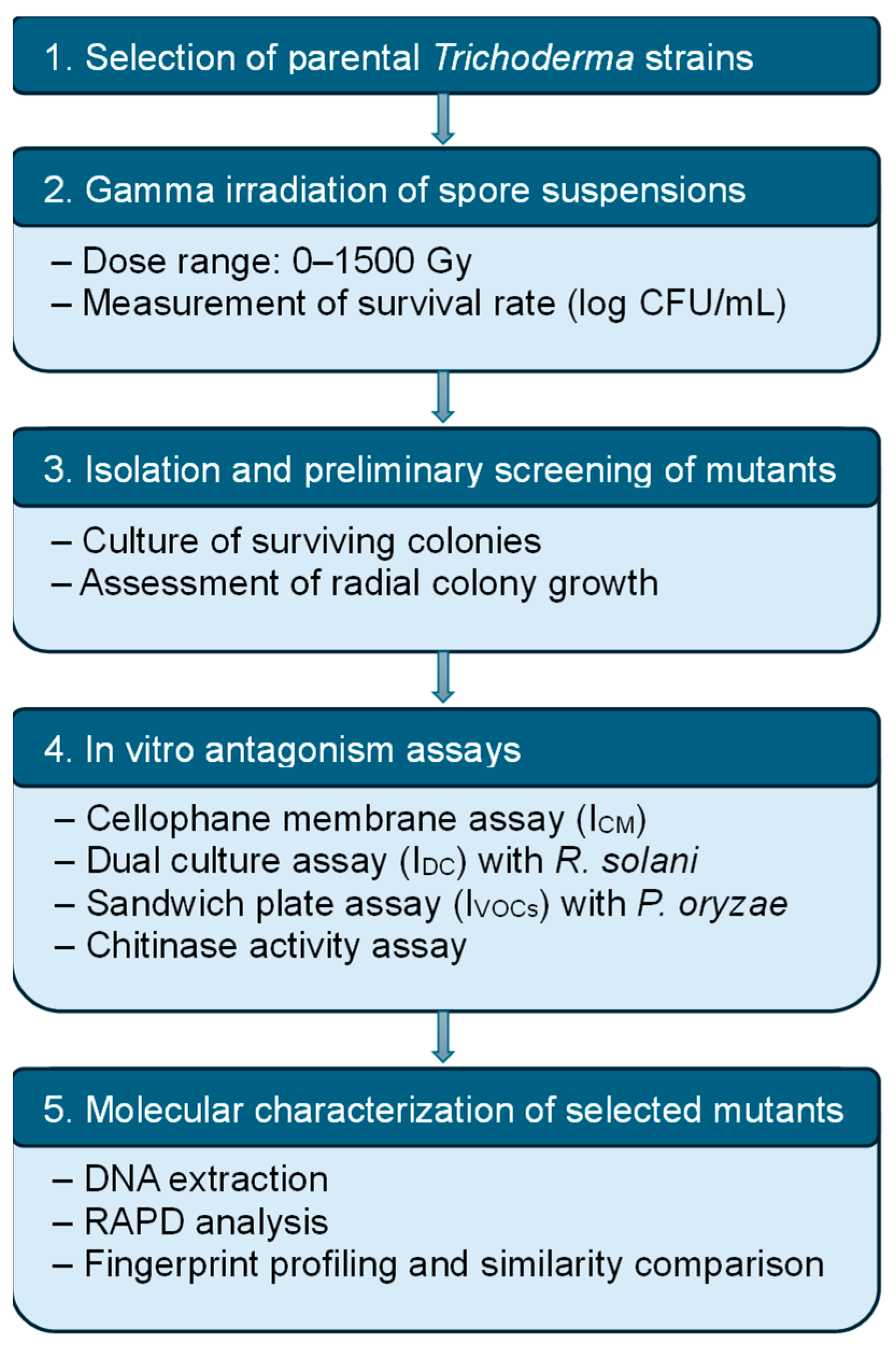
Figure 1. Experimental workflow for the development and characterization of gamma-irradiated Trichoderma mutants.
Applbiosci 04 00041 g001
Figure 2. Classification of the strains TVN-H0 and TVN-A0 by internal transcribed spacer sequences. (A) Analysis of PCR products amplified ITS genes from genomes of strain TVN-H0 and TVN-A0. (B) Analysis of phylogenetic relationships of TVN-H0 and TVN-A0 and Trichoderma species, Hyponyces species using ITS nucleotide sequence alignment by maximum likelihood method with 1000 bootstrap replications. Tricho. is abbreviation name of Trichoderma in the phylogenic tree. The species name in the phylogenetic tree included accession codes of their gene sequences in the GenBank, prefixed with “NR” followed by the species names. The black triangles mark the strains in this study.
Applbiosci 04 00041 g002
Figure 3. Correlation between the number of surviving spores of Trichoderma strains and radiation dose. Data represent means ± standard errors from three independent replicates.
Applbiosci 04 00041 g003
Figure 4. Comparison of the antagonistic activity of TVN-A0 and its mutant AM2 against pathogenic fungi on PDA medium using two culture methods. Dual culture: (A) R. solani alone, (B) R. solani + TVN-A0, (C) R. solani + AM2; sandwich system: (D) P. oryzae alone, (E) P. oryzae + TVN-A0, and (F) P. oryzae + AM2.
Applbiosci 04 00041 g004
Figure 5. Comparison of the antagonistic activity of TVN-H0 and its mutant HM2 against pathogenic fungi on PDA medium using two culture methods. Dual culture: (A) R. solani alone, (B) R. solani + TVN-H0, (C) R. solani + HM2; sandwich system: (D) P. oryzae alone, (E) P. oryzae + TVN-H0, and (F) P. oryzae + HM2.
Applbiosci 04 00041 g005
Figure 6. Chitinase activity of wild-type and potential mutant Trichoderma strains over the cultivation period. Values represent means of three independent replicates, with error bars indicating standard errors. Different letters above the bars denote statistically significant differences among strains at the same time point, as determined by one-way ANOVA followed by Duncan’s multiple range test (p < 0.05).
Applbiosci 04 00041 g006
Figure 7. Investigation of genetic differences between wild-type strains and their irradiated mutants by RAPD and dendrogram analysis. (AH) analysis of genomic DNA extracted from two wild-type strains and 10 mutants; PCR products amplified by OPA-10, Eric1R, Eric2, BoxA1R, M13, OPA-16, Crip1 primers from the DNA genomes, respectively, by electrophoresis in the agarose gel 1.5%. (I,J) Heatmap and dendrogram analysis of Crip1 fingerprints of 2 parental strains and their mutants derived from TVN-H0, TVN-A0, respectively. 1–5: mutants numbered 1 to 5 corresponding to HM1-HM5 or AM1-AM5; W1: Trichoderma sp. TVN-H0; W2: Trichoderma sp. TVN-A0; arrows indicate the different bands observed by naked eyes. Red triangles indicate the differentiated DNA bands between TVN-H0 derivative mutants and TVN-A0 derivative mutants, or discriminated DNA band between HM4 mutant and its parent.
Applbiosci 04 00041 g007
Table 1. Inhibitory efficacy against R. solani and P. oryzae through antagonistic mechanisms of the wild-type strains and five selected high-performing mutants from TVN-A0 and TVN-H0.
Fungal Strain/Potential MutantICM (%)IDC (%)IVOCs(%)
R. solaniP. oryzaeR. solaniP. oryzae
Trichoderma sp. TVN-A061.43 a ± 0.8454.73 a ± 1.6357.71 a ± 1.1955.62 a ± 1.59
AM190.63 d ± 1.2175.38 c ± 1.4780.75 e ± 1.8881.10 c ± 0.66
AM296.71 e ± 0.9792.57 d ± 1.0687.76 g ± 1.2983.57 d ± 1.15
AM3 81.91 b ± 1.2871.43 b ± 1.3667.58 b ± 1.7273.39 b ± 0.83
AM4 85.26 c ± 1.4070.85 b ± 1.8974.38 d ± 0.6475.49 b ± 1.31
AM5 87.26 c ± 0.3369.25 b ± 0.5271.67 c ± 1.3073.36 b ± 1.62
Trichoderma sp. TVN-H063.25 a ± 0.5456.43 a ± 1.2360.26 a ± 0.6858.82 a ± 0.48
HM1 100.00 e ± 0.0074.75 b ± 1.3968.67 b ± 1.2178.42 d ± 1.25
HM2 95.33 d ± 0.5185.28 d ± 1.2491.24 e ± 1.2279.33 d ± 0.44
HM3 93.26 c ± 1.0483.51 d ± 0.9875.41 c ± 1.4669.29 b ± 0.70
HM4 95.06 d ± 1.3777.65 c ± 0.9184.54 d ± 1.7971.57 c ± 0.54
HM5 86.73 b ± 0.9573.24 b ± 0.5883.39 d ± 0.6867.79 b ± 0.63
Means with different superscript letters in the same column are significantly different (Duncan’s test, one-way ANOVA, and p < 0.05). The bold values exhibit the strongest antifungal activities against both R. solani and P. oryzae among mutants derived from each parental strain.
Disclaimer/Publisher’s Note: The statements, opinions and data contained in all publications are solely those of the individual author(s) and contributor(s) and not of MDPI and/or the editor(s). MDPI and/or the editor(s) disclaim responsibility for any injury to people or property resulting from any ideas, methods, instructions or products referred to in the content.