Review

Plant Latex Proteases in Hemostasis: Beyond Thrombin-like Activity

by
Linesh-Kumar Selvaraja
and
Siti-Balqis Zulfigar
*
Bioprocess Technology Division, School of Industrial Technology, Universiti Sains Malaysia, Gelugor 11800, Penang, Malaysia
*
Author to whom correspondence should be addressed.
Appl. Biosci. 2025, 4(3), 37; https://doi.org/10.3390/applbiosci4030037
Submission received: 6 June 2025 / Revised: 10 July 2025 / Accepted: 21 July 2025 / Published: 1 August 2025
(This article belongs to the Special Issue Plant Natural Compounds: From Discovery to Application (2nd Edition))

Abstract

Plant latex is a rich source of proteolytic enzymes with potential biomedical applications, particularly in hemostasis. Among them, thrombin-like enzymes (TLEs) have garnered interest in their ability to mimic thrombin by catalyzing the conversion of fibrinogen to fibrin, facilitating clot formation. While TLEs from snake venoms have been well-characterized and applied clinically, their plant-derived counterparts remain underexplored. This review critically examines the structural and functional characteristics of TLEs from plant latex, comparing them to animal-derived TLEs and evaluating their role in both procoagulant and fibrinolytic processes. Emphasis is placed on dual fibrinogenolytic and fibrinolytic activities exhibited by latex proteases, which often vary with concentration, incubation time, and protease type. In vitro coagulation assays and electrophoretic analyses are discussed as critical tools for characterizing their multifunctionality. By addressing the knowledge gaps and proposing future directions, this paper positions plant latex proteases as promising candidates for development in localized hemostatic and thrombolytic therapies.

Graphical Abstract

1. Introduction

Plant latex has long served as a traditional remedy for wound healing across various cultures, owing to its procoagulant properties [1]. Modern biochemical research has identified proteolytic enzymes within these latexes, some of which exhibit thrombin-like activity, whereby they mimic thrombin by catalyzing the conversion of fibrinogen to fibrin [2,3,4]. While thrombin-like enzymes (TLEs) have predominantly been characterized from snake venoms and have found applications in surgical adhesives and diagnostics tools, plant-derived TLEs offer a promising, biocompatible, and ethically sustainable alternative [5,6]. Their potential in biomedical settings, particularly in the development of topical hemostatic agents and antithrombotic therapeutics, positions these enzymes as underexplored yet highly relevant to the field of biocatalysts.
TLEs are proteases with the capacity to induce the formation of a fibrin clot from fibrinogen and their activity resembles the catalytic actions of thrombin [7]. These proteases mimic the action of thrombin by cleaving at specific sites of the Aα and Bβ fibrinogen subunits, liberating fibrinopeptide A (FpA) and fibrinopeptide B (FpB), respectively [8]. Fibrin clots formed from TLEs are commonly abnormal in their structure, which leads to easy defibrinogenation in the subsequent fibrinolysis process [9]. The hemostatic properties of TLEs—particularly their ability to promote clot formation—make them promising candidates for use in wound healing and bleeding control, with ongoing studies investigating their therapeutic potential. In addition to their clinical applications, TLEs, especially those derived from snake venoms, are also employed in diagnostic assays, such as those measuring fibrinogen levels, fibrin degradation products, and fibrinogen functional abnormalities [10].
Despite this potential, the classification, characterization, and mechanistic understanding of plant TLEs remain fragmented. Reports suggest these enzymes may not only induce clot formation but also contribute to fibrinolysis and platelet modulation—indicating a multifunctionality that may be context-dependent [11,12,13,14]. This dual activity, if properly understood and harnessed, could inform the development of tunable enzyme-based formulations for both procoagulant and fibrinolytic therapies.
This review aims to critically examine the structural and functional properties of plant latex-derived TLEs, evaluate their mechanistic divergence from canonical snake venom TLEs, and discuss the analytical methods used to assess their activity. By consolidating current knowledge and highlighting research gaps, we explore their translational potential in therapeutic enzyme design and localized hemostasis applications.

2. Overview of the Blood Coagulation Cascade

A foundational understanding of the blood coagulation cascade is essential to contextualize the hemostatic roles of plant latex proteases. The following section outlines the key molecular events and components involved in coagulation, serving as a basis for the subsequent discussion on thrombin-like and related enzymatic activities. The model from McFarlane [15] delineates the blood coagulation cascade into two primary pathways: the intrinsic and extrinsic pathways (Figure 1). The intrinsic pathway will be initiated by Factor XII upon contact with exposed collagen at the site of vascular injury. Activated Factor XII will catalyze the conversion of Factor XI into its active form (Factor XIa), which subsequently activates Factor IX. Factor IX, together with Factor VIIIa and calcium ions, will form a complex that effectively converts Factor X into its active state (Factor Xa), marking the convergence of the intrinsic and extrinsic pathways, which is known as the common pathway. On the other hand, the extrinsic pathway will be initiated by the tissue factor released from damaged tissues. Tissue factor, in association with Factor VII, activates Factor X. This series of enzyme activations ultimately results in the activation of thrombin (Factor II), a serine protease that will convert fibrinogen (Factor I) in circulation into fibrin molecules, which subsequently polymerize with each other to form a fibrin mesh at the site of injury [16]. The fibrin polymers will be hydrolyzed by another specialized serine protease known as plasmin, once healing is attained.
A more recent cell-based model links the enzymatic factors with specific cells that accumulate at the site of the wound [17,18]. This model recognizes tissue factor (TF) as the primary trigger for coagulation, which complements the McFarlane Cascade Model by providing a more physiologically relevant and clinically applicable representation of the coagulation process. In this cell-based model, thrombin does not only facilitate fibrin formation from fibrinogen but also acts as an amplification factor, activating Factors V, VIII, and XIII. Factor XIIIa plays a crucial role in stabilizing the fibrin clot.

2.1. Thrombin and Fibrinogen

Thrombin is a serine protease that plays pivotal role in blood hemostasis both in coagulation and anticoagulation pathways. The crystal structure of thrombin is highly homologous to other serine proteases such as chymotrypsin, with the catalytic triad of S195, H57, and D189 residues at its active site. Although thrombin is highly specific in terms of its cleavage specificity, this enzyme is amazingly multifunctional, attributed to its overall structural features consisting of anion exosites (exosite I and exosite II), which mediates interaction with negatively charged patches on cofactors and various substrates [19].
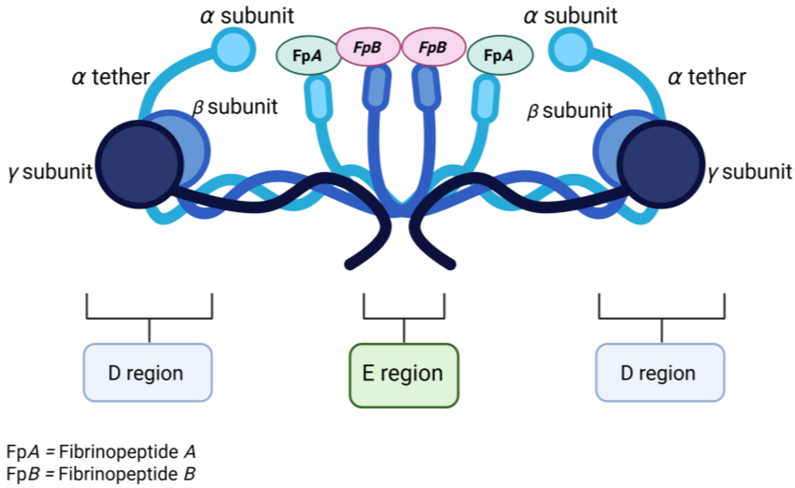
Meanwhile fibrinogen, a 340 kDa dimeric glycoprotein that exists in blood circulation at a concentration of 1.5–4.0 g/L [19], can be converted into a fibrin clot when cleaved at specific sites by thrombin. The fibrinogen molecule is composed of three pairs of subunits denoted as the Aα, Bβ, and γ chains, with molecular masses of 66.2, 54.2 and 48.3 kDa, respectively [20]. Fibrinogen has two identical D regions located at opposite terminals with one E region at the center of the molecule [21] (Figure 2).
The N-terminals of all three subunits (Aα, β, and γ) are held together at the central E region by five disulfide bridges. Meanwhile, the C-terminals of all subunits are extended to the opposite sides where each subunit ends as an individual globular domain [20]. The globular domains (C-terminals) of Bβ and γ are positioned close to each other at the terminals, whereas the C-terminal of Aα chain (known as the αC region) folds back to the center of the molecule. Using structural elucidations, the αC region was observed to consist of two components: the αC tether (Aα240-410) and the αC nodule (Aα411-629) [22]. Both αC regions were reported to be highly susceptible towards proteolytic attacks [21]. The αC region plays various important roles with regards to polymerizations, fibrinolysis, and crosslinking processes [23]. Meanwhile, the N-terminals of the Aα chain and Bβ chain denoted as fibrinopeptide A (FpA) and fibrinopeptide B (FpB) will be cleaved off by thrombin during fibrinogen–fibrin transformation.

2.2. Role of Thrombin in Fibrinogen–Fibrin Transformation

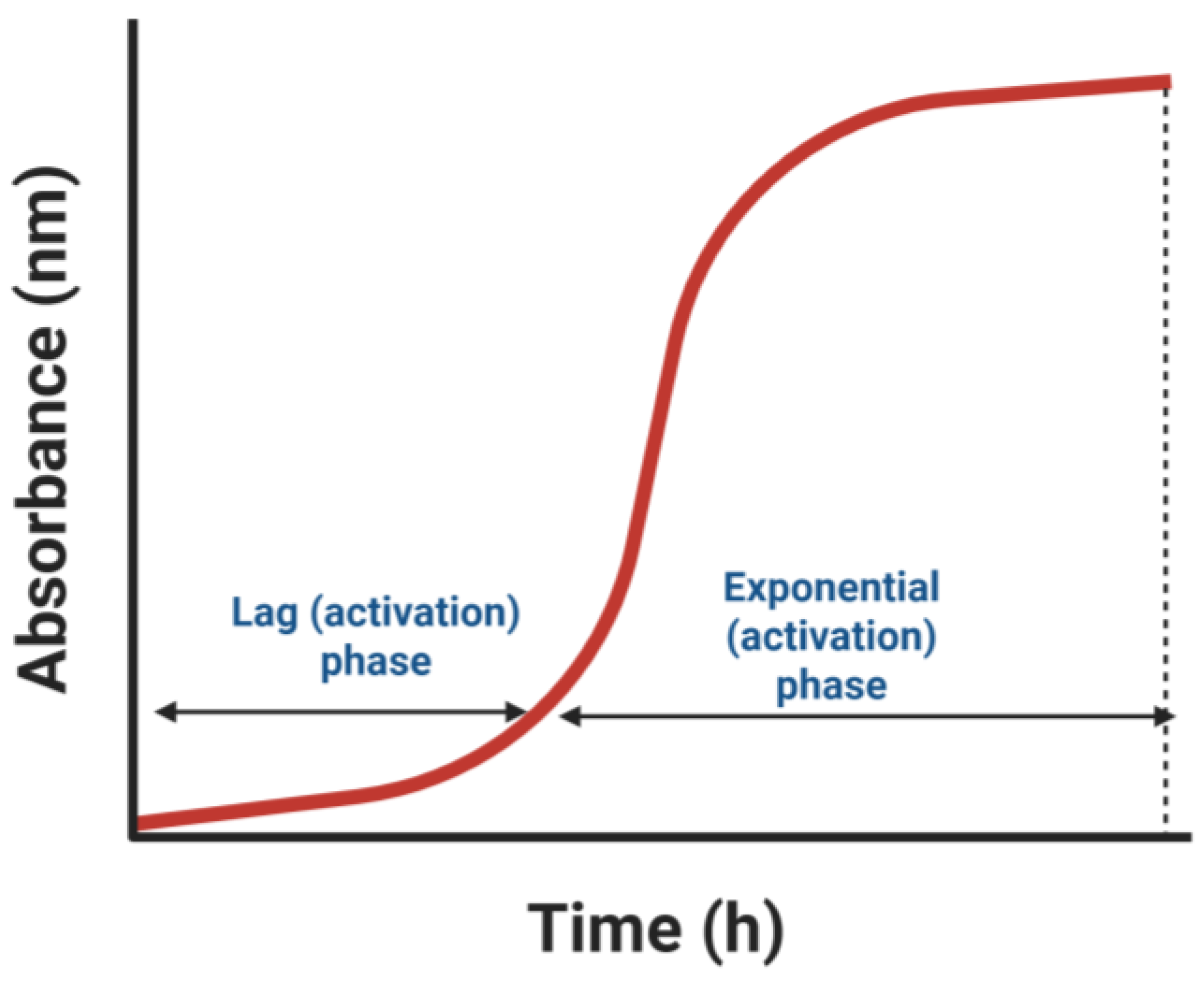
Based on the classical model proposed by Blomback [24], the transformation of fibrinogen into fibrin can be broadly divided into two main stages: (1) the activation of fibrinogen and (2) the polymerization of the activated fibrin monomers. This process is commonly monitored using spectrophotometric methods, which generate a characteristic gelation curve featuring two distinct phases: a lag phase, corresponding to fibrinogen activation, and an exponential phase, representing fibrin polymerization [25,26].
During the initial activation stage, thrombin cleaves two small peptides—fibrinopeptide A (FpA) and fibrinopeptide B (FpB)—from the N-terminal regions of the Aα and Bβ chains of fibrinogen, respectively. This cleavage initiates conformational changes that facilitate fibrin monomer formation and subsequent polymerization. The lag phase observed in the gelation curve reflects the time required for these initial cleavages and the accumulation of activated fibrin monomers. Thrombin preferentially cleaves the Aα chain at the Arg35–Gly36 bond, resulting in the rapid release of FpA, whereas cleavage of the Bβ chain at Arg44–Gly45 and the subsequent release of FpB occurs more slowly [26] (Figure 3).
The subsequent polymerization phase begins when newly exposed N-terminal sequences, referred to as knobs, interact with complementary binding sites, or holes, on adjacent fibrinogen molecules. Specifically, the cleavage of fibrinopeptide A (FpA) exposes a new N-terminal sequence (knob A, Gly-Pro-Arg) that is complementary to hole a, located within the γ-chain C-terminal domain of another fibrinogen molecule. This interaction facilitates the longitudinal, half-staggered alignment and elongation of fibrin polymers [20,21]. Similarly, the cleavage of fibrinopeptide B (FpB) exposes knob B (Gly-His-Arg-Pro), which is complementary to hole b located in the β-nodule of another fibrinogen molecule. These interactions promote lateral or side-to-side associations between fibrin molecules, contributing to fiber thickening and the development of a robust three-dimensional network. This structural transition corresponds to the exponential phase of the gelation curve, characterized by a rapid increase in absorbance due to fibrin aggregation [27].
It is important to note, however, that proteases other than thrombin may exhibit different substrate specificities, thereby influencing the cleavage pattern and subsequent polymerization behavior. For instance, gabonase, a thrombin-like enzyme from the venom of Bitis gabonica, has been shown to preferentially cleave the Bβ chain over the Aα chain, potentially altering the kinetics and structural properties of the resulting fibrin network [28].

3. Exogenous Hemostatic Factors

Given the complexity of coagulation disorders and trauma-induced bleeding, there has been a growing demand for exogenous agents capable of restoring hemostasis effectively. This section highlights various external hemostatic factors—both synthetic and biologically derived—that have laid the foundation for the exploration of plant-based alternatives. Exogenous hemostatic factors refer to external substances or agents that enhance the body’s natural clotting mechanisms to promote hemostasis. These compounds, often of natural origin, can interact with various components of the hemostatic system and serve diverse biological functions in their native organisms, including roles in defense, prey immobilization, or feeding. Such factors have been isolated from a wide range of biological sources, including insects [29,30,31], fungi [32,33], bacteria [34,35], plants [2,36,37], and animal venoms, particularly those of snakes [38,39].
Among these, snake venoms represent a major source of exogenous hemostatic factors and are known to contain a diverse array of active proteins and peptides with potent effects on the coagulation cascade [40]. Generally, exogenous hemostatic agents can be classified into two categories: procoagulants and anticoagulants, as summarized in Figure 4. Thrombin-like enzymes fall under the procoagulant category, alongside other agents such as platelet activators and clotting factor activators.

4. Thrombin-like Enzymes

Although thrombin is a multifunctional enzyme involved in various aspects of blood hemostasis, TLEs are a group of proteases that specifically mimic thrombin’s catalytic activity by converting fibrinogen into fibrin [8,41]. Among these, snake venom serine proteases (SVSPs) that possess fibrinogenolytic activity and induce coagulopathy or hypofibrinogenemia are classified as snake venom thrombin-like enzymes (SVTLEs). These SVTLEs have been reported to be expressed in members of the Viperidae family, particularly within the Viperinae and Crotalinae subfamilies [42].
While TLEs functionally resemble thrombin, they differ in several key aspects. TLEs typically cleave only specific sites on the Aα or Bβ chains of fibrinogen and lack the ability to activate coagulation factors V, VIII, and XIII, which are essential for fibrin stabilization and clot amplification. Cleavage at Arg35–Gly36 on the Aα chain releases fibrinopeptide A (FpA), whereas cleavage at Arg44–Gly45 on the Bβ chain releases fibrinopeptide B (FpB). Based on their cleavage specificity, TLEs are classified into three categories: TLE-AB, TLE-A, and TLE-B. TLE-AB enzymes release both FpA and FpB, while TLE-A and TLE-B selectively release either FpA or FpB from their respective subunits [43].
Fibrin clots formed by TLE-A or TLE-B are structurally abnormal—characterized by reduced density and a translucent appearance—due to insufficient fibrin crosslinking. These fragile clots are more susceptible to degradation during fibrinolysis, resulting in rapid defibrinogenation [44]. TLEs constitute approximately 10% to 24% of the total protein content in crude snake venom, making them the second most abundant class of enzymes in many venom profiles [7].

Thrombin-like Enzymes Structural Properties

Despite their ability to catalyze fibrin clot formation, TLEs exhibit a unique sequence profile, sharing only a low degree of sequence identity with human thrombin but showing greater similarity to trypsin (36–43%), while still conserving the classical catalytic triad—His57, Asp102, and Ser195—characteristic of serine proteases such as trypsin and thrombin [45]. In addition to the active site residues, regions responsible for substrate binding, particularly the subsites labeled S1 to S4, are also partially conserved. The S1 and S2 subsites in TLEs retain key residues Asp189 and Gly216, which are critical for substrate recognition and cleavage, and they are generally synthesized as inactive proenzymes (zymogens), with the first 18 amino acids constituting the signal peptide required for secretion [7].
A distinctive biochemical feature of TLEs is their exceptional stability, particularly under thermal stress. This stability is thought to be conferred in part by glycosylation, as carbohydrates may account for up to 30% of the total protein mass in snake venom serine proteases [44]. One proposed mechanism suggests that glycosylation stabilizes protein structures through interactions between sugar moieties and specific amino acid residues, thereby enhancing structural integrity. Several studies have reported a reduction or complete loss of enzymatic activity following deglycosylation, emphasizing the importance of glycosylation in preserving catalytic function. For example, deglycosylation of elegaxobin II significantly reduced its ability to cleave fibrinogen and resulted in a 30% decrease in kinin-releasing activity compared to the native enzyme [7,39].

5. Plant Latex Proteases

Building upon the structural and functional insights into thrombin-like enzymes, the discussion now shifts toward plant latex—a rich, underexplored source of proteolytic enzymes with significant hemostatic and thrombin-like properties. Latex is exuded from laticiferous plants upon wounding, where it rapidly forms clots at the injury site as part of the plant’s defense mechanism. This clotting process bears a striking resemblance to mammalian blood coagulation, in which fibrin clots are formed to seal wounds. Latex derived from various families, including Euphorbiaceae, Moraceae, Apocynaceae, Asclepiadaceae, and Papaveraceae, is highly valued in traditional medicine for its exogenous hemostatic properties and is widely applied as remedy for wound related cases. Plant latex proteases predominantly belong to the cysteine and serine protease families, with only a limited number classified under the aspartic and metalloprotease groups [45]. Latex is a biologically active fluid enriched with hydrolytic enzymes, known to regulate over 20 physiological processes in plants [7]. The concentration of proteases in latex has been reported to be several folds higher than in other plant tissues, underscoring its critical role in plant defense mechanisms against pathogens [7].
Cysteine proteases are characterized by a cysteine residue at their catalytic site, which plays a pivotal role in the enzyme’s nucleophilic attack on peptide bonds [46]. Their catalytic mechanism involves the formation of a covalent enzyme–substrate complex, facilitated by the thiol (-SH) group of the cysteine residue. These enzymes typically have molecular weights ranging from 20 to 50 kDa, with those isolated from latex commonly falling within the 20–30 kDa range. However, cysteine proteases are highly sensitive to oxidation due to the redox activity of the catalytic cysteine, leading to rapid inactivation upon exposure to air [47].
In contrast, serine proteases are more resistant to oxidative inactivation and are characterized by the presence of a serine residue at the active site, which acts as a nucleophile during the cleavage of peptide bonds [48]. Their molecular weights span a broad range of 19–110 kDa, with most plant-derived serine proteases typically falling between 60 and 80 kDa [49]. These enzymes are generally more stable across a wide pH range and at elevated temperatures. Moreover, plant serine proteases are frequently glycosylated, a post-translational modification that contributes to their higher molecular weights and enhanced stability under extreme environmental conditions [49].

5.1. Latex Proteases: Hemostatic Activities and Analysis

In recent years, growing scientific interest has been directed toward the hemostatic potential of plant latexes, owing to their traditional medicinal use and bioactive enzymatic components [2,3,37,50]. Studies investigating the procoagulant and fibrin(ogen)olytic properties of latex-derived proteases have employed various analytical approaches to uncover their mechanisms of action. These include in vitro hemostatic assays to evaluate clotting activity [37,51], electrophoretic techniques to assess substrate specificity and fibrinogen degradation patterns [2,52], and mass spectrometry-based analyses to characterize protease composition and post-translational modifications [11,49,52]. These methodologies provide a comprehensive understanding of the functional and structural attributes of plant latex proteases with potential therapeutic value.

5.1.1. In Vitro Hemostatic Assays

The clotting capacity of a substance can be evaluated through coagulation assays. These laboratory tests measure the time it takes for a clot to form in the presence of a specific trigger, providing valuable insights into the mechanisms of how an exogenous factor affects blood hemostasis. Commonly used coagulation assays include the Prothrombin Time (PT), Activated Partial Thromboplastin Time (APTT), and Thrombin Clotting Time (TCT), among others. The PT test evaluates the extrinsic pathway of coagulation, while the APTT test assesses the intrinsic pathway [53,54]. Meanwhile the TCT test specifically measures the ability of fibrinogen to be converted to fibrin in the presence of thrombin [53].
In context with the evaluation hemostatic activity of plants, coagulation assays have been utilized to assess the procoagulant and anticoagulant properties of a substance providing a valuable tool for the investigation of the mechanisms and the development of therapeutic interventions [53]. Thrombin-like enzymes are categorized under direct procoagulants, which prompts the transformation of fibrinogen to fibrin without any intermediate reaction. Direct procoagulant activities results to an immediate reduction in clotting time, which can be observed through thrombin time (TT), APTT, and PT assays. TT in particular, is a more straightforward assay for determining the thrombin-like activity as it focuses on the final common pathway step of the conversion from fibrinogen to fibrin. In contrast, indirect procoagulant activities involve more gradual changes or dependency on other factors, which might be revealed through alterations in these assays under specific conditions or through modulation of thrombin generation profiles [55].
Research findings from Osoniyi and Onajobi [56] on the latex of Jathropa curcas Linn. revealed that the results from PT and APTT assays support the presence of both coagulant and anticoagulant activities in the Jatropha curcas latex. The findings demonstrated that the J. curcas latex exhibits both coagulant and anticoagulant properties, with procoagulant activity dominating at higher concentrations and anticoagulant effects emerging at lower concentrations. This suggests the presence of multiple active components within the latex, each exerting distinct effects under different conditions. The study suggests that the coagulant and anticoagulant activities may be mediated by different factors within the latex, each functioning optimally at varying concentrations, thus leading to the observed dual activities. Meanwhile, Antiquorin (Aqn), a thrombin-like protease from Euphorbia antiquorum, was shown to facilitate clot formation by acting on the intrinsic and common pathways of the coagulation cascade, while also inducing platelet aggregation through the PAR1-Akt/p38 signaling axis [2]. In another study, Procerain A and B—cysteine proteases from the latex of Calotropis procera—exhibited both thrombin-like and plasmin-like activities, influencing clot formation and fibrinolysis. Latex protein fractions LP PII and LP PIII further demonstrated a dose-dependent reduction in clotting time, as observed in APTT assays, indicating their potential as procoagulant agents [57].
The recalcification time assay, on the other hand, is a general coagulation test used to measure the time required for clot formation in platelet-poor plasma following the addition of calcium ions (Ca2+), often in the presence of an external factor. A reduction in clotting time in response to plant latex indicates procoagulant activity, reflecting the latex’s ability to influence either the intrinsic or extrinsic pathways of the coagulation cascade. This assay provides valuable insight into the overall hemostatic potential of latex-derived compounds. Previous studies have demonstrated that many plant latexes can significantly shorten the recalcification time. For instance, the latex of Asclepias curassavica L. was shown to markedly reduce plasma clotting time [4], while Jatropha curcas latex exhibited coagulant properties by inducing clot formation, particularly in its concentrated form [14]. A reduction in clotting time in the presence of latex suggests the presence of bioactive components—most likely proteases—that can modulate coagulation processes. Although the assay does not specifically distinguish between the intrinsic and extrinsic pathways, it offers insight into the overall capacity of the latex to influence the coagulation cascade.

5.1.2. Electrophoretic Analysis and Fibrinogenolytic Activity

Electrophoretic analysis is a powerful analytical technique widely employed in biochemistry and molecular biology to separate molecules, primarily proteins, based on properties such as molecular weight, charge, and shape. In the study of fibrin(ogen)olytic enzymes, these techniques offer a visual representation of protein degradation, facilitating the identification and characterization of proteases by evaluating their cleavage preferences and the order of fibrinogen subunit degradation. The three most common electrophoretic techniques used in the study of proteolytic enzymes, particularly in fibrinogenolytic, fibrinolytic, and thrombin-like activities, include Sodium Dodecyl Sulfate Polyacrylamide Gel Electrophoresis (SDS-PAGE), Zymography, and Native PAGE [58,59]. Gel electrophoresis qualitative data are often integrated with densitometric or band-intensity analyses that convert band images into quantitative information. Band-intensity analysis measures protein degradation or enzyme activity by quantifying the disappearance or appearance of electrophoretic bands using specialized image processing softwares such as ImageJ and GelAnalyzer [60,61]. The resulting densitometric data allows for precise, reproducible, and comparative assessments of enzyme activities under different experimental conditions.
SDS-PAGE is the most frequently applied electrophoretic technique to characterize fibrinogenolytic activities of plant latex proteases. Due to its robust and sensitive nature, SDS-PAGE displays degradation patterns and allows for precise molecular weight estimations of proteolytic fragments. The fibrinogenolytic activity of a protease plays a crucial role in clotting process by facilitating the breakdown of fibrinogen, exposing the newly formed N-terminal that initiates fibrin polymerization. These analyses have demonstrated that most plant latex proteases are capable of degrading all three fibrinogen subunits, typically in the order of Aα ≥ Bβ > γ [2,4,36]. This degradation sequence influences the structural characteristics of the resulting fibrin clot, which often differs from those formed by thrombin. The fibrinogen molecule comprises three distinct polypeptide chains—Aα (~66 kDa), Bβ (~58 kDa), and γ (~47 kDa)—that can be resolved by SDS-PAGE based on their molecular weights [20].
The extent of fibrinogen degradation by plant latex proteases is commonly observed to be both dose- and time-dependent. A consistent pattern was observed where the Aα chain of fibrinogen (~66 kDa) was the most susceptible to proteolytic degradation, typically being cleaved rapidly even at low enzyme concentrations or short incubation periods [3,62,63]. Meanwhile the Bβ chain (~52–58 kDa) followed in susceptibility but generally required higher concentrations of enzyme or longer exposure for visible degradation. On the other hand, the γ chain (~47 kDa) was the most resistant, with minimal degradation observed under moderate enzymatic conditions; it only showed noticeable breakdown under prolonged incubation or with potent enzymes. There were cases of several thrombin-like enzymes, such as those from Euphorbia drupifera (Drupin) and Pseuderanthemum latifolium, which demonstrated selective degradation of the Aα and Bβ chains while sparing the γ chain, reflecting thrombin’s specificity [62]. In contrast, enzymes from Plumeria alba and papain from Calotropic procera showed more extensive degradation across all three chains, indicating plasmin-like, broad-spectrum fibrinogenolytic activity [3,57].
It is evident that among the three fibrinogen subunits, the Aα chain—lacking glycosylation—is more structurally flexible and thus is highly susceptible to proteolytic cleavage, even at low enzyme concentrations and short incubation periods [21]. In contrast, the Bβ chain exhibits moderate resistance, while the γ chain is typically the most resistant, requiring higher enzyme concentrations or prolonged incubation to achieve significant degradation [25]. These degradation patterns from plant proteases stand in contrast to the action of other thrombin-like enzymes, which selectively cleave only the Aα and Bβ chains and do not alter the γ chain structure, reflecting thrombin’s limited substrate specificity. This distinction emphasizes the broader substrate specificity of plant-derived proteases compared to the highly selective activity of TLEs. These observations not only reveal the differential susceptibility of fibrinogen subunits but also highlight the protease-specific cleavage preferences, which are crucial for determining their suitability in therapeutic or biomedical applications, such as antithrombotic agents.
The degradation sequence of fibrinogen subunits, which occurs in the order of Aα ≥ Bβ > γ—commonly observed in plant latex proteases—reflects the differential susceptibility of these chains to proteolytic cleavage. This order is not merely an electrophoretic observation but bears mechanistic relevance to clot architecture. Clinically, this degradation pattern may influence the clot’s mechanical properties and potential therapeutic applications. These distinct clot structures produced from thrombin-like enzymes can be taken as an advantage when developing new materials with specific properties, such as specific porosity, fiber thickness, and degree of branching, to fit their intended use [64].

6. Thrombin-like Activity of Plant Latex

According to Mackessy et al. [28], thrombin-like enzymes are limited to proteases that induce fibrinogen clotting and mimic thrombin’s specificity by releasing FpA and FpB during their catalytic activity. The fibrinogen clotting assay is often employed to assess the capacity of a protease to catalyze the conversion of fibrinogen to fibrin, mirroring the final step of coagulation where a reduction in clotting time relative to controls is indicative of procoagulant or thrombin-like activity [65]. While most TLEs studied to date originate from snake venoms, a plant-derived cysteine protease, pergularain e I, from Pergularia extensa latex, has demonstrated thrombin-like activity by inducing fibrinogen clotting, and more importantly, releasing peptide fragments of a similar mass to FpA and FpB [11]. Unlike thrombin however, pergularain e I generated additional peptide species upon prolonged incubation, suggesting broader substrate specificity or extended proteolytic action. Several other studies have also demonstrated that plant latex proteases—such as proteases from Carica papaya, Calotropis gigantea, and Asclepias currasavica L.—can induce fibrin clot formation or plasma coagulation, suggesting a functional resemblance to thrombin-like enzymes [4,66,67].
However, the classification of such plant-derived proteases with thrombin-like activity remains contentious. In the literature on plant proteases, the term “thrombin-like activity” is often attributed to any protease capable of inducing fibrinogen gelation or clot formation, even when its mechanism of action differs substantially from that of thrombin. This broad functional interpretation, although convenient, risks conflating distinct enzymatic behaviors. Thrombin is a highly specific serine protease that initiates clotting through selective cleavage of the Aα and Bβ chains to release FpA and FpB, leading to well-regulated fibrin polymerization [20]. In contrast, most plant-derived enzymes tend to exhibit broader substrate specificity and may promote gelation through non-canonical mechanisms such as partial degradation or transpeptidation, as observed with papain [67]. These enzymes often cleave multiple regions within fibrinogen and may also remove charged peptide segments that modulate aggregation. Therefore, while such plant enzymes may be functionally described as having thrombin-like activity with clot-inducing potential, a more precise and mechanistically grounded classification would improve scientific clarity and support more accurate functional annotation in protease research.
In terms of fibrin clot formation, Yang et al. implied that apart from the knob–hole A:a and B:b interactions, which arise through the release of FpA and FpB, the aggregation of protofibrils could also occur through associations at the αC regions, the C-terminals of the α chains, and the β nodules [27,68,69]. It is also possible that the cleavage of peptides from the fibrinogen molecules removes repulsive charges on the protein, thereby facilitating self-association and assembly [70,71]. These notions, however, require further investigation and structural characterizations to gain better insight into the protein assembly process. Elucidation of fibrino(geno)lytic activities at the proteomics level could shed new light on the molecular mechanisms underlying fibrinogen-to-fibrin conversion by plant latex proteases. The identification of generated peptides is of particular significance, as it may provide a broader understanding of the clotting mechanism induced by these enzymes. Therefore, careful interpretation and characterization of the peptide sequences generated from such analyses are essential to substantiate the presence of FpA, FpB, and other bioactive fragments.
To address the ambiguity, we propose that the term “thrombin-like” be reserved for plant proteases that meet at least three key criteria: (1) the confirmed release of FpA and/or FpB via peptide mapping or mass spectrometry, (2) preferential cleavage patterns at thrombin-recognized sites in the Aα and Bβ chains, and (3) functional mimicry of thrombin in clotting assays, ideally benchmarked against standard thrombin activity. Incorporating structural data such as the presence of conserved catalytic domains (e.g., serine or cysteine protease triads) would further strengthen this classification. Such a framework will allow for more accurate differentiation between true thrombin mimetics and broader fibrinogenolytic enzymes, thereby advancing the biochemical understanding and translational potential of these plant-derived proteases.

7. Beyond Thrombin-like Activity: Multifunctionality in Plant Latex

While thrombin-like activity remains one of the central focus in characterizing the hemostatic potentials of plant latex proteases, accumulating evidence suggests that these enzymes often exhibit multifunctional properties that extend beyond fibrin clot formation. Studies have indicated that plant latexes demonstrated the ability to both initiate coagulation and promote fibrinolysis, highlighting a dual role that is dependent on enzyme concentration, reaction conditions, or the presence of distinct protease classes within the latex. This multifunctionality, increasingly observed across species, may stem from the coexistence of thrombin-like, fibrinolytic, and even plasmin-like activities within the same latex matrix.
Several studies have highlighted the dual functionality exhibited by plant latex proteases, wherein the same enzymatic preparation can promote both clot formation and fibrin degradation. Proteases derived from plant latex often show thrombin-like activity by rapidly degrading the Aα and Bβ chains of fibrinogen, leading to fibrin clot formation. However, upon prolonged incubation or at higher concentrations, these enzymes have also been observed to hydrolyze existing fibrin networks, indicating underlying fibrinolytic or plasmin-like activity [3,12,13]. This functional duality is thought to arise from the coexistence of multiple enzyme classes within the latex—such as serine and cysteine proteases—each contributing differently to coagulation and fibrinolysis. Additionally, this bidirectional modulation of coagulation may be influenced by internal regulatory components within the latex, including endogenous inhibitors or activators [14]. In many cases, the procoagulant or anticoagulant effect appears to be concentration-dependent, with higher enzyme levels accelerating clot formation and lower levels exerting anticoagulant effects by interfering with normal coagulation pathways [67]. Such context-dependent behavior supports their potential for dual application in promoting coagulation or facilitating fibrinolysis, depending on the physiological or experimental conditions.
As a whole, the observed multifunctionality of plant latexes may derive several hypotheses, which include the following: (1) the coexistence of distinct enzymes with opposing functions (e.g., thrombin-like vs. plasmin-like), (2) the concentration-dependent switch in enzyme behavior, and (3) the influence of endogenous protease inhibitors or activators within the latex matrix. These findings emphasize the complex enzymatic environment of plant latex and underscore its biomedical potential. Moving beyond classical thrombin-like actions, further characterization of fibrinolytic activity using substrate-specific assays, zymography, and mass spectrometry will be essential to map out the full spectrum of latex protease functionality.

8. Future Perspective and Research Opportunities

Future investigations into plant latex-derived proteases should emphasize comprehensive proteomic profiling to elucidate the diversity and specificity of enzyme classes involved in thrombin-like and fibrinolytic activities. Advanced analytical platforms, particularly high-resolution mass spectrometry coupled with peptide mapping, are essential to confirm the release of fibrinopeptides A and B (FpA and FpB) and to distinguish genuine thrombin-mimetic functions from broader proteolytic actions. Additionally, it is essential to incorporate global hemostasis assays that simulate physiological environments more accurately. Conventional in vitro assays, while informative, often overlook the contribution of cellular and mechanical components of the coagulation system. Viscoelastic methods such as thromboelastography (TEG) or rotational thromboelastometry (ROTEM) offer dynamic assessments of clot formation, stability, and lysis by integrating the roles of platelets, fibrin networks, and red blood cells. Incorporating such methods could provide a more holistic understanding of the therapeutic potential of latex-derived enzymes in both procoagulant and fibrinolytic contexts [16,21]. Detailed dose- and time-dependent studies are also necessary to delineate the concentration thresholds at which proteases transition from procoagulant to fibrinolytic behavior. These studies should be supported by robust in vitro assays—such as chromogenic substrate assays, which can provide quantifiable metrics of coagulation and fibrinolysis under controlled conditions.

9. Conclusions

Plant latex proteases have emerged as promising candidates in the field of exogenous hemostatic agents, primarily due to their thrombin-like and fibrinolytic activities. This review has explored their unique structural properties, substrate specificity, and multifunctionality, emphasizing their potential to act not only in clot formation but also in clot dissolution—a property that mirrors the dynamic needs of wound healing. While much progress has been made in characterizing their biochemical behavior through electrophoresis and mass spectrometry-based analyses, many questions remain regarding their mechanism of actions. Critically, the distinction between thrombin mimicry and broader fibrinogenolytic activity must be clarified to better position these enzymes within the hemostatic toolbox. Future investigations integrating proteomics, molecular modelling, and translational studies will be the key to unlocking the therapeutic value of these plant-derived biocatalysts. A more holistic understanding of latex proteomes, their synergistic interactions and regulatory dynamics may transform them from folkloric remedies into scientifically validated biomedical innovations.

Author Contributions

Conceptualization, S.-B.Z. and L.-K.S.; writing—original draft preparation, L.-K.S.; writing—review and editing, S.-B.Z.; visualization, S.-B.Z.; supervision, S.-B.Z.; project administration, S.-B.Z.; funding acquisition, S.-B.Z. All authors have read and agreed to the published version of the manuscript.

Funding

This research was funded by the Universiti Sains Malaysia Short Term Grant (304/PTEKIND/6315530).

Institutional Review Board Statement

Not applicable.

Informed Consent Statement

Not applicable.

Data Availability Statement

Not applicable.

Acknowledgments

During the preparation of this manuscript, the authors used ChatGPT (OpenAI, GPT-4o, 2024) for the purposes of refining scientific language, improving structural clarity, and enhancing academic writing flow. Additionally, BioRender (biorender.com) was used to generate scientific illustrations for conceptual visualization. The authors have reviewed and edited all output generated using these tools and take full responsibility for the content of this publication.

Conflicts of Interest

The authors declare no conflicts of interests.

References

  1. Bordoloi, C.; Kumar, S.; Barbhuiya, A.M.; Kushari, S.; Kalita, J.M.; Sahu, B.P.; Laloo, D. Herbal Medicine Used for Wound Healing by the Tribes of the North Eastern States of India: A Comprehensive Review. J. Herb. Med. 2023, 41, 100697. [Google Scholar] [CrossRef]
  2. Urs, A.P.; Manjuprasanna, V.N.; Rudresha, G.V.; Hiremath, V.; Sharanappa, P.; Rajaiah, R.; Vishwanath, B.S. Molecular Cell Research Thrombin-like Serine Protease, Antiquorin from Euphorbia Antiquorum Latex Induces Platelet Aggregation via PAR1-Akt/P38 Signaling Axis. Biochim. Biophys. Acta Mol. Cell Res. 2021, 1868, 118925. [Google Scholar] [CrossRef]
  3. Kusuma, C.G.; Gubbiveeranna, V.; Sumachirayu, C.K.; Bhavana, S.; Ravikumar, H.; Nagaraju, S. Thrombin- and Plasmin-like and Platelet-Aggregation-Inducing Activities of Plumeria alba L. Latex: Action of Cysteine Protease. J. Ethnopharmacol. 2021, 273, 114000. [Google Scholar] [CrossRef]
  4. Shivaprasad, H.V.; Rajesh, R.; Nanda, B.L.; Dharmappa, K.K.; Vishwanath, B.S. Thrombin like Activity of Asclepias curassavica L. Latex: Action of Cysteine Proteases. J. Ethnopharmacol. 2009, 123, 106–109. [Google Scholar] [CrossRef]
  5. Seon, G.M.; Lee, M.H.; Kwon, B.-J.; Kim, M.S.; Koo, M.-A.; Kim, D.; Seomun, Y.; Kim, J.-T.; Park, J.-C. Functional Improvement of Hemostatic Dressing by Addition of Recombinant Batroxobin. Acta Biomater. 2017, 48, 175–185. [Google Scholar] [CrossRef]
  6. Green, D. Hematology Products from Snake Venoms. Thromb. Res. 2025, 245, 109215. [Google Scholar] [CrossRef]
  7. Ullah, A.; Masood, R.; Ali, I.; Ullah, K.; Ali, H.; Akbar, H.; Betzel, C. Thrombin-like Enzymes from Snake Venom: Structural Characterization and Mechanism of Action. Int. J. Biol. Macromol. 2018, 114, 788–811. [Google Scholar] [CrossRef]
  8. Swenson, S.D.; Stack, S.; Markl, F.S. Thrombin-like serine proteinases in reptile venoms. In Handbook of Venoms and Toxins of Reptiles; CRC Press: Boca Raton, FL, USA, 2021; pp. 351–362. [Google Scholar]
  9. Vu, T.T.; Stafford, A.R.; Leslie, B.A.; Kim, P.Y.; Fredenburgh, J.C.; Weitz, J.I. Batroxobin Binds Fibrin with Higher Affinity and Promotes Clot Expansion to a Greater Extent than Thrombin*. J. Biol. Chem. 2013, 288, 16862–16871. [Google Scholar] [CrossRef] [PubMed]
  10. Moore, G.W. Snake Venoms in Diagnostic Hemostasis and Thrombosis. Semin. Thromb. Hemost. 2021, 48, 145–160. [Google Scholar] [CrossRef] [PubMed]
  11. Shivaprasad, H.V.; Rajaiah, R.; Frey, B.M.; Frey, F.J.; Vishwanath, B.S. ‘Pergularain e I’—A Plant Cysteine Protease with Thrombin-like Activity from Pergularia Extensa Latex. Thromb. Res. 2010, 125, e100–e105. [Google Scholar] [CrossRef] [PubMed]
  12. Shivaprasad, H.V.; Riyaz, M.; Venkatesh Kumar, R.; Dharmappa, K.K.; Tarannum, S.; Siddesha, J.M.; Rajesh, R.; Vishwanath, B.S. Cysteine Proteases from the Asclepiadaceae Plants Latex Exhibited Thrombin and Plasmin like Activities. J. Thromb. Thrombolysis 2009, 28, 304–308. [Google Scholar] [CrossRef] [PubMed]
  13. Hamed, M.B.; El-Badry, M.O.; Kandil, E.I.; Borai, I.H.; Fahmy, A.S. A Contradictory Action of Procoagulant Ficin by a Fibrinolytic Serine Protease from Egyptian Ficus Carica Latex. Biotechnol. Rep. 2020, 27, e00492. [Google Scholar] [CrossRef] [PubMed]
  14. Osoniyi, O.; Onajobi, F. Coagulant and Anticoagulant Activities in Jatropha Curcas Latex. J. Ethnopharmacol. 2003, 89, 101–105. [Google Scholar] [CrossRef] [PubMed]
  15. Macfarlane, R.G. An Enzyme Cascade in the Blood Clotting Mechanism, and Its Function as a Biochemical Amplifier. Nature 1964, 202, 498–499. [Google Scholar] [CrossRef]
  16. Negrier, C.; Shima, M.; Hoffman, M.; Pradel, L. Blood Reviews The Central Role of Thrombin in Bleeding Disorders. Blood Rev. 2019, 38, 100582. [Google Scholar] [CrossRef]
  17. Hoffman, M.; Monroe, D.M., III. A Cell-Based Model of Hemostasis. Thromb. Haemost. 2001, 85, 958–965. [Google Scholar] [CrossRef]
  18. Monroe, D.M.; Hoffman, M. What Does It Take to Make the Perfect Clot? Arterioscler. Thromb. Vasc. Biol. 2006, 26, 41–48. [Google Scholar] [CrossRef]
  19. Crawley, J.T.B.; Zanardelli, S.; Chion, C.K.N.K.; Lane, D.A. The Central Role of Thrombin in Hemostasis. J. Thromb. Haemost. 2007, 5, 95–101. [Google Scholar] [CrossRef]
  20. Litvinov, R.I.; Pieters, M.; Lange-Loots, Z.D.; Weisel, J.D. Fibrinogen and fibrin. In Macromolecular Protein Complexes III: Structure and Function; Springer: Berlin/Heidelberg, Germany, 2021; pp. 471–501. [Google Scholar]
  21. Weisel, J.W.; Litvinov, R.I. Fibrin Formation, Structure and Properties. In Fibrous Proteins: Structures and Mechanisms; Parry, D.A.D., Squire, J.M., Eds.; Springer: Berlin/Heidelberg, Germany, 2017; pp. 405–456. ISBN 9783319496740. [Google Scholar]
  22. Protopopova, A.D.; Barinov, N.A.; Zavyalova, E.G.; Kopylov, A.M.; Sergienko, V.I. Visualization of Fibrinogen a C Regions and Their Arrangement during Fibrin Network Formation by High-Resolution AFM. J. Thromb. Haemost. 2015, 13, 570–579. [Google Scholar] [CrossRef]
  23. Ping, L.; Huang, L.; Cardinalli, B.; Profumo, A.; Gorkun, O.V.; Lord, S.T. Substitution of the Human AC Region with the Analogous Chicken Domain Generates a Fibrinogen with Severely Impaired Lateral Aggregation: Fibrin Monomers Assemble into Protofibrils but Protofibrils Do Not Assemble into Fibers. Biochemistry 2011, 50, 9066–9075. [Google Scholar] [CrossRef] [PubMed]
  24. Blomback, B. Fibrinogen and Fibrin Transformation. In Fibrinolytics and Antifibrinolytics; Springer: Berlin/Heidelberg, Germany, 1978; pp. 49–79. [Google Scholar]
  25. Kattula, S.; Byrnes, J.R.; Wolberg, A.S. Fibrinogen and Fibrin in Hemostasis and Thrombosis. Arter. Thromb. Vasc. Biol. 2017, 37, e13–e21. [Google Scholar] [CrossRef]
  26. Blomb, B.; Bark, N. Fibrinopeptides and Fibrin Gel Structure. Biophys. Chem. 2004, 112, 147–151. [Google Scholar] [CrossRef]
  27. Yang, Z.; Mochalkin, I.; Doolittle, R.F. A Model of Fibrin Formation Based on Crystal Structures of Fibrinogen and Fibrin Fragments Complexed with Synthetic Peptides. Proc. Natl. Acad. Sci. USA 2000, 97, 14146–14161. [Google Scholar] [CrossRef]
  28. Kornalik, F. Toxins affecting blood coagulation and fibrinolysis. In Handbook of Toxinology; CRC Press: Boca Raton, FL, USA, 2020; pp. 683–760. [Google Scholar]
  29. Yeon, B.; Sik, K.; Yong, K.; Joo, H.; Rae, B. Anti-Fibrinolytic Activity of a Metalloprotease Inhibitor from Bumblebee (Bombus ignitus) Venom. Comp. Biochem. Physiol. Part C 2021, 245, 109042. [Google Scholar] [CrossRef]
  30. Sheng, X.; Gao, L.; Lu, X.; Wang, Y.; Han, Y.; Meng, P.; Chen, W.; Lu, Q. Expression and Purification Expression and Characterization of a Fibrinogenolytic Enzyme from Horse Fly Salivary Gland. Protein Expr. Purif. 2017, 129, 135–142. [Google Scholar] [CrossRef] [PubMed]
  31. Sik, K.; Yeon, B.; Joo, H.; Soo, Y.; Rae, B. Secapin, a Bee Venom Peptide, Exhibits Anti- Fi Brinolytic, Anti-Elastolytic, and Anti-Microbial Activities. Dev. Comp. Immunol. 2016, 63, 27–35. [Google Scholar] [CrossRef]
  32. Liu, X.; Kopparapu, N.; Li, Y.; Deng, Y.; Zheng, P.X. Biochemical Characterization of a Novel Fibrinolytic Enzyme from Cordyceps Militaris. Int. J. Biol. Macromol. 2017, 94, 793–801. [Google Scholar] [CrossRef]
  33. Guo, R.; Zhang, Y.; Duan, D.; Fu, Q.; Zhang, X.; Yu, X. Fibrinolytic Evaluation of Compounds Isolated from a Marine Fungus Stachybotrys Longispora FG216. Chin. J. Chem. 2016, 34, 1194–1198. [Google Scholar] [CrossRef]
  34. Anusree, M.; Swapna, K.; Aguilar, C.N.; Sabu, A. Optimization of Process Parameters for the Enhanced Production of Fibrinolytic Enzyme by a Newly Isolated Marine Bacterium. Bioresour. Technol. Rep. 2020, 11, 100436. [Google Scholar] [CrossRef]
  35. Barzkar, N.; Jahromi, S.T.; Fabio, V. Marine Microbial Fibrinolytic Enzymes: An Overview of Source, Production, Biochemical Properties and Thrombolytic Activity. Mar. Drugs 2022, 20, 46. [Google Scholar] [CrossRef]
  36. Manjuprasanna, V.N.; Urs, A.P.; Rudresha, G.V.; Milan Gowda, M.D.; Jayachandra, K.; Hiremath, V.; Rajaiah, R.; Vishwanath, B.S. Drupin, a Thrombin-like Protease Prompts Platelet Activation and Aggregation through Protease—Activated Receptors. J. Cell. Biochem. 2021, 122, 870–881. [Google Scholar] [CrossRef]
  37. Uday, P.; Maheshwari, M.; Sharanappa, P.; Nafeesa, Z.; Kameshwar, V.H.; Priya, B.S.; Nanjunda Swamy, S. Exploring Hemostatic and Thrombolytic Potential of Heynein—A Cysteine Protease from Ervatamia Heyneana Latex. J. Ethnopharmacol. 2017, 199, 316–322. [Google Scholar] [CrossRef] [PubMed]
  38. Sanchez, E.F.; Flores-Ortiz, R.J.; Alvarenga, V.G.; Eble, J.A. Direct Fibrinolytic Snake Venom Metalloproteinases Affecting Hemostasis: Structural, Biochemical Features and Therapeutic Potential. Toxins 2017, 9, 392. [Google Scholar] [CrossRef] [PubMed]
  39. Oyama, E.; Takahashi, H. Structures and Functions of Snake Venom Metalloproteinases (SVMP) from Protobothrops Venom Collected in Japan. Molecules 2017, 22, 1305. [Google Scholar] [CrossRef]
  40. Matsui, T.; Fujimura, Y.; Titani, K. Snake Venom Proteases Affecting Hemostasis and Thrombosis. Biochim. Biophys. Acta 2000, 1477, 146–156. [Google Scholar] [CrossRef]
  41. Mackessy, S. Handbook of Venoms and Toxins of Reptiles; CRC Press: Boca Raton, FL, USA, 2016. [Google Scholar]
  42. Castro, H.C.; Zingali, R.B.; Albuquerque, M.G.; Pujol-Luz, M.; Rodrigues, C.R. Snake Venom Thrombin-like Enzymes: From Reptilase to Now. Cell. Mol. Life Sci. 2004, 61, 843–856. [Google Scholar] [CrossRef]
  43. Pradniwat, P.; Rojnuckarin, P. Snake Venom Thrombin-like Enzymes. Toxin Rev. 2014, 33, 16–22. [Google Scholar] [CrossRef]
  44. Kini, R.M.; Cho, Y.K. Metalloproteases Affecting Blood Coagulation, Fibrinolysis and Platelet Aggregation from Snake Venoms: Definition and Nomenclature of Interaction Sites. Toxins 2016, 8, 284. [Google Scholar] [CrossRef]
  45. Castro, H.C.; Silva, D.M.; Craik, C.; Zingali, R.B. Structural Features of a Snake Venom Thrombin-like Enzyme: Thrombin and Trypsin on a Single Catalytic Platform? Biochim. Biophys. Acta-Protein Struct. Mol. Enzymol. 2001, 1547, 183–195. [Google Scholar] [CrossRef]
  46. Sharma, A.; Sharma, D.; Verma, S.K. In Silico Study of Iron, Zinc and Copper Binding Proteins of Pseudomonas syringae pv. lapsa: Emphasis on Secreted Metalloproteins. Front. Microbiol. 2018, 9, 1838. [Google Scholar] [CrossRef]
  47. Verma, S.; Dixit, R.; Pandey, K.C. Cysteine Proteases: Modes of Activation and Future Prospects as Pharmacological Targets. Front. Pharmacol. 2016, 7, 107. [Google Scholar] [CrossRef] [PubMed]
  48. Hedstrom, L. Serine Protease Mechanism and Specificity. Chem. Rev. 2002, 102, 4501–4523. [Google Scholar] [CrossRef]
  49. Siti-Balqis, Z.; Rosma, A.; Kim-Teck, L.; Ismail, M.N. Artocarpus Altilis Latex Polypeptides: An Insight into Its Fibrino(Geno)Lytic Activity. Biocatal. Agric. Biotechnol. 2018, 16, 467–475. [Google Scholar] [CrossRef]
  50. Singh, M.K.; Bindhu, O.S. Plant latex: A rich source of haemostatic proteases. In Herbal Medicine in India; Springer: Berlin/Heidelberg, Germany, 2020; pp. 143–153. [Google Scholar]
  51. Siritapetawee, J.; Khunkaewla, P.; Thumanu, K. Chemico-Biological Interactions Roles of a Protease from Euphorbia Resinifera Latex in Human Anticoagulant and Antithrombotic Activities. Chem. Biol. Interact. 2020, 329, 109223. [Google Scholar] [CrossRef]
  52. Singh, M.K.; Rajagopalan, A.; Tanimu, H.; Omana, B. Purification, Characterization and Fibrino (Geno) Lytic Activity of Cysteine Protease from Tabernaemontana Divaricata Latex. 3 Biotech 2021, 11, 106. [Google Scholar] [CrossRef]
  53. Kijkriengkraikul, N.; Nuchprayoon, I. Study of Optimum Condition for Rapid Preparation of Thrombin Using Russell’s Viper Venom Factor X Activator. Open Biotechnol. J. 2018, 12, 95–103. [Google Scholar] [CrossRef]
  54. Momot, A.P. The Dynamics of the Hemostatic Parameters in Physiological Pregnancy and After Delivery. Hematol. Blood Transfus. Disord. 2016, 3, 10-18411. [Google Scholar] [CrossRef] [PubMed]
  55. Winter, W.E.; Flax, S.D.; Harris, N.S. Coagulation Testing in the Core Laboratory. Lab. Med. 2017, 48, 295–313. [Google Scholar] [CrossRef]
  56. Ventakesha, S.H.; Rajaiah, R.; Vishwanath, B.S. Hemostatic Interference of Plant Latex. SM J. Clin. Pathol. 2016, 1, 1002. [Google Scholar]
  57. Ramos, M.V.; Viana, C.A.; Silva, A.F.B.; Freitas, C.D.T.; Figueiredo, I.S.T.; Oliveira, R.S.B.; Alencar, N.M.N.; Lima-Filho, J.V.M.; Kumar, V.L. Proteins Derived from Latex of C. Procera Maintain Coagulation Homeostasis in Septic Mice and Exhibit Thrombin- and Plasmin-like Activities. Naunyn. Schmiedebergs. Arch. Pharmacol. 2012, 385, 455–463. [Google Scholar] [CrossRef]
  58. Laemmli, U.K. Cleavage of Structural Proteins during the Assembly of the Head of Bacteriophage T4. Nature 1970, 227, 680–685. [Google Scholar] [CrossRef] [PubMed]
  59. Choi, N.-S.; Kim, S.-H. Two Fibrin Zymography Methods for Analysis of Plasminogen Activators on Gels. Anal. Biochem. 2000, 281, 236–238. [Google Scholar] [CrossRef]
  60. Pereira, B.; Brazón, J. Aqueous Extract from Brownea Grandiceps Flowers with Effect on Coagulation and Fibrinolytic System. J. Ethnopharmacol. 2015, 160, 6–13. [Google Scholar] [CrossRef]
  61. Lazar, I., Jr.; Lazar, I., Sr. Gel Analyzer 23.1.1: Freeware 1D Gel Electrophoresis Image Analysis Software. Available online: http://www.gelanalyzer.com (accessed on 20 February 2021).
  62. Van Truong, L.; Paulsen, B.S.; Bac, V.H. A Novel Serine Protease from Pseuderanthemum Latifolium B. Hansen: Characterization and Fibrino(Geno)Lytic Activities. Nat. Prod. Res. 2021, 35, 2640–2646. [Google Scholar] [CrossRef]
  63. Siritapetawee, J.; Teamtisong, K.; Limphirat, W.; Charoenwattanasatien, R.; Attarataya, J.; Mothong, N. Identification and Characterization of a Protease (EuRP-61) from Euphorbia Resinifera Latex. Int. J. Biol. Macromol. 2020, 145, 998–1007. [Google Scholar] [CrossRef]
  64. Brown, A.C.; Barker, T.H. Fibrin-Based Biomaterials: Modulation of Macroscopic Properties through Rational Design at the Molecular Level. Acta Biomater. 2014, 10, 1502–1514. [Google Scholar] [CrossRef]
  65. Denson, K.W.E. Coagulant and Anticoagulant Action of Snake Venoms. Toxicon 1969, 7, 5–11. [Google Scholar] [CrossRef] [PubMed]
  66. Rajesh, R.; Shivaprasad, H.V.; Raghavendra Gowda, C.D.; Nataraju, A.; Dhananjaya, B.L.; Vishwanath, B.S. Comparative Study on Plant Latex Proteases and Their Involvement in Hemostasis: A Special Emphasis on Clot Inducing and Dissolving Properties. Planta Med. 2007, 73, 1061–1067. [Google Scholar] [CrossRef]
  67. Doolittle, R.F. Clotting of Mammalian Fibrinogens by Papain: A Re-Examination. Biochemistry 2014, 53, 6687–6694. [Google Scholar] [CrossRef]
  68. Litvinov, R.I.; Gorkun, O.V.; Galanakis, D.K.; Yakovlev, S.; Medved, L.; Shuman, H.; Weisel, J.W. Polymerization of Fibrin: Direct Observation and Quantification of Individual B:B Knob-Hole Interactions. Blood 2006, 109, 130–138. [Google Scholar] [CrossRef] [PubMed]
  69. Litvinov, R.I.; Yakovlev, S.; Tsurupa, G.; Gorkun, O.V.; Medved, L.; Weisel, J.W. Direct Evidence for Specific Interactions of the Fibrinogen AC-Domains with the Central E Region and with Each Other. Biochemistry 2007, 46, 9133–9142. [Google Scholar] [CrossRef] [PubMed]
  70. Chiti, F.; Calamai, M.; Taddei, N.; Stefani, M.; Ramponi, G.; Dobson, C.M. Studies of the Aggregation of Mutant Proteins in Vitro Provide Insights into the Genetics of Amyloid Diseases. Proc. Natl. Acad. Sci. USA 2002, 99, 16419–16426. [Google Scholar] [CrossRef] [PubMed]
  71. Dobson, C.M. Principles of Protein Folding, Misfolding and Aggregation. Semin. Cell Dev. Biol. 2004, 15, 3–16. [Google Scholar] [CrossRef] [PubMed]
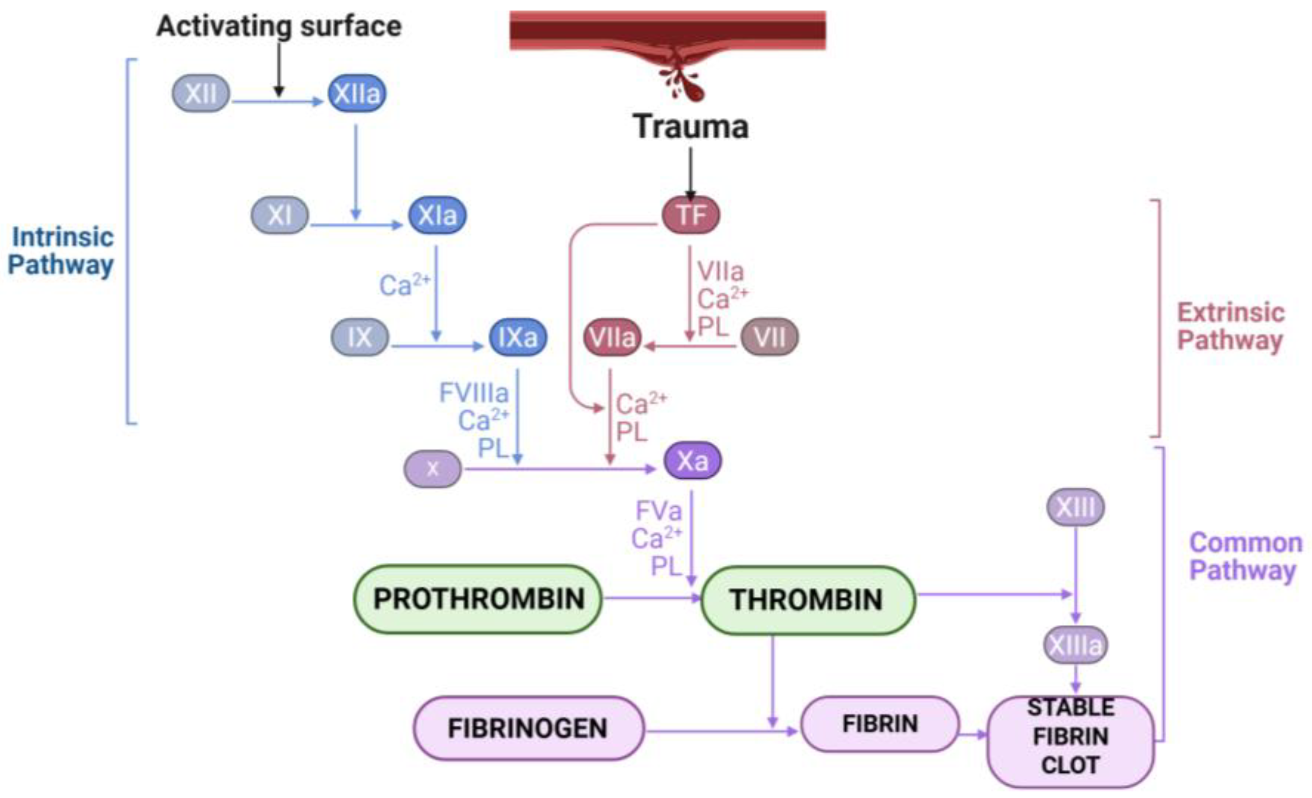
Figure 1. Schematic diagram depicting the roles of thrombin in the classical blood coagulation pathways.
Applbiosci 04 00037 g001
Figure 2. Schematic diagram of human fibrinogen.
Applbiosci 04 00037 g002
Figure 3. Typical fibrinogen gelation profile observed through spectrophotometric method.
Applbiosci 04 00037 g003
Figure 4. Exogenous hemostatic factors and their sub-divisions.
Applbiosci 04 00037 g004
Disclaimer/Publisher’s Note: The statements, opinions and data contained in all publications are solely those of the individual author(s) and contributor(s) and not of MDPI and/or the editor(s). MDPI and/or the editor(s) disclaim responsibility for any injury to people or property resulting from any ideas, methods, instructions or products referred to in the content.