Review

Nanobiotechnology in Bone Tissue Engineering Applications: Recent Advances and Future Perspectives

1
School of Chemical and Process Engineering, University of Leeds, Leeds LS2 9JT, UK
2
Anya Biopharm India LLP, Atal Incubation Centre, Ahmedabad 380009, India
3
Institute of Chemical Technology, Matunga (East), Mumbai 400019, India
4
RAS Life Science Solutions, Stresemannallee 61, 60596 Frankfurt, Germany
5
School of Biological and Chemical Sciences, University of Galway, H91W2TY Galway, Ireland
6
Indian Institute of Technology, Powai, Mumbai 400076, India
7
Leeds Institute of Rheumatic and Musculoskeletal Medicine, University of Leeds, Leeds LS9 7JT, UK
*
Author to whom correspondence should be addressed.
These authors contributed equally to this work.
Appl. Biosci. 2023, 2(4), 617-638; https://doi.org/10.3390/applbiosci2040039
Submission received: 25 August 2023 / Revised: 17 October 2023 / Accepted: 10 November 2023 / Published: 15 November 2023
(This article belongs to the Special Issue Anatomy and Regenerative Medicine: From Methods to Applications)

Abstract

:
Bone regeneration and repair are complex processes with the potential of added complications, like delayed repair, fracture non-union, and post-surgical infections. These conditions remain a challenge globally, pressurizing the economy and patients suffering from these conditions. Applications of nanotechnology (NBT) in the field of medicine have provided a medium for several approaches to support these global challenges. Tissue engineering is one such field that has been on the rise in the last three decades through the utilization of NBT for addressing the challenges related to bone regeneration. First, NBT enables the formation of scaffolds at the nanoscale needed for bone tissue engineering (BTE) using natural and synthetic polymers, as well as with minerals and metals. Then, it aids the development of the nano-formulation strategized to deliver antimicrobial drugs and/or growth factors through various ways to enhance bone repair through the scaffold. Third, NBT facilitates the use of specialized nanoparticles to image and track cellular events in vitro as well as in vivo. This review is an effort to bring together the current knowledge in the field of BTE and present the scope of ever-evolving NBT, a contribution towards precision medicine.

1. Introduction

Bone regeneration post-diseased states, like osteoarthritis (OA) and osteoporosis (OP), or after trauma related bone defects, remains a clinical challenge worldwide [1,2,3]. Delayed or impaired bone healing in the case of trauma or fracture may lead to non-union, which further increases the number of procedures required to aid bone healing and incur additional financial and societal pressures [4,5,6]. In 2019, 178 million new fractures were recorded globally, significantly increasing fractures in the older population [5]. A more thorough look into bone-related diseases indicate that about 200 million women suffer from OP, leading to nearly 8.9 million fractures yearly [3]. Similarly in 2020, just considering knee OA, over 654.1 million individuals over 40 years old were found to suffer from the disease, making them more vulnerable to fractures [7].
While surgical intervention is the primary approach for the treatment of these conditions, surgeries are invasive, expensive in many countries, and require considerable time for repair post-surgery. Another challenge faced is the chance of post-surgical infection at the surgical site [8]. To combat these challenges, in recent decades, the applications of tissue engineering and regenerative medicine for the purpose of bone repair have increased [9,10,11,12]. At its core, tissue engineering for the bone has four principal requirements—a mechanical substrate or environment, osteoconductive scaffolds, osteogenic cells, and appropriate growth factors to enable bone formation or osteogenesis. These four factors form the ‘diamond concept’ for bone tissue engineering (BTE) [13,14]. Mechanical environment refers to the base where the osteogenic scaffold is placed. The osteogenic cells that are usually bone marrow (BM)-derived mesenchymal stromal cells (MSCs) or progenitor cells are added to the scaffold in the presence of growth factors, like bone morphogenic factor-2 (BMP-2) and vascular endothelial growth factor (VEGF). These promote the process of osteogenesis needed for bone regeneration and repair [14,15,16]. The source of cells—autogenic or allogenic—and the age of donors and/or patients can impact the application stage [17,18]. Along with these four factors, vasculature is often challenging to replicate using methods for BTE and needs special consideration. Additionally host factors, that is, the effect of the environment of the host, is also a necessary factor to be considered for successful BTE applications and, ultimately, for bone regeneration [14,19]. These are outlined below in Figure 1.
The global cost of bone fractures is estimated at US 5.5 billion per year; the total annual cost of bone repair is estimated at US 17 billion worldwide, as of 2020 [20]. Bone by itself is a complicated organ with a dynamic and multifaceted BM and highly intricate microenvironment. Within the BM various cells, like MSCs, hematopoietic stem cells (HSCs), osteogenic cells, adipogenic cells, immune cells, and macrophages, several growth factors and extracellular matrix (ECM) co-exist and communicate with each other providing an optimal environment for their function and survival [21,22,23]. Bone regeneration is a complex and intricate process that requires a specific sequence of events, depending upon the trauma or diseased state [24]. Thus, the intricacies and the level of complexity involved in bone regeneration and repair makes it extremely challenging to mimic the organ in vitro or ex vivo [25].
Technological advancements aimed at better replicating the anatomy and physiology of the bone have paved the way for the use of nanoscale materials for the purpose of BTE [26,27,28,29]. Nanomedicine as a field has enabled the formulation of a given material in various forms, like nanoparticles (NPs), nanofibers (NFs), nanosheets (NSs), nanotubes (NTs), and nanorods (NRs). These nanoscale formulations exhibit unique features that make them ideal candidates for several applications in BTE. The nanoscale structures have the potential to mimic the bone complexity and 3D architecture. They have increasingly been utilized not just for scaffold preparation [30,31], but also for the delivery of drugs, like antibacterials for the prevention of post-operative infections, through the scaffold [32,33,34]. Additionally, nanotechnology has enabled the delivery of growth factors and proteins via NPs within scaffolds. Moreover, they can be used for labeling and targeting cells in the scaffolds for cellular tracing and functional analysis [26,35]. Therefore, these techniques are being extensively used by scientist and medical professionals due to their edge over traditional methods [36].
This article brings together the current knowledge of nanobiotechnology (NBT) in the field of BTE since the last two decades. Here, we discuss the three major applications of NBT in BTE—(a) nanoscale materials for scaffold structure and function, (b) nanomedicine and drug delivery in scaffolds for BTE, and (c) nanotechnology for cell targeting and labeling for BTE. Furthermore, we critically review these applications and evaluate the recent scenario of this ever-expanding field. We finally outline the challenges faced by NBT in replicating the ever-elusive bone, BM, and the process of bone regeneration while predicting the foreseeable future of BTE with the help of nanotechnology.

2. Nanoscale Materials for Scaffold Structure and Function

Nanoscale materials have emerged as pivotal players in BTE due to their inherent advantages, including biocompatibility, biodegradability, mechanical tunability, and their capacity to mimic the natural bone ECM [28,37,38,39,40]. Several types of materials are being used either individually or more commonly in combination with materials providing complementary benefits towards the final scaffold. For example, several polymers, including natural polymers such as chitosan, gelatin, and collagen, are used in hydrogel preparation for BTE due to their biochemical and biophysical properties [41]. Other polymers, discussed below, were also found to be beneficial at nanoscale for applications in drug delivery and tissue engineering [42]. They may also be used in combination with harder materials, like bioactive glass, or metals, like titanium, zinc, strontium, and others in their nanoscale forms, offering biocompatibility, enhanced mechanical strength, and biodegradability towards scaffold formulation [43,44]. The various materials used for BTE, as shown in Figure 2, broadly explores polymeric and non-polymeric materials that have been used in BTE applications, specifically for scaffold formation.

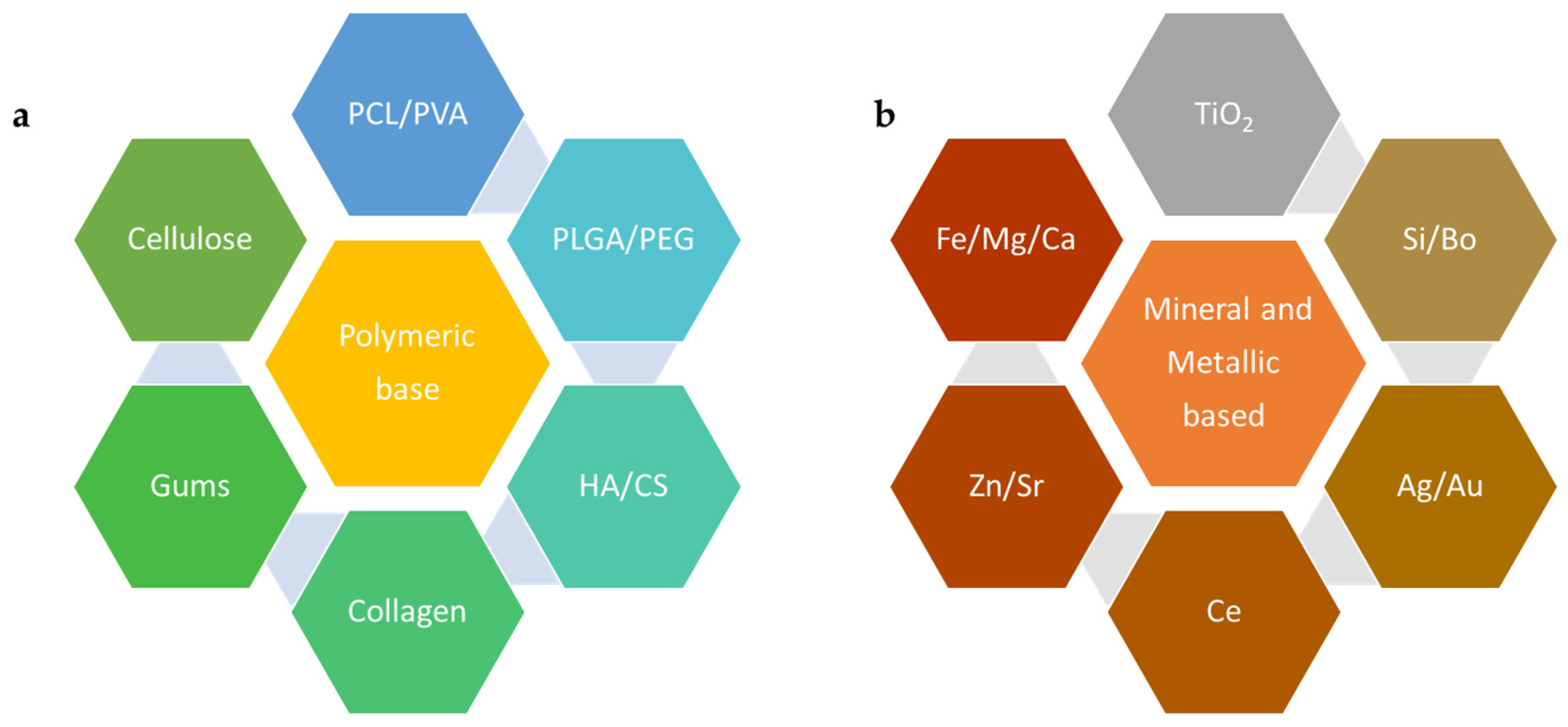
2.1. Polymeric Materials Used at the Nanoscale for BTE

Over the last two to three decades, polymeric materials have been explored, optimized, and utilized for various applications, including the formulation of scaffolds for BTE [45,46]. With a wide variety of materials and characteristics, these can be improvised based on the requirements of the target tissues. Figure 2a lists some of these well-known and commonly used polymers that have contributed to our knowledge of biomaterials that may be used for BTE. Additionally, at the nanoscale, the materials have been found to be promising due to their biodegradability and biocompatibility on account of their enhanced porosity [47]. This section explores some of these polymeric materials with examples outlining their role in BTE at the nanoscale.

2.1.1. Synthetic Polymers

Polycaprolactone (ε-PCL) is a biodegradable, biocompatible polyester that has attracted substantial attention in BTE due to its adaptable biodegradation and ease of manipulation into NFs, NPs, and scaffolds [48]. A PCL fiber mesh fabricated via electrospinning demonstrated enhanced osteoblast cell proliferation and differentiation, whereby the structure mimicked the natural ECM, facilitating cell adhesion and growth essential for successful BTE [49]. Poly(lactic-co-glycolic acid) (PLGA), a copolymer of lactic and glycolic acids, is another example of a biodegradable and biocompatible polymer commonly used in BTE. Nanostructured PLGAs can be formed into NPs for drug delivery, NFs, or scaffolds for cell growth and have each contributed unique benefits to BTE [50,51].
PLGA NFs combined with nano hydroxyapatite (HA), a major inorganic component of bone, exhibited improved cell attachment, proliferation, and differentiation [52]. Polyethylene glycol (PEG) is another polymer that is often utilized in nanoscale form due to its excellent biocompatibility and resistance to protein adsorption. PEGylated gelatin nanospheres have been used to deliver BMP-2, which enhances bone regeneration in vivo, signifying the importance of PEG in nanoscale drug delivery systems for BTE [53]. Other nanoscale polymers, such as poly(lactic acid) (PLA), poly(glycolic acid) (PGA), poly(vinyl alcohol) (PVA), and polyurethane (PU), are also being explored for their potential in BTE and have been discussed in great detail elsewhere [37,54,55]. When crafted into nanoscale forms, such as fibers or particles, these polymers show promising potential in promoting cell attachment, proliferation, differentiation, and delivering therapeutic agents, thereby boosting bone regeneration [43,44,56,57,58].

2.1.2. Natural Polymers

Chitosan (CS) is another example of a biopolymer exhibiting a multitude of favorable properties, including non-toxicity, biodegradability, biocompatibility [59,60], antifungal, antibacterial [61,62], and wound healing abilities. Therefore, CS is widely utilized in the biomedical, biotechnology, and pharmaceutical fields [63,64]. CS is a prime candidate for crafting potential bone scaffolds, particularly when paired with osteoconductive substances, such as HA [59,65] and poly(acrylic acid) (PAA) [66]. Studies have shown that CS-based bone scaffolds foster cell adhesion and osteoblast cell proliferation, creating mineralized bone matrices in vitro [67]. An enhancement in osteoblast cell proliferation was observed for CS composites that incorporated nanoHA, ultimately triggering bone regeneration within eight weeks as validated by micro-computed tomography [68]. The mechanical characteristics of CS scaffolds can be enhanced by crosslinking with substances possessing a minimum of two reactive functional groups, such as calcium phosphates, composites (nanozirconia and nanocalcium zirconate), and bioglass, leading to a superior performance compared to constructs consisting of only CS [69].
Cellulose is another widespread linear biopolymer that contributes significant tensile strength to trees and is present in various organisms, including marine species, bacteria, fungi, and even amoebas [70]. It is usually characterized by long fibrils that comprise various crystalline and amorphous regions. By subjecting the cellulose pulp to mechanical or chemical alterations, it is feasible to extract tiny crystalline cellulose components. These diminutive cellulose polymers, referred to as cellulose nanocrystals (CNCs), typically exhibit diameters of 2–20 nm and a length distribution of 100–600 nm [71]. The functionalization of CNCs with sulphate or phosphate ester groups can be achieved through sulfuric or phosphoric acid hydrolyses [72]. These modified scaffolds reveal superior crosslinking and, subsequently, enhanced mechanical properties compared to those that are unmodified.
Furthermore, compared with phosphate ester-modified aerogels, aerogels crafted from sulphate ester-treated CNCs display greater compressive strength, porosity, and crosslinking [73]. These sulphate ester-modified CNCs (S–CNCs) possess in vivo osteoconductive properties, enabling bone growth at the defect location. After three weeks, S–CNC aerogels can create a bone volume percentage that exceeds controls by 33% and can see a size increase of 50% after twelve weeks. Comprehensive research into cell–interface interactions and in vivo scaffold degradation is crucial. Scaffolds constructed using modified CNCs with bone tissue-enhancing properties are emerging as promising solutions for BTE applications [47]. Various methods, like electrospinning or 3D printing, may be applied to fabricate polymeric scaffolds and the recent advances in these applications have been outlined elsewhere [74,75].

2.2. Mineral-Based and Metallic Nanoscale Materials Used for BTE

NPs obtained from minerals, such as zinc, silica, or calcium phosphate particles, are classified as nanoscale materials where their primary role is to provide vital ions for tissue generation. They do so by enhancing the mechanical properties of 3D-printed scaffolds. Mineral-derived nanomaterials can be produced by several techniques, such as plasma spraying, milling, and precipitation from solution [76]. Similarly, metallic nanoscale formulations of gold (Au), silver (Ag), and titanium (Ti) have been explored at length for their enhanced functionalities that are discussed in this section.

2.2.1. Mineral Nanoparticles

Calcium Phosphate

Adding mineral-based nanomaterials to both the surface and bulk of 3D-printed scaffolds has been shown to improve the overall functionality of the scaffolds. Nanomaterials derived from calcium phosphate (CaP) are often added to the 3D-printed scaffolds since bone is inherently composed of CaP crystals (70%) and 30% of organic collagen fibrils [77]. In 2017, Chen et al. generated a biomimetic composite scaffold of collagen and biphasic calcium phosphate nanoparticles (BCP NPs). They engineered these scaffolds to release dexamethasone (DEX) during preparation and hybridized this with collagen scaffolds. The subcutaneous implantation of the composite scaffolds at the dorsal side of athymic nude mice demonstrated the regeneration of the ectopic bone tissue. When used for the 3D culture of human BM-derived MSCs, these scaffolds demonstrated enhanced biocompatibility and promoted the osteogenic differentiation of hMSCs [78]. Another study by Sokolova et al. in 2020 used scaffolds of PLGA and nanohydroxyapatite (nHAP) (85:15) combined with DNA-loaded CaP NPs. The results suggested increased cytocompatibility and rate of gene transfection into cells indicating their favorable application as a scaffold for BTE or a bone substitution material [79].

Silicon Dioxide (SiO2)

Silica NPs are an example of inorganic NPs. Silica particles have been shown to promote osteoblast differentiation while inhibiting osteoclast differentiation [80]. A recent study by Echazú et al. in 2022 [81] combined soluble silica particles and a CS polymer to engineer a potential bone substitute. This was implanted into the medullary compartment of both tibiae in Wistar rats and investigated for cytotoxicity and biocompatibility. The results indicated successful new bone formation at the tissue–biocomposite interface (osseointegration) [81]. Another study conducted in 2023 by Shuai et al. used poly(L-lactic acid) (PLLA)-based bone scaffolds and combined SiO2 NPs with graphene oxide (GO) NS to promote the dispersion of GO on the biopolymer bone scaffold. The optimization of the adequate dispersion of GO with SiO2 enhanced the mechanical properties and cytocompatibility of the scaffold, making it a potential candidate for BTE applications [82].

Zinc Oxide (ZnO)

Zinc is one of the minerals present in bone in the form of trace elements. It promotes bone density and the prevention of bone loss [83]. It also activates proteins involved in bone homeostasis as a part of inorganic minerals. ZnO NPs exhibit low toxicity, act as antibacterial agents, and exhibit optimal biological compatibility and chemical stability. They stimulate osteogenesis and hence have the potential to accelerate bone growth and mineralization [84,85]. In 2020, Cho et al. investigated the cell proliferation and antibacterial activities of a PCL/nanoHA scaffold doped with ZnO. Their results indicated that the scaffolds with 100 nm-thick ZnO coatings showed enhanced antibacterial cell proliferation activities and mechanical properties [86]. In 2017, Forero et al. studied the effect of CS/gelatin/nanoHA scaffolds containing a nano-copper–zinc alloy for BTE. These scaffolds increased the proliferation and adhesion of mouse embryonic fibroblasts and induced osteogenic differentiation. After an in vivo subcutaneous implant, they induced the growth of surrounding tissues and promoted granulation tissue formation [87]. The use of ZnO NPs in the field of BTE is very limited, but promising, with novel advancements being made using hybrid mix of compatible biopolymers.

2.2.2. Metallic NPs

A high penetrating ability and surface area coupled with improved cell adhesion, differentiation, and growth suggests that metal NPs are one of the ideal candidates for scaffold fabrication in BTE. Ag and Au are the most common osteogenic agents used in this category. They exhibit a high surface area, enhanced antibacterial properties, biocompatibility, and surface reactive features in order to identify pathogenic viruses [88] and intracellular targets [89].

Silver (Ag)

Silver (metal or salt) has been used as an antibacterial agent in implants [90,91]. Ag ions can penetrate the bacterial cell wall and cause growth inhibition. Ag-based scaffolds are highly potent with enhanced cell adhesion and cytocompatibility, improved osteogenesis and osteo-conductivity, acceptable mechanical strength, an ability to bridge oxygen molecules, and low toxicity [92,93,94]. Akturk et al. optimized the protocol to generate an antibacterial gelatin nanocomposite membrane for BTE applications. They used soluble starch-coated Ag NPs and bioactive glass particles incorporated into gelatin to fabricate a nanohybrid nanocomposite fiber [95]. Another study investigated the use of CS-silver polymeric scaffolds in BTE. These scaffolds demonstrated enhanced matrix mineralization and osteogenic differentiation (marked by the upregulation of marker genes Runt-related transcription factor 2 (Runx2), type-1 collagen (Col-I), alkaline phosphatase (ALP) activity, and secreted osteocalcin (OCN)) [96].

Gold (Au)

Gold NPs exhibit low toxicity, high antibacterial effects, and biocompatibility in the scaffold environment [97]. While some studies indicate dose-dependent cytotoxicity in human cell lines marked by membrane damage and reactive oxidative species (ROS) generation [98,99], this property of Au NPs has often been useful to destroy cancer cells [100]. Thus, ensuring the appropriate size and dosing of Au NP plays a crucial role in BTE applications. Nekounam et al. demonstrated an optimized protocol for the fabrication of a carbon NF/Au NP-based scaffold that supported cell proliferation and indicated low toxicity in bone cells [101]. Au NPs that are smaller in size (<10 nm) have been used for miRNA delivery to the nucleus. Yu et al. in 2017 created nanohybrids using polyethylenimine, liposomes coated on the surface of Au NPs. After the release from endosomes inside the cells, miR-5106 was able to facilitate BM stem cell osteoblastic differentiation, which further aided in mineralization [102].

Titanium Dioxide (TiO2)

TiO2 NPs are bioinert cell carrier materials with improved permeability and high biocompatibility. They are available in the form of nanocrystals and exhibit antibacterial and antiseptic properties making them a promising scaffolding material for bone tissue repair [103]. A study published in 2018 described the methodology to fabricate a lightweight and economical TiO2/CS scaffold by freeze-drying [104]. An in vitro study conducted by Pattanashetti et al. in 2020 determined the ideal weight percentages of TiO2 NPs that needed to be crosslinked in a PVA matrix. These nanofibrous scaffolds were developed using electrospinning and the results indicated that 0.1 g of TiO2 exhibited enhanced mechanical stability and a subsequent slow rate of degradation in the scaffolds. These scaffolds were found to be viable and non-toxic to the cells, as observed by the MTT assay using MG-63 osteosarcoma cell lines [105].

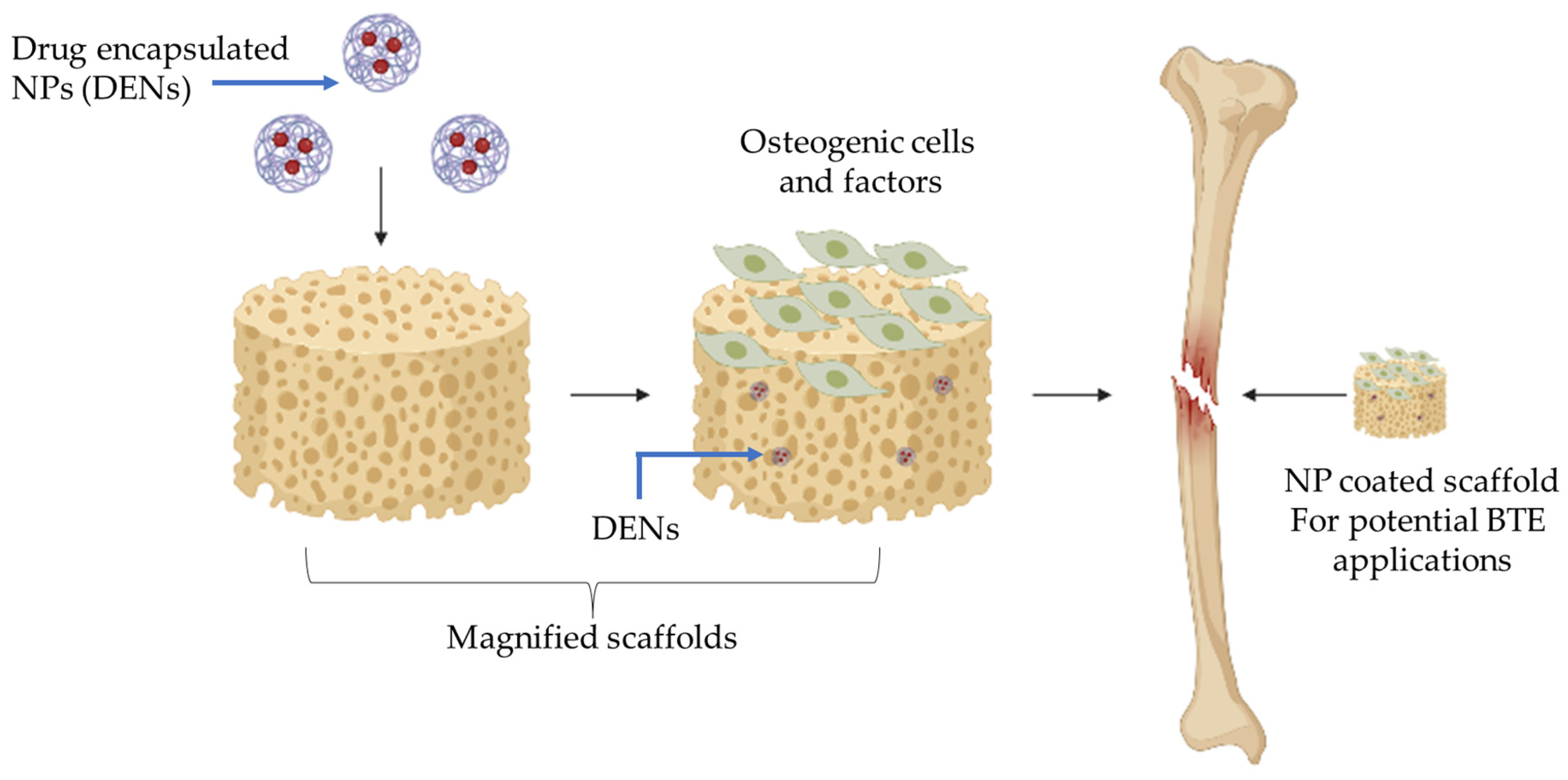
3. Nanomedicine and Drug Delivery in Scaffolds for BTE

NPs have demonstrated their potential as vehicles for delivering bioactive molecules; they are particularly suited for encapsulating and delivering growth factors and genes that stimulate bone regeneration [106,107]. Additionally, bone repair often demands the presence of a microenvironment that can foster bone formation and may additionally require pharmaceutical agents to prevent complications, like post-surgical infections. These complex physical and biological properties are met through the constructs reinforced with various pharmaceutical agents, like antibiotics, drugs, genes, and growth factors. Precise control over the carrier vehicle is needed for the efficient loading, targeting, and delivery of these agents. This is where nanotechnology offers exceptional opportunities ranging from the modulation of biomaterial surfaces to the targeted and controlled release of the payload. Figure 3 shows the schematic representation of drug-loaded NPs embedded in a scaffold aimed for BTE.

3.1. Pharmaceutical Agents in Scaffolds

Scaffolds are usually designed considering the mechanical and functional properties of the site of application. Scaffolds developed for bone regeneration are osteoconductive matrices, as they provide optimum signaling for the attachment of osteogenic cells with subsequent proliferation, crucial for successful integration at the site of interest. Thus, an ideal scaffold should be adaptable and provide suitable mechanical support that may or may not be resorbable (depending upon the end goal). Furthermore, the scaffold should promote cell growth and differentiation by offering appropriate signaling cues and microenvironments, without the risk of rejection by the host tissue [27].
The first impediment to the acceptance of a foreign material by the human body is microbial infection. The introduction of medical devices or scaffolds is associated with a high risk of microbial infection, which may appear months or even years after the graft, resulting in fatal consequences [8,108]. The primary prevention techniques include environmental control, surface coatings, sterilization, and antibiotic therapy. However, effective and long-term antimicrobial materials are necessary for combating the onslaught of bacteria, especially for antibiotic-resistant strains. This has led to the emergence of biomaterials imbued with antimicrobial properties. Antimicrobial properties can be imparted by the incorporation of antibiotics, metal ions and metal-based NPs, antimicrobial peptides, and bacteriophages [109]. Scaffolds impregnated with antibiotics, like ciprofloxin, gentamycin, and vancomycin, directly or in the form of NPs, have been used for localized antibiotic delivery [110,111].
Due to the declining effectiveness of conventional antibiotics, constructs enhanced with metal-based nanomaterials and antimicrobial peptides are being explored for long-term infection management [92]. Silver, gold, copper, zinc, cerium, and their oxides are some of the most-used materials for the synthesis of metal NPs [61,91,94]. These NPs exhibit toxicity by the impairment of the cell membrane, generation of ROS, damaging the biomolecules (lipids, proteins, and DNA), thereby interfering with their function and, ultimately, critical cellular pathways. The triggering of multiple antibacterial mechanisms combined with the nano-size of the particles makes it difficult for the bacteria to develop resistance against metal NPs [112,113,114]. However, an overexpression of proteins, like flagellin, or the production of extracellular polymeric substances may alter the size and zeta potential of NPs leading to their agglomeration and thereby inactivation. Conversely, a reduction in the expression of proteins, like porins, which are involved in the uptake of NPs, may also be adopted by bacteria as a resistance mechanism against metal NPs. Although, the inactivation of NPs through agglomeration, the reduction in expression of surface porins, the upregulation of metal sequestration, and bio-precipitation, as well as the increased production of scavenger and detoxification enzymes, has been reported in some cases [115].
The co-delivery of antibiotics with growth factors or drugs presents a lucrative prospect in BTE. For example, core–shell mesoporous silica NFs with BMP-2 at the core and gentamycin in the shell demonstrated enhanced ontogenetic regeneration abilities with advanced antibacterial properties [110]. The mesoporous silica NP and BMP-2 combination was also used with deferoxamine to trigger angiogenesis [32]. The bioavailability of pharmaceutical agents can be improved by loading them in liposomes—enclosed vesicles with a bilayer structure [116]. These nontoxic, self-assembling, spherical vesicles can hold both hydrophilic and hydrophobic payloads in their bilayer membranes and aqueous centers, respectively. The encapsulation and subsequent release of curcumin, a hydrophobic compound, from liposomes incorporated onto 3D calcium phosphate scaffolds increased its bioavailability. The released curcumin showed selective cytotoxicity towards osteosarcoma cells while promoting the proliferation of osteoblasts [116]. Lee et al. synthesized oxysterol-based non-phospholipid liposomes and loaded them with a smoothened agonist (SAG). This tailor-made delivery system was able to stimulate osteogenic differentiation based on the intrinsic osteo-inductive properties of the oxysterol 20S-hydroxycholesterol as well as the drug cargo, SAG, which is a small molecule activator of Hedgehog signaling, thus resulting in enhanced bone repair [117]. Liposomes have been used as a delivery system for several hydrophilic and hydrophobic drugs, proteins, and growth factors, and as a non-viral delivery system for genes through transfection [118].
Depending on the specific requirements of BTE, several types of drug release strategies viz. controlled, sustained, programmed, responsive, transfection, and surface presentation have been developed [119,120,121]. Physical adsorption and chemical conjugation are primary tools to achieve these modifications. Layering nanomaterials onto scaffolds has led to the generation of multifunctional scaffolds that provide control over the spatiotemporal release of pharmaceutical agents. A layer-by-layer (L-B-L) approach was used to enhance the biocompatibility of oxysterol liposome-loaded porous PLGA by coating the liposome layer with polydopamine [117]. The release behavior of small and large molecules can be influenced through the sequential L-B-L coating of CS and sodium hyaluronate on porous scaffolds [122]. The sequential delivery of BMP-2 and alendronate (ALN) resulted in the osteogenic differentiation of MSCs followed by an inhibition of osteoclastic activity leading to a synergistic effect on bone regeneration [123].
The scaffold itself can be modulated on a nano- or microscale to assist bone repair. The interface between ECM and cells is composed of nanoscale morphological features that guide cellular properties, like adhesion, growth, and differentiation. Mimicking bone’s inherent nano-topography by creating hierarchical nanostructures has been employed to guide cell shape, expression patterns, and signaling, thereby influencing cell fate. The cell microenvironment can be further enhanced by the chemical modification of scaffold surfaces. These include a controlled release of metal ions, NPs, and miRNAs [54]. Antimicrobial and osteoconductive abilities were shown by the nano-functionalization of titanium scaffolds with TiO2 and γFe2O3 [124]. Another example of surface functionalization was shown by the surface phosphorylation of polyethylene terephthalate followed by coating with hydroxyethyl methacrylate loaded with ciprofloxacin [111]. The presence of anionic hydrophilic phosphate groups led to better biomineralization and yielded higher cell densities on the surface functionalized biomaterial as compared to the bare PET matrix. Moreover, coating with hydroxyethyl methacrylate (HEMA) further improved the hydrophilicity and compatibility of the biomaterial without hampering the accessibility of phosphate groups [111]. Harnessing the potential of nanoscale engineering, in the form of NPs or surface modifications, can lead to the development of smart/novel drug delivery systems that promote the optimal conditions for tissue growth and regeneration.

3.2. Protein Functionalization for Scaffold Surfaces

As outlined above, bone regeneration and the regulation of cellular behavior are influenced by the nanoscale morphology on the BTE scaffold [125]. A nanoscale approach helps in mimicking the natural ECM composition of a tissue by forming a 3D scaffold for cells, by offering mechanical strength, monitoring cellular events, and delivering various bioactive agents to the targeted bone tissue [126]. The synthesis of protein-based NPs for functionalizing scaffolds utilizes proteins from animal sources, like collagen, albumin, elastin, silk protein, and fibronectin, as well as from plant sources, like soy protein, zein, and wheat gluten. The choice of protein depends on the application of the scaffold and related properties of proteins.
The commonly used methods for the synthesis of protein-based scaffolds include electrospinning, sol-gel, freeze-drying, and solvent casting [46]. Electrospinning involves the conversion of a polymer into a viscous solution through solvent addition, increasing the voltage source to extend the polymer into a thin stream across an electrostatic field, and then obtaining the formed NFs on the collector [26]. The sol-gel process involves the mixing of colloidal particles in the desired solvent of a low viscosity, allowing it to form the colloidal solution (sol). This solution is kept in an appropriate mold for casting and then gelation (gel) occurs to form 3D networks from colloidal particles. The freeze-drying method, also known as lyophilization, involves the dehydration of a frozen solution under a low-pressure vacuum, forming an anhydrous 3D structure, yielding 90% porosity in the scaffold. In the solvent casting method, solvent in contact with the polymer is allowed to undergo evaporation forming porous scaffolds. In the salt-leaching method, water-soluble salts (e.g., sodium chloride, sodium citrate, and sodium acetate), porogen, or sugar particles are mixed together with a biodegradable polymer solution and allowed to solidify into a mold of a desired shape. In lyophilization, the solvent is removed at low temperature under vacuum, resulting in the leaching off of salt flakes and leaving behind a porous structure [127]. The thermal crosslinking method involves the use of the gelation method, which can be performed using a microwave [46]. In the solvothermal method, nanostructured materials are synthesized via a heterogeneous reaction conducted in a non-aqueous medium under high-pressure and -temperature conditions around the critical point [128]. Biomimetic mineralization is a methodology that mimics the natural process of the mineralization of material surfaces. It uses a supersaturated solution of simulated body fluids (SBFs) containing ions at concentrations similar to those of blood plasma. This coating method is conducted under biological conditions (temperature, pH, and pressure) forming carbonated apatite on a substrate, which imitates bone mineral [76]. Apart from the traditional methods of scaffold synthesis, 3D-printing technology allows the fabrication of scaffolds with precision, using chemical deposition, electron beam lithography, and laser lithography [129]. These techniques are combined with mechano-transduction approaches for an appropriate stimulation of cells into biochemical signals, thereby, regulating cell behavior on these scaffolds [125].
Of the two main types of proteins used for NP synthesis, animal protein-based NPs are known for their high bioavailability, non-toxicity, biodegradability, ease of tunability, extended shelf lives, and in vivo half-lives. On the other hand, plant proteins have gained attention on account of their low immunogenicity as compared to animal proteins [46]. Additionally, their low molecular weight and high polar nature as compared to the animal proteins manifests their natural hydrophilicity and efficient cell attachment. Furthermore, plant tissue features are aptly utilized in fabricating scaffolds, on account of their inherent vascular networks, unified porosity, high surface area, suitable mechanical property, and remarkable water-absorption ability [130]. These features of both types of proteins are suitable to offer a plethora of alternatives for scaffold functionalization, essential for BTE applications (Table 1).

3.2.1. Plant Proteins

The potential of zein/HA NFs has been investigated for their application in BTE by a group of researchers. Zein, a core maize endosperm protein, is biocompatible with human liver cells, mouse fibroblast cells, and human umbilical vein endothelial cells (HUVEC). This biocompatibility makes it a suitable candidate for a therapeutic carrier and design engineered scaffold. Despite the poor mechanical properties and structural instability of zein in an aqueous environment, the extensive usage of zein occurs through assembling NFs using the electrospinning process. The blend of zein fibers with synthetic polymers or crosslinkers imparts to them suitable aqueous and mechanical stability properties [131]. Natural ECM comprises various growth factors, polysaccharides, proteins, and other components, building a network of cells for attachment, growth, and proliferation [132]. HA, a key constituent of bones and teeth, in its nanocrystalline form is favorable for fabricating bio-composite scaffolds for BTE aspects [131].
In another study, zein/HA NF synthesis was achieved by two techniques: magnetic stirring and ultrasonication. The homogenous distribution of HA and increased wettability of NFs were achieved by ultrasonication, while balanced strength and elongation were achieved by the magnetic stirring approach. Furthermore, the tensile strength and mechanical properties of fibers obtained by both methods were found to be desirable. Cell seeding on the zein/HA scaffold electrospun using a magnetic stirrer with a 5.0 weight% HA indicated notably higher proliferation as compared to those seeded on the control zein fibers on the seventh day. This study demonstrated that the zein/HA NF membranes designed with a high biological efficacy possessed a noteworthy BTE application potential [133].
Another plant protein, soy, was used in the form of porous scaffolds, synthesized using sol-gel and crosslinking techniques. The CS–soy blend prepared using the sol-gel approach and utilizing tetraethyl orthosilicate as a crosslinker, was found to improve the mechanical stability, rate of degradation, surface energy, and porosity, thereby increasing the water uptake. This blend is suitable for soft tissue engineering [134]. Furthermore, fibrous soy scaffolds can be prepared by electrospinning and melt-spinning methods, with the former being more suitable for drug loading and cell adhesion [131]. In a recent report by Chien et al., soy scaffolds were synthesized using freeze-drying as well as the 3D-printing approach. Freeze-dried scaffolds with 1 and 3% soy quantities exhibited the highest volume percentage of pore sizes while the bioprinted ones demonstrated a larger pore volume at a 5 µm pore size on account of the printing channels and their interconnections. It was found that the porosity, density, and degradation rate of these scaffolds remarkably influenced the in vivo response [135].

3.2.2. Animal Proteins

The animal source protein, collagen, has also been approved as an efficient BTE material as it is effective in preserving biomolecule activity, thereby assisting the osteogenic differentiation of BM MSCs. Additionally, HA NPs’ incorporation into collagen encourages bone regeneration and improvises the mechanical properties of collagen scaffolds [136]. The synergistic effect of two bioactive molecules, BMP-2 and ALN, was demonstrated by Lee et al. through the sequential release of these molecules from collagen–HA scaffolds. BMP-2 was loaded onto PLGA microspheres encapsulating ALN. The synergistic effect of bone regeneration was demonstrated by the foremost release of BMP-2, facilitating the osteogenic differentiation of MSCs, and eventually, the secondary release of ALN inhibiting osteoclastic activity [123]. The effects of the sequential release of BMP-2 and ALN on bone regeneration were evaluated in vitro and in vivo, compared to those of the single or concomitant release of BMP-2 and ALN using different and complementary assays. These scaffolds indicated increased osteogenic activity on account of the synergistic effect of molecules. Enhanced bone regeneration was identified at eight weeks post-implantation in the rat with an 8 mm critical-sized defect. These outcomes are indicative of the sequential drug delivery from the scaffolds, resulting in a synergistic effect on bone regeneration [123].
Table 1. Protein functionalization used for scaffold surfaces aimed at BTE.
ProteinSourceScaffold FormSynthesis MethodCell/Animal ModelStudy TypeReference
ZeinPlantHAP/zein nanofibersSolvothermalMouse MSCsIn vitro[137]
Zein/Ca phosphate nanofibrous matsElectrospinningAdipose-derived stem cellsIn vitro[132]
Zein porous scaffoldSalt-leachingRabbit MSCs in nude miceIn vivo[138]
Zein/chitosan/nanohydroxyapatite porous scaffoldFreeze-dryingMG-63In vitro[139]
SoyPlantSoya protein isolate/polyethylene oxide nanofiber membraneCrosslinkingRat MSCsIn vivo[140]
Soya protein isolate/β-tricalcium phosphate/graphene oxideFreeze-dryingRat MSCsIn vivo[141]
Soy 3D scaffoldCrosslinking/freeze-dryinghMSCsIn vitro[142]
Soy 3D scaffoldElectrospinningAdipose-derived stem cellsIn vitro[143]
CollagenAnimalCollagen hydrogel scaffoldEncapsulationhMSCsIn vitro[144]
Collagen/chitosan/hyaluronic acid hydrogelCrosslinkingMG-63In vitro[145]
Collagen/alginate/nanosilica hydrogelCrosslinkingMG-63In vitro[146]
Collagen /hydroxyapatiteBiomimetic mineralizationRabbit ribIn vivo[147]
SilkAnimalSilk fibroinLyophilizationMale rabbitIn vivo[148]
polycaprolactone/aloe vera/silk fibroin–hydroxyapatite nanofibrous scaffoldsElectrospinningAdipose-derived stem cellsIn vitro[149]
Collagen/dECM/silk fibroin (SF)3D printingPre-osteoblast MC3T3-E1 cellsIn vitro[150]
Chitosan-silk sericin 24/hydroxyapatiteBiomimetic mineralizationMG-63In vitro[151]
One of the studies on the HA scaffold with protein nanorods involved 3D-printed bone tissue. This study validated the nanoscale maneuvering of cellular signals for rabbit femur restoration. Here, the researchers integrated 3D printing with hydrothermal treatment for the fabrication of the scaffold, which encouraged osteogenesis through its macro- and nano-topology. HA scaffolds prepared by 3D printing were sintered at 1200 °C for enhanced mechanical properties. Furthermore, hydrothermally prepared nanorods were coated onto the scaffold, which activated the Yes-associated protein (YAP), myosin II, and integrin subunit, thereby promoting osteogenesis. In vitro studies exhibited accelerated cell proliferation and osteogenic differentiation due to the nanorods (dia. 30 nm). YAP implicated the cell-sensing system, which regulated the cellular structure and gene expression. These results suggest that the surface nanoscale morphology prompts mechanotransduction-related signals for encouraging osteogenesis. In vivo experiments indicated that the 3D-printed scaffolds with a nanorod coating assisted in bone regeneration, even in the absence of exogenous cells and any growth factors. This work depicted the potential strategy for the personalized repair of bone tissues [125].

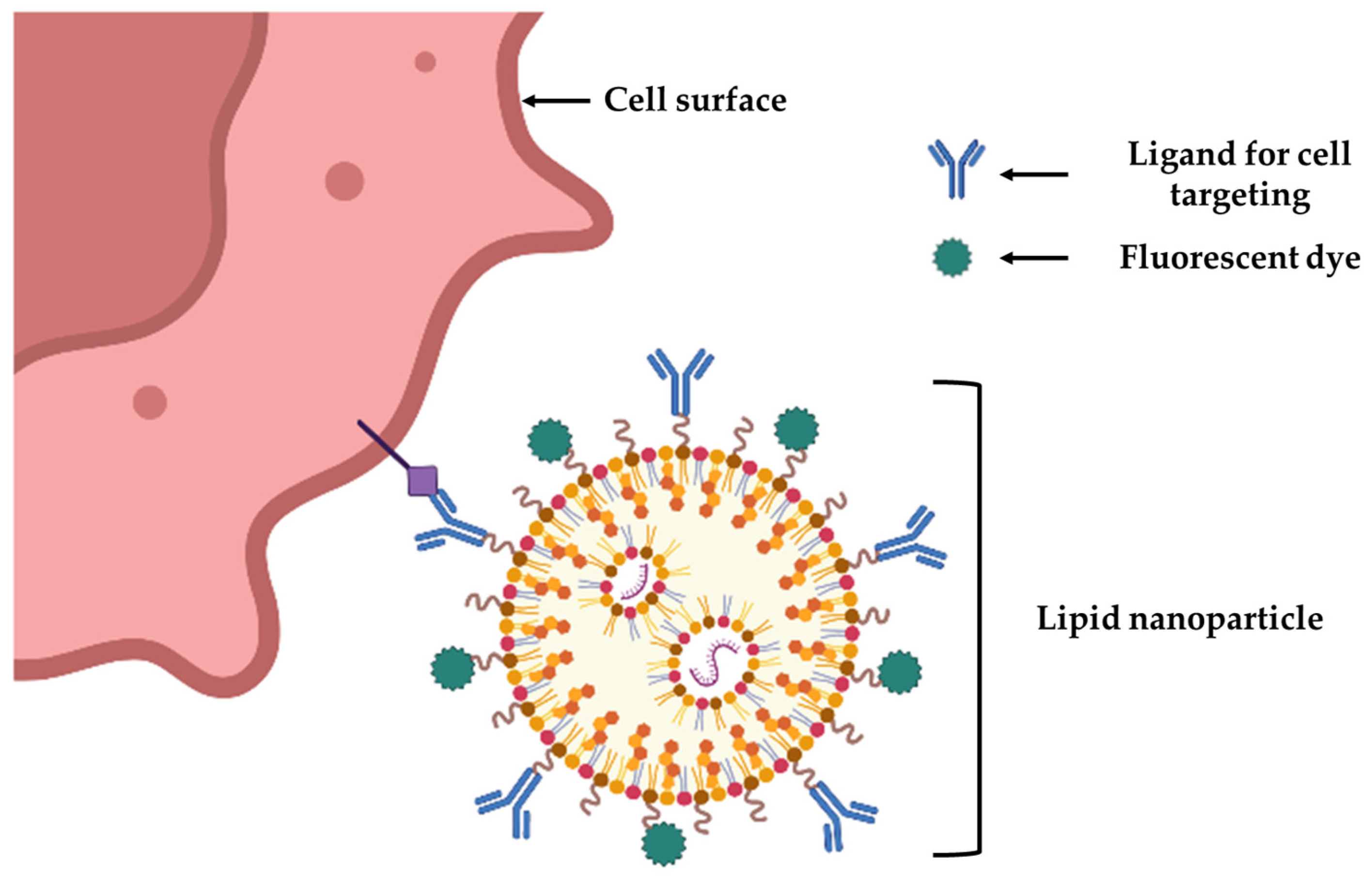
4. Nanotechnology for Cell Targeting and Labeling for BTE

In addition to the application of nanotechnology in BTE previously discussed, it is also being extensively explored for its potential role as a biosensor. The technology is used to perform continuous cell tracking and the monitoring of cell fate in BTE applications. Conceptually, NPs prepared using Au or materials that can be traced are used, which specifically help to visualize the cells in vitro or in vivo (Figure 4), enabling scientists to visualize the integration of cells in BTE in a non-invasive manner [29]. Imaging techniques can significantly help test host graft interactions and immune responses to implants, scaffolds, and viable grafts, as well as study the signaling behavior. Thus, in vivo, monitoring is essential to advance tissue engineering to repair or regenerate bone tissue [152].
A variety of NPs, like quantum dots, superparamagnetic iron oxide nanoparticles (SPIONs), silica NPs, and gold NPs [40], are used as contrast agents for several imaging modalities, like fluorescence imaging, magnetic resonance imaging (MRI), computed tomography (CT), and photoacoustic imaging.
The colloidal NPs known as quantum dots (QDs) have exceptional optical properties, making them ideal for the fluorescence imaging of biological systems. They have unique optical and electrical features due to quantum confinement effects [153]. This produces QDs with superior stability, biocompatibility, size-modulated absorbance and emissions, brightness, and persistent luminescence. These properties have enabled them to be employed as long-term cell-labeling and tracking probes for dynamic processes [40]. They have broad excitation and narrow emission ranges, allowing multiplexed imaging with a single excitation source. Jahed and colleagues synthesized QD-histidine-βcyclodextrin to label human adipose stem cells (ASCs) and track their differentiation into bone tissue in a 3D cell-laden CS hydrogel scaffold [154]. Another group injected lead sulfide QDs (PbS QDs) encapsulated by ribonuclease A (RNase A) with a shortwave infrared (SWIR) emission in Balb/C nude mice. It achieved the accurate long-term imaging of various bone structures in a 3D configuration [155]. Despite their numerous advantages in imaging, QDs have presented significant challenges of toxicity associated with the materials used in their core, i.e., cadmium (Cd) (II) and lead (Pb)(II) ions, and their eventual accumulation in organs. Furthermore, focused studies using high dosages of specific combinations of QDs need to be conducted to construe the cytotoxicity issue [153].
Recently, inorganic magnetic NPs have emerged as a potential MRI probe for cell tracking. SPION nanocomposites are NPs with a functionalized shell surrounding an iron oxide core [156]. Clusters with different aggregation states can be adapted by adjusting the composition of composite materials or the ratio between organic and inorganic matter to improve the uptake by the live cells in vivo for better visibility [157]. SPIONs are effective contrast agents, owing to their superparamagnetic properties and high sensitivity in MRIs, requiring lower concentrations and thus reducing their side effects [158]. SPIONs surfaces can be functionalized to recognize specific targets. Hence, they may be applied for processes, including cellular imaging, cellular tracking, biosensor applications, as well as for guided drug and gene delivery [40]. Jing et al. used MSCs in the articular cartilage of rabbit knee joints using the Feridex–protamine sulfate complex as the SPION and transfection agents [159]. Kim et al. demonstrated a PAA backbone conjugated with 2-aminoethyl-trimethyl ammonium (TMA) by 1-ethyl-3-(3-dimethylaminopropyl)-carbodiimide (EDC) initiation to produce particles called “TMA–SPION” for an in vivo mouse model and were able to visualize them using MRI at day 7 [160]. However, different studies on human and animal cells have revealed that, at the cellular level, SPION uptake can change gene expression and produce ROS. High levels of ferric ions in the cell can create an osmotic imbalance leading to membrane leakage and cytotoxicity [156].
Silica NP synthesized as core/shell silica NPs (C/S, SiNPs) and mesoporous silica NPs (MSNPs) are often used in imaging agent delivery. In core/shell silica NPs, the core composed of imaging agents, such as fluorescent probes, AuNPs, and SPIONs, is shielded by the silica shell, reducing the amount of photobleaching and allowing for the long-term monitoring of the labeled material [40]. MSNPs also offer various attractive physicochemical properties owing to their tunable shape, size, morphology, large surface area/pore volume, high colloidal property, thermal stability, biocompatibility, and ease of surface modification [161].
On the other hand, metal NPs are used for optical detection due to their tunable strength, bandwidth, and frequency. These characteristics, attributed to the collective oscillation mode, make the metal NPs ideal for optical applications [40]. Au NPs are being explored as contrast agents in combination with organic and inorganic polymers in bioimaging processes. Recently, their application in imaging has generated interest owing to their favorable properties of biocompatibility, easy synthesis, surface plasmon resonance (SPR), and surface ligand functionalization [39]. Additionally, their efficient uptake by the cells allows their use to track cells in vivo. They can be adapted to absorb in the near-infrared range (NIR), thus enhancing deep-tissue visualization by imaging techniques. Additionally, substantial X-ray attenuation driven by the high atomic number helps boost computed tomography (CT) contrast. A two-photon imaging (TPI) agent was created by Gunnlaugsson and colleagues using Eu(III) complexes that were covalently bonded to the surface of Au NPs. This substance enabled the 3D imaging of small-scale bone injuries by binding to exposed calcium sites (i.e., microcracks) within the injured bone surface [162]. As 19F magnetic resonance imaging (MRI) and CT contrast agents, it was demonstrated in a study that labeling CPC with perfluoro-15-crown-5-ether-loaded (PFCE) PLGA NPs and AuNPs (diameter: 40 nm) could improve the image contrast and accurately identify and locate the scaffold [163].
However, Au NPs’ practical application for in vivo imaging is still challenging due to the low penetration depth of the scattered light in tissue samples. Also, the clinical application of Au NPs and the long-term toxicity associated with their in vivo usage remains elusive. In spite of these challenges, the evidence presented above indicates its potential application in the field of BTE and bone repair.

5. Challenges and Future Perspectives

While NBT in BTE applications have promising potentials, we are yet to overcome certain challenges. The systemic toxicity of metallic NPs and immunogenic responses, especially those required at higher doses, require special attention. This is because it can negatively affect cell adhesion and architecture. More comprehensive, reliable toxicity assessment studies for defined compositions of NPs are vital for their success. Reports of the toxicity response to NPs in the scaffolds due to the degradation of the implants and heavy metals in carbon NTs increase the need for further research into technologies aimed at mimicking the local and systematic biological functions of these NPs. These results would provide a better cognizance of the biocompatibility of these systems and would be the guiding light for making the process economical and sustainable. In regard to the NPs used in imaging and cell tracing, future studies on biomedical applications with in vivo systems are warranted to ensure the safe application of SPIONs in the field of BTE imaging.
A combination of several materials to form nano-scaffolds is another area that needs more research as it can open more avenues concerning the optimum formula for scaffolds [164]. Another limitation that requires more study is implant mediated infection, an unwanted reaction as a result of a foreign body and a loss of the implant material due to degradation. The use of electrospun scaffolds for BTE has documented cases of infections leading to failed bone tissue regeneration [165]. Another challenge of designing scaffolds in general is obtaining mechanical properties and vascularity similar to that of bone [35]. Innovative fabrication techniques, like 3D functional constructs in conjugation with NPs, can help modulate the properties of the microstructure [166]. The overall standardization of protocols and carefully defining permitted limits or the concentration of materials used would bring us a step closer to the safety and efficacy of these formulations.
Despite these challenges, the field is ever-expanding and providing innovative solutions to problems that were previously unapproachable. A new class of NPs is increasingly becoming known to scientists and these are known as cell-membrane-coated NPs. This approach uses the bionic cell membrane as a coating technology for designing NPs for formulating the multi-functional biomimetic drug delivery system discussed by several authors, including Liu and colleagues and Ye and colleagues [167,168]. Furthermore, futuristic applications of NBT in cancer has been indicated by the use of bio-inspired nanoplatelets and nano-sponges for chemotherapy. Both the nanoplatelets as well as the nanosponges were found to not only ablate the primary tumor in vivo, but also inhibit breast cancer metastasis [169,170].
Currently, there are 508 clinical trials recorded just for the use of NPs as per the data accessed from ClinicalTrials.gov. These include the use of nanoscale applications ranging from treatment to diagnostic purposes and from jaw bone to leukemia applications. Out of these, there 25 studies used NBT in some form or the other for bone-related conditions in these trials. The most relevant ones reflecting the aim of this article are outlined in Table 2.
Modifications of scaffolds using NPs potentially translates to better biological and structural mimicking properties of the natural bone environment. This provides a favorable environment for cellular attachment, division, and bone formation. The three main targets of osteogenic cell survival, osteoblastic differentiation, or modulation of immunological response warrant nano-structural changes to polymer surfaces. The persistent quest to better understand the interactions between nanoscale surface topography and the biological system into which it is introduced remains a consistent discussion of research with foreseeable noteworthy developments.

6. Conclusions

NBT provides novel and technologically advanced tools to engineer scaffolds using nano-scale materials to create drug delivery devices with controlled spatial and temporal release patterns. Different types of formulations at the nanoscale using natural (proteins and polysaccharides) or synthetic (metals, ceramics, magnetic/paramagnetic materials, and polymers) materials can provide a strong platform for tissue-engineered artificial organ functionalization. Although these advancements provide opportunities to harness NBT to enhance tissue repair, numerous questions and challenges still remain to be addressed. We are yet to utilize the full potential of the mechanisms by which variations in the nano-scaling, orientation, and co-presentation of these formulations modulate cell responses. The field of nanotechnology, along with the integration of growth factors, metabolites, and biomaterials of appropriate mechanical properties will help in attaining timely and effective bone regeneration. In summary, the future of medicine with the use of NBT is promising for BTE and orthopedic applications, especially with the advancement of nanomedicine and NBT as a tool in precision medicine [171].

Author Contributions

Conceptualization, P.G.; software, T.P., N.R., N.I., A.G., M.L., A.D. and P.G.; resources, P.G.; validation, A.D.; writing—original draft preparation, N.R., T.P., N.I., A.G., M.L. and P.G.; writing—review and editing, A.D., N.R., T.P., N.I., A.G., M.L. and P.G.; visualization, P.G.; funding acquisition, P.G. All authors have read and agreed to the published version of the manuscript.

Funding

This work received no external funding.

Conflicts of Interest

Author Tejal Pant was employed by the company Anya Biopharm India LLP and author Abhishek Goyal was employed by the company RAS Life Science Solutions. The remaining authors declare that the work was conducted in the absence of any commercial or financial relationships that could be construed as a potential conflict of interest.

References

  1. Einhorn, T.A.; Gerstenfeld, L.C. Fracture healing: Mechanisms and interventions. Nat. Rev. Rheumatol. 2015, 11, 45–54. [Google Scholar] [CrossRef] [PubMed]
  2. Perez, J.R.; Kouroupis, D.; Li, D.J.; Best, T.M.; Kaplan, L.; Correa, D. Tissue Engineering and Cell-Based Therapies for Fractures and Bone Defects. Front. Bioeng. Biotechnol. 2018, 6, 105. [Google Scholar] [CrossRef] [PubMed]
  3. de Villiers, T.J.; Goldstein, S.R. Bone health 2022: An update. Climacteric J. Int. Menopause Soc. 2022, 25, 1–3. [Google Scholar] [CrossRef] [PubMed]
  4. Mills, L.A.; Aitken, S.A.; Simpson, A.H.R.W. The risk of non-union per fracture: Current myths and revised figures from a population of over 4 million adults. Acta Orthop. 2017, 88, 434–439. [Google Scholar] [CrossRef] [PubMed]
  5. Wu, A.-M.; Bisignano, C.; James, S.L.; Abady, G.G.; Abedi, A.; Abu-Gharbieh, E.; Alhassan, R.K.; Alipour, V.; Arabloo, J.; Asaad, M.; et al. Global, regional, and national burden of bone fractures in 204 countries and territories, 1990–2019: A systematic analysis from the Global Burden of Disease Study 2019. Lancet Healthy Longev. 2021, 2, e580–e592. [Google Scholar] [CrossRef] [PubMed]
  6. Cross, M.; Smith, E.; Hoy, D.; Nolte, S.; Ackerman, I.; Fransen, M.; Bridgett, L.; Williams, S.; Guillemin, F.; Hill, C.L.; et al. The global burden of hip and knee osteoarthritis: Estimates from the global burden of disease 2010 study. Ann. Rheum. Dis. 2014, 73, 1323–1330. [Google Scholar] [CrossRef]
  7. Cui, A.; Li, H.; Wang, D.; Zhong, J.; Chen, Y.; Lu, H. Global, regional prevalence, incidence and risk factors of knee osteoarthritis in population-based studies. EClinicalMedicine 2020, 29–30, 100587. [Google Scholar] [CrossRef]
  8. Steinmetz, S.; Wernly, D.; Moerenhout, K.; Trampuz, A.; Borens, O. Infection after fracture fixation. EFORT Open Rev. 2019, 4, 468–475. [Google Scholar] [CrossRef]
  9. Henkel, J.; Woodruff, M.; Epari, D.; Steck, R.; Glatt, V.; Dickinson, I.C.; Choong, P.; Schuetz, M.A.; Hutmacher, D.W. Bone Regeneration Based on Tissue Engineering Conceptions—A 21st Century Perspective. Bone Res. 2013, 1, 216–248. [Google Scholar] [CrossRef]
  10. Amini, A.R.; Laurencin, C.T.; Nukavarapu, S.P. Bone tissue engineering: Recent advances and challenges. Crit. Rev. Biomed. Eng. 2012, 40, 363–408. [Google Scholar] [CrossRef]
  11. Braddock, M.; Houston, P.; Campbell, C.; Ashcroft, P. Born again bone: Tissue engineering for bone repair. Physiology 2001, 16, 208–213. [Google Scholar] [CrossRef] [PubMed]
  12. Salgado, A.J.; Coutinho, O.P.; Reis, R.L. Bone tissue engineering: State of the art and future trends. Macromol. Biosci. 2004, 4, 743–765. [Google Scholar] [CrossRef] [PubMed]
  13. Calori, G.M.; Giannoudis, P.V. Enhancement of fracture healing with the diamond concept: The role of the biological chamber. Injury 2011, 42, 1191–1193. [Google Scholar] [CrossRef] [PubMed]
  14. Andrzejowski, P.; Giannoudis, P.V. The ‘diamond concept’ for long bone non-union management. J. Orthop. Traumatol. 2019, 20, 21. [Google Scholar] [CrossRef]
  15. Bartold, M.; Gronthos, S.; Haynes, D.; Ivanovski, S. Mesenchymal stem cells and biologic factors leading to bone formation. J. Clin. Periodontol. 2019, 46 (Suppl. S21), 12–32. [Google Scholar] [CrossRef]
  16. Arthur, A.; Gronthos, S. Clinical Application of Bone Marrow Mesenchymal Stem/Stromal Cells to Repair Skeletal Tissue. Int. J. Mol. Sci. 2020, 21, 9759. [Google Scholar] [CrossRef]
  17. Ganguly, P.; El-Jawhari, J.J.; Giannoudis, P.V.; Burska, A.N.; Ponchel, F.; Jones, E.A. Age-related Changes in Bone Marrow Mesenchymal Stromal Cells: A Potential Impact on Osteoporosis and Osteoarthritis Development. Cell Transplant. 2017, 26, 1520–1529. [Google Scholar] [CrossRef]
  18. Ganguly, P.; El-Jawhari, J.J.; Burska, A.N.; Ponchel, F.; Giannoudis, P.V.; Jones, E.A. The Analysis of In Vivo Aging in Human Bone Marrow Mesenchymal Stromal Cells Using Colony-Forming Unit-Fibroblast Assay and the CD45 low CD271 + Phenotype. Stem Cells Int. 2019, 2019, 1–14. [Google Scholar] [CrossRef]
  19. Iijima, K.; Otsuka, H. Cell Scaffolds for Bone Tissue Engineering. Bioengineering 2020, 7, 119. [Google Scholar] [CrossRef]
  20. Sallent, I.; Capella-Monsonís, H.; Procter, P.; Bozo, I.Y.; Deev, R.V.; Zubov, D.; Vasyliev, R.; Perale, G.; Pertici, G.; Baker, J.; et al. The Few Who Made It: Commercially and Clinically Successful Innovative Bone Grafts. Front. Bioeng. Biotechnol. 2020, 8, 952. [Google Scholar] [CrossRef]
  21. Chen, J.; Hendriks, M.; Chatzis, A.; Ramasamy, S.K.; Kusumbe, A.P. Bone Vasculature and Bone Marrow Vascular Niches in Health and Disease. J. Bone Miner. Res. Off. J. Am. Soc. Bone Miner. Res. 2020, 35, 2103–2120. [Google Scholar] [CrossRef] [PubMed]
  22. Morrison, S.J.; Scadden, D.T. The bone marrow niche for haematopoietic stem cells. Nature 2014, 505, 327–334. [Google Scholar] [CrossRef]
  23. Reagan, M.R.; Rosen, C.J. Navigating the bone marrow niche: Translational insights and cancer-driven dysfunction. Nat. Rev. Rheumatol. 2016, 12, 154–168. [Google Scholar] [CrossRef] [PubMed]
  24. Dimitriou, R.; Jones, E.; McGonagle, D.; Giannoudis, P.V. Bone regeneration: Current concepts and future directions. BMC Med. 2011, 9, 66. [Google Scholar] [CrossRef]
  25. Rausch, M.; Iqbal, N.; Pathak, S.; Owston, H.E.; Ganguly, P. Organoid Models and Next-Generation Sequencing for Bone Marrow and Related Disorders. Organoids 2023, 2, 123–139. [Google Scholar] [CrossRef]
  26. Walmsley, G.G.; McArdle, A.; Tevlin, R.; Momeni, A.; Atashroo, D.; Hu, M.S.; Feroze, A.H.; Wong, V.W.; Lorenz, P.H.; Longaker, M.T.; et al. Nanotechnology in bone tissue engineering. Nanomed. Nanotechnol. Biol. Med. 2015, 11, 1253–1263. [Google Scholar] [CrossRef]
  27. Saiz, E.; Zimmermann, E.A.; Lee, J.S.; Wegst, U.G.; Tomsia, A.P. Perspectives on the role of nanotechnology in bone tissue engineering. Dent. Mater. Off. Publ. Acad. Dent. Mater. 2013, 29, 103–115. [Google Scholar] [CrossRef]
  28. Simchi, A.; Mazaheri, M.; Eslahi, N.; Ordikhani, F.; Tamjid, E. Nanomedicine applications in orthopedic medicine: State of the art. Int. J. Nanomed. 2023, 10, 6039–6054. [Google Scholar] [CrossRef]
  29. Griffin, M.F.; Kalaskar, D.M.; Seifalian, A.; Butler, P.E. An update on the Application of Nanotechnology in Bone Tissue Engineering. Open Orthop. J. 2016, 10, 836–848. [Google Scholar] [CrossRef]
  30. Schofer, M.D.; Roessler, P.P.; Schaefer, J.; Theisen, C.; Schlimme, S.; Heverhagen, J.T.; Voelker, M.; Dersch, R.; Agarwal, S.; Fuchs-Winkelmann, S.; et al. Electrospun PLLA nanofiber scaffolds and their use in combination with BMP-2 for reconstruction of bone defects. PLoS ONE 2011, 6, e25462. [Google Scholar] [CrossRef]
  31. Prabha, R.D.; Kraft, D.C.E.; Harkness, L.; Melsen, B.; Varma, H.; Nair, P.D.; Kjems, J.; Kassem, M. Bioactive nano-fibrous scaffold for vascularized craniofacial bone regeneration. J. Tissue Eng. Regen. Med. 2018, 12, e1537–e1748. [Google Scholar] [CrossRef] [PubMed]
  32. Yao, Q.; Liu, Y.; Selvaratnam, B.; Koodali, R.T.; Sun, H. Mesoporous silicate nanoparticles/3D nanofibrous scaffold-mediated dual-drug delivery for bone tissue engineering. J. Control. Release Off. J. Control. Release Soc. 2018, 279, 69–78. [Google Scholar] [CrossRef] [PubMed]
  33. Mouriño, V.; Boccaccini, A.R. Bone tissue engineering therapeutics: Controlled drug delivery in three-dimensional scaffolds. J. R. Soc. Interface 2010, 7, 209–227. [Google Scholar] [CrossRef] [PubMed]
  34. Jayaraman, P.; Gandhimathi, C.; Venugopal, J.R.; Becker, D.L.; Ramakrishna, S.; Srinivasan, D.K. Controlled release of drugs in electrosprayed nanoparticles for bone tissue engineering. Adv. Drug Deliv. Rev. 2015, 94, 77–95. [Google Scholar] [CrossRef] [PubMed]
  35. Hajiali, H.; Ouyang, L.; Llopis-Hernandez, V.; Dobre, O.; Rose, F.R.A.J. Review of emerging nanotechnology in bone regeneration: Progress, challenges, and perspectives. Nanoscale 2021, 13, 10266–10280. [Google Scholar] [CrossRef]
  36. Wen, J.; Cai, D.; Gao, W.; He, R.; Li, Y.; Zhou, Y.; Klein, T.; Xiao, L.; Xiao, Y. Osteoimmunomodulatory Nanoparticles for Bone Regeneration. Nanomaterials 2023, 13, 692. [Google Scholar] [CrossRef]
  37. Scott, T.G.; Blackburn, G.; Ashley, M.; Bayer, I.S.; Ghosh, A.; Biris, A.S.; Biswas, A. Advances in bionanomaterials for bone tissue engineering. J. Nanosci. Nanotechnol. 2013, 13, 1–22. [Google Scholar] [CrossRef]
  38. Qiao, K.; Xu, L.; Tang, J.; Wang, Q.; Lim, K.S.; Hooper, G.; Woodfield, T.B.F.; Liu, G.; Tian, K.; Zhang, W.; et al. The advances in nanomedicine for bone and cartilage repair. J. Nanobiotechnol. 2022, 20, 141. [Google Scholar] [CrossRef]
  39. Babuska, V.; Kasi, P.B.; Chocholata, P.; Wiesnerova, L.; Dvorakova, J.; Vrzakova, R.; Nekleionova, A.; Landsmann, L.; Kulda, V. Nanomaterials in Bone Regeneration. Appl. Sci. 2022, 12, 6793. [Google Scholar] [CrossRef]
  40. Vieira, S.; Vial, S.; Reis, R.L.; Oliveira, J.M. Nanoparticles for bone tissue engineering. Biotechnol. Prog. 2017, 33, 590–611. [Google Scholar] [CrossRef]
  41. Guillén-Carvajal, K.; Valdez-Salas, B.; Beltrán-Partida, E.; Salomón-Carlos, J.; Cheng, N. Chitosan, Gelatin, and Collagen Hydrogels for Bone Regeneration. Polymers 2023, 15, 2762. [Google Scholar] [CrossRef] [PubMed]
  42. Begines, B.; Ortiz, T.; Pérez-Aranda, M.; Martínez, G.; Merinero, M.; Argüelles-Arias, F.; Alcudia, A. Polymeric Nanoparticles for Drug Delivery: Recent Developments and Future Prospects. Nanomaterials 2020, 10, 1403. [Google Scholar] [CrossRef] [PubMed]
  43. Khan, M.U.A.; Al-Arjan, W.S.; Binkadem, M.S.; Mehboob, H.; Haider, A.; Raza, M.A.; Razak, S.I.A.; Hasan, A.; Amin, R. Development of Biopolymeric Hybrid Scaffold-Based on AAc/GO/nHAp/TiO2 Nanocomposite for Bone Tissue Engineering: In-Vitro Analysis. Nanomaterials 2021, 11, 1319. [Google Scholar] [CrossRef] [PubMed]
  44. Matos, R.J.R.; Silva, J.C.; Soares, P.I.P.; Borges, J.P. Polyvinylpyrrolidone Nanofibers Incorporating Mesoporous Bioactive Glass for Bone Tissue Engineering. Biomimetics 2023, 8, 206. [Google Scholar] [CrossRef] [PubMed]
  45. Chahal, S.; Kumar, A.; Hussian, F.S.J. Development of biomimetic electrospun polymeric biomaterials for bone tissue engineering. A review. J. Biomater. Sci. Polym. Ed. 2019, 30, 1308–1355. [Google Scholar] [CrossRef] [PubMed]
  46. Samrot, A.V.; Sathiyasree, M.; Rahim, S.B.A.; Renitta, R.E.; Kasipandian, K.; Shree, S.K.; Rajalakshmi, D.; Shobana, N.; Dhiva, S.; Abirami, S.; et al. Scaffold Using Chitosan, Agarose, Cellulose, Dextran and Protein for Tissue Engineering—A Review. Polymers 2023, 15, 1525. [Google Scholar] [CrossRef]
  47. Sagadevan, S.; Schirhagl, R.; Rahman, Z.; Bin Ismail, M.F.; Lett, J.A.; Fatimah, I.; Kaus, N.H.M.; Oh, W.-C. Recent advancements in polymer matrix nanocomposites for bone tissue engineering applications. J. Drug Deliv. Sci. Technol. 2023, 82, 104313. [Google Scholar] [CrossRef]
  48. Dwivedi, R.; Kumar, S.; Pandey, R.; Mahajan, A.; Nandana, D.; Katti, D.S.; Mehrotra, D. Polycaprolactone as biomaterial for bone scaffolds: Review of literature. J. Oral Biol. Craniofacial Res. 2020, 10, 381–388. [Google Scholar] [CrossRef]
  49. Al-Maawi, S.; Dohle, E.; Lim, J.; Weigl, P.; Teoh, S.H.; Sader, R.; Ghanaati, S. Biologization of Pcl-Mesh Using Platelet Rich Fibrin (Prf) Enhances Its Regenerative Potential In Vitro. Int. J. Mol. Sci. 2021, 22, 2159. [Google Scholar] [CrossRef]
  50. Jin, S.; Xia, X.; Huang, J.; Yuan, C.; Zuo, Y.; Li, Y.; Li, J. Recent advances in PLGA-based biomaterials for bone tissue regeneration. Acta Biomater. 2021, 127, 56–79. [Google Scholar] [CrossRef]
  51. Hassan, M.; Sulaiman, M.; Yuvaraju, P.D.; Galiwango, E.; Rehman, I.u.; Al-Marzouqi, A.H.; Khaleel, A.; Mohsin, S. Biomimetic PLGA/Strontium-Zinc Nano Hydroxyapatite Composite Scaffolds for Bone Regeneration. J. Funct. Biomater. 2022, 13, 13. [Google Scholar] [CrossRef] [PubMed]
  52. Chen, L.; Zhu, W.-M.; Fei, Z.-Q.; Chen, J.-L.; Xiong, J.-Y.; Zhang, J.-F.; Duan, L.; Huang, J.; Liu, Z.; Wang, D.; et al. The study on biocompatibility of porous nHA/PLGA composite scaffolds for tissue engineering with rabbit chondrocytes in vitro. BioMed Res. Int. 2013, 2013, 412745. [Google Scholar] [CrossRef] [PubMed]
  53. Wang, H.; Zou, Q.; Boerman, O.C.; Nijhuis, A.W.; Jansen, J.A.; Li, Y.; Leeuwenburgh, S.C. Combined delivery of BMP-2 and bFGF from nanostructured colloidal gelatin gels and its effect on bone regeneration in vivo. J. Control. Release Off. J. Control. Release Soc. 2013, 166, 172–181. [Google Scholar] [CrossRef] [PubMed]
  54. Zhu, L.; Luo, D.; Liu, Y. Effect of the nano/microscale structure of biomaterial scaffolds on bone regeneration. Int. J. Oral Sci. 2020, 12, 6. [Google Scholar] [CrossRef] [PubMed]
  55. Abbas, M.; Alqahtani, M.S.; Alhifzi, R. Recent Developments in Polymer Nanocomposites for Bone Regeneration. Int. J. Mol. Sci. 2023, 24, 3312. [Google Scholar] [CrossRef] [PubMed]
  56. Castro, J.I.; Llano, C.H.V.; Tenorio, D.L.; Saavedra, M.; Zapata, P.; Navia-Porras, D.P.; Delgado-Ospina, J.; Chaur, M.N.; Hernández, J.H.M.; Grande-Tovar, C.D. Biocompatibility Assessment of Polylactic Acid (PLA) and Nanobioglass (n-BG) Nanocomposites for Biomedical Applications. Molecules 2022, 27, 3640. [Google Scholar] [CrossRef]
  57. Suaza, M.L.M.; Rivera, J.C.L.; Padilla, M.C.R.; Acevedo, M.E.M.; Orozco, C.P.O.; Triviño, D.G.Z. Poly(vinyl alcohol)/Silk Fibroin/Ag-NPs Composite Nanofibers as a Substrate for MG-63 Cells’ Growth. Polymers 2023, 15, 1838. [Google Scholar] [CrossRef]
  58. Wang, Y.-J.; Jeng, U.-S.; Hsu, S.-H. Biodegradable Water-Based Polyurethane Shape Memory Elastomers for Bone Tissue Engineering. ACS Biomater. Sci. Eng. 2018, 4, 1397–1406. [Google Scholar] [CrossRef]
  59. Anitha, A.; Sowmya, S.; Kumar, P.S.; Deepthi, S.; Chennazhi, K.; Ehrlich, H.; Tsurkan, M.; Jayakumar, R. Chitin and chitosan in selected biomedical applications. Prog. Polym. Sci. 2014, 399, 1644–1667. [Google Scholar] [CrossRef]
  60. Aljawish, A.; Chevalot, I.; Jasniewski, J.; Scher, J.; Muniglia, L. Enzymatic synthesis of chitosan derivatives and their potential applications. J. Mol. Catal. B Enzym. 2015, 112, 25–39. [Google Scholar] [CrossRef]
  61. Yildizbakan, L.; Iqbal, N.; Ganguly, P.; Kumi-Barimah, E.; Do, T.; Jones, E.; Giannoudis, P.V.; Jha, A. Fabrication and Characterisation of the Cytotoxic and Antibacterial Properties of Chitosan-Cerium Oxide Porous Scaffolds. Antibiotics 2023, 12, 1004. [Google Scholar] [CrossRef] [PubMed]
  62. Iqbal, N.; Braxton, T.M.; Anastasiou, A.; Raif, E.M.; Chung, C.K.Y.; Kumar, S.; Giannoudis, P.V.; Jha, A. Dicalcium Phosphate Dihydrate Mineral Loaded Freeze-Dried Scaffolds for Potential Synthetic Bone Applications. Materials 2022, 15, 6245. [Google Scholar] [CrossRef] [PubMed]
  63. Younes, I.; Rinaudo, M. Chitin and chitosan preparation from marine sources. Structure, properties and applications. Mar. Drugs 2015, 13, 1133–1174. [Google Scholar] [CrossRef]
  64. Garg, U.; Chauhan, S.; Nagaich, U.; Jain, N. Current Advances in Chitosan Nanoparticles Based Drug Delivery and Targeting. Adv. Pharm. Bull. 2019, 9, 195–204. [Google Scholar] [CrossRef] [PubMed]
  65. Rodríguez-Vázquez, M.; Vega-Ruiz, B.; Ramos-Zúñiga, R.; Saldaña-Koppel, D.A.; Quiñones-Olvera, L.F. Chitosan and Its Potential Use as a Scaffold for Tissue Engineering in Regenerative Medicine. BioMed Res. Int. 2015, 2015, 821279. [Google Scholar] [CrossRef]
  66. Ibrahim, H.M.; Zairy, E.M.R.E. Chitosan as a Biomaterial—Structure, Properties, and Electrospun Nanofibers. In Concepts, Compounds and the Alternatives of Antibacterials; IntechOpen: London, UK, 2015. [Google Scholar] [CrossRef]
  67. Levengood, S.K.L.; Zhang, M. Chitosan-based scaffolds for bone tissue engineering. J. Mater. Chem. B 2014, 2, 3161–3184. [Google Scholar] [CrossRef]
  68. Lee, J.S.; Baek, S.D.; Venkatesan, J.; Bhatnagar, I.; Chang, H.K.; Kim, H.T.; Kim, S.-K. In vivo study of chitosan-natural nano hydroxyapatite scaffolds for bone tissue regeneration. Int. J. Biol. Macromol. 2014, 67, 360–366. [Google Scholar] [CrossRef]
  69. Sultankulov, B.; Berillo, D.; Sultankulova, K.; Tokay, T.; Saparov, A. Progress in the Development of Chitosan-Based Biomaterials for Tissue Engineering and Regenerative Medicine. Biomolecules 2019, 9, 470. [Google Scholar] [CrossRef]
  70. Mehdi Jorfi, E.J.F. Recent advances in nanocellulose for biomedical applications. J. Appl. Polym. Sci. 2014, 132, 41719. [Google Scholar] [CrossRef]
  71. Md Nazrul Islam, F.R. Chapter 6—Production and modification of nanofibrillated cellulose composites and potential applications. In Green Composites for Automotive Applications; Woodhead Publishing Series in Composites Science and Engineering; Elsevier: Amsterdam, The Netherlands, 2019; pp. 115–141. [Google Scholar] [CrossRef]
  72. Osorio, D.A.; Lee, B.E.J.; Kwiecien, J.M.; Wang, X.; Shahid, I.; Hurley, A.L.; Cranston, E.D.; Grandfield, K. Cross-linked cellulose nanocrystal aerogels as viable bone tissue scaffolds. Acta Biomater. 2019, 87, 152–165. [Google Scholar] [CrossRef]
  73. Baldwin, P.; Li, D.J.; Auston, D.A.; Mir, H.S.; Yoon, R.S.; Koval, K.J. Autograft, Allograft, and Bone Graft Substitutes: Clinical Evidence and Indications for Use in the Setting of Orthopaedic Trauma Surgery. J. Orthop. Trauma 2019, 33, 203–213. [Google Scholar] [CrossRef] [PubMed]
  74. Ganguly, P.; Jones, E.; Panagiotopoulou, V.; Jha, A.; Blanchy, M.; Antimisiaris, S.; Anton, M.; Dhuiège, B.; Marotta, M.; Marjanovic, N.; et al. Electrospun and 3D printed polymeric materials for one-stage critical-size long bone defect regeneration inspired by the Masquelet technique: Recent Advances. Injury 2022, 53 (Suppl. S2), s2–s12. [Google Scholar] [CrossRef] [PubMed]
  75. Di Marzio, N.; Eglin, D.; Serra, T.; Moroni, L. Bio-Fabrication: Convergence of 3D Bioprinting and Nano-Biomaterials in Tissue Engineering and Regenerative Medicine. Front. Bioeng. Biotechnol. 2020, 8, 326. [Google Scholar] [CrossRef] [PubMed]
  76. Shin, K.; Acri, T.; Geary, S.; Salem, A.K. Biomimetic Mineralization of Biomaterials Using Simulated Body Fluids for Bone Tissue Engineering and Regenerative Medicine. Tissue Eng. Part A 2017, 23, 1169–1180. [Google Scholar] [CrossRef] [PubMed]
  77. Hindy, O.A.; Goker, M.; Yilgor Huri, P. Nanoscale agents within 3D-printed constructs: Intersection of nanotechnology and personalized bone tissue engineering. Emergent Mater. 2022, 5, 195–205. [Google Scholar] [CrossRef]
  78. Chen, Y.; Kawazoe, N.; Chen, G. Preparation of dexamethasone-loaded biphasic calcium phosphate nanoparticles/collagen porous composite scaffolds for bone tissue engineering. Acta Biomater. 2018, 67, 341–353. [Google Scholar] [CrossRef]
  79. Sokolova, V.; Kostka, K.; Shalumon, K.T.; Prymak, O.; Chen, J.-P.; Epple, M. Synthesis and characterization of PLGA/HAP scaffolds with DNA-functionalised calcium phosphate nanoparticles for bone tissue engineering. J. Mater. Sci. Mater. Med. 2020, 31, 102. [Google Scholar] [CrossRef]
  80. Ha, S.-W.; Viggeswarapu, M.; Habib, M.M.; Beck, G.R. Bioactive effects of silica nanoparticles on bone cells are size, surface, and composition dependent. Acta Biomater. 2018, 82, 184–196. [Google Scholar] [CrossRef]
  81. Echazú, M.I.A.; Renou, S.J.; Alvarez, G.S.; Desimone, M.F.; Olmedo, D.G. Synthesis and Evaluation of a Chitosan–Silica-Based Bone Substitute for Tissue Engineering. Int. J. Mol. Sci. 2022, 23, 13379. [Google Scholar] [CrossRef]
  82. Shuai, C.; Yang, F.; Shuai, Y.; Peng, S.; Chen, S.; Deng, Y.; Feng, P. Silicon dioxide nanoparticles decorated on graphene oxide nanosheets and their application in poly(l-lactic acid) scaffold. J. Adv. Res. 2023, 48, 175–190. [Google Scholar] [CrossRef]
  83. Li, Y.; Yang, Y.; Qing, Y.; Li, R.; Tang, X.; Guo, D.; Qin, Y. Enhancing ZnO-NP Antibacterial and Osteogenesis Properties in Orthopedic Applications: A Review. Int. J. Nanomed. 2020, 15, 6247–6262. [Google Scholar] [CrossRef] [PubMed]
  84. Kandasamy, S.; Narayanan, V.; Sumathi, S. Zinc and manganese substituted hydroxyapatite/CMC/PVP electrospun composite for bone repair applications. Int. J. Biol. Macromol. 2020, 145, 1018–1030. [Google Scholar] [CrossRef] [PubMed]
  85. Singh, T.A.; Sharma, A.; Tejwan, N.; Ghosh, N.; Das, J.; Sil, P.C. A state of the art review on the synthesis, antibacterial, antioxidant, antidiabetic and tissue regeneration activities of zinc oxide nanoparticles. Adv. Colloid Interface Sci. 2021, 295, 102495. [Google Scholar] [CrossRef] [PubMed]
  86. Cho, Y.S.; Kim, H.-K.; Ghim, M.-S.; Hong, M.W.; Kim, Y.Y.; Cho, Y.-S. Evaluation of the Antibacterial Activity and Cell Response for 3D-Printed Polycaprolactone/Nanohydroxyapatite Scaffold with Zinc Oxide Coating. Polymers 2020, 12, 2193. [Google Scholar] [CrossRef]
  87. Forero, J.C.; Roa, E.; Reyes, J.G.; Acevedo, C.; Osses, N. Development of Useful Biomaterial for Bone Tissue Engineering by Incorporating Nano-Copper-Zinc Alloy (nCuZn) in Chitosan/Gelatin/Nano-Hydroxyapatite (Ch/G/nHAp) Scaffold. Materials 2017, 10, 1177. [Google Scholar] [CrossRef]
  88. Mokhtarzadeh, A.; Eivazzadeh-Keihan, R.; Pashazadeh, P.; Hejazi, M.; Gharaatifar, N.; Hasanzadeh, M.; Baradaran, B.; de la Guardia, M. Nanomaterial-based biosensors for detection of pathogenic virus. Trends Anal. Chem. TRAC 2017, 97, 445–457. [Google Scholar] [CrossRef]
  89. Chenab, K.K.; Eivazzadeh-Keihan, R.; Maleki, A.; Pashazadeh-Panahi, P.; Hamblin, M.R.; Mokhtarzadeh, A. Biomedical applications of nanoflares: Targeted intracellular fluorescence probes. Nanomed. Nanotechnol. Biol. Med. 2019, 17, 342–358. [Google Scholar] [CrossRef]
  90. Qing, Y.; Cheng, L.; Li, R.; Liu, G.; Zhang, Y.; Tang, X.; Wang, J.; Liu, H.; Qin, Y. Potential antibacterial mechanism of silver nanoparticles and the optimization of orthopedic implants by advanced modification technologies. Int. J. Nanomed. 2018, 13, 3311–3327. [Google Scholar] [CrossRef]
  91. Wang, Y.-C.; Lin, S.-H.; Chien, C.-S.; Kung, J.-C.; Shih, C.-J. In Vitro Bioactivity and Antibacterial Effects of a Silver-Containing Mesoporous Bioactive Glass Film on the Surface of Titanium Implants. Int. J. Mol. Sci. 2022, 23, 9291. [Google Scholar] [CrossRef]
  92. Eivazzadeh-Keihan, R.; Noruzi, E.B.; Chenab, K.K.; Jafari, A.; Radinekiyan, F.; Hashemi, S.M.; Ahmadpour, F.; Behboudi, A.; Mosafer, J.; Mokhtarzadeh, A.; et al. Metal-based nanoparticles for bone tissue engineering. J. Tissue Eng. Regen. Med. 2020, 14, 1687–1714. [Google Scholar] [CrossRef]
  93. Xu, L.; Wang, Y.-Y.; Huang, J.; Chen, C.-Y.; Wang, Z.-X.; Xie, H. Silver nanoparticles: Synthesis, medical applications and biosafety. Theranostics 2020, 10, 8996–9031. [Google Scholar] [CrossRef] [PubMed]
  94. Padmanabhan, V.P.; Sivashanmugam, P.; Kulandaivelu, R.; Sagadevan, S.; Sridevi, B.; Govindasamy, R.; Thiruvengadam, M. Biosynthesised Silver Nanoparticles Loading onto Biphasic Calcium Phosphate for Antibacterial and Bone Tissue Engineering Applications. Antibiotics 2022, 11, 1780. [Google Scholar] [CrossRef] [PubMed]
  95. Akturk, A.; Taygun, M.E.; Goller, G. Optimization of the electrospinning process variables for gelatin/silver nanoparticles/bioactive glass nanocomposites for bone tissue engineering—Akturk—2020—Polymer Composites—Wiley Online Library. Polym. Compos. 2020, 41, 2411–2425. [Google Scholar] [CrossRef]
  96. Vaidhyanathan, B.; Vincent, P.; Vadivel, S.; Karuppiah, P.; Al-Dhabi, N.A.; Sadhasivam, D.R.; Vimalraj, S.; Saravanan, S. Fabrication and Investigation of the Suitability of Chitosan-Silver Composite Scaffolds for Bone Tissue Engineering Applications. Process Biochem. 2021, 100, 178–187. [Google Scholar] [CrossRef]
  97. Lopes, J.; Ferreira-Gonçalves, T.; Ascensão, L.; Viana, A.S.; Carvalho, L.; Catarino, J.; Faísca, P.; Oliva, A.; Barros, D.P.C.d.; Rodrigues, C.M.P.; et al. Safety of Gold Nanoparticles: From In Vitro to In Vivo Testing Array Checklist. Pharmaceutics 2023, 15, 1120. [Google Scholar] [CrossRef] [PubMed]
  98. Tang, Y.; Shen, Y.; Huang, L.; Lv, G.; Lei, C.; Fan, X.; Lin, F.; Zhang, Y.; Wu, L.; Yang, Y. In vitro cytotoxicity of gold nanorods in A549 cells. Environ. Toxicol. Pharmacol. 2015, 39, 871–878. [Google Scholar] [CrossRef]
  99. Pooja, D.; Panyaram, S.; Kulhari, H.; Rachamalla, S.S.; Sistla, R. Xanthan gum stabilized gold nanoparticles: Characterization, biocompatibility, stability and cytotoxicity. Carbohydr. Polym. 2014, 110, 1–9. [Google Scholar] [CrossRef]
  100. Ko, W.-C.; Wang, S.-J.; Hsiao, C.-Y.; Hung, C.-T.; Hsu, Y.-J.; Chang, D.-C.; Hung, C.-F. Pharmacological Role of Functionalized Gold Nanoparticles in Disease Applications. Molecules 2022, 27, 1551. [Google Scholar] [CrossRef]
  101. Nekounam, H.; Allahyari, Z.; Gholizadeh, S.; Mirzaei, E.; Shokrgozar, M.A.; Faridi-Majidi, R. Simple and robust fabrication and characterization of conductive carbonized nanofibers loaded with gold nanoparticles for bone tissue engineering applications. Mater. Sci. Eng. C Mater. Biol. Appl. 2020, 117, 111226. [Google Scholar] [CrossRef]
  102. Yu, M.; Lei, B.; Gao, C.; Yan, J.; Ma, P.X. Optimizing surface-engineered ultra-small gold nanoparticles for highly efficient miRNA delivery to enhance osteogenic differentiation of bone mesenchymal stromal cells. Nano Res. 2016, 10, 49–63. [Google Scholar] [CrossRef]
  103. Hou, Y.; Cai, K.; Li, J.; Chen, X.; Lai, M.; Hu, Y.; Luo, Z.; Ding, X.; Xu, D. Effects of titanium nanoparticles on adhesion, migration, proliferation, and differentiation of mesenchymal stem cells. Int. J. Nanomed. 2013, 8, 3619–3630. [Google Scholar] [CrossRef]
  104. Kumar, P. Nano-TiO2 Doped Chitosan Scaffold for the Bone Tissue Engineering Applications. Int. J. Biomater. 2018, 2018, 6576157. [Google Scholar] [CrossRef] [PubMed]
  105. Pattanashetti, N.A.; Hiremath, C.; Naik, S.R.; Heggannavar, G.B.; Kariduraganavar, M.Y. Development of nanofibrous scaffolds by varying the TiO2 content in crosslinked PVA for bone tissue engineering. New J. Chem. 2020, 44, 2111–2121. [Google Scholar] [CrossRef]
  106. Yang, J.; Jia, C.; Yang, J. Designing Nanoparticle-based Drug Delivery Systems for Precision Medicine. Int. J. Med. Sci. 2021, 18, 2943–2949. [Google Scholar] [CrossRef] [PubMed]
  107. Wilczewska, A.Z.; Niemirowicz, K.; Markiewicz, K.H.; Car, H. Nanoparticles as drug delivery systems. Pharmacol. Rep. 2012, 64, 1020–1037. [Google Scholar] [CrossRef]
  108. Lu, V.; Zhang, J.; Patel, R.; Zhou, A.K.; Thahir, A.; Krkovic, M. Fracture Related Infections and Their Risk Factors for Treatment Failure—A Major Trauma Centre Perspective. Diagnostics 2022, 12, 1289. [Google Scholar] [CrossRef]
  109. Johnson, C.T.; García, A.J. Scaffold-based anti-infection strategies in bone repair. Ann. Biomed. Eng. 2015, 43, 515–528. [Google Scholar] [CrossRef]
  110. Xu, C.; Cao, Y.; Lei, C.; Li, Z.; Kumeria, T.; Meka, A.K.; Xu, J.; Liu, J.; Yan, C.; Luo, L.; et al. Polymer–Mesoporous Silica Nanoparticle Core–Shell Nanofibers as a Dual-Drug-Delivery System for Guided Tissue Regeneration. Appl. Nano Mater. 2020, 3, 1457–1467. [Google Scholar] [CrossRef]
  111. Sreeja, S.; Muraleedharan, C.; Varma, P.H.; Sailaja, G. Surface-transformed osteoinductive polyethylene terephthalate scaffold as a dual system for bone tissue regeneration with localized antibiotic delivery. Mater. Sci. Eng. C Mater. Biol. Appl. 2020, 109, 110491. [Google Scholar] [CrossRef]
  112. Sánchez-López, E.; Gomes, D.; Esteruelas, G.; Bonilla, L.; Lopez-Machado, A.L.; Galindo, R.; Cano, A.; Espina, M.; Ettcheto, M.; Camins, A.; et al. Metal-Based Nanoparticles as Antimicrobial Agents: An Overview. Nanomaterials 2020, 10, 292. [Google Scholar] [CrossRef]
  113. Shaikh, S.; Nazam, N.; Rizvi, S.M.D.; Ahmad, K.; Baig, M.H.; Lee, E.J.; Choi, I. Mechanistic Insights into the Antimicrobial Actions of Metallic Nanoparticles and Their Implications for Multidrug Resistance. Int. J. Mol. Sci. 2019, 20, 2468. [Google Scholar] [CrossRef] [PubMed]
  114. Nisar, P.; Ali, N.; Rahman, L.; Ali, M.; Shinwari, Z.K. Antimicrobial activities of biologically synthesized metal nanoparticles: An insight into the mechanism of action. J. Biol. Inorg. Chem. JBIC A Publ. Soc. Biol. Inorg. Chem. 2019, 24, 929–941. [Google Scholar] [CrossRef] [PubMed]
  115. Godoy-Gallardo, M.; Eckhard, U.; Delgado, L.M.; de Roo Puente, Y.J.; Hoyos-Nogués, M.; Gil, F.J.; Perez, R.A. Antibacterial approaches in tissue engineering using metal ions and nanoparticles: From mechanisms to applications. Bioact. Mater. 2021, 6, 4470–4490. [Google Scholar] [CrossRef]
  116. Sarkar, N.; Bose, S. Liposome-Encapsulated Curcumin-Loaded 3D Printed Scaffold for Bone Tissue Engineering. ACS Appl. Mater. Interfaces 2019, 11, 17184–17192. [Google Scholar] [CrossRef]
  117. Lee, C.-S.; Hsu, G.C.-Y.; Sono, T.; Lee, M.; James, A.W. Development of a Biomaterial Scaffold Integrated with Osteoinductive Oxysterol Liposomes to Enhance Hedgehog Signaling and Bone Repair. Mol. Pharm. 2021, 18, 1677–1689. [Google Scholar] [CrossRef] [PubMed]
  118. Cheng, R.; Liu, L.; Xiang, Y.; Lu, Y.; Deng, L.; Zhang, H.; Santos, H.A.; Cui, W. Advanced liposome-loaded scaffolds for therapeutic and tissue engineering applications. Biomaterials 2020, 232, 119706. [Google Scholar] [CrossRef]
  119. Porter, J.R.; Ruckh, T.T.; Popat, K.C. Bone tissue engineering: A review in bone biomimetics and drug delivery strategies. Biotechnol. Prog. 2009, 25, 1539–1560. [Google Scholar] [CrossRef]
  120. Makvandi, P.; Josic, U.; Delfi, M.; Pinelli, F.; Jahed, V.; Kaya, E.; Ashrafizadeh, M.; Zarepour, A.; Rossi, F.; Zarrabi, A.; et al. Drug Delivery (Nano)Platforms for Oral and Dental Applications: Tissue Regeneration, Infection Control, and Cancer Management. Adv. Sci. 2021, 8, 2004014. [Google Scholar] [CrossRef]
  121. Farokhi, M.; Mottaghitalab, F.; Shokrgozar, M.A.; Ou, K.-L.; Mao, C.; Hosseinkhani, H. Importance of dual delivery systems for bone tissue engineering. J. Control. Release Off. J. Control. Release Soc. 2016, 225, 152–169. [Google Scholar] [CrossRef]
  122. Chen, S.; Shi, Y.; Luo, Y.; Ma, J. cLayer-by-layer coated porous 3D printed hydroxyapatite composite scaffolds for controlled drug delivery. Colloids Surf. B Biointerfaces 2019, 179, 121–127. [Google Scholar] [CrossRef]
  123. Lee, D.; Wufuer, M.; Kim, I.; Choi, T.H.; Kim, B.J.; Jung, H.G.; Jeon, B.; Lee, G.; Jeon, O.H.; Chang, H.; et al. Sequential dual-drug delivery of BMP-2 and alendronate from hydroxyapatite-collagen scaffolds for enhanced bone regeneration. Sci. Rep. 2021, 11, 746. [Google Scholar] [CrossRef] [PubMed]
  124. Calabrese, G.; Franco, D.; Petralia, S.; Monforte, F.; Condorelli, G.G.; Squarzoni, S.; Traina, F.; Conoci, S. Dual-Functional Nano-Functionalized Titanium Scaffolds to Inhibit Bacterial Growth and Enhance Osteointegration. Nanomaterials 2021, 11, 2634. [Google Scholar] [CrossRef] [PubMed]
  125. Wu, L.; Pei, X.; Zhang, B.; Su, Z.; Gui, X.; Gao, C.; Guo, L.; Fan, H.; Jiang, Q.; Zhao, L.; et al. 3D-printed HAp bone regeneration scaffolds enable nano-scale manipulation of cellular mechanotransduction signals. Chem. Eng. J. 2023, 455, 140699. [Google Scholar] [CrossRef]
  126. Fathi-Achachelouei, M.; Knopf-Marques, H.; da Silva, C.E.R.; Barthès, J.; Bat, E.; Tezcaner, A.; Vrana, N.E. Use of Nanoparticles in Tissue Engineering and Regenerative Medicine. Front. Bioeng. Biotechnol. 2019, 7, 113. [Google Scholar] [CrossRef]
  127. Soundarya, S.P.; Menon, A.H.; Chandran, S.V.; Selvamurugan, N. Bone tissue engineering: Scaffold preparation using chitosan and other biomaterials with different design and fabrication techniques. Int. J. Biol. Macromol. 2018, 119, 1228–1239. [Google Scholar] [CrossRef]
  128. Baig, N.; Kammakakam, I.; Falath, W. Nanomaterials: A review of synthesis methods, properties, recent progress, and challenges. Mater. Adv 2021, 2, 1821–1871. [Google Scholar] [CrossRef]
  129. Biggs, M.J.P.; Richards, R.G.; Dalby, M.J. Nanotopographical modification: A regulator of cellular function through focal adhesions. Nanomed. Nanotechnol. Biol. Med. 2010, 6, 619–633. [Google Scholar] [CrossRef]
  130. Iravani, S.; Varma, R.S. Plants and plant-based polymers as scaffolds for tissue engineering. Green Chem. 2019, 21, 4839–4867. [Google Scholar] [CrossRef]
  131. Jahangirian, H.; Azizi, S.; Rafiee-Moghaddam, R.; Baratvand, B.; Webster, T.J. Status of Plant Protein-Based Green Scaffolds for Regenerative Medicine Applications. Biomolecules 2019, 9, 40. [Google Scholar] [CrossRef]
  132. Lian, H.; Liu, X.; Meng, Z. Enhanced mechanical and osteogenic differentiation performance of hydroxyapatite/zein composite for bone tissue engineering. J. Mater. Sci. 2018, 54, 719–729. [Google Scholar] [CrossRef]
  133. Zhang, M.; Liu, Y.; Jia, Y.; Han, H.; Sun, D. Preparation and Evaluation of Electrospun Zein/HA Fibers Based on Two Methods of Adding HA Nanoparticles. J. Bionic Eng. 2014, 11, 115–124. [Google Scholar] [CrossRef]
  134. Silva, S.S.; Oliveira, J.M.; Mano, J.F.; Reis, R.L. Physicochemical Characterization of Novel Chitosan-Soy Protein/TEOS Porous Hybrids for Tissue Engineering Applications. Mater. Sci. Forum 2006, 514–516, 1000–1004. [Google Scholar] [CrossRef]
  135. Chien, K.B.; Aguado, B.A.; Bryce, P.J.; Shah, R.N. In vivo acute and humoral response to three-dimensional porous soy protein scaffolds. Acta Biomater. 2013, 9, 8983–8990. [Google Scholar] [CrossRef] [PubMed]
  136. Villa, M.M.; Wang, L.; Huang, J.; Rowe, D.W.; Wei, M. Bone tissue engineering with a collagen-hydroxyapatite scaffold and culture expanded bone marrow stromal cells. J. Biomed. Mater. Res. Part B Appl. Biomater. 2015, 103, 243–253. [Google Scholar] [CrossRef]
  137. Zhang, C.-Y.; Zhang, W.; Mao, L.-B.; Zhao, Y.; Yu, S.-H. Biomimetic mineralization of zein/calcium phosphate nanocomposite nanofibrous mats for bone tissue scaffolds. CrystEngComm 2014, 16, 9513–9519. [Google Scholar] [CrossRef]
  138. Tu, J.; Wang, H.; Li, H.; Dai, K.; Wang, J.; Zhang, X. The in vivo bone formation by mesenchymal stem cells in zein scaffolds. Biomaterials 2009, 30, 4369–4376. [Google Scholar] [CrossRef]
  139. Shahbazarab, Z.; Teimouri, A.; Chermahini, A.N.; Azadi, M. Fabrication and characterization of nanobiocomposite scaffold of zein/chitosan/nanohydroxyapatite prepared by freeze-drying method for bone tissue engineering. Int. J. Biol. Macromol. 2018, 108, 1017–1027. [Google Scholar] [CrossRef]
  140. Lee, H.-J.; Abueva, C.D.; Padalhin, A.R.; Lee, B.-T. Soya protein isolate-polyethylene oxide electrospun nanofiber membrane with bone marrow-derived mesenchymal stem cell for enhanced bone regeneration. J. Biomater. Appl. 2020, 34, 1142–1149. [Google Scholar] [CrossRef]
  141. Liu, F.; Liu, C.; Zheng, B.; He, J.; Liu, J.; Chen, C.; Lee, I.-s.; Wang, X.; Liu, Y. Synergistic Effects on Incorporation of β-Tricalcium Phosphate and Graphene Oxide Nanoparticles to Silk Fibroin/Soy Protein Isolate Scaffolds for Bone Tissue Engineering. Polymers 2020, 12, 69. [Google Scholar] [CrossRef]
  142. Chien, K.B.; Shah, R.N. Novel soy protein scaffolds for tissue regeneration: Material characterization and interaction with human mesenchymal stem cells. Acta Biomater. 2012, 8, 694–703. [Google Scholar] [CrossRef]
  143. Xu, H.; Cai, S.; Sellers, A.; Yang, Y. Intrinsically water-stable electrospun three-dimensional ultrafine fibrous soy protein scaffolds for soft tissue engineering using adipose derived mesenchymal stem cells. RSC Adv. 2014, 4, 15451–15457. [Google Scholar] [CrossRef]
  144. Nguyen, B.B.; Moriarty, R.A.; Kamalitdinov, T.; Etheridge, J.M.; Fisher, J.P. Collagen hydrogel scaffold promotes mesenchymal stem cell and endothelial cell coculture for bone tissue engineering. J. Biomed. Mater. Res. Part A 2017, 105, 1123–1131. [Google Scholar] [CrossRef] [PubMed]
  145. Gilarska, A.; Lewandowska-Łańcucka, J.; Horak, W.; Nowakowska, M. Collagen/chitosan/hyaluronic acid—Based injectable hydrogels for tissue engineering applications—Design, physicochemical and biological characterization. Colloids Surf. B Biointerfaces 2018, 170, 152–162. [Google Scholar] [CrossRef] [PubMed]
  146. Khatami, N.; Khoshfetrat, A.B.; Khaksar, M.; Zamani, A.R.N.; Rahbarghazi, R. Collagen-alginate-nano-silica microspheres improved the osteogenic potential of human osteoblast-like MG-63 cells. J. Cell. Biochem. 2019, 120, 15069–15082. [Google Scholar] [CrossRef] [PubMed]
  147. Liu, F.-Z.; Wang, D.-W.; Zhang, Y.-J.; Lv, Z.Y.; Sun, X.-D.; Li, K.-Y.; Zhang, B.; Wang, X.-M.; Cui, F.-Z. Comparison of rabbit rib defect regeneration with and without graft. J. Mater. Sci. Mater. Med. 2017, 28, 2. [Google Scholar] [CrossRef] [PubMed]
  148. Cai, Y.; Guo, J.; Chen, C.; Yao, C.; Chung, S.-M.; Yao, J.; Lee, I.-S.; Kong, X. Silk fibroin membrane used for guided bone tissue regeneration. Mater. Sci. Eng. C Mater. Biol. Appl. 2017, 70, 148–154. [Google Scholar] [CrossRef]
  149. Shanmugavel, S.; Reddy, V.J.; Ramakrishna, S.; Lakshmi, B.; Dev, V.G. Precipitation of hydroxyapatite on electrospun polycaprolactone/aloe vera/silk fibroin nanofibrous scaffolds for bone tissue engineering. J. Biomater. Appl. 2014, 29, 46–58. [Google Scholar] [CrossRef]
  150. Lee, H.; Yang, G.H.; Kim, M.; Lee, J.; Huh, J.; Kim, G. Fabrication of micro/nanoporous collagen/dECM/silk-fibroin biocomposite scaffolds using a low temperature 3D printing process for bone tissue regeneration. Mater. Sci. Eng. C Mater. Biol. Appl. 2018, 84, 140–147. [Google Scholar] [CrossRef]
  151. Chen, L.; Hu, J.; Ran, J.; Shen, X.; Tong, H. A novel nanocomposite for bone tissue engineering based on chitosan–silk sericin/hydroxyapatite: Biomimetic synthesis and its cytocompatibility. RCS Adv. 2015, 69, 56410–56422. [Google Scholar] [CrossRef]
  152. Fragogeorgi, A.E.; Rouchota, M.; Georgiou, M.; Velez, M.; Bouziotis, P.; Loudos, G. In vivo imaging techniques for bone tissue engineering. J. Tissue Eng. 2019, 10, 1–13. [Google Scholar] [CrossRef]
  153. Gil, H.M.; Price, T.W.; Chelani, K.; Bouillard, J.-S.G.; Calaminus, S.D.; Stasiuk, G.J. NIR-quantum dots in biomedical imaging and their future. iScience 2021, 24, 102189. [Google Scholar] [CrossRef] [PubMed]
  154. Jahed, V.; Vasheghani-Farahani, E.; Bagheri, F.; Zarrabi, A.; Jensen, H.H.; Larsen, K.L. Quantum dots-βcyclodextrin-histidine labeled human adipose stem cells-laden chitosan hydrogel for bone tissue engineering. Nanomed. Nanotechnol. Biol. Med. 2020, 27, 102217. [Google Scholar] [CrossRef] [PubMed]
  155. Che, Y.; Feng, S.; Guo, J.; Hou, J.; Zhu, X.; Chen, L.; Yang, H.; Chen, M.; Li, Y.; Chen, S.; et al. In vivo live imaging of bone using shortwave infrared fluorescent quantum dots. Nanoscale 2020, 12, 22022–22029. [Google Scholar] [CrossRef] [PubMed]
  156. Jaswal, N.; Justa, P.; Kumar, H.; Deepshikha; Krishna; Pani, B.; Kumar, P. Biomedical Applications of Superparamagnetic Iron Oxide Nanoparticles (SPIONS) as a Theranostic Agent. In Iron Ores and Iron Oxide; IntechOpen: London, UK, 2023. [Google Scholar] [CrossRef]
  157. Cai, Z.; Wu, C.; Yang, L.; Wang, D.; Ai, H. Assembly-Controlled Magnetic Nanoparticle Clusters as MRI Contrast Agents. ACS Biomater. Sci. Eng. 2020, 6, 2533–2542. [Google Scholar] [CrossRef] [PubMed]
  158. Seifalian, A.M.; Bull, E.; Madani, S.Y.; Sheth, R.; Green, M.; Seifalian, A. Stem cell tracking using iron oxide nanoparticles. Int. J. Nanomed. 2014, 9, 1641–1653. [Google Scholar] [CrossRef]
  159. Jing, X.-H.; Yang, L.; Duan, X.-J.; Xie, B.; Chen, W.; Li, Z.; Tan, H.-B. In vivo MR imaging tracking of magnetic iron oxide nanoparticle labeled, engineered, autologous bone marrow mesenchymal stem cells following intra-articular injection. Jt. Bone Spine 2008, 75, 432–438. [Google Scholar] [CrossRef]
  160. Kim, H.; Dae, H.-M.; Park, C.; Kim, E.O.; Kim, D.; Kim, I.-H.; Kim, Y.-H.; Choi, Y. A highly sensitive magnetite nanoparticle as a simple and rapid stem cell labelling agent for MRI tracking. J. Mater. Chem. 2011, 21, 7742–7747. [Google Scholar] [CrossRef]
  161. Yuan, D.; Ellis, C.M.; Davis, J.J. Mesoporous Silica Nanoparticles in Bioimaging. Materials 2020, 13, 3795. [Google Scholar] [CrossRef]
  162. Wu, Y.; Ali, M.R.; Chen, K.; Fang, N.; El-Sayed, M.A. Gold nanoparticles in biological optical imaging. Nano Today 2019, 24, 120–140. [Google Scholar] [CrossRef]
  163. Bouché, M.; Hsu, J.C.; Dong, Y.C.; Kim, J.; Taing, K.; Cormode, D.P. Recent Advances in Molecular Imaging with Gold Nanoparticles. Bioconjugate Chem. 2020, 31, 303–314. [Google Scholar] [CrossRef]
  164. Raghavendran, H.R.B.; Puvaneswary, S.; Talebian, S.; Murali, M.R.; Naveen, S.V.; Krishnamurithy, G.; McKean, R.; Kamarul, T. A comparative study on in vitro osteogenic priming potential of electron spun scaffold PLLA/HA/Col, PLLA/HA, and PLLA/Col for tissue engineering application. PLoS ONE 2014, 9, e104389. [Google Scholar] [CrossRef] [PubMed]
  165. Xue, J.; He, M.; Liu, H.; Niu, Y.; Crawford, A.; Coates, P.D.; Chen, D.; Shi, R.; Zhang, L. Drug loaded homogeneous electrospun PCL/gelatin hybrid nanofiber structures for anti-infective tissue regeneration membranes. Biomaterials 2014, 35, 9395–9405. [Google Scholar] [CrossRef] [PubMed]
  166. Heo, D.N.; Castro, N.J.; Lee, S.-J.; Noh, H.; Zhu, W.; Zhang, L.G. Enhanced bone tissue regeneration using a 3D printed microstructure incorporated with a hybrid nano hydrogel. Nanoscale 2017, 9, 5055–5062. [Google Scholar] [CrossRef] [PubMed]
  167. Liu, H.; Su, Y.-Y.; Jiang, X.-C.; Gao, J.-Q. Cell membrane-coated nanoparticles: A novel multifunctional biomimetic drug delivery system. Drug Deliv. Transl. Res. 2023, 13, 716–737. [Google Scholar] [CrossRef]
  168. Fang, R.H.; Kroll, A.V.; Gao, W.W.; Zhang, L.F. Cell Membrane Coating Nanotechnology. Adv. Mater. 2018, 30, e1706759. [Google Scholar] [CrossRef]
  169. Ye, H.; Wang, K.; Wang, M.; Liu, R.; Song, H.; Li, N.; Lu, Q.; Zhang, W.; Du, Y.; Yang, W.; et al. Bioinspired nanoplatelets for chemo-photothermal therapy of breast cancer metastasis inhibition. Biomaterials 2019, 206, 1–12. [Google Scholar] [CrossRef]
  170. Ye, H.; Wang, K.; Lu, Q.; Zhao, J.; Wang, M.; Kan, Q.; Zhang, H.; Wang, Y.; He, Z.; Sun, J. Nanosponges of circulating tumor-derived exosomes for breast cancer metastasis inhibition. Biomaterials 2020, 242, 119932. [Google Scholar] [CrossRef]
  171. Alghamdi, M.A.; Fallica, A.N.; Virzì, N.; Kesharwani, P.; Pittalà, V.; Greish, K. The Promise of Nanotechnology in Personalized Medicine. J. Pers. Med. 2022, 12, 673. [Google Scholar] [CrossRef]
Figure 1. The diamond concept for BTE.
Applbiosci 02 00039 g001
Figure 2. Example of materials used in nanoscale forms for BTE: (a)—polymeric materials and (b)—metallic elements, PCL—poly(ε-caprolactone), PVA—poly(vinyl alcohol), PLGA—poly(lactic-co-glycolic acid), PEG—polyethylene glycol, HA—hyaluronic acid, CS—chitosan, TiO2—titanium dioxide, Si—silicon, Bo—boron, Ag—argentum (silver), Au—aurum (gold), Ce—cerium, Zn—zinc, Sr—strontium, Fe—ferrous/ferric (Iron), Mg—magnesium, Ca—calcium.
Applbiosci 02 00039 g002
Figure 3. Drug-encapsulated NP delivery in scaffold for BTE.
Applbiosci 02 00039 g003
Figure 4. Conceptual figure of a cell targeting using a ligand-targeted lipid nanoparticle.
Applbiosci 02 00039 g004
Table 2. Current clinical trials using NBT for bone-related applications.
Trial idTitlePhaseNBT UsedTarget
NCT04316091 A Phase I Clinical Trial of Neoadjuvant Chemotherapy With/Without SPIONs/SMF for Patients with Osteosarcomas ISPIONSOsteosarcoma/bone cancer
NCT01323894 Osteogenic Effects on Human Mesenchymal Stem Cells Enhanced by Wnt Signaling (using HA NPs) ObservationalNPOsteoblastogenesis of human MSCs
NCT05258006 Assessment of Autogenous Dentin Graft in Treatment of Infra-bony Defect (using demineralized Dentin NPs) NANPStage III periodontitis
NCT03678883 9-ING-41 in Patients with Advanced Cancers IINPCancers (including bone)
NCT04803500 Simvastatin Around Immediate Implant (using simvastatin gel (1.2 mg/0.1 mL of solid lipid nanoparticles))IILipid NPsAlveolar bone regeneration
NCT03140657 The Effects of Nanocurcumin on Treg Cells and Th17 Cells Responses in Ankylosing Spondylitis Patients IINanocurcuminIntervertebral and sacroiliac joints
NCT05906563 Evaluations of Melatonin and Metformin Loaded Nanoparticles in the Treatment of Periodontal Intra-bony DefectsIINPBone loss in the jaw
NCT05101655 Construction of Microfluidic Exosome Chip for Diagnosis of Lung Metastasis of Osteosarcoma (using NP tracking analysis (NTA)) ObservationalNPOsteosarcomas, pulmonary metastases
Disclaimer/Publisher’s Note: The statements, opinions and data contained in all publications are solely those of the individual author(s) and contributor(s) and not of MDPI and/or the editor(s). MDPI and/or the editor(s) disclaim responsibility for any injury to people or property resulting from any ideas, methods, instructions or products referred to in the content.

