Article

A Survey of Zoonotic Bacteria in the Spleen of Six Species of Rodents in Panama

by
Gleydis García
1,2,3,†,
Anakena M. Castillo
4,5,†,
Publio González
2,
Blas Armien
2,5,* and
Luis C. Mejía
3,5,6,7,*
1
Programa de Maestría en Ciencias Biológicas, Universidad de Panamá, Apartado 3366, Panama
2
Departamento de Investigación de Enfermedades Emergentes y Zoonóticas (DIEEZ), Instituto Conmemorativo Gorgas de Estudios de la Salud (ICGES), Ciudad de Panamá 0816-02593, Panama
3
Centro de Biodiversidad y Descubrimiento de Drogas, Instituto de Investigaciones Científicas y Servicios de Alta Tecnología (INDICASAT-AIP), Clayton 0843-01103, Panama
4
Departamento de Investigación en Entomología Médica, Instituto Conmemorativo Gorgas de Estudios de la Salud (ICGES), Ciudad de Panamá 0816-02593, Panama
5
Sistema Nacional de Investigación (SNI), Secretaria Nacional de Ciencia Tecnología e Innovación (SENACYT), Ciudad de Panamá 0816-02852, Panama
6
Smithsonian Tropical Research Institute, Balboa Ancón 0843-03092, Panama
7
Departamento de Genética y Biología Molecular, Universidad de Panamá, Apartado 3366, Panama
*
Authors to whom correspondence should be addressed.
These authors contributed equally to this work.
Zoonotic Dis. 2024, 4(2), 162-173; https://doi.org/10.3390/zoonoticdis4020015
Submission received: 7 January 2024 / Revised: 10 May 2024 / Accepted: 11 May 2024 / Published: 3 June 2024

Abstract

:

Simple Summary

In this study, we performed microbial community ecology analyses of bacteria present in the spleen of six species of rodents in Panama in order to identify taxa with zoonotic potential in the country. Genera of bacteria containing species with zoonotic potential detected in this study included Acinetobacter, Bartonella, Cutibacterium, Enterococcus, and Staphylococcus. The results obtained are of value for estimating the prevalence and relative abundance of the bacteria found and the potential of different species of rodents as reservoirs of bacterial zoonosis. This study provides information for comparative studies in the Neotropics and other regions of the world and to generate knowledge on the conditions that may drive zoonosis in different rural vs. suburban environmental settings.

Abstract

Emerging zoonotic diseases are one of the main threats to human and animal health. Among the agents with the potential for zoonoses, those of bacterial origin have great relevance in Public Health. Rodents are considered one of the main reservoirs of pathogens that represent a risk to human health or animal species. We used massive 16S ribosomal RNA gene amplicon sequencing to survey bacteria present in the spleen of six species of rodents in Panama in order to identify bacterial taxa with zoonotic potential in the country. We found 3352 bacterial Amplicon Sequence Variants (ASVs, i.e., phylogenetic species) in the spleen of six rodent species surveyed (Liomys adspersus, Melanomys caliginosus, Mus musculus, Proechimys semispinosus, Rattus rattus, Zygodontomys brevicauda). This bacterial community was represented by 25 phyla, 55 classes, 140 orders, 268 families, and 508 genera. The three predominant phyla were Actinobacteria, Firmicutes, and Proteobacteria, and the five predominant classes were Actinobacteria, Alpha- and Gammaproteobacteria, Bacilli, and Clostridia. There were seven high-abundance genera: Acinetobacter, Bartonella, Cutibacterium, Enterococcus, Sarcina, Staphylococcus, and Wolbachia. Genera found with less abundance included Bradyrhizobium, Chryseobacterium, Clostridium, Corynebacterium, Lactobacillus, Pseudonocardia, Rhodococcus, and Sphingomonas. Some of these genera (high or low abundance) have clinical importance. The identification of bacterial taxa with zoonotic potential in rodent species performed here allows us to have surveillance mechanisms for these pathogens and to be able to recognize localities to be prioritized for prevention of transmission and outbreaks, thus being of value for public health in Panama.

1. Introduction

Emerging zoonotic diseases are one of the main threats to human and animal health [1,2,3]. According to recent estimations, there are about 1407 human pathogens, of which 58% are zoonotic, and 13% are classified as emerging or reemerging [4]. The infectious agents that involve emerging and reemerging zoonotic diseases include viruses, parasites, fungi, and bacteria, among others [3,5]. Among these etiological agents of zoonoses, those with a bacterial origin have great relevance in public health [3,6]. Of the 1407 human pathogens reported, 538 are bacteria, and 54 of them (10%) are considered emerging or reemerging. Most of them originated from an animal or other sources (i.e., water) and are considered to be zoonoses [7]. Zoonotic bacteria can be transmitted by different animals, rodents being considered major hosts of pathogens [6,8,9], and cause risk to human health or animal species when they act as reservoirs or amplifying hosts for these microorganisms [10,11,12]. This participation of rodents in the epidemiology of human pathogenic bacteria is also favored because they constitute one of the most abundant and diversified groups of mammals [8,9,13,14] and because of their ability to successfully colonize a wide range of habitats, where they often interact with humans, but also with other animal species [14,15]. In addition, from the ecological perspective, the transmission of diseases by rodents also involves other factors, including alterations of the ecosystem (anthropogenic or natural) and changes in the number of available hosts and vectors [16,17].
For instance, several authors have also pointed out that the destruction of habitats as a consequence of human expansion and land use across the globe are among the main factors that have led to a defaunation that includes the global reduced abundant of mammals [18,19], which in turn causes an increase in the population of rodents and their pathogens, as observed in the indirect transmission systems of Bartonella spp. from Africa [18]. Therefore, an increase in the prevalence of rodent-borne diseases occurs as a result of changes in the abundance of susceptible hosts (rodents) and by closer human–rodent contact [10,20,21]. Therefore, taxonomic surveys of microbial communities in different species of rodents can contribute to understanding the natural occurrence and dynamics of pathogenic bacteria in them, and this information is valuable in the development of more precise risk models for these diseases [10,21].
A review of the diversity of rodents that make up the wild mammal fauna of Panama has shown the existence of several species of rodents that in other countries have been reported as reservoirs and hosts of zoonotic agents, which are frequently closely related to the human environment (synanthropic) [10,21]. Therefore, understanding the presence of rodents in Panama with the capacity to act as a reservoir for pathogenic bacteria will provide information on the epidemiological links in the country for the circulation and transmission of bacterial zoonoses. On the other hand, the continuous deforestation, land use, and unplanned urbanization in Panama have increased human contact with rodents, which has intensified the number of infections transmitted by rodents in the human population [20].
Based on the above and considering the increase in the cases of zoonoses in various rural and suburban areas with the consequent cases of death [20,22], it is important to carry out studies that allow for the identification of pathogenic bacteria present in different rodents in order to derive the prevalence, co-infection, and interaction of these bacteria and their distribution in natural populations of rodents, and in this way, to know the potential that these animals have to directly or indirectly transmit zoonoses.
In this study, we used massive 16S ribosomal RNA gene amplicon sequencing and microbial community ecology analyses to survey bacteria present in the spleens of six species of rodents in Panama in order to identify and list bacteria with zoonotic potential in the country. This study contributes to generating information on the potential of different species of rodents as reservoirs of bacterial zoonosis in Panama.

2. Materials and Methods

2.1. The Materials Rodent Surveys and Sample Collection

Six species of rodents (Liomys adspersus, Melanomys caliginosus, Mus musculus, Proechimys semispinosus, Rattus rattus, Zygodontomys brevicauda) were collected from seven sites along Panama: Cañazas Chiriquí Grande (8°54′24″ N, 82°14′19″ W, CCG); Comarca Ngäbe-Buglé (8°46′11″ N, 81°44′02″ W, CNB); Divalá (8°25′12” N, 82°43′12″ W); Mercado de Abasto de Curundú (8°59′12″ N, 79°32′11″ W, MAC); Mercado Público de David (8°26′00″ N, 82°26′00″ W, MPD); Oajaca Chiguirí Arriba (8°38′12″ N, 80°12′22″ W, OCA); and Panama Port Balboa (8°57′27″ N, 79°33′40″ W, PPB), (Table 1, Figure 1). The traps were placed according to Armien’s methodology [20]. Trapping grids were separated by a minimum distance of 500 m. All trapping grids were georeferenced with a Global Positioning System (GPS) receiver (Garmin 60 CSx) using the WGS 84 / UTM zone 17 N system, and their central points (centroids) were selected. For the distribution map, we used ArcMap 10.7.1. Mammals were handled according to recommendations by Mills and others [23]. The rodent species collected were identified using taxonomic keys, morphometry, and reference books [24,25,26]. Information on reproductive stage, age, morphometric measurements, and habitat was recorded for each specimen. The animals were euthanized with inhaled isoflurane. Blood and samples of the spleen, liver, kidneys, heart, and lungs were collected in separate, labeled cryovials, using clean, sterilized instruments for each animal. All biological samples were immediately placed into liquid nitrogen. Data collected for all individuals were captured according to previous work [20].

2.2. DNA Extraction

Total DNA was extracted from the spleens using the DNeasy Blood and Tissue Kit (Qiagen, Chatsworth, CA, USA) following the manufacturer’s protocol and with final DNA elution in 200 µL of AE buffer. A total of 26 samples were processed.

2.3. DNA Amplification

Primers 799 F and 1115R [27,28] were used to amplify a portion of the V5 and V6 regions of the 16S rRNA gene. We use these primers because we can reduce the number of chloroplasts in our sequences [27,28], knowing that rodents are consumers of many plants and a wide range of crops [29,30]. These primers contained read adapters for a second PCR needed for DNA library preparation. Each sample was amplified in triplicate PCR using 2.0 µL of DNA, 2.5 µL of 10 X PCR buffer, 1.5 µL 25 mM MgCl2, 2.0 µL of 10 mM dNTPs, 0.75 µL of 10 µM of primers (799 F and 1115 R), 0.5 µL of Taq DNA polymerase (Taq DNA polymerase kit of Qiagen (Product catalog 201203 (Qiagen, Valencia, CA, USA), and 15 µL of molecular grade water to obtain a total volume of 25 µL. Amplifications were conducted as follows: denaturation at 94 °C for 3 min, followed by 35 cycles of denaturation at 94 °C for 45 s; annealing at 50 °C for 60 s; elongation of 72 °C for 90 s; and final extension of 72 °C for 10 min. We run 2 µL of PCR products on 1% agarose gel to verify amplification using GelRed stain.

2.4. DNA Library Preparation

The three PCR replicates of each sample were pooled and used as templates for a second PCR conducted with complementary primers to read primer adapters and containing indexes and flow cell adapters for Illumina® DNA sequencing by synthesis technology. Reactions were conducted as follows: 14.75 µL of molecular grade water; 2 µL of 10 X Buffer; 1.5 µL of 25 mM MgCl2; 2 µL of 10 mM dNTPs; 1 µL of 5 µM of each index primer (forward and reverse); 0.25 µL of Taq; and 2 µL of pooled DNA template. PCR reaction started with a denaturation step of 94 °C for 3 min, followed by six cycles of 94 °C for 45 s, 50 °C for 60 s, 72 °C for 1.5 min, and a final extension of 30 s at 72 °C. PCR samples were combined, concentrated, and later purified using Agencourt AMPure XP, following the manufacturer’s instructions (Beckman Coulter International, Nyon, Switzerland). The DNA library was quantified using a Qubit fluorometer (Invitrogen, Waltham, MA, USA), and quality was determined on a BioAnalyzer (Agilent Technologies, Santa Clara, CA, USA). Finally, the DNA library was sequenced on an Illumina MiSeq sequencing platform following a 2 × 250 bp Paired-End sequencing (Illumina Inc., San Diego, CA, USA).

2.5. Data Analysis

Using the QIIME 2TM bioinformatics pipeline [31,32,33], we dereplicated and quality-filtered DNA sequences using the Divisive Amplicon Denoising Algorithm (DADA2) [34,35]. Read 1 (R1) was used for subsequent analyses because the sequence quality for Read 2 was low. Continuously, we trained the sequence classifier for our specific region (V5 and V6) using the SILVA database (v.138 for bacteria, www.arb-silva.de, accessed on 17 January 2021) [36,37] that was used to taxonomically annotate amplicon sequence variants (ASVs). DNA sequences of mitochondria, chloroplasts, and unassigned bacterial taxa, as well as ASVs with less than 10 counts, were excluded for further analyses. Community ecology analyses were performed using QIIME 2.0 as well as the R software for subsequent plotting [38].

2.6. Bacterial Diversity and Community Composition

For diversity estimation analysis, sequence data from each sample were rarefied to a depth of 3000. Alpha diversity from rodent species and localities was estimated using Faith’s phylogenetic diversity (Faith’s PD) and analyzed by non-parametric Kruskal–Wallis to determine statistical differences. Faith’s PD was used to compare bacterial diversity associated with M. musculus from two sites, as these were the rodent species with a comparable number of samples from two sites. Beta diversity between species was estimated based on weighted UniFrac distance using PERMANOVA and ANOSIM analyses in the vegan package [39,40] and visualized using Principle Coordinates Analysis (PCoA) phyloseq [35] and ggplot2 package [41]. We did not estimate beta diversity between localities due to two sites (Comarca Ngäbe-Buglé and Divalá) being represented by only one rodent specimen (Table 1).

3. Results

We obtained a total of 403,188 sequence reads (per sample Min = 6828; Median = 12,830; Maximum = 41,263; Mean = 15,507), from which 3352 (ASVs, i.e., putative bacterial species) were detected. Rarefaction curves captured the majority of the bacterial diversity dataset in this study (Figure S1A,B).

The Spleen Microbiome by Rodent Species and Locality

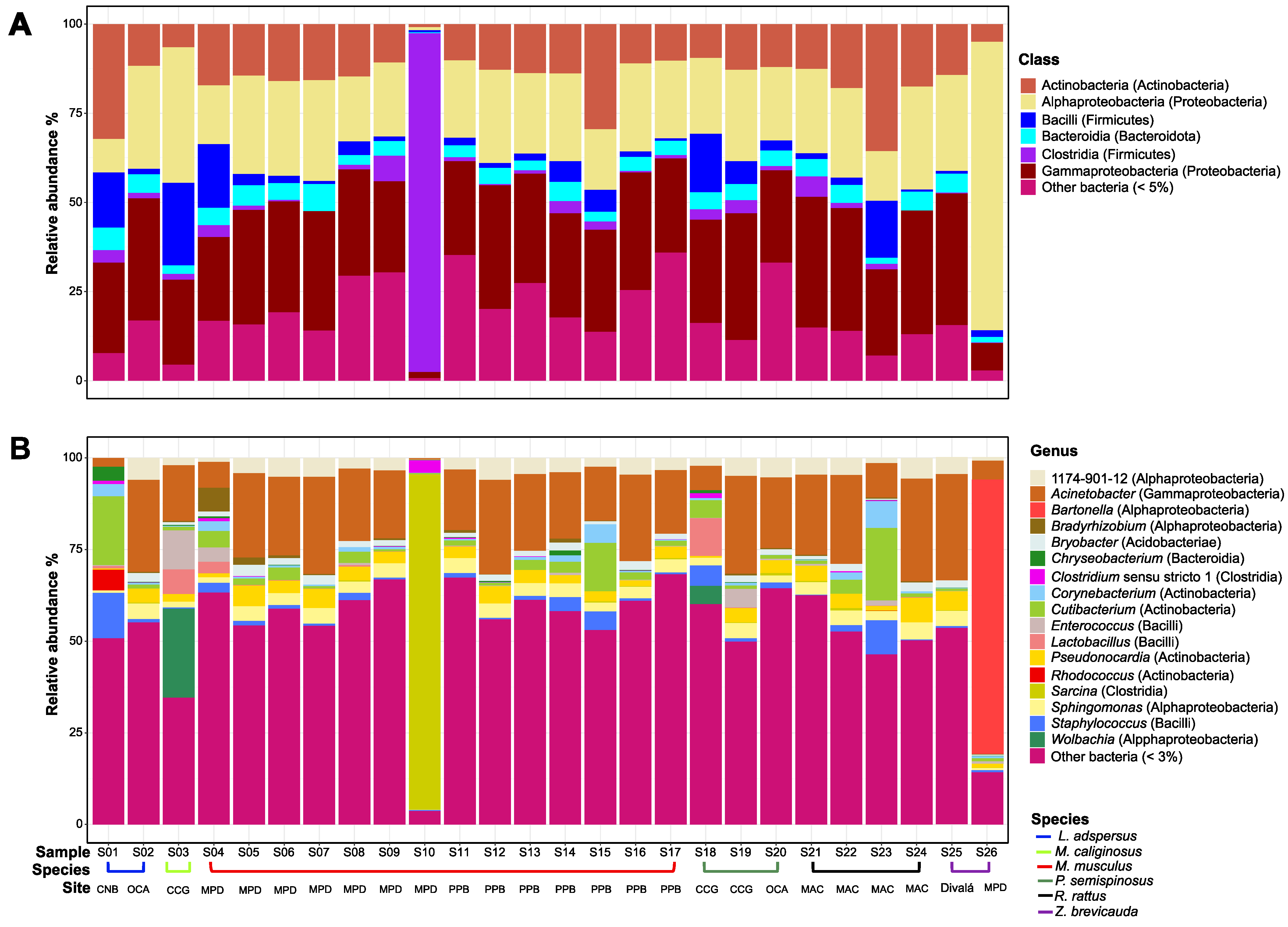
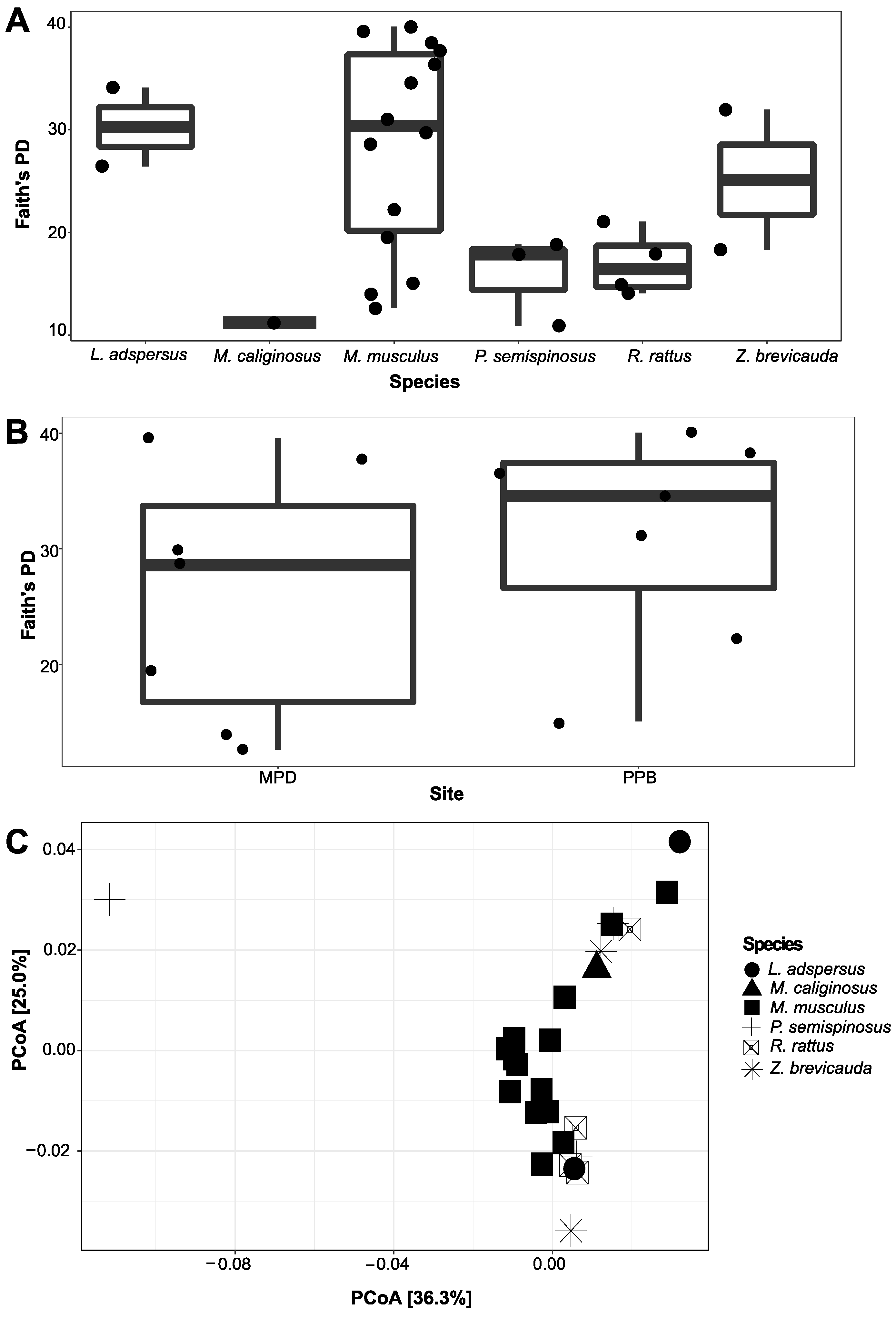
The spleen microbiome of six species of rodents was composed of 3352 ASVs. This bacterial community was represented by 25 phyla, 55 classes, 140 orders, 268 families, and 508 genera. The three predominant phyla were Actinobacteria, Firmicutes, and Proteobacteria (Figure 2A). The five predominant classes were Actinobacteria, Alpha- and Gammaproteobacteria, Bacilli, and Clostridia (Figure 2A). There were seven most dominant genera: Acinetobacter; Bartonella; Cutibacterium; Enterococcus; Sarcina; Staphylococcus; and Wolbachia. However, there were other bacterial taxa groups in less abundance, such as Bradyrhizobium, Chryseobacterium, Clostridium, Corynebacterium, Lactobacillus, Pseudonocardia, Rhodococcus, and Sphingomonas. Overall, some of them could be of clinical importance (Table 2, Figure 2B). No statistical significance in Alpha diversity was observed by species and site (Kruskal–Wallis: H = 9.38, p > 0.05, Figure 3A). Additionally, the spleen microbiome of M. musculus did not show significant differences between sites (Kruskal–Wallis: H = 1.18, p > 0.05, Figure 3B). We found a significant difference in Beta diversity between species (Adonis statistic: R2 = 0.30, p < 0.001; Anosim statistic: R = 0.59, p < 0.001, Figure 3C).

4. Discussion

A survey of the spleen microbiome of species of rodents in Panama represents an important opportunity to explore potential zoonosis bacteria in these small mammals, which are considered one of the main hosts of pathogens. Here, we assessed the bacterial community associated with six closely related species of small mammals in Panama using a massive 16S ribosomal RNA gene amplicon sequencing for the first time.

Bacterial Composition in the Spleen of Six Species of Rodents, Role in Rodents, and Implications in Zoonotic Diseases

Overall, we found 3352 ASVs associated with the six species of rodents. The most common and abundant bacterial taxa included the classes of Actinobacteria, Alpha- and Gammaproteobacteria, Bacilli, Clostridia, and Bacteroidia. Here, such genera as Acinetobacter, Bartonella, Cutibacterium, Enterococcus, Sarcina, Staphylococcus, and Wolbachia showed high abundance either in species of rodents or sites. Some of these bacterial taxa are pathogens responsible for several zoonotic diseases in humans and animals (i.e., domestic and wild animals) [10,42,43,44,45], and some genera or some species belonging to these genera are also found in rodents, which are major reservoirs [10,44,45]. For instance, Acinetobacter, which we found in all our rodent species and sites, was previously isolated from laboratory mice and rodents [46] and is associated with infections, and some species showed high drug resistance [47]. Bartonella, which was only found in high abundance in Z. brevicauda, is a common bacteria found in rodents worldwide [10,45,48,49,50,51], and some species are associated with many clinical manifestations, including endocarditis [52,53], neurologic disorders [54], and meningitis [55], among others [45]. Additionally, a study showed evidence of the transmission of Bartonella from rodents by fleas [45]. Cutibacterium, which was found in L. adspersus, contains species (i.e., Cutibacterium acnes) that are known as skin infection bacteria [56,57]. Enterococcus was found in high abundance in M. caliginosus; this bacterium is found in different animals, including free-living raptors [44], and it is a commensal organism, an opportunistic pathogen associated with the mortality of humans and animals [44,58]. On the other hand, previous studies showed that species such as Enterococcus faecalis were associated with small rodents [43,59], and it has caused inflammatory disease in mice [43]. Another observation is Sarcina, which was found in high abundance in M. musculus. Studies have shown that some species belonging to Sarcina (i.e., Sarcina ventriculi) are Gram-positive bacteria, able to survive in extremely low pH environments [60], and it is an important pathogen that is associated with a lethal disease in sanctuary chimpanzees [61]. Staphylococcus was found in high abundance in L. adspersus. It contains some species, such as Staphylococcus aureus; this species is a commensal bacteria of the human skin and gastrointestinal tract, which causes infections [62]. Finally, Wolbachia was found in the spleen of rodents, and it was also found in high abundance in M. caliginosus. This bacterium is associated with insects [63,64,65,66] and has implications for ecology and reproduction in various insects [63,67,68,69].
This study is the first step in screening bacterial taxa with the potential for zoonosis in the rodents surveyed in Panama. Although we found several pathogenic bacteria, more studies are needed to accurately estimate their potential for zoonosis in the country. Further research is also needed to assess the core microbiome associated with different species of rodents and which ones have a higher potential for zoonosis.

5. Conclusions

This study resulted in the identification and relative abundance of important bacterial taxa with the potential for zoonosis in six rodent species in a neotropical country. More studies are needed to determine which of the rodent species studied have a higher potential for bacterial zoonosis and which environmental conditions, for example, rural vs. suburban or urban settings, may drive bacterial zoonosis.

Supplementary Materials

The following supporting information can be downloaded at https://www.mdpi.com/article/10.3390/zoonoticdis4020015/s1, Figure S1: Rarefaction curves of bacterial phylogenetic diversity (Faith’s PD, ±SE) associated with species of rodents from Panama (A) and associated with M. musculus from two sites (B). Inner plot showed rarefaction curves from other sites collected.

Author Contributions

Conceptualization, G.G., A.M.C., B.A. and L.C.M.; methodology and data generation, G.G., P.G., B.A. and L.C.M.; formal analysis, A.M.C., B.A. and L.C.M.; writing—original draft preparation, G.G., A.M.C., P.G., B.A. and L.C.M.; writing—review and editing, G.G., A.M.C., P.G., B.A. and L.C.M.; funding acquisition, B.A. and L.C.M. All authors have read and agreed to the published version of the manuscript.

Funding

This research was funded by the Ministry of Economy and Finance of Panama (Grant: FPI-MEF-056), “Epidemiología y ecología de Hantavirus, otras enfermedades zoonóticas y transmitidas por vectores (emergentes y re-emergentes) en Panamá”, Fase-I y Fase II (PhoEZyTV I-II) to B.A. 2010–2014, and the Ministry of Economy and Finance of Panama [Grant number 111130150.501.274], B.A., 2014–2019. A.M.C., B.A. and L.C.M. acknowledge support from the Sistema Nacional de Investigación (SNI-SENACYT). L.C.M. participation was also supported by SENACYT grant IDDS15-208 and INDICASAT internal grant LM13-2020.

Institutional Review Board Statement

This study was evaluated and approved by the Institutional Animal Care and Use Committee of the Gorgas Memorial Institute for Health Studies (# 001/05 CIUCAL/ICGES, 4 July 2005), using the criteria established in the “International Guiding Principles for Biomedical Research Involving Animals developed by the Council for International Organizations of Medical Sciences (CIOMIS). This study was in accordance with Law No. 23 of 15 January 1997 (Animal Welfare Assurance) of the Republic of Panama.

Informed Consent Statement

Not applicable.

Data Availability Statement

The original contributions presented in the study are included in the article/supplementary material, further inquiries can be directed to the corresponding authors.

Acknowledgments

We thank the Panamanian Ministry of Environmental Affairs, the Gorgas Committee for Animal Care and Use, the Gorgas Memorial Institute for Health Studies (GMISH), the Ministry of Health, the Ministry of Agricultural Development, and the Ministry of Environment for their support. We also thank individuals from the communities and areas surveyed, sever state organization, and the rodent ecology team of the Ministry of Health and GMISH, especially Eustiquio Broce, Omar Vargas (R.I.P), Francisco Crespo, Carlos Falcon, Ricardo Rodriguez, Juan Bosco Navarro, Santiago Bosh, Ricardo Cedeño, Daniel Gonzalez, Carlos Falconet, Jose Miguel Montenegro, Miguel Vergara, Victor Dominguez, Ariel Perez, Joel Gonzalez, Algis Vergara, Heriberto Rivera, Cesar Lombardo, Domingo Archibold, Leonel Castillo, Alexis Ortiz, Donderis Soto, Carmelo Ortiz (R.I.P), Alexis Araúz, Juán De Leon, Juán Díaz, Ramón García, Jose Santizo (R.I.P), Juan Francisco Tello, Jorge Garzón, Gilberto Niño, Ricardo Rodríguez, Dagoberto Olivares, Jony Castillo, Candelario Olivares, Emilio Salazar, Pablo Gutiérrez, Lorenzo Aldobán, Olmedo Castro, José Valencia (R.I.P), Armando Mepaquito, Silvio Bethancourt, Enrique Ramos (R.I.P), and Rafael Figueroa, for support during fieldwork. We thank Claudia Dominguez, database manager. We also thank Rosa de Vargas and Iris Reyes for the significant administrative support given as part of the Department of Research in Emerging and Zoonotic Infectious Diseases, Gorgas Memorial Institute of Health Studies.

Conflicts of Interest

The authors declare no conflicts of interest.

References

  1. Morens, D.M.; Folkers, G.K.; Anthony, S. Fauci The challenge of emerging and re-emerging infectious diseases. Nature 2004, 430, 242–249. [Google Scholar] [CrossRef] [PubMed]
  2. Magouras, I.; Brookes, V.J.; Jori, F.; Martin, A.; Pfeiffer, D.U.; Dürr, S. Emerging Zoonotic Diseases: Should We Rethink the Animal–Human Interface? Front. Vet. Sci. 2020, 7, 582743. [Google Scholar] [CrossRef] [PubMed]
  3. Rahman, M.T.; Sobur, M.A.; Islam, M.S.; Ievy, S.; Hossain, M.J.; Zowalaty, M.E.E.; Rahman, A.M.M.T.; Ashour, H.M. Zoonotic diseases: Etiology, impact, and control. Microorganisms 2020, 8, 1405. [Google Scholar] [CrossRef] [PubMed]
  4. Woolhouse, M.E.J.; Gowtage-Sequeria, S. Host range and emerging and reemerging pathogens. Emerg. Infect. Dis. 2005, 11, 1842–1847. [Google Scholar] [CrossRef] [PubMed]
  5. Wang, W.-H.; Thitithanyanont, A.; Urbina, A.N.; Wang, S.-F. Emerging and re-emerging virus. Pathogens 2021, 10, 827. [Google Scholar] [CrossRef] [PubMed]
  6. Cantas, L.; Suer, K. Review: The important bacterial zoonoses in “One Health” concept. Front. Public Health 2014, 2, 144. [Google Scholar] [CrossRef] [PubMed]
  7. Vouga, M.; Greub, G. Emerging bacterial pathogens: The past and beyond. Clin. Microbiol. Infect. 2015, 1, 12–21. [Google Scholar] [CrossRef] [PubMed]
  8. Johnson, C.K.; Hitchens, P.L.; Pandit, P.S.; Rushmore, J.; Evans, T.S.; Young, C.C.W.; Doyle, M.M. Global shifts in mammalian population trends reveal key predictors of virus spillover risk. Proc. R. Soc. B Biol. Sci. 2020, 287, 20192736. [Google Scholar] [CrossRef] [PubMed]
  9. Naveed, A.; Sara, H.; Muhammad, I.; Faheem, N.; Sadaf, B. Rodents human zoonotic pathogens transmission: Historical background and future prospects. In Rodents and Their Role in Ecology, Medicine and Agriculture; IntechOpen Limited: London, UK. [CrossRef]
  10. Dahmana, H.; Granjon, L.; Diagne, C.; Davoust, B.; Fenollar, F.; Mediannikov, O. Rodents as hosts of pathogens and related zoonotic disease risk. Pathogens 2020, 9, 202. [Google Scholar] [CrossRef]
  11. Mawanda, P.; Rwego, I.; Kisakye, J.J.; Sheil, D. Rodents as potential hosts and reservoirs of parasites along the edge of a central african forest: Bwindi impenetrable national park, South Western Uganda. Afr. Health Sci. 2020, 20, 1168–1178. [Google Scholar] [CrossRef]
  12. Luis, A.D.; Hayman, D.T.S.; O’Shea, T.J.; Cryan, P.M.; Gilbert, A.T.; Pulliam, J.R.C.; Mills, J.N.; Timonin, M.E.; Willis, C.K.R.; Cunningham, A.A.; et al. A comparison of bats and rodents as reservoirs of zoonotic viruses: Are bats special? Proc. R. Soc. B Biol. Sci. 2013, 280, 20122753. [Google Scholar] [CrossRef]
  13. Han, B.A.; Schmidt, J.P.; Bowden, S.E.; Drake, J.M. Rodent reservoirs of future zoonotic diseases. Proc. Natl. Acad. Sci. USA 2015, 112, 7039–7044. [Google Scholar] [CrossRef]
  14. Assefa, A.; Chelmala, S. Correction to: Comparison of rodent community between natural and modified habitats in Kafta-Sheraro National Park and its adjoining villages, Ethiopia: Implication for conservation. J. Basic Appl. Zool. 2019, 80, 59. [Google Scholar] [CrossRef]
  15. Hassell, J.M.; Begon, M.; Ward, M.J.; Fèvre, E.M. Urbanization and Disease Emergence: Dynamics at the Wildlife–Livestock–Human Interface. Trends Ecol. Evol. 2017, 32, 55–67. [Google Scholar] [CrossRef] [PubMed]
  16. Wasserberg, G.; Abramsky, Z.; Kotler, B.P.; Ostfeld, R.S.; Yarom, I.; Warburg, A. Anthropogenic disturbances enhance occurrence of cutaneous leishmaniasis in Israel deserts: Patterns and mechanisms. Ecol. Appl. 2003, 13, 868–881. [Google Scholar] [CrossRef]
  17. Friggens, M.M.; Beier, P. Anthropogenic disturbance and the risk of flea-borne disease transmission. Oecologia 2010, 164, 809–820. [Google Scholar] [CrossRef]
  18. Young, H.S.; Dirzo, R.; Helgen, K.M.; McCauley, D.J.; Billeter, S.A.; Kosoy, M.Y.; Osikowicz, L.M.; Salkeld, D.J.; Young, T.P.; Dittmar, K. Declines in large wildlife increase landscape-level prevalence of rodent-borne disease in Africa. Proc. Natl. Acad. Sci. USA 2014, 111, 7036–7041. [Google Scholar] [CrossRef] [PubMed]
  19. Andermann, T.; Faurby, S.; Turvey, S.T.; Antonelli, A.; Silvestro, D. The past and future human impact on mammalian diversity. Sci. Adv. 2020, 6, eabb2313. [Google Scholar] [CrossRef]
  20. Armién, A.G.; Armién, B.; Koster, F.; Pascale, J.M.; Avila, M.; Gonzalez, P.; De La Cruz, M.; Zaldivar, Y.; Mendoza, Y.; Gracia, F.; et al. Hantavirus infection and habitat associations among rodent populations in agroecosystems of Panama: Implications for human disease risk. Am. J. Trop. Med. Hyg. 2009, 81, 59–66. [Google Scholar] [CrossRef]
  21. Jahan, N.A.; Lindsey, L.L.; Larsen, P.A. The Role of Peridomestic Rodents as Reservoirs for Zoonotic Foodborne Pathogens. Vector-Borne Zoonotic Dis. 2021, 21, 133–148. [Google Scholar] [CrossRef]
  22. Armién, B.; Muñoz, C.; Cedeño, H.; Salazar, J.R.; Salinas, T.P.; González, P.; Trujillo, J.; Sánchez, D.; Mariñas, J.; Hernández, A.; et al. Hantavirus in Panama: Twenty Years of Epidemiological Surveillance Experience. Viruses 2023, 15, 1395. [Google Scholar] [CrossRef] [PubMed]
  23. Mills, J.N.; Yates, T.L.; Childs, J.E.; Parmenter, R.R.; Ksiazek, T.G.; Rollin, P.E.; Peters, C.J. Guidelines for Working with Rodents Potentially Infected with Hantavirus. J. Mammal. 1995, 76, 716. [Google Scholar] [CrossRef]
  24. Eisenberg, J.F. Mammals of the Neotropics, Volume 1; University of Chicago Press: Chicago, IL, USA, 1989. [Google Scholar]
  25. Mendez, E. Los Roedores de Panama; Impresora Pacifico, S.A.: Apartado, Panama, 1993. [Google Scholar]
  26. Reid, F.A. A Field Guide to the Mammals of Central America and Southeast Mexico. J. Mammal. 1997, 81, 912–914. [Google Scholar] [CrossRef]
  27. Hanshew, A.S.; Mason, C.J.; Raffa, K.F.; Currie, C.R. Minimization of chloroplast contamination in 16S rRNA gene pyrosequencing of insect herbivore bacterial communities. J. Microbiol. Methods 2013, 95, 149–155. [Google Scholar] [CrossRef] [PubMed]
  28. Kembel, S.W.; O’Connor, T.K.; Arnold, H.K.; Hubbell, S.P.; Wright, S.J.; Green, J.L. Relationships between phyllosphere bacterial communities and plant functional traits in a neotropical forest. Proc. Natl. Acad. Sci. USA 2014, 111, 13715–13720. [Google Scholar] [CrossRef] [PubMed]
  29. Bekele, A.; Leirs, H. Population ecology of rodents of maize fields and grassland in central Ethiopia. Belg. J. Zool. 1997, 127, 39–48. [Google Scholar]
  30. Meheretu, Y.; Sluydts, V.; Welegerima, K.; Bauer, H.; Teferi, M.; Yirga, G.; Mulungu, L.; Haile, M.; Nyssen, J.; Deckers, J.; et al. Rodent abundance, stone bund density and its effects on crop damage in the Tigray highlands, Ethiopia. Crop. Prot. 2014, 55, 61–67. [Google Scholar] [CrossRef]
  31. Caporaso, J.G.; Kuczynski, J.; Stombaugh, J.; Bittinger, K.; Bushman, F.D.; Costello, E.K.; Fierer, N.; Peña, A.G.; Goodrich, J.K.; Gordon, J.I.; et al. QIIME allows analysis of high-throughput community sequencing data. Nat. Methods 2010, 7, 335–336. [Google Scholar] [CrossRef] [PubMed]
  32. Bokulich, N.A.; Kaehler, B.D.; Rideout, J.R.; Dillon, M.; Bolyen, E.; Knight, R.; Huttley, G.A.; Gregory Caporaso, J. Optimizing taxonomic classification of marker-gene amplicon sequences with QIIME 2’s q2-feature-classifier plugin. Microbiome 2018, 6, 90. [Google Scholar] [CrossRef]
  33. Bolyen, E.; Rideout, J.R.; Dillon, M.R.; Bokulich, N.A.; Abnet, C.; Al-Ghalith, G.A.; Alexander, H.; Alm, E.J.; Arumugam, M.; Asnicar, F.; et al. QIIME 2: Reproducible, interactive, scalable, and extensible microbiome data science. PeerJ Prepr. 2018, 6, e27295v1. [Google Scholar] [CrossRef]
  34. Callahan, B.J.; McMurdie, P.J.; Rosen, M.J.; Han, A.W.; Johnson, A.A.; Holmes, S.P. DADA2: High resolution sample inference from Illumina amplicon data. Nat. Methods 2016, 13, 4–5. [Google Scholar] [CrossRef]
  35. Callahan, B.J.; Sankaran, K.; Fukuyama, J.A.; McMurdie, P.J.; Holmes, S.P. Bioconductor Workflow for Microbiome Data Analysis: From raw reads to community analyses. F1000Research 2016, 5, 1492. [Google Scholar] [CrossRef]
  36. Quast, C.; Pruesse, E.; Yilmaz, P.; Gerken, J.; Schweer, T.; Yarza, P.; Peplies, J.; Glöckner, F.O. The SILVA ribosomal RNA gene database project: Improved data processing and web-based tools. Nucleic Acids Res. 2013, 41, 590–596. [Google Scholar] [CrossRef] [PubMed]
  37. Yilmaz, P.; Parfrey, L.W.; Yarza, P.; Gerken, J.; Pruesse, E.; Quast, C.; Schweer, T.; Peplies, J.; Ludwig, W.; Glöckner, F.O. The SILVA and “all-species Living Tree Project (LTP)” taxonomic frameworks. Nucleic Acids Res. 2014, 42, 643–648. [Google Scholar] [CrossRef] [PubMed]
  38. R Development Core R: A Language and Environment for Statistical Computing; R Foundation for Statistical Computing: Vienna, Austria, 2008.
  39. Clarke, K.R. Non-parametric multivariate analyses of changes in community structure. Aust. J. Ecol. 1993, 18, 117–143. [Google Scholar] [CrossRef]
  40. Oksanen, J.; Blanchet, F.G.; Friendly, M.; Kindt, R.; Legendre, P.; Mcglinn, D.; Minchin, P.R.; O’hara, R.B.; Simpson, G.L.; Solymos, P.; et al. Vegan: Community Ecology Package. R Package Version 2.4-2. Available online: https://cran.r-project.org/package=vegan (accessed on 17 January 2021).
  41. Wickham, H. ggplot2—Elegant Graphics for Data Analysis; Springer : New York, NY, USA, 2017; Available online: https://link.springer.com/book/10.1007/978-0-387-98141-3 (accessed on 17 January 2021).
  42. Liyai, R.; Kimita, G.; Masakhwe, C.; Abuom, D.; Mutai, B.; Onyango, D.M.; Waitumbi, J. The spleen bacteriome of wild rodents and shrews from Marigat, Baringo County, Kenya. PeerJ 2021, 9, e12067. [Google Scholar] [CrossRef] [PubMed]
  43. Balish, E.; Warner, T. Enterococcus faecalis induces inflammatory bowel disease in interleukin-10 knockout mice. Am. J. Pathol. 2002, 160, 2253–2257. [Google Scholar] [CrossRef]
  44. Marrow, J.; Whittington, J.K.; Mitchell, M.; Hoyer, L.L.; Maddox, C. Prevalence and antibiotic-resistance characteristics of Enterococcus spp. isolated from free-living and captive raptors in central illinois. J. Wildl. Dis. 2009, 45, 302–313. [Google Scholar] [CrossRef]
  45. Gutiérrez, R.; Krasnov, B.; Morick, D.; Gottlieb, Y.; Khokhlova, I.S.; Harrus, S. Bartonella infection in rodents and their flea ectoparasites: An overview. Vector-Borne Zoonotic Dis. 2015, 15, 27–39. [Google Scholar] [CrossRef]
  46. Benga, L.; Feßler, A.T.; Benten, W.P.M.; Engelhardt, E.; Köhrer, K.; Schwarz, S.; Sager, M. Acinetobacter species in laboratory mice: Species survey and antimicrobial resistance. Lab. Anim. 2019, 53, 470–477. [Google Scholar] [CrossRef]
  47. Maragakis, L.L.; Perl, T.M. Acinetobacter baumannii: Epidemiology, antimicrobial resistance, and treatment options. Clin. Infect. Dis. 2008, 46, 1254–1263. [Google Scholar] [CrossRef] [PubMed]
  48. Pérez-Martínez, L.; Venzal, J.M.; González-Acuña, D.; Portillo, A.; Blanco, J.R.; Oteo, J.A. Bartonella rochalimae and other Bartonella spp. in fleas, Chile. Emerg Infect Dis. 2009, 15, 1150–1152. [Google Scholar] [CrossRef] [PubMed]
  49. Theonest, N.O.; Carter, R.W.; Amani, N.; Doherty, S.L.; Hugho, E.; Keyyu, J.D.; Mable, B.K.; Shirima, G.M.; Tarimo, R.; Thomas, K.M.; et al. Molecular detection and genetic characterization of Bartonella species from rodents and their associated ectoparasites from northern Tanzania. PLoS ONE 2019, 14, e0223667. [Google Scholar] [CrossRef] [PubMed]
  50. Landaeta-Aqueveque, C.; Moreno Salas, L.; Henríquez, A.L.; Silva-de la Fuente, M.C.; González-Acuña, D. Parasites of Native and Invasive Rodents in Chile: Ecological and Human Health Needs. Front. Vet. Sci. 2021, 8, 643742. [Google Scholar] [CrossRef] [PubMed]
  51. Saengsawang, P.; Morand, S.; Desquesnes, M.; Yangtara, S.; Inpankaew, T. Molecular detection of bartonella species in rodents residing in urban and suburban areas of central Thailand. Microorganisms 2021, 9, 2588. [Google Scholar] [CrossRef] [PubMed]
  52. Daly, J.S.; Worthington, M.G.; Brenner, D.J.; Moss, C.W.; Hollis, D.G.; Weyant, R.S.; Steigerwalt, A.G.; Weaver, R.E.; Daneshvar, M.I.; O’Connor, S.P. Rochalimaea elizabethae sp. nov. isolated from a patient with endocarditis. J. Clin. Microbiol. 1993, 31, 872–881. [Google Scholar] [CrossRef] [PubMed]
  53. Fenollar, F.; Sire, S.; Raoult, D. Bartonella vinsonii subsp. arupensis as an agent of blood culture-negative endocarditis in a human. J. Clin. Microbiol. 2005, 43, 945–947. [Google Scholar] [CrossRef] [PubMed]
  54. Welch, D.F.; Carroll, K.C.; Hofmeister, E.K.; Persing, D.H.; Robison, D.A.; Steigerwalt, A.G.; Brenner, D.J. Isolation of a new subspecies, Bartonella vinsonii subsp. arupensis, from a cattle rancher: Identity with isolates found in conjunction with Borrelia burgdorferi and Babesia microti among naturally infected mice. J. Clin. Microbiol. 1999, 37, 2598–2601. [Google Scholar] [CrossRef] [PubMed]
  55. Probert, W.; Louie, J.K.; Tucker, J.R.; Longoria, R.; Hogue, R.; Moler, S.; Graves, M.; Palmer, H.J.; Cassady, J.; Fritz, C.L. Meningitis due to a “Bartonella washoensis”-like human pathogen. J. Clin. Microbiol. 2009, 47, 2332–2335. [Google Scholar] [CrossRef]
  56. Corvec, S. Clinical and biological features of Cutibacterium (Formerly propionibacterium) avidum, an underrecognized microorganism. Clin. Microbiol. Rev. 2018, 31, e00064-17. [Google Scholar] [CrossRef]
  57. Elston, M.J.; Dupaix, J.P.; Opanova, M.I.; Atkinson, R.E. Cutibacterium acnes (formerly Proprionibacterium acnes) and Shoulder Surgery. Hawai’i J. Health Soc. Welf. 2019, 78, 3–5. [Google Scholar]
  58. Ellerbroek, L.; Mac, K.N.; Peters, J.; Hultquist, L. Hazard Potential from Antibiotic-resistant Commensals like Enterococci. J. Vet. Med. 2004, 51, 393–399. [Google Scholar] [CrossRef] [PubMed]
  59. Mallon, D.J.P.; Corkill, J.E.; Hazel, S.M.; Sian Wilson, J.; French, N.P.; Bennett, M.; Hart, C.A. Excretion of vancomycin-resistant enterococci by wild mammals. Emerg. Infect. Dis. 2002, 8, 636–638. [Google Scholar] [CrossRef] [PubMed]
  60. Marcelino, L.P.; Valentini Junior, D.F.; Machado, S.M.D.S.; Schaefer, P.G.; Rivero, R.C.; Osvaldt, A.B. Sarcina ventriculi a rare pathogen. Autops. Case Rep. 2021, 11, e2021337. [Google Scholar] [CrossRef] [PubMed]
  61. Owens, L.A.; Colitti, B.; Hirji, I.; Pizarro, A.; Jaffe, J.E.; Moittié, S.; Bishop-Lilly, K.A.; Estrella, L.A.; Voegtly, L.J.; Kuhn, J.H.; et al. A Sarcina bacterium linked to lethal disease in sanctuary chimpanzees in Sierra Leone. Nat. Commun. 2021, 12, 763. [Google Scholar] [CrossRef] [PubMed]
  62. Kim, H.K.; Missiakas, D.; Schneewind, O. Mouse models for infectious diseases caused by Staphylococcus aureus. Physiol. Behav. 2018, 176, 139–148. [Google Scholar] [CrossRef]
  63. Poinsot, D.; Charlat, S.; Merçot, H. On the mechanism of Wolbachia-induced cytoplasmic incompatibility: Confronting the models with the facts. BioEssays 2003, 25, 259–265. [Google Scholar] [CrossRef] [PubMed]
  64. Hedges, L.M.; Brownlie, J.C.; O’Neill, S.L.; Johnson, K.N. Wolbachia and Virus Protection in Insects. Science 2008, 322, 702. [Google Scholar] [CrossRef] [PubMed]
  65. Castillo, A.M.; Saltonstall, K.; Arias, C.F.; Chavarria, K.A.; Ramírez-Camejo, L.A.; Mejía, L.C.; De León, L.F. The microbiome of neotropical water striders and its potential role in codiversification. Insects 2020, 11, 758. [Google Scholar] [CrossRef]
  66. Castillo, A.M.; Chavarria, K.A.; Saltonstall, K.; Arias, C.F.; Mejía, L.C. Salinity effects on the microbiome of a Neotropical water strider. Hydrobiologia 2021, 850, 3705–3717. [Google Scholar] [CrossRef]
  67. Moran, N.A.; McCutcheon, J.P.; Nakabachi, A. Genomics and Evolution of Heritable Bacterial Symbionts. Annu. Rev. Genet. 2008, 42, 165–190. [Google Scholar] [CrossRef] [PubMed]
  68. Tang, X.; Xie, G.; Shao, K.; Bayartu, S.; Chen, Y.; Gao, G. Influence of salinity on the bacterial community composition in Lake Bosten, a large oligosaline lake in arid Northwestern China. Appl. Environ. Microbiol. 2012, 78, 4748–4751. [Google Scholar] [CrossRef] [PubMed]
  69. Schuler, H.; Egan, S.P.; Hood, G.R.; Busbee, R.W.; Driscoe, A.L.; Ott, J.R. Diversity and distribution of Wolbachia in relation to geography, host plant affiliation and life cycle of a heterogonic gall wasp. BMC Evol. Biol. 2018, 18, 37. [Google Scholar] [CrossRef] [PubMed]
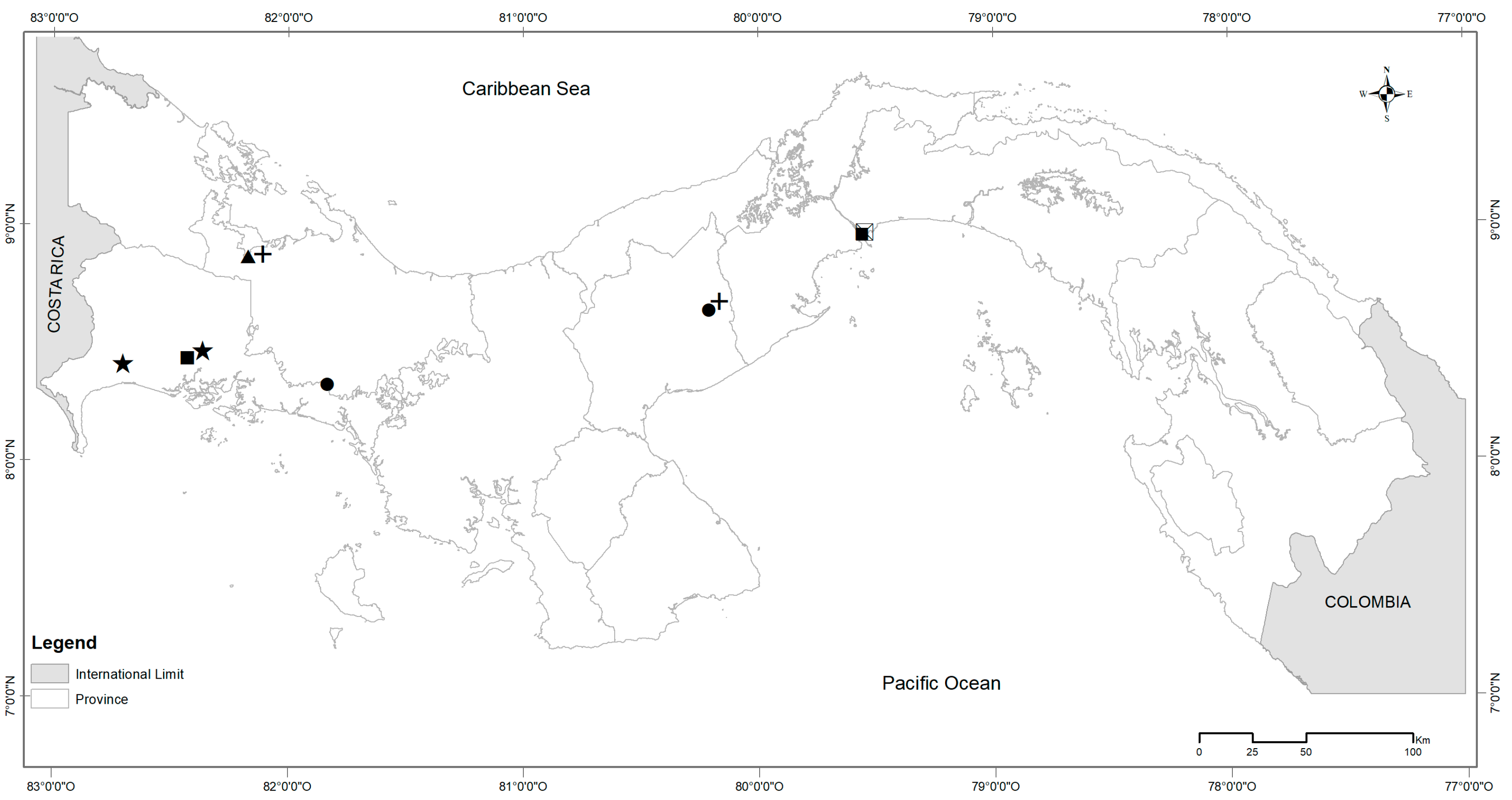
Figure 1. Sampling sites of six species of rodents in Panama. Symbols represent L. adspersus (circle), M. caliginosus (triangle), M. musculus (filled square), P. semispinosus (plus), R. rattus (square across), and Z. brevicauda (star).
Zoonoticdis 04 00015 g001
Figure 2. Relative abundance of dominant bacteria taxa associated with rodents from Panama. Abundance was estimated at the level of bacterial class (A) and the level of bacterial genus (B). The x-axis shows the sample, species, and site of rodents.
Zoonoticdis 04 00015 g002
Figure 3. Bacterial diversity associated with rodents from Panama. Graphs represent estimates of alpha diversity-based Faith’s PD for each species (A) and sites (for M. musculus) (B). Beta diversity PCoA based on weighted Unifrac distance among species (C).
Zoonoticdis 04 00015 g003
Table 1. Species of rodents collected in this study, sites, and sample size (n).
SpeciesSiten
L. adspersusComarca Ngäbe-Buglé (CNB)1
L. adspersusOajaca Chiguirí Arriba (OCA)1
M. caliginosusCañazas Chiriquí Grande (CCG)1
M. musculusMercado Público de David (MPD)7
M. musculusPanama Port Balboa (PPB)7
P. semispinosusCañazas Chiriquí Grande (CCG)2
P. semispinosusOajaca Chiguirí Arriba (OCA)1
R. rattusMercado de Abasto Curundú (MAC)4
Z. brevicaudaDivalá1
Z. brevicaudaMercado Público de David (MPD)1
Table 2. Relative abundance (%) of bacteria at the level of genus in the six species of rodents. Sites are shown in abbreviation with exception of Divala. Number of sample sizes are shown in n. Only M. musculus showed relative abundance with same number of n.
BacteriaSpecies of Rodents
GenusL.M.M.P.R.Z.
adspersuscaliginosusmusculussemispinosusrattusbrevicauda
CNBOCACCGMPDPPBCCGOCAMACDivaláMPD
n = 1n = 1n = 1n = 7n = 7n = 2n = 1n = 4n = 1n = 1
1174-901-12 (Alphaproteobacteria)0.005.971.973.163.973.545.314.114.590.66
Acinetobacter (Gammaproteobacteria)2.4625.1915.2416.7019.6216.6719.3620.9029.015.16
Bartonella (Alphaproteobacteria)0.000.000.000.000.000.000.000.000.0074.87
Bradyrhizobium (Alphaproteobacteria)0.000.360.381.450.310.250.270.220.000.22
Bryobacter (Acidobacteria)0.002.330.711.731.600.911.551.442.030.30
Chryseobacterium (Bacteroidia)3.830.110.310.120.290.540.000.030.030.11
Clostridium sensu stricto 1 (Clostridia)0.900.140.000.650.090.660.000.140.000.00
Corynebacterium (Actinobacteria)3.280.380.240.881.290.670.072.520.190.56
Cutibacterium (Actinobacteria)18.850.921.012.053.502.871.026.410.640.93
Enterococcus (Bacilli)0.160.0010.560.640.172.500.390.460.060.63
Lactobacillus (Bacilli)0.620.006.790.580.015.270.000.070.000.00
Pseudonocardia (Actinobacteria)0.334.062.083.173.042.223.443.975.101.10
Rhodococcus (Actinobacteria)5.650.000.000.000.000.000.000.020.000.00
Sarcina (Clostridia)0.000.240.0013.100.110.060.710.260.190.07
Sphingomonas (Alphaproteobacteria)0.714.111.512.853.473.121.903.664.180.58
Staphylococcus (Bacilli)12.361.070.311.191.823.261.582.860.490.59
Wolbachia (Alphaproteobacteria)0.000.0024.260.000.002.470.000.000.000.00
Other bacteria (<3%)50.8555.1234.6351.7360.7154.9964.4052.9353.4914.22
Disclaimer/Publisher’s Note: The statements, opinions and data contained in all publications are solely those of the individual author(s) and contributor(s) and not of MDPI and/or the editor(s). MDPI and/or the editor(s) disclaim responsibility for any injury to people or property resulting from any ideas, methods, instructions or products referred to in the content.