Abstract
Monkeypox (MPX) is a relatively unknown and minor resurgent viral zoonotic disease caused by the monkeypox virus (MPXV). The disease can spread from person to person or from animal to person. The disease is most prevalent in the tropical rainforests of West and Central Africa. The first MPXV outbreak was recorded in a monkey during 1958 as a small pox-like disease causing flu-like symptoms, such as chills and fever, as well as a rash, and the first MPXV case in a human was in a 9-month-old child in the Democratic Republic of the Congo on 1 September 1970. There were 16,016 laboratory confirmed cases of MPXV infection and five deaths reported in 75 countries/territories/areas across all six WHO Regions as of 22 July 2022. MPXV has a wide host range, including humans, squirrels, mice, rabbits, hamsters, porcupines, non-human primates (orangutans, chimps, sooty mangabeys, cynomolgus monkeys), black-tailed prairie dogs, African brush-tailed porcupines, rats, and shrews. MPXV replicates at the site of inoculation, the respiratory or oropharyngeal mucosa, and spreads to other organs, such as the skin, lungs, and gastrointestinal tract, where clinical signs and symptoms of the disease manifest. Before the rash appears, most patients have prominent lymphadenopathy, which distinguishes human MPX from small pox. This is followed by macules, papules, vesicles, pustules, umbilication, scabbing, and desquamation. Laboratory tools, such as virus isolation, PCR-based assays, haemagglutination inhibition assays, electron microscopy, ELISA, Western blotting, or immunohistochemistry, have been used to confirm diagnoses. Following a confirmatory diagnosis, tecovirimat, an FDA-approved antiviral drug, is currently available to treat severe cases of MPXV infection, along with symptomatic and supportive therapies. Physical and close contact activities, such as sleeping in the same room or on the same bed as the infected person, intimate contact with an infected partner, living in the same house as infected people, and sharing the same cups and plates, must be avoided to prevent the spread of the disease. Vaccination with vaccinia virus against monkeypox is approximately 85% effective and may protect against MPXV infection if administered within 4 days and up to 14 days (without showing any symptoms) after initial contact with a confirmed monkeypox case.
Keywords:
monkeypox; monkeypox virus; zoonosis; human; biosafety; diagnosis; vaccine; risk management 1. Introduction
Monkeypox (MPX) is a relatively unknown viral zoonotic disease caused by the monkeypox virus (MPXV). The disease has been most common in the West and Central Africa’s tropical rainforests. MPXV is a highly pathogenic virus that causes lesions and clinical symptoms similar to smallpox [1,2]. It has caused sporadic outbreaks in various countries in the past, and it is now re-emerging globally, causing flu-like symptoms, such as chills and fever, as well as a rash. Its re-emergence could be due to wildlife spill over. In Africa, the case fatality rate ranges from 1 to 10%, with young children dying at a higher rate [3]. However, the case-fatality rate in children who have not been immunised against smallpox ranges from 1% to 14% [4,5].
As the number of MPX cases grows on a daily basis around the world [6], current knowledge of MPX is essential. In this context, this review provides an in-depth understanding of MPX, including epidemiology, aetiology, diagnosis, treatment, biosafety and biosecurity measures, prevention and control, and so on.
2. Epidemiology and Its History
Monkeypox has most likely been present in Sub-Saharan Africa for thousands of years, ever since humans became infected with the MPXV virus from infected animals [7]. The first MPXV outbreak in a monkey was recorded in 1958 as a small pox-like disease [8,9], and the first MPXV case in a human was a 9-month-old child in the Democratic Republic of Congo on 1 September 1970, where a MPXV-like virus was isolated [10], and several cases of MPXV were recorded [11,12]. The current outbreak of MPXV is a strange and unsettling reminder of poxviruses, a largely forgotten threat since the last natural case of small pox was in Somalia in 1977, and WHO declared eradication on 8 May 1980. The virus is found naturally in West and Central Africa near tropical jungles [3]. Nigerian travellers brought the disease into Israel, Singapore, and the United Kingdom in 2018–2019 [13,14,15,16]. The disease arrived in Dallas, TX (USA) in July 2021 from Lagos, Nigeria [17].
Monkeypox affects people of all age groups, but children under the age of 16 account for the majority of cases [4]. It may happen in small villages where children under the age of 16 hunt and eat squirrels, which are thought to be a reservoir for MPXV [18]. MPXV isolates from West Africa appear to be less virulent and/or transmissible to humans and nonhuman primates than those from Central Africa’s Congo Basin. Furthermore, the discontinuation of smallpox vaccination appears to have increased humans’ susceptibility to severe monkeypox. The first human case of monkeypox was discovered in 1970 in a 9-month-old child in the Democratic Republic of the Congo (formerly Zaïre), in a region where smallpox had been eradicated in 1968 [10,19]. Six more cases of human monkeypox infection were reported in Liberia, Sierra Leone, and Nigeria the following year [20]. Between 1970 and 1979, 47 human cases of monkeypox were identified, with Zaïre accounting for 38 of them [3,7]. Between 1981 and 1986, 338 cases were reported in the Democratic Republic of the Congo, and more than 400 cases were reported between February 1996 and October 1997 [21,22]. After an outbreak in the Midwestern United States (Illinois, Indiana, Kansas, Missouri, Ohio, and Wisconsin) caused by the importation of MPXV-infected West African rodents from Ghana, the first cases of human monkeypox in the Western Hemisphere were reported in 2003 [3,23,24,25]. A timeline of monkeypox outbreaks globally at various intervals is depicted in Figure 1. As of 7 August 2022, there were a total of 27,814 laboratory confirmed cases of MPXV infection and 11 deaths reported in 89 countries/territories/areas across all six WHO Regions [6] (Figure 2 and Figure 3).
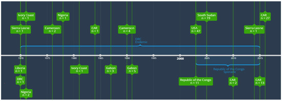
Figure 1.
Timeline of monkeypox outbreaks globally at various interval [26,27,28,29,30] Data used for making timeline from Centers for Disease Control and Prevention, 2016, Formenty et al. (2010), Learned et al. (2005), International Federation of Red Cross and Red Crescent Societies (2016), Damon et al. 2006.
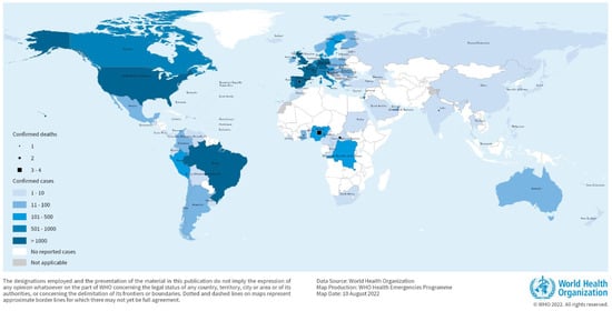
Figure 2.
Geographic distribution of confirmed cases of monkeypox reported to or identified by WHO from official public sources from 1 January 2022 to 7 August 17:00 CEST (adopted from WHO official site, https://www.who.int/publications/m/item/multi-country-outbreak-of-monkeypox--external-situation-report--3---10-august-2022) accessed on 13 August 2022 [6].
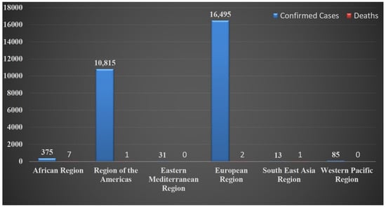
Figure 3.
Number of confirmed monkeypox cases and deaths reported as on 7 August 17:00 CEST by WHO (Numerical value taken from WHO official site).
3. Etiology, Host, and Reservoir
MPX disease is caused by MPXV, a pathogen classified as risk group 3 [31], which belongs to the family Poxviridae, subfamily Chordopoxvirinae, and genus Orthopoxvirus [7,23,32]. MPXV is a 200–250 nm brick-shaped or oval enveloped virus with characteristic surface tubules and a dumbbell-shaped core component [33,34]. The MPXV genome is made up of 197 kb of linear double-stranded DNA that can replicate in the cytoplasm but not in the nucleus [3]. The Poxviridae family is further subdivided into two sub-families, Chorodopoxvirinae and Entomopoxvirinae (Table 1).

Table 1.
Classification of pox group virus belongs to Family Poxviridae [35].
MPXV shares antigens with the variola and vaccinia viruses [19]; 30 proteins each for these three members were shown by polyacrylamide gel electrophoresis [36]. Variola, vaccinia, and MPXV are considered to be an important member of subgroup Orthopoxvirus because they cause infection in human beings [37]. Following the eradication of the smallpox virus, MPXV is regarded as the most important Orthopoxvirus infection in humans. It is divided into two clades based on genetic and phenotypic analysis: West African and Congo Basin (Central African). Clinical signs are nearly identical in infections caused by West African and Congo Basin clades viruses [38], but mortality rates vary from 1–3.6% with no direct human-to-human transmission for the West African clade (WAC) in comparison to the Central African clade (CAC) with a higher mortality rate of 10% with human-to-human transmission [39,40,41]. WAC is less virulent than CAC, which could be attributed to deletions and fragmentation in the MPXV open reading frame. These open reading frames are thought to be involved in viral life cycle, host range, or immune evasion changes, or to be virulence factors [40]. Furthermore, MPXV of CAC has been shown to suppress the host response through apoptosis [42].
Humans, squirrels, mice, rabbits, hamsters, porcupines, non-human primates (orangutans, chimps, sooty mangabeys, cynomolgus monkeys), black-tailed prairie dogs, African brush-tailed porcupines, rats, and shrews are all hosts for MPXV [3,7,10,20,21,22,23,24,25,41,43,44,45,46,47]. Although, the natural reservoir for MPXV is unknown, arboreal squirrels Funisciurus and Heliosciurus are thought to act as a reservoir for the monkeypox virus [19,21,43,48].
4. Mode of Transmission
Infections are spread through lesion fluids or crusts, respiratory secretions, and infected host tissues [7,38,49,50]. The exact mode of MPXV transmission is still unknown; however, the possible modes of MPXV transmission, animal-to-human or human-to-human transmission, are shown in Table 2.

Table 2.
Mode of transmission of Monkeypox virus.
R0 values for MPXV, or the reproductive ratio or degree of disease transmissibility, range from 1.10 to 2.40 in countries where exposure to Orthopoxvirus species is minimal [58]. This R0 implies that each infected individual has the ability to infect one to two other people. Because of its transmissibility, it is necessary to take special precautions to maintain social distance and to quarantine oneself. The transmission pattern of MPX in endemic and non-endemic settings are depicted in Figure 4.
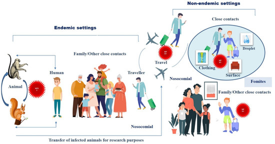
Figure 4.
Transmission of human monkeypox in endemic and non-endemic settings, where monkeypox transmission leads to an outbreak of this disease. Zoonotic factors are very important in endemic settings, while in non-endemic settings, transmission could be the result of transferring infected animals from one place to another. Human-to-human transmission occurs both in endemic and non-endemic settings (adopted form Titanji et al., 2022 [59] with minor modification).
Previously, it was thought that human-to-human transmission was not sustainable, however, the rate of transmission is increasing, with a secondary attack rate of about 10% [3,5]. MPXV can transmit from person to person; a chain of up to six sequential human-to-human transmission events has been documented [3,7,22,60,61]. Previous research has also stated that sexual transmission and spread are the most common in men who have sex with men (MSM) [62,63] (Figure 5).
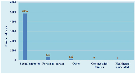
Figure 5.
Mode of transmission of monkeypox [6].
MPV cases increased 20-fold in the Democratic Republic of the Congo between the 1980s and the mid-2000s due to sexual transmission [64]. Other common risk factors for person-to-person transmission of MPXV infection include sleeping in the same room or bed with an infected person, sharing the same cups and plates [65], handling or eating dead bush meat or monkeys [66,67], and sleeping on the floor in an affected area [54].
In the current scenario, a potential threat of reverse zoonotic transmission is being monitored, as are new MPXV reservoirs, which, due to their broad host range, may result in repeated transmission to humans. Keeping rodents isolated and monitored to prevent possible transmission is a concerning situation.
5. Infectious Dose, Incubation Period and Communicability Period
MPXV infectious dose is not well understood. This virus’s incubation period ranges between 7 and 17 days [68]. It is communicated to other people 1–2 days before the rash appears and continues until all of the scabs fall off or subside.
6. Pathology and Pathophysiology
MPX pathogenesis began with MPXV transmission in the host, which could be the result of human-to-human or animal-to-human transmission (Figure 6). MPXV, like the smallpox virus, infiltrates the host system. MPXV begins to replicate at the site of inoculation, which is the respiratory or oropharyngeal mucosa. The virus enters the bloodstream after replication, resulting in primary viraemia. During primary viraemia, the virus spreads to the local lymph node via monocytic cells and replicates again. Following replication, the virus enters the bloodstream, causing secondary viraemia, and the virus spreads to other organs, such as the skin, lungs, and gastrointestinal tract, where clinical signs and symptoms of the disease manifest [68]. Infected people are thought to be most infectious during the secondary viraemic phase, also known as the prodromal phase.
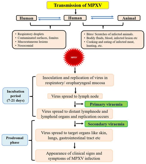
Figure 6.
Pathogenesis of monkeypox virus.
Symptoms, such as fever, myalgias, lymphadenopathy, common cold-like symptoms, and so on, appear during the prodromal phase. The enlargement of auxiliary, cervical, maxillary, and inguinal lymph nodes appear during fever onset, and this could be due to a host immune response [69]. Rashes would appear first on the face and spread across the body in a centrifugal manner after the onset of fever and/or lymphadenopathy [42,69], which means that lesions appear on the extremities and the face rather than the abdominal region. MPX rashes are vesiculopustular in nature and go through several developmental stages before desquamation. These developmental stages are enanthem, macule, papule, vesicle, pustule, and crust [42,61]. In the early stages, the lesion shows central epidermis necrosis and may extend to the superficial layer of dermis in humans [70,71]. Later on, a necrotic area surrounded by oedema and clefts develops in the interstitial space of cells, and cellular debris and fluid are deposited in this space [69,70]. Non-human primates have also been reported to have lesions, such as pustules, ulcerations, necrosis, and interstitial hyperplasia [72]. Furthermore, bronchopneumonia has been documented in humans [69], whereas non-human primates’ lungs have been shown to have fulminant bronchopneumonia, focal necrosis, and diffused pulmonary consolidation [70].
7. Diagnosis
Initially, it was thought that the clinical presentation of human MPX resembled that of smallpox in terms of clinical symptoms, severity, and mortality [73]. MPX causes mild disease symptoms and is less fatal than smallpox [74], but it can cause severe illness in young children, pregnant women, and immunocompromised individuals [75]. During the prodromal period of 2 to 3 days, important symptoms include fever, headache, body aches, and exhaustion [3,23,24].
Most patients have prominent lymphadenopathy (it may be unilateral or bilateral in submandibular, cervical, postauricular, axillary, and inguinal lymph nodes) before the onset of the rash (which distinguishes human MPX from small pox [3,23,24,76]), followed by macules, papules, vesicles, pustules, umbilication, scabbing, and desquamation [3,7]. These processes last for approximately 2–4 weeks [3,48,56,77,78].
The rash usually appears in a centrifugal pattern, spreading to the palms and soles of the feet [3]. Mucous membranes, conjunctivae, the mouth, the tongue, and the genitalia can all develop lesions [7]. Monkeypox has a similar clinical presentation to smallpox, with the exception of pronounced lymphadenopathy and generally milder symptoms [4,19]. As a result, lymphadenopathy is regarded as a key distinguishing feature of monkeypox [4,5,7]. MPX should be considered in the differential diagnosis of mucosal lesions or unusual eruptive skin rashes associated with distinct lymphadenopathy, gastrointestinal symptoms, and hematologic abnormalities in endemic settings [79]. Some complications are also reported after MPXV infection, and these complications are encephalitis [79,80], severe dehydration due to vomiting and/or diarrhoea [42,80], tonsilitis, pharyngitis [60], conjunctivitis and oedema of eyelids [60], bronchopneumonia [42], and pitted scarring [26,42]. The frequency of symptoms in reported cases of monkeypox globally are presented in Figure 7.
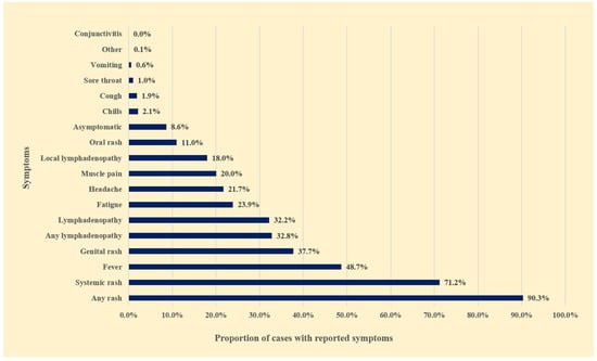
Figure 7.
Frequency of symptoms in reported cases of monkeypox globally, as of 22 July 2022 (n = 9099) (data were taken from WHO official site) [75].
Laboratory tools, such as virus isolation, PCR-based assays, haemagglutination inhibition assays, electron microscopy, ELISA, Western blotting, or immunohistochemistry, were used to confirm the diagnosis [3,5,7,19,24,32,43]. Skin lesions, such as the roof, and fluid from vesicle and pustules, should be collected in sterile containers for MPXV diagnosis. Clinical samples can also include dry crusts and blood. Samples should be stored in dry ice or at −20 °C after collection. Serology and antigen detection methods are ineffective for detecting exclusive MPXV infection, because the virus is serologically cross reactive to the other members of Orthopoxvirus; thus, antigen and antibody detection methods do not provide MPX specific confirmation [81]. However, anti-Orthopoxvirus IgG and IgM assays can be used to assess a recent or remote exposure to the Orthopoxvirus family. In cases of MPX, leukocytosis, increased transaminase levels, decreased blood urea nitrogen, and hypoalbuminemia have all been observed [79].
8. Treatment
At the moment, there is no systematic treatment for MPX disease, but antiviral drugs may be used after a confirmatory diagnosis of MPXV. Tecovirimat, an FDA-approved antiviral drug, is currently available to treat severe cases of MPXV infection [69]. This tecovirimat (ST-246 or TPOXX, an oral medication) is a small-molecule compound that targets a gene that produces the viral envelop protein VP37, a major envelope protein required for extracellular virus production [https://www.sciencedirect.com/topics/pharmacology-toxicology-and-pharmaceutical-science/tecovirimat, accessed on 25 September 2022] that aids in viraemia reduction and rapid recovery with no side effects [69,76]. Cidofovir is also being considered as a potential therapeutic agent for MPXV infections, as it has been shown to have in vitro activity against many DNA viruses, including MPXV, by inhibiting viral DNA polymerase [42,82]. Other symptomatic and supportive therapies, in addition to the above-mentioned MPX disease treatment, have been required for early recovery. Respiratory distress, bronchopneumonia, sepsis, ulcer, fever, skin lesion, and other symptoms may necessitate supportive care.
9. Prevention and Control
Contact with infected or vulnerable people should be avoided. Physical and close contact activities include sleeping in the same room or on the same bed with an infected person, intimate contact with an infected partner, living in the same house with infected people, sharing the same cups and plates, and so on. When dried, Orthopoxviruses are stable at room temperature [49]. Heat (autoclaving and incineration) inactivates it [83]. Chloroxylenol-based household disinfectants, 0.5 percent sodium hypochlorite, glutaraldehyde, formaldehyde, and paraformaldehyde are all effective against Orthopoxviruses [83].
10. Immunization and Prophylaxis
Vaccination against monkeypox with vaccinia virus (a live attenuated vaccine against smallpox virus) is approximately 85 percent effective [3,49,84]. Smallpox vaccines may protect against MPXV infection if administered within 4 days and up to 14 days (in absence of any symptoms) after initial contact with a confirmed monkeypox case [6,7,49]. Vaccination against smallpox is currently declining globally, which could be the cause of the global resurgence of MPXV infections. WHO recommends pre-exposure vaccination for health workers, laboratory personnel, outbreak response team members, and people who have multiple sexual partners [6].
11. Biosafety and Biosecurity Measures
MPXV is a risk group 3 pathogen, also known as a security sensitive biological agent (SSBA), and is thus governed by the Human Pathogens and Toxins Act (HPTA) and the Health of Animals Act (HAA). Work involving regulated infectious materials, animals, or cultures necessitates Biosafety level 3 containment facilities. Personnel entering the laboratory should remove their street clothes and jewellery, and change into laboratory-specific clothing and shoes, or wear full-coverage protective clothing (i.e., completely covering all street clothing). When infectious materials are directly handled, additional protection, such as solid-front gowns with tight-fitting wrists, gloves, and respiratory protection, should be worn over laboratory clothing. When there is a known or potential risk of splashing, eye protection must be worn [85]. All infectious material activities should be carried out in a biological safety cabinet (BSC) or other appropriate primary containment devices, along with personal protective equipment. Infected materials must be centrifuged in closed containers placed in sealed safety cups, or in rotors loaded and unloaded in a biological safety cabinet. Needles, syringes, and other sharp objects should be used with extreme caution. Waterproof dressings should be applied to open wounds, cuts, scratches, and grazes. Additional precautions should be taken when working with animals or engaging in large-scale activities [85].
In the case of a spill, allow aerosols to settle before gently covering the spill with paper towels and applying a suitable disinfectant, beginning at the perimeter and working towards the centre while wearing protective clothing. Allow enough time for contact before cleaning (30 min) [85]. Decontaminate all disposal materials using steam sterilisation, chemical disinfection, and/or incineration [85].
12. Conclusions
Monkeypox disease is a neglected viral zoonotic disease that has been reported in over 75 countries around the world. Everyone should be aware of the pathogen, clinical signs and symptoms, transmission, prevention, control, biosafety, and biosecurity in order to manage and control the disease effectively. This review provides a concise understanding of MPX. MPX surveillance, vaccination, and antiviral use can all aid in the reduction of MPX cases.
Author Contributions
Conceptualization, R.R.; Analysis, R.R.; data curation, R.R., J.K.B.; writing first draft of manuscript, R.R.; writing—review & editing, R.R., J.K.B. All authors have read and agreed to the published version of the manuscript.
Funding
This research received no external funding.
Institutional Review Board Statement
Not applicable.
Informed Consent Statement
Not applicable.
Data Availability Statement
Not applicable. All data presented in this report has been collected from public domain with proper citation and/or from media sources.
Acknowledgments
Authors are thankful to all who have supported to make this review.
Conflicts of Interest
Authors declare no conflict of interest.
References
- MacNeil, A.; Reynolds, M.; Braden, Z.; Carroll, D.S.; Bostik, V.; Karem, K.; Smith, S.K.; Davidson, W.; Li, Y.; Moundeli, A.; et al. Transmission of atypical varicella-zoster virus infections involving palm and sole manifestations in an area with monkeypox endemicity. Clin. Infect. Dis. 2009, 48, e6–e8. [Google Scholar] [CrossRef] [PubMed]
- Radonić, A.; Metzger, S.; Dabrowski, P.W.; Couacy-Hymann, E.; Schuenadel, L.; Kurth, A.; Mätz-Rensing, K.; Boesch, C.; Leendertz, F.H.; Nitsche, A. Fatal monkeypox in wild-living sooty mangabey, Cote d’Ivoire, 2012. Emerg. Infect. Dis. 2014, 20, 1009–1011. [Google Scholar] [CrossRef] [PubMed]
- Parker, S.; Nuara, A.; Buller, R.M.L.; Schultz, D.A. Human monkeypox: An emerging zoonotic disease. Future. Microbiol. 2007, 2, 17–34. [Google Scholar] [CrossRef]
- Heymann, D.L. Control of Communicable Diseases Manual, 19th ed.; American Public Health Association: Washington, DC, USA, 2008. [Google Scholar]
- Weber, D.J.; Rutala, W.A. Risks and prevention of nosocomial transmission of rare zoonotic diseases. Clin. Infect. Dis. 2001, 32, 446–456. [Google Scholar] [CrossRef] [PubMed]
- Multi-Country Outbreak of Monkeypox, External Situation Report #3—10 August 2022. Available online: https://www.who.int/publications/m/item/multi-country-outbreak-of-monkeypox--external-situation-report--3---10-august-2022 (accessed on 13 August 2022).
- Nalca, A.; Rimoin, A.W.; Bavari, S.; Whitehouse, C.A. Re-emergence of monkeypox: Prevalence, diagnostics, and countermeasures. Clin. Infect. Dis. 2005, 41, 1765–1771. [Google Scholar] [CrossRef]
- Petersen, E.; Kantele, A.; Koopmans, M.; Asogun, D.; Yinka-Ogunleye, A.; Ihekweazu, C.; Zumla, A. Human monkeypox: Epidemiologic and clinical characteristics, diagnosis, and prevention. Infect. Dis. Clin. N. Am. 2019, 33, 1027–1043. [Google Scholar] [CrossRef]
- Von Magnus, P.; Andersen, E.K.; Petersen, K.B.; Birch-Andersen, A. A pox-like disease in cynomolgus monkeys. Acta Pathol. Microbiol. Scand. 1959, 46, 156–176. [Google Scholar] [CrossRef]
- Ladnyj, I.D.; Ziegler, P.; Kima, E. A human infection caused by monkeypox virus in Basankusu Territory, Democratic Republic of the Congo. Bull. World Health Organ. 1972, 46, 593–597. [Google Scholar]
- Sklenovska, N.; Van Ranst, M. Emergence of monkeypox as the most important orthopoxvirus infection in humans. Front. Public Health 2018, 6, 241. [Google Scholar] [CrossRef]
- Heymann, D.L.; Simpson, K. The evolving epidemiology of human monkeypox: Questions still to be answered. J. Infect. Dis. 2021, 223, 1839–1841. [Google Scholar] [CrossRef]
- Sejvar, J.J.; Chowdary, Y.; Schomogyi, M.; Stevens, J.; Patel, J.; Karem, K.; Fischer, M.; Kuehnert, M.J.; Zaki, S.R.; Paddock, C.D.; et al. Human monkeypox infection: A family cluster in the midwestern United States. J. Infect. Dis. 2004, 190, 1833–1840. [Google Scholar] [CrossRef] [PubMed]
- Erez, N.; Achdout, H.; Milrot, E.; Schwartz, Y.; Wiener-Well, Y.; Paran, N.; Politi, B.; Tamir, H.; Israely, T.; Weiss, S.; et al. Diagnosis of imported monkeypox, Israel, 2018. Emerg. Infect. Dis. 2019, 25, 980–983. [Google Scholar] [CrossRef] [PubMed]
- Vaughan, A.; Aarons, E.; Astbury, J.; Brooks, T.; Chand, M.; Flegg, P.; Hardman, A.; Harper, N.; Jarvis, R.; Mawdsley, S.; et al. Human-to-human transmission of monkeypox virus, United Kingdom, October 2018. Emerg. Infect. Dis. 2020, 26, 782–785. [Google Scholar] [CrossRef]
- Yong, S.E.F.; Ng, O.T.; Ho, Z.J.M.; Mak, T.M.; Marimuthu, K.; Vasoo, S.; Yeo, T.W.; Ng, Y.K.; Cui, L.; Ferdous, Z.; et al. Imported monkeypox, Singapore. Emerg. Infect. Dis. 2020, 26, 1826–1830. [Google Scholar] [CrossRef] [PubMed]
- Rao, A.K.; Schulte, J.; Chen, T.-H.; Hughes, C.M.; Davidson, W.; Neff, J.M.; Markarian, M.; Delea, K.C.; Wada, S.; Liddell, A.; et al. Monkeypox in a traveler returning from Nigeria—Dallas, Texas, July 2021. MMWR Morb. Mortal. Wkly. Rep. 2022, 71, 509–516. [Google Scholar] [CrossRef]
- Khodakevich, C.; Jezek, Z.; Messinger, D. Monkeypox virus: Ecology and public health significance. Bull. World Health Organ. 1988, 66, 742–752. [Google Scholar]
- Acha, P.N.; Szyfres, B. Zoonoses and Communicable Diseases Common to Man and Animals, 3rd ed.; Pan American Health Organization: Washington, DC, USA, 2003; ISBN 9275119910. Available online: https://www3.paho.org/hq/index.php?option=com_content&view=article&id=2237:2010-zoonoses-communicable-diseases-common-man-animals-3rd-edition-three-volumes&Itemid=1894&lang=en#gsc.tab=0 (accessed on 25 September 2022).
- Foster, S.O.; Brink, E.W.; Hutchins, D.L.; Pifer, J.M.; Lourie, B.; Moser, C.R.; Cummings, E.C.; Kuteyi, O.E.; Eke, R.E.; Titus, J.B.; et al. Human monkeypox. Bull. World. Health. Organ. 1972, 46, 569–576. [Google Scholar]
- Pattyn, S.R. Monkeypoxvirus Infections; Revue Scientifique et Technique Office International des Epizooties: 2000. Available online: https://research.itg.be/en/publications/monkeypoxvirus-infections (accessed on 25 September 2022).
- Hutin, Y.J.; Williams, R.J.; Malfait, P.; Pebody, R.; Loparev, V.N.; Ropp, S.L.; Rodriguez, M.; Knight, J.C.; Tshioko, F.K.; Khan, A.S.; et al. Outbreak of human monkeypox, Democratic Republic of Congo, 1996 to 1997. Emerg. Infect. Dis. 2001, 7, 434–438. [Google Scholar] [CrossRef]
- Reynolds, M.G.; Davidson, W.B.; Curns, A.T.; Conover, C.S.; Huhn, G.; Davis, J.P.; Wegner, M.; Croft, D.R.; Newman, A.; Obiesie, N.N.; et al. Spectrum of infection and risk factors for human monkeypox, United States, 2003. Emerg. Infect. Dis. 2007, 13, 1332–1339. [Google Scholar] [CrossRef]
- Croft, D.R.; Sotir, M.J.; Williams, C.J.; Kazmierczak, J.J.; Wegner, M.V.; Rausch, D.; Graham, M.B.; Foldy, S.L.; Wolters, M.; Damon, I.K.; et al. Occupational risks during a monkeypox outbreak, Wisconsin, 2003. Emerg. Infect. Dis. 2007, 13, 1150–1157. [Google Scholar] [CrossRef]
- Update: Multistate outbreak of monkeypox—Illinois, Indiana, Kansas, Missouri, Ohio, and Wisconsin, 2003. Morb. Mortal. Wkly. Rep. 2003, 52, 642–646.
- Centers for Disease Control and Prevention. Available online: http://www.cdc.gov/poxvirus/monkeypox/index.html (accessed on 13 May 2016).
- Formenty, P.; Muntasir, M.O.; Damon, I.; Chowdhary, V.; Opoka, M.L.; Monimart, C.; Mutasim, E.M.; Manuguerra, J.-C.; Davidson, W.B.; Karem, K.L.; et al. Human monkeypox outbreak caused by novel virus belonging to Congo Basin clade, Sudan, 2005. Emerg. Infect. Dis. 2010, 16, 1539–1545. [Google Scholar] [CrossRef] [PubMed]
- Learned, L.A.; Bolanda, J.D.; Li, Y.; Reynolds, M.; Moudzeo, H.; Wassa, D.W.; Libama, F.; Harvey, J.M.; Likos, A.; Formenty, P.; et al. Extended interhuman transmission of monkeypox in a hospital community in the Republic of the Congo, 2003. Am. J. Trop. Med. Hyg. 2005, 73, 428–434. [Google Scholar] [CrossRef] [PubMed]
- Damon, I.K.; Roth, C.E.; Chowdhary, V. Discovery of monkeypox in Sudan. N. Engl. J. Med. 2006, 355, 962–963. [Google Scholar] [CrossRef] [PubMed]
- International Federation of Red Cross and Red Crescent Societies. Emergency Plan of Action (EPoA) CAR Monkey-Pox Epidemic Outbreak. Available online: http://reliefweb.int/sites/reliefweb.int/files/resources/MDRCF020.pdf (accessed on 20 May 2016).
- Human pathogens and toxins act. S.C. 24, Second Session, Fortieth Parliament, 57–58 Elizabeth II, 2009. Available online: https://laws.justice.gc.ca/eng/acts/H-5.67/20090623/P1TT3xt3.html (accessed on 25 September 2022).
- Dubois, M.E.; Slifka, M.K. Retrospective analysis of monkeypox infection. Emerg. Infect. Dis. 2008, 14, 592–599. [Google Scholar] [CrossRef] [PubMed]
- McFadden, G. Poxvirus tropism. Nat. Rev. Microbiol. 2005, 3, 201–213. [Google Scholar] [CrossRef] [PubMed]
- Oliveira, G.P.; Rodrigues, R.A.L.; Lima, M.T.; Drumond, B.P.; Abrahao, J.S. Poxvirus host range genes and virus-host spectrum: A critical review. Viruses 2017, 9, 331. [Google Scholar] [CrossRef]
- Hughes, A.L.; Irausquin, S.; Friedman, R. The evolutionary biology of poxviruses. Infect. Genet. Evol. 2010, 10, 50–59. [Google Scholar] [CrossRef]
- Esposito, J.J.; Obijeski, J.F.; Nakano, J.H. The virion and soluble antigen proteins of variola, monkeypox, and vaccinia viruses. J. Med. Virol. 1977, 1, 95–110. [Google Scholar] [CrossRef]
- Baxby, D. Identification and interrelationships of the variola/ vaccinia subgroup of poxviruses. Prog. Virol. Med. 1975, 19, 215–246. [Google Scholar]
- Yinka-Ogunleye, A.; Aruna, O.; Dalhat, M.; Ogoina, D.; McCollum, A.; Disu, Y.; Mamadu, I.; Akinpelu, A.; Ahmad, A.; Burga, J.; et al. Outbreak of human monkeypox in Nigeria in 2017–18: A clinical and epidemiological report. Lancet. Infect. Dis. 2019, 19, 872–879. [Google Scholar] [CrossRef]
- Likos, A.M.; Sammons, S.A.; Olson, V.A.; Frace, A.M.; Li, Y.; Olsen-Rasmussen, M.; Davidson, W.; Galloway, R.; Khristova, M.L.; Reynolds, M.G.; et al. A tale of two clades: Monkeypox viruses. J. Gen. Virol. 2005, 86, 2661–2672. [Google Scholar] [CrossRef] [PubMed]
- Reynolds, M.G.; Damon, I.K. Outbreaks of human monkeypox after cessation of smallpox vaccination. Trends Microbiol. 2012, 20, 80–87. [Google Scholar] [CrossRef] [PubMed]
- Bunge, E.M.; Hoet, B.; Chen, L.; Lienert, F.; Weidenthaler, H.; Baer, L.R.; Steffen, R. The changing epidemiology of human monkeypox-A potential threat? A systematic review. PLoS Negl. Trop. Dis. 2022, 16, e0010141. [Google Scholar] [CrossRef] [PubMed]
- McCollum, A.M.; Damon, I.K. Human monkeypox. Clin. Infect. Dis. 2014, 58, 260–267. [Google Scholar] [CrossRef]
- Mukinda, V.B.; Mwema, G.; Kilundu, M.; Heymann, D.L.; Khan, A.S.; Esposito, J.J. Re-emergence of human monkeypox in Zaire in 1996. Lancet 1997, 349, 1449–1450. [Google Scholar] [CrossRef]
- Doty, J.B.; Malekani, J.M.; Kalemba, L.N.; Stanley, W.T.; Monroe, B.P.; Nakazawa, Y.U.; Mauldin, M.R.; Bakambana, T.L.; Liyandja, T.L.D.; Braden, Z.H.; et al. Assessing monkeypox virus prevalence in small mammals at the human-animal interface in the Democratic Republic of the Congo. Viruses 2017, 9, 283. [Google Scholar] [CrossRef]
- Silva, N.I.O.; de Oliveira, J.S.; Kroon, E.G.; Trindade, G.S.; Drumond, B.P. Here, there, and everywhere: The wide host range and geographic distribution of zoonotic Orthopoxviruses. Viruses 2021, 13, 43. [Google Scholar] [CrossRef]
- Diaz, J.H. The Disease Ecology, Epidemiology, Clinical Manifestations, Management, Prevention, and Control of Increasing Human Infections with Animal Orthopoxviruses. Wilderness Environl. Med. 2021, 32, 528–536. [Google Scholar] [CrossRef]
- Patrono, L.V.; Pléh, K.; Samuni, L.; Ulrich, M.; Röthemeier, C.; Sachse, A.; Muschter, S.; Nitsche, A.; Couacy-Hymann, E.; Boesch, C.; et al. Monkeypox virus emergence in wild chimpanzees reveals distinct clinical outcomes and viral diversity. Nat. Microbiol. 2020, 5, 955–965. [Google Scholar] [CrossRef]
- Thomassen, H.A.; Fuller, T.; Asefi-Najafabady, S.; Shiplacoff, J.A.G.; Mulembakani, P.M.; Blumberg, S.; Johnston, S.C.; Kisalu, N.K.; Kinkela, T.L.; Fair, J.N.; et al. Pathogen-host associations and predicted range shifts of human monkeypox in response to climate change in central Africa. PLoS ONE 2013, 8, e66071. [Google Scholar] [CrossRef] [PubMed]
- Centers for Disease Control and Prevention. Biosafety in Microbiological and Biomedical Laboratories (BMBL), 5th ed.; Richmond, J.Y., McKinney, R.W., Eds.; Centers for Disease Control and Prevention: Washingtion, DC, USA, 2007.
- Kaler, J.; Hussain, A.; Flores, G.; Kheiri, S.; Desrosiers, D. Monkeypox: A Comprehensive Review of Transmission, Pathogenesis, and Manifestation. Cureus 2022, 14, e26531. [Google Scholar] [CrossRef] [PubMed]
- Mutombo, M.W.; Jezek, Z.; Arita, I.; Jezek, Z. Human monkeypox transmitted by a chimpanzee in a tropical rain-forest area of Zaire. Lancet 1983, 1, 735–737. [Google Scholar] [CrossRef]
- Brown, K.; Leggat, P.A. Human Monkeypox: Current State of Knowledge and Implications for the Future. Trop. Med. Infect. Dis. 2016, 1, 8. [Google Scholar] [CrossRef] [PubMed]
- Parker, S.; Buller, R.M. A review of experimental and natural infections of animals with monkeypox virus between 1958 and 2012. Futur. Virol. 2013, 8, 129–157. [Google Scholar] [CrossRef]
- Nolen, L.D.; Tamfum, J.-J.M.; Kabamba, J.; Likofata, J.; Katomba, J.; McCollum, A.M.; Monroe, B.; Kalemba, L.; Mukadi, D.; Bomponda, P.L.; et al. Introduction of monkeypox into a community and household: Risk factors and zoonotic reservoirs in the Democratic Republic of the Congo. Am. J. Trop. Med. Hyg. 2015, 93, 410–415. [Google Scholar] [CrossRef]
- Ellis, C.K.; Carroll, D.S.; Lash, R.R.; Peterson, A.T.; Damon, I.K.; Malekani, J.; Formenty, P. Ecology and geography of human monkeypox case occurrences across Africa. J. Wildl. Dis. 2012, 48, 335–347. [Google Scholar] [CrossRef]
- Yinka-Ogunleye, A.; Aruna, O.; Ogoina, D.; Aworabhi, N.; Eteng, W.; Badaru, S.; Mohammed, A.; Agenyi, J.; Etebu, E.; Numbere, T.-W.; et al. Reemergence of human monkeypox in Nigeria, 2017. Emerg. Infect. Dis. 2018, 24, 1149–1151. [Google Scholar] [CrossRef]
- Ihekweazu, C.; Yinka-Ogunleye, A.; Lule, S.; Ibrahim, A. Importance of epidemiological research of monkeypox: Is incidence increasing? Expert Rev. Anti-Infect. Ther. 2020, 18, 389–392. [Google Scholar] [CrossRef]
- Hussain, A.; Kaler, J.; Tabrez, E.; Tabrez, S.; Tabrez, S.S. Novel COVID-19: A comprehensive review of transmission, manifestation, and pathogenesis. Cureus 2020, 12, e8184. [Google Scholar] [CrossRef]
- Titanji, B.K.; Tegomoh, B.; Nematollahi, S.; Konomos, M.; Kulkarni, P.A. Monkeypox: A Contemporary Review for Healthcare Professionals. Open Forum Infect. Dis. 2022, 9, ofac310. [Google Scholar] [CrossRef] [PubMed]
- Jezek, Z.; Grab, B.; Szczeniowski, M.V.; Paluku, K.M.; Mutombo, M. Human monkeypox: Secondary attack rates. Bull. World Health Organ. 1988, 66, 465–470. [Google Scholar] [PubMed]
- Di Giulio, D.B.; Eckburg, P.B. Human monkeypox: An emerging zoonosis. Lancet Infect. Dis. 2004, 4, 15–25. [Google Scholar] [CrossRef]
- Antinori, A.; Mazzotta, V.; Vita, S.; Carletti, F.; Tacconi, D.; Lapini, L.E.; D’Abramo, A.; Cicalini, S.; Lapa, D.; Pittalis, S.; et al. Epidemiological, clinical and virological characteristics of four cases of monkeypox support transmission through sexual contact, Italy, May 2022. Eurosurveillance 2022, 27, 2200421. [Google Scholar] [CrossRef]
- Heskin, J.; Belfield, A.; Milne, C.; Brown, N.; Walters, Y.; Scott, C.; Bracchi, M.; Moore, L.S.; Mughal, N.; Rampling, T.; et al. Transmission of monkeypox virus through sexual contact—A novel route of infection. J. Infect. 2022, 85, 334–363. [Google Scholar] [CrossRef]
- Rimoin, A.W.; Mulembakani, P.M.; Johnston, S.C.; Smith, J.O.L.; Kisalu, N.K.; Kinkela, T.L.; Blumberg, S.; Thomassen, H.A.; Pike, B.L.; Fair, J.N.; et al. Major increase in human monkeypox incidence 30 years after smallpox vaccination campaigns cease in the Democratic Republic of Congo. Proc. Natl. Acad. Sci. USA 2010, 107, 16262–16267. [Google Scholar] [CrossRef]
- Nolen, L.D.; Osadebe, L.; Katomba, J.; Likofata, J.; Mukadi, D.; Monroe, B.; Doty, J.; Hughes, C.M.; Kabamba, J.; Malekani, J.; et al. Extended human-to-human transmission during a monkeypox outbreak in the Democratic Republic of the Congo. Emerg. Infect. Dis. 2016, 22, 1014–1021. [Google Scholar] [CrossRef]
- Meyer, H.; Perrichot, M.; Stemmler, M.; Emmerich, P.; Schmitz, H.; Varaine, F.; Shungu, R.; Tshioko, F.; Formenty, P. Outbreaks of disease suspected of being due to human monkeypox virus infection in the Democratic Republic of Congo in 2001. J. Clin. Microbiol. 2002, 40, 2919–2921. [Google Scholar] [CrossRef]
- Nakouné, E.; Kazanji, M. Monkeypox detection in maculopapular lesions in two young Pygmies in the Central African Republic. Int. J. Infect. Dis. 2012, 16, e266–e267. [Google Scholar] [CrossRef]
- Moore, M.; Zahra, F. Monkeypox; Stat Pearls: Treasure Island, FL, USA, 2022. [Google Scholar]
- Okyay, R.A.; Bayrak, E.; Kaya, E.; Sahin, A.R.; Kocyigit, B.F.; Tasdogan, A.M.; Avci, A.; Sumbul, H.E. Another epidemic in the shadow of Covid 19 pandemic: A review of monkeypox. EJMO 2022, 6, 95–99. [Google Scholar] [CrossRef]
- Reynolds, M.G.; McCollum, A.M.; Nguete, B.; Lushima, R.S.; Petersen, B.W. Improving the care and treatment of monkeypox patients in low-resource settings: Applying evidence from contemporary biomedical and smallpox biodefense research. Viruses 2017, 9, 380. [Google Scholar] [CrossRef] [PubMed]
- Stagles, M.; Watson, A.; Boyd, J.; More, I.; McSeveney, D. The histopathology and electron microscopy of a human monkeypox lesion. Trans. R. Soc. Trop. Med. Hyg. 1985, 79, 192–202. [Google Scholar] [CrossRef]
- Afshar, Z.M.; Rostami, H.N.; Hosseinzadeh, R.; Janbakhsh, A.; Pirzaman, A.T.; Babazadeh, A.; Aryanian, Z.; Sio, T.T.; Barary, M.; Ebrahimpour, S. The reemergence of monkeypox as a new potential health challenge: A critical review. Authorea 2022. [Google Scholar] [CrossRef]
- Chen, N.; Li, G.; Liszewski, M.K.; Atkinson, J.P.; Jahrling, P.B.; Feng, Z.; Schriewer, J.; Buck, C.; Wang, C.; Lefkowitz, E.J.; et al. Virulence differences between monkeypox virus isolates from West Africa and the Congo Basin. Virology 2005, 340, 46–63. [Google Scholar] [CrossRef]
- Rubins, K.H.; Hensley, L.E.; Relman, D.A.; Brown, P.O. Stunned silence: Gene expression programs in human cells infected with monkeypox or vaccinia virus. PLoS ONE 2011, 6, e15615. [Google Scholar] [CrossRef] [PubMed]
- Multi-Country Outbreak of Monkeypox, External Situation Report #2—25 July 2022. Available online: https://www.who.int/publications/m/item/multi-country-outbreak-of-monkeypox--external-situation-report--2---25-july-2022 (accessed on 25 July 2022).
- Damon, I.K. Status of human monkeypox: Clinical disease, epidemiology and research. Vaccine 2011, 29, D54–D59. [Google Scholar] [CrossRef]
- Jezek, Z.; Szczeniowski, M.; Paluku, K.M.; Mutombo, M. Human monkeypox: Clinical features of 282 patients. J. Infect. Dis. 1987, 156, 293–298. [Google Scholar] [CrossRef]
- Breman, J.G. Monkeypox: An emerging infection for humans? In Emerging Infections 4; American Society of Microbiology: Washington, DC, USA, 2000; pp. 45–67. [Google Scholar]
- Huhn, G.D.; Bauer, A.M.; Yorita, K.; Graham, M.B.; Sejvar, J.; Likos, A.; Damon, I.K.; Reynolds, M.G.; Kuehnert, M.J. Clinical characteristics of human monkeypox, and risk factors for severe disease. Clin. Infect. Dis. 2005, 41, 1742–1751. [Google Scholar] [CrossRef]
- Roess, A.A.; Monroe, B.P.; Kinzoni, E.A.; Gallagher, S.; Ibata, S.R.; Badinga, N.; Molouania, T.M.; Mabola, F.S.; Mombouli, J.V.; Carroll, D.S.; et al. Assessing the effectiveness of a community intervention for monkeypox prevention in the Congo basin. PLoS Negl. Trop. Dis. 2011, 5, e1356. [Google Scholar] [CrossRef]
- Quarleri, J.; Delpino, M.V.; Galvan, V. Monkeypox: Considerations for the understanding and containment of the current outbreak in non-endemic countries. Geroscience 2022, 20, 1–9. [Google Scholar] [CrossRef]
- De Clercq, E. Cidofovir in the treatment of poxvirus infections. Antivir. Res. 2002, 55, 1–13. [Google Scholar] [CrossRef]
- Butcher, W.; Ulaeto, D. Contact inactivation of Orthopoxviruses by household disinfectants. J. Appl. Microbiol. 2005, 99, 279–284. [Google Scholar] [CrossRef] [PubMed]
- Fine, P.E.; Jezek, Z.; Grab, B.; Dixon, H. The transmission potential of monkeypox virus in human populations. Int. J. Epidemiol. 1988, 17, 643–650. [Google Scholar] [CrossRef] [PubMed]
- Public Health Agency of Canada. Canadian Biosafety Standard (CBS); Government of Canada: Ottawa, ON, Canada, 2015.
Publisher’s Note: MDPI stays neutral with regard to jurisdictional claims in published maps and institutional affiliations. |
© 2022 by the authors. Licensee MDPI, Basel, Switzerland. This article is an open access article distributed under the terms and conditions of the Creative Commons Attribution (CC BY) license (https://creativecommons.org/licenses/by/4.0/).
