Case Report

Leishmania Infection during Chemotherapy in a Dog Diagnosed with Multicentric Large B-Cell Lymphoma—A Diagnostic Challenge

Department of Veterinary Sciences, Veterinary Teaching Hospital “Mario Modenato”, University of Pisa, Via Livornese (Lato Monte) 1, San Piero a Grado, 56122 Pisa, Italy
*
Author to whom correspondence should be addressed.
Zoonotic Dis. 2021, 1(1), 25-36; https://doi.org/10.3390/zoonoticdis1010003
Submission received: 1 September 2021 / Revised: 27 October 2021 / Accepted: 10 November 2021 / Published: 30 November 2021
(This article belongs to the Special Issue Feature Papers of Zoonotic Diseases 2021–2022)

Abstract

:

Simple Summary

To the best of the authors’ knowledge, the comorbidity of lymphoma and subsequent Leishmania infection, occurring as a complication during chemotherapy, has not been previously reported. Lymphoma, the side effects of chemotherapy, and leishmaniasis can lead to overlapping laboratory abnormalities. This uncommon and complex condition is thus a diagnostic challenge. This especially applies to veterinarians working in regions characterized by low or no endemicity and who might be unfamiliar with correct diagnostic and therapeutic approaches. We therefore believe that the present case report may be deemed of particular educational value.

Abstract

Dogs with lymphoma are at risk of developing clinical complications due to immunosuppression and side effects of chemotherapy. Clinical reports of concurrent lymphoma and leishmaniasis are rare and confined to single cases of comorbidity at presentation. Herein, we describe a case of lymphoma during maintenance chemotherapy in which bone marrow cytology showed myelodysplasia associated with leishmaniasis. The dog was a seven-year-old intact female Parson Russel Terrier with a two-week history of generalized lymphadenopathy. Diagnosis of multicentric high-grade B-cell lymphoma stage Va was carried out with cytological and cytofluorimetric assays of external lymph nodes, abdominal ultrasound, chest radiology, and lymphoid blasts blood smear examination. The dog lived and had traveled in endemic areas of Leishmania with uninterrupted prevention against sand fly bites by an insecticide-impregnated collar and presented seronegativity to Leishmania at presentation. Chemotherapy for lymphoma was successful and the patient achieved complete remission. Approximately eight months after the diagnosis, a persistent pancytopenia was assessed. Unexpectedly, Leishmania amastigotes were identified in the bone marrow. Combined treatment rounds were administered with antileishmanial and antineoplastic drugs for approximately eight months. Eventually, lymphoma relapsed and became unresponsive to chemotherapy, and the dog was euthanatized. Canine lymphoma overlapping with subsequent Leishmania infection as a complication is rare and lacks specific clinical manifestations. A delayed diagnosis of leishmaniasis may occur. We suggest considering leishmaniasis as part of the differential diagnosis of persistent pancytopenia in dogs with lymphoma, particularly in dogs who reside or travel to endemic areas, when treatment fails or abnormal laboratory findings are present.

1. Introduction

Lymphomas are a group of blood malignancies originating from lymphocytes that can affect all lymphoid organs, as well as other tissues. There are more than 30 different known types of canine lymphoma, varying in aggressiveness, survival rates, and clinical signs. The four most common types of lymphoma in dogs are typically characterized by the site of occurrence: (1) Multicentric, which occurs in multiple sites; (2) alimentary, which originates in the digestive system; (3) mediastinal, which arises within the chest; and (4) extranodal, which can involve the kidneys, central nervous system, skin, or other tissues. Multicentric lymphoma is by far the most common in dogs, accounting for more than 80% of diagnosed canine lymphomas [1,2]. In this type of lymphoma, lymph nodes throughout the body are affected, and, in many cases, the most obvious clinical manifestation is their rapid enlargement. Later in the course of the disease, when there are multiple large lymph nodes, dogs may show general signs of illness, including weakness, fever, loss of appetite, and dehydration. Grade and stage are important prognostic factors for lymphoma. Indolent or low-grade forms develop slowly, and dogs may show very few signs of the disease [3,4,5].
Leishmaniasis is a vector-borne parasitic infection caused by the protozoa of the genus Leishmania that includes a group of more than 20 species, mainly affecting humans and dogs. Many studies on leishmaniases have emphasized that dogs are primary hosts that naturally infect insect vectors and transmit the infection; however, a number of other vertebrate species, including cats, can also be infected [6,7]. In Mediterranean countries, including Italy, dogs are infected by the species Leishmania infantum. Dogs are the most important reservoir of this zoonotic species and transmission from dog-to-dog or from dog-to-human occurs through bites of infected female sand flies [8]. Non-vectorial, less common routes of transmission between dogs include venereal and transplacental transmission or infection through blood transfusion from infected donors [8]. Macrophages are considered the main host cells for the parasite. Leishmania amastigotes live and multiply within macrophages by binary fission. Infected cells can be lysed by multiplying amastigotes, which then spread toward uninfected cells [9]. Immune-mediated mechanisms, associated with a marked antibody response that does not confer protective immunity, are responsible for most of the pathological features in canine leishmaniasis (CanL) [9]. The clinical picture ranges from inapparent subclinical infections to systemic disease, with a highly variable spectrum of clinical manifestations. These may include weight loss, exercise intolerance, lethargy, epistaxis, skin abnormalities, lymphadenomegaly, ocular disease (keratoconjunctivitis, uveitis), splenomegaly, and onychogryphosis. Skin abnormalities may include localized or generalized exfoliative dermatitis, cutaneous ulcers, and hyperkeratosis. Other clinical findings may include polyuria and polydipsia due to kidney involvement and lameness due to joint injuries [9,10]. The disease is potentially fatal [9,10]. The prevalence of CanL is expected to increase in some countries over the next decades. In fact, some studies have reported the presence of suitable insect vectors and autochthonous cases in previously Leishmania-free European regions, such as Germany [8], Hungary [11], and northeast Italy [12]. This phenomenon is mainly caused by the introduction and local transmission of the parasite due to a northward vector expansion toward traditionally vector-free areas [10,11,12].
Although multicentric lymphoma and leishmaniasis are the main differential diagnoses in dogs presenting with generalized lymphadenomegaly [13], little attention has been paid to the pathological implications of their comorbidity. A search in the PubMed database revealed that the prevalence of Leishmania infection in dogs diagnosed with lymphoma is unknown at present. In single cases previously reported in the literature, lymphoma and leishmaniasis existed simultaneously at presentation [2,14,15,16]. Therefore, the appearance of leishmaniasis during the chemotherapy regimen remains a rare clinical scenario in veterinary medicine. The presence of one disease may eclipse another and thereby provide a challenge to small animal veterinary clinicians. Herein, we present a case of an association between multicentric large B-cell lymphoma and subsequent CanL that occurred as a clinical complication during chemotherapy for lymphoma with a 252-day follow-up period. The aim of this case report is to improve the current knowledge concerning this medical condition.

2. Case Report

2.1. Case History

At the end of January 2019, a seven-year-old intact female Parson Russel Terrier was referred to our veterinary teaching hospital, as two weeks previously, her veterinarian had found a generalized lymphadenopathy. The dog lived in Tuscany, a region of central Italy known to be an endemic area for L. infantum, and wore a slow release imidacloprid/flumethrin impregnated collar without interruption.

2.2. Clinical and Laboratory Findings and Treatment

Upon clinical examination, the dog showed no particular clinical signs related to the main problem. The diagnostic plan included a complete blood count (CBC; ProCyte Dx® IDEXX blood cell counter, IDEXX Laboratories, Milan, Italy) that revealed the occurrence of lymphoid blasts in the peripheral blood (1.63 K/µL over 7.30 K/µL of total WBC; RR 5.05–16.7) and mild thrombocytopenia (110 K/µL RR 148–484), although the white blood cell (WBC) count was within the reference interval (WRI). The serum biochemical profile (Analyzer SAT 450®, Medical System, Guidonia, Rome, Italy, with their dedicated kits) revealed a mild increase in aspartate amino transferase (AST 88 U/L; RR 15–40) and alanine amino transferase (ALT 137 U/L; RR 20–70), a consistent increase in lactic dehydrogenase (LDH 736 U/L; RR 20–160) and iron (282 mcg/dL; RR 80–190), and a decrease in total protein (5.5 g/dL; RR 5.8–7.8) and globulins (1.5 g/dL; RR 2.5–4.5). The serum protein electrophoresis (Interlab Pretty, agarose gel, Sebia, Bagno a Ripoli, FI, Italy), coagulation profile (STA Compact DOS Stago, Milan, Italy; dedicated kits), and urinalysis (IDEXX VetLab UA, IDEXX Laboratories, Milan, Italy, and manual microscopic examination of the urine sediment performed by an experienced clinical pathologist), including the protein/creatinine ratio (Analyzer SAT 450®, Medical System, Guidonia, Rome, Italy, with their dedicated kits) were WRI. Several explorable lymph nodes were biopsied and the cytological exam was compatible with large cell high-grade lymphoma (all the cytology was stained with an automatic slide stainer Aerospray Wescor, Delcon, Milan, Italy, using May–Grünwald–Giemsa staining and evaluated by an experienced clinical pathologist).
A cytofluorimetric assay (Beckman Coulter® Cytomics FC 500, Beckman Coulter Life Sciences, Cassina dei Pecchi, MI, Italy, and dedicated reagents and kits) on the fine-needle biopsies of external lymph nodes [17] revealed the prevalence of B-cell lymphocytes (positive to CD79 94.3% and CD21 92.6%). An abdominal ultrasound evidenced an increased volume of all abdominal lymph nodes (meseraic, portal, pancreatic, duodenal, and medial iliac). The chest radiology highlighted an increase in the sternal and cranial mediastinal lymph nodes. The serology screenings with the indirect fluorescence antibody test (IFAT) for Ehrlichia canis and L. infantum were negative. The IFAT protocols employed for E. canis and L. infantum were performed as previously described by other authors [18,19]. The final diagnosis was multicentric large B-cell high-grade lymphoma stage Va [3,5].
In February 2019, the dog began treatment with chemotherapy: CHOP protocol, namely, cyclophosphamide, doxorubicin, vincristine, and prednisolone as induction phase for nine weeks, then vincristine, chlorambucil, and melphalan for maintenance [20].
Eight months later (at the beginning of September), during a routine monthly check with the dog in clinical complete remission (CR) for lymphoma, a CBC revealed a peripheral blood pancytopenia. Side effects due to antineoplastic drugs used in maintenance chemotherapy were suspected. The pancytopenia continued to be consistently persistent for nearly another two months (see footnote of Table 1), but the owner was not experienced enough to easily recognize the progression of this disorder. Finally, at the end of October, additional clinicopathological tests were performed, including serum biochemistry, coagulation profile, serum protein electrophoresis (SPE), serum symmetric dimethylarginine, urinalysis, and bone marrow cytology (BMC). The results of all these investigated parameters were WRI or unremarkable. For BMC examination, smears were stained with May–Grünwald–Giemsa and examined by an experienced clinical pathologist. The BMC showed adequate cellularity for megakaryocyte, myeloid, and erythroid cells. Moreover, macrophages showed increased erythrophagocytosis, while a large number were infected by Leishmania amastigotes, which were also found extracellularly (Figure 1a,b). Real-time polymerase chain reaction (RT-PCR) on the BMC sample was carried out to identify the Leishmania species and to evaluate the parasite burden following the method reported by Trotta et al. [21]. In addition to L. infantum, other vector-borne diseases were investigated, including Anaplasma phagocytophilum/platys, Babesia spp., Bartonella spp., Ehrlichia canis, Hepatozoon spp., and Rickettsia spp. according to the methods described by Trotta et al. [21,22] and Solano-Gallego et al. [23]. As expected, the PCR resulted positive for L. infantum (347.6 K/mL copies of kinetoplasts), while other vector-borne diseases were not detected. Anti-Leishmania antibodies showed an IFAT titer of 1:1280. Therefore, the final diagnosis was consistent with myelodysplasia and CanL. The dog was then treated with meglumine antimoniate (100 mg/kg/day divided into two equal doses daily, for four weeks, SC) and allopurinol (10 mg/kg BID, PO). In addition, a minimal dose of antineoplastic drugs (chlorambucil 0.2 mg/kg, twice a week, PO), together with supplements (B-complex vitamins and omega-3, PO), were administered.

2.3. Clinical Follow-Up and Treatment

In order to progressively monitor the dog’s health status after the unexpected diagnosis of overlapping CanL (D0) as a complication, a series of occasional check-up visits were planned as follows: D14, D24, D35, D62, D85, D115, D128, D207, and D250. The laboratory results at each scheduled follow-up visit are shown in Table 1.
After two weeks of leishmanicidal treatment (D14), the CBC was still showing pancytopenia, while the urinalysis showed a urine specific gravity (USG) of 1011 and no crystalluria due to allopurinol therapy.
After a further 10 days (D24), a check-up was scheduled, and the CBC again showed persistent pancytopenia, while the urinalysis was equivalent to the previous one (USG of 1011).
Four days after the end of the treatment with antimonials (D35), the CBC showed an improvement in erythrocyte and thrombocyte values, the biochemical profile and SPE were unremarkable, and the IFAT titer for Leishmania decreased to 1:320. At this point, filgrastim was added to the treatment protocol for four days (due to the leukopenia), keeping the administration of allopurinol unchanged. After one week, a CBC was performed by the referring veterinarian, which showed a slow but gradual improvement in erythrocyte and leucocyte numbers (RBC 5.24 K/μL, RR 5.50–8.50; WBC 3.51 K/μL, RR 6.00–17.00; PLT 53 K/μL, RR 200–500).
Approximately two months after the CanL diagnosis (D62), the CBC showed a further improvement in erythrocyte and leucocyte counts, the PLT count was almost unchanged, and for the first time microcytosis was observed. At this time point, domperidone (0.5 mg/kg/day, PO) was prescribed for 28 days. The explorable lymph nodes were still of normal size.
Nearly three months after CanL diagnosis (D85), the CBC showed a further improvement in erythrocyte, mild leukopenia neutropenia related, a decrease in PLT count, and worsening of microcytosis. The BMC was thus performed again. The cellularity was still good and the myeloid/erythroid ratio was mildly increased. Several clusters of macrophages infected by Leishmania amastigotes were found again. In comparison to the previous BMC, extracellular amastigotes were not detected. The RT-PCR for Leishmania on this BMC sample showed a lower number of copies (128.8 K/mL) and the IFAT titer had risen to 1:1280. Another anti-Leishmania drug was introduced (miltefosine 2 mg/kg/day, four weeks, PO), in addition to allopurinol (10 mg/kg BID, PO), chlorambucil (0.2 mg/kg, twice a week, PO) and supplements (B-complex vitamins and omega-3, PO). After approximately three weeks of domperidone treatment that started at D62, the dog was still in CR from lymphoma.
At D115 (last day of miltefosine therapy), the physical examination revealed a relapse of lymphadenopathy in submandibular and popliteal lymph nodes. In the CBC, mild neutropenia and thrombocytopenia were still present, as well as microcytosis. The lymphoma relapse was confirmed with lymph nodes cytology and an intravenous treatment with vincristine at 0.6/sqm was resumed after approximately five months of IV chemotherapy interruption. At that time, the CD4/CD8 ratio was carried out (0.9, RI 2–1.05) and the PCR for Leishmania on lymph node aspirate was positive.
At D128 (two weeks after the vincristine administration), the lymphadenopathy was still present (stable disease), as well as microcytosis, severe neutropenia, and thrombocytopenia. In order to manage the relapse of lymphoma, L-asparaginase was introduced at 400 UI/kg three times every three weeks. In addition, a new cycle of meglumine antimoniate (100 mg/kg/day divided into two equivalent doses, for four weeks, SC) was given and the treatment with allopurinol was never interrupted after T0 (10 mg/kg BID, PO). As part of the clinical follow-up and because of the COVID-19 lockdown, the referring veterinarian was contacted by telephone for updates on the dog’s health condition. According to the telephone reports, treatment with L-asparaginase led to CR of lymphoma.
Nearly seven months after the CanL diagnosis (D207), a mild lymphadenopathy was found and cytology of the lymph nodes by a May–Grünwald–Giemsa stain smear confirmed the relapse of high-grade multicentric lymphoma (Figure 2a,b). The CBC showed microcytosis, though both the RBC and WBC parameters were WRI, while thrombocytopenia was still present. A few circulating lymphoid blasts were also found. The results from the serum biochemical profile, SPE, and urine were unremarkable. The IFAT titer had decreased to 1:160. In addition to the continuous administration of allopurinol (10 mg/kg BID, PO) and supplements (B-complex vitamins and omega-3, PO), a combination of nucleotide and hexose PO was used. For the lymphoma, lomustine was prescribed (60 mg/sqm every three weeks, PO) as a rescue therapy. Due to the current difficulty of finding this drug in Italy, the first dose was administered three weeks after the prescription.
Finally, more than 18 months after the diagnosis of lymphoma and approximately eight and a half months after the diagnosis of CanL (D250), the CBC showed microcytosis, mild thrombocytopenia, and some circulating lymphoblasts. Although the treatment with lomustine initially reduced the size of lymph nodes for approximately two weeks, a new relapse of lymphoma followed, with severe lymphadenopathy (progressive disease) and severe upper airway dyspnea, as observed upon clinical examination on D250. A few days later, the owner decided to interrupt any type of further diagnostic and therapeutic procedures and the dog was euthanized by the referring veterinarian.
Survival in dogs with primary nodal diffuse large B-cell lymphoma is defined as the time in days from the initiation of CHOP to death from any cause [24]. Overall, the dog in this case report survived 538 days from the start of CHOP until death by euthanasia, including 252 days with the coexistence of lymphoma and leishmaniosis. In this period of approximately eight and half months, the quality of life was well-maintained, although moderate lymphadenopathy was still present.

3. Discussion

As reviewed by Schwing et al. [25], the association between Leishmania and malignancies has been previously reported in humans and, to a much lesser extent, in dogs. In a literature review on humans evaluating 37 cases and involving 44 patients, Kopterides et al. [26] found the following four types of associations between Leishmania and malignant disorders: (1) Leishmaniasis mimicking a malignant disorder, such as lymphoma; (2) leishmaniasis developing as a difficult infection to diagnose and treat among patients receiving chemotherapy for various malignancies; (3) simultaneous diagnosis of leishmaniasis and a neoplastic disorder in the same tissue samples of immunocompromised patients; and (4) direct involvement of leishmaniasis in the pathogenesis and occurrence of malignant lesions, especially of the skin and mucous membranes. By arbitrarily applying the classification of Kopterides et al. [26] to dogs, the comorbidity of multicentric B-cell lymphoma and leishmaniasis observed in our study can thus be included within the second group. Previous cases in veterinary medicine have mainly reported an association between Leishmania and canine transmissible venereal tumor [27,28,29,30,31]. Other sporadic cases have reported the simultaneous presence of Leishmania and fibrosarcoma [14], perianal or adrenocortical adenoma [14,32], blood malignancies such as multiple myeloma [33] and acute myeloid leukemia [34], or different types of lymphomas located in various body sites [2,14,15,16]. To the best of our knowledge, this is the first reported case of leishmaniasis as an unexpected diagnosis during the evaluation of pancytopenia in a dog undergoing chemotherapy.
It has been shown that L. infantum triggers a series of bone marrow abnormalities in infected dogs, including dysplastic changes and erythrophagocytosis [15]. It has thus been suggested that the prolonged antigenic stimulation and chronic immunosuppression that typically occurs in dogs chronically infected with Leishmania can play a crucial role in the etiopathogenesis of some hematopoietic malignancies, such as T-cell lymphoma [15] and acute myeloid leukemia [34]. However, all dogs in previously reported cases were found to be chronically infected with Leishmania at the same time as the diagnosis of the tumor [2,14,15,16]. Therefore, since infections with Leishmania and tumors were diagnosed concurrently, it is difficult to speculate whether Leishmania infection might have occurred before or after the development of the tumoral forms. In contrast, the above-mentioned hypothesis cannot be applied to our case, since the initial seronegativity to IFAT for Leishmania at the time of lymphoma diagnosis clearly indicated that a multicentric B-cell lymphoma was pre-existing to the infection. It is possible that in our case, the association between lymphoma and subsequent leishmaniasis may have occurred simply by chance. However, an increased index of suspicion is needed in endemic areas for dogs taking drugs that strongly suppress the reproduction of cancer cells. It is possible that the comorbidity of lymphoma and Leishmania is seriously underestimated in dogs, as its epidemiology has not been widely verified. Guidelines do not recommend routine diagnostic screening for Leishmania in lymphoma-affected dogs. Instead, our case report suggests that the possible overlap of subsequent leishmaniasis should be suspected and properly investigated in dogs with multicentric large B-cell lymphoma and possible exposure to Leishmania. Leishmaniasis should therefore be ruled out during the treatment of tumoral forms and, if found, should be treated accordingly. Prospective and larger studies are needed to further define whether there are clinical pictures that justify adding leishmaniasis to the list of possible clinical complications occurring in the course of lymphoma and chemotherapy in dogs.
Pancytopenia is a medical condition in which there is a decrease in the number of circulating red blood cells, white blood cells, and platelets. Pancytopenia can develop in dogs for many different causes, including myelosuppression due to the side effects of chemotherapy [35,36], leishmaniasis [36,37], and primary or secondary myelodysplasia [38,39]. Moreover, malignant lymphoma is one of the major conditions associated with secondary myelodysplastic syndromes in dogs, which can be directly associated with the disease or be the result of chemotherapeutic drugs [39]. Analysis of morphological changes in the bone marrow of dogs with leishmaniasis has shown that megakaryocytes presented marked dysplasia [37]. In the present case report, multicentric lymphoma, chemotherapy treatment, and complication with subsequent CanL were three conditions present simultaneously, all together leading to the development and worsening of both myelodysplasia and consequent pancytopenia. Initially, pancytopenia was misdiagnosed as a consequence of chemotherapy drugs administered for seven months. Afterward, the underlying causes of pancytopenia were investigated by BMC and were found to be consistent with myelodysplasia and Leishmania infection. Other possible causes of myelodysplasia and pancytopenia in dogs (Anaplasma phagocytophilum/platys, Babesia spp., Bartonella spp., Ehrlichia canis, Hepatozoon spp., and Rickettsia spp.), were ruled out with RT-PCR. Our case report shows that the presence of persistent pancytopenia in dogs treated for lymphoma in endemic areas makes it necessary to rule out leishmaniasis. This underlines that the presentation and clinical course of leishmaniasis may be overlooked in lymphoma-affected dogs, resulting in delayed diagnosis and treatment. Moreover, diagnosis of leishmaniasis may be delayed or even totally missed, especially during chemotherapy, due to the initial negativity of serological tests unless specific diagnostic methods are routinely employed. This emphasizes the need to systematically monitor dogs treated with antineoplastic drugs for Leishmania infection when pancytopenia is present. This might help to accelerate establishing a diagnosis.
The diagnosis of CanL mainly relies on several serological techniques that determine antibody levels, PCR-based tests developed to detect and amplify Leishmania DNA in blood and tissues, and the demonstration of parasites by direct visualization with BMC [9,10]. Bone marrow biopsy is the gold standard for the diagnosis of leishmaniasis, as well as for ruling out other causes of pancytopenia [40]. Early diagnosis of CanL is crucial for the treatment and outcome of the infection. It is likely that the cell-to-cell spread of Leishmania amastigotes within macrophages of lymphoma affected dogs might be enhanced by the humoral and cellular immunosuppression status caused, in turn, by both the lymphoma and chemotherapy, thus decreasing the life expectancy. However, despite the overlap of subsequent leishmaniasis occurring during chemotherapy, the survival time (538 days) of the dog of this case report was close to or even longer than the average life expectancy of dogs diagnosed with lymphoma. In fact, median survival times of 6–12 months [4], less than two years in more than 89% of cases [41], and 218 days [3] have been reported in the literature, depending on treatment plans and lymphoma types. In another study on 98 dogs with chemotherapy protocols, 43.9% and 10.2% of them were still alive after one and two years from the initiation of CHOP, respectively, with a median overall survival time of 341 days (range 41–1068) [24].
Initially, the dog in this case report was seronegative to anti-Leishmania antibodies by IFAT. Later, CanL was unexpectedly diagnosed at the end of October, almost two months after the initial onset of pancytopenia at the beginning of September. It is likely that the infection had only been ongoing for a few months. Indeed, the activity season of sand flies and, consequently, the risk of L. infantum transmission in Italy span from spring (April–May) to autumn (October–November), depending on regional climatic conditions, when adults are found in large numbers and bite suitable vertebrate reservoir host species of the parasite for a blood meal [42,43]. Our patient had always lived in central Italy, which is an area traditionally considered endemic for CanL, and previous trips to other Italian areas where the infection is endemic were also reported. For this reason, the dog received uninterrupted preventive treatment from sand fly bites with an insecticide-impregnated collar. The effectiveness of collars impregnated with insecticide for reducing the incidence of CanL has been evaluated in field studies (with a protection range of 69–96%), which emphasize the importance of their uninterrupted use, constituting a more effective method than commercial vaccines [42,43,44,45,46]. In our case, the dog regularly wore a slow-release imidacloprid/flumethrin collar with a reported efficacy of 93.4% (range 90.5–100%) [43]; thus, the clinical suspicion of Leishmania infection was overlooked. Furthermore, the dog in this case report shared the same home environment and traveled together with its mother, which continuously wore the same type of collar to prevent sand fly bites and was seronegative to IFAT for Leishmania. This suggests that the protection from a dog collar in definitely preventing or reducing the risk of Leishmania transmission varies from dog-to-dog. Moreover, the residual risk of infected sand fly bites can never be fully eliminated, especially in immune-depressed patients. To the best of the authors’ knowledge, whether the effectiveness of insecticide-impregnated collars to prevent Leishmania infection is reduced in lymphoma-affected dogs has not been explored.
CanL has been detected in kenneled foxhounds from 18 USA states and two Canadian provinces [47]. Moreover, a likely autochthonous case has been reported in the U.K. [48]. Although sand fly species that transmit Leishmania are not present in these regions [47,49,50], dog-to-dog transmission has been established [47,48]. This strongly suggests that Leishmania infection can maintain itself within groups of infected dogs. In addition, dog owners and breeders increasingly travel with their pets or import animals from abroad. The risk of delaying or missing CanL diagnosis in highly susceptible dogs must not be underestimated. Indeed, a low index of suspicion is to be expected among small animal veterinary clinicians in areas characterized by low or no endemicity, mostly with respect to dogs lacking a history of residence in or travel to known endemic areas. Moreover, since veterinary practitioners may be unfamiliar with specific diagnostic tests and drug treatments in these regions, the diagnosis and treatment of CanL could be complicated or even unfeasible.

4. Conclusions

This case report illustrates how sometimes, a disease may hide another one, leading to misdiagnosis or delayed diagnosis because of similar clinical signs and laboratory abnormalities. Multicentric large B-cell lymphoma, side effects of chemotherapy, and Leishmania infection may be characterized by pancytopenia, which makes it difficult to distinguish between them. The comorbidity of different etiologies for pancytopenia can be diagnostically challenging. In this case report, there was no evidence to suspect the presence of underlying leishmaniasis when the dog was evaluated seven months after the diagnosis of lymphoma. At first, pancytopenia was incorrectly attributed to chemotherapy treatment. However, the persistence of pancytopenia for approximately two months led us to consider alternative causes. The diagnosis of underlying myelodysplasia and Leishmania infection by BMC explained the pancytopenia. The overall survival time of this dog affected with both lymphoma and Leishmania falls within the range reported in previous studies on multicentric lymphoma [3,4,41], and the coexistence of leishmaniosis did not significantly affect its survival.
To summarize, the aim of this case report was to raise awareness regarding the increased risk of exposure to leishmaniasis in dogs affected by hematological malignancies that live or travel through Leishmania endemic areas. An additional aim was to emphasize the pathological implications associated with the coexistence of multicentric B-cell lymphoma, chemotherapy, and the overlapping subsequent Leishmania infection as a clinical complication. The concurrence of lymphoma and subsequent Leishmania infection can lead to a misdiagnosis in dogs with the onset of pancytopenia during chemotherapy, thus delaying diagnosis and appropriate treatment for leishmaniasis and favoring L. infantum transmission. Specific diagnostic procedures are essential for the final diagnosis and should be performed upon early suspicion.

Author Contributions

Conceptualization and investigation, G.D.F., P.S. and G.L.; supervision and visualization, G.L.; writing—original draft, G.L. and R.A.P.; funding acquisition, R.A.P.; writing—review and editing, R.A.P. All authors have read and agreed to the published version of the manuscript.

Funding

The publication of the study was financially supported by the budget “Fondi di Ateneo 2020” allocated by the University of Pisa and assigned to Roberto Amerigo Papini.

Institutional Review Board Statement

The client-owned dog was treated by chemotherapy and with leishmanicidal drugs after informing the owner of the inherent risks. The owner consented and signed a release form to permit the treatments.

Informed Consent Statement

Informed consent was obtained from all subjects involved in the study.

Data Availability Statement

The raw data from this case report are available upon request to George Lubas.

Acknowledgments

The authors sincerely thank the “San Marco” Veterinary Clinic and Laboratory (Veggiano, Padova, Italy), where all serological and molecular analyses were performed, for providing its excellent technical support.

Conflicts of Interest

The authors declare no conflict of interest.

References

  1. Martineau, M.M.C. Multicentric canine lymphoma in a 12-year-old keeshond: Chemotherapy options. Can. Vet. J. 2002, 43, 709–711. [Google Scholar]
  2. Peixoto, T.C.; Freitas, J.L.; Farias, S.S.; Vieira-Filho, C.H.; Laranjeira, D.F.; Mascarenhas, M.B.; Nogueira, V.A.; Barrouin-Melo, S.M. Linfoma primário cardíaco associado à leishmaniose visceral em cão—Relato de caso. Rev. Bras. Med. Vet. 2016, 38 (Suppl. 1), 47–54. [Google Scholar]
  3. Valli, V.E.; Kass, P.H.; San Myint, M.; Scott, F. Canine lymphomas: Association of classification type, disease stage, tumor subtype, mitotic rate, and treatment with survival. Vet. Pathol. 2013, 50, 738–748. [Google Scholar] [CrossRef] [PubMed]
  4. Vail, D.M.; Pinkerton, M.; Young, K.M. Section A: Canine Lymphoma and Lymphocytic Leukemias. Lymphoma. In Hemato-poietic Tumors. Withrow and MacEwen’s Small Animal Clinical Oncology, 6th ed.; Vail, D., Thamm, D.H., Liptak, J.M., Eds.; Elsevier: St. Louis, MI, USA, 2019; pp. 688–772. Available online: https://www.ncbi.nlm.nih.gov/pmc/articles/PMC7161413/ (accessed on 13 November 2021).
  5. Zandvliet, M. Canine lymphoma: A review. Vet. Q. 2000, 36, 76–104. [Google Scholar] [CrossRef] [PubMed]
  6. Roque, A.L.; Jansen, A.M. Wild and synanthropic reservoirs of Leishmania species in the Americas. Int. J. Parasitol. Parasites Wildl. 2014, 3, 251–262. [Google Scholar] [CrossRef] [PubMed] [Green Version]
  7. Alemayehu, B.; Alemayehu, M. Leishmaniasis: A Review on Parasite, Vector and Reservoir. Host. Health Sci. J. 2017, 11, 519. [Google Scholar] [CrossRef]
  8. Naucke, T.J.; Menn, B.; Massberg, D.; Lorentz, S. Sandflies and leishmaniasis in Germany. Parasitol. Res. 2008, 103, 65–68. [Google Scholar] [CrossRef]
  9. Paltrinieri, S.; Solano-Gallego, L.; Fondati, A.; Lubas, G.; Gradoni, L.; Castagnaro, M.; Crotti, A.; Maroli, M.; Oliva, G.; Roura, X.; et al. Guidelines for diagnosis and clinical classification of leishmaniasis in dogs. J. Am. Vet. Med. Assoc. 2010, 236, 1184–1191. [Google Scholar] [CrossRef] [PubMed]
  10. Solano-Gallego, L.; Miró, G.; Koutinas, A.; Cardoso, L.; Pennisi, M.G.; Ferrer, L.; Bourdeau, P.; Oliva, G.; Baneth, G. LeishVet guidelines for the practical management of canine leishmaniosis. Parasites Vectors 2011, 4, 86. [Google Scholar] [CrossRef] [Green Version]
  11. Tánczos, B.; Balogh, N.; Király, L.; Biksi, I.; Szeredi, L.; Gyurkovsky, M.; Scalone, A.; Fiorentino, E.; Gramiccia, M.; Farkas, R. First record of autochthonous canine Leishmaniasis in Hungary. Vector Borne Zoonotic Dis. 2012, 12, 588–594. [Google Scholar] [CrossRef] [Green Version]
  12. Morosetti, G.; Toson, M.; Trevisiol, K.; Idrizi, I.; Natale, A.; Lucchese, L.; Michelutti, A.; Ceschi, P.; Lorenzi, G.; Piffer, C.; et al. Canine leishmaniosis in the Italian northeastern Alps: A survey to assess serological prevalence in dogs and distribution of phlebotomine sand flies in the Autonomous Province of Bolzano-South Tyrol, Italy. Vet. Parasitol. Reg. Stud. Rep. 2020, 21, 100432. [Google Scholar] [CrossRef] [PubMed]
  13. Melendez-Lazo, A.; Jasensky, A.K.; Jolly-Frahija, I.T.; Kehl, A.; Müller, E.; Mesa-Sánchez, I. Clonality testing in the lymph nodes from dogs with lymphadenomegaly due to Leishmania infantum infection. PLoS ONE 2019, 14, e0226336. [Google Scholar] [CrossRef]
  14. Ferro, S.; Palmieri, C.; Cavicchioli, L.; De Zan, G.; Aresu, L.; Benali, S.L. Leishmania amastigotes in neoplastic cells of 3 nonhistiocytic canine tumors. Vet. Pathol. 2013, 50, 749–752. [Google Scholar] [CrossRef] [PubMed] [Green Version]
  15. Foglia Manzillo, V.; Restucci, B.; Pagano, A.; Gradoni, L.; Oliva, G. Pathological changes in the bone marrow of dogs with leishmaniosis. Vet. Rec. 2006, 158, 690–694. [Google Scholar] [CrossRef]
  16. Loiola, M.T.A.; Silva, M.C.; Albuquerque, B.F.; Vago, P.B. Desenvolvimento de linfoma em cão com leishmaniose visceral. Ci. Anim. 2019, 29, 128–136. [Google Scholar]
  17. Sözmen, M.; Tasca, S.; Carli, E.; De Lorenzi, D.; Furlanello, T.; Caldin, M. Use of fine needle aspirates and flow cytometry for the diagnosis, classification, and immunophenotyping of canine lymphomas. J. Vet. Diagn. Investig. 2005, 17, 323–330. [Google Scholar] [CrossRef] [PubMed]
  18. Mendoza-Roldan, J.A.; Benelli, G.; Bezerra-Santos, M.A.; Nguyen, V.-L.; Conte, G.; Latta, R.; Furlanello, T.; Otranto, D. Seropositivity to canine tick-borne pathogens in a population of sick dogs in Italy. Parasites Vectors 2021, 14, 292. [Google Scholar] [CrossRef] [PubMed]
  19. Mendoza-Roldan, J.; Benelli, G.; Panarese, R.; Iatta, R.; Furlanello, T.; Beugnet, F.; Zatelli, A.; Otranto, D. Leishmania infantum and Dirofilaria immitis infections in Italy, 2009–2019: Changing distribution patterns. Parasites Vectors 2020, 13, 193. [Google Scholar] [CrossRef] [PubMed] [Green Version]
  20. Gavazza, A.; Lubas, G.; Fridman, A.; Peruzzi, D.; Impellizeri, J.A.; Luberto, L.; Marra, E.; Roscilli, G.; Ciliberto, G.; Aurisicchio, L. Safety and efficacy of a genetic vaccine targeting telomerase plus chemotherapy for the therapy of canine B-cell lymphoma. Hum. Gene Ther. 2013, 24, 728–738. [Google Scholar] [CrossRef]
  21. Trotta, M.; Nicetto, M.; Fogliazza, A.; Montarsi, F.; Caldin, M.; Furlanello, T.; Solano-Gallego, L. Detection of Leishmania infantum, Babesia canis, and rickettsiae in ticks removed from dogs living in Italy. Ticks Tick Borne Dis. 2012, 3, 293–296. [Google Scholar] [CrossRef]
  22. Trotta, M.; Fogliazza, A.; Furlanello, T.; Solano-Gallego, L. A molecular and serological study of exposure to tick-borne pathogens in sick dogs from Italy. Clin. Microbiol. Infect. 2009, 15 (Suppl. 2), 62–63. [Google Scholar] [CrossRef] [PubMed] [Green Version]
  23. Solano-Gallego, L.; Caprì, A.; Pennisi, M.G.; Caldin, M.; Furlanello, T.; Trotta, M. Acute febrile illness is associated with Rickettsia spp infection in dogs. Parasites Vectors 2015, 8, 216. [Google Scholar] [CrossRef] [Green Version]
  24. Childress, M.O.; Ramos-Vara, J.A.; Ruple, A. Retrospective analysis of factors affecting clinical outcome following CHOP-based chemotherapy in dogs with primary nodal diffuse large B-cell lymphoma. Vet. Comp. Oncol. 2017, 16, E159–E168. [Google Scholar] [CrossRef] [PubMed]
  25. Schwing, A.; Pomares, C.; Majoor, A.; Boyer, L.; Marty, P.; Michel, G. Leishmania infection: Misdiagnosis as cancer and tumor-promoting potential. Acta Trop. 2019, 197, 104855. [Google Scholar] [CrossRef] [PubMed]
  26. Kopterides, P.; Mourtzoukou, E.G.; Skopelitis, E.; Tsavaris, N.; Falagas, M.E. Aspects of the association between leishmaniasis and malignant disorders. Trans. R. Soc. Trop. Med. Hyg. 2007, 101, 1181–1189. [Google Scholar] [CrossRef] [PubMed]
  27. Albanese, F.; Poli, A.; Millanta, F.; Abramo, F. Primary cutaneous extragenital canine transmissible venereal tumour with Leishmania-laden neoplastic cells: A further suggestion of histiocytic origin? Vet. Dermatol. 2002, 13, 243–246. [Google Scholar] [CrossRef]
  28. Catone, G.; Marino, G.; Poglayen, G.; Gramiccia, M.; Ludovisi, A.; Zanghì, A. Canine transmissible venereal tumour parasitized by Leishmania infantum. Vet. Res. Commun. 2003, 27, 549–553. [Google Scholar] [CrossRef]
  29. Marino, G.; Gaglio, G.; Zanghì, A. Clinicopathological study of canine transmissible venereal tumour in leishmaniotic dogs. J. Small Anim. Pract. 2012, 53, 323–327. [Google Scholar] [CrossRef] [PubMed]
  30. Kegler, K.; Habierski, A.; Hahn, K.; Amarilla, S.P.; Seehusen, F.; Baumgärtner, W. Vaginal canine transmissible venereal tumour associated with intra-tumoural Leishmania spp. amastigotes in an asymptomatic female dog. J. Comp. Pathol. 2013, 149, 156–161. [Google Scholar] [CrossRef]
  31. Carreira, V.S.; Ferrari, H.F.; Langohr, I.M.; Mackenzie, C.; Montezzo, L.C.; Taira, E.; Floeter-Winter, L.M.; Luvizotto, M.C.R. Leishmania sp. amastigotes identification in canine transmissible venereal tumor. Case Rep. Vet. Med. 2014, 2014, 603852. [Google Scholar] [CrossRef] [Green Version]
  32. Di Mattia, D.; De Bellis, F. Leishmania spp. in perianal adenoma in a dog: A case report. Top. Companion Anim. Med. 2019, 34, 33–35. [Google Scholar] [CrossRef] [PubMed]
  33. Antognoni, M.T.; Birettoni, F.; Miglio, A.; Lalli, P.; Porciello, F.; Mangili Pecci, V. Monoclonal gammopathy associated with multiple myeloma and visceral leishmaniasis in the dog: A comparison of two cases. Vet. Res. Commun. 2010, 34 (Suppl. 1), 97–101. [Google Scholar] [CrossRef] [PubMed] [Green Version]
  34. Bezerra, J.A.B.; Rodrigues, R.T.G.A.; Lima, I.O.; Vale, A.M.; Filgueira, K.D. Acute myeloid leukemia in a dog chronically infected with Leishmania spp. and other infectious agents. Acta Sci. Vet. 2018, 46 (Suppl. 1), 260. [Google Scholar] [CrossRef] [Green Version]
  35. Weiss, D.J.; Evanson, O.A.; Sykes, J. A retrospective study of canine pancytopenia. Vet. Clin. Pathol. 1999, 28, 83–88. [Google Scholar] [CrossRef]
  36. Frezoulis, P.S.; Angelidou, E.; Karnezi, D.; Oikonomidis, I.L.; Kritsepi-Konstantinou, M.; Kasabalis, D.; Mylonakis, M.E. Canine pancytopoenia in a Mediterranean region: A retrospective study of 119 cases (2005 to 2013). J. Small Anim. Pract. 2017, 58, 395–402. [Google Scholar] [CrossRef] [PubMed]
  37. Momo, C.; Jacintho, A.P.; Moreira, P.R.; Munari, D.P.; Machado, G.F.; Vasconcelos, R.O. Morphological changes in the bone marrow of the dogs with visceral leishmaniasis. Vet. Med. Int. 2014, 2014, 150582. [Google Scholar] [CrossRef] [PubMed]
  38. Weiss, D.; Smith, S. Primary Myelodysplastic Syndromes of Dogs: A Report of 12 Cases. J. Vet. Intern. Med. 2000, 14, 491–494. [Google Scholar] [CrossRef] [PubMed]
  39. Weiss, D.; Aird, B. Cytologic evaluation of primary and secondary syndromes in the dog. Vet. Clin. Pathol. 2001, 30, 67–75. [Google Scholar] [CrossRef] [PubMed]
  40. Paparcone, R.; Fiorentino, E.; Cappiello, S.; Gizzarelli, M.; Gradoni, L.; Oliva, L.; Foglia Manzillo, V. Sternal aspiration of bone marrow in dogs: A practical approach for canine leishmaniasis diagnosis and monitoring. J. Vet. Med. 2013, 2013, 217314. [Google Scholar] [CrossRef] [Green Version]
  41. Marconato, L.; Stefanello, D.; Valenti, P.; Bonfanti, U.; Comazzi, S.; Roccabianca, P.; Caniatti, M.; Romanelli, G.; Massari, F. Predictors of long-term survival in dogs with high-grade multicentric lymphoma. J. Am. Vet. Med. Assoc. 2011, 238, 480–485. [Google Scholar] [CrossRef]
  42. Maroli, M.; Mizzon, V.; Siragusa, C.; D’Orazi, A.; Gradoni, L. Evidence for an impact on the incidence of canine leishmaniasis by the mass use of deltamethrin-impregnated dog collars in southern Italy. Med. Vet. Entomol. 2001, 15, 358–363. [Google Scholar] [CrossRef]
  43. Brianti, E.; Gaglio, G.; Napoli, E.; Falsone, L.; Prudente, C.; Solari Basano, F.; Latrofa, M.S.; Tarallo, V.D.; Dantas-Torres, F.; Capelli, G.; et al. Efficacy of a slow-release imidacloprid (10%)/flumethrin (4.5%) collar for the prevention of canine leishmaniosis. Parasites Vectors 2014, 7, 327. [Google Scholar] [CrossRef] [PubMed] [Green Version]
  44. Coura-Vital, W.; de Almeida Leal, G.G.; Araújo Marques, L.; da Costa Pinheiro, A.; Carneiro, M.; Reis, A.B. Effectiveness of deltamethrin-impregnated dog collars on the incidence of canine infection by Leishmania infantum: A large scale intervention study in an endemic area in Brazil. PLoS ONE 2018, 13, e0208613. [Google Scholar] [CrossRef] [PubMed] [Green Version]
  45. Lopes, E.G.; Sevá, A.P.; Ferreira, F.; Nunes, C.M.; Keid, L.B.; Hiramoto, R.M.; Ferreira, H.L.; Oliveira, T.M.F.S.; Ovallos, F.G.; Galati, E.A.B.; et al. Vaccine effectiveness and use of collar impregnated with insecticide for reducing incidence of Leishmania infection in dogs in an endemic region for visceral leishmaniasis, in Brazil. Epidemiol. Infect. 2018, 146, 401–406. [Google Scholar] [CrossRef] [PubMed] [Green Version]
  46. Courtenay, O.; Dilger, E.; Calvo-Bado, L.A.; Kravar-Garde, L.; Carter, V.; Bell, M.J.; Alves, G.B.; Goncalves, R.; Makhdoomi, M.M.; González, M.A.; et al. Sand fly synthetic sex-aggregation pheromone co-located with insecticide reduces the incidence of infection in the canine reservoir of visceral leishmaniasis: A stratified cluster randomised trial. PLoS Negl. Trop. Dis. 2019, 13, e0007767. [Google Scholar] [CrossRef] [PubMed]
  47. Duprey, Z.H.; Steurer, F.J.; Rooney, J.A.; Kirchhoff, L.V.; Jackson, J.E.; Rowton, E.D.; Schantz, P.M. Canine visceral leishmaniasis, United States and Canada, 2000–2003. Emerg. Infect. Dis. 2006, 12, 440–446. [Google Scholar] [CrossRef] [PubMed]
  48. McKenna, M.; Charalampos, A.; Tasker, S.; Augusto, M. Leishmaniosis in a dog with no travel history outside of the UK. Vet. Rec. 2019, 184, 441. [Google Scholar] [CrossRef] [PubMed] [Green Version]
  49. Shaw, S.E.; Langton, D.A.; Hillman, T.J. Canine leishmaniosis in the United Kingdom: A zoonotic disease waiting for a vector? Vet. Parasitol. 2009, 163, 281–285. [Google Scholar] [CrossRef]
  50. Silvestrini, P.; Batchelor, D.; Allenspach, K.; Maunder, C.; Seth, M.; Mas, A.; Hill, T.; Serrano, G.; Roura, X.; Planellas, M.; et al. Clinical leishmaniasis in dogs living in the UK. J. Small Anim. Pract. 2016, 57, 453–458. [Google Scholar] [CrossRef] [PubMed] [Green Version]
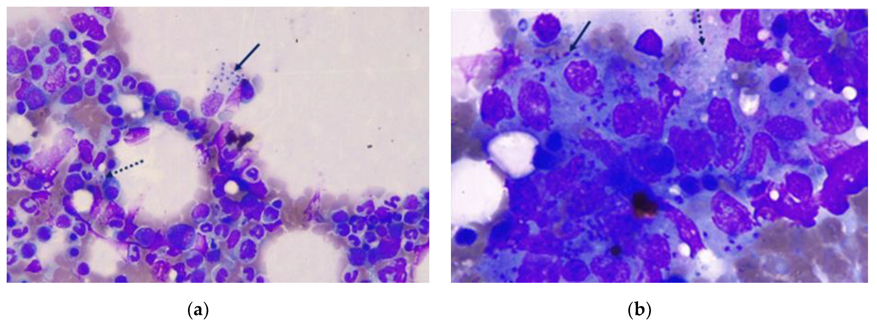
Figure 1. Bone marrow cytology in a seven-year-old intact female Parson Russel Terrier diagnosed with multicentric B-cell lymphoma eight months earlier, showing macrophages with many intracellular Leishmania infantum amastigotes at 400× (a) and 1000× (b) magnification (black solid arrows). Extracellular amastigotes (black punctuated arrows) can also be seen (May–Grünwald–Giemsa stain).
Zoonoticdis 01 00003 g001
Figure 2. Lymph node cytology in a seven-year-old intact female Parson Russell Terrier diagnosed with multicentric B-cell lymphoma 15 months earlier, showing a large amount of medium and large centroblastic lymphoid cells, along with a few normal lymphocytes and several damaged cells as a sign of lymphoma relapse at 400× (a) and 1000× (b) magnification (May–Grünwald–Giemsa stain).
Zoonoticdis 01 00003 g002
Table 1. Complete blood count results found in a seven-year-old intact female Parson Russel Terrier first diagnosed with multicentric large B-cell high-grade lymphoma and eight months later diagnosed with unexpected Leishmania infection (CanL), which occurred as a clinical complication during chemotherapy. The results are presented starting from the time of CanL diagnosis to the time of euthanasia, during a 252-day follow-up period.
Parameters and UnitsReference RangesD0 D14 D24D35D62D85D115D128D207D250
RBC M/µL5.65–8.874.744.484.585.225.836.206.355.927.006.72
HCT%37.3–61.731.429.329.032.735.737.138.736.041.940.2
HGB g/dL13.1–20.510.59.69.510.711.612.212.912.214.114.1
MCV fL61.6–73.566.765.463.362.661.259.860.960.859.959.8
MCH pg21.2–25.922.221.420.720.519.919.720.320.620.121.0
MCHC g/dL32.0–37.933.432.832.832.732.532.933.333.933.735.1
RDW%13.6–21.719.819.520.422.022.924.124.123.523.324.3
Retics K/µL10.0–110.035.619.329.349.129.745.330.526.042.728.9
Retic-HGB pg22.3–29.619.221.019.421.219.220.223.523.722.425.3
WBC K/µL5.05–16.762.592.962.542.474.033.564.341.845.0515.34
NEU seg K/µL3.7–11.91.762.431.931.982.742.783.390.964.1710.74
NEU band K/µL0.0–0.30.000.000.050.000,000.000.040.000.050.00
EOS K/µL0.1–1.350.000.000.000.100.000.070.040.000.050.00
BAS K/µL0.0–0.10.000.000.000.000.000.000.000.000.000.00
LYM K/µL0.7–5.10.360.360.460.301.050.430.690.660.504.14
MON K/µL0.2–1.50.470.180.100.100.240.280.170.220.250.46
PLT K/µL148–484376772817743414711668
MPV fL8.7–13.213.912.913.912.912.312.111.010.311.210.0
PLT estimateAdequateInadequateInadequateInadequateInadequateInadequateInadequateInadequateInadequateInadequateInadequate
D0: Day of CanL diagnosis, CBC reported at D0 is similar to values of three previous CBCs at weekly intervals; D14 onward: Days after D0; in italics are the values outside the reference ranges.
Publisher’s Note: MDPI stays neutral with regard to jurisdictional claims in published maps and institutional affiliations.