
Article

Linking Genomic Virulence and Antimicrobial Resistance Determinants to Host-Interaction Phenotypes in the Emerging Bovine Mastitis Pathogen Enterococcus lactis

by
María F. Cerioli
1,
Franco D. Fernández
2,
Melina V. Moliva
1,
Mishael Sánchez-Pérez
3,
Federico Serral
4,5,
Darío Fernandez Do Porto
4,5 and
Elina B. Reinoso
1,*
1
Institute of Environmental Biotechnology and Health (INBIAS-CONICET), Department of Microbiology and Immunology, National University of Río Cuarto, Route 36 Km 601, Río Cuarto X5804ZAB, Argentina
2
Unit of Phytopathology and Agricultural Modeling, Institute of Plant Pathology (IPAVE), Center for Agricultural Research (CIAP), National Institute of Agricultural Technology (INTA), Camino 60 Cuadras Km 5.5, Córdoba X5020ICA, Argentina
3
Faculty of Sports Science, Autonomous University of the State of Morelos, Av. Universidad 1001 Col. Chamilpa, Cuernavaca 62209, Morelos, Mexico
4
Institute of Calculus, Faculty of Exact and Natural Sciences, University of Buenos Aires, Buenos Aires C1428EGA, Argentina
5
Department of Biological Chemistry, Faculty of Exact and Natural Sciences, University of Buenos Aires, Buenos Aires C1428EGA, Argentina
*
Author to whom correspondence should be addressed.
Bacteria 2026, 5(2), 26; https://doi.org/10.3390/bacteria5020026
Submission received: 6 February 2026 / Revised: 30 March 2026 / Accepted: 24 April 2026 / Published: 9 May 2026

Abstract

Enterococcus lactis is increasingly recognized as an emerging mastitis pathogen, yet the functional basis of its virulence and associated health risks remain poorly defined. This study presents an integrated genomic and phenotypic characterization of E. lactis strain EL-A150 isolated from bovine subclinical mastitis. Whole-genome sequencing revealed a 2.49 Mb circular chromosome encoding multiple genes associated with adhesion (acm, bepA, fms, sagA), biofilm formation (empB, empC) and antimicrobial resistance, including determinants related to aminoglycosides and macrolides. Phenotypic assays demonstrated rapid growth, strong biofilm-forming capacity and high adhesion to bovine mammary epithelial cells, while internalization remained low and intracellular persistence was transient. Comparative genomic analyses confirmed the taxonomic placement of the strain within the E. lactis clade (ANI up to 99.5% against reference genomes) and revealed a limited resistome composed of chromosomally encoded genes, with no detectable plasmids or major mobile genetic elements. Collectively, these findings demonstrate that E. lactis EL-A150 possesses a coordinated set of traits conducive to intramammary colonization, supporting its classification as an opportunistic pathogen. The convergence of virulence potential and clinically relevant antimicrobial resistance within a single isolate underscores a One Health concern and highlights the need for surveillance frameworks that integrate functional validation with genomic risk assessment.

1. Introduction

Bovine mastitis remains one of the most impactful diseases in dairy production, generating substantial economic losses and compromising milk quality. This condition is caused by a wide range of environmental and contagious bacterial pathogens capable of establishing persistent infections in both clinical and subclinical forms [1,2]. Effective management depends on the accurate identification of the pathogens, a challenge compounded by the limitations of conventional phenotypic methods for discriminating closely related taxa [3]. The advent of whole-genome sequencing (WGS) has largely overcome this diagnostic hurdle, enabling precise species-level identification and revealing previously overlooked pathogens in the bovine mammary niche.
Among these emerging agents is Enterococcus lactis, a species phylogenetically close to E. faecium but now recognized as genetically and metabolically distinct [4,5]. While historically associated with dairy products, E. lactis has recently been detected in bovine mastitis cases, confirming its presence in this clinical context [6]. This shift from an environmental commensal to a putative intramammary pathogen highlights critical, unanswered questions regarding its pathogenic mechanism. Specifically, the virulence determinants that enable E. lactis to colonize and potentially damage the mammary gland remain uncharacterized. Furthermore, the functional and clinical relevance of its antimicrobial resistance repertoire is poorly understood.
Current knowledge of E. lactis in mastitis, while increasing, remains fragmented. Previous studies, including a recent genomic and phenotypic characterization from Argentina [6], have confirmed its presence and provided valuable strain-specific information. However, the contribution of genomic virulence and resistance determinants to key steps of mastitis pathogenesis has not yet been systematically evaluated within an integrated framework. Moreover, the clinical relevance, evolutionary origin, and dissemination potential of antimicrobial resistance genes within dairy ecosystems remain unresolved, limiting risk assessment from a One Health perspective at the animal–human–environment interface.
The aim of this work was to investigate the pathogenic framework of E. lactis beyond single-strain characterization. To this end, we pursued an integrated approach designed to functionally assess key virulence traits relevant to mastitis pathogenesis, including biofilm formation and adhesion to bovine mammary epithelial cells; correlate these phenotypic findings with the strain’s genomic virulence repertoire; and contextualize its antimicrobial resistance profile by evaluating the genetic support and potential dissemination risk of resistance determinants. This integrated genomic and phenotypic analysis provides new insights into the pathogenic potential of E. lactis and contributes to a broader understanding of its role as an emerging agent of bovine mastitis.

2. Materials and Methods

2.1. Bacterial Isolation, Phenotypic Identification and Storage

The E. lactis EL-A150 strain was isolated from a composite milk sample obtained from a dairy cow with subclinical mastitis in the central basin region of Argentina. Subclinical mastitis was confirmed by the veterinary service of the farm based on somatic cell count (SCC > 200,000 cells/mL) and a positive California Mastitis Test (CMT) [7]. Sample was cultured on tryptic soy agar supplemented with 5% defibrinated bovine blood and incubated aerobically at 37 °C for 24 h. A single colony with homogeneous morphology was selected and stored at −20 °C in Brain Heart Infusion (BHI) broth containing 20% glycerol.
The isolate was initially characterized by using conventional microbiological and biochemical tests for Enterococcus spp., including Gram staining, catalase test, bile esculin hydrolysis, growth in 6.5% NaCl, and growth at 45 °C, as previously described [8]. Based on these phenotypic traits, the isolate was presumptively identified as belonging to the Enterococcus faecium group. To complement this preliminary identification, the 16S rRNA gene was amplified using the universal bacterial primers 27F (5′-AGAGTTTGATCMTGGCTCAG-3′) and 1492R (5′-TACGGYTACCTTGTTACGACTT-3′). PCR was performed in a 25 µL reaction mixture containing 1× PCR buffer, 1.5 mM MgCl2, 0.2 mM each dNTP, 0.5 µM of each primer, 1 U of Taq DNA polymerase, and approximately 50 ng of DNA template. The thermal cycling conditions were: initial denaturation at 95 °C for 5 min; 30 cycles of 95 °C for 30 s, 55 °C for 30 s, and 72 °C for 90 s; and a final extension at 72 °C for 10 min. The PCR product was purified and sequenced in both directions. The resulting amplicon was sequenced and compared with reference sequences in public databases. Definitive species-level identification was subsequently achieved by whole-genome sequencing.

2.2. Phenotypic Assays

2.2.1. Bacterial Growth Curve

Ten isolated colonies grown on Trypticase Soy agar (TS; Britania, Argentina) were inoculated into 5 mL of TS broth and incubated at 37 °C with shaking (180 rpm) for 18 h. Overnight cultures were diluted 1:100 into fresh TS broth and incubated under the same conditions. Samples were collected every 2 h over a 72 h period. Total cell density was monitored by measuring OD660, and viable counts were obtained by serial dilution and plating following the standard viable count method [9]. Each growth curve assay was independently repeated twice.

2.2.2. Biofilm Formation Assay

Biofilm formation on abiotic surfaces was assessed using the standardized microtiter plate method described by [10], a widely adopted approach for initial screening and quantification of biofilm biomass that allows high-throughput assessment across multiple experimental conditions, with minor modifications. Briefly, diluted bacterial cultures were inoculated into 96-well flat-bottom plates containing TS broth and incubated at 37 °C for 24 h. Wells were washed, fixed, and stained with 0.1% crystal violet. After solubilization, absorbance was measured at 560 nm. Each isolate was tested in quadruplicate across two independent experiments. Staphylococcus epidermidis was used as the positive control and sterile TS broth served as negative control.
To assess the temporal dynamics of biofilm formation, a separate kinetic assay was conducted over a period of 72 h.
To evaluate the effect of different milk components on biofilm formation, the standard protocol described above was modified by supplementing TS broth with distinct carbon sources: glucose (0.5% w/v), lactose (0.5% w/v), or casein (0.5% w/v). Biofilm formation was assessed after 24 h of incubation at 37 °C under static conditions, following the same staining and quantification procedure.

2.2.3. Adherence, Internalization and Intracellular Survival in MAC-T Cells

Adherence and internalization assays were performed using MAC-T bovine mammary epithelial cells following [11]. Cells were cultured in DMEM supplemented with hydrocortisone (4 μg/mL), heat-inactivated fetal bovine serum (10%; GIBCO), and antibiotic mixture (ATM 100×; GIBCO). Upon reaching full confluence, monolayers were infected with bacterial suspensions at the defined multiplicity of infection (MOI). After incubation, cells were washed, lysed, and plated on TSA to quantify adhered bacteria.
For internalization assays, gentamicin was applied after infection to eliminate extracellular bacteria. Cells were washed, lysed, and plated to enumerate internalized bacteria.
To assess intracellular survival, infected and gentamicin-treated MAC-T cells were incubated for up to 96 h. At 24 h intervals, cells were lysed and viable intracellular bacteria were quantified by serial dilution. Escherichia coli was included as a negative control, following established protocols for epithelial cell interaction assays with enterococci [11,12]. All assays were performed in triplicate and repeated independently three times.

2.2.4. Antibiotic Susceptibility Testing

Antibiotic susceptibility testing was performed using the disk diffusion method on Mueller–Hinton agar. The bacterial inoculum was prepared by adjusting the turbidity to a 0.5 McFarland standard (approximately 1–2 × 10⁸ CFU/mL), following CLSI guidelines (2020) [13]. Antibiotic disks commonly used in bovine mastitis therapy were tested, including β-lactams [ampicillin (10 μg) and penicillin (10 IU)], macrolides [erythromycin (15 μg)], aminoglycosides [gentamicin (10 μg) and streptomycin (15 μg)], amphenicols [chloramphenicol (30 μg)] and glycopeptides [teicoplanin (30 μg) and vancomycin (30 μg)]. Enterococcus faecalis ATCC 29212 served as the quality control strain.

2.3. Genomic DNA Extraction, Sequencing, Assembly and Species Confirmation

High-molecular-weight genomic DNA was extracted from overnight cultures grown in TSB (37 °C, 250 rpm) using the Wizard® Genomic DNA Purification Kit (Promega Corporation, Madison, WI, USA). DNA integrity was verified by agarose gel electrophoresis, whereas purity and concentration were quantified using NanoDrop™ and Quantus™ fluorometry (Promega Corporation, Madison, WI, USA). Illumina libraries were prepared with the TruSeq DNA Nano kit and sequenced on a NovaSeq 6000 platform (2 × 150 bp; Macrogen, Republic of Korea).
Hybrid genome assembly was generated with Trycycler, integrating multiple long-read subsamples and polished with two iterative rounds of Medaka (long-read polishing) followed by Polypolish (short-read correction). Assembly quality metrics, including completeness and contamination, were assessed with BUSCO (lactobacillales_odb10) and CheckM v1.4.0. Structural and functional annotation was performed using the NCBI PGAP pipeline. KEGG ortholog assignment and pathway reconstruction were obtained via KAAS using the BBH (bi-directional best hit) method.
Definitive species identification was achieved through a polyphasic genomic approach: average nucleotide identity (ANI) and digital DNA–DNA hybridization (dDDH) were calculated using OrthoANIu and the TYGS server, respectively [14,15]. Phylogenomic placement was confirmed via a maximum-likelihood tree constructed with single-copy orthologous genes identified by OrthoFinder v2.5.2.

2.4. In-Depth Genomic Characterization and Comparative Analysis

Virulome profiling was conducted by querying predicted coding sequences against the Virulence Factor Database (VFDB) core dataset. Antimicrobial resistance determinants were identified using the Comprehensive Antibiotic Resistance Database (CARD) Resistance Gene Identifier (RGI) (Perfect/Strict hits) [16] and validated with ResFinder [17]. The genomic context of resistance genes (plasmidic or chromosomal location, associated with mobile genetic elements) was manually inspected in the annotated assembly using Artemis v18.0.0 and BLAST 2.17.0 comparisons against public plasmid databases.
CRISPR arrays and Cas operons were characterized using CRISPRCasFinder, while intact, questionable, and incomplete prophage regions were detected and annotated with PHASTER [18].
In silico multilocus sequence typing (MLST) was conducted using the assembled genome of EL-A150 by applying the Enterococcus faecium MLST scheme implemented in the Center for Genomic Epidemiology (CGE). Housekeeping gene sequences were identified directly from the genome assembly and compared against the PubMLST database. Sequence type (ST) assignment was based on the allelic profile defined by the seven-locus MLST scheme.
Orthogroup inference between the strain and reference Enterococcus genomes was performed using OrthoFinder v2.5.2 to identify core and accessory genomic components. Gene annotations for comparative genomics were standardized using Prokka. Multiple sequence alignments of core genes were generated with MAFFT (L-INS-i algorithm), and maximum-likelihood phylogenies were constructed with IQ-TREE employing ModelFinder for substitution model selection and 1000 ultrafast bootstrap replicates.

2.5. Statistical Analysis

Data are expressed as mean ± standard error of the mean (SEM). All experiments were performed in triplicate and repeated in three independent assays. Statistical analyses were conducted using InfoStat software (UNC, Córdoba, Argentina) [19]. Differences among groups were evaluated by one-way analysis of variance (ANOVA), followed by Tukey’s multiple comparison test. Differences were considered statistically significant at p < 0.05.

3. Results

3.1. Strain Identification

Based on conventional microbiological and biochemical characterization, the isolate exhibited phenotypic characteristics typical of enterococci, including Gram-positive morphology, catalase negativity, bile-esculin hydrolysis, growth in the presence of 6.5% NaCl and at 45 °C.
Partial analysis of the 16S rRNA gene sequence showed high sequence similarity (>99%) with members of the genus Enterococcus; however, this marker did not allow unequivocal discrimination between closely related species within the E. faecium–E. lactis group. To resolve its taxonomy definitively, the isolate was subjected to whole-genome sequencing. Comparative genomic analyses, detailed in the following section, confirmed the strain as E. lactis.

3.2. Phenotypic Characterization

The growth kinetics of E. lactis EL-A150 revealed a short lag phase, followed by a well-defined exponential growth phase between 2 and 6 h post-inoculation (Figure 1). Maximum cell density was achieved at approximately 8 h, after which cultures entered and remained in stationary phase. The generation time, calculated using viable counts during exponential growth, was 34.1 min, indicating rapid growth under the tested conditions.
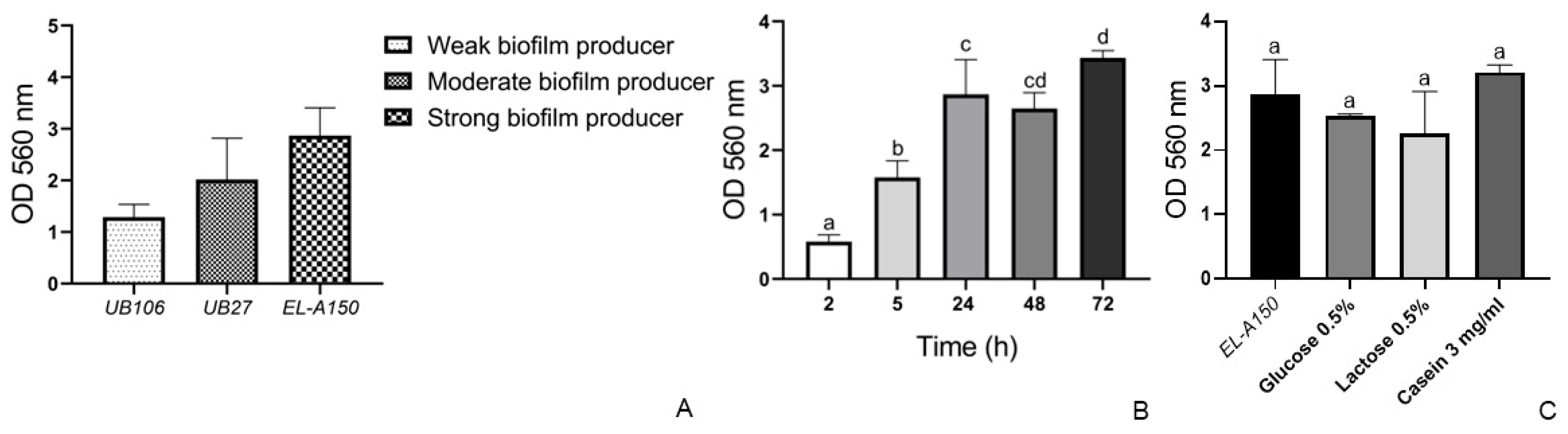
Consistent with this growth profile, EL-A150 exhibited a strong biofilm-forming capacity and was therefore classified as a high biofilm producer (Figure 2A). In addition, the biofilm formation followed a clear time-dependent kinetic pattern. At early incubation times, low levels of attached biomass were detected, corresponding to the initial adhesion phase. This was followed by a marked increase in biofilm biomass at intermediate time points, indicative of active biofilm development. At later stages, the highest levels of biomass were observed, consistent with the establishment of a mature biofilm (Figure 2B).
Furthermore, biofilm formation was enhanced in the presence of casein, with absorbance values exceeding those of the control (p < 0.05). In contrast, supplementation with lactose or glucose resulted in a reduction in biofilm biomass (p < 0.05) (Figure 2C).
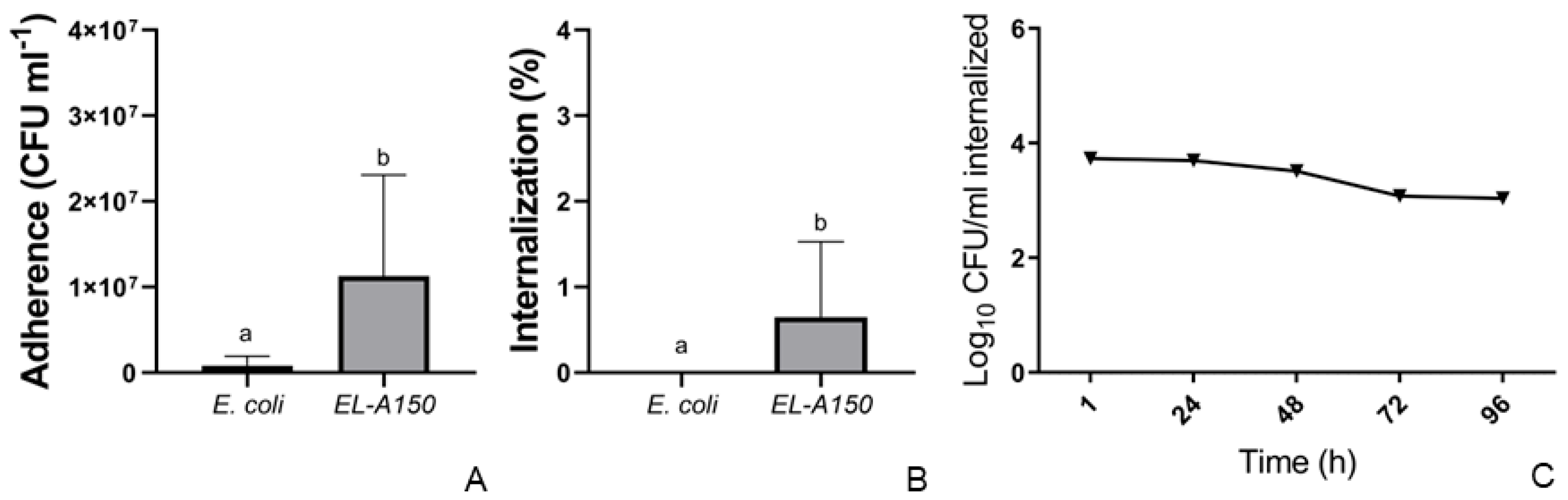
In MAC-T cell interaction assays, EL-A150 exhibited high adhesion capacity (8.7 × 10⁶ CFU/well) but low internalization (0.65%). The strain survived intracellularly for up to 24 h, after which a progressive decline in viable counts occurred. No viable bacteria were detected after 72 h, indicating limited long-term persistence within MAC-T cells (Figure 3).

3.3. Genome Assembly and Key Features of Enterococcus lactis EL-A150

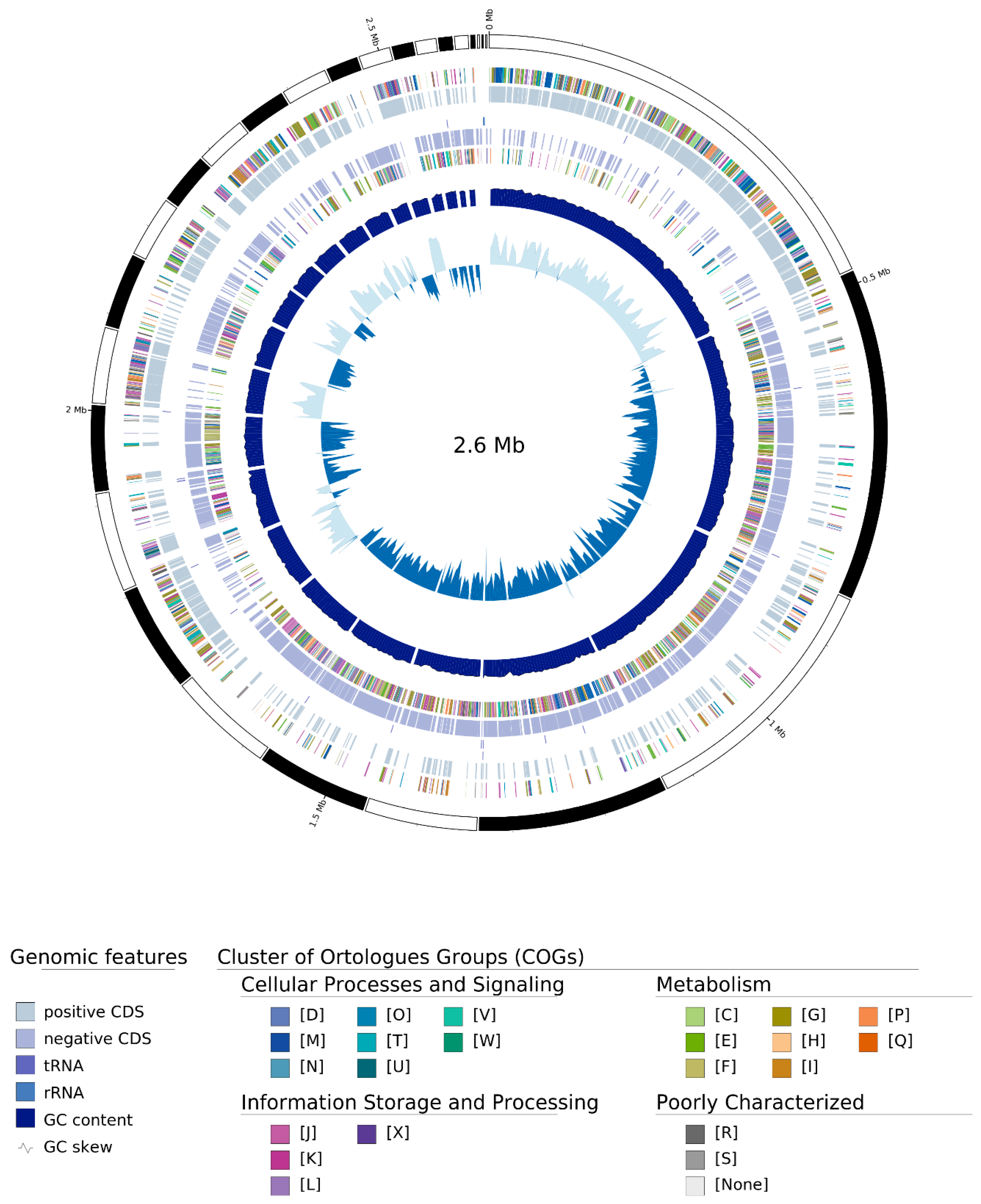
The genome assembly consisted of a single circular chromosome of 2,486,471 bp with a G + C content of 38.60%. Genome annotation predicted 2480 coding sequences (CDSs), including 67 hypothetical proteins, as well as one complete rRNA operon and 21 tRNA genes.
Genome completeness assessed with BUSCO v5.1.1 yielded 98.4% completeness using the bacteria_odb10 dataset (121/124 complete BUSCOs) and 99.0% completeness using the Lactobacillales_odb10 dataset (398/402 complete BUSCOs), confirming the high quality of the draft genome (Figure 4). The complete genome sequence of Enterococcus lactis EL-A150 has been deposited in GenBank under the accession number GCA_054404755.1 (WGS project JBTIZM01).

3.4. Genomic Confirmation and Phylogenetic Analysis

Whole-genome sequencing provided strong evidence for the taxonomic assignment of strain EL-A150. As shown in Table 1, EL-A150 exhibited the highest average nucleotide identity (ANI) values with E. lactis reference strains, reaching 99.50% with strain BT159. This value is well above the commonly accepted species boundary of 95–96% ANI, supporting its classification as E. lactis. In contrast, ANI values with other Enterococcus species, such as E. faecium (95.37%) and Enterococcus durans (77.38%), were clearly lower.
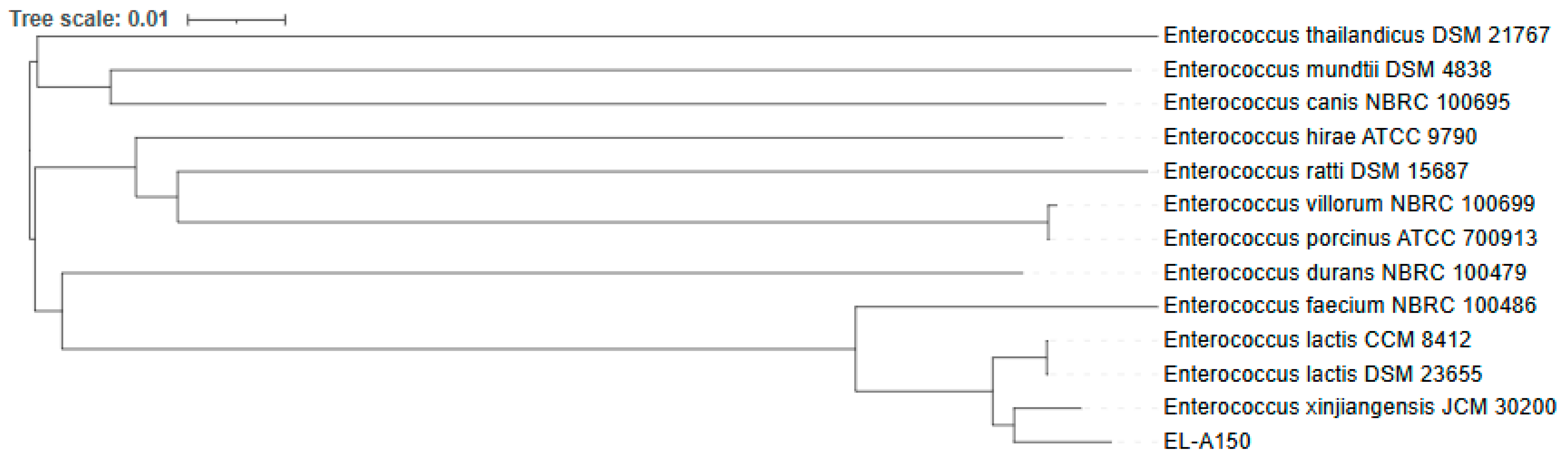
To determine the phylogenetic placement of EL-A150, a phylogenomic analysis was performed using 12 reference Enterococcus genomes together with EL-A150. OrthoFinder identified 536 single-copy core genes (SCGs) shared across all genomes. A maximum-likelihood tree inferred from the concatenated SCG alignment (603,863 bp) placed EL-A150 within the lineage containing the E. lactis reference strains, with strong bootstrap support (100) (Figure 5). EL-A150 clustered with strains assigned to the E. lactis/E. xinjiangensis lineage, consistent with previous studies proposing E. xinjiangensis as a later heterotypic synonym of E. lactis [20]. Together with ANI and dDDH results, these data support the assignment of EL-A150 to E. lactis.
dDDH analysis using the TYGS/GGDC platform yielded values of 83.4% (formula d4) between EL-A150 and the E. lactis reference strains DSM 23655 and CCM 8412, with negligible G + C content difference (<0.1%) (Table 2). In contrast, non-lactis Enterococcus species showed substantially lower dDDH values and greater G + C content differences, further supporting the assignment of EL-A150 to E. lactis.
Together, these genomic and phylogenomic analyses strongly support the classification of EL-A150 as Enterococcus lactis and highlight its relevance within this emerging group of bovine-associated Enterococcus strains.

3.5. Identification of Potential Virulence Genes

Comparative genomic analysis identified 18 putative virulence genes with ≥90% identity to known determinants in E. lactis and E. faecium. These included genes associated with adhesion (acm, bepA, fms, fnm, sagA), metabolic regulation (ccpA) and biofilm formation (empB, empC). Several genes showed homology to hospital-associated E. faecium strains (e.g., Efm_DO), while others aligned with community- or environment-derived isolates such as E. faecium C59 (Table 3).
Genes were selected based on their established or putative role in bacterial pathogenesis and their relevance to establishing infection in a mucosal environment like the mammary gland.
In addition, a targeted search for genes associated with intracellular survival and oxidative stress resistance, including sodA (superoxide dismutase), katA (catalase), and gadB (glutamate decarboxylase), revealed their absence in the EL-A150 genome.

3.6. Antimicrobial Resistance Genes

The resistome of EL-A150 included genes conferring resistance to aminoglycosides (aac(6′)-Ii), macrolides (ermB), and glycopeptides (a vanY gene fragment). The phenotypic resistance to erythromycin was consistent with the presence of the ermB gene. Similarly, susceptibility to glycopeptides (vancomycin, teicoplanin) aligned with the detection of only a truncated, low-identity vanY sequence, suggesting a lack of functional resistance. Full annotations of the antimicrobial resistance genes are provided in Table 4.

3.7. CRISPR-Cas System Analysis

A CRISPR-Cas element was detected, consisting of a 28-nt direct repeat (TGAGGAAGAAGGTGTTGTTTCTGCTGCA) located between positions 11,070–11,163. A single spacer was identified. Although the orientation of the array could not be fully resolved, both the repeat (96.43% identity) and spacer (100% identity) showed high conservation.

3.8. Prophage Regions

PHASTER analysis identified multiple prophage regions in the EL-A150 genome. One intact prophage of 31.9 kb (GC 35.71%, integrity score 150%) was located between positions 80–32,046 bp and contained 58 phage-related CDSs. An additional incomplete prophage region of 13.2 kb (GC 36.03%, score 30) was detected between positions 1601–14,821 bp, comprising 10 CDSs. No insertion sequences or transposases were found within these prophage elements.

3.9. Multilocus Sequence Typing

MLST analysis assigned EL-A150 to sequence type ST902.

3.10. PlasmidFinder Analysis

PlasmidFinder analysis did not identify complete plasmid replicons in the E. lactis EL-A150 genome. A single replicon-related sequence, repUS43, was detected with 100% identity within a large chromosomal contig. No replicons belonging to Inc18, RepA_N (repUS15), or other Enterococcus-associated plasmid families were identified.

4. Discussion

This integrated genomic and phenotypic study moves beyond the initial reporting of E. lactis in bovine mastitis to elucidate the mechanistic underpinnings of its potential as an emerging intramammary pathogen. By combining high-resolution phylogenomics, functional host-interaction assays and an analysis of adaptive gene content, our findings suggest that the bovine mastitis isolate EL-A150 occupies a position within the broader ecological and evolutionary continuum of E. lactis, a species that straddles the line between environmental commensal and opportunistic pathogen.
Genome-wide analyses classified EL-A150 as E. lactis, in agreement with recent large-scale comparative genomic studies demonstrating that E. lactis and E. faecium are distinct, albeit closely related, species with partially overlapping ecological niches [21,22]. Notably, these studies demonstrated that E. lactis strains do not segregate into origin-specific phylogenetic groups, unlike E. faecium, whose human-adapted subclade II exhibits clear genomic features of clinical specialization. The placement of EL-A150 within a non-host-restricted E. lactis clade is consistent with the view that cattle-associated isolates represent ecologically flexible members of the species, rather than descendants of human-adapted lineages. At the phenotypic level, EL-A150 displayed a high-adhesion, strong-biofilm profile conducive to mammary gland colonization, contrasting with the moderate biofilm and low adhesion reported for the previously characterized mastitis-associated strain SU-B46 [6]. This striking phenotypic heterogeneity between two mastitis isolates underscores that E. lactis does not rely on a single, conserved virulence strategy. Instead, it suggests a species-level capacity for niche adaptation, where different genetic backgrounds can converge on a clinical outcome through distinct combinations of traits related to persistence and host interaction.
A key finding is the apparent paradox between the low internalization rate (0.65%) and the ability of EL-A150 to survive intracellularly for up to 24 h. Genomic analysis revealed the absence of classical intracellular survival determinants, including sodA, katA, and gadB, indicating that EL-A150 lacks active mechanisms for oxidative stress resistance or lysosomal evasion. Instead, transient survival is likely a quantitative consequence of high initial adhesion (8.7 × 10⁶ CFU/well), which yields a sufficient number of internalized bacteria despite the low internalization percentage. The progressive decline in viable counts after 24 h, with no detectable bacteria by 72 h, supports eventual clearance by host lysosomal pathways. Thus, intracellular persistence in EL-A150 appears driven by the magnitude of adhesion rather than active subversion of host defenses.
Further supporting this concept of environmental modulation, our analysis of biofilm formation in the presence of milk components revealed that while glucose, a readily fermentable carbohydrate, promotes robust biofilm formation in EL-A150, the major milk components lactose and casein exert a suppressive effect on biofilm development under the tested conditions. This suggests that biofilm formation by E. lactis in the bovine mammary gland may be modulated by the local nutritional environment, with implications for colonization dynamics during mastitis.
The repertoire of virulence-associated genes in EL-A150 further supports this perspective. Genes involved in adhesion, biofilm formation and metabolic regulation were detected, overlapping with genetic categories described in both mastitis-associated and non-clinical E. lactis isolates. Chaichana [23] reported similar functional traits for E. lactis RB10 isolated from goat feces, where such genes were linked to colonization fitness and ecological competitiveness, rather than overt pathogenicity. Taken together, these observations suggest that potential virulence genes in E. lactis likely represent niche-adaptive factors shared between commensal and opportunistic lifestyles, which can be co-opted for infection in a permissive host environment like the mammary gland. The genomic landscape of EL-A150 thus reflects an intermediate adaptive, equipped with niche-adaptive traits but lacking the specialized virulence repertoire of highly pathogenic enterococci.
This pathogenic potential contrasts sharply with that of food-derived E. lactis strains. For instance, E. lactis IDCC 2105, isolated from cheese, harbors chromosomal resistance genes but exhibits a phenotypically avirulent and plasmid-free profile, supporting its classification as safe [24]. In contrast, EL-A150 expresses a high-adhesion, strong-biofilm phenotype despite a similarly stable genome architecture. This divergence underscores the decisive role of ecological context, suggesting that clinical environments may activate latent pathogenic traits encoded within the core genome and highlighting that risk in E. lactis is fundamentally context dependent.
While it carries clinically relevant resistance genes (e.g., ermB, aac(6′)-Ii), its resistome is less extensive than those of hospital-adapted E. faecium clones, aligning with its animal-environment origin. Importantly, its mobilome architecture differed markedly from that of other mastitis isolates. Unlike strain SU-B46, which harbors a large plasmid, EL-A150 lacked detectable autonomous plasmids, and its prophage regions showed no evidence of recent transposition activity. Nevertheless, the detection of a CRISPR-Cas system and integrated prophage regions confirms prior exposure to mobile elements, while the absence of insertion sequences within these elements supports genomic stability. This observation aligns with population genomics studies indicating that E. lactis generally exhibits lower rates of horizontal gene transfer and a more stable genome than pathogenic E. faecium lineages [21,22]. The absence of detectable plasmids and transferable resistance genes in EL-A150 aligns with the genomic observations of Ahmed [25], who reported a consistent safety profile for the E. lactis species in strains of intestinal origin. However, the isolation of EL-A150 from a clinical context, together with the report of the SU-B46 strain which does harbor a plasmid [6], highlights significant genomic and ecological heterogeneity within mastitis-associated E. lactis. These findings suggest that intramammary pathogenicity in this species may arise through distinct genomic strategies: either via exploitation of a resident chromosomal virulence repertoire, as observed in EL-A150, or through plasmid-mediated gene acquisition, as in SU-B46.
Analysis of the EL-A150 resistome revealed mixed genotype-phenotype correlations that refine its risk assessment. While the ermB gene perfectly predicted erythromycin resistance, the aac(6)-Ii gene did not confer gentamicin resistance, suggesting it is non-functional or unexpressed. Likewise, susceptibility to glycopeptides corresponded with a truncated, non-functional vanY remnant and the absence of the remaining van cluster genes, suggesting that this element is non-functional. This fragment may represent a vestigial remnant of an ancestral van-related locus or the result of an incomplete acquisition event. These findings indicate that EL-A150 harbors a limited, predominantly chromosomal resistome with low mobilization risk, aligning with adaptation to low-antibiotic-pressure environments like dairy herds.
MLST analysis further highlighted the genetic diversity of cattle-associated E. lactis. EL-A150 was assigned to ST902, unlike ST296 reported for other mastitis-associated isolates [26]. This diversity is in line with population-level analyses describing E. lactis as a genetically heterogeneous species lacking strong clonal structuring by host or source [21,22]. The occurrence of multiple sequence types among mastitis-associated isolates suggests repeated, independent ecological adaptation events, rather than clonal expansion of a single pathogenic lineage.
Therefore, our findings collectively argue that E. lactis should not be viewed merely as an occasional mastitis isolate, but as an ecologically versatile species with latent potential for opportunistic pathogenesis. The case of EL-A150 demonstrates that a core chromosomal arsenal for adhesion, biofilm formation and antimicrobial resistance can be sufficient for establishing intramammary infection, even in the absence of major mobile genetic elements.
Accordingly, our results highlight the need to incorporate functional genomic profiling into surveillance programs, shifting the focus from descriptive detection to actionable risk stratification. Future surveillance efforts should prioritize the identification of key adaptive traits, whether chromosomal or mobile, to better predict and manage the emergence of E. lactis in dairy herds. Such an approach is essential for safeguarding animal health and mitigating potential zoonotic and antimicrobial resistance risks within a robust One Health framework.
Finally, the pathogenic profile of E. lactis EL-A150, characterized by chromosomal virulence factors and a lack of mobile resistance genes, illustrates the spectrum of risk potential that exists within the genus Enterococcus, a group of bacteria famously described as a ‘double-edged sword’ for their dual roles as both opportunistic pathogens and potential probiotics [27]. By employing an integrated genomic and phenotypic framework, this study moves beyond the mere detection of virulence genes to functionally validate their role in host interaction. This approach provides a practical model for the rigorous, strain-specific assessments required to distinguish harmless commensals from emerging pathogens and to guide informed risk evaluation in animal health and beyond. From a practical perspective, these findings support the inclusion of E. lactis in routine PCR-based mastitis screening panels. Given the heterogeneity among E. lactis strains demonstrated here, strain-level evaluation and routine phenotypic susceptibility testing are essential to guide treatment decisions and prevent the emergence of resistant populations.

5. Conclusions

This study provides an integrated genomic and functional characterization of E. lactis EL-A150, supporting its role as an opportunistic pathogen in bovine mastitis. Our findings demonstrate that E. lactis possesses a stable genomic structure with ecologically niche-adaptive features that can be exploited for context-dependent intramammary colonization. By linking genomic characteristics with experimentally validated phenotypes, this work establishes a framework for strain-level risk assessment of emerging mastitis-associated enterococci. Future surveillance efforts should integrate genomic profiling with functional validation to improve mastitis management and inform One Health strategies at the animal–environment interface.

Author Contributions

Conceptualization, E.B.R.; methodology, E.B.R., M.F.C. and M.V.M.; software, F.D.F., D.F.D.P., M.F.C., M.S.-P. and F.S.; validation, E.B.R., F.D.F., M.S.-P. and D.F.D.P.; formal analysis, M.S.-P. and M.F.C.; investigation, E.B.R. and M.F.C.; resources, E.B.R.; data curation, E.B.R.; writing—original draft preparation, E.B.R. and M.F.C.; writing—review and editing, E.B.R., M.F.C. and M.V.M.; visualization, E.B.R.; supervision, E.B.R.; project administration, E.B.R.; funding acquisition, E.B.R. All authors have read and agreed to the published version of the manuscript.

Funding

This research was funded by CONICET, grant number 11220200102826CO, PIP 2021-2023 GI.

Institutional Review Board Statement

This study was conducted in accordance with ethical standards and received approval from the Comité de Ética de la Investigación (COEDI) (Approval Code: 393/22, Approval Date: 21 March 2022).

Informed Consent Statement

Not applicable.

Data Availability Statement

The genome assembly generated in this study is available in GenBank under accession GCA_054404755.1. The associated whole-genome shotgun project is JBTIZM01 (BioProject PRJNA1392908, BioSample SAMN54275740). The raw sequencing reads are available via the Sequence Read Archive (SRA) under the same WGS project. The raw data supporting the conclusions of this article will be made available by the authors upon request.

Acknowledgments

M.V. Moliva, D. Fernandez Do Porto and E. Reinoso are members of the research career of CONICET. M.F. Cerioli is a fellow doctoral, F. Serral is a fellow post-doctoral at CONICET.

Conflicts of Interest

The authors declare no conflicts of interest.

References

  1. Arikan, M.S.; Mat, B.; Alkan, H.; Çevrimli, M.B.; Akin, A.C.; Başar, E.K.; Tekindal, M.A. Determination of subclinical mastitis prevalence in dairy cows in Türkiye through meta-analysis and production loss calculation. Pak. Vet. J. 2024, 44, 391–399. [Google Scholar] [CrossRef]
  2. Reinoso, E.B. Bovine mastitis caused by Streptococcus uberis: Virulence factors and biofilm. J. Microb. Biochem. Technol. 2017, 9, 237–243. [Google Scholar] [CrossRef]
  3. Di Domenico, E.G.; Toma, L.; Prignano, G.; Pelagalli, L.; Police, A.; Cavallotti, C.; Torelli, R.; Sanguinetti, M.; Ensoli, F. Misidentification of Streptococcus uberis as a human pathogen: A case report and literature review. Int. J. Infect. Dis. 2015, 33, 79–81. [Google Scholar] [CrossRef] [PubMed]
  4. Morandi, S.; Cremonesi, P.; Povolo, M.; Brasca, M. Enterococcus lactis sp. nov., from Italian raw milk cheeses. Int. J. Syst. Evol. Microbiol. 2012, 62, 1992–1996. [Google Scholar] [CrossRef]
  5. Sukhodolets, V.V.; Botina, S.G.; Lysenko, A.M.; Trenina, M. The lactic acid enterococci Enterococcus faecium and Enterococcus durans: Nucleotide sequence diversity in 16S rRNA genes. Microbiology 2005, 74, 704–708. [Google Scholar] [CrossRef]
  6. Cerioli, M.F.; Fernández, F.D.; Moliva, M.V.; Serral, F.; Fernandez Do Porto, D.; Reinoso, E.B. Genome characterization of Enterococcus lactis strain isolated from bovine subclinical mastitis. Microb. Pathog. 2025, 206, 107846. [Google Scholar] [CrossRef]
  7. Tommasoni, C.; Fiore, E.; Lisuzzo, A.; Gianesella, M. Mastitis in Dairy Cattle: On-Farm Diagnostics and Future Perspectives. Animals 2023, 13, 2538. [Google Scholar] [CrossRef]
  8. Teixeira, L.M.; Siqueira Carvalho, M.G.; Lynn Shewmaker, P.  Enterococcus. In Manual of Clinical Microbiology; ASM Press: Washington, DC, USA, 2011. [Google Scholar]
  9. Green, L.H.; Goldman, E. Practical Handbook of Microbiology; CRC Press: Boca Raton, FL, USA, 2021. [Google Scholar]
  10. Moliva, M.V.; Cerioli, F.; Reinoso, E.B. Evaluation of environmental and nutritional factors and sua gene on in vitro biofilm formation of Streptococcus uberis isolates. Microb. Pathog. 2017, 107, 144–148. [Google Scholar] [CrossRef]
  11. Moliva, M.V.; Campra, N.; Ibañez, M.; Cristofolini, A.L.; Merkis, C.I.; Reinoso, E.B. Capacity of adherence, invasion and intracellular survival of Streptococcus uberis biofilm-forming strains. J. Appl. Microbiol. 2022, 132, 1751–1759. [Google Scholar] [CrossRef]
  12. Almeida, R.A.; Luther, D.A.; Kumar, S.J.; Calvinho, L.F.; Bronze, M.S.; Oliver, S.P. Adherence of Streptococcus uberis to bovine mammary epithelial cells and to extracellular matrix proteins. J. Vet. Med. Ser. B 1993, 43, 385–392. [Google Scholar] [CrossRef]
  13. Clinical and Laboratory Standards Institute (CLSI). Performance Standards for Antimicrobial Susceptibility Testing, 30th ed.; CLSI: Wayne, PA, USA, 2020. [Google Scholar]
  14. Yoon, S.H.; Ha, S.M.; Lim, J.M.; Kwon, S.J.; Chun, J. A large-scale evaluation of algorithms to calculate average nucleotide identity. Antonie Van Leeuwenhoek 2017, 110, 1281–1286. [Google Scholar] [CrossRef] [PubMed]
  15. Meier-Kolthoff, J.P.; Göker, M. TYGS is an automated high-throughput platform for state-of-the-art genome-based taxonomy. Nat. Commun. 2019, 10, 2182. [Google Scholar] [CrossRef]
  16. Alcock, B.P.; Huynh, W.; Chalil, R.; Smith, K.W.; Raphenya, A.R.; Wlodarski, M.A.; Edalatmand, A.; Petkau, A.; A Syed, S.; Tsang, K.K.; et al. CARD 2023: Expanded curation, support for machine learning, and resistome prediction at the Comprehensive Antibiotic Resistance Database. Nucleic Acids Res. 2023, 51, D690–D699. [Google Scholar] [CrossRef] [PubMed]
  17. Florensa, A.F.; Kaas, R.S.; Clausen, P.T.L.C.; Aytan-Aktug, D.; Aarestrup, F.M. ResFinder—An open online resource for identification of antimicrobial resistance genes in next-generation sequencing data and prediction of phenotypes from genotypes. Microb. Genom. 2022, 8, 000748. [Google Scholar] [CrossRef]
  18. Arndt, D.; Grant, J.; Marcu, A.; Sajed, T.; Pon, A.; Liang, Y.; Wishart, D.S. PHASTER: A better, faster version of the PHAST phage search tool. Nucleic Acids Res. 2016, 44, W16–W21. [Google Scholar] [CrossRef] [PubMed]
  19. Di Renzo, J.A.; Casanoves, F.; Balzarini, M.G.; Gonzalez, L.; Tablada, M.; Robledo, C.W. InfoStat Version 2018; Centro de Transferencia InfoStat, FCA, Universidad Nacional de Córdoba: Córdoba, Argentina, 2018; Available online: http://www.infostat.com.ar (accessed on 3 December 2025).
  20. Li, Y.Q.; Gu, C.T. Proposal of Enterococcus xinjiangensis Ren et al. 2020 as a later heterotypic synonym of Enterococcus lactis Morandi et al. 2012. Int. J. Syst. Evol. Microbiol. 2021, 71, 004716. [Google Scholar] [CrossRef]
  21. Ross, T.A.; Janice, J.; Arredondo-Alonso, S.; Löhr, I.H.; Holsbø, E.; Corander, J.; Pöntinen, A.K.; Kampffmeyer, M.; Hegstad, K. Enterococcus lactis is ecologically and genetically distinct from the major opportunistic pathogen Enterococcus faecium. Microb. Genom. 2025, 11, 001420. [Google Scholar] [CrossRef]
  22. Lebreton, F.; van Schaik, W.; McGuire, A.M.; Godfrey, P.; Griggs, A.; Mazumdar, V.; Corander, J.; Cheng, L.; Saif, S.; Young, S.; et al. Emergence of epidemic multidrug-resistant Enterococcus faecium from animal and commensal strains. mBio 2013, 4, e00534-13. [Google Scholar] [CrossRef]
  23. Chaichana, N.; Suwannasin, S.; Boonsan, J.; Yaikhan, T.; Klaysubun, C.; Singkhamanan, K.; Wonglapsuwan, M.; Pomwised, R.; Konglue, S.; Chema, R.; et al. Genomic characterization and safety evaluation of Enterococcus lactis RB10 isolated from goat feces. Antibiotics 2025, 14, 612. [Google Scholar] [CrossRef]
  24. Parks, D.H.; Imelfort, M.; Skennerton, C.T.; Hugenholtz, P.; Tyson, G.W. CheckM: Assessing the quality of microbial genomes recovered from isolates, single cells, and metagenomes. Genome Res. 2015, 25, 1043–1055. [Google Scholar] [CrossRef]
  25. Ahmed, N.A.; Khattab, R.A.; Ragab, Y.M.; Hassan, M. Safety assessment of Enterococcus lactis strains complemented with comparative genomics analysis reveals probiotic and safety characteristics of the entire species. BMC Genom. 2023, 24, 667. [Google Scholar] [CrossRef]
  26. Ocejo, M.; Mugica, M.; Oporto, B.; Lavín, J.L.; Hurtado, A. Whole-genome long-read sequencing to unveil Enterococcus antimicrobial resistance in dairy cattle farms exposed a widespread occurrence of Enterococcus lactis. Microbiol. Spectr. 2024, 12, e0367223. [Google Scholar] [CrossRef]
  27. Ben Braïek, O.; Smaoui, S. Enterococci: Between emerging pathogens and potential probiotics. BioMed Res. Int. 2019, 2019, 5938210. [Google Scholar] [CrossRef]
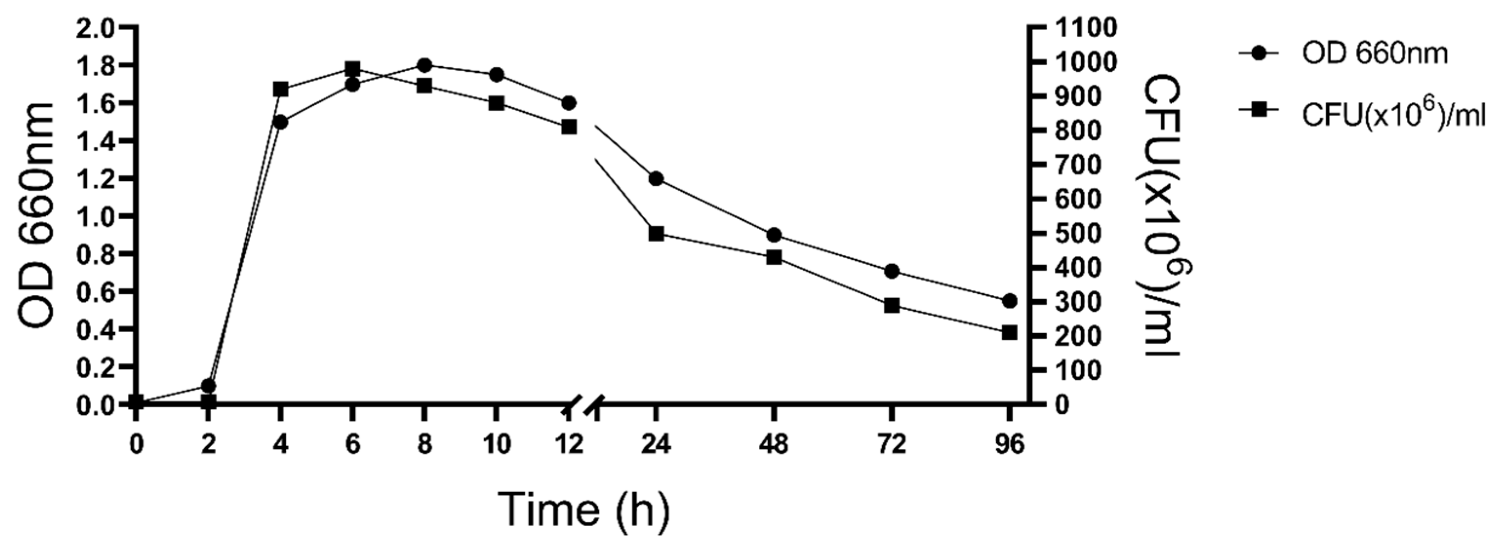
Figure 1. Growth kinetics of Enterococcus lactis EL-A150.
Bacteria 05 00026 g001
Figure 2. (A) Biofilm-forming ability and (B) kinetics of biofilm formation at different incubation times of Enterococcus lactis EL-A150. (C) Effect of milk components on biofilm formation after 24 h of incubation in TSB supplemented with glucose (0.5% w/v), lactose (0.5% w/v) or casein (3 mg/mL). The bars represent the mean ± the standard error of the mean (SEM). Average of triplicates of three independent assays is presented. Statistically significant differences are shown with different letters (p < 0.05).
Bacteria 05 00026 g002
Figure 3. Interaction of Enterococcus lactis EL-A150 with MAC-T epithelial cells. The graph shows bacterial adhesion (A), internalization (B) and intracellular survival (C) at different incubation times. Escherichia coli was included as a negative control. The bars represent the mean ± the standard error of the mean (SEM). Average of triplicates of three independent assays is presented. Significant differences are shown with different letters (p < 0.05).
Bacteria 05 00026 g003
Figure 4. Circular representation of the genome of Enterococcus lactis EL-A150. From outer to inner rings: (i) genomic coordinates (Mb scale), (ii) coding sequences (CDS) on the forward strand, (iii) CDS on the reverse strand, (iv) tRNA and rRNA genes, (v) functional annotation based on COG categories, (vi) GC content, and (vii) GC skew. Colors represent COG functional categories as indicated in the legend. GC content and GC skew are shown as deviations from the genome average.
Bacteria 05 00026 g004
Figure 5. Phylogenomic analysis of Enterococcus lactis EL-A150. Maximum-likelihood tree inferred from the concatenated alignment of 536 single-copy core genes (603,863 bp) identified in 12 reference Enterococcus genomes and EL-A150. Bootstrap support values (based on 1000 replicates) are shown at major nodes.
Bacteria 05 00026 g005
Table 1. Average nucleotide identity (ANI) values of strain EL-A150 against reference genomes of closely related Enterococcus species.
Query StrainReference StrainSpeciesAccession No. (RefSeq)ANI (%)
EL-A150BT159E. lactisGU98369799.50
EL-A150CCM 8412E. lactisGCF_015751045.198.05
EL-A150JCM 30200E. xinjiangensis *GCF_015751065.198.30
EL-A150NBRC 100486E. faeciumGCF_001544255.195.37
EL-A150NBRC 100479E. duransGCF_001544215.177.38
EL-A150ATCC 9790E. hiraeGCF_000271405.277.27
EL-A150ATCC 700913E. porcinusGCF_000407205.177.08
EL-A150NBRC 100699E. villorumGCF_007990225.176.85
EL-A150NBRC 100695E. canisGCF_001544375.171.02
EL-A150DSM 4838E. mundtiiGCF_001886035.176.07
EL-A150DSM 15687E. rattiGCF_001886195.176.07
EL-A150DSM 21767E. thailandicusGCF_001886265.176.32
ANI values were calculated using whole-genome sequences retrieved from public databases. Species delineation was interpreted according to the commonly accepted 95–96% ANI threshold. * Enterococcus xinjiangensis has been proposed as a later heterotypic synonym of E. lactis based on comparative genomic analyses of the type strains [20].
Table 2. Digital DNA-DNA hybridization (dDDH) values between Enterococcus lactis EL-A150 and type strains of related Enterococcus species.
Reference StraindDDH (Formula d4)
[%]
CI d4
[%]
dDDH (d0)
[%]
dDDH (d6)
[%]
G + C Diff.
[%]
E. lactis DSM 2365583.480.6–85.979.683.20.09
E. lactis CCM 841283.480.6–85.979.583.10.10
E. xinjiangensis JCM 3020085.683.0–88.086.689.30.10
E. faecium NBRC 10048663.160.2–65.983.081.90.15
E. durans NBRC 10047922.220.0–24.719.519.00.43
E. hirae ATCC 979022.219.9–24.619.719.21.32
E. porcinus ATCC 70091321.919.7–24.418.618.33.20
E. villorum NBRC 10069921.719.4–24.118.618.33.40
E. canis NBRC 10069521.619.3–24.013.113.53.64
E. mundtii DSM 483821.419.1–23.817.217.00.11
E. ratti DSM 1568720.918.7–23.417.217.03.93
E. thailandicus DSM 2176720.918.7–23.419.318.71.62
dDDH values were calculated using GGDC, as implemented in TYGS. CI, confidence interval; d4, recommended formula for species delineation.
Table 3. Key virulence-associated genes identified in the Enterococcus lactis EL-A150 genome.
Virulence CategoryGene(s)Relevance to Mammary Gland Pathogenesis/Predicted RoleLocus Tag(s) in EL-A150
Adhesion and colonizationacmAdhesion to collagenorf01788
ebpABC, srtCEndocarditis- and biofilm-associated (Ebp) pilus system.orf00061-64; orf00431,33,85-87
efaACell wall adhesinorf02362
Biofilm formationbopDBiofilm matrix proteinorf00887
Immune evasion and surface protectioncpsA, cpsBCapsular polysaccharide biosynthesis.orf00782, orf00783
Regulation and stress responsestp (homolog)Serine/threonine phosphatase.orf02022
Protease activityhtrA/degP (homolog)Stress-responsive serine protease.orf01014
Virulence-associated genes were identified by in silico annotation and homology-based inference. Functional relevance was assigned based on published enterococcal virulence factors. Locus tags refer to the EL-A150 genome.
Table 4. Antimicrobial resistance determinants identified in the Enterococcus lactis EL-A150 genome and their correlation with phenotype.
Resistance GeneDrug Class FafectedPredicted Mechanism% Amino Acid Identity *Genomic Ocntext/Notes
ermBMacrolides23S rRNA methylation (target modification)99.5Chromosomal. Phenotype: Concordant (Resistant).
*aac(6′)-Ii*Aminoglycosides Antibiotic acetylation (inactivation)98.9Chromosomal. Phenotype: Discordant (Susceptible to gentamicin).
vanY (fragment)GlycopeptidesD,D-carboxypeptidase (target alteration)34.5Chromosomal remnant. Low identity to functional vanB cluster. Phenotype: Concordant (Susceptible).
tet(M)TetracyclinesRibosomal protection (target protection)94.05Chromosomal. Phenotype: Concordant (Susceptible)
Antimicrobial resistance determinants were identified by in silico analysis and compared with reference resistance genes. * Percentage of amino acid identity indicates similarity to functional reference proteins. Phenotype–genotype correlation was determined by antimicrobial susceptibility testing; “concordant” denotes agreement between genotype and phenotype, whereas “discordant” denotes lack of phenotypic resistance despite gene presence.
Disclaimer/Publisher’s Note: The statements, opinions and data contained in all publications are solely those of the individual author(s) and contributor(s) and not of MDPI and/or the editor(s). MDPI and/or the editor(s) disclaim responsibility for any injury to people or property resulting from any ideas, methods, instructions or products referred to in the content.