Article

Complete Genome Analysis of a Flower-Associated Leuconostoc suionicum JNUCC 76 from Prunus yedoensis

Department of Chemistry and Cosmetics, Jeju National University, Jeju 63243, Republic of Korea
*
Author to whom correspondence should be addressed.
These authors contributed equally to this work.
Bacteria 2026, 5(2), 25; https://doi.org/10.3390/bacteria5020025
Submission received: 25 December 2025 / Revised: 31 January 2026 / Accepted: 21 April 2026 / Published: 7 May 2026

Abstract

Leuconostoc suionicum strain JNUCC 76 (=CH10) was isolated from cherry blossom flowers (Prunus yedoensis) collected on Jeju Island, Republic of Korea, representing a flower-associated strain of L. suionicum. To clarify its taxonomic position and genomic characteristics, whole-genome sequencing was performed using a hybrid PacBio–Illumina approach. The complete genome was assembled into a single circular chromosome of 2.20 Mb with a GC content of 36.8% and high sequencing depth, indicating a high-quality, closed genome assembly. Genome annotation revealed a compact gene repertoire dominated by functions related to carbohydrate transport and metabolism, amino acid utilization, and core cellular processes, consistent with adaptation to plant-derived, sugar-rich environments. Genome-based phylogenomic analyses using average nucleotide identity (ANI), digital DNA–DNA hybridization (dDDH), and Genome BLAST Distance Phylogeny (GBDP) placed strain JNUCC 76 within the species L. suionicum. Genome-based metrics clearly exceeded the accepted species thresholds, supporting the assignment of the strain to L. suionicum. Secondary metabolite gene cluster analysis identified a limited number of low-complexity and precursor-oriented biosynthetic gene clusters, including RiPP-like, type III polyketide synthase, and terpene-precursor clusters, suggesting that the ecological fitness of the strain relies primarily on primary metabolism rather than extensive secondary metabolite production. Overall, this study expands current knowledge of flower-associated Leuconostoc lineages and provides a high-quality genomic framework for future comparative and functional studies. The genomic features of strain JNUCC 76 highlight floral environments as underexplored reservoirs of lactic acid bacteria diversity and support further evaluation of flower-derived Leuconostoc strains as potential postbiotic or fermentation-based resources for cosmetic and related biotechnological applications.

1. Introduction

The genus Leuconostoc comprises heterofermentative lactic acid bacteria that are well known for their roles in fermented foods, where they contribute to flavor development, texture formation, and preservation through the production of organic acids, aroma-related metabolites, and exopolysaccharides (EPSs). In recent years, the application scope of fermentation-derived microorganisms has expanded beyond the food sector toward skin microbiome-friendly cosmetics, low-irritation formulations, and naturally derived functional ingredients. In this context, Leuconostoc strains have gained renewed attention as promising resources for cosmetic and postbiotic applications.
Notably, Leuconostoc-based materials are not merely of academic interest but have already been implemented as commercial cosmetic ingredients. A representative example is Leuconostoc/Radish Root Ferment Filtrate, which is produced by fermenting plant-derived substrates with Leuconostoc and is widely used as a naturally derived preservative-supporting ingredient in skincare formulations. Previous studies have reported that antimicrobial and preservative-related activities observed in fermented radish products are associated with fermentation-derived metabolites, supporting the rationale for their use in cosmetic applications [1,2,3]. Because these ingredients are applied in the form of fermentation filtrates rather than live microorganisms, they offer advantages in formulation stability, reproducibility, and regulatory acceptance, aligning well with the current postbiotic-oriented strategy in the cosmetic industry.
Another key industrial value of Leuconostoc lies in its capacity to produce EPS, particularly dextran and α-glucan-type polysaccharides. EPS derived from Leuconostoc species have been extensively investigated for their physicochemical properties, including viscosity modulation, moisture retention, and film-forming ability, all of which are desirable features for cosmetic formulations. Recent work demonstrated efficient EPS production by Leuconostoc mesenteroides during sugarcane juice fermentation, highlighting the applicability of these biopolymers beyond food systems and into pharmaceutical and cosmetic domains [4]. Furthermore, the structural diversity and strain-dependent characteristics of lactic acid bacteria-derived EPS have been shown to influence their functional and biological properties, underscoring the importance of strain-specific characterization and molecular-level evidence when developing EPS-based cosmetic ingredients [5].
Beyond fermentation products and polysaccharides, emerging studies have extended the application of Leuconostoc to microbial-derived extracellular vesicles (EVs), often referred to as exosomes. In particular, EVs isolated from L. mesenteroides subsp. DB-21, a strain obtained from Camellia japonica flowers, were reported to exhibit anti-inflammatory and anti-melanogenic activities in skin-related experimental models [6]. This finding provides a compelling example that Leuconostoc strains associated with plant or floral niches may serve as next-generation postbiotic or microbial signaling materials for cosmetic applications, offering a non-viable yet biologically active alternative to conventional probiotics.
In addition, Leuconostoc species have attracted attention as biocatalytic platforms for the production of functional cosmetic ingredients. Enzymes derived from Leuconostoc, such as glucansucrases, have been employed for transglycosylation reactions that improve the physicochemical stability and usability of polyphenols, enabling their development as functional cosmetic agents [7,8]. Moreover, α-glucosyl glycerol (glucosyl glycerol, GG), a natural osmolyte associated with skin hydration and protection, is now produced through established biocatalytic processes and is already utilized in cosmetic and healthcare products [9,10,11]. These examples illustrate that Leuconostoc is relevant not only as a fermentation organism but also as an enzymatic resource for cosmetic ingredient manufacturing.
Despite these advances, current knowledge of Leuconostoc remains largely biased toward strains isolated from food-related environments such as kimchi, dairy products, and sourdough. In contrast, Leuconostoc strains originating from natural ecosystems—particularly flowers—have been far less explored. Floral niches are characterized by dynamic sugar availability, exposure to plant secondary metabolites, ultraviolet radiation, and interactions with pollinators, all of which impose unique selective pressures on associated microorganisms. Microbes adapted to such environments may therefore possess distinct physiological and genetic traits compared to their food-derived counterparts.
Floral environments such as nectar-rich blossoms represent highly selective and transient microbial habitats. These niches are characterized by high sugar concentrations, frequent exposure to ultraviolet (UV) radiation, desiccation stress, plant-derived antimicrobial secondary metabolites, and repeated dispersal through pollinator visitation. Such environmental conditions are expected to impose distinct selective pressures on resident microbial populations compared with food-associated or host-associated habitats.
Based on these ecological characteristics, we hypothesized that flower-associated Leuconostoc strains would exhibit genomic signatures consistent with (i) enhanced carbohydrate uptake and metabolism to exploit nectar sugars, (ii) stress response and oxidative defense systems to tolerate UV and environmental fluctuations, and (iii) traits related to surface adhesion or biofilm formation facilitating persistence on floral tissues and interaction with plant surfaces.
The present genome-based study was therefore designed to test whether a flower-derived L. suionicum strain harbors genomic features consistent with adaptation to these floral niche-specific selective pressures. By integrating complete genome sequencing with functional and comparative genomic analyses, we aimed to explore the ecological genomic traits of a flower-associated lineage and evaluate how they differ from the well-studied food-associated members of the genus.
In bacterial systematics and functional assessment, whole-genome sequencing has become an indispensable tool, as 16S rRNA gene-based analysis alone often lacks sufficient resolution for species-level discrimination within lactic acid bacteria. Genome-based approaches, including average nucleotide identity (ANI), digital DNA–DNA hybridization (dDDH), and phylogenomics, provide robust frameworks for taxonomic placement, while complete genome information enables comprehensive evaluation of carbohydrate metabolism, EPS biosynthesis, environmental adaptation, and safety-related genetic features. Such genome-level insights are increasingly regarded as essential for assessing the suitability of specific strains as postbiotic or cosmetic ingredient candidates [12].
Against this background, the present study aims to obtain and describe the complete genome sequence of a flower-associated strain of L. suionicum, thereby providing fundamental genomic information and a genome-based framework for its taxonomic placement and overall characterization. By focusing on a flower-associated Leuconostoc strain, this work contributes to expanding the ecological and genomic landscape of the genus and establishes a foundational resource for future studies exploring its potential applications in cosmetics and related industries.

2. Materials and Methods

2.1. Isolation, Cultivation, and Genomic DNA Extraction

Strain JNUCC 76 (=CH10), identified as Leuconostoc suionicum, was isolated from cherry blossom flowers collected on 26 April 2024 on Jeju Island, Republic of Korea (33.448244° N, 126.568930° E). Floral samples were aseptically transferred to sterile containers, suspended in sterile saline (0.85% NaCl), and serially diluted. Aliquots of the diluted suspensions were spread onto de Man–Rogosa–Sharpe (MRS) agar (BD Difco™, Sparks, MD, USA) plates.
The plates were incubated under anaerobic conditions at 28 °C using an AnaeroJar™ 2.5 L system (Oxoid, Thermo Fisher Scientific, Basingstoke, UK) with a GasPak anaerobic system (BD, Sparks, MD, USA), which is suitable for the cultivation of lactic acid bacteria. After incubation, morphologically distinct colonies were selected and repeatedly streaked onto fresh MRS agar plates to obtain a pure culture. Culture purity was confirmed based on consistent colony morphology following successive subculturing. The purified isolate was routinely maintained on MRS agar and preserved as glycerol stocks at −80 °C for long-term storage.
For genomic analyses, genomic DNA (gDNA) was extracted from freshly grown pure cultures using a commercially available bacterial genomic DNA isolation kit, according to the manufacturer’s instructions. The quantity and purity of the extracted gDNA were assessed spectrophotometrically, and DNA integrity was confirmed by agarose gel electrophoresis. High-quality gDNA was subsequently used for whole-genome sequencing.

2.2. Genome Sequencing, Assembly, and Annotation

Whole-genome sequencing was performed by Macrogen (Seoul, Republic of Korea) using a hybrid sequencing strategy that combined PacBio long-read and Illumina short-read platforms [13]. For PacBio sequencing, microbial SMRTbell libraries were prepared using a PacBio microbial library preparation workflow on the Pacific Biosciences platform (PacBio, Menlo Park, CA, USA) [13]. For Illumina sequencing, paired-end libraries with an average insert size of approximately 350 bp were constructed using the TruSeq Nano DNA Library Prep Kit (Illumina, San Diego, CA, USA) and sequenced according to the manufacturer’s standard protocols [14].
Raw sequencing reads were subjected to quality assessment prior to assembly. Illumina reads were evaluated using FastQC (v0.11.7) [15], and adapter sequences and low-quality bases were trimmed using Trimmomatic (v0.38) [16] to minimize potential biases in downstream analyses. PacBio reads were processed using the standard SMRT Link workflow [13].
De novo genome assembly was conducted using PacBio long reads with the Microbial Genome Analysis pipeline implemented in SMRT Link (SMRTLINK_13.1.0.221970) [1]. Assembly accuracy was further improved by correcting potential assembly errors using Inspector (v1.0.1) and polishing the assembled contigs with Illumina short reads using Pilon (v1.22) [17]. The assembly pipeline also included automated evaluation of contig circularity and rotation of the contig start position to the predicted origin of replication, where applicable.
Genome assembly quality was assessed using multiple complementary approaches. K-mer-based genome size estimation was performed using Jellyfish (v1.1.12) and GenomeScope [18]. Illumina reads were mapped back to the assembled genome using BWA (v0.7.17-r1188), while PacBio reads were mapped using pbmm2 (v12.1) to evaluate sequencing depth, coverage, and mapping consistency. Genome completeness was assessed using BUSCO (v5.1.3) with the bacterial lineage dataset [19]. For taxonomic proximity and genomic relatedness analyses, BLAST+ searches and ANI calculations were performed using pyani v0.2.12 [20].
Gene prediction and primary genome annotation were carried out using Prokka (v1.14.6) [21]. Functional annotation and orthology-based classification were further refined using eggNOG (v4.5) and InterProScan (v5.34-73.0) [22]. A circular representation of the genome was generated using Circos (v0.69.9) [23].
In addition, DNA base modifications were inferred from PacBio sequencing kinetic information using ipdSummary, and methylation motif discovery was performed using motifMaker, both implemented within the SMRT Link framework [13]. CpG/5-methylcytosine (5mC) methylation probabilities were evaluated using pb-CpG-tools (v1.1.0) [24].

2.3. Genome-Based Phylogenomic and Taxonomic Analysis

Genome-based taxonomic and phylogenomic analyses of strain JNUCC 76 (=CH10) were performed using the Type (Strain) Genome Server (TYGS), a high-throughput platform for state-of-the-art prokaryotic genome-based taxonomy (https://tygs.dsmz.de, accessed on 31 October 2025) [25]. The analysis was conducted using default parameters and incorporated the most recent methodological updates implemented in TYGS at the time of analysis.
To identify closely related taxa, the genome sequence of strain JNUCC 76 was initially compared against all available type strain genomes in the TYGS database using the MASH algorithm, which provides a rapid estimation of intergenomic relatedness based on MinHash distances [26]. In parallel, 16S rRNA gene sequences were extracted from the genome using RNAmmer [27], and similarity searches were performed against the 16S rRNA gene sequences of all type strains in the TYGS database using BLAST+ [28]. The top-matching reference strains from both approaches were combined to construct a dataset of the most closely related type strains for downstream analyses.
Pairwise whole-genome comparisons between the query genome and the selected type strain genomes were carried out using the Genome BLAST Distance Phylogeny (GBDP) method [29]. Intergenomic distances were calculated under the ‘trimming’ algorithm with distance formula d5, and 100 distance replicates were generated for each comparison. dDDH values and their corresponding confidence intervals were calculated using the Genome-to-Genome Distance Calculator (GGDC) version 4.0, applying the recommended settings for prokaryotic species delineation [25].
Phylogenomic trees based on both whole-genome sequences and 16S rRNA gene sequences were inferred using FastME v2.1.6.1, employing a balanced minimum evolution approach with subtree pruning and regrafting (SPR) postprocessing [30]. Branch support values were estimated from 100 pseudo-bootstrap replicates, and all trees were rooted at the midpoint. Tree visualization and graphical outputs were generated using PhyD3, with publication-ready figures exported in vector format (SVG) [31].
Species-level clustering was conducted using a 70% dDDH threshold, which corresponds to the generally accepted genomic boundary for prokaryotic species delineation [32], while subspecies-level clustering was evaluated using a 79% dDDH threshold, as previously proposed [33]. Nomenclatural information and taxonomic metadata for reference strains were retrieved from the List of Prokaryotic names with Standing in Nomenclature (LPSN), which is fully integrated within the TYGS framework [34].

2.4. Secondary Metabolite Biosynthetic Gene Cluster Analysis

The secondary metabolite biosynthetic potential of L. suionicum strain JNUCC 76 was investigated through the prediction of biosynthetic gene clusters (BGCs) using the bacterial version of antiSMASH (v8.0.4) [35]. Genome annotation files generated with Prokka v1.14.6 were used as input in GenBank format. BGC identification was conducted using the relaxed detection strictness setting to allow sensitive detection of both complete and partial clusters, including atypical or fragmented loci lacking canonical core biosynthetic genes.
To enhance functional annotation and comparative interpretation, several optional antiSMASH analytical modules were enabled, including KnownClusterBlast, SubClusterBlast, ActiveSiteFinder, RREFinder, and transcription factor binding site (TFBS) analysis [35]. In addition, Pfam domain–based annotation was applied to identify conserved catalytic and accessory domains within predicted biosynthetic loci. These analytical settings were selected to improve the detection and characterization of low-abundance or cryptic BGCs that may not be identified under more stringent prediction conditions.
Predicted BGCs were subsequently compared with reference clusters deposited in the Minimum Information about a Biosynthetic Gene cluster (MIBiG) database to assess similarity to experimentally characterized biosynthetic pathways [36,37]. The gene composition, organization, and predicted domain architectures of individual clusters were examined using the visualization and comparison tools integrated within the antiSMASH platform.
Overall, this in silico workflow provided a standardized and reproducible framework for the systematic identification and preliminary classification of secondary metabolite biosynthetic gene clusters encoded in the genome of L. suionicum strain JNUCC 76.

2.5. Antimicrobial Resistance Gene Screening

The genome of strain JNUCC 76 was screened for antimicrobial resistance genes using the Comprehensive Antibiotic Resistance Database (CARD) and the Resistance Gene Identifier (RGI) tool with default parameters. Only perfect and strict hits were considered in the interpretation.

3. Results and Discussion

3.1. Genomic Features

The complete genome of L. suionicum strain JNUCC 76 was assembled into a single circular chromosome of 2,196,993 bp, with no detectable plasmids, indicating a closed and structurally simple genome organization (Table 1). The assembly consisted of a single contig, with the contig N50 equal to the full genome length, and was supported by a high sequencing depth of approximately 509×, reflecting excellent assembly continuity and reliability. Such high-quality genome reconstruction was achieved using a hybrid long- and short-read sequencing strategy based on PacBio Sequel II and Illumina NovaSeq platforms, which are well established for accurate microbial genome assembly and polishing [13,14,15,16,17,18].
The overall GC content of the genome was 36.8%, which is consistent with previously reported values for members of the genus Leuconostoc and related lactic acid bacteria [12]. Genome annotation predicted a total of 2214 genes, including 2110 protein-coding genes, 18 pseudogenes, 71 tRNA genes, and 12 rRNA genes organized into four complete rRNA operons (5S, 16S, and 23S), together with three ncRNA genes (Table 1). This gene composition reflects the compact and efficient genomic architecture typical of lactic acid bacteria, which are generally adapted to nutrient-rich and host-associated environments such as fermented foods and plant-derived niches [1,2,12].
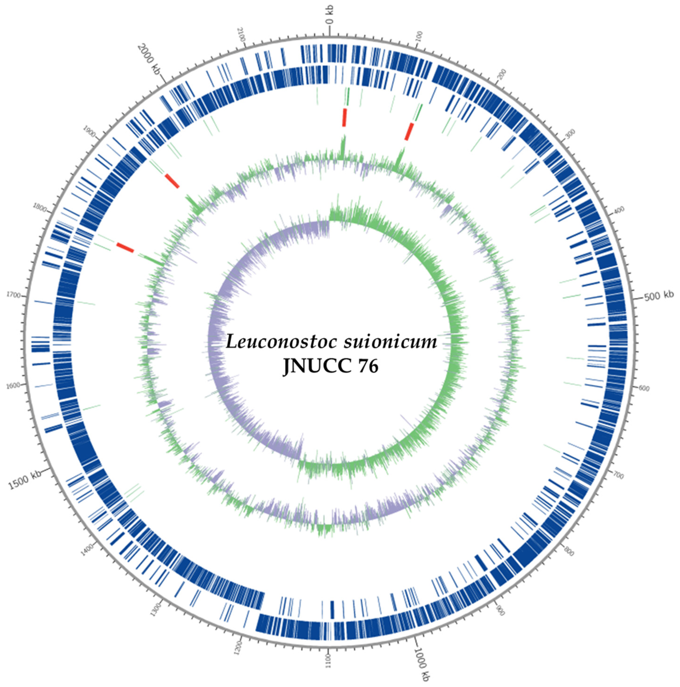
The global genomic organization of strain JNUCC 76 is illustrated in the circular genome map (Figure 1), which was generated using Circos-based visualization [23]. Coding sequences are evenly distributed on both forward and reverse strands across the chromosome, while tRNA and rRNA genes are discretely positioned without extensive clustering. GC content and GC skew profiles show moderate local fluctuations around the genome-wide average, without pronounced polarity that would clearly define the origin or terminus of replication. Such patterns are commonly observed in complete genomes of Leuconostoc species and other lactic acid bacteria, supporting a stable chromosomal structure without evidence of large-scale rearrangements or atypical compositional bias [12].
Overall, the genomic features summarized in Table 1 and Figure 1 demonstrate that L. suionicum JNUCC 76 possesses a high-quality, complete genome with structural and compositional characteristics representative of the genus. This well-resolved genomic framework provides a robust basis for subsequent comparative genomic, phylogenomic, and functional analyses aimed at elucidating niche adaptation and potential biotechnological relevance of flower-associated Leuconostoc strains [12,25,29].
Members of the genus Leuconostoc, including L. suionicum, are generally described as Gram-positive, non-spore-forming, coccoid to ovoid lactic acid bacteria that occur singly or in pairs and are considered non-motile. Consistent with these characteristics, no genes related to flagellar biosynthesis or motility were detected in the genome of strain JNUCC 76.
Species of Leuconostoc are also known for their ability to produce EPS, particularly dextran and related α-glucans, during growth on sucrose-rich substrates. EPS production in this genus is often associated with smooth and sometimes slightly moist or mucoid colony textures. In the genome of strain JNUCC 76, genes putatively involved in polysaccharide and carbohydrate metabolism were identified, supporting the general metabolic capacity of this lineage for EPS-related biosynthetic processes. These features are consistent with established descriptions of Leuconostoc biology reported in the literature.

3.2. Phylogenetic Relationships

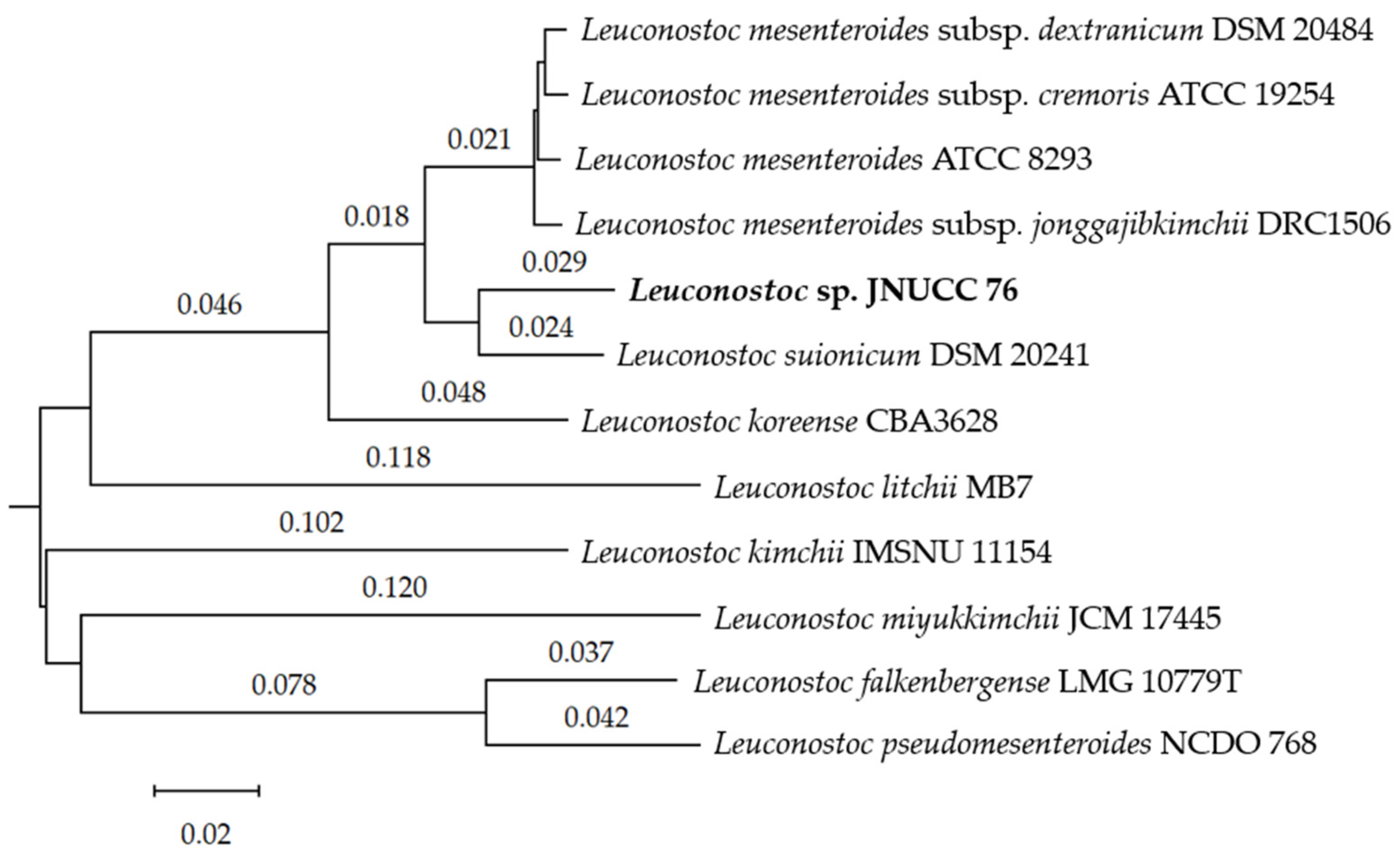
Genome-based phylogenetic analyses consistently placed strain JNUCC 76 within L. suionicum, forming a robust and well-supported clade with the type strain L. suionicum DSM 20241. Phylogenetic reconstruction based on 16S rRNA gene sequences using the GBDP method showed that strain JNUCC 76 clusters closely with L. suionicum and L. mesenteroides-related taxa, while remaining clearly separated from more distantly related species such as L. kimchii, L. falkenbergense, and L. pseudomesenteroides [25,29]. Although 16S rRNA gene-based phylogeny provides a useful initial framework for taxonomic placement, its limited resolution among closely related Leuconostoc taxa necessitates complementary genome-scale analyses [32].
Whole-genome sequence-based phylogenetic analysis using the GBDP approach yielded a topology largely congruent with the 16S rRNA gene tree but with improved discriminatory power. In the genome-based tree, strain JNUCC 76 formed a distinct and robust lineage most closely associated with L. suionicum, supported by high bootstrap values, while remaining clearly differentiated from other members of the L. mesenteroides group [25,29,30]. The concordance between gene-based and genome-based phylogenies supports the stability of the phylogenetic placement of strain JNUCC 76 within the species L. suionicum (Figure 2).
Quantitative genome similarity metrics further clarified the taxonomic relationship of strain JNUCC 76 to its closest relatives. dDDH values between JNUCC 76 and L. suionicum DSM 20241 reached 79.1% (d0) and 77.9% (d6), exceeding the conventional 70% species delineation threshold [32,33], whereas comparisons with other Leuconostoc taxa yielded substantially lower dDDH values (≤72.8%) (Table 2). These results indicate a close genomic relationship between JNUCC 76 and L. suionicum, while also highlighting measurable divergence from other members of the genus.
Consistently, OrthoANIu analysis revealed an average nucleotide identity of 95.09% between strain JNUCC 76 and L. suionicum, exceeding the widely accepted 95% species boundary for prokaryotic species delineation (Table 3) [20,32]. ANI values in this range are commonly observed among closely related lactic acid bacteria and are considered consistent with intraspecies genomic diversity driven by ecological or niche-specific adaptation rather than indicating species-level separation [12].
Taken together, the congruent results obtained from 16S rRNA gene phylogeny, whole-genome GBDP analysis, dDDH, and OrthoANIu assessments consistently support the assignment of strain JNUCC 76 to the species L. suionicum. The strain forms a robust and well-supported clade with the type strain L. suionicum DSM 20241, clearly distinguished from other closely related Leuconostoc taxa. This integrative phylogenetic framework provides a robust basis for future studies addressing the ecological specialization and functional potential of flower-associated Leuconostoc lineages.

3.3. Functional Annotation and COG Classification

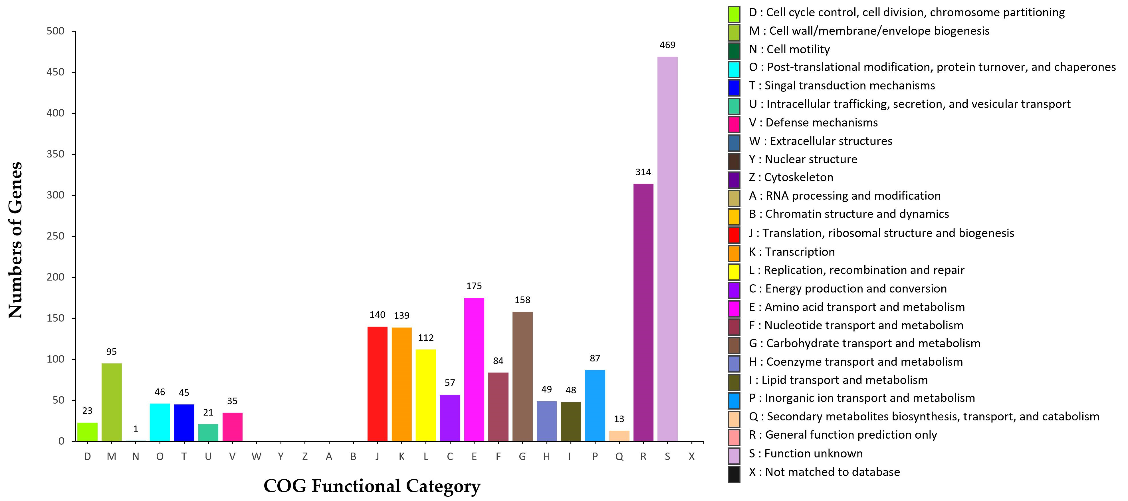
Functional annotation of the L. suionicum strain JNUCC 76 genome was performed using the EggNOG database, and the predicted protein-coding genes were classified into Clusters of Orthologous Groups (COG) functional categories (Figure 3) [22]. A substantial proportion of genes were assigned to metabolic and housekeeping-related functions, reflecting the streamlined genomic organization typical of lactic acid bacteria adapted to nutrient-rich environments [12].
Among the annotated COG categories, genes involved in carbohydrate transport and metabolism (G) constituted a major fraction (158 genes; 7.48%), highlighting the metabolic versatility of strain JNUCC 76 in utilizing diverse carbohydrates. This feature is consistent with the ecological lifestyle of Leuconostoc species, which frequently inhabit plant- and food-associated niches where complex carbohydrates are abundant [2,12]. Genes related to amino acid transport and metabolism (E) were also well represented (175 genes; 8.29%), underscoring the importance of amino acid uptake and biosynthesis for cellular growth and adaptation in lactic acid bacteria [12].
Core cellular processes were supported by a considerable number of genes assigned to translation, ribosomal structure, and biogenesis (J) (140 genes; 6.63%), transcription (K) (139 genes; 6.58%), and replication, recombination, and repair (L) (112 genes; 5.31%), indicating a well-maintained genetic machinery for genome stability and protein synthesis [21,22]. In addition, cell wall/membrane/envelope biogenesis (M) accounted for 95 genes (4.50%), reflecting structural adaptations important for environmental persistence and host- or plant-associated lifestyles [12].
Genes involved in energy production and conversion (C) (57 genes; 2.70%), lipid transport and metabolism (I) (48 genes; 2.27%), and coenzyme transport and metabolism (H) (49 genes; 2.32%) further contribute to the central metabolic framework of strain JNUCC 76, consistent with patterns reported for Leuconostoc and related lactic acid bacteria [12]. Notably, only a small number of genes were assigned to secondary metabolite biosynthesis, transport, and catabolism (Q) (13 genes; 0.62%), suggesting limited specialization toward secondary metabolite production, a characteristic commonly observed in Leuconostoc and other lactic acid bacteria [12].
A substantial fraction of genes fell into the categories function unknown (S) (469 genes; 22.22%) and general function prediction only (R) (314 genes; 14.87%). The prevalence of these categories indicates that a significant portion of the genome encodes proteins with conserved but poorly characterized functions, highlighting the potential for future functional studies to uncover novel biological roles relevant to ecological adaptation or biotechnological applications [22]. In the context of a flower-associated habitat, some of these poorly characterized genes may be involved in niche-specific stress responses, surface interactions, or metabolic processes that are not yet well represented in current functional databases.
Overall, the COG functional profile of L. suionicum JNUCC 76 is dominated by genes associated with carbohydrate metabolism, amino acid utilization, and essential cellular processes, consistent with its placement within the genus Leuconostoc [12]. This functional landscape supports its adaptation to plant-derived niches such as floral environments and provides a genomic basis for further investigations into strain-specific metabolic capabilities.
Floral environments are increasingly recognized as ecological niches that harbor specialized communities of lactic acid bacteria. Several independent studies have reported the isolation of Leuconostoc species from flowers, including Camellia japonica, and have demonstrated their functional relevance in postbiotic and extracellular vesicle research [6,38,39,40]. These findings suggest that Leuconostoc strains associated with floral niches are not incidental contaminants but may represent stable members of plant-associated microbial communities.
In this context, the genome of L. suionicum strain JNUCC 76 encodes diverse carbohydrate transport systems, glycoside hydrolases, oxidative stress response genes, and EPS biosynthesis pathways. These genomic features are consistent with adaptation to sugar-rich, UV-exposed, and environmentally dynamic floral habitats. Although direct colonization assays were beyond the scope of this genome-focused study, the combination of ecological precedent from previous flower-derived Leuconostoc reports and the genomic traits identified here supports the interpretation that strain JNUCC 76 represents a flower-adapted lineage.
In addition to metabolic and functional gene categories, the genome of L. suionicum strain JNUCC 76 was screened for antimicrobial resistance determinants using the CARD database with the RGI tool. No acquired antibiotic resistance genes or clinically relevant resistance determinants were detected. The few hits associated with vancomycin-related gene clusters showed low sequence identity (approximately 33–35%) and are consistent with intrinsic glycopeptide resistance commonly observed in lactic acid bacteria such as Leuconostoc, rather than indicating the presence of transferable resistance elements. These findings support the safety profile of strain JNUCC 76 and its suitability for applications involving fermentation-derived or postbiotic materials.

3.4. Secondary Metabolite Gene Clusters

The secondary metabolite biosynthetic potential of L. suionicum strain JNUCC 76 was investigated using antiSMASH based on the complete chromosome sequence (GenBank accession CM132206.1) [35,36]. The analysis identified six putative biosynthetic gene clusters (BGCs), corresponding to RiPP-like, lincosamide-like, T3PKS, terpene-precursor, cytokinin-like, and betalactone-like clusters (Table 4) [35,36]. Overall, the detected BGC repertoire was limited in number and diversity, consistent with the compact genome architecture and primary metabolism-oriented lifestyle typical of lactic acid bacteria [41,42].
It should be noted that all BGC annotations in this study are based solely on in silico prediction using sequence similarity and domain architecture. No transcriptomic, metabolomic, or biochemical analyses were performed to confirm gene expression or metabolite production. Therefore, the functional interpretations presented below should be regarded as tentative and exploratory rather than definitive evidence of secondary metabolite biosynthesis.
A RiPP-like cluster (Region 1) was identified, indicating sequence features consistent with ribosomally synthesized and post-translationally modified peptides [43]. However, the cluster showed low similarity to well-characterized RiPP families, and no hallmark tailoring enzymes indicative of known antimicrobial RiPPs were detected, indicating that the functional product of this locus cannot be reliably inferred from sequence data alone [43].
A lincosamide-like cluster (Region 2) was predicted, although canonical genes required for the biosynthesis of clinically relevant lincosamide antibiotics were incomplete or absent [41]. This observation suggests that the cluster is unlikely to direct the production of a classical lincosamide compound and may instead represent a gene set whose biosynthetic role remains unclear based solely on current annotation [44].
The type III polyketide synthase (T3PKS) cluster (Region 3) encodes a simple PKS system typically associated with the synthesis of small aromatic polyketides [45]. T3PKS clusters are sporadically reported in lactic acid bacteria and are generally linked to low-molecular-weight metabolites rather than complex secondary products, although the actual metabolic output of this locus in strain JNUCC 76 remains to be experimentally determined [42,45].
In addition, a terpene-precursor-type cluster (Region 4) was identified, likely involved in the generation of isoprenoid precursors rather than fully elaborated terpene secondary metabolites [46]. The absence of downstream terpene cyclases or modification enzymes supports the interpretation that this locus primarily contributes to essential cellular functions requiring isoprenoid intermediates [46], although its precise metabolic role in this strain has not been experimentally verified.
A cytokinin-like cluster (Region 5) was also predicted [47]. While cytokinin biosynthesis genes are occasionally detected in plant-associated bacteria, the functional relevance of such clusters in Leuconostoc remains unclear [48]. The ecological or physiological significance of this locus in strain JNUCC 76 cannot be determined without targeted functional studies [42,47].
Finally, a betalactone-like cluster (Region 6) was detected near the terminal region of the chromosome [35,36]. Betalactone-associated enzymes are typically involved in the synthesis of small bioactive molecules or intermediates, but the lack of strong similarity to characterized clusters suggests that this locus may encode a structurally simple or functionally divergent metabolite [35,36]. However, this remains a prediction based solely on sequence features.
Taken together, the secondary metabolite gene cluster profile of L. suionicum JNUCC 76 indicates limited biosynthetic capacity for complex secondary metabolites [41,42]. The identified clusters are predominantly small, incomplete, or precursor-oriented, supporting the view that the ecological success of this strain relies primarily on carbohydrate metabolism and core cellular processes rather than extensive secondary metabolism [38,39]. Several clusters with low similarity to characterized systems were identified; however, their functional roles remain uncertain and would require targeted transcriptomic and metabolomic studies for validation [42,48].
From a cosmetic and postbiotic perspective, however, the presence of these low-complexity and precursor-oriented BGCs remains noteworthy. Lactic acid bacteria are not typically producers of large antibiotic-type secondary metabolites, but rather generate small bioactive peptides, simple polyketide-derived molecules, and isoprenoid-related metabolites during growth and fermentation. Such low-molecular-weight and fermentation-associated metabolites are increasingly recognized as functional components in cosmetic applications, particularly in postbiotic formulations aimed at supporting skin microbiome balance, antioxidative protection, and barrier function [49,50]. For example, RiPP-like peptides may contribute to microbiome-modulating or preservative-supporting activities [51,52], while T3PKS-related small aromatic compounds and isoprenoid-derived metabolites are often associated with redox and stress-response functions [46,47]. Although experimental validation is required, the genome of strain JNUCC 76 therefore encodes a metabolic repertoire consistent with the types of bioactive small molecules and fermentation-derived fractions that are currently of interest in cosmetic science.

4. Conclusions

In this study, we report the complete genome sequence and comprehensive genome-based characterization of L. suionicum strain JNUCC 76, isolated from cherry blossom flowers (Prunus yedoensis) collected on Jeju Island, Republic of Korea. The genome was assembled into a single circular chromosome with high sequencing depth and completeness, providing a robust foundation for taxonomic and functional interpretation.
Genome-based phylogenomic analyses using ANI, dDDH, and GBDP consistently placed strain JNUCC 76 within the species L. suionicum, with all genome similarity metrics exceeding the accepted species delineation thresholds. The combined genome-based evidence therefore supports the classification of strain JNUCC 76 as L. suionicum. This interpretation is consistent with current best practices in genome-based bacterial systematics, particularly for closely related lactic acid bacteria.
Functional annotation revealed a streamlined genome dominated by genes involved in carbohydrate transport and metabolism, amino acid utilization, and core cellular processes, consistent with adaptation to plant-associated, sugar-rich environments. The limited but diverse set of predicted biosynthetic gene clusters—including RiPP-like, T3PKS, terpene-precursor, and other low-complexity clusters—suggests that the ecological fitness of strain JNUCC 76 relies primarily on primary metabolism rather than extensive secondary metabolite production. Nevertheless, these interpretations are based on a single genome, and the presence of cryptic and low-similarity clusters should not be taken as representative of all flower-associated Leuconostoc strains. Instead, they highlight features specific to strain JNUCC 76 that merit future experimental investigation and broader comparative studies.
Collectively, these findings provide genomic insight into one flower-associated L. suionicum strain and suggest that floral environments may harbor underexplored diversity within this genus. However, conclusions regarding the genomic characteristics of flower-associated Leuconostoc should be considered preliminary until additional strains from similar ecological niches are investigated. The high-quality genome of L. suionicum JNUCC 76 provides a valuable reference for comparative genomics and offers a foundational framework for future studies assessing its suitability as a postbiotic or fermentation-derived resource in cosmetic and related biotechnological application.

Author Contributions

Conceptualization, C.-G.H.; methodology, K.-A.H., M.N.K., and J.-H.K.; investigation, K.-A.H. and J.-H.K.; resources, K.-A.H.; data curation J.-H.K.; formal analysis, C.-G.H.; writing—original draft preparation, C.-G.H.; writing—review and editing, C.-G.H.; supervision, C.-G.H.; project administration, C.-G.H.; funding acquisition, C.-G.H. All authors have read and agreed to the published version of the manuscript.

Funding

This research was supported by the Regional Innovation System & Education (RISE) program through the Jeju RISE Center, funded by the Ministry of Education (MOE) and the Jeju Special Self-Governing Province, Republic of Korea (Grant number: 2026-RISE-17-001).

Institutional Review Board Statement

Not applicable.

Informed Consent Statement

Not applicable.

Data Availability Statement

The complete genome sequence of Leuconostoc suionicum strain JNUCC 76 has been deposited in the NCBI GenBank and RefSeq databases under the accession numbers GCA_053667755.1 (GenBank) and GCF_053667755.1 (RefSeq), corresponding to the genome assembly ASM5366775v1. The genome consists of a single circular chromosome with a total length of 2,196,993 bp and a GC content of 36.8%. The associated BioSample is available under accession number SAMN52655647, and the whole-genome shotgun project has been registered under the accession JBSJYA01. Genome annotation was performed using the NCBI Prokaryotic Genome Annotation Pipeline (PGAP). All data are publicly available in the NCBI database without restriction.

Conflicts of Interest

The authors declare no conflicts of interest. The funders had no role in the design of the study; in the collection, analyses, or interpretation of data; in the writing of the manuscript; or in the decision to publish the results.

References

  1. Li, J.; Chaytor, J.L.; Findlay, B.; McMullen, L.M.; Smith, D.C.; Vederas, J.C. Identification of didecyldimethylammonium salts and salicylic acid as antimicrobial compounds in commercial fermented radish kimchi. J. Agric. Food Chem. 2015, 63, 3053–3058. [Google Scholar] [CrossRef] [PubMed]
  2. Bartkiene, E.; Lele, V.; Ruzauskas, M.; Domig, K.J.; Starkute, V.; Zavistanaviciute, P.; Bartkevics, V.; Pugajeva, I.; Klupsaite, D.; Juodeikiene, G.; et al. Lactic Acid Bacteria Isolation from Spontaneous Sourdough and Their Characterization Including Antimicrobial and Antifungal Properties Evaluation. Microorganisms 2019, 8, 64. [Google Scholar] [CrossRef]
  3. Zhang, J.; Xiao, Y.; Wang, H.; Zhang, H.; Chen, W.; Lu, W. Lactic acid bacteria-derived exopolysaccharide: Formation, immunomodulatory ability, health effects, and structure-function relationship. Microbiol. Res. 2023, 274, 127432. [Google Scholar] [CrossRef]
  4. Wang, Z.; Yang, Y.; Yu, W.; Zhou, B.; Qiu, Y.; Chen, Z.; Du, R. Analysis of the metabolic process of sugarcane juice fermented by Leuconostoc mesenteroides and identification of exopolysaccharides. Food Res. Int. 2025, 220, 117098. [Google Scholar] [CrossRef]
  5. Ge, Z.; Wang, D.; Zhao, W.; Wang, P.; Dai, Y.; Dong, M.; Wang, J.; Zhao, Y.; Zhao, X. Structural and functional characterization of exopolysaccharide from Leuconostoc citreum BH10 discovered in birch sap. Carbohydr. Res. 2024, 535, 108994. [Google Scholar] [CrossRef]
  6. Choi, B.M.; Lee, G.; Hong, H.; Park, C.M.; Yeom, A.; Chi, W.J.; Kim, S.Y. Whitening and Anti-Inflammatory Activities of Exosomes Derived from Leuconostoc mesenteroides subsp. DB-21 Strain Isolated from Camellia japonica Flower. Molecules 2025, 30, 1124. [Google Scholar] [CrossRef]
  7. Nam, S.H.; Park, J.; Jun, W.; Kim, D.; Ko, J.A.; Abd El-Aty, A.M.; Choi, J.Y.; Kim, D.I.; Yang, K.Y. Transglycosylation of gallic acid by using Leuconostoc glucansucrase and its characterization as a functional cosmetic agent. AMB Express 2017, 7, 224. [Google Scholar] [CrossRef] [PubMed]
  8. Queiroz, M.F.; Sabry, D.A.; Sassaki, G.L.; Rocha, H.A.O.; Costa, L.S. Gallic Acid-Dextran Conjugate: Green Synthesis of a Novel Antioxidant Molecule. Antioxidants 2019, 8, 478. [Google Scholar] [CrossRef]
  9. Martinić Cezar, T.; Marđetko, N.; Trontel, A.; Paić, A.; Slavica, A.; Teparić, R.; Žunar, B. Engineering Saccharomyces cerevisiae for the production of natural osmolyte glucosyl glycerol from sucrose and glycerol through Ccw12-based surface display of sucrose phosphorylase. J. Biol. Eng. 2024, 18, 69. [Google Scholar] [CrossRef] [PubMed]
  10. Schwaiger, K.N.; Cserjan-Puschmann, M.; Striedner, G.; Nidetzky, B. Whole cell-based catalyst for enzymatic production of the osmolyte 2-O-α-glucosylglycerol. Microb. Cell Fact. 2021, 20, 79. [Google Scholar] [CrossRef]
  11. Bolivar, J.M.; Luley-Goedl, C.; Leitner, E.; Sawangwan, T.; Nidetzky, B. Production of glucosyl glycerol by immobilized sucrose phosphorylase: Options for enzyme fixation on a solid support and application in microscale flow format. J. Biotechnol. 2017, 257, 131–138. [Google Scholar] [CrossRef]
  12. Endo, A.; Tanizawa, Y.; Tanaka, N.; Maeno, S.; Kumar, H.; Shiwa, Y.; Okada, S.; Yoshikawa, H.; Dicks, L.; Nakagawa, J.; et al. Comparative genomics of Fructobacillus spp. and Leuconostoc spp. reveals niche-specific evolution of Fructobacillus spp. BMC Genom. 2015, 16, 1117. [Google Scholar] [CrossRef]
  13. Rhoads, A.; Au, K.F. PacBio Sequencing and Its Applications. Genom. Proteom. Bioinform. 2015, 13, 278–289. [Google Scholar] [CrossRef]
  14. Rhodes, J.; Beale, M.A.; Fisher, M.C. Illuminating choices for library prep: A comparison of library preparation methods for whole genome sequencing of Cryptococcus neoformans using Illumina HiSeq. PLoS ONE 2014, 9, e113501. [Google Scholar] [CrossRef]
  15. Wingett, S.W.; Andrews, S. FastQ Screen: A tool for multi-genome mapping and quality control. F1000Research 2018, 7, 1338. [Google Scholar] [CrossRef] [PubMed]
  16. Sewe, S.O.; Silva, G.; Sicat, P.; Seal, S.E.; Visendi, P. Trimming and Validation of Illumina Short Reads Using Trimmomatic, Trinity Assembly, and Assessment of RNA-Seq Data. Methods Mol. Biol. 2022, 2443, 211–232. [Google Scholar] [CrossRef]
  17. Walker, B.J.; Abeel, T.; Shea, T.; Priest, M.; Abouelliel, A.; Sakthikumar, S.; Cuomo, C.A.; Zeng, Q.; Wortman, J.; Young, S.K.; et al. Pilon: An integrated tool for comprehensive microbial variant detection and genome assembly improvement. PLoS ONE 2014, 9, e112963. [Google Scholar] [CrossRef] [PubMed]
  18. Vurture, G.W.; Sedlazeck, F.J.; Nattestad, M.; Underwood, C.J.; Fang, H.; Gurtowski, J.; Schatz, M.C. GenomeScope: Fast reference-free genome profiling from short reads. Bioinformatics 2017, 33, 2202–2204. [Google Scholar] [CrossRef]
  19. Tegenfeldt, F.; Kuznetsov, D.; Manni, M.; Berkeley, M.; Zdobnov, E.M.; Kriventseva, E.V. OrthoDB and BUSCO update: Annotation of orthologs with wider sampling of genomes. Nucleic Acids Res. 2025, 53, D516–D522. [Google Scholar] [CrossRef] [PubMed]
  20. Larralde, M.; Zeller, G.; Carroll, L.M. PyOrthoANI, PyFastANI, and Pyskani: A suite of Python libraries for computation of average nucleotide identity. NAR Genom. Bioinform. 2025, 7, lqaf095. [Google Scholar] [CrossRef]
  21. Seemann, T. Prokka: Rapid prokaryotic genome annotation. Bioinformatics 2014, 30, 2068–2069. [Google Scholar] [CrossRef]
  22. Jones, P.; Binns, D.; Chang, H.Y.; Fraser, M.; Li, W.; McAnulla, C.; McWilliam, H.; Maslen, J.; Mitchell, A.; Nuka, G.; et al. InterProScan 5: Genome-scale protein function classification. Bioinformatics 2014, 30, 1236–1240. [Google Scholar] [CrossRef]
  23. Krzywinski, M.; Schein, J.; Birol, I.; Connors, J.; Gascoyne, R.; Horsman, D.; Jones, S.J.; Marra, M.A. Circos: An information aesthetic for comparative genomics. Genome Res. 2009, 19, 1639–1645. [Google Scholar] [CrossRef]
  24. Ni, P.; Nie, F.; Zhong, Z.; Xu, J.; Huang, N.; Zhang, J.; Zhao, H.; Zou, Y.; Huang, Y.; Li, J.; et al. DNA 5-methylcytosine detection and methylation phasing using PacBio circular consensus sequencing. Nat. Commun. 2023, 14, 4054. [Google Scholar] [CrossRef]
  25. Meier-Kolthoff, J.P.; Göker, M. TYGS is an automated high-throughput platform for state-of-the-art genome-based taxonomy. Nat. Commun. 2019, 10, 2182. [Google Scholar] [CrossRef]
  26. Ondov, B.D.; Treangen, T.J.; Melsted, P.; Mallonee, A.B.; Bergman, N.H.; Koren, S.; Phillippy, A.M. Mash: Fast genome and metagenome distance estimation using MinHash. Genome Biol. 2016, 17, 132. [Google Scholar] [CrossRef] [PubMed]
  27. Lagesen, K.; Hallin, P.; Rødland, E.A.; Staerfeldt, H.H.; Rognes, T.; Ussery, D.W. RNAmmer: Consistent and rapid annotation of ribosomal RNA genes. Nucleic Acids Res. 2007, 35, 3100–3108. [Google Scholar] [CrossRef] [PubMed]
  28. Camacho, C.; Coulouris, G.; Avagyan, V.; Ma, N.; Papadopoulos, J.; Bealer, K.; Madden, T.L. BLAST+: Architecture and applications. BMC Bioinform. 2009, 10, 421. [Google Scholar] [CrossRef]
  29. Meier-Kolthoff, J.P.; Auch, A.F.; Klenk, H.P.; Göker, M. Genome sequence-based species delimitation with confidence intervals and improved distance functions. BMC Bioinform. 2013, 14, 60. [Google Scholar] [CrossRef]
  30. Lefort, V.; Desper, R.; Gascuel, O. FastME 2.0: A Comprehensive, Accurate, and Fast Distance-Based Phylogeny Inference Program. Mol. Biol. Evol. 2015, 32, 2798–2800. [Google Scholar] [CrossRef] [PubMed]
  31. Kreft, L.; Botzki, A.; Coppens, F.; Vandepoele, K.; Van Bel, M. PhyD3: A phylogenetic tree viewer with extended phyloXML support for functional genomics data visualization. Bioinformatics 2017, 33, 2946–2947. [Google Scholar] [CrossRef]
  32. Wayne, L.G.; Brenner, D.J.; Colwell, R.R.; Grimont, P.A.D.; Kandler, O.; Krichevsky, M.I.; Moore, L.H.; Moore, W.E.C.; Murray, R.G.E.; Stackebrandt, E.; et al. Report of the Ad Hoc Committee on Reconciliation of Approaches to Bacterial Systematics. Int. J. Syst. Bacteriol. 1987, 37, 463–464. [Google Scholar] [CrossRef]
  33. Meier-Kolthoff, J.P.; Göker, M.; Spröer, C.; Klenk, H.P. When should a DDH experiment be mandatory in microbial taxonomy? Arch. Microbiol. 2013, 195, 413–418. [Google Scholar] [CrossRef] [PubMed]
  34. Parte, A.C.; Sardà Carbasse, J.; Meier-Kolthoff, J.P.; Reimer, L.C.; Göker, M. List of Prokaryotic names with Standing in Nomenclature (LPSN) moves to the DSMZ. Int. J. Syst. Evol. Microbiol. 2020, 70, 5607–5612. [Google Scholar] [CrossRef]
  35. Blin, K.; Shaw, S.; Vader, L.; Szenei, J.; Reitz, Z.L.; Augustijn, H.E.; Cediel-Becerra, J.D.D.; de Crécy-Lagard, V.; Koetsier, R.A.; Williams, S.E.; et al. antiSMASH 8.0: Extended gene cluster detection capabilities and analyses of chemistry, enzy-mology, and regulation. Nucleic Acids Res. 2025, 53, W32–W38. [Google Scholar] [CrossRef] [PubMed]
  36. Blin, K.; Shaw, S.; Medema, M.H.; Weber, T. The antiSMASH database version 5. Nucleic Acids Res. 2025, 18, gkaf1210. [Google Scholar] [CrossRef]
  37. Kautsar, S.A.; Blin, K.; Shaw, S.; Navarro-Muñoz, J.C.; Terlouw, B.R.; van der Hooft, J.J.J.; van Santen, J.A.; Tracanna, V.; Suarez Duran, H.G.; Pascal Andreu, V.; et al. MIBiG 2.0: A repository for biosynthetic gene clusters of known function. Nucleic Acids Res. 2020, 48, D454–D458. [Google Scholar] [CrossRef]
  38. Baek, J.; Cho, S.; Lee, G.; Ki, H.; Kim, S.Y.; Choi, G.M.; Kim, J.H.; Kim, J.W.; Park, C.M.; Kim, S.Y.; et al. Modulation of Moisturizing and Barrier Related Molecular Markers by Extracellular Vesicles Derived from Leuconostoc mesenteroides DB-21 Isolated from Camellia japonica Flower. Curr. Issues Mol. Biol. 2025, 47, 1022. [Google Scholar] [CrossRef]
  39. Choi, B.M.; Park, T.J.; Lee, H.H.; Hong, H.; Chi, W.J.; Kim, S.Y. Inhibition of Melanin Synthesis and Inflammation by Exosomes Derived from Leuconostoc mesenteroides DB-14 Isolated from Camellia japonica Flower. J. Microbiol. Biotechnol. 2025, 35, e2411080. [Google Scholar] [CrossRef]
  40. Behare, P.V.; Ali, S.A.; McAuliffe, O. Draft Genome Sequences of Fructobacillus fructosus DPC 7238 and Leuconostoc mesenteroides DPC 7261, Mannitol-Producing Organisms Isolated from Fructose-Rich Honeybee-Resident Flowers on an Irish Farm. Microbiol. Resour. Announc. 2020, 9, e01297-20. [Google Scholar] [CrossRef]
  41. Medema, M.H.; Kottmann, R.; Yilmaz, P.; Cummings, M.; Biggins, J.B.; Blin, K.; de Bruijn, I.; Chooi, Y.H.; Claesen, J.; Coates, R.C.; et al. Minimum Information about a Biosynthetic Gene cluster. Nat. Chem. Biol. 2015, 11, 625–631. [Google Scholar] [CrossRef]
  42. Banicod, R.J.S.; Tabassum, N.; Javaid, A.; Kim, Y.M.; Khan, F. Lactic Acid Bacteria-Derived Secondary Metabolites: Emerging Natural Alternatives for Food Preservation. Probiotics Antimicrob. Proteins 2025, 18, 3113–3150. [Google Scholar] [CrossRef]
  43. Arnison, P.G.; Bibb, M.J.; Bierbaum, G.; Bowers, A.A.; Bugni, T.S.; Bulaj, G.; Camarero, J.A.; Campopiano, D.J.; Challis, G.L.; Clardy, J.; et al. Ribosomally synthesized and post-translationally modified peptide natural products: Overview and recommendations for a universal nomenclature. Nat. Prod. Rep. 2013, 30, 108–160. [Google Scholar] [CrossRef]
  44. Spížek, J.; Řezanka, T. Lincosamides: Chemical structure, biosynthesis, mechanism of action, resistance, and applications. Biochem. Pharmacol. 2017, 133, 20–28. [Google Scholar] [CrossRef]
  45. Katsuyama, Y.; Ohnishi, Y. Type III polyketide synthases in microorganisms. Methods Enzymol. 2012, 515, 359–377. [Google Scholar] [CrossRef] [PubMed]
  46. Cane, D.E.; Ikeda, H. Exploration and mining of the bacterial terpenome. Acc. Chem. Res. 2012, 45, 463–472. [Google Scholar] [CrossRef]
  47. Frébortová, J.; Frébort, I. Biochemical and Structural Aspects of Cytokinin Biosynthesis and Degradation in Bacteria. Microorganisms 2021, 9, 1314. [Google Scholar] [CrossRef] [PubMed]
  48. Afzaal, M.; Saeed, F.; Shah, Y.A.; Hussain, M.; Rabail, R.; Socol, C.T.; Hassoun, A.; Pateiro, M.; Lorenzo, J.M.; Rusu, A.V.; et al. Human gut microbiota in health and disease: Unveiling the relationship. Front. Microbiol. 2022, 13, 999001. [Google Scholar] [CrossRef]
  49. Lew, L.C.; Liong, M.T. Bioactives from probiotics for dermal health: Functions and benefits. J. Appl. Microbiol. 2013, 114, 1241–1253. [Google Scholar] [CrossRef]
  50. Salminen, S.; Collado, M.C.; Endo, A.; Hill, C.; Lebeer, S.; Quigley, E.M.M.; Sanders, M.E.; Shamir, R.; Swann, J.R.; Szajewska, H.; et al. The International Scientific Association of Probiotics and Prebiotics (ISAPP) consensus statement on the definition and scope of postbiotics. Nat. Rev. Gastroenterol. Hepatol. 2021, 18, 649–667. [Google Scholar] [CrossRef]
  51. Cotter, P.D.; Ross, R.P.; Hill, C. Bacteriocins—A viable alternative to antibiotics? Nat. Rev. Microbiol. 2013, 11, 95–105. [Google Scholar] [CrossRef]
  52. Li, H.; Ding, W.; Zhang, Q. Discovery and engineering of ribosomally synthesized and post-translationally modified peptide (RiPP) natural products. RSC Chem. Biol. 2023, 5, 90–108. [Google Scholar] [CrossRef]
Figure 1. Circular genome map of L. suionicum strain JNUCC 76. The circular map shows, from outer to inner rings, coding sequences (CDSs) on the forward and reverse strands, tRNA genes, rRNA genes, GC content, and GC skew. Forward- and reverse-strand CDSs are displayed as blocks, tRNA genes are indicated in light green, and rRNA genes in red. GC content and GC skew are shown as deviations from the genome-wide average, with outward and inward peaks representing relative enrichment patterns.
Bacteria 05 00025 g001
Figure 2. Phylogenetic tree of Leuconostoc species based on whole-genome sequence-based using the GBDP method.
Bacteria 05 00025 g002
Figure 3. COG functional classification of L. suionicum strain JNUCC 76. Predicted protein-coding genes were classified into Clusters of Orthologous Groups (COG) functional categories based on EggNOG annotation. The distribution shows a predominance of genes related to carbohydrate transport and metabolism (G), amino acid transport and metabolism (E), and core cellular processes such as translation (J), transcription (K), and replication, recombination, and repair (L). A substantial proportion of genes were assigned to the categories function unknown (S) and general function prediction only (R), while only a small number of genes were associated with secondary metabolite biosynthesis, transport, and catabolism (Q).
Bacteria 05 00025 g003
Table 1. General Genomic Features of L. suionicum Strain JNUCC 76.
L. suionicum Strain JNUCC 76
Genome size (bp)2,196,993
Total number of contigs1
Contigs N50 (bp)2,196,993
Plasmid0
G+C content (%)36.8
Genome coverage509.3×
Number of chromosomes1
Total number of predicted genes2214
Total number of protein coding genes2110
Total number of pseudo genes18
Total number of tRNA-coding genes71
Total number of rRNA-coding genes (5S, 16S, 23S)12 (4, 4, 4)
Total number of ncRNA-coding genes3
Table 2. dDDH and G+C Content Differences Between L. suionicum Strain JNUCC 76 and Closely Related Type Strains.
Subject StraindDDH
(d0, In %)
C.I.
(d0, In %)
dDDH
(d4, In %)
C.I.
(d4, In %)
dDDH
(d6, In %)
C.I.
(d6, In %)
G+C Content
Difference (In %)
Leuconostoc suionicum DSM 2024179.1[75.2–82.6]59.9[57.0–62.6]77.9[74.4–81.0]0.75
Leuconostoc mesenteroides subsp. dextranicum DSM 2048465.3[61.5–69.0]53.3[50.6–56.0]64.4[61.1–67.6]1.21
Leuconostoc mesenteroides subsp. jonggajibkimchii DRC150669.4[65.5–73.1]53.1[50.4–55.8]67.8[64.4–71.0]0.85
Leuconostoc mesenteroides subsp. cremoris ATCC 1925451.4[48.0–54.9]52.8[50.1–55.5]52[48.9–55.1]1.03
Leuconostoc mesenteroides ATCC 829372.8[68.8–76.4]51.9[49.3–54.6]70.3[66.8–73.5]0.84
Leuconostoc koreense CBA362860.9[57.2–64.5]38.7[36.2–41.2]56[52.8–59.1]0.17
Leuconostoc kimchii IMSNU 1115415.3[12.4–18.8]23.8[21.5–26.3]15.5[13.0–18.4]1.08
Leuconostoc litchii MB748.6[45.2–52.0]22.7[20.4–25.1]39.5[36.6–42.6]1.07
Leuconostoc falkenbergense LMG 10779T19.6[16.5–23.2]21.1[18.9–23.5]19[16.3–22.0]2.2
Leuconostoc miyukkimchii JCM 1744515.9[13.0–19.4]20.6[18.4–23.0]15.9[13.4–18.8]0.48
Leuconostoc pseudomesenteroides NCDO 76818.9[15.8–22.5]20.3[18.1–22.7]18.4[15.7–21.4]2.01
The table summarizes dDDH estimates calculated using formulas d0, d4, and d6, together with their confidence intervals (C.I.), as well as differences in genomic G+C content. These metrics were used to assess genomic relatedness and taxonomic boundaries between strain JNUCC 76 and phylogenetically related Leuconostoc type strains.
Table 3. OrthoANIu results between L. suionicum Strain JNUCC 76 and L. suionicum.
Metric(a) L. suionicum JNUCC 76(b) L. suionicum DSM 20241
Genome length (bp)2,196,0602,048,160
Aligned length (bp)1,218,776-
Coverage (%)55.5059.51
OrthoANIu value (%)95.09-
The table summarizes genome size, aligned genome length, genome coverage, and OrthoANIu values used to assess genomic relatedness between strain JNUCC 76 and its closest phylogenetic relative, L. suionicum.
Table 4. Biosynthetic Gene Clusters (BGCs) predicted in the genome of strain L. suionicum JNUCC 76 by antiSMASH.
RegionTypeFromTo
1RiPP-like62,49974,646
2Lincosamides454,523519,720
3T3PKS1,249,2281,290,385
4Terpene-precursor1,749,0501,769,964
5Cytokinin1,773,2661,803,998
6Betalactone2,134,1572,166,266
Disclaimer/Publisher’s Note: The statements, opinions and data contained in all publications are solely those of the individual author(s) and contributor(s) and not of MDPI and/or the editor(s). MDPI and/or the editor(s) disclaim responsibility for any injury to people or property resulting from any ideas, methods, instructions or products referred to in the content.