Article

Biotechnological Potential of Native Thermotolerant Bacteria Isolated from Geothermal Springs in Northwestern Mexico


by
Leticia Isabel Peñuelas-Castro
1,
Jesús Guadalupe Luna-Valdez
1,
Analila Luna-Valenzuela
1,
Imelda Noehmi Monroy-García
2,
Héctor Alejandro Leyva-Hernández
1,
Marlet Marchena-Peñuelas
3,
Guadalupe Arlene Mora-Romero
3 and
Lelie Denise Castro-Ochoa
2,*
1
Departamento de Ingeniería y Tecnología, Unidad Regional Los Mochis, Universidad Autónoma de Occidente, Los Mochis C.P. 81223, Sinaloa, Mexico
2
Departamento de Ingeniería Química y Bioquímica, Tecnológico Nacional de México/Instituto Tecnológico de Los Mochis, Juan de Dios Bátiz y 20 de Noviembre, Los Mochis C.P. 81259, Sinaloa, Mexico
3
Unidad de Investigación en Ambiente y Salud, Unidad Regional Los Mochis, Universidad Autónoma de Occidente, Los Mochis C.P. 81223, Sinaloa, Mexico
*
Author to whom correspondence should be addressed.
Bacteria 2026, 5(2), 21; https://doi.org/10.3390/bacteria5020021
Submission received: 5 December 2025 / Revised: 2 February 2026 / Accepted: 23 March 2026 / Published: 7 April 2026

Abstract

Bacteria adapted to elevated temperatures are commonly associated with geothermal environments and are recognized for their functional diversity. In this study, cultivable bacteria were isolated from a geothermal spring in northern Sinaloa, Mexico, and characterized through physicochemical analysis, molecular identification, growth kinetics, and functional screening. The isolates were identified as Bacillus licheniformis (strains J1, J3, and J8) and Brevibacillus borstelensis (strains J6 and J9). Growth analyses showed that, in nutrient broth at 45 °C, the evaluated strains exhibited specific growth rates ranging from 1.25 to 1.78 h−1 and short doubling times between 23 and 33 min, with B. borstelensis J6 displaying the highest rate. At 50 °C, μmax values ranged from 0.77 to 1.08 h−1, indicating sustained growth at elevated temperatures. Functional assays demonstrated extracellular proteolytic, amylolytic, and cellulolytic activities, mainly associated with B. licheniformis strains, in addition to tolerance to the pesticides fluazinam and benomyl. Antagonistic tests showed that B. licheniformis J8 inhibited the phytopathogenic fungi Sclerotinia sclerotiorum and Sclerotium rolfsii, while qualitative mineral solubilization assays indicated the ability of selected isolates to mobilize phosphate and potassium. These findings highlight geothermal ecosystems as valuable reservoirs of thermotolerant bacteria with enzymatic versatility and environmental relevance, supporting further molecular and process-optimization studies.

1. Introduction

Terrestrial geothermal springs represent extreme environments characterized by elevated temperatures and distinctive physicochemical conditions that strongly influence microbial community structure and function [1]. These systems have long been recognized as natural laboratories for studying microbial adaptation to thermal stress and as reservoirs of microorganisms with specialized physiological and metabolic traits [2,3]. Temperature, pH, and geochemical gradients are among the primary factors shaping bacterial diversity in geothermal environments. Numerous studies have demonstrated that bacterial community composition varies along thermal gradients and that cultivable microorganisms recovered from hot springs frequently exhibit thermotolerant or moderately thermophilic behavior rather than extreme thermophily [4,5]. In many geothermal systems, particularly those with temperatures above 60 °C, microorganisms belonging to the domain Archaea tend to dominate, while bacterial isolates often display optimal growth at lower temperatures, reflecting adaptive strategies that allow persistence under fluctuating thermal conditions [6,7].
Beyond their ecological relevance, bacteria isolated from geothermal environments have attracted considerable attention due to their capacity to produce thermostable enzymes and bioactive compounds of industrial interest [8,9,10,11]. Members of the genus Bacillus, in particular, are frequently reported in geothermal systems and are well known for secreting extracellular hydrolases and secondary metabolites with applications in biotechnology, food processing, and environmental remediation [12,13]. In addition to industrial applications, recent studies have explored the functional potential of bacteria from hot springs in agro-environmental contexts, including nutrient solubilization, plant growth-promoting traits, and antagonistic activity against phytopathogens [14,15]. Although this line of research is still emerging, available evidence suggests that geothermal environments may constitute a source of bacteria with multifunctional traits relevant to sustainable agriculture and biological control strategies [16,17,18].
Although geothermal springs have been extensively investigated worldwide as sources of temperature-adapted microorganisms and functional traits [19,20,21], studies conducted in Mexico remain comparatively fewer and are often focused on specific sites or particular microbial components. Available reports from Mexican geothermal environments have highlighted the influence of physicochemical variability on microbial communities and documented cultivable bacterial isolates with potential biotechnological relevance [22,23,24,25,26,27]. However, further site-specific studies integrating microbial isolation, taxonomic identification, and functional screening are still needed to broaden the understanding of bacterial diversity and functional potential across different geothermal regions of the country.
Accordingly, the present study focuses on the isolation and characterization of cultivable bacteria recovered from the El Jípago geothermal spring in northwestern Mexico. The selected isolates were evaluated for their growth behavior at elevated temperatures and for functional traits including extracellular enzyme production, mineral solubilization capacity, antagonistic activity against phytopathogenic fungi, and tolerance to selected pesticides, aiming to contribute to the understanding of the functional potential of bacteria inhabiting geothermal environments in the region.

2. Materials and Methods

2.1. Chemicals

Nutrient broth and nutrient agar were purchased from BD Bioxon (Mexico City, Mexico), Luria–Bertani (LB) broth from Sigma-Aldrich (St. Louis, MO, USA), blood agar plates from Productos Biológicos HYLA (Mexico City, Mexico), and glycerol and sodium chloride from J.T. Baker (Xalostoc, Mexico). Skimmed milk powder was obtained from Nestlé (Vevey, Switzerland). All other reagents used in physicochemical analyses, enzymatic assays, molecular analyses, mineral solubilization tests, and pesticide tolerance assays were of analytical grade and were purchased from Sigma-Aldrich (St. Louis, MO, USA) or Invitrogen (Thermo Fisher Scientific, Waltham, MA, USA), unless otherwise stated.

2.2. Sampling and Physicochemical Water Analysis

A geothermal spring was sampled at El Jípago, located in the town of El Fuerte, Sinaloa, Mexico (26°29′27″ N, 108°31′49″ W). The sampling site was selected in an area characterized by high water mobility, adjacent to zones of subsurface water inflow and discharge. The water sample was transported in insulated coolers and thermally isolated to maintain their physicochemical integrity until laboratory analysis.
Water temperature and pH were measured in situ using a calibrated digital thermometer and a digital pH meter with automatic temperature compensation, according to NMX-AA-007-SCFI-2013 [28] and NMX-AA-008-SCFI-2016 [29], respectively. Total dissolved solids were determined by the gravimetric method after filtration and evaporation of the filtrate at 180 °C to constant mass, following NMX-AA-034-SCFI-2015 [30]. Alkalinity was quantified by acid–base titration using standardized 0.02 N sulfuric acid with methyl orange as the indicator to a pH endpoint of 4.5, in accordance with NMX-AA-036-SCFI-2001 [31]. Turbidity was measured using the nephelometric method under the conditions defined in NMX-AA-038-SCFI-2001 [32]. Chloride concentration was determined by the argentometric (Mohr) method using silver nitrate and potassium chromate as the endpoint indicator, according to NMX-AA-073-SCFI-2001 [33], and total hardness was determined by complexometric titration with EDTA using Eriochrome Black T at a buffered pH of 10, as established in NMX-AA-072-SCFI-2001 [34]. All analyses were conducted in triplicate, and results were expressed as the mean ± standard deviation.

2.3. Bacterial Isolation and Morphological Characterization

For bacterial enrichment, 250 mL Erlenmeyer flasks containing 50 mL of nutrient broth composed of 5 g/L peptone and 3 g/L beef extract dissolved in distilled water were prepared and sterilized by autoclaving at 121 °C for 15 min. Each flask was inoculated with 5 mL of water sample collected from the hot spring and incubated at 55 °C for 12 h, corresponding to the in situ temperature of the sampling site. Subsequently, decimal serial dilutions were prepared up to 10−6, and 100 µL aliquots from dilutions 10−3 to 10−6 were spread onto nutrient agar plates using the spread-plate method. The plates were incubated at 55 °C for 12 h. Colonies displaying distinct morphology or pigmentation were selected and purified by sub-culturing by streaking on nutrient agar. The purified isolates were preserved in nutrient broth supplemented with 10% (v/v) glycerol at −20 °C until further analysis.
Morphological characterization was performed through macroscopic and microscopic observation. The isolated colonies in nutrient agar were visually evaluated considering size, shape, margin, elevation, texture, surface color, and optical characteristics. For the microscopic analysis, Gram’s staining and endospore staining using the malachite green–safranin method was performed, and the preparations were observed under an optical microscope at 100X with immersion oil [35,36].

2.4. Hemolysis Trial

To assess the potential virulence of the bacterial isolates, their hemolytic activity was determined as a preliminary biosafety criterion. The isolates were cultivated overnight in Lysogeny Broth (LB) medium at 55 °C. Afterwards, the cultures were inoculated onto blood agar using the streak-plate method under aseptic conditions and incubated at 37 °C for 24 h. After incubation, the hemolytic patterns were classified as α-hemolysis (greenish halo, partial lysis of erythrocytes), β-hemolysis (clear areas, complete lysis), and γ-hemolysis (lack of lysis). Only the isolates with γ-hemolysis were kept, for further analysis, as α and β activities are generally associated with potential pathogens [37].

2.5. Molecular Identification of Thermophilic Bacteria

Individual bacterial colonies were inoculated into LB broth and incubated overnight at 45 °C with agitation. After incubation, 1 mL of each culture was centrifuged at 8000 rpm for 5 min, and the resulting cellular sediment was collected. Genomic DNA was extracted using the cetyltrimethylammonium bromide (CTAB) method [38]. The extraction buffer contained 2% CTAB, 100 mM Tris-HCl (pH 8.0), 20 mM EDTA, and 1.4 M NaCl. The extracted DNA was stored at −20 °C for later use. The 16S rRNA gene was amplified by polymerase chain reaction (PCR). Each reaction mixture of 25 µL contained 1 µL of the genomic DNA (40–50 ng), 2.5 µL of the 10X PCR buffer, 0.75 µL of dNTP (10 mM), 0.75 µL of MgCl2 (50 mM), 0.62 µL of each primer (10 µM), and 0.066 µL of Taq DNA polymerase (5 U/µL Invitrogen). Primers F2C (5′-AGA GTT TGA TCA TGG CTC-3′) and C (5′-ACG GGC GGT GTG TAC-3′) were used to amplify a ~1400 bp fragment of the 16S rRNA gene [39]. The PCR amplification was done in a Bio-Rad C1000 Touch thermal cycler in the following conditions: initial denaturing at 95 °C for 5 min; 35 denaturing cycles at 95 °C for 50 s, hybridization at 60 °C for 50 s, and extension at 72 °C for 90 s; and a final extension at 72 °C for 10 s. The PCR success was verified by loading 3 µL of each product onto a 1% (w/v) agarose gel prepared in a 1X TAE buffer and stained with ethidium bromide (1 µL for 30 mL). Electrophoresis was performed at 90 V for 30 min, and the bands were visualized using a gel documentation system. Afterwards, the PCR products were purified and sequenced by Macrogen, Inc. (Seoul, Republic of Korea).

2.6. Phylogenetic Analysis

The consensus sequences obtained for each bacterial isolate were edited in BioEdit and compared with available sequences in the National Center for Biotechnology Information (NCBI) using the BLASTn algorithm 2.17.0 [40]. The resulting sequences were sent to GenBank to obtain the accession numbers. The edited sequences were aligned with reference sequences using MUSCLE, implemented in MEGA version 12 [41]. The phylogenetic trees were built using the maximum likelihood method, applying the most appropriate evolutionary model based on the corrected Akaike information criterion (AICc). The robustness of the resulting topologies was evaluated through a bootstrap analysis with 1000 replicates.

2.7. Qualitative Production of Enzyme Activity

The production of proteolytic, amylolytic, and cellulolytic enzymes by the isolated bacterial strains was evaluated through qualitative plate assays.
Protease activity was determined on agar plates containing 1% (w/v) skimmed milk. The wells (6 mm in diameter) were filled with 50 µL of centrifuged culture supernatant (14,000 rpm, 10 min), and the plates were incubated at 45 °C for 24 h. The appearance of clear halos around the wells indicated proteolytic activity and protein degradation [42].
Amylase activity was evaluated on a modified starch agar medium composed (per liter) of: casein peptone (0.1% w/v), MgSO4·7H2O (0.03% w/v), FeSO4·7H2O (0.001% w/v), (NH4)2SO4 (0.14% w/v), CaCl2·2H2O (0.03% w/v), KH2PO4 (0.2% w/v), starch (2% w/v), and agar (2% w/v). The plates were incubated at 45 °C for 24 h, starch hydrolysis was visualized by flooding the plate surface with 0.01 M Gram’s iodine solution. The formation of clear halos around the colonies indicated amylolytic activity [43].
Cellulase activity was evaluated using a supplemented basal minimal saline medium containing 0.5% (w/v) carboxymethylcellulose (CMC) and 2% (w/v) agar. The wells (6 mm diameter) were filled with 50 µL cell-free supernatant bacterial extract, and the plates were incubated at 45 °C for 24 h. Cellulose hydrolysis was visualized via staining with 0.1% (w/v) Congo red for 15 min, followed by a 1 M NaCl wash. The presence of clear halos around the wells indicated cellulolytic activity [44].
Calculation of the relative enzyme activity index.
Proteolytic, amylolytic, and cellulolytic activities were expressed as a relative enzyme activity index (EAI), calculated from plate-based qualitative assays based on halo formation around the inoculation wells [45,46]. After incubation, the total diameter of the hydrolysis zone, including the well, was measured in millimeters. The diameter of the wells was 6 mm. The enzyme activity index (EAI) was calculated according to the following equation:
E A I = T D D W .
where
EAI is the Enzyme Activity Index.
TD represents the Total Diameter of the hydrolysis zone (mm).
DW represents the Diameter of the Well (6 mm).
All measurements were performed in triplicate, and enzyme activity indices were expressed as the mean ± standard deviation.

2.8. Mineral Solubilization Capacity of Thermophilic Bacteria

The capacity of isolated bacteria to solubilize phosphate, potassium, and zinc was evaluated using qualitative plate assays following the methods of Pradhan et al., Velázquez et al., and Bapiri et al., respectively [47,48,49]. Phosphate solubilization was evaluated on Pikovskaya agar supplemented with tricalcium phosphate (5 g/L) and bromocresol purple (10 mg/L) as a pH indicator. Potassium solubilization was evaluated using Pikovskaya agar that contained bromocresol blue (10 mg/L). Isolates capable of releasing potassium from insoluble minerals acidify the medium, producing a visible halo and a color shift from blue to yellow. Zinc solubilization was assessed using Pikovskaya agar supplemented with insoluble zinc compounds and bromocresol green (10 mg/L) as a pH indicator. Zinc-solubilizing strains were identified by the formation of yellow or colorless halos around the colonies, resulting from the dissolution of zinc compounds. All culture media were adjusted to pH 7.2, sterilized by autoclaving, and poured into Petri dishes. The plates were inoculated by streaking and incubated at 45 °C for 24 h. Mineral solubilization was qualitatively determined by the presence of hydrolysis halos and associated color shifts, indicating the production of organic acids and other metabolites involved in nutrient mobilization.
All experiments were performed in triplicate.

2.9. Thermophilic Bacteria Antagonism Assay

In this assay, Sclerotinia sclerotiorum and Sclerotium rolfsii, phytopathogenic fungi previously identified by molecular methods, were obtained from the fungal collection of the Environment and Health Research Unit at the Universidad Autonoma de Occidente [50]. The antagonistic activity of the isolated thermophilic bacterial strains against both fungi was evaluated using the dual culture technique described by Tapwal et al. [51]. A 6 mm diameter agar plug of active fungal mycelium was placed in the center of a Potato Dextrose Agar (PDA) plate. Subsequently, 10 µL of an overnight bacterial culture (grown in LB broth at 45 °C) was inoculated at a designated position on the same plate. The plates were incubated at 20 °C for S. sclerotiorum and 25 °C for S. rolfsii. Fungal growth inhibition was determined by measuring the mycelial expansion radius; control plates (without bacterial inoculation) were used to monitor complete fungal colonization. The percentage of radial growth inhibition (L) was calculated using the following equation:
L   ( % ) =   C T C   ×   100
where
L is the percentage of inhibition of radial growth.
C represents the pathogen’s radial growth in the control.
T represents the pathogen’s radial growth in the presence of the bacterial isolate.
Each treatment was performed in quadruplicate, and all assays were conducted twice in independent experiments. Results were expressed as mean ± standard deviation.

2.10. Pesticide Tolerance Assay

The growth capacity of the isolated bacterial strains was evaluated in a minimal saline medium supplemented with different pesticides as the unique sources of carbon and/or nitrogen. Four treatments were established: (1) pesticide as the sole carbon source, (2) pesticide as the sole nitrogen source, (3) pesticide as the sole source of both carbon and nitrogen, and (4) a control medium containing glucose and ammonium sulfate as standard carbon and nitrogen sources. The tested pesticides (fluazinam, benomyl, and azoxystrobin) were added to a final concentration of 500 ppm after sterilization. All media were adjusted to pH 7.0. The bacterial isolates were inoculated using the simple streak plate method and incubated at 45 °C for 7 days [52]. Tolerance was qualitatively determined by observing visible colony development compared to the control group. All experiments were performed in triplicate.

2.11. Growth Kinetics of Thermophilic Bacterial Isolates

The growth kinetics of the selected thermophilic bacterial isolates were evaluated in liquid culture using nutrient broth. Each isolate was pre-cultured overnight at 45 °C with orbital shaking at 120 rpm. Subsequently, the fresh nutrient broth (50 mL in 250 mL Erlenmeyer flasks) was inoculated with 1% (v/v) of the pre-culture, standardized to an initial OD600 of approximately 0.02. Cultures were incubated at two different temperatures (45 °C and 55 °C) under constant shaking (120 rpm). Bacterial growth was monitored by measuring the optical density at 600 nm (OD600) using a spectrophotometer at 1 h intervals until the stationary phase was reached. The specific growth rate (μmax, h−1) was calculated during the exponential phase of the growth curves by linear regression of the natural logarithm (ln) of OD600 versus time, according to the equation:
μmax = (ln X2 − ln X1)/(t2 − t1)
where X1 and X2 represent the OD600 values at times t1 and t2, respectively. The doubling time (td, h) was determined as td = ln (2)/μmax, following established microbial growth kinetics models [53,54].
All experiments were performed in triplicate, and results were expressed as the mean ± standard deviation.

3. Results

3.1. Physicochemical Profile of the Geothermal Spring

The physicochemical characterization of the El Jípago geothermal spring is summarized in Table 1. The water exhibited a high thermal profile (66.4 °C) and a slightly alkaline pH (7.8). Turbidity values were notably low (0.408 ± 0.03 NTU), indicating a minimal concentration of suspended particles. Total hardness was determined to be 186 ± 14.11 mg/L as CaCO3, classifying the water as moderately hard, whereas alkalinity was relatively low (3.96 ± 0.006 mg/L as CaCO3). Chloride concentrations were below the detection limit under the analytical conditions employed. Total Dissolved Solids (TDS) were 330 ± 15.87 mg/L, reflecting a moderate mineral content characteristic of geothermal systems in this region.

3.2. Isolation and Morphological Characterization of Thermophilic Bacteria

A total of 17 bacterial isolates were initially obtained from the geothermal water samples under thermophilic cultivation conditions. Based on similarities in colony morphology, microscopic characteristics, and hemolytic activity, the isolates were systematically screened; redundant or β-hemolytic strains were excluded from further study. Consequently, five non-hemolytic thermophilic isolates (J1, J3, J6, J8, and J9) with distinct morphological features were retained for subsequent functional and molecular analyses.
The macroscopic and microscopic characteristics of these five selected isolates are summarized in Table 2. Colony size ranged from small (J1) to large (J8 and J9), while J3 and J6 exhibited medium-sized colonies. Colony shapes varied from rhizoid (J1) and circular (J3, J6) to irregular (J8, J9), with edges ranging from filamentous to lobulated. Further differences were observed in colony elevation, surface texture, consistency, pigmentation, and optical properties. Microscopic examination revealed that all selected isolates were Gram positive and capable of endospore formation, indicating morphological homogeneity at the cellular level despite the variability observed in colony morphology.

3.3. Molecular Identification and Phylogenetic Analysis

Molecular identification of the selected thermophilic bacterial isolates was performed based on partial 16S rRNA gene sequence analysis. A comparative analysis between the obtained sequences and reference sequences deposited in GenBank is summarized in Table 3. The amplified fragments ranged from 1403 to 1540 bp, and all sequences showed high similarity to previously reported thermophilic bacterial species, with query coverage values of 100%.
Isolates J1, J3, and J8 were identified as Bacillus licheniformis, exhibiting sequence identities ranging from 99.92 to 100%. In contrast, isolates J6 and J9 showed 100% sequence identity with Brevibacillus borstelensis.
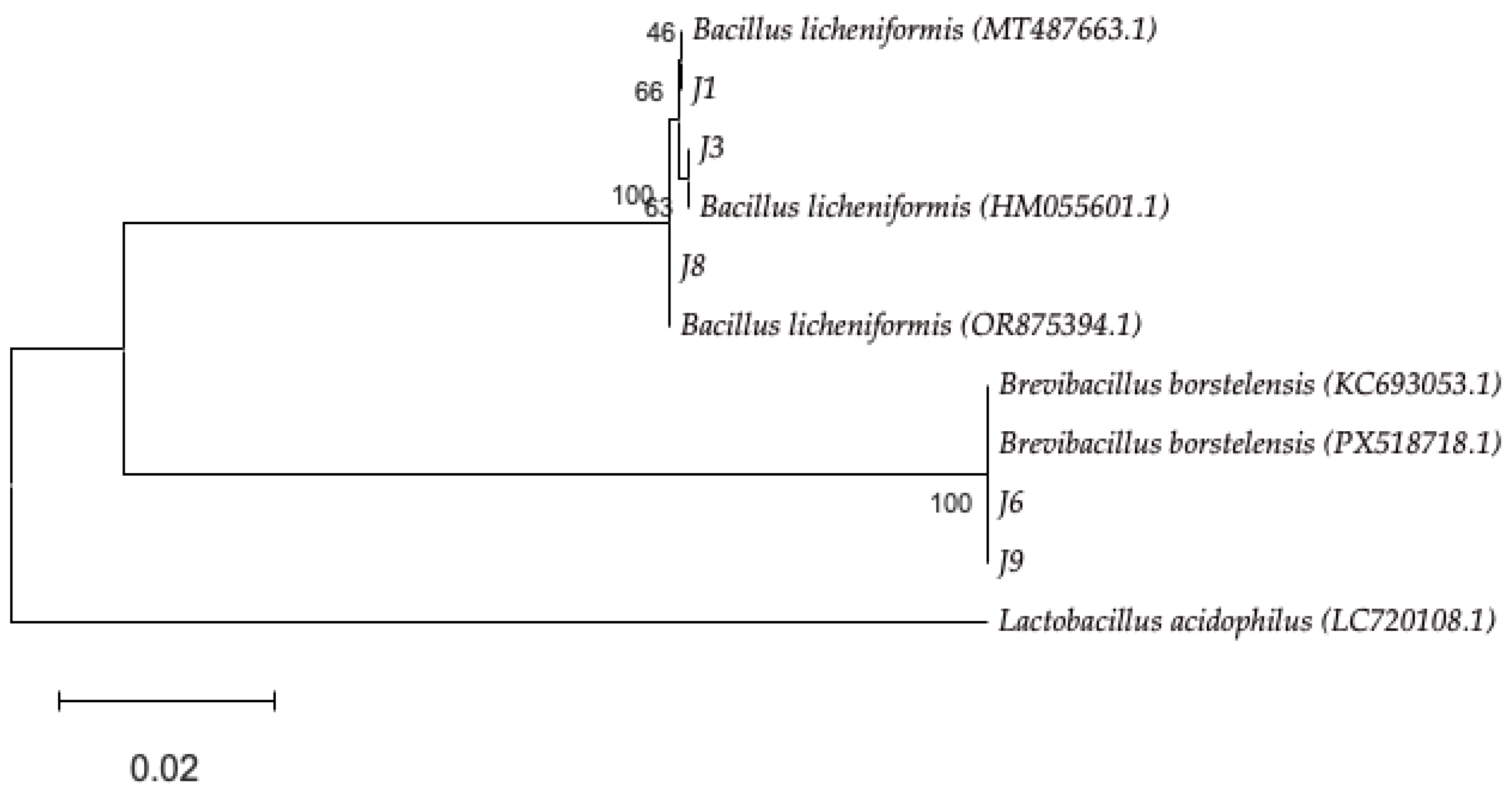
Phylogenetic analysis based on 16S rRNA gene sequences further supported these identifications. The Maximum Likelihood tree (Figure 1) clustered the isolates into two well-defined clades. Isolates J1, J3, and J8 clustered with reference sequences of B. licheniformis, whereas isolates J6 and J9 grouped within a distinct clade associated with B. borstelensis. The branching patterns were strongly supported by bootstrap values at the corresponding internal nodes.
A sequence from Lactobacillus acidophilus was included as an outgroup to provide a root for the phylogenetic tree, confirming the clear separation between the Bacillus and Brevibacillus lineages. The final phylogenetic reconstruction was based on a multiple sequence alignment of 1261 nucleotide positions after the removal of sites with less than 95% coverage.

3.4. Analysis of Growth Kinetics in Thermophilic Bacterial Isolates

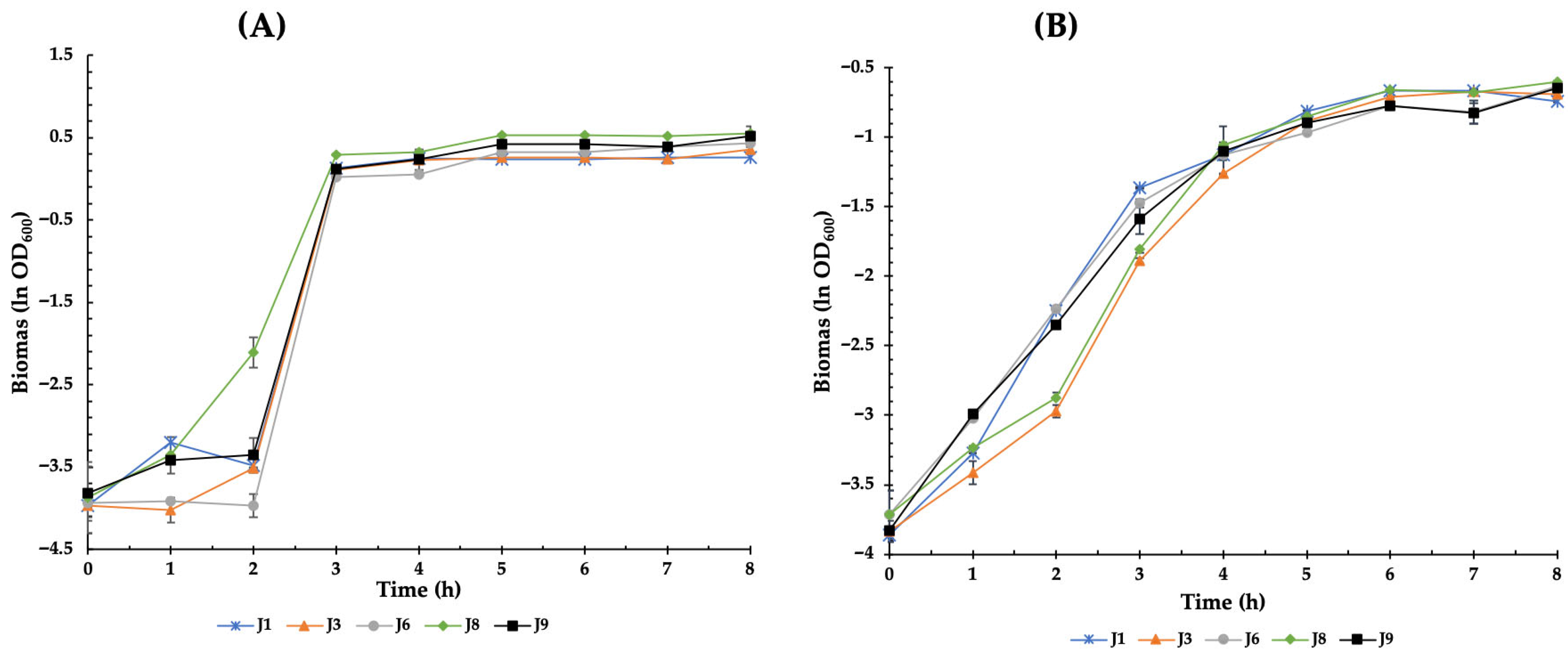
Bacterial growth curves obtained at 45 °C and 50 °C, expressed as ln OD600 as a function of time, revealed clear differences associated with both incubation temperature and taxonomic identity of the evaluated strains (Bacillus licheniformis and Brevibacillus borstelensis) (Figure 2, Table 4). At 45 °C, strains identified as B. licheniformis (J1, J3, and J8) and B. borstelensis (J6 and J9) exhibited a short lag phase, extending from 0 to 2 h, followed by a well-defined exponential phase. B. licheniformis strains entered the exponential phase slightly earlier, displaying steeper growth slopes between 2 and 4 h (Figure 2A), which resulted in higher ln OD600 values during this period. Consistently, these strains showed specific growth rates (μmax) ranging from 1.25 to 1.46 h−1, with doubling times between 28.40 and 33.30 min, with strain J1 exhibiting the highest μmax value within this group (1.46 h−1). Under the same thermal condition, B. borstelensis strains entered the exponential phase more gradually; however, they exhibited comparable or superior kinetic performance. Strain J6 showed the highest specific growth rate of the entire study (μmax = 1.78 h−1) and the shortest doubling time (23.31 min), indicating high growth efficiency at 45 °C. Despite these differences during the exponential phase, both species reached similar optical density values during the stationary phase, which began at approximately 5 h of incubation.
At 50 °C, a general prolongation of the lag phase was observed across all strains, being more pronounced in B. licheniformis, where the lag phase extended up to approximately 3 h (Figure 2 B). Correspondingly, μmax values decreased markedly, ranging from 0.77 to 1.08 h−1, with extended doubling times (38.46–54.31 min). Strain J3 showed the greatest thermal sensitivity, exhibiting the lowest μmax value (0.79 h−1) and the longest doubling time (52.78 min). In contrast, B. borstelensis strains (J6 and J9) displayed a better adaptation to increased temperature, characterized by an earlier transition to the exponential phase and slightly higher growth slopes compared to B. licheniformis. At 50 °C, B. borstelensis maintained relatively stable μmax values (1.07 h−1 for J6 and 0.83 h−1 for J9), with doubling times of 38.70 and 50.20 min, respectively. The stationary phase was reached at approximately 6 h, with final ln OD600 values lower than those observed at 45 °C, particularly in B. licheniformis strains.

3.5. Production of Hydrolytic Enzymes

The enzymatic potential of the thermophilic isolates was quantified using the Enzymatic Activity Index (EAI), as summarized in Table 5. All B. licheniformis isolates (J1, J3, and J8) exhibited a broad enzymatic profile, showing positive results for proteolytic, amylolytic, and cellulolytic assays. For these strains, EAI values ranged from 1.9 to 2.4 for proteolysis, 2.1 to 2.5 for amylolysis, and 2.8 to 3.6 for cellulolysis. Notably, isolate J8 exhibited the highest cellulolytic index (3.6 ± 0.10) among all tested strains.
In contrast, B. borstelensis isolates showed limited enzymatic capacity. Isolate J6 did not exhibit detectable activity for any of the enzymes under the evaluated conditions. Isolate J9 showed only proteolytic activity with a low EAI (1.1 ± 0.19), while amylolytic and cellulolytic activities were absent. Overall, the cellulolytic assays yielded the highest activity indices among the enzymatically active isolates.

3.6. Mineral Solubilization by Thermophilic Bacteria

The mineral solubilization capacity of the thermophilic isolates was evaluated through qualitative plate assays, as summarized in Table 6. Phosphate and potassium solubilization were observed in isolates J1, J3, J6, and J8, characterized by the formation of distinct halos and characteristic color changes in the indicator media. In contrast, none of the isolates exhibited detectable zinc solubilization under the evaluated conditions.
Isolate J9 did not show solubilization activity for any of the tested minerals. Overall, the ability to mobilize phosphate and potassium was more prevalent among the analyzed strains than zinc solubilization, suggesting a specialized metabolic potential for nutrient mobilization in these geothermal isolates.

3.7. Antagonistic Activity of the Thermophilic Bacteria

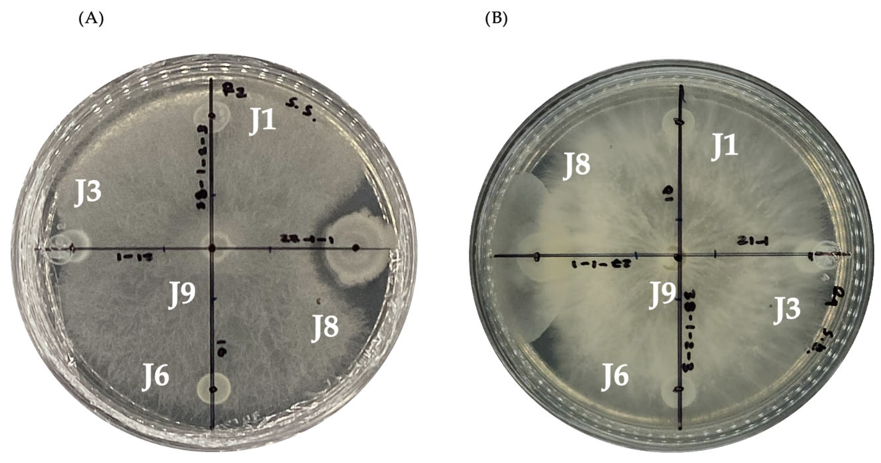
The antagonistic potential of the thermotolerant isolates against the phytopathogenic fungi Sclerotinia sclerotiorum and Sclerotium rolfsii was evaluated using the dual culture technique. Among all evaluated strains, only isolate J8 exhibited inhibitory activity under the assay conditions. A visible inhibition halo of approximately 3 mm was observed around the bacterial colony in both cases. The reduction in mycelial radial growth was 6.00 ± 0.21% for Sclerotinia sclerotiorum and 6.07 ± 0.49% for Sclerotium rolfsii (mean ± SD, n = 8) compared to the control plates (Figure 3).

3.8. Evaluation of Pesticide Tolerance Potential

The growth response of the thermophilic isolates under nutrient-limited conditions supplemented with pesticides is summarized in Table 7. All bacterial strains exhibited robust growth in the control medium (glucose and ammonium sulfate). In contrast, Azoxystrobin exerted a total inhibitory effect, with no growth observed regardless of whether it was supplied as a sole source of carbon, nitrogen, or both.
Fluazinam and Benomyl revealed a differential metabolic plasticity among the isolates. Specifically, B. licheniformis J1 and J3, and B. borstelensis J9, demonstrated the ability to utilize Fluazinam as a source of carbon, nitrogen, or as a combined C/N source. However, isolate J6 was unable to utilize Fluazinam as a sole nitrogen source, and isolate J8 showed no growth when it was provided as the sole carbon source, although both thrived under the combined C/N treatment. Regarding Benomyl, B. borstelensis (J6 and J9) and B. licheniformis J8 exhibited the highest metabolic versatility, growing under all evaluated nutritional conditions. Conversely, B. licheniformis J1 and J3 were able to utilize Benomyl as an individual source of C or N, but their growth was completely inhibited when the pesticide was supplied as the sole source of both elements (C/N).

4. Discussion

Geothermal activity drives heated water to the surface, transporting dissolved minerals and reduced chemical species that define the microbial diversity, which is modulated by temperature, pH, and nutrient availability. In this study, the El Jípago spring exhibited a thermal profile of 66 °C and a slightly alkaline pH (7.8), conditions conducive to the establishment of specialized high-temperature microbial communities. This temperature range is consistent with geothermal systems worldwide, such as Indian hot springs (32–67 °C) and the Deulajhari complex (43–65 °C) [55,56]. Similarly, the observed pH aligns with reports from the Dongnae hot springs in South Korea (pH 7.6) and the Araró region in Mexico, where neutral to moderately alkaline values remain stable regardless of seasonal variations [25].
The chemical framework at El Jípago, characterized by total hardness (186 mg/L) and total dissolved solids (330 mg/L), reflects a moderately mineralized freshwater system according to USGS/EPA and Davis and DeWiest criteria [57]. The relatively elevated TDS may be linked to the high-water temperature, which enhances mineral dissolution and ionic solubility [58,59]. These physicochemical parameters provide a stable ecological niche that supports the survival of the cultivable thermophilic bacteria identified in this work.
The isolation and selection of bacteria from this geothermal spring resulted in the identification of cultivable representatives belonging to Bacillus licheniformis and Brevibacillus borstelensis. Both genera have been repeatedly reported in hot spring environments worldwide. Brevibacillus has been isolated from hot springs and sediments in Ethiopia, which demonstrates its thermotolerance and adaptability to high-temperature ecosystems [60]. In northern Algeria, their presence has been confirmed in multiple studies: Aireche et al. (2025) identified Brevibacillus strains in two extreme hot springs using MALDI-TOF MS and 16S ribosomal RNA sequencing; Benammar et al. reported their presence in hot springs at 40.6–96 °C; and Gomri et al. described aerobic thermophilic endospore forming bacteria in hot springs in northern Algeria [61,62,63]. In India, Brevibacillus has also been identified as part of the microbial community inhabiting more than 100 hot springs in the northeast region [64]. In México, B. licheniformis was isolated from Espinazo hot springs in Nuevo León, confirming its presence in geothermal ecosystems in northern México [65]. Similar findings have been reported in Asia. Lakra and Sharma isolated B. licheniformis from hot springs of Surajkund and Ramkund (50–70 °C) in Jharkhand, India, and identified it as a thermophilic strain exopolysaccharide secretor [66]. In Europe and Western Asia, Ulucay et al. documented the presence of B. licheniformis in seven geothermal hot springs from east and southeast Anatolia, Turkey (40–85 °C) [67]. In general, previous studies show that B. licheniformis commonly colonizes hot spring habitats worldwide. Its persistence in different geothermal niches may be attributed to its high thermal resistance, metabolic flexibility, and endospore formation, which altogether confer a strong ecological advantage in extreme environments.
The integration of growth curves and kinetic parameters demonstrates that temperature plays a critical role in modulating bacterial growth kinetics, affecting lag phase duration, specific growth rate, and doubling time. At 45 °C, both B. licheniformis and B. borstelensis exhibited rapid adaptation and high growth rates, indicating that this temperature is close to the optimal growth range for the evaluated strains. The early exponential onset and relatively high μmax values observed in B. licheniformis at 45 °C are consistent with previous reports describing this species as thermotolerant and metabolically active at elevated temperatures [68,69]. However, the superior performance of B. borstelensis strain J6, which exhibited the highest μmax and shortest doubling time, suggests a greater efficiency in biomass production under these conditions. At 50 °C, the pronounced reduction in μmax values and the extension of doubling times reflect the impact of thermal stress on cellular metabolism. This effect was more evident in B. licheniformis, suggesting that 50 °C approaches the upper functional temperature limit for this species. The prolonged lag phase and reduced growth rates are indicative of increased metabolic costs associated with cellular adaptation, including enzyme stabilization and stress-response mechanisms. Conversely, B. borstelensis maintained relatively high μmax values at 50 °C, particularly strain J6, supporting its classification as a highly thermotolerant bacterium. The ability of this species to sustain growth under elevated temperatures suggests the presence of effective physiological adaptations that mitigate thermal damage and maintain metabolic activity [70]. Overall, the results indicate that 45 °C represents a near-optimal growth condition for both species, whereas 50 °C constitutes a limiting temperature, with a more pronounced inhibitory effect on B. licheniformis. In contrast, B. borstelensis, especially strain J6, displayed greater kinetic robustness and thermal tolerance, highlighting its potential suitability for biotechnological applications requiring operation at elevated temperatures.
The bacterial isolates were additionally evaluated for their biotechnological potential through assays assessing antagonistic activity against phytopathogenic fungi, pesticide tolerance, mineral solubilization, and extracellular enzyme production.
The traditional management of diseases caused by these pathogens relies heavily on chemical pesticides, which can lead to environmental contamination and the development of resistant pathogen populations. Consequently, there is an increasing need for alternative and sustainable disease management strategies, such as biological control approaches. Phytopathogenic fungi such as Sclerotinia sclerotiorum and Sclerotium rolfsii are responsible for economically important plant diseases affecting a wide range of crops, including potato and other horticultural and legume species [71]. Among the analyzed strains, B. licheniformis J8 exhibited antagonistic activity (6%) against Sclerotinia sclerotiorum and Sclerotium rolfsii. Although the level of inhibition observed in this study was limited, it suggests the secretion of diffusible antifungal metabolites capable of halting mycelial expansion before physical contact between the colonies. Previous studies have also reported antifungal activity for other B. licheniformis strains. For example, Malakar et al. described B. licheniformis SCV1 as a strong antagonist against S. sclerotiorum (76.3% inhibition), as well as against Colletotrichum gloeosporioides (53%), Fusarium verticillioides (51%), and Corynespora cassicola (36%) [72]. The lower inhibition observed in the present study may be attributed to strain-specific differences and the non-optimized, exploratory nature of the assay. On the other hand, Brevibacillus strains did not exhibit antagonistic activity under the conditions evaluated, despite reports indicating that members of this genus can produce antimicrobial metabolites [73].
Pesticide residues linger in soil and water, affecting non-target organisms and posing toxicological risks to humans [74]. Bioremediation using microorganisms capable of degrading or tolerating such compounds offers a sustainable alternative. In this study, B. licheniformis and Brevibacillus strains grew in the presence of fluazinam and benomyl, tolerating these pesticides. As per our knowledge, this is the first report of such activity in these genera. Benomyl degradation has previously been attributed to Pseudomonas spp., which can use its metabolite, carbendazim, as a carbon source [75]. Even though the isolates didn’t grow in the presence of azoxystrobin, previous research shows that B. licheniformis TAB7 may metabolize this compound, converting it into azoxystrobin amine, which is less toxic [76]. In a similar way, other Bacillus species (B. cereus, B. megaterium, and B. weihenstephanensis, included) that have been associated with the resistance or degradation of the azoxystrobin in soil bioaugmentation systems [77].
Mineral solubilization assays were performed as an exploratory functional screening. Qualitative tests showed phosphate and potassium solubilization in several B. licheniformis isolates (J1, J3, and J8), whereas zinc solubilization was not detected under the tested conditions. These findings are consistent with previous studies describing the capacity of B. licheniformis to mobilize inorganic minerals [78,79]. In Brevibacillus, solubilization was limited to strain J6, in agreement with the comparatively fewer reports of mineral-mobilizing activity for B. borstelensis [80]. Given the qualitative nature of the assays, these results should be interpreted as preliminary indicators rather than evidence of plant growth-promoting potential.
Proteolytic enzymes are among the most commonly used biocatalysts in industry and account for approximately 60% of the international enzyme market. The proteases traditionally used as milk coagulants in cheese production play an essential role in a range of sectors, including detergents, leather processing, food and drink, paper, pharmaceutical products, diagnostics, textiles, bioremediation, and even silver recovery [81]. In this research, all B. licheniformis strain isolates showed protease activity, consistent with numerous reports describing this species as a prolific producer of thermostable proteases with broad industrial relevance. Ji et al. (2020) characterized a serine protease of B. licheniformis W10 with antifungal activity; Ling et al. (2022) identified a similar enzyme from strain TG116; and Abdella and Ahmed (2025) described an alkaline protease of the MA1 strain [82,83,84]. On the contrary, the production of Brevibacillus spp. proteases have been described less frequently [85]. In this study, one of the three Brevibacillus isolates (strain J9) exhibited detectable proteolytic activity. However, the genomic analysis of B. borstelensis S8 identified at least 42 putative peptidases, including pepA, pepF, pepP, pepQ, pepV, and pepT, indicating substantial genetic potential for protease synthesis [86]. Proteolytic activity was assessed using a semi-quantitative enzyme activity index (EAI). B. licheniformis isolates showed EAI values ranging from 1.9 to 2.4, with strain J8 exhibiting the highest activity (2.4 ± 0.19). These values are comparable to or slightly higher than those reported by Prihanto et al. (2021) for Bacillus sp. (proteolytic index up to 2.09 ± 0.41) [87]. In contrast, B. borstelensis isolates showed low or undetectable proteolytic activity (EAI ≈ 1.1), which falls within the lower range reported in screening studies such as that of Behera et al. (2021), REI 1.05–1.64 [88]. Overall, the detected activities are consistent with preliminary screening-level enzymatic potential.
Amylases account for approximately 25% of the global enzyme market and are extensively used in the food, detergent, pharmaceutical, paper, and textile industries [89]. In the present study, all B. licheniformis isolates (J1, J3, and J8) exhibited amylase activity, in agreement with previous studies describing B. licheniformis as a robust producer of thermostable amylases [90,91,92]. In contrast, no detectable amylase activity was observed in the Brevibacillus isolates (J6 and J9), although amylase production has been documented in certain B. borstelensis strains, suggesting strain-dependent expression of this enzymatic trait [93]. The amylolytic activity detected in B. licheniformis isolates yielded enzyme activity index (EAI) values ranging from 2.1 to 2.5, exceeding those reported for wild-type microorganisms in previous screening studies. For example, Verma and Verma (2018) described relative enzyme activity (REA) values between 1.028 and 1.224 for fungal isolates evaluated using qualitative plate assays [94]. These results remain below the highest enzymatic indices described for thermotolerant bacteria; for instance, Cotta et al. (2021) reported a maximum amylase EI of 4.11 in B. licheniformis isolates [93], indicating that enzymatic expression may vary substantially among strains and cultivation conditions.
Cellulase production in B. licheniformis (J1, J3 and J8) was also detected. The cellulolytic capacity of this species has been associated with various biotechnological applications, including clarification and yield improvement of fruit juice, valorization of agro-industrial waste, and production of molecular-weight polysaccharides for food and pharmaceutical uses [95,96,97]. Although no cellulase activity was detected among the analyzed Brevibacillus isolates, previous research has reported that Brevibacillus strains are cellulase producers. For example, Khosravi et al. (2021) identified B. borstelensis strains in soil samples and leaves with significant cellulase activity, and Wang et al. (2024) characterized the A24 strain (B. borstelensis) as one of the most efficient cellulose degraders in their analysis [98,99]. The cellulolytic activity observed in the B. licheniformis isolates yielded enzyme activity index (EAI) values ranging from 2.8 to 3.6. These values are higher than those documented in previous screening studies. Dewiyanti et al. (2021) described low cellulolytic activity in bacteria isolated from mangrove soils, with cellulolytic index (CI) values between 0.07 and 0.80, whereas Nelson et al. (2021) reported Bacillus strains exhibiting cellulolytic index (ICA) values of 0.844 ± 0.092 and 1.146 ± 0.109 [100,101]. Likewise, higher cellulolytic EI values have been described in other thermotolerant Bacillus species, such as Bacillus altitudinis (EI = 6.40) reported by Cotta et al. (2021), suggesting that cellulase production is strongly strain-dependent and influenced by experimental parameters [93].

5. Conclusions

The thermotolerant bacterial isolates obtained from the El Jípago geothermal spring demonstrated diverse functional traits, with Bacillus licheniformis strains showing the most consistent extracellular enzymatic activities and mineral-mobilizing capacity, while Brevibacillus borstelensis exhibited comparatively lower but detectable functional responses. Although antagonistic effects against phytopathogenic fungi were limited, the observed pesticide tolerance and enzymatic versatility highlight the adaptive potential of these bacteria to high-temperature environments. These findings reinforce geothermal ecosystems as valuable sources of functionally diverse microorganisms and support future quantitative and molecular studies to further define their potential applications in agrobiotechnological and environmental processes.

Author Contributions

L.I.P.-C., G.A.M.-R. and L.D.C.-O. conceived and planned the experiments. M.M.-P. and L.I.P.-C. carried out the experiments. G.A.M.-R., J.G.L.-V., H.A.L.-H., I.N.M.-G., A.L.-V. and L.D.C.-O. contributed to the interpretation of the results. L.I.P.-C. and L.D.C.-O. took the lead in writing the manuscript. All authors provided critical feedback and helped shape the research, analysis, and manuscript. All authors have read and agreed to the published version of the manuscript.

Funding

This research was funded by Universidad Autónoma de Occidente under the Institutional Program for the Strengthening of Research and Graduate Studies (PIFIP 2024).

Institutional Review Board Statement

Not applicable.

Informed Consent Statement

Not applicable.

Data Availability Statement

The genomic sequences generated in this study have been deposited in the National Center for Biotechnology Information (NCBI) database under accession number(s) [PX401984, PX401986, PX401987, PX401989, PX401990]. All other data supporting the findings of this study are available from the corresponding author upon reasonable request.

Acknowledgments

The authors gratefully acknowledge the Universidad Autónoma de Occidente for the facilities and financial support provided through the project PIFIP-2024 during the development of this research.

Conflicts of Interest

The authors declare no conflicts of interest.

References

  1. Des Marais, D.J.; Walter, M.R. Terrestrial hot spring systems: Introduction. Astrobiology 2019, 19, 1419–1432. [Google Scholar] [CrossRef] [PubMed]
  2. Mehta, D.; Satyanarayana, T. Diversity of Hot Environments and Thermophilic Microbes. In Thermophilic Microbes in Environmental and Industrial Biotechnology; Satyanarayana, T., Littlechild, J., Kawarabayasi, Y., Eds.; Springer: Dordrecht, The Netherlands, 2013; pp. 3–60. [Google Scholar] [CrossRef]
  3. Uribe-Lorío, L.; Brenes-Guillén, L.; Hernández-Ascencio, W.; Mora-Amador, R.; González, G.; Ramírez-Umaña, C.J.; Díez, B.; Pedrós-Alió, C. The influence of temperature and pH on bacterial community composition of microbial mats in hot springs from Costa Rica. Microbiologyopen 2019, 8, e893. [Google Scholar] [CrossRef] [PubMed]
  4. Merino, N.; Aronson, H.S.; Bojanova, D.P.; Feyhl-Buska, J.; Wong, M.L.; Zhang, S.; Giovannelli, D. Living at the extremes: Extremophiles and the limits of life in a planetary context. Front. Microbiol. 2019, 10, 780. [Google Scholar] [CrossRef]
  5. Ishaq, K.; Shah, A.H.; Fariq, A.; Rasheed, S.; Jannat, S. Diversity of culturable thermophilic bacteria from Tata Pani hotspring of Kotli Azad Jammu and Kashmir. Biodivers. Data J. 2023, 11, e99224. [Google Scholar] [CrossRef]
  6. Kumar, S.; Das, S.; Jiya, N.; Sharma, A.; Saha, C.; Sharma, P.; Tamang, S.; Thakur, N. Bacterial diversity along the geothermal gradients: Insights from the high-altitude Himalayan hot spring habitats of Sikkim. Curr. Res. Microb. Sci 2024, 7, 100310. [Google Scholar] [CrossRef]
  7. Rekadwad, B.; Gonzalez, J.M. Multidisciplinary involvement and potential of thermophiles. Folia Microbiol. 2019, 64, 389–406. [Google Scholar] [CrossRef]
  8. Giordano, D. Bioactive Molecules from Extreme Environments. Mar. Drugs 2020, 18, 640. [Google Scholar] [CrossRef]
  9. Pandey, A.; Dhakar, K. Extreme thermal environments: Reservoirs of industrially important thermozymes. Front. Microbiol. 2026, 16, 1739143. [Google Scholar] [CrossRef]
  10. Arbab, S.; Ullah, H.; Khan, M.I.U.; Khattak, M.N.K.; Zhang, J.; Li, K.; Hassan, I.U. Diversity and distribution of thermophilic microorganisms and their applications in biotechnology. J. Basic Microbiol. 2022, 62, 95–108. [Google Scholar] [CrossRef]
  11. Gallo, G.; Imbimbo, P.; Aulitto, M. The Undeniable Potential of Thermophiles in Industrial Processes. Int. J. Mol. Sci 2024, 25, 7685. [Google Scholar] [CrossRef] [PubMed]
  12. Panosyan, H.; Margaryan, A.; Birkeland, N.K. Geothermal springs in Armenia and Nagorno-Karabakh: Potential sources of hydrolase-producing thermophilic bacilli. Extremophiles 2020, 24, 519–536. [Google Scholar] [CrossRef]
  13. Harwood, C.R.; Mouillon, J.M.; Pohl, S.; Arnau, J. Secondary metabolite production and the safety of industrially important members of the Bacillus subtilis group. FEMS Microbiol. Rev. 2018, 42, 721–738. [Google Scholar] [CrossRef]
  14. Verma, J.P.; Jaiswal, D.K.; Krishna, R.; Prakash, S.; Yadav, J.; Singh, V. Characterization and Screening of Thermophilic Bacillus strains for developing plant growth promoting consortium from hot spring of Leh and Ladakh Region of India. Front. Microbiol. 2018, 9, 1293. [Google Scholar] [CrossRef]
  15. Kumar, A.; Sahu, A.; Jaiswal, K.; Prasad, R. Thermophilic Microbes: Their Role in Plant Growth Promotion and Mitigation of Biotic Stress. In Extremophiles for Sustainable Agriculture and Soil Health Improvement; Ranjan, A., Rajput, V.D., Chauhan, A., Prazdnova, E.V., Minkina, T., Zargar, S.M., Eds.; Springer: Cham, Switzerland, 2024; pp. 337–360. [Google Scholar] [CrossRef]
  16. Patel, K.S.; Naik, J.H.; Chaudhari, S.; Amaresan, N. Characterization of culturable bacteria isolated from hot springs for plant growth promoting traits and effect on tomato (Lycopersicon esculentum) seedling. Comptes Rendus Biol. 2017, 340, 244–249. [Google Scholar] [CrossRef]
  17. Pawar, R.; Borkar, S.G. Isolation and characterization of thermophilic bacteria from different habitats and their assessment for antagonism against soil-borne fungal plant pathogens. Afr. J. Microbiol. Res. 2018, 12, 556–566. [Google Scholar] [CrossRef]
  18. Abdulsalam, L.; Ganiyu, S.A.; Khalil, A.B. Purification and Identification of Antimicrobial Peptides Produced by Thermophilic Bacteria Isolated from Hot Springs. Probiotics Antimicrob. Proteins 2025, 1–26. [Google Scholar] [CrossRef] [PubMed]
  19. Meslé, M.M.; Mueller, R.C.; Peach, J.; Eilers, B.; Tripet, B.P.; Bothner, B.; Copié, V.; Peyton, B.M. Isolation and Characterization of Lignocellulose-Degrading Geobacillus thermoleovorans from Yellowstone National Park. Appl. Environ. Microbiol. 2022, 88, e00958-21. [Google Scholar] [CrossRef]
  20. Saghatelyan, A.; Margaryan, A.; Panosyan, H.; Birkeland, N.K. Microbial Diversity of Terrestrial Geothermal Springs in Armenia and Nagorno-Karabakh: A Review. Microorganisms 2021, 9, 1473. [Google Scholar] [CrossRef] [PubMed]
  21. Ruhl, I.A.; Sheremet, A.; Smirnova, A.V.; Sharp, C.E.; Grasby, S.E.; Strous, M.; Dunfield, P.F. Microbial Functional Diversity Correlates with Species Diversity along a Temperature Gradient. mSystems 2022, 7, e00991-21. [Google Scholar] [CrossRef] [PubMed]
  22. Pinzón-Martínez, D.L.; Rodríguez-Gómez, C.; Miñana-Galbis, D.; Carrillo-Chávez, J.A.; Valerio-Alfaro, G.; Oliart-Ros, R. Thermophilic bacteria from Mexican thermal environments: Isolation and potential applications. Environ. Technol. 2010, 31, 957–966. [Google Scholar] [CrossRef]
  23. Prieto-Barajas, C.M.; Alfaro-Cuevas, R.; Valencia-Cantero, E.; Santoyo, G. Effect of seasonality and physicochemical parameters on bacterial communities in two hot spring microbial mats from Araró, Mexico. Rev. Mex. Biodiv. 2017, 88, 616–624. [Google Scholar] [CrossRef]
  24. Ovando-Chacon, S.L.; Tacias-Pascacio, V.G.; Ovando-Chacon, G.E.; Rosales-Quintero, A.; Rodriguez-Leon, A.; Ruiz-Valdiviezo, V.M.; Servin-Martinez, A. Characterization of thermophilic microorganisms in the geothermal water flow of El Chichón Volcano Crater Lake. Water 2020, 12, 2172. [Google Scholar] [CrossRef]
  25. Ortega-Villar, R.; Escalante, A.; Astudillo-Melgar, F.; Lizárraga-Mendiola, L.; Vázquez-Rodríguez, G.A.; Hidalgo-Lara, M.E.; Coronel-Olivares, C. Isolation and characterization of thermophilic bacteria from a hot spring in the state of Hidalgo, Mexico, and geochemical analysis of the thermal water. Microorganisms 2024, 12, 1066. [Google Scholar] [CrossRef]
  26. Castelán-Sanchez, H.G.; Fernández, J.V.; Rojas-Vargas, J.; Martínez-Ocampo, F.; Hurtado-Ramírez, J.M.; Ríos-Vázquez, D.I.; Sánchez-Alonso, P.; Vazquez-Cruz, C.; Rojas-Ruiz, N.E. Thermophilic Pseudomonas aeruginosa strain Ch39 isolated from Chignahuapan hot springs in Puebla, Mexico. Lett. Appl. Microbiol 2025, 78, ovaf059. [Google Scholar] [CrossRef]
  27. Juárez-Aragón, M.C.; Pantoja-Irys, J.R.; de la Rosa-Manzano, E.; Garrido-Olvera, L.; Mujica-Sánchez, H.; Trejo-De León, C.R.; Vázquez-Lobo, A. Microbiome diversity across physicochemical gradient in low-medium enthalpy springs at the Sierra Madre Oriental eastern flank, northeastern Mexico. Front. Microbiol. 2025, 16, 1663000. [Google Scholar] [CrossRef]
  28. Secretaría de Economía. Análisis de agua-Determinación de la temperatura en aguas naturales, residuales y residuales tratadas-Método de prueba [Water Analysis-Determination of Temperature in Natural, Waste and Treated Wastewaters-Test Method] (NMX-AA-007-SCFI-2013). Diario Oficial de la Federación, 2013. Available online: https://www.gob.mx/cms/uploads/attachment/file/166766/nmx-aa-007-scfi-2013.pdf (accessed on 17 July 2023).
  29. Secretaría de Economía. Análisis de agua-Determinación del valor de potencial de hidrógeno (pH)-Método de prueba [Water Analysis-Determination of the Hydrogen Potential Value (pH)-Test Method] (NMX-AA-008-SCFI-2016). Diario Oficial de la Federación, 2016. Available online: https://www.gob.mx/cms/uploads/attachment/file/166767/NMX-AA-008-SCFI-2016.pdf (accessed on 17 July 2023).
  30. Secretaría de Economía. Análisis de agua-Determinación de sólidos y sales disueltas en aguas naturales, residuales y residuales tratadas-Método de prueba [Water Analysis-Determination of Solids and Dissolved Salts in Natural, Waste and Treated Wastewaters-Test Method] (NMX-AA-034-SCFI-2015). Diario Oficial de la Federación, 2015. Available online: https://www.gob.mx/cms/uploads/attachment/file/166146/nmx-aa-034-scfi-2015.pdf (accessed on 17 July 2023).
  31. Secretaría de Economía. Análisis de agua-Determinación de alcalinidad total en aguas naturales, residuales y residuales tratadas-Método de prueba [Water Analysis-Determination of Total Alkalinity in Natural, Waste and Treated Wastewaters-Test Method] (NMX-AA-036-SCFI-2001). Diario Oficial de la Federación, 2001. Available online: https://www.gob.mx/cms/uploads/attachment/file/166776/NMX-AA-036-SCFI-2001.pdf (accessed on 17 July 2023).
  32. Secretaría de Economía. Análisis de agua-Determinación de turbiedad en aguas naturales, residuales y residuales tratadas-Método de prueba [Water Analysis-Determination of Turbidity in Natural, Waste and Treated Wastewaters-Test Method] (NMX-AA-038-SCFI-2001). Diario Oficial de la Federación, 2001. Available online: https://www.gob.mx/cms/uploads/attachment/file/166777/NMX-AA-038-SCFI-2001.pdf (accessed on 17 July 2023).
  33. Secretaría de Economía. Análisis de agua-Determinación de cloruros totales en aguas naturales, residuales y residuales tratadas-Método de prueba [Water Analysis-Determination of Total Chlorides in Natural, Waste and Treated Wastewaters-Test Method] (NMX-AA-073-SCFI-2001). Diario Oficial de la Federación, 2001. Available online: https://www.gob.mx/cms/uploads/attachment/file/166789/NMX-AA-073-SCFI-2001.pdf (accessed on 17 July 2023).
  34. Secretaría de Economía. Análisis de agua-Determinación de dureza total en aguas naturales, residuales y residuales tratadas-Método de prueba [Water Analysis-Determination of Total Hardness in Natural, Waste and Treated Wastewaters-Test Method] (NMX-AA-072-SCFI-2001). Diario Oficial de la Federación, 2001. Available online: https://www.gob.mx/cms/uploads/attachment/file/166788/NMX-AA-072-SCFI-2001.pdf (accessed on 17 July 2023).
  35. Ogodo, A.C.; Agwaranze, D.I.; Daji, M.; Aso, R.E. Chapter 13 Microbial techniques and methods: Basic techniques and microscopy. In Analytical Techniques in Biosciences from Basics to Applications; Egbuna, C., Patrick-Iwuanyanwu, K.C., Shah, M.A., Ifemeje, J.C., Rasul, A., Eds.; Academic Press: Cambridge, MA, USA, 2022; pp. 201–220. [Google Scholar] [CrossRef]
  36. Schaeffer, A.B.; Fulton, M.D. A simplified method of staining endospores. Science 1933, 77, 194. [Google Scholar] [CrossRef]
  37. Moreno, I.; Marasca, E.T.G.; de Sá, P.B.Z.R.; Moitinho, J.S.; Marquezinil, M.G.; Alves, M.R.C.; Bromberg, R. Evaluation of probiotic potential of bacteriocinogenic lactic acid bacteria strains isolated from meat products. Probiotics Antimicrob. Proteins 2018, 10, 762–774. [Google Scholar] [CrossRef]
  38. Doyle, J. DNA Protocols for plants. In Molecular Techniques in Taxonomy; NATO ASI Series; Hewitt, G.M., Johnston, A.W.B., Young, J.P.W., Eds.; Springer: Berlin/Heidelberg, Germany, 1991. [Google Scholar] [CrossRef]
  39. Weisburg, W.G.; Barns, S.M.; Pelletier, D.A.; Lane, D.J. 16S ribosomal DNA amplification for phylogenetic study. J. Bacteriol. 1991, 173, 697–703. [Google Scholar] [CrossRef]
  40. Camacho, C.; Boratyn, G.M.; Joukov, V.; Vera Alvarez, R.; Madden, T.L. ElasticBLAST: Accelerating sequence search via cloud computing. BMC Bioinform. 2023, 24, 117. [Google Scholar] [CrossRef] [PubMed]
  41. Stecher, G.; Suleski, M.; Tao, Q.; Tamura, K.; Kumar, S. MEGA 12.1: Cross-Platform Release for macOS and Linux Operating Systems. J. Mol. Evol. 2025, 94, 14–18. [Google Scholar] [CrossRef] [PubMed]
  42. Jones, B.V.; Sun, F.; Marchesi, J.R. Using skimmed milk agar to functionally screen a gut metagenomic library for proteases may lead to false positives. Lett. Appl. Microbiol. 2007, 45, 418–420. [Google Scholar] [CrossRef]
  43. Mishra, S.; Behera, N. Amylase activity of a starch degrading bacteria isolated from soil receiving kitchen wastes. Afr. J. Biotechnol. 2008, 7, 3326–3331. [Google Scholar] [CrossRef]
  44. Carder, J.H. Detection and quantitation of cellulase by Congo red staining of substrates in a cup-plate diffusion assay. Anal. Biochem. 1986, 153, 75–79. [Google Scholar] [CrossRef]
  45. Bradner, J.; Gillings, M.; Nevalainen, K. Qualitative assessment of hydrolytic activities in antarctic microfungi grown at different temperatures on solid media. World J. Microbiol. Biotechnol. 1999, 15, 131–132. [Google Scholar] [CrossRef]
  46. Abe, C.A.L.; Faria, C.B.; De Castro, F.F.; De Souza, S.R.; Santos, F.C.d.; Da Silva, C.N.; Tessmann, D.J.; Barbosa-Tessmann, I.P. Fungi Isolated from Maize (Zea mays L.) Grains and Production of Associated Enzyme Activities. Int. J. Mol. Sci. 2015, 16, 15328–15346. [Google Scholar] [CrossRef] [PubMed]
  47. Pradhan, N.; Sukla, L.B. Solubilization of inorganic phosphates by fungi isolated from agriculture soil. Afr. J. Biotechnol. 2005, 5, 850–854. [Google Scholar]
  48. Velázquez-Gurrola, A.; Ramos-Alegría, M.P. Beneficios de microorganismos solubilizadores de P y K en la recuperación y mantenimiento de suelos agrícolas. In Proceedings of the Acta Proceedings, VIII Congreso Mundial de la Palta 2015, Lima, Perú, 13–18 September 2015. [Google Scholar]
  49. Bapiri, A.; Asgharzadeh, A.; Mujallali, H.; Khavazi, K.; Pazira, E. Evaluation of zinc solubilization potential by different strains of fluorescent Pseudomonas. J. Appl. Sci. Environ. Manag. 2012, 16, 295–298. [Google Scholar] [CrossRef]
  50. Urias-Lugo, D.A.; Martínez-Ereva, O.E.; Romero-Urías, C.D.L.A.; Ibarra-Sarmiento, C.R.; Estrada-Díaz, S.A.; Félix-Gastélum, R.; Leyva-Madrigal, K.Y.; Mora-Romero, G.A. Actividad antifúngica in vitro de extractos acuosos de Datura discolor obtenidos por procesamiento de alta presión. Rev. Mex. Fitopatol. 2024, 42. [Google Scholar] [CrossRef]
  51. Tapwal, A.; Singh, U.; Singh, G.; Garg, S.; Kumar, R. In vitro antagonism of Trichoderma viride against five phytopathogens. Pest Technol. 2011, 5, 59–62. [Google Scholar]
  52. Bano, N.; Musarrat, J. Characterization of a novel carbofuran degrading Pseudomonas spp. with collateral biocontrol and plant growth promoting potential. FEMS Microbiol. Lett. 2004, 23, 13–17. [Google Scholar] [CrossRef]
  53. Monod, J. The growth of bacterial cultures. Annu. Rev. Microbiol. 1949, 3, 371–394. [Google Scholar] [CrossRef]
  54. Madigan, M.T.; Bender, K.S.; Buckley, D.H.; Sattley, W.M.; Stahl, D.A. Brock Biology of Microorganisms, 16th ed.; Pearson: New York, NY, USA, 2021; pp. 144–177. [Google Scholar]
  55. Narsing Rao, M.P.; Dong, Z.Y.; Luo, Z.H.; Li, M.M.; Liu, B.B.; Guo, S.X.; Hozzein, W.N.; Xiao, M.; Li, W.J. Physicochemical and microbial diversity analyses of Indian hot springs. Front. Microbiol. 2021, 2, 627200. [Google Scholar] [CrossRef]
  56. Dixit, S.; Gaur, M.; Subudhi, E.; Sahoo, R.K.; Dey, S.; Mahapatra, L.D.; Mandal, S.; Senthil Kumar, N.; Anirudh, H. Bacterial diversity and CAZyme potential revealed in Pandanus rich thermal spring cluster of India: A non-cultivable 16S rRNA sequencing approach. Front. Microbiol. 2021, 12, 760573. [Google Scholar] [CrossRef]
  57. Davis, S.N.; De Weist, R.J.M. Hydrogeology; John Wiley and Sons: New York, NY, USA, 1966; p. 463. [Google Scholar]
  58. Temizel, E.H.; Gültekin, F.; Ersoy, A.F.; Gülbay, R.K. Multi-isotopic (O, H, C, S, Sr, B, Li) characterization of waters in a low-enthalpy geothermal system in Havza (Samsun), Turkey. Geothermics 2021, 97, 102240. [Google Scholar] [CrossRef]
  59. Yang, K.; Zhou, L.; Chen, Z.; Zhang, W.; Zhou, W. Experimental Investigations on the Dissolution Process of Dolomite by Sulfate-Rich Geothermal Water: A Case Study of the Shuijing Hot Springs in Guizhou Province. Minerals 2025, 15, 9. [Google Scholar] [CrossRef]
  60. Guta, M.; Abebe, G.; Bacha, K.; Cools, P. Screening and characterization of thermostable enzyme-producing bacteria from selected hot springs of Ethiopia. Microbiol. Spectr. 2024, 12, e03710-23. [Google Scholar] [CrossRef]
  61. Aireche, M.; Merzoug, M.; Hammadi, A.I.; Zater, Z.Y.; Bendida, K.; Brakna, C.N.; Berrazeg, M.; Aireche, A.Y.; Saidi, Y.; Todorov, S.D.; et al. Taxonomic diversity and antimicrobial potential of thermophilic bacteria from two extreme algerian hot springs. Microorganisms 2025, 13, 1425. [Google Scholar] [CrossRef] [PubMed]
  62. Benammar, L.; İnan Bektaş, K.; Menasria, T.; Beldüz, A.O.; Güler, H.I.; Bedaida, I.K.; Gonzalez, J.M.; Ayachi, A. Diversity and enzymatic potential of thermophilic bacteria associated with terrestrial hot springs in Algeria. Braz. J. Microbiol. 2020, 51, 1987–2007. [Google Scholar] [CrossRef] [PubMed]
  63. Gomri, M.A.; El Moulouk Khaldi, T.; Kharroub, K. Analysis of the diversity of aerobic, thermophilic endospore-forming bacteria in two Algerian hot springs using cultural and non-cultural methods. Ann. Microbiol. 2018, 68, 915–929. [Google Scholar] [CrossRef]
  64. Verma, J.; Sourirajan, A.; Dev, K. Bacterial diversity in 110 thermal hot springs of Indian Himalayan Region (IHR). 3 Biotech 2022, 12, 238. [Google Scholar] [CrossRef] [PubMed]
  65. Silva-Salinas, A.; Rodríguez-Delgado, M.; Gómez-Treviño, J.; López-Chuken, U.; Olvera-Carranza, C.; Blanco-Gámez, E.A. Novel thermotolerant amylase from Bacillus licheniformis strain LB04: Purification, characterization and agar-agarose. Microorganisms 2021, 9, 1857. [Google Scholar] [CrossRef]
  66. Lakra, U.; Sharma, S.R. Production of exopolysaccharides from Bacillus licheniformis, a thermophilic bacteria from hot spring of Jharkhand. Nat. Prod. Res. 2024, 38, 819–828. [Google Scholar] [CrossRef]
  67. Ulucay, O.; Gormez, A.; Ozic, C. Identification, characterization and hydrolase producing performance of thermophilic bacteria: Geothermal hot springs in the Eastern and Southeastern Anatolia Regions of Turkey. Antonie Van Leeuwenhoek 2022, 115, 253–270. [Google Scholar] [CrossRef]
  68. Rodrigo, W.W.P.; Magamulla, L.S.; Thiwanka, M.S.; Yapa, Y.M.S.M. Optimization of growth conditions to identify the superior Bacillus strain which produces high yield of thermostable alpha amylase. Adv. Enzym. Res. 2022, 10, 1–22. [Google Scholar] [CrossRef]
  69. Khalil, A.B.; Sivakumar, N.; Arslan, M.; Saleem, H.; Qarawi, S. Insights into Brevibacillus borstelensis AK1 through Whole Genome Sequencing: A Thermophilic Bacterium Isolated from a Hot Spring in Saudi Arabia. Biomed. Res. Int. 2018, 2018, 5862437. [Google Scholar] [CrossRef] [PubMed]
  70. Bashir, R.; Syed, Q.; Mumtaz, M.W.; Anwar, F.; Saari, N.; Adnan, A. Growth kinetics, purification and characterization of α-amylase produced from Bacillus licheniformis DSM-1969 using lignocellulosic Banana waste as an Elicitor. Bioresources 2014, 9, 6791–6804. [Google Scholar] [CrossRef]
  71. Wang, S.Y.; Zhang, Y.J.; Chen, X.; Shi, X.C.; Herrera-Balandrano, D.D.; Liu, F.Q.; Laborda, P. Biocontrol methods for the management of Sclerotinia sclerotiorum in legumes: A review. Phytopathology 2024, 114, 1447–1457. [Google Scholar] [CrossRef]
  72. Malakar, C.; Barman, D.; Kalita, M.C.; Deka, S. A biosurfactant-producing novel bacterial strain isolated from vermicompost having multiple plant growth-promoting traits. J. Basic Microbiol. 2023, 63, 746–758. [Google Scholar] [CrossRef]
  73. Yang, X.; Yousef, A.E. Antimicrobial peptides produced by Brevibacillus spp.: Structure, classification and bioactivity: A mini review. World J. Microbiol. Biotechnol. 2018, 34, 57. [Google Scholar] [CrossRef]
  74. Garud, A.; Pawar, S.; Patil, M.S.; Kale, S.R.; Patil, S. A Scientific Review of Pesticides: Classification, Toxicity, Health Effects, Sustainability, and Environmental Impact. Cureus 2024, 16, e67945. [Google Scholar] [CrossRef]
  75. Fuchs, A.; de Vries, F.W. Bacterial breakdown of benomyl I pure cultures. Antonie Van Leeuwenhoek 1978, 44, 283–292. [Google Scholar] [CrossRef]
  76. Mpofu, E.; Alias, A.; Tomita, K.; Suzuki-Minakuchi, C.; Tomita, K.; Chakraborty, J.; Malon, M.; Ogura, Y.; Takikawa, H.; Okada, K.; et al. Azoxystrobin amine: A novel azoxystrobin degradation product from Bacillus licheniformis strain TAB7. Chemosphere 2021, 273, 129663. [Google Scholar] [CrossRef] [PubMed]
  77. Baćmaga, M.; Wyszkowska, J.; Kucharski, J. Bioaugmentation of soil contaminated with azoxystrobin. Water Air Soil Pollut. 2017, 228, 19. [Google Scholar] [CrossRef]
  78. Sharma, R.; Chandel, S.; Chauhan, A.; Shirkot, C.K. Enhanced phosphorus solubilization by Bacillus licheniformis CKA1 using central composite design and response surface methodology. J. Pure Appl. Microbiol. 2015, 9, 3131–3141. [Google Scholar]
  79. Raji, M.; Thangavelu, M. Isolation and screening of potassium solubilizing bacteria from saxicolous habitat and their impact on tomato growth in different soil types. Arch. Microbiol. 2021, 203, 3147–3161. [Google Scholar] [CrossRef]
  80. Yadav, R.C.; Sharma, S.K.; Varma, A.; Singh, U.B.; Kumar, A.; Bhupenchandra, I.; Rai, J.P.; Sharma, P.K.; Singh, H.V. Zinc-solubilizing Bacillus spp. in conjunction with chemical fertilizers enhance growth, yield, nutrient content, and zinc biofortification in wheat crop. Front. Microbiol. 2023, 14, 1210938. [Google Scholar] [CrossRef]
  81. Razzaq, A.; Shamsi, S.; Ali, A.; Ali, Q.; Sajjad, M.; Malik, A.; Ashraf, M. Microbial proteases applications. Front. Bioeng. Biotechnol. 2019, 7, 110. [Google Scholar] [CrossRef]
  82. Ji, Z.L.; Peng, S.; Chen, L.L.; Liu, Y.; Yan, C.; Zhu, F. Identification and characterization of a serine protease from Bacillus licheniformis W10: A potential antifungal agent. Int. J. Biol. Macromol. 2020, 145, 594–603. [Google Scholar] [CrossRef]
  83. Ling, L.; Cheng, W.; Jiang, K.; Jiao, Z.; Luo, H.; Yang, C.; Pang, M.; Lu, L. The antifungal activity of a serine protease and the enzyme production of characteristics of Bacillus licheniformis TG116. Arch. Microbiol. 2022, 204, 601. [Google Scholar] [CrossRef] [PubMed]
  84. Abdella, M.A.A.; Ahmed, S.A. Stable protease from Bacillus licheniformis-MA1 strain: Statistical production optimization, kinetic and thermodynamic characterization, and application in silver recovery from used X-ray films. Microb. Cell Fact. 2025, 24, 98. [Google Scholar] [CrossRef] [PubMed]
  85. Rai, S.K.; Mukherjee, A.K. Optimization of the production of an oxidant and detergent-stable alkaline beta-keratinase from Brevibacillus sp. strain AS-S10-11: Application of enzyme in laundry detergent formulations and in the leather industry. Biochem. Eng. J. 2011, 54, 47–56. [Google Scholar] [CrossRef]
  86. Liang, A.; Wang, J.; Ding, L.; Zou, L.; Wang, D.; Zhu, C.; Tang, J. Probiotic properties, whole-genome sequence analysis, and safety assessment of Brevibacillus borstelensis S8. LWT 2024, 210, 116800. [Google Scholar] [CrossRef]
  87. Prihanto, A.A.; Aninta, K.D.; Trisnaningrum, S. Protease production from Bacillus sp. isolated from gastrointestinal tract of Catfish (Clarias sp.) with different medium. J. Aquac. Fish Health 2021, 10, 186–191. [Google Scholar] [CrossRef]
  88. Behera, B.C.; Sethi, B.K.; Mohapatra, S.; Thatoi, H.; Mishra, R.R. Bio-production of alkaline protease by Trichoderma longibrachiatum and Penicillium rubidurum using different agro-industrial products. Nov. Res. Microbiol. J. 2021, 5, 1241–1255. [Google Scholar]
  89. Gopinath, S.C.; Anbu, P.; Arshad, M.K.; Lakshmipriya, T.; Voon, C.H.; Hashim, U.; Chinni, S.V. Biotechnological processes in microbial amylase production. Biomed. Res. Int. 2017, 2017, 1272193. [Google Scholar] [CrossRef]
  90. Suthar, S.; Joshi, D.; Patel, H.; Patel, D.; Kikani, B.A. Optimization and purification of a novel calcium-independent thermostable, α-amylase produced by Bacillus licheniformis UDS-5. World J. Microbiol. Biotechnol. 2024, 40, 385. [Google Scholar] [CrossRef] [PubMed]
  91. Aras, S.; Altıntas, R.; Aygun, E.; Goren, G.; Sisecioglu, M. Purification of α-amylase from thermophilic Bacillus licheniformis SO5 by using a novel method, alternating current magnetic-field assisted three-phase partitioning: Molecular docking and bread quality improvement. Food Chem. 2025, 484, 144258. [Google Scholar] [CrossRef]
  92. Fan, S.; Lü, X.; Wei, X.; Lü, R.; Feng, C.; Jin, Y.; Yan, M.; Yang, Z. Computational design of α-amylase from Bacillus licheniformis to increase its activity and stability at high temperatures. Comput. Struct. Biotechnol. J. 2024, 13, 982–989. [Google Scholar] [CrossRef]
  93. Cotta, S.P.M.; Marins, M.S.; Marriel, I.E.; Lana, U.G.P.; Gomes, E.A.; Figueiredo, J.E.F.; Oliveira-Paiva, C.A. Thermoresistant enzyme-producing microorganisms isolated from composting. Braz. J. Biol. 2021, 83, e244205. [Google Scholar] [CrossRef]
  94. Verma, P.; Verma, R.K. Qualitative estimation of amylase enzyme activity of fungal species isolated from iron ore mined overburden soil. Trop. Plant Res. 2018, 5, 396–404. [Google Scholar] [CrossRef]
  95. Abdella, M.A.A.; Ibrahim, G.E. Application of statistical designs strategy to improve cellulase production using agro-waste residue by a novel isolate Bacillus licheniformis strain-MA1 and assessing the enzyme effect on apple juice quality. BMC Microbiol. 2024, 29, 511. [Google Scholar] [CrossRef]
  96. Zhu, T.; Liu, S.; Gao, Y.; Sun, X.; Hu, H.; Zhang, Z.; Zhang, W.; Xiong, L.; Liu, H. Functional characterization of a highly efficient endoglucanase from Bacillus licheniformis BJ2022 and its application in the preparation of low-molecular-weight konjac glucomannan. J. Agric. Food Chem. 2025, 73, 8367–8380. [Google Scholar] [CrossRef]
  97. Okoth, B.A.; Makonde, H.M.; Bosire, C.M.; Kibiti, C.M. Characterization and enzymatic potential of bacteria and fungi from Mwakirunge Dumpsite, Kenya. Int. J. Microbiol. 2025, 2025, 7818433. [Google Scholar] [CrossRef]
  98. Khosravi, F.; Khaleghi, M.; Naghavi, H. Screening and identification of cellulose-degrading bacteria from soil and leaves at Kerman province, Iran. Arch. Microbiol. 2021, 204, 88. [Google Scholar] [CrossRef]
  99. Wang, J.; Bao, F.; Wei, H.; Zhang, Z. Screening of cellulose-degrading bacteria and optimization of cellulase production from Bacillus cereus A49 through response surface methodology. Sci. Rep. 2024, 14, 7755. [Google Scholar] [CrossRef]
  100. Dewiyanti, I.; Darmawi, D.; Muchlisin, Z.A.; Helmi, T.Z.; Arisa, I.I.; Rahmiati, R.; Destri, E. Cellulase enzyme activity of the bacteria isolated from mangrove ecosystem in Aceh Besar and Banda Aceh. In IOP Conference Series: Earth and Environmental Science; IOP Publishing: Bristol, UK, 2022; Volume 951, p. 012113. [Google Scholar]
  101. Nelson, K.; Muge, E.; Wamalwa, B. Cellulolytic Bacillus species isolated from the gut of the desert locust Schistocerca gregaria. Sci. Afr. 2021, 11, e00665. [Google Scholar] [CrossRef]
Figure 1. Maximum Likelihood phylogenetic tree based on partial 16S rRNA gene sequences of the bacterial isolates, with Lactobacillus acidophilus as the outgroup. Bootstrap values (1000 replicates) are shown at branch nodes.
Bacteria 05 00021 g001
Figure 2. Bacterial growth kinetics of thermotolerant strains. (A) Growth curves obtained at 45 °C for Bacillus licheniformis (J1, J3, J8) and Brevibacillus borstelensis (J6, J9); (B) Growth curves obtained at 50 °C for the same bacterial strains. Values are expressed as the natural logarithm of optical density (ln OD600) over time. Specific growth rates (μmax) and doubling times (td) were determined during the exponential phase (2–4 h).
Bacteria 05 00021 g002
Figure 3. In vitro antagonism of thermophilic bacteria against phytopathogenic fungi: (A) Sclerotinia sclerotiorum and (B) Sclerotium rolfsii, demonstrating the inhibitory activity of strain J8. where strains J1, J3 and J8 were identified as B. licheniformis, and strains J6 and J9 as B. borstelensis.
Bacteria 05 00021 g003
Table 1. Physicochemical properties of the geothermal water sample.
PropertyValue
Temperature (°C)66.4
pH7.8
Turbidity (NTU)0.408 ± 0.03
Total hardness (mg/L as CaCO3)186 ± 14.11
Alkalinity (mg/L as CaCO3)3.96 ± 0.006
Chlorides (mg/L)ND
Total solids (mg/L)330 ± 15.87
Note: (ND) Not detected. Data are expressed as the mean ± standard deviation (SD) of three independent replicates (n = 3).
Table 2. Macroscopic and microscopic morphological characteristics of the isolates.
Property Isolate
J1J3J6J8J9
Colony SizeSmallMediumMediumLargeLarge
ShapeRhizoidCircularCircularIrregularIrregular
Margin (Edges)FilamentousWavyWavyLobulatedLobulated
ElevationConvexFlatConvexFlatFlat
SurfaceRugoseSmoothSmoothRugoseSmooth
ConsistencyMembranousMembranousCreamyMembranousCreamy
ColorBeigeCreamCreamBeigeBeige
Transmitted LightOpaqueOpaqueTranslucentOpaqueOpaque
Reflected LightOpaqueTranslucentOpaqueOpaqueShiny
Gram StainingGram positiveGram positiveGram positiveGram positiveGram positive
Endospore formation+++++
Note: (+) Positive result.
Table 3. Comparative analysis between the obtained 16 rRNA gene sequence and reference sequence deposited in GenBank.
IsolateMicroorganismAccession Number (Reference)Sequence Length (Bp)Query Cover (%)Identity (%)GenBank
Accession (This Study)
J1Bacillus licheniformisMT487663.11403100100PX401987
J3Bacillus licheniformisHM055601.1145410099.92PX401989
J6Brevibacillus borstelensisKC693053.11436100100PX401984
J8Bacillus licheniformisOR875394.1154010099.92PX401990
J9Brevibacillus borstelensisPX518718.11405100100PX401986
Table 4. Kinetic parameters of thermophilic isolates at different temperatures.
Temperature
(°C)
ParameterB. licheniformis
J1
B. licheniformis J3B. borstelensis J6B. licheniformis
J
B. borstelensis J9
45μmax (h−1)1.46 ± 0.011.33 ± 0.011.78 ± 0.041.25 ± 0.011.26 ± 0.01
td (min)28.40 ± 0.1931.35 ± 0.2023.31 ± 0.4433.30 ± 0.1232.87 ± 0.10
50μmax (h−1)1.08 ± 0.010.79 ± 0.011.07 ± 0.010.77 ± 0.010.83 ± 0.01
td (min)38.46 ± 0.2152.78 ± 0.1338.70 ± 0.1054.31 ± 0.2850.20 ± 0.20
Note: μmax: Specific growth rate (h−1); td: Doubling time (min); Data are presented as the mean ± standard deviation (SD) of three independent replicates (n = 3).
Table 5. Enzymatic activity assays.
IsolateProteolytic
Activity
Amylolytic
Activity
Cellulolytic
Activity
Proteolytic
EAI
Amylolytic
EAI
Cellulolytic
EAI
B. licheniformis J1+++1.9 ± 0.102.1 ± 0.102.9 ± 0.11
B. licheniformis J3+++2.0 ± 0.002.3 ± 0.292.8 ± 0.09
B. borstelensis J6NDNDND
B. licheniformis J8+++2.4 ± 0.192.5 ± 0.293.6 ± 0.10
B. borstelensis J91.1 ± 0.19NDND
Note: (EAI) Enzyme Activity Index, (+) Positive result; (−) Negative result; (ND) Not detected. Data are expressed as the mean ± standard deviation (SD) of three replicates. (EAI) Enzyme Activity Index.
Table 6. Mineral solubilization assays.
IsolatePhosphate
Solubilization
Potassium
Solubilization
Zinc
Solubilization
B. licheniformis J1++
B. licheniformis J3++
B. borstelensis J6++
B. licheniformis J8++
B. borstelensis J9
Note: (+) solubilization detected; (−) no solubilization detected.
Table 7. Pesticide tolerance of B. licheniformis and B. bortelensis isolates strains tested with different treatments.
IsolateControlAzoxystrobinFluazinamBenomyl
CNC/NCNC/NCNC/N
B. licheniformis J1++++++
B. licheniformis J3++++++
B. borstelensis J6++++++
B. licheniformis J8++++++
B. borstelensis J9+++++++
Note: (+) indicates visible colony growth; (−) indicates no growth. (C) sole carbon source; (N) sole nitrogen source; (C/N) sole carbon and nitrogen source.
Disclaimer/Publisher’s Note: The statements, opinions and data contained in all publications are solely those of the individual author(s) and contributor(s) and not of MDPI and/or the editor(s). MDPI and/or the editor(s) disclaim responsibility for any injury to people or property resulting from any ideas, methods, instructions or products referred to in the content.