Next Article in Journal
Optimizing Material Ratios and Moisture Content for Potassium-Solubilizing Purple Nonsulfur Bacteria-Inoculated Compost Production
Previous Article in Journal
Unveiling the Antibacterial Potential of Melaleuca cajuputi Essential Oils Against MRSA: Integrating In Vitro Efficacy and In Silico Mechanistic Insights
 
 
bacteria-logo
Article Menu
Review

Genetic Elements That Contribute to Antibiotic Resistance in Bacteria of Clinical Importance

by
Benjamín Abraham Ayil-Gutiérrez
1,†,
Erika Acosta-Cruz
2,†,
Juan Manuel Bello-López
3,
Yesseny Vásquez-Martínez
4,
Marcelo Cortez-San Martin
5,
Lorenzo Felipe Sánchez-Teyer
6,
Luis Carlos Rodríguez-Zapata
6,
Francisco Alberto Tamayo-Ordoñez
7,
Esmeralda Cázares-Sánchez
8,
Víctor Hugo Ramos-García
9,
Eric Sánchez-López
10,
Hernan de Jesús Villanueva-Alonzo
11,
Virgilio Bocanegra-García
12,
Humberto Martínez-Montoya
9,
Grethel Díaz-Palafox
13,
María José García-Castillo
6,
María Concepción Tamayo-Ordoñez
2,* and
Yahaira de Jesús Tamayo-Ordoñez
12,*
1
SECIHTI, Centro de Biotecnología Genómica, Instituto Politécnico Nacional, Reynosa 88710, Mexico
2
Facultad de Ciencias Químicas, Universidad Autónoma de Coahuila, Saltillo 25280, Mexico
3
Hospital Juárez de México, Mexico City 07760, Mexico
4
Escuela de Medicina, Facultad de Ciencias Médicas, Universidad de Santiago de Chile, Santiago 9170022, Chile
5
Laboratorio de Virología Molecular y Control de Patógenos, Facultad de Química y Biología, Universidad de Santiago de Chile, Santiago 9170022, Chile
6
Unidad de Biotecnología, Centro de Investigación Científica de Yucatán A.C., Mérida 97200, Mexico
7
Facultad de Química, Universidad Autónoma del Carmen, Ciudad del Carmen 24180, Mexico
8
Tecnológico Nacional de México, Instituto Tecnológico de la Zona Maya, Chetumal 77960, Mexico
9
Unidad Académica Multidisciplinaria Reynosa-Aztlán, Universidad Autónoma de Tamaulipas, Reynosa 88740, Mexico
10
Universidad Tecnológica de Tamaulipas Norte, Reynosa 88770, Mexico
11
SECIHTI, Centro de Investigaciones Regionales “Dr. Hideyo Noguchi”, Universidad Autónoma de Yucatán, Mérida 97000, Mexico
12
Centro de Biotecnología Genómica, Instituto Politécnico Nacional, Reynosa 88710, Mexico
13
Facultad de Enfermería y Nutrición U.L., Universidad Autónoma de Coahuila, Torreón 27298, Mexico
*
Authors to whom correspondence should be addressed.
These authors contributed equally to this work.
Bacteria 2026, 5(1), 14; https://doi.org/10.3390/bacteria5010014
Submission received: 20 November 2025 / Revised: 19 January 2026 / Accepted: 22 February 2026 / Published: 5 March 2026

Abstract

Antimicrobial resistance (AMR) poses a severe threat to global health by limiting treatment options and increasing clinical and economic burdens. This review synthesizes evidence showing that resistance evolution is strongly shaped by antibiotic pressure, leading to the accumulation of adaptive mutations, activation of efflux systems, and widespread dissemination of resistance determinants across clinical, animal, and environmental settings. We highlight recent genomic, metagenomic, and structural findings that elucidate the molecular basis of AMR, with particular emphasis on horizontal gene transfer mediated by mobile genetic elements such as plasmids, integrons, and transposons. Analyses across One Health interfaces reveal extensive sharing of antimicrobial resistance genes among humans, livestock, and environmental reservoirs, identifying Enterobacteriaceae and ESKAPE pathogens as key hubs of resistance dissemination. Special focus is placed on Acinetobacter baumannii, where phylogenetic and three-dimensional structural analyses of class D β-lactamases OXA-23 and OXA-24/40 demonstrate a conserved catalytic framework coupled with substantial sequence and conformational variability. These structural differences likely influence carbapenem specificity and resistance levels. Collectively, the findings underscore how genetic diversity, mobile elements, and structural adaptation converge to drive AMR, reinforcing the need for integrated genomic and structural approaches to guide surveillance and antimicrobial development.

1. Introduction

Antimicrobial resistance (AMR), particularly to antibiotics, has become a serious global public health problem. It is estimated that by 2050, approximately 1.91 million deaths will be directly attributable to AMR, and an additional 8.22 million deaths will be associated with this condition [1]. This phenomenon represents one of the greatest threats to modern medicine, as it compromises the effectiveness of current treatments and contributes to increased mortality, hospital costs, and the duration of illnesses.
The economic cost of conventional antibiotic therapy, particularly regimens that require hospitalization and intensive monitoring such as intravenous vancomycin, represents a substantial and often unsustainable burden for many healthcare institutions. Economic evaluations consistently demonstrate that the direct medical costs of vancomycin therapy extend far beyond the relatively modest acquisition price of the drug itself (approximately US $9.01 per 1 g dose), with comprehensive cost models showing that total per-patient expenditures for a course of treatment can reach US $22,493–US $48,925 when hospital length of stay and associated care are included, depending on the infection type (e.g., skin and soft tissue infection, bacteremia, infective endocarditis, or hospital-acquired pneumonia)—an order of magnitude higher than drug cost alone owing to inpatient care expenses [2].
These high hospitalization costs are reinforced by comparative analyses indicating that the total cost of vancomycin treatment per patient (including inpatient and outpatient components) can exceed US $11,000, with a significant proportion attributable to medical services, length of stay, specialist consultations, and therapeutic drug monitoring rather than the antibiotic itself [3]. f. In many health systems, per-day general ward costs alone can exceed US $1300–US $1900, with intensive care unit rates markedly higher, intensifying the economic strain of extended inpatient antibiotic therapy [3].
The magnitude of these costs has important implications for resource-limited hospitals, where budgets may not support prolonged vancomycin-based treatment without adverse impacts on other services. Analyses of broader national expenditures further highlight that the majority of antibiotic-associated costs are derived from prolonged hospital stays rather than pharmaceutical costs per se, underscoring how conventional therapies that necessitate inpatient administration can rapidly outpace available funding in smaller or under-resourced facilities [4]. Consequently, the economic burden entailed by standard antibiotic regimens like vancomycin is not only a matter of drug pricing but a complex interplay of clinical, logistical, and financial factors that may exceed the fiscal capacity of some hospitals to provide sustainable care.
Current research in this field focuses on the development of new antibacterial agents that act through mechanisms distinct from those of conventional antibiotics [5,6]. In parallel, the use of combinations of antimicrobial agents with non-antibiotic compounds has emerged as a promising strategy to enhance therapeutic efficacy and mitigate the emergence of resistance [7,8,9].
In 2024, the World Health Organization (WHO) published an updated list of priority antibiotic-resistant pathogens, identifying the twelve bacterial families that pose the greatest threat to human health [10]. This list categorizes pathogens into three priority levels—critical, high, and medium—based on the urgency with which new antibiotics are needed. The critical-priority group includes multidrug-resistant bacteria that pose significant risks in healthcare settings such as hospitals and nursing homes, particularly among patients requiring invasive devices such as ventilators or intravenous catheters. This group comprises Acinetobacter baumannii, Pseudomonas aeruginosa, Mycobacterium tuberculosis, and several Enterobacteriaceae species, including Klebsiella pneumoniae, Escherichia coli, Serratia marcescens, and Proteus mirabilis. These pathogens can cause severe and often life-threatening infections, including bacteremia and pneumonia. The high-priority group includes Salmonella Typhi, Shigella spp., Enterococcus faecium, Neisseria gonorrhoeae, and Staphylococcus aureus, whereas the medium-priority group comprises macrolide-resistant Streptococcus pyogenes (group A), Streptococcus pneumoniae, ampicillin-resistant Haemophilus influenzae, and macrolide-resistant Streptococcus agalactiae (group B) [10].
These bacteria have developed resistance to a broad range of antibiotics, including carbapenems and third-generation cephalosporins, which are considered among the most effective options for treating multidrug-resistant infections [11,12]. Within this context, a particularly concerning cluster of microorganisms—collectively known by the acronym ESKAPE—has gained attention. These pathogens are notorious for their rapid acquisition of antimicrobial resistance across multiple drug classes, as well as their ability to survive on dry surfaces for prolonged periods, facilitating their dissemination in hospital environments, especially during the COVID-19 pandemic [13,14]. Given the profound clinical and social impact of bacterial resistance, it is essential to deepen our understanding of the genetic and molecular mechanisms underpinning this phenomenon. Accordingly, this research aims to explore recent advances in genetic and molecular strategies designed to counteract antibiotic resistance in clinically relevant bacteria, thereby contributing to the development of new therapeutic approaches and diagnostic tools.

2. Factors Contributing to Antibiotic Resistance in Bacteria

Antibiotic resistance occurs when a drug no longer effectively inhibits bacterial growth or fails to kill bacteria. Bacteria that survive or replicate in the presence of an antimicrobial agent that should prevent their multiplication are termed resistant bacteria. Although resistance can arise naturally as a result of evolutionary selection, its emergence and dissemination have been markedly accelerated by the inappropriate or excessive use of antibiotics in the population [15,16]. Indeed, resistance often appears shortly after the introduction of new antimicrobial compounds into clinical practice.
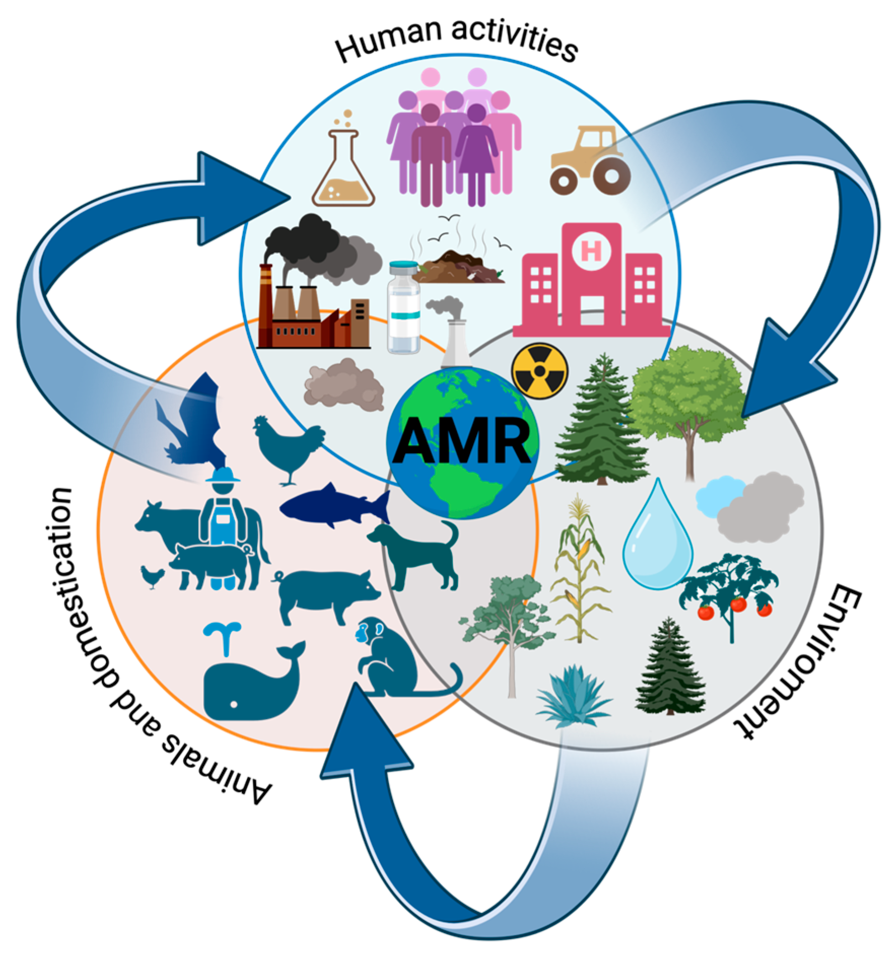
Among the multiple factors contributing to the development of resistance (Figure 1), agricultural and livestock practices are particularly significant. The increasing global demand for animal-derived food products has driven the widespread use of antibiotics as growth promoters in livestock intended for human consumption. It is estimated that per kilogram of animal produced, 45 mg/kg are used in cattle, 148 mg/kg in chickens, and 172 mg/kg in pigs [17]. This practice has facilitated the emergence of resistant pathogens within the commensal microbiota of animals, creating the potential for resistance transmission along the food chain—either through direct consumption or contact with animals and their waste. The populations most vulnerable to such infections include immunocompromised individuals, infants, and the elderly [18].
Several studies involving metagenomic analyses have documented the presence of shared antimicrobial resistance genes (ARGs) between human and animal microbiomes, as well as potential horizontal gene transfer events. Napit et al. [19] assessed antimicrobial resistance (AMR) dynamics in a temporary settlement in Kathmandu through shotgun metagenomic analysis of human, avian, and environmental samples. The results demonstrated the co-occurrence of pathogenic bacteria, virulence factors, and antimicrobial resistance genes (ARGs) across all compartments, with shared bacterial taxa between humans and animals. A total of 72 virulence factor genes and 53 ARG subtypes were identified, with poultry samples showing the highest ARG diversity, suggesting a link with antibiotic use in poultry production. Yang et al. [20] investigated the microbiome, resistome, and mobility of antimicrobial resistance genes (ARGs) within a Chinese wet market system using metagenomic assembly and binning approaches. The study identified 1080 ARG subtypes across 36 metagenomes, of which 221 were shared among human feces, chicken feces, chicken carcasses, and environmental samples, indicating substantial overlap of ARGs across compartments. ARG distribution was strongly influenced by mobile genetic elements and bacterial community composition. Notably, 89 ARG-carrying genomes were detected, including several opportunistic pathogenic species harboring multiple ARGs and mobile genetic elements, such as Escherichia coli, Acinetobacter johnsonii, Klebsiella variicola, Klebsiella pneumoniae, and Citrobacter freundii. These findings highlighted these bacterial species as key reservoirs and vectors for ARG mobility. In addition, numerous putative horizontal gene transfer events were reported, supporting the role of wet market environments in facilitating ARG dissemination among humans, animals, and the environment. Cao et al. [21] analyzed more than 1000 human, swine, and chicken gut metagenomes worldwide to investigate the similarity and transferability of acquired antimicrobial resistance genes (ARGs) between humans and food animals. The study reported a higher abundance and diversity of ARGs in swine compared with humans, highlighting food animals as important reservoirs of resistance. Commensal bacteria, particularly members of the order Clostridiales, were identified as major contributors of ARGs associated with mobile genetic elements (MGEs) in both humans and animals. Notably, specific MGEs, including Tn4451/Tn4453 and TnAs3, were found to play a central role in ARG sharing between hosts, with the site-specific recombinase TnpX facilitating gene mobility. Although ARG transferability showed greater variability in humans, the average transferability was higher in swine, indicating potential resistance hotspots in food animals. These findings underscore the importance of global surveillance of ARGs in livestock within a One Health framework to prevent the emergence and spread of antimicrobial resistance.
Another major contributing factor is the inappropriate prescription and administration of antibiotics, including self-medication, incomplete treatment regimens, or overuse, all of which exert strong selective pressure on bacterial populations [22]. Compounding this issue are poor sanitation conditions, inadequate control of drug distribution, and substandard water and hygiene systems in healthcare facilities—factors that collectively promote the global spread of antimicrobial resistance (AMR) [22,23]. In hospital environments, the continuous surveillance of resistant strains is essential, as many bacteria possess the capacity to transfer genetic material, such as plasmids and transposons, to other host cells [24]. For instance, colistin-resistant Escherichia coli strains carrying the mcr-1 gene have been isolated [25], as well as Klebsiella pneumoniae strains exhibiting resistance to aminoglycosides and quinolones, along with intrinsic resistance to penicillin [26,27].
Several plasmids have been identified in clinical isolates from hospital settings. Among them, pFBAOT and pIE420 confer resistance to oxytetracycline [28], while blaKPC and blaNDM are associated with resistance to carbapenems, a class of broad-spectrum antibiotics [29]. Plasmids harboring virulence genes have also been described, such as KpVP-1, which contributes to the pathogenicity of K. pneumoniae [30]. The identification and characterization of resistant plasmids and bacterial strains remain a top priority, particularly in intensive care units (ICUs), where up to 50% of vulnerable patients may develop infections caused by multidrug-resistant microorganisms, leading to severe complications or death. Therefore, monitoring resistant isolates from hospital samples is crucial for assessing the incidence of nosocomial infections and improving therapeutic decision-making [31,32].
Beyond clinical settings, improper disposal of antibiotics contributes to environmental contamination. Large quantities of antibiotic residues and resistant bacteria are released into aquatic ecosystems, facilitating the dissemination of resistance genes. Plasmids carrying such genes can be horizontally transferred to other bacteria, promoting the spread of resistance among soil and waterborne microorganisms [33]. Wastewater environments are particularly important reservoirs for resistant bacteria, as they contain elevated levels of carbon and nutrients that support microbial survival [34,35]. Wastewater treatment plants have therefore become critical points for isolating resistant bacteria. In these environments, plasmids such as pRSB107, which confer resistance to multiple antibiotics—including ampicillin, penicillin, chloramphenicol, erythromycin, kanamycin, neomycin, streptomycin, sulfonamides, tetracycline, and trimethoprim—have been identified in microorganisms isolated from biofilms within treatment facilities [36]. It has been suggested that the prevalence of resistant bacteria in wastewater may exceed that observed in hospital environments, underscoring their ecological and epidemiological significance.
Hospital wastewater systems constitute a significant and under-monitored reservoir for antibiotic-resistant bacteria and associated resistance genes. A recent one-year seasonal study of untreated effluent from five discharge points at the Hospital Juárez de México found a high taxonomic diversity dominated by phyla such as Proteobacteria, Firmicutes and Bacteroidetes, with clinically relevant genera identified in approximately 73% of the samples [37]. Among the isolates, members of the ESKAPE group (for example, Enterobacter and Escherichia-Shigella) were prevalent, and seven out of eight targeted antimicrobial-resistance genes (including blaKPC, blaOXA-40 and mcr-1) were detected, with clear seasonal differences in their occurrence [37]. The study also found that physicochemical parameters such as total and dissolved solids, temperature, nitrate concentration and pH significantly influenced the bacterial community composition in the wastewater [37]. Moreover, in parallel research the authors documented the release of antibiotics including meropenem, cefepime and ceftazidime via the hospital wastewater stream—further amplifying the risk of selection and dissemination of resistant pathogens [38].

3. Mechanisms of Antibiotic Resistance in Bacteria

Bacteria have developed the ability to resist antibiotics that were once effective in treating infections. The mechanisms by which bacteria acquire resistance include mutations leading to drug inactivation (e.g., β-lactamases), modification of previously essential biosynthetic pathways, alteration of antibiotic target sites (such as those involved in cell wall synthesis, folic acid biosynthesis, or protein biosynthesis), and active drug efflux from the cell. In addition, horizontal gene transfer through transformation, transduction, or conjugation contributes significantly to the dissemination of these genetic alterations. A particularly concerning group of pathogens is the ESKAPE, as mentioned above. These multidrug-resistant organisms are major causes of nosocomial infections that are difficult to treat. They have developed resistance to nearly all available classes of antibacterial agents, including tetracyclines, erythromycins, methicillin, gentamicin, vancomycin, imipenem, ceftazidime, levofloxacin, linezolid, daptomycin, and ceftaroline.
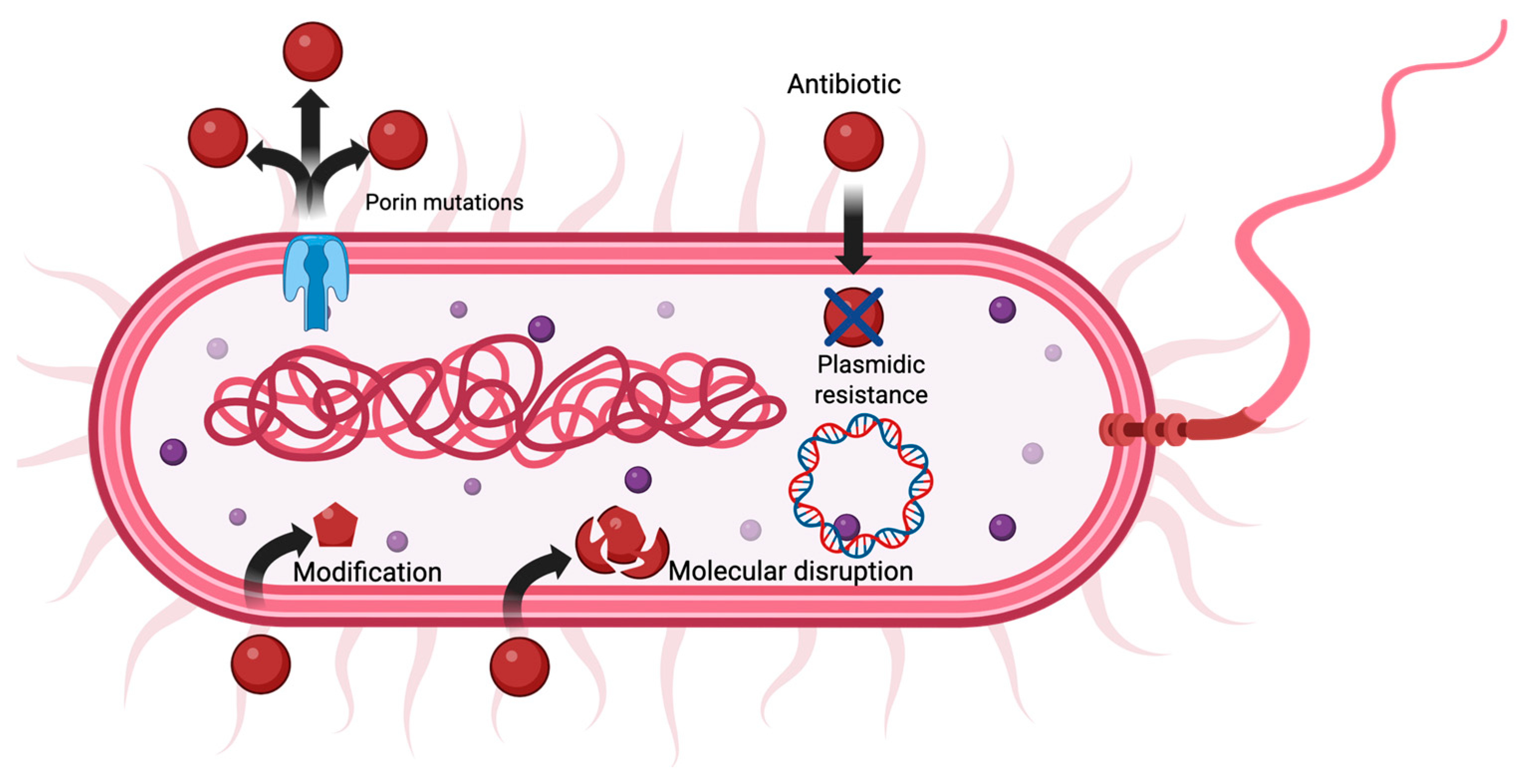
The success of bacterial antibiotic resistance lies in the transcription and translation of specific resistance proteins. One of the most common mechanisms is the production of enzymes that chemically modify or destroy antibiotics [39]. Another mechanism involves alteration or overexpression of antibiotic target sites, which prevents the drug from binding effectively and diminishes its activity (Figure 2) [40]. Resistance mechanisms can also occur at the cell membrane level. For example, reduced porin expression decreases membrane permeability, thereby preventing antibiotics from entering the bacterial cell. Additionally, the presence of efflux pumps actively expels antibiotics from the intracellular environment to the outside medium, significantly lowering the intracellular drug concentration (Figure 2) [39,40].
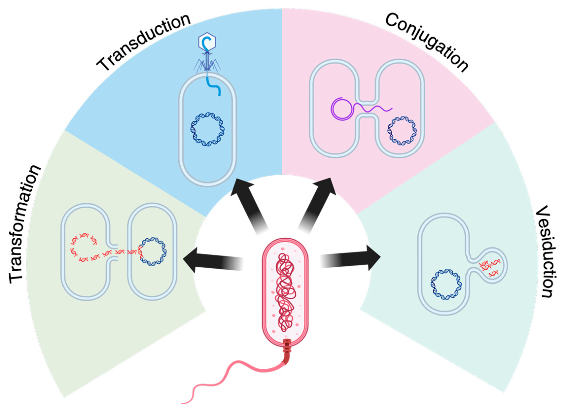
Biofilm formation is another important factor contributing to bacterial resistance. Biofilms form when bacteria adhere to the surface and produce an extracellular polymeric matrix that enhances their protection. This structure increases bacterial resistance to antibiotics by multiple means. The extracellular matrix may contain enzymes that degrade antibiotics, such as β-lactamases, and its low permeability can restrict antibiotic diffusion, resulting in a resistant phenotype even in bacteria that are susceptible under planktonic conditions [41]. Currently, four main mechanisms of horizontal gene transfer (HGT) are recognized in bacteria (see Figure 3) [42]. The first is transformation, which involves the uptake of free DNA from the environment; the second is conjugation, which requires direct physical contact between microbial cells; and the third is transduction, a process mediated by bacteriophages that transfer genetic material during infection of bacterial cells. The fourth mechanism, more recently described, is known as “vesiduction”, referring to DNA transfer mediated by extracellular vesicles [43].
All these mechanisms reduce the effectiveness of antibiotic therapy and have increased the incidence of infections caused by antibiotic-resistant or multidrug-resistant microorganisms. Latin America is currently one of the regions with the highest incidence of outbreaks caused by organisms resistant to one or more antibiotics. Therefore, research focused on the isolation and genetic characterization of resistant microorganisms—particularly the identification of resistance plasmids—as well as understanding other molecular mechanisms such as membrane modifications or enzymatic inactivation, is crucial for improving medical treatments. The knowledge generated through these studies will be key to developing effective therapeutic protocols and new antimicrobial drugs, which could greatly benefit healthcare systems in regions most vulnerable to infections caused by multidrug-resistant strains.

4. Regulatory Networks Modulating Cell Wall Integrity and Antimicrobial Resistance in Staphylococcus aureus and Quorum Sensing in Pseudomonas aeruginosa

The adaptive capacity of bacterial pathogens to resist environmental stressors and antimicrobial agents is fundamentally governed by complex regulatory networks that coordinate gene expression in response to external cues. In Staphylococcus aureus, regulatory proteins and enzymes modulate virulence factor production, cell wall integrity, and the interplay with host innate immunity. Among these, the low-molecular-weight protein arginine phosphatase PtpB has been recently characterized as a contributor to regulatory pathways influencing cell wall integrity and stress responsiveness [44].
PtpB, encoded within the core genome of S. aureus, participates in growth phase-dependent transcriptional regulation of key virulence-associated genes and small regulatory RNAs, including the agr quorum-sensing effector RNAIII. Functional ablation of ptpB results in significant attenuation of extracellular nuclease and protease activities, reduced capacity to withstand Triton X-100-induced autolysis, and increased susceptibility to phagocytic clearance by polymorphonuclear leukocytes, collectively implicating PtpB in maintaining cell wall robustness and evasion of host defenses [44]. Transcriptomic analyses further link PtpB to modulation of stress response gene networks controlling reactive oxygen species detoxification and metabolic adaptation during intracellular survival, indicating that PtpB integrates environmental signals into regulatory circuits that reinforce bacterial cell envelope integrity and pathogenic persistence. These findings expand the conceptual framework of S. aureus regulatory systems beyond classical two-component and quorum-sensing elements to include phosphatase-dependent post-translational control mechanisms with direct implications for cell wall function under hostile conditions [45].
In Gram-negative pathogens such as Pseudomonas aeruginosa, antibiotic resistance and virulence are coordinated through elaborate quorum-sensing (QS) networks that respond to population density and environmental stress. The LasR transcriptional regulator lies at the apex of the LasI/LasR QS circuit, which controls a hierarchy of downstream effectors including the Rhl and PQS (Pseudomonas quinolone signal) systems. Mutations in lasR are frequently encountered in clinical multidrug-resistant (MDR) isolates and are associated with altered expression of virulence determinants, efflux pump genes, and phenotypic diversification that enhances survival in chronic infection contexts [46]. Loss of LasR function perturbs the QS hierarchy, leading to compensatory upregulation of alternative virulence pathways and increased efflux activity, which together contribute to the persistence of P. aeruginosa in antibiotic-challenged environments. Moreover, clinical studies demonstrate that lasR mutations correlate with heightened virulence factor production and exacerbated tissue damage in host models, underscoring the dual role of LasR in modulating both pathogenicity and adaptive resistance phenotypes [47].
Beyond single-gene effects, recent research highlights that the QS network in P. aeruginosa regulates a broad spectrum of biological functions including biofilm formation, secretion systems, motility, and stress responses, revealing a high degree of integration between regulatory circuits governing virulence and antibiotic resistance [48]. The dynamic interplay between LasR, efflux systems, and other regulatory modules underscores the complexity of transcriptional control strategies that facilitate phenotypic plasticity and survival under antimicrobial pressure.
Collectively, insights into regulatory proteins such as PtpB and transcriptional modulators like LasR illustrate that bacterial resistance and virulence are not solely attributable to static gene content but emerge from dynamic regulatory networks capable of reprogramming cellular physiology in response to environmental and host-derived signals. Understanding these regulatory layers offers promising avenues for novel therapeutic interventions that target network nodes rather than individual pathways, potentially mitigating resistance by disrupting bacterial adaptability itself.

5. Evidence of Horizontal Gene Transfer Mechanisms Described in Clinically Relevant Bacteria

5.1. Acinetobacter baumannii

Acinetobacter baumannii is an opportunistic pathogen primarily associated with infections in critically ill patients hospitalized in Intensive Care Units (ICUs). The most frequent nosocomial infections caused by A. baumannii include ventilator-associated pneumonia and bacteremia, both of which are associated with considerable morbidity and mortality. Other clinical manifestations include skin and soft tissue infections (particularly in burn patients), wound infections, urinary tract infections, and, less frequently, secondary meningitis [49]. Acinetobacter baumannii exhibits resistance to the majority of antibiotic classes, including β-lactams, particularly carbapenems (imipenem, meropenem, biapenem, ertapenem, and doripenem), and in some cases even colistin [50]. Many blaOXA genes and certain metallo-β-lactamase (MBL) determinants, such as blaNDM, have been found on plasmids, highlighting the central role of conjugation in the dissemination of carbapenemase genes.
A. baumannii strains produce oxacillinases, represented mainly by the OXA-51/69 variants [51]. Conjugation of plasmids carrying the blaOXA-51 gene from A. nosocomialis to A. baumannii has been experimentally observed [52]. Similarly, the transfer of a plasmid carrying blaOXA-23 from A. baumannii to A. baylyi has been reported [53,54]. Additionally, the mobilization of a plasmid encoding blaOXA-58 has been demonstrated during conjugation involving a self-transmissible plasmid from A. pittii to A. baumannii [55]. It has been suggested that in A. baumannii, plasmids encoding blaOXA-58 can be mobilized by self-conjugative plasmids belonging to the replicon group GR6 [56].
Regarding metallo-β-lactamase (MBL) genes, these are often inserted within integrons, which may themselves be integrated into mobile genetic elements such as transposons. Some of these integrons are chromosomally encoded, while others are plasmid-borne, facilitating their dissemination through conjugation. Class 1 integrons carrying blaIMP genes in A. baumannii can be located either on the chromosome [57] or on plasmids [58].
Conjugative transfer of plasmids carrying blaIMP-5 from A. bereziniae to A. baumannii has been reported to be unsuccessful [59]. However, intraspecific conjugation of blaIMP between A. baumannii strains was demonstrated by Takahashi et al. [60]. Experimental evidence also showed that a chromosomal class 1 integron carrying blaIMP-5 in a clinical strain (A. baumannii 65FFC) [61] could be transferred via natural transformation to an integron-carrying A. baylyi derivative (strain SD2 of BD413). The gene was incorporated into the recipient’s chromosome through homologous recombination between conserved regions of the integron. These findings suggest that carbapenem-resistance genes located within class 1 integrons may be acquired by naturally compatible bacterial species that do not restrict DNA uptake to their own species.
The New Delhi metallo-β-lactamase (NDM) gene, blaNDM-1, has been identified on a ~47-kb plasmid capable of conjugative transfer into Escherichia coli J53 [62]. Moreover, blaNDM-1 can also be chromosomally located, with transposon Tn125 serving as the major vehicle for its dissemination in A. baumannii [63]. Experimental evidence further supports horizontal transfer of Tn125 carrying blaNDM-1 via transduction from a carbapenem-resistant A. baumannii donor.
Finally, genomic analysis of A. baumannii strains harboring blaKPC-3 revealed its insertion within Tn4401b located on the chromosome, embedded in a 26.5-kb fragment containing a KQ-like element similar to that previously described in a Klebsiella pneumoniae plasmid. This finding suggests that blaKPC-3 acquisition likely resulted from an ISEcp1-mediated transposition event [64].

5.2. Pseudomonas spp.

Pseudomonas aeruginosa is the third most common opportunistic pathogen in hospitals. It is characterized by its high resistance to most antibiotics and causes severe infections in immunocompromised patients (such as those with cancer, HIV, or organ transplants), burn patients, and individuals with cystic fibrosis. This pathogen employs diverse antibiotic resistance mechanisms, including the acquisition of resistance plasmids and integrons, adaptive mutations, active efflux systems, and the production of drug-metabolizing enzymes. Furthermore, mutations in antibiotic target sites contribute significantly to its overall resistance capacity [65]. Metallo-β-lactamases (MBLs), such as blaVIM and blaIMP, as well as serine carbapenemases (blaKPC), are encoded on plasmids and often associated with class 1 integrons. The IncP-6 plasmid, for instance, harbors the blaPER-1 and blaGES-5 genes, which can be transferred by conjugation.
A comprehensive analysis of the P. aeruginosa pangenome, based on 1311 high-quality genomes, revealed that horizontal gene transfer (HGT) plays a crucial role in the acquisition of antimicrobial resistance. A total of 3010 complete or fragmented plasmids were identified, of which 5% and 12% contained genes associated with antibiotic resistance or virulence, respectively. Among the resistance determinants identified were genes related to aminocoumarins, aminoglycosides, fluoroquinolones, β-lactamases, phenicols, fosfomycin, macrolides, polymyxins, sulfonamides, and tetracyclines [65].
Likewise, single-nucleotide polymorphisms (SNPs) associated with rifampicin resistance (rpoB) and fluoroquinolone resistance (gyrA, gyrB, parE, and parC) were detected. In addition, 4209 genes (7.8% of the 54,272 genes comprising the pangenome) showed homology with sequences from the PHAST database, suggesting that transduction may also have contributed to HGT events in P. aeruginosa [65]. Within the genus Pseudomonas, plasmids with an average size of approximately 113 kb have been described. Examples include: pGLE121P3 (39,583 bp) from Pseudomonas sp. GLE121, which carries a UV radiation resistance module (a homolog of the rulAB operon) potentially involved in adaptation to high-UV environments, such as Antarctic regions. P27494_1 (135,475 bp) from Pseudomonas antarctica PAMC 27494, which encodes a type III secretion system. KOPRI126573 from Pseudomonas sp. MC1, which includes genetic modules related to naphthalene degradation, biofilm formation, and UV radiation resistance [65].
Romaniuk et al. [66] reported the presence of 15 novel plasmids in Pseudomonas populations isolated from Antarctic soils. Three replicons (pA6H3, pA46H2, and pA62H1) contained pilA-like genes associated with biofilm formation. Functional analyses demonstrated that the genetic modules of pA62H1 and pA6H3 significantly enhanced the biofilm-forming capacity of host strains, whereas the PIL module of pA46H2 was inactive in the tested strain. Comparative genomic analyses revealed that Antarctic Pseudomonas plasmids are significantly (p < 0.0001) more like one another than to plasmids from mesophilic bacteria of the same genus. These findings highlight the presence of unique genetic modules, providing new insights into horizontal gene transfer mechanisms in extreme environments. Finally, in Pseudomonas spp., mutations in gyrA (S83L) and parC (S87L), combined with the overexpression of MexXY-OprM efflux pumps, have been shown to reduce intracellular antibiotic accumulation, thereby reinforcing multidrug resistance [67].

5.3. Mycobacterium spp.

Mycobacterium tuberculosis (Mtb) has developed extensive drug resistance and, in some cases, complete resistance, resulting in untreatable forms of tuberculosis [68]. Many slow-growing strains, with a doubling time of approximately 24 h, are pathogenic. These include the M. tuberculosis complex (MTBC), consisting of M. africanum, M. bovis, M. caprae, M. microti, M. mungi, M. pinnipedii, and M. tuberculosis. Treatment of tuberculosis relies on combinations of first-line drugs, including rifampicin (RIF), isoniazid (INH), pyrazinamide (PZA), and ethambutol (EMB). When these antibiotics lose effectiveness, second-line drugs are employed, such as fluoroquinolones—including ofloxacin (OFX), levofloxacin (LEV), moxifloxacin (MOX), and ciprofloxacin (CIP)—and injectable agents, such as kanamycin (KAN), amikacin (AMK), and capreomycin (CAP). Due to the rapid acquisition of resistance, the World Health Organization (WHO) reported in 2020 the emergence of multidrug-resistant (MDR) isolates, resistant to the most potent antituberculosis drugs (RIF and INH), as well as extensively drug-resistant (XDR) isolates, defined until 2021 as MDR with additional resistance to any aminoglycoside and fluoroquinolone.
Although mutations in the core genome represent the main mechanism of drug resistance in Mtb, recent studies have revealed complex mutational patterns. Reshetnikov et al. [69], using ABESS (Algorithm for Solving the Best-Subset Selection Problem) and HHS (Hungry, Hungry SNPos), identified mutations associated with drug-specific resistance within genes related to resistance to other antibiotics. The study found a higher number of mutations (9329–11,913) in genes linked to resistance to first-line drugs (RIF, INH, PZA, EMB), whereas the fewest mutations were observed in genes associated with ciprofloxacin and prothionamide. These findings suggest that the accumulation of mutations during Mycobacterium evolution may have promoted resistance to first-line antibiotics. Strong correlations were observed between rrs, tlyA, and gidB genes and aminoglycoside resistance, and between gyrA and gyrB genes and fluoroquinolone resistance. Specific mutations were identified in genes conferring resistance to various drugs: rifampicin (eccC2, rpoB), isoniazid (fabG1), pyrazinamide (embB, fadA, pncA, Rv0658c), ethambutol (embB, embA, Rv0012), streptomycin (gid), kanamycin (gyrA), capreomycin (rrs), ciprofloxacin (gyrA), ethionamide (Rv0221), and prothionamide (ethA) [69].
In addition, a gene transfer mechanism called Distributive Conjugal Transfer (DCT) has been described. This process involves the transfer of chromosomal DNA between mycobacteria, generating transconjugants with mosaic genomes derived from parental strains. Multiple segments of donor chromosomal DNA can be transferred simultaneously, independent of their location or selective pressure, resulting in transconjugant genomes containing numerous donor-derived segments, hence the term DCT [70].

5.4. Klebsiella pneumoniae

Klebsiella pneumoniae is an opportunistic pathogen characterized by its ability to cause a wide range of extraintestinal infections, including septicemia and endocarditis, as well as potentially fatal diseases such as pneumonia and septic shock. In addition, it significantly contributes to severe community-acquired infections, including necrotizing pneumonia, pyogenic liver abscesses, and endogenous endophthalmitis. This species can be classified into two major pathotypes: classical K. pneumoniae (cKP) and hypervirulent K. pneumoniae (hvKP). The public health concern regarding this pathogen lies in the emergence of strains resistant to carbapenems, identified as carbapenem-resistant K. pneumoniae (CRKP) and carbapenem-resistant hypervirulent K. pneumoniae (CR-hvKP) (quizas aqui falte una referencia).
K. pneumoniae exhibits resistance to carbapenems, cephalosporins, aminoglycosides, and fosfomycin [71]. Antibiotic resistance in this species is primarily mediated by horizontal gene transfer (HGT) [71]. Transformation contributes to the acquisition of antibiotic resistance genes (ARGs), particularly in biofilm-associated infections where extracellular DNA from lysed bacterial cells is readily incorporated into the genome. Transduction, mediated by bacteriophages, facilitates additional gene exchange among K. pneumoniae strains. Recently, outer membrane vesicles (OMVs) have emerged as a novel mechanism of HGT, encapsulating and releasing genetic material that enhances both resistance and virulence [72]. Furthermore, K. pneumoniae exhibits an open pangenome, continuously incorporating new genetic material through plasmids, transposons, and integrons. This genetic plasticity accelerates its evolutionary rate, particularly in clinical settings, and promotes the rapid emergence of antibiotic-resistant phenotypes.
Fluoroquinolone resistance is predominantly driven by mutations in the gyrA and parC genes, which encode DNA gyrase and topoisomerase IV, respectively. Mutations within the quinolone resistance-determining regions (QRDRs) reduce drug-binding affinity, diminishing fluoroquinolone efficacy and leading to high-level resistance. Carbapenem resistance frequently results from mutations in or loss of outer membrane porins, particularly OmpK35 and OmpK36. Structural modifications or downregulation of these porins decrease membrane permeability, thereby limiting antibiotic influx. The production of extended-spectrum β-lactamases (ESBLs) and AmpC β-lactamases constitutes the primary mechanism of cephalosporin resistance [73,74]. Conjugative plasmids harboring blaCTX-M, blaSHV, and blaTEM genes confer ESBL production, while plasmids carrying blaKPC, blaNDM, and blaOXA-48 genes are associated with carbapenem resistance [74]. Additionally, Class 1 integrons, bacteriophage-mediated transduction, and OMV-mediated gene transfer further facilitate the dissemination of resistance and virulence determinants. The most prevalent and clinically relevant ESBL genes belong to the CTX-M, TEM, and SHV families, with CTX-M enzymes being the most globally widespread. ESBLs hydrolyze a broad range of penicillins and cephalosporins, significantly compromising the therapeutic efficacy of β-lactam antibiotics. Moreover, K. pneumoniae can produce carbapenemases, leading to colistin resistance, which poses a major therapeutic challenge [75].
Strains carrying KPC-1 display moderate to high levels of carbapenem resistance, whereas those harboring KPC-2 and KPC-3 exhibit high-level resistance only when specific outer membrane porins are lost or downregulated [76]. Furthermore, K. pneumoniae isolates may contain metallo-β-lactamases (IMP, VIM, NDM), plasmid-mediated β-lactamases inhibited by clavulanic acid (NmcA, IMI, SME, GES) and expanded-spectrum oxacillinases (OXA-48), all of which contribute to its multidrug-resistant phenotype.

6. Genes and Mutations Involved in Antibiotic Resistance in Bacteria

Daruka et al. [77] subjected ESKAPE bacteria to experimental evolution for 120 generations (60 days) under antibiotic exposure (specific antibiotics not specified) and demonstrated the accumulation of mutations that contributed to enhanced bacterial resistance. The median antibiotic resistance level in the evolved lines was approximately 64-fold higher compared to their ancestral strains. The results indicated that the initial genetic composition of the bacterial population had a significant impact on the evolution of resistance, although this effect was largely antibiotic-specific. Ten of the evolved lines exhibited elevated genomic mutation rates, a phenomenon frequently observed in bacterial populations subjected to antibiotic stress in both clinical and laboratory environments. Across 506 evolved lines, 1817 unique mutational events were identified, including 1212 single-nucleotide polymorphisms (SNPs) and 605 insertions.
A significant excess of nonsynonymous over synonymous mutations was observed, suggesting that the accumulation of SNPs within protein-coding regions was largely driven by positive selection for increased resistance. Among the detected mutations, 19.7% generated premature stop codons, frameshift mutations, or disrupted start codons—indicative of loss-of-function events, which represent a key mechanism contributing to antibiotic resistance. Target-site mutations were detected in genes encoding efflux pumps (acrB) and their corresponding regulatory genes (acrR, adeN, and nfxB), as well as in tetA and tetX, which encode tetracycline-specific efflux pumps. These mutations were found to confer resistance compatible with omadacycline. Additionally, mutations in baeS and crp, two regulatory genes involved in antibiotic efflux, emerged in response to cefiderocol exposure. Furthermore, the carbapenemases NDM-15, NDM-22, and NDM-27 were demonstrated to confer resistance to cefiderocol.
Eravacycline was specifically designed to overcome common tetracycline resistance mechanisms, including efflux and ribosomal protection. Although eravacycline exhibits a broad antimicrobial spectrum, resistance to this compound can evolve rapidly in vitro through the modification of efflux pump activity. Eravacycline shows enhanced antibacterial activity against various bacterial pathogens; however, it remains highly prone to resistance development via genomic mutations and horizontal gene transfer. Consequently, the future application of this antibiotic against initially susceptible pathogens may yield variable outcomes depending on the organism’s adaptive capacity to develop resistance.
Resistance to the peptide antibiotic SPR-206 also emerged rapidly through genomic mutations. In K. pneumoniae, up to a 128-fold increase in resistance levels was observed, associated with single mutations in the BasS/BasR two-component regulatory system [77].

7. Rates and Genetic Vehicles of Antimicrobial Resistance Gene Transfer Across One Health Interfaces

The dissemination of antimicrobial resistance (AMR) across humans, animals, hospital settings, and environmental reservoirs is fundamentally driven by horizontal gene transfer (HGT) and the mobility of mobile genetic elements (MGEs), particularly plasmids, integrons, and insertion sequences. Recent genomic analyses indicate that plasmid-mediated transfer is ubiquitous and plays a central role in AMR propagation, with approximately 70.2% of clinically relevant plasmids harboring antibiotic resistance genes (ARGs) and showing evidence of inter-plasmid transfer among diverse bacterial hosts and environments, facilitated by elements such as IS26 and class 1 integrons. These elements alone accounted for more than 63% of detected ARG transfer events among plasmids in systematic surveys of 2420 plasmid genomes, highlighting mechanisms that underlie the assembly and dissemination of multidrug resistance determinants in pathogens spanning clinical and environmental niches [78].
Plasmids of the IncI2, IncX4, IncHI2, and IncX3 incompatibility groups have been repeatedly identified as major vectors of ARGs across One Health compartments. For example, mcr-1-bearing IncX4 and IncI2 plasmids demonstrate high conjugation efficiency and broad host range in Enterobacterales, supporting the dissemination of plasmid-mediated colistin resistance among Escherichia coli, Klebsiella pneumoniae, and other Enterobacteriaceae from humans, food animals, and environmental sources. Experimental conjugation studies showed that IncX4 plasmids carrying mcr-1 frequently transferred in vitro to recipient strains, emphasizing their potential role in global AMR spread [79].
Quantitative data on plasmid transfer frequencies further illustrate the dynamics of HGT. Studies of IncI2 plasmids carrying mcr-1 revealed transfer frequencies ranging from approximately 5 × 10⁻⁴ to 7.9 × 10⁻² in E. coli donors, with lower rates observed for interspecies transfers to Salmonella and K. pneumoniae, suggesting that plasmid dissemination can vary significantly depending on host compatibility and ecological context [80]. Additional evidence from wastewater environments confirms the role of plasmid families including IncP, IncN, IncQ2, and IncU as vehicles for ARGs linked with class 1 integrons and transposons such as Tn402 and IS6100, reinforcing the notion that environmental reservoirs such as biosolids and sewage treatment plants act as hotspots for horizontal exchange of resistance determinants [81].
Integration of metagenomic and culture-based approaches across One Health interfaces has further clarified how ARGs co-occur and potentially co-transfer among compartments. Studies spanning human, animal, and environmental microbiomes detected shared ARG subtypes and MGEs across domains, with network analyses revealing associations between resistance genes (e.g., qnr, ermD, arnA, aac) and diverse bacterial taxa, including members of Enterobacteriaceae and other opportunistic pathogens, suggesting interconnected resistome networks that transcend ecosystem boundaries [19].
Evidence from regional surveillance also highlights the movement of resistance genes across human and food ecosystems. In China, large-scale metagenomic and isolate analyses identified 743 ARG subtypes distributed among humans, food, and environmental samples, with Enterobacteriaceae, plasmids, and bacteriophages implicated as key carriers in regional ARG flow. In these systems, HGT and strain transmission acted either independently or synergistically to disseminate carbapenemase genes such as OXA-347 across reservoirs mediating AMR spread [82].
Despite these advances, many studies still lack explicit quantification of transfer and co-transfer rates between compartments, detailed characterization of the specific plasmid vehicles involved, and clear linkage between ARGs and their bacterial hosts in situ. Reporting such metrics—including quantitative conjugation efficiencies, host range, plasmid incompatibility groups, and associated MGEs—is essential for accurately assessing the epidemiological relevance of HGT events, refining One Health surveillance strategies, and identifying targets for intervention. Only by integrating detailed genetic, ecological, and quantitative data can research fully elucidate the complex pathways by which antimicrobial resistance spreads across humans, animals, clinical settings, and the broader environment.

8. Diversity of OXA-23 and OXA-24/40 Genes in A. baumannii and Their Potential Influence on Differential Carbapenem Resistance

The diversity of blaOXA-23 and blaOXA-24/40 genes in A. baumannii may influence differential resistance to carbapenems. Resistance to various antibiotics in bacterial strains can arise from strain-specific differences in their initial susceptibility to a given antibiotic, the presence of efflux pumps, or the influence of specific “enhancer” genes that facilitate nonconventional mutational pathways toward resistance through epistatic interactions with existing resistance mutations. Further investigations are required to elucidate the molecular pathways that promote mutations in specific genes conferring resistance to different antibiotics in bacteria of medical importance. In the future, such knowledge may contribute to the development of species-specific therapeutic strategies to counteract the rapid evolution and acquisition of antibiotic resistance.
Future studies should also aim to determine the precise role of identified mutations by reintroducing them individually or in combination into wild-type genetic backgrounds, followed by assessing their impact on the susceptibility of the resulting mutant strains to novel antibiotics. Moreover, resistance to antibiotic candidates specifically targeting Gram-positive bacteria and combination therapies involving new antimicrobials should be further explored. To contribute to this purpose, we analyzed the blaOXA-23 and blaOXA-24/40 proteins and genes. The bioinformatics methodology applied in this study is described in detail in the Supplementary Materials [83,84,85,86], where all computational pipelines, software tools, and analytical parameters are provided to ensure transparency and reproducibility.
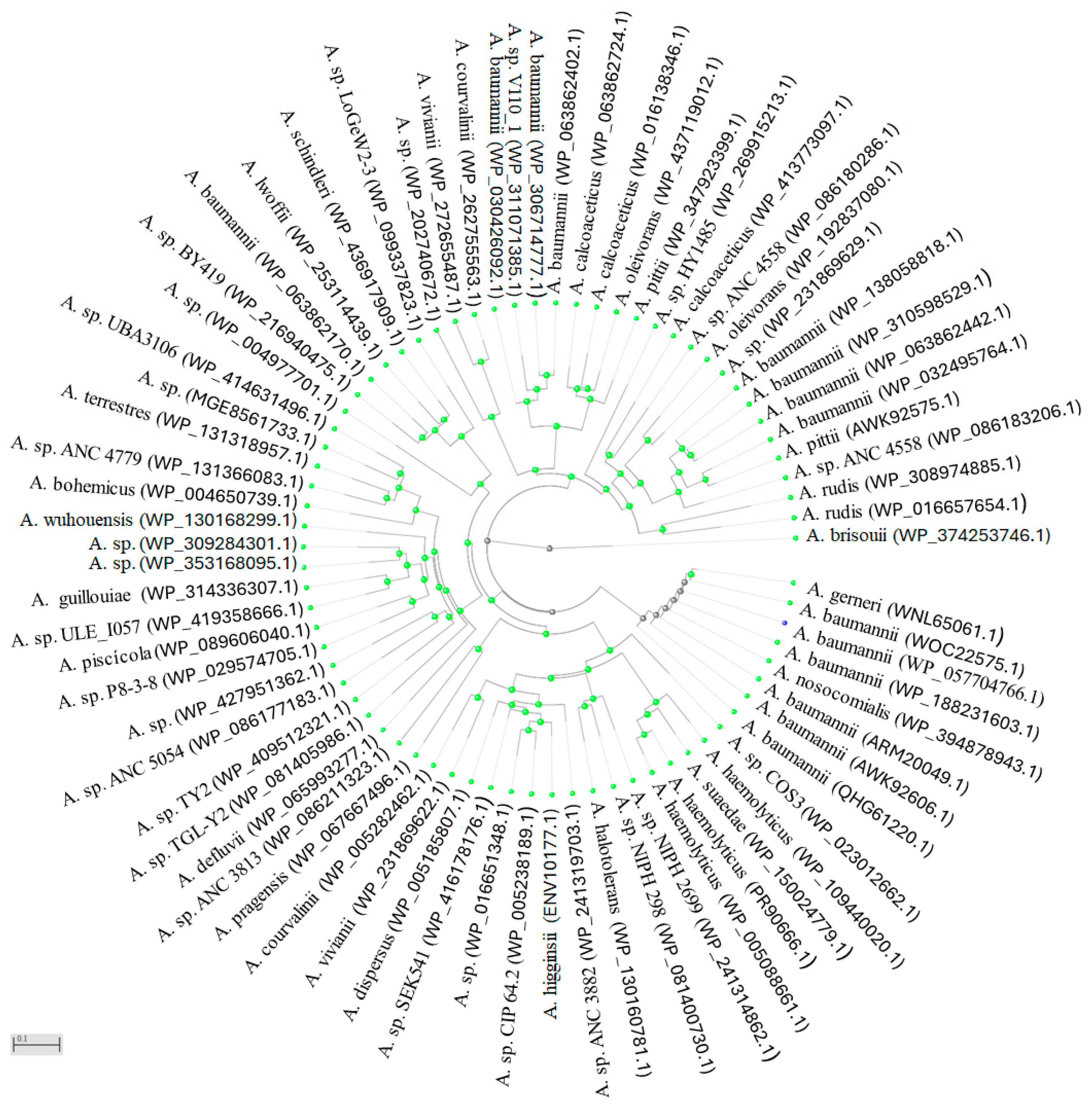
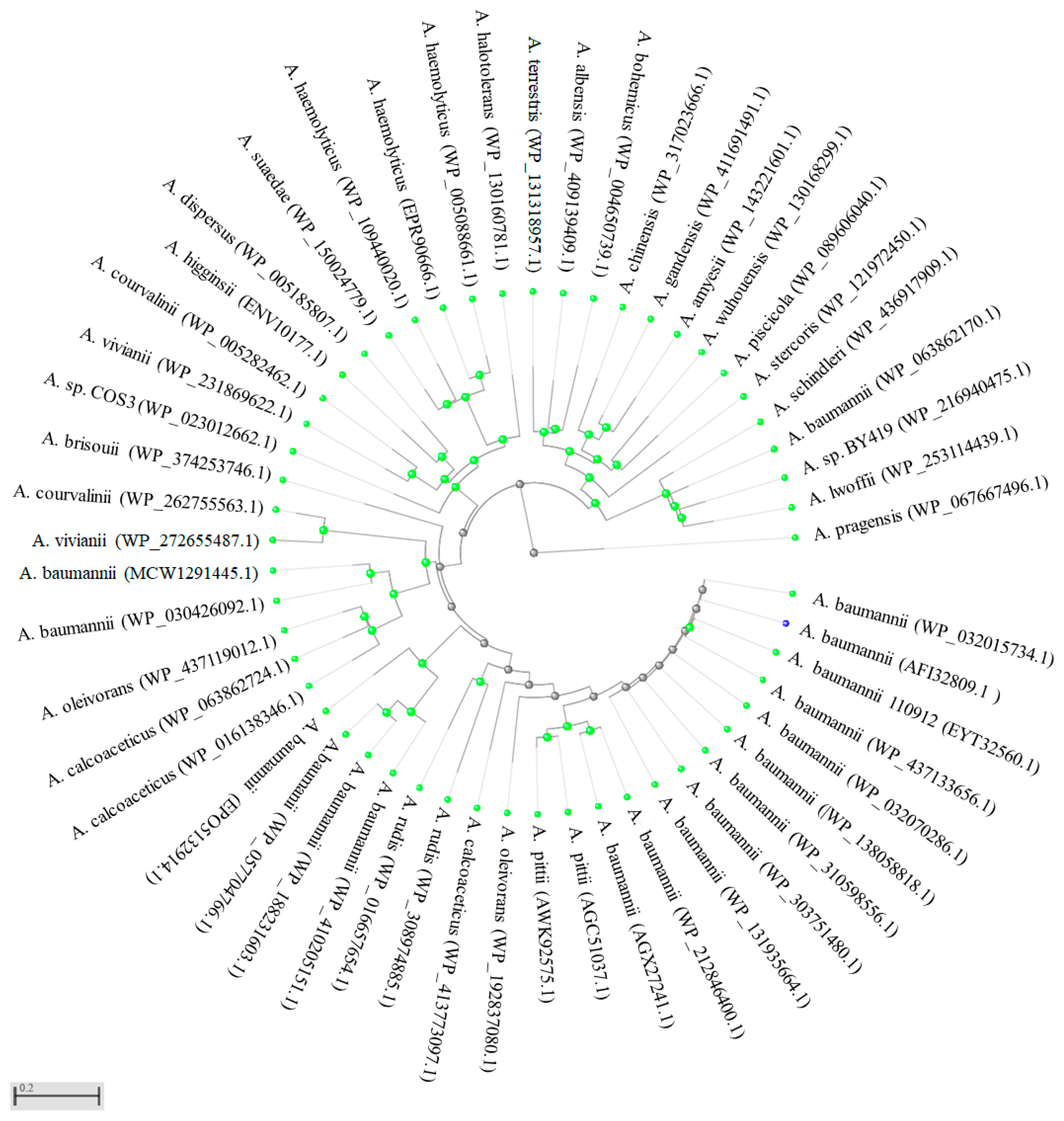
Evaluated how potential nonsynonymous substitutions could affect antibiotic resistance. A phylogenetic analysis was performed using protein sequences encoded by blaOXA-23 and blaOXA-24/40 genes associated with carbapenem resistance. The blaOXA-23 analysis included 24 Acinetobacter species. Several accessions of A. baumannii (WOC22575.1; WP_057704766.1; WP_188231603.1; ARM20049.1; AWK92606.1; QHG61220.1), A. nosocomialis (WP_394878943.1), and A. gerneri (WNL65061.1) were found to cluster outside the main clade, forming an outgroup (Figure 4). The gray branching points among these accessions suggest possible divergence events of the blaOXA-23 gene over evolutionary time.
Two principal groups were identified. The first group included A. baumannii, A. rudis, A. brisouii, A. pittii, A. oleovorans, A. calcoaceticus, A. vivianii, and A. courvalinii. The second group formed two subclusters. The first subcluster comprised A. baumannii, A. schindleri, A. lwoffii, A. terrestris, A. bohemicus, A. wuhouensis, A. guillouiae, A. piscicola, and A. defluvii. The second subcluster included A. pragensis, A. courvalinii, A. vivianii, A. dispersus, A. higginsii, A. halotolerans, A. haemolyticus, and A. suaedae. In general, most Acinetobacter species have been shown to occupy water-associated habitats. For example, A. pittii is ubiquitous and found in various soil types, freshwater, and marine ecosystems. A. calcoaceticus primarily inhabits soil and diverse aquatic environments, including freshwater, seawater, and estuaries. A. vivianii has been identified in wastewater and sludge, while A. courvalinii is commonly found in natural ecosystems, particularly in soil and water.
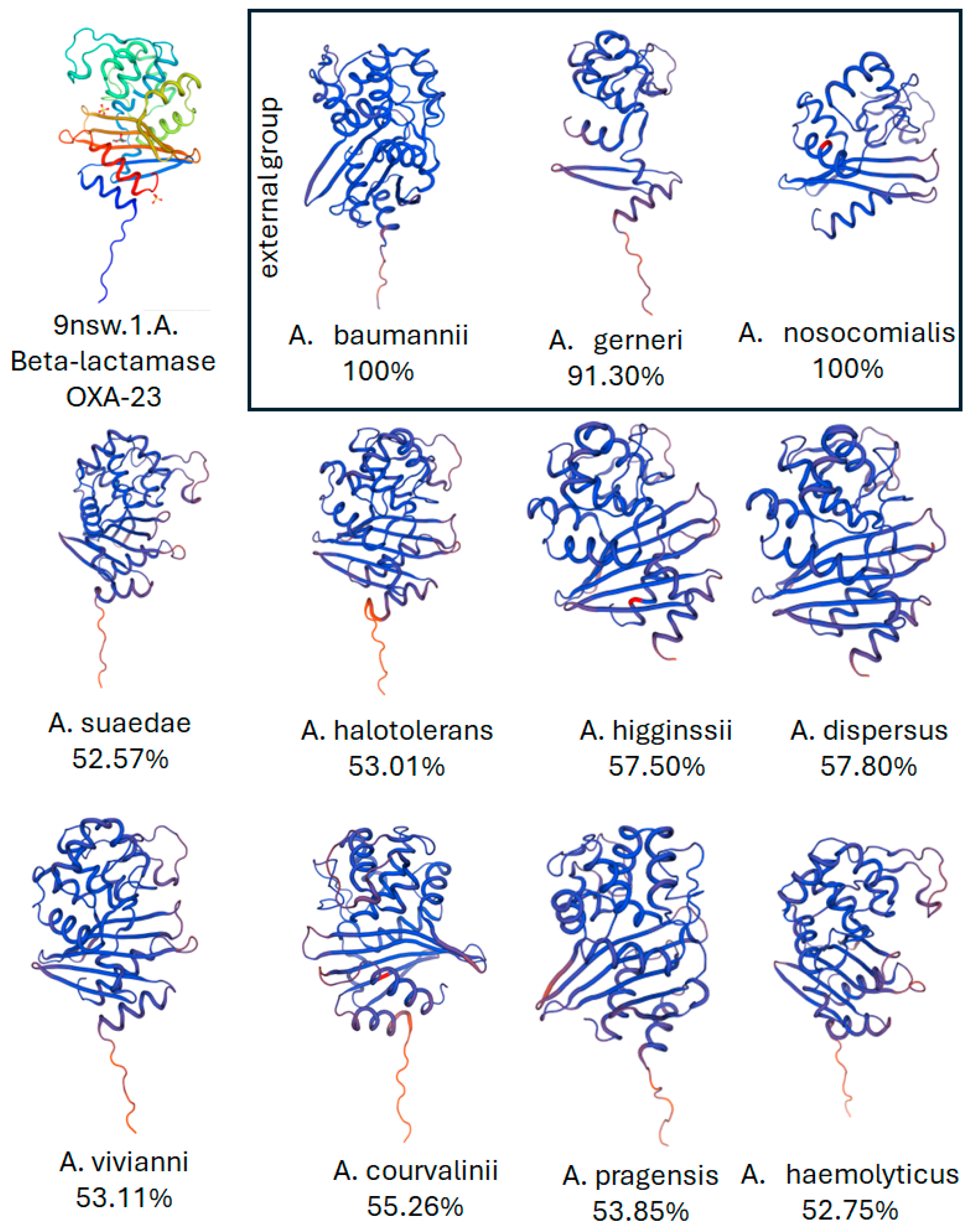
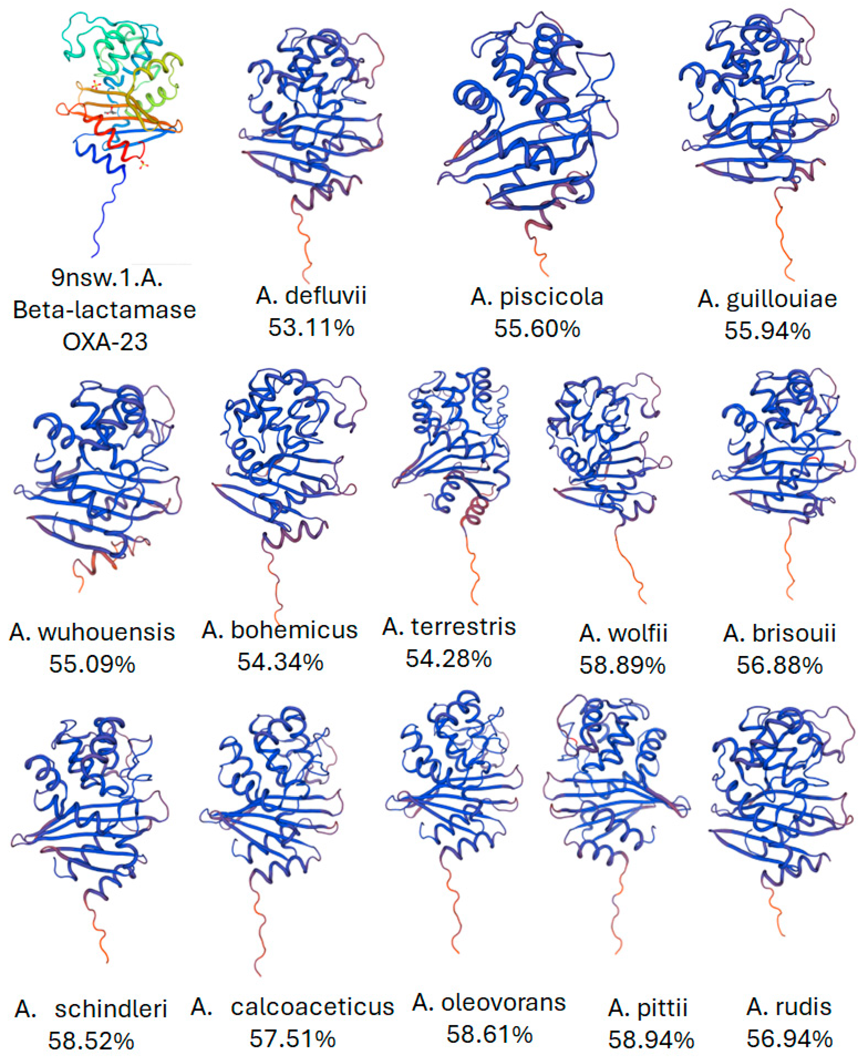
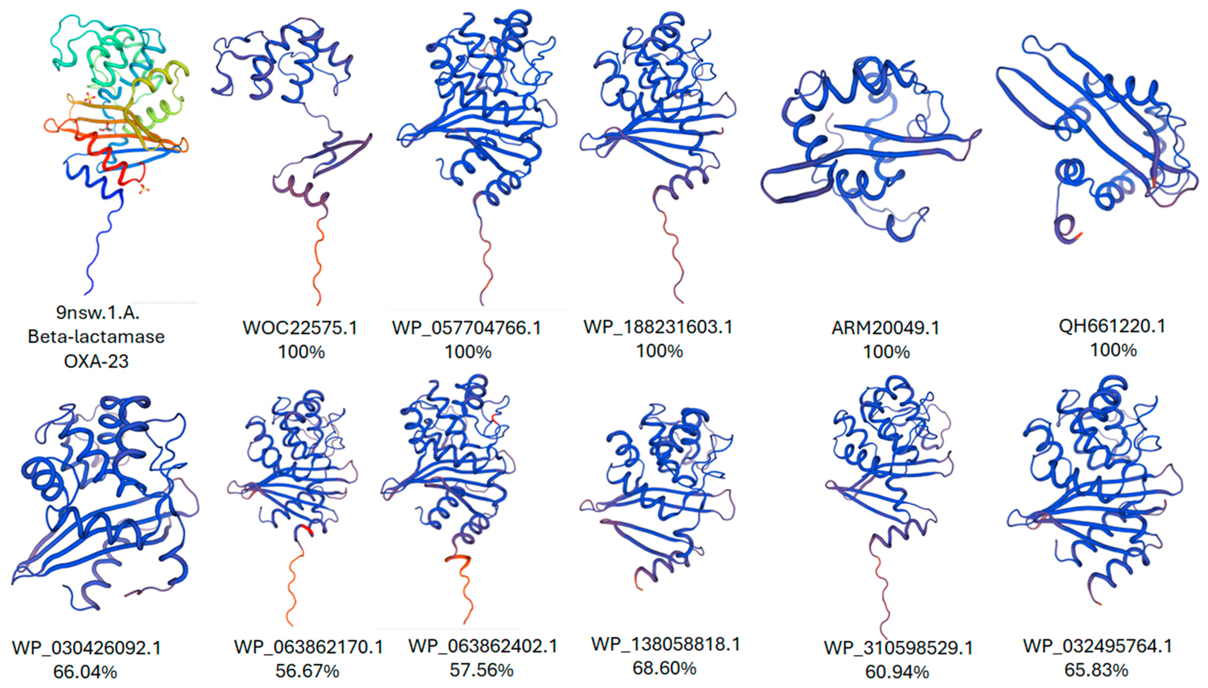
Several species have also been isolated from hospital environments, including A. baumannii, A. haemolyticus, A. wuhouensis, A. piscicola, A. higginsii, A. defluvii, A. dispersus, A. courvalinii, A. lwoffii, and A. pittii. A three-dimensional modeling analysis of the β-lactamase OXA-23 enzyme revealed protein sequence identities ranging from 52% to 100% relative to the reference model 9NSW.1.A [87]. The tertiary structures of the analyzed species demonstrated structural rearrangements within their 3D conformations. The first clade—including A. baumannii, A. rudis, A. brisouii, A. pittii, A. oleovorans, A. calcoaceticus, A. vivianii, and A. courvalinii—showed sequence identities of 53–70% relative to the reference model (Figure 5 and Figure 6). Species in the second group, such as A. baumannii, A. schindleri, A. lwoffii, A. terrestris, A. bohemicus, A. wuhouensis, A. guillouiae, A. piscicola, A. defluvii, A. pragensis, A. courvalinii, A. vivianii, A. dispersus, A. higginsii, A. halotolerans, A. haemolyticus, and A. suede, exhibited sequence identities of 52–59% relative to the reference model (Figure 5 and Figure 6).
Accessions of A. baumannii were present both as outgroups and within both clades, suggesting the existence of OXA-23 variants within this highly pathogenic species. The 3D modeling of these accessions indicated protein identities ranging from 56% to 100% relative to the reference model (Figure 7). Structural rearrangements observed among these proteins may influence the enzyme’s substrate specificity toward carbapenems. In these peptide sequences, it was not possible to identify the carbapenem-binding motif reported in the reference model, suggesting that these OXA-23 protein variants may exhibit specificity toward other carbapenem-type antibiotics.
The phylogenetic analysis of the OXA-24/40 protein included 24 Acinetobacter species and revealed the formation of two main clades (Figure 8). Species such as A. lwoffii, A. baumannii, A. schindleri, A. stercoris, A. piscicola, A. wuhouensis, A. amyesii, A. gandensis, A. bohemicus, A. chinensis, A. albensis, and A. terrestris clustered together within the first clade. In the second main clade, two subgroups were observed: the first comprised A. halotolerans, A. haemolyticus, A. suaedae, A. dispersus, A. higginsii, A. courvalinii, and A. vivianii, while the second included A. brisouii, A. courvalinii, A. baumannii, A. oleovorans, A. calcoaceticus, A. rudis, and A. pittii. Similar to the results obtained in the phylogenetic analysis of the OXA-23 protein, A. baumannii accessions were present in both major groups. The observed branching points may explain how the blaOXA-24/40 gene has diverged over evolutionary time. It appears that A. baumannii may represent the Acinetobacter species that has accumulated key mutations marking points of divergence within this lineage.
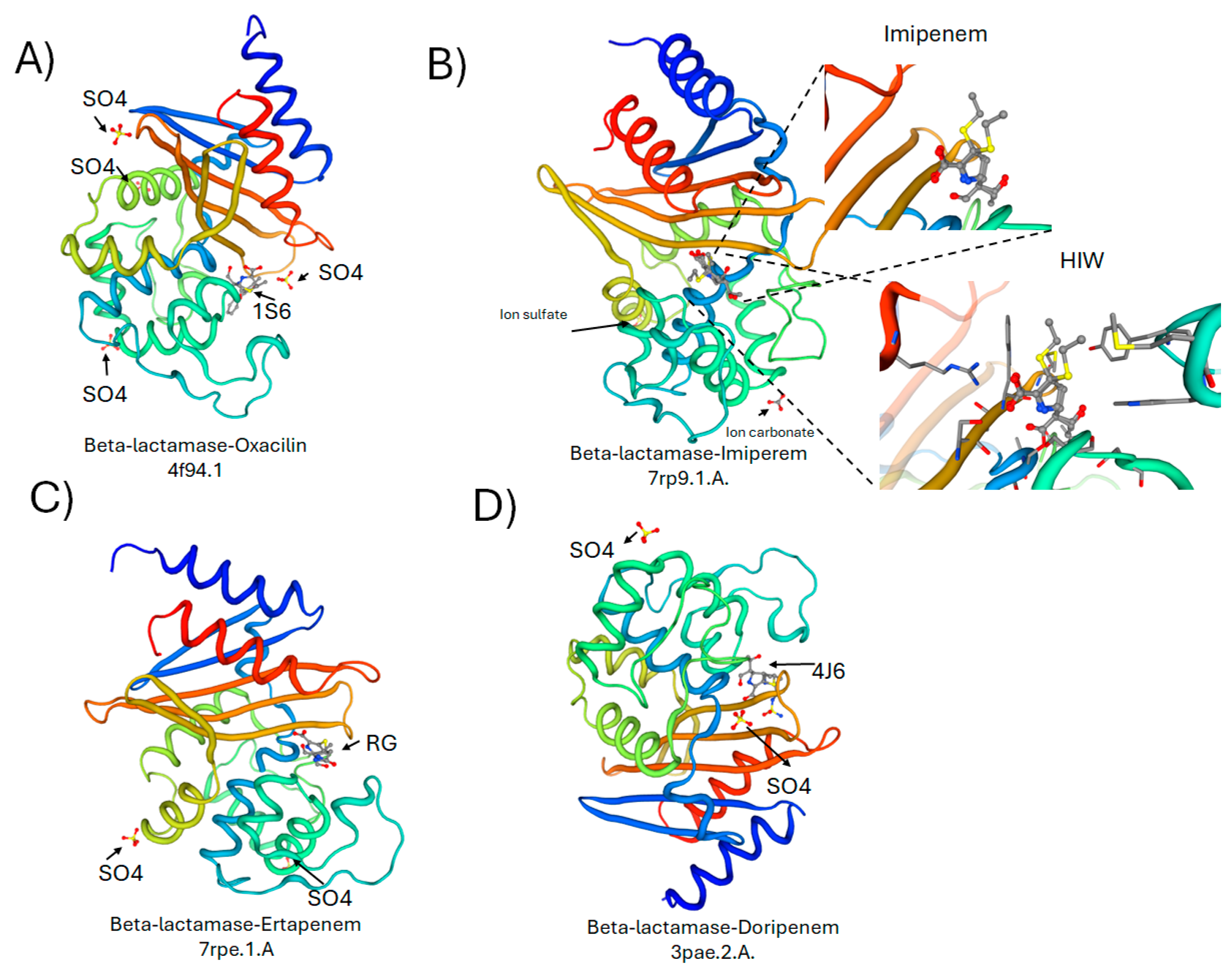
For the three-dimensional structure analysis of OXA-24/40, four established β-lactamase models with substrate specificity for B-lactamase-oxacillin (4f94.1.A), Beta-lactamase-imipenem (7rp9.1.A), B-lactamase-ertapenem (7rpe.1.A), and Beta-lactamase-doripenem (3pae.2.A) were included (Figure 9) [88,89]. The structural models included—oxacillin β-lactamase (4F94.1.A), β-lactamase-imipenem (7RP9.1.A), β-lactamase-ertapenem (7RPE.1.A), and β-lactamase-doripenem (3PAE.2.A)—represent different variants of class D β-lactamases that confer resistance to β-lactam antibiotics through hydrolysis of the β-lactam ring. These enzymes share a conserved serine-based catalytic mechanism but exhibit structural adaptations that influence substrate specificity and affinity for different carbapenems and oxacillin derivatives. Comparative analysis of these models highlights key variations in the active-site configuration, loop flexibility, and electrostatic surface potential that modulate antibiotic binding and turnover efficiency, providing insights into the molecular basis of β-lactam resistance observed in clinical isolates.
The three-dimensional structures of twenty-five Acinetobacter species were modeled and showed sequence identity between 57.61–99.18%, 0–99.18%, 0–99.59% and 0–99.18% with respect to the models of B-lactamase-oxacillin(4f94.1.A), B-lactamase-imipenem (7rp9.1.A), B-lactamase-ertapenem (7rpe.1.A), and β-lactamase doripenem (3pae.2.A), respectively (Table 1). Specific substrates for the enzyme were only identified in A. baumannii (AFI32809.1). The modeling of the three-dimensional structures of the twenty-five Acinetobacter species, and their comparison with known β-lactamase complexes, provides valuable insights into the molecular mechanisms underlying antibiotic resistance within this genus. The observed sequence identity ranges—particularly the high homology (up to ~99%) with β-lactamase models complexed with oxacillin, imipenem, ertapenem, and doripenem—suggest that many Acinetobacter species conserve key structural motifs essential for β-lactam hydrolysis. This conservation indicates a shared evolutionary pathway that favors resistance mechanisms against a broad spectrum of β-lactam antibiotics. However, the presence of lower identity values (as low as 0%) in some comparisons reflects significant divergence among certain species or strains, which may result in altered substrate specificity or reduced catalytic efficiency toward β-lactams.
Interestingly, only A. baumannii (AFI32809.1) displayed identifiable specific substrates, highlighting its distinctive enzymatic efficiency and adaptability. This finding aligns with previous reports describing A. baumannii as one of the most clinically relevant and multidrug-resistant Acinetobacter species. The unique substrate recognition profile of A. baumannii β-lactamases could stem from subtle conformational changes in the active site, affecting drug binding and turnover rates. These structural differences may provide an adaptive advantage, allowing the bacterium to hydrolyze a wider range of β-lactam antibiotics, including carbapenems, which are often considered last-resort treatments.
Overall, these results underscore the importance of structural modeling in elucidating the evolutionary dynamics of β-lactamase enzymes. The combination of high conservation and species-specific variations suggests that while Acinetobacter β-lactamases share a common catalytic framework, adaptive mutations fine-tune their activity, driving the emergence of diverse resistance phenotypes. Further biochemical and kinetic studies are warranted to validate these structural predictions and to explore potential inhibitors capable of targeting conserved catalytic residues across different β-lactamase variants.
The three-dimensional structures of twenty-five Acinetobacter species were modeled, revealing sequence identity ranging from 57.61–99.18%, 0–99.18%, 0–99.59%, and 0–99.18% with respect to the reference models β-lactamase–oxacillin (4f94.1.A), β-lactamase–imipenem (7rp9.1.A), β-lactamase–ertapenem (7rpe.1.A), and β-lactamase–doripenem (3pae.2.A), respectively (Table 1).
Similarly, the modeled three-dimensional structures of fourteen A. baumannii accessions exhibited sequence identity values ranging from 61.14–99.55%, 0–99.55%, 61–100%, and 0–99.18% relative to the same β-lactamase reference models (Table 2).
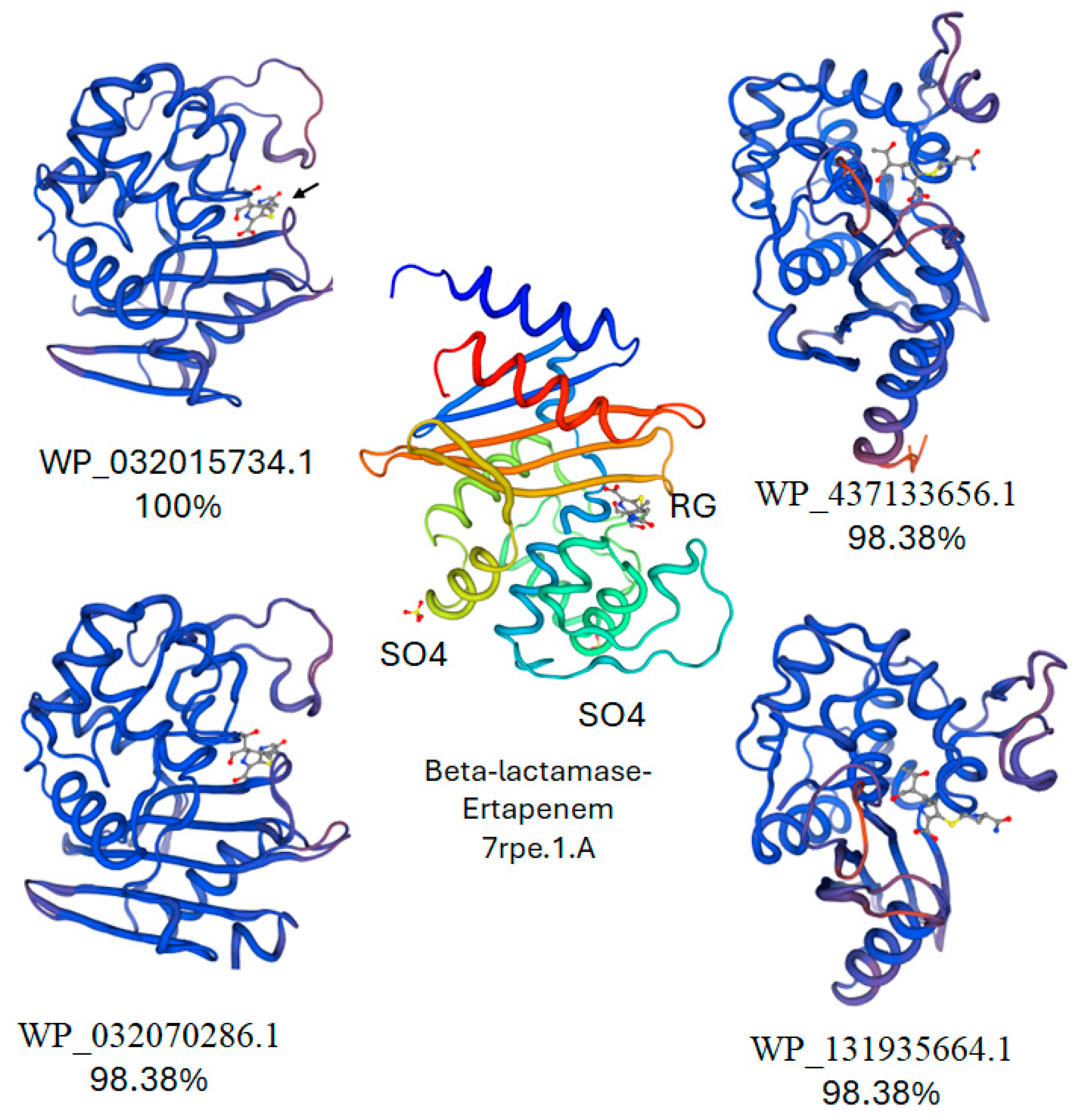
It was possible to identify ertapenem-binding ligands in four A. baumannii accessions, which may suggest that point mutations could influence the substrate (antibiotic) binding of the β-lactamase enzyme (Figure 10).
Similar to the results obtained in the phylogenetic analysis of the OXA-23 protein, A. baumannii accessions were present in both major groups. The observed branching points may explain how the blaOXA-24/40 gene has diverged over evolutionary time. It appears that A. baumannii may represent the Acinetobacter species that has accumulated key mutations marking points of divergence within this lineage.

9. Antibiotic Formulations with Natural Products to Combat Bacterial Resistance

The increasing prevalence of bacterial resistance to conventional antibiotics has become a major global health concern, prompting the search for alternative therapeutic strategies. Natural products, derived from plants, microorganisms, and marine organisms, represent a valuable source of bioactive compounds with antimicrobial potential. These substances often exhibit diverse chemical structures and unique mechanisms of action that differ from those of traditional antibiotics, reducing the likelihood of cross-resistance. Furthermore, natural compounds can act synergistically with existing antibiotics, enhancing their efficacy and overcoming bacterial defense mechanisms such as efflux pumps and biofilm formation. The study and development of natural products as complementary or alternative antimicrobial agents provide a promising avenue for addressing the growing challenge of multidrug-resistant bacterial infections [90,91].
Recent research has increasingly focused on natural products as potential alternatives to conventional antibiotics in the fight against multidrug-resistant (MDR) bacteria. According to Franco and Vázquez [92], natural compounds exhibit structural diversity and unique mechanisms of action that reduce cross-resistance, offering new opportunities for antimicrobial drug development. A comprehensive review by Elmaidomy et al. [93] reported that flavonoids, alkaloids, terpenoids, tannins, and phenolic compounds isolated from plants, fungi, and marine organisms have shown significant antibacterial activity against pathogens such as Staphylococcus aureus, Escherichia coli, Pseudomonas aeruginosa, and Klebsiella pneumoniae. Specifically, flavonoids and phenolic extracts from Moringa oleifera and Azadirachta indica demonstrated inhibitory effects on S. aureus and E. coli, while tannins derived from propolis exhibited strong antibacterial activity against P. aeruginosa and K. pneumoniae [94,95], and this observation is supported by recent systematic reviews summarizing plant-derived antimicrobials [90].
Likewise, terpenoids from Dodonaea viscosa and alkaloids from Sophora alopecuroides have been shown to inhibit resistant strains of E. coli, S. aureus (including MRSA), and P. aeruginosa through mechanisms such as efflux pump inhibition. Ethnobotanical approaches have further supported the discovery of plant-derived metabolites as promising leads for antibiotic development [96,97]. Additionally, natural compounds have demonstrated antibiofilm properties, contributing to the disruption of bacterial defense mechanisms. Collectively, these studies underscore the therapeutic potential of natural bioactive compounds as sustainable and effective agents to counteract bacterial resistance.

10. Conclusions

Future research aimed at combating antibiotic resistance in bacteria must adopt a multidisciplinary and integrative approach that combines molecular biology, structural bioinformatics, and systems-level analyses. It is essential to deepen the understanding of enzymatic mechanisms responsible for antibiotic degradation, including β-lactamases and other resistance determinants, through advanced computational modeling and experimental validation. Furthermore, efforts should focus on the discovery of new antimicrobial agents, particularly natural products and synthetic derivatives, capable of overcoming current resistance mechanisms. The integration of genomic surveillance, protein engineering, and drug design technologies will be key to identifying novel therapeutic targets and preventing the dissemination of resistant strains. Equally important is the promotion of responsible antibiotic use and the development of public health policies that limit the selective pressure favoring resistant pathogens. Altogether, these strategies represent the foundation for a sustainable and effective response to the growing global challenge of antimicrobial resistance.

Supplementary Materials

The following supporting information can be downloaded at: https://www.mdpi.com/article/10.3390/bacteria5010014/s1, Supplementary Material: Bioinformatic analysis methodology.

Author Contributions

Conceptualization, B.A.A.-G. and E.A.-C.; methodology, M.C.-S.M. and Y.V.-M.; software, J.M.B.-L. and L.F.S.-T.; formal analysis, L.C.R.-Z. and F.A.T.-O.; investigation, E.C.-S., V.H.R.-G. and H.M.-M.; writing—original draft preparation, E.S.-L., H.d.J.V.-A. and V.B.-G.; writing—review and editing, Y.d.J.T.-O., M.C.T.-O., Y.V.-M. and M.C.-S.M.; visualization, G.D.-P. and M.J.G.-C.; supervision, M.C.-S.M. All authors have read and agreed to the published version of the manuscript.

Funding

This research received no external funding.

Institutional Review Board Statement

Not applicable.

Informed Consent Statement

Not applicable.

Data Availability Statement

The original contributions presented in this study are included in the article. Further inquiries can be directed to the corresponding authors.

Conflicts of Interest

The authors declare no conflicts of interest.

References

  1. Naghavi, M.; Vollset, S.E.; Ikuta, K.S.; Bisignano, C.; Safiri, S.; Karamouzian, M.; Aliyari, R.; Aali, A.; Abbasian, M.; Abbasi-Kangevari, M.; et al. Global burden of bacterial antimicrobial resistance 1990–2021: A systematic analysis with forecasts to 2050. Lancet 2024, 404, 1199–1226. [Google Scholar] [CrossRef] [Scilit]
  2. Shah, N.P.; Reddy, P.; Paladino, J.A.; McKinnon, P.S.; Klepser, M.E.; Pashos, C.L. Direct medical costs associated with using vancomycin in methicillin-resistant Staphylococcus aureus infections: An economic model. Curr. Med. Res. Opin. 2004, 20, 779–790. [Google Scholar] [CrossRef] [Scilit]
  3. Stephens, J.M.; Gao, X.; Patel, D.A.; Verheggen, B.G.; Shelbaya, A.; Haider, S. Economic burden of inpatient and outpatient antibiotic treatment for methicillin-resistant Staphylococcus aureus complicated skin and soft-tissue infections: A comparison of linezolid, vancomycin, and daptomycin. Clin. Outcomes Res. 2013, 5, 447–457. [Google Scholar] [CrossRef] [Scilit]
  4. Asensio, Á.; Bouza, E.; Grau, S.; Rubio-Rodríguez, D.; Rubio-Terrés, C. Coste de la diarrea asociada a Clostridium difficile en España. Rev. Esp. Salud Publica 2013, 87, 25–33. [Google Scholar] [CrossRef] [Scilit] [PubMed]
  5. Sadeer, N.B.; Mahomoodally, M.F. Antibiotic potentiation of natural products: A promising target to fight pathogenic bacteria. Curr. Drug Targets 2021, 22, 555–572. [Google Scholar] [CrossRef] [Scilit] [PubMed]
  6. Vitiello, G.; Silvestri, B.; Luciani, G. Learning from nature: Bioinspired strategies towards antimicrobial nanostructured systems. Curr. Top. Med. Chem. 2018, 18, 22–41. [Google Scholar] [CrossRef] [Scilit] [PubMed]
  7. Tiwana, G.; Cock, I.E.; Taylor, S.M.; Cheesman, M.J. Beyond antibiotics: Repurposing non-antibiotic drugs as novel anti-bacterial agents to combat resistance. Int. J. Mol. Sci. 2025, 26, 9880. [Google Scholar] [CrossRef] [Scilit]
  8. Gajic, I.; Tomic, N.; Lukovic, B.; Jovicevic, M.; Kekic, D.; Petrovic, M.; Djukic, S.; Zdravkovic, N.; Milovanovic, J.; Kostic, M.A. A comprehensive overview of antibacterial agents for combating multidrug-resistant bacteria. Antibiotics 2025, 14, 221. [Google Scholar] [CrossRef] [Scilit]
  9. Rahman, M.U.; Shah, J.A.; Khan, M.N.; Bilal, H.; Zhu, D.; Du, Z.; Mu, D.S. Innovative approaches to combat antimicrobial resistance: A review of emerging therapies and technologies. Probiotics Antimicrob. Proteins, 2025; Online ahead of print. [CrossRef] [Scilit]
  10. Jesudason, T. WHO publishes updated list of bacterial priority pathogens. Lancet Microbe 2024, 5, 100940. [Google Scholar] [CrossRef] [Scilit] [PubMed]
  11. Giurazza, R.; Mazza, M.C.; Andini, R.; Sansone, P.; Pace, M.C.; Durante-Mangoni, E. Emerging treatment options for multi-drug-resistant bacterial infections. Life 2021, 11, 519. [Google Scholar] [CrossRef] [Scilit]
  12. Tacconelli, E.; Carrara, E.; Savoldi, A.; Harbarth, S.; Mendelson, M.; Monnet, D.L.; Pulcini, C.; Kahlmeter, G.; Kluytmans, J.; Carmeli, Y. Discovery, research, and de-velopment of new antibiotics: The WHO priority list of antibiotic-resistant bacteria and tuberculosis. Lancet Infect. Dis. 2018, 18, 318–327. [Google Scholar] [CrossRef] [Scilit]
  13. Woh, P.Y.; Zhang, X. The burden of ESKAPE pathogen-related hospital-acquired infections: Clinical and financial perspective from a systematic review. J. Hosp. Infect. 2025, 162, 377–385. [Google Scholar] [CrossRef] [Scilit]
  14. Cureño-Díaz, M.A.; Plascencia-Nieto, E.S.; Loyola-Cruz, M.Á.; Cruz-Cruz, C.; Nolasco-Rojas, A.E.; Durán-Manuel, E.M.; García-Mena, J.; Hernández-Castro, R.; Xicohtencatl-Cortés, J.; Bocanegra-Ibarias, P.; et al. Gram-negative ESKAPE bacteria surveillance in COVID-19 pandemic exposes high-risk sequence types of Aci-netobacter baumannii MDR. Pathogens 2024, 13, 50. [Google Scholar] [CrossRef] [Scilit]
  15. Cantón, R.; Horcajada, J.P.; Oliver, A.; Garbajosa, P.R.; Vila, J. Inappropriate use of antibiotics in hospitals: The complex relationship between antibiotic use and antimicrobial resistance. Enferm. Infecc. Microbiol. Clin. 2013, 31, 3–11. [Google Scholar] [CrossRef] [Scilit]
  16. Mulchandani, R.; Tiseo, K.; Nandi, A.; Klein, E.; Gandra, S.; Laxminarayan, R.; Van Boeckel, T. Global trends in inap-propriate use of antibiotics, 2000–2021. BMJ Public Health 2025, 3, e002411. [Google Scholar] [CrossRef] [Scilit]
  17. Van Boeckel, T.P.; Brower, C.; Gilbert, M.; Grenfell, B.T.; Levin, S.A.; Robinson, T.P.; Teillant, A.; Laxminarayan, R.; Dunn, R.R.; Eisenberg, J.N.S.; et al. Global trends in antimicrobial use in food animals. Proc. Natl. Acad. Sci. USA 2015, 112, 5649–5654. [Google Scholar] [CrossRef] [Scilit]
  18. Dropulic, L.K.; Lederman, H.M. Overview of infections in the immunocompromised host. Diagn. Microbiol. Immunocompromised Host 2016, 4, 1–50. [Google Scholar] [CrossRef] [Scilit]
  19. Napit, R.; Gurung, A.; Poudel, A.; Chaudhary, A.; Manandhar, P.; Sharma, A.N.; Kafle, S.; Banjara, M.R.; Adhikari, B.; Subedi, D.; et al. Metagenomic analysis of human, animal, and environmental samples identifies potential emerging pathogens, profiles antibiotic resistance genes, and reveals horizontal gene transfer dynamics. Sci. Rep. 2025, 15, 12156. [Google Scholar] [CrossRef] [Scilit]
  20. Yang, J.; Wang, L.; Liang, Q.; Wang, Y.; Yang, X.; Wu, X.; Pei, X. Microbiome, resistome, and potential transfer of antibiotic resistance genes in Chinese wet market under One Health sectors. BMC Microbiol. 2025, 25, 406. [Google Scholar] [CrossRef] [Scilit]
  21. Cao, H.; Bougouffa, S.; Park, T.J.; Lau, A.; Tong, M.K.; Chow, K.H.; Ho, P.L. Sharing of antimicrobial resistance genes between humans and food animals. mSystems 2022, 7, e0077522. [Google Scholar] [CrossRef] [Scilit]
  22. Sachdev, C.; Anjankar, A.; Agrawal, J. Self-medication with antibiotics: An element increasing resistance. Cureus 2022, 14, e30844. [Google Scholar] [CrossRef] [Scilit]
  23. Weets, C.M.; Katz, R. Global approaches to tackling antimicrobial resistance: A comprehensive analysis of water, sanitation and hygiene policies. BMJ Glob. Health 2024, 9, e013855. [Google Scholar] [CrossRef] [Scilit]
  24. Lerminiaux, N.A.; Cameron, A.D. Horizontal transfer of antibiotic resistance genes in clinical environments. Can. J. Microbiol. 2019, 65, 34–44. [Google Scholar] [CrossRef] [Scilit]
  25. Ugarte Silva, R.G.; Olivo López, J.M.; Corso, A.; Pasteran, F.; Albornoz, E.; Sahuanay Blácido, Z.P. Resistencia a colistín mediado por el gen mcr-1 identificado en cepas de Escherichia coli y Klebsiella pneumoniae. An. Fac. Med. 2018, 79, 213–217. [Google Scholar] [CrossRef] [Scilit]
  26. González, A.; Nieves, B. Resistencia a aminoglucósidos y quinolonas en cepas de Klebsiella pneumoniae. Med. UIS 2016, 29, 21–30. [Google Scholar] [CrossRef] [Scilit]
  27. Wyres, K.L.; Holt, K.E. Klebsiella pneumoniae as a key trafficker of drug resistance genes. Curr. Opin. Microbiol. 2018, 45, 131–139. [Google Scholar] [CrossRef] [Scilit]
  28. Rhodes, G.; Huys, G.; Swings, J.; McGann, P.; Hiney, M.; Smith, P.; Pickup, R.W. Distribution of oxytetracycline resistance plasmids between aeromonads. Appl. Environ. Microbiol. 2000, 66, 3883–3890. [Google Scholar] [CrossRef] [Scilit]
  29. Bai, J.; Liu, Y.; Kang, J.; Song, Y.; Yin, D.; Wang, S.; Li, X.; Zhang, H.; Chen, Y.; Zhao, L.; et al. Antibiotic resistance and virulence of carbapenem-resistant K. pneumoniae. Microb. Pathog. 2023, 175, 105969. [Google Scholar] [CrossRef] [Scilit]
  30. Turton, J.; Davies, F.; Perry, C.; Payne, Z.; Pike, R. Hybrid resistance and virulence plasmids in “high-risk” Klebsiella pneumoniae clones. Microorganisms 2019, 7, 326. [Google Scholar] [CrossRef] [Scilit]
  31. Ramatla, T.; Nkhebenyane, J.; Lekota, K.E.; Thekisoe, O.; Monyama, M.; Achilonu, C.C.; Khasapane, G. Global prevalence and antibiotic resistance profiles of carbapenem-resistant Pseudomonas aeruginosa. Front. Microbiol. 2025, 16, 1599070. [Google Scholar] [CrossRef] [Scilit]
  32. Cerini, P.; Meduri, F.R.; Tomassetti, F.; Polidori, I.; Brugneti, M.; Nicolai, E.; Bressan, R.; Codeluppi, M.; Di Bella, S.; Tumbarello, M.; et al. Trends in antibiotic resistance of noso-comial and community-acquired infections in Italy. Antibiotics 2023, 12, 651. [Google Scholar] [CrossRef] [Scilit]
  33. Patra, M.; Dubey, S.K. Antimicrobial resistance transmission in environmental matrices. Rev. Environ. Contam. Toxicol. 2025, 263, 20. [Google Scholar] [CrossRef] [Scilit]
  34. Roberts, M.C. One Health: The role of wastewater treatment plants as reservoirs of antibiotic resistance. In Antimicrobial Resistance in Wastewater Treatment Processes; Manaia, C.M., Rocha, J., Scaccia, N., Eds.; Springer: Cham, Switzerland, 2017; pp. 35–53. [Google Scholar] [CrossRef] [Scilit]
  35. Meradji, S.; Basher, N.S.; Sassi, A.; Ibrahim, N.A.; Idres, T.; Touati, A. The role of water as a reservoir for antibiotic-resistant bacteria. Antibiotics 2025, 14, 763. [Google Scholar] [CrossRef] [Scilit]
  36. Szczepanowski, R.; Braun, S.; Riedel, V.; Schneiker, S.; Krahn, I.; Pühler, A.; Schluter, A. The 120 kb IncF plasmid pRSB107 isolated from sewage encodes multiple resistance determinants. Microbiology 2005, 151, 1095–1111. [Google Scholar] [CrossRef] [Scilit]
  37. Cruz-Cruz, C.; Gaytán-Cervantes, J.; González-Torres, C.; Nolasco-Rojas, A.E.; Loyola-Cruz, M.Á.; Delgado-Balbuena, L.; Durán-Manuel, E.M.; Plascencia-Nieto, E.S.; Cureño-Díaz, M.A.; Bocanegra-Ibarias, P.; et al. Profiling of bacterial communities of hospital wastewater reveals clinically relevant genera. Microorganisms 2025, 13, 1316. [Google Scholar] [CrossRef] [Scilit]
  38. Nolasco-Rojas, A.E.; Cruz-Del-Agua, E.; Cruz-Cruz, C.; Loyola-Cruz, M.Á.; Ayil-Gutiérrez, B.A.; Tamayo-Ordóñez, M.C.; Durán-Manuel, E.M.; Delgado-Balbuena, L.; Plascencia-Nieto, E.S.; Cureño-Díaz, M.A.; et al. Microbiological risks to health associated with hospital wastewater. Pathogens 2025, 14, 402. [Google Scholar] [CrossRef] [Scilit]
  39. Schaenzer, A.J.; Wright, G.D. Antibiotic resistance by enzymatic modification of antibiotic targets. Trends Mol. Med. 2020, 26, 768–782. [Google Scholar] [CrossRef] [Scilit]
  40. Elshobary, M.E.; Badawy, N.K.; Ashraf, Y.; El-Sayed, H.S.; Ibrahim, D.; El-Baz, A.M.; El-Sayed, A.; Hassan, S.E.-D.; Abdelkader, M.S.; El-Gendy, A.O.; et al. Combating antibiotic resistance: Mechanisms, multidrug-resistant path-ogens, and novel therapeutic approaches: An updated review. Pharmaceuticals 2025, 18, 402. [Google Scholar] [CrossRef] [Scilit]
  41. Almatroudi, A. Biofilm resilience: Molecular mechanisms driving antibiotic resistance in clinical contexts. Biology 2025, 14, 165. [Google Scholar] [CrossRef] [Scilit]
  42. Shchit, I.Y.; Kuznetsov, A.V. Concepts of Horizontal Gene Transfer at the Turn of the 20th and 21st Centuries. Russ. J. Genet. 2025, 61, 642–650. [Google Scholar] [CrossRef] [Scilit]
  43. Wachino, J.I. Horizontal Gene Transfer Systems for Spread of Antibiotic Resistance in Gram-Negative Bacteria. Microbiol. Immunol. 2025, 69, 367–376. [Google Scholar] [CrossRef] [Scilit]
  44. Elhawy, M.I.; Molle, V.; Becker, S.L.; Bischoff, M. The Low-Molecular Weight Protein Arginine Phosphatase PtpB Affects Nuclease Production, Cell Wall Integrity, and Uptake Rates of Staphylococcus aureus by Polymorphonuclear Leukocytes. Int. J. Mol. Sci. 2021, 22, 5342. [Google Scholar] [CrossRef] [Scilit]
  45. Elhawy, M.I.; Huc-Brandt, S.; Pätzold, L.; Gannoun-Zaki, L.; Abdrabou, A.M.M.; Bischoff, M.; Molle, V. The phosphoarginine phosphatase PtpB from Staphylococcus aureus is involved in bacterial stress adaptation during infection. Cells 2021, 10, 645. [Google Scholar] [CrossRef] [Scilit]
  46. Ding, F.; Han, L.; Xue, Y.; Yang, I.T.; Fan, X.; Tang, R.; Zhang, Y.; Li, H.; Wang, J.; Liu, Q.; et al. Multidrug-resistant Pseudomonas aeruginosa is predisposed to lasR mutation through up-regulated activity of efflux pumps in non-cystic fibrosis bronchiectasis patients. Front. Cell. Infect. Microbiol. 2022, 12, 934439. [Google Scholar] [CrossRef] [Scilit]
  47. Pan, X.; Yin, L.; Zhou, D.; Jin, Y.; Cheng, Z.; Ha, U.H.; Wu, W.; Liang, H.; Wu, M.; Shen, L.; et al. Pseudomonas aeruginosa lasR-deficient mutant contributes to bacterial virulence through enhancing the PhoB-mediated pathway in response to host environment. mBio 2025, 16, e0178825. [Google Scholar] [CrossRef] [Scilit]
  48. Zhang, X.; Zhang, D.; Zhou, D.; Zheng, S.; Li, S.; Hou, Q.; Liu, Y.; Wang, J.; Chen, H.; Zhao, Y.; et al. A comprehensive review of the pathogenic mechanisms of Pseudomonas aeruginosa: Synergistic effects of virulence factors, quorum sensing, and biofilm formation. Front. Microbiol. 2025, 16, 1619626. [Google Scholar] [CrossRef] [Scilit] [PubMed]
  49. Luchian, N.; Olaru, I.; Pleșea-Condratovici, A.; Duceac, M.; Mătăsaru, M.; Dabija, M.G.; Elkan, E.M.; Dabija, V.A.; Eva, L.; Duceac, L.D. Clinical and Epidemiological Aspects on Healthcare-Associated Infections with Acinetobacter spp. in a Neurosurgery Hospital in North-East Romania. Medicina 2025, 61, 990. [Google Scholar] [CrossRef] [Scilit] [PubMed]
  50. Nguyen, M.; Joshi, S.G. Carbapenem resistance in Acinetobacter baumannii, and their importance in hospital-acquired infections: A scientific review. J. Appl. Microbiol. 2021, 131, 2715–2738. [Google Scholar] [CrossRef] [Scilit]
  51. Gupta, N.; Angadi, K.; Jadhav, S. Molecular characterization of carbapenem-resistant Acinetobacter baumannii with special reference to carbapenemases: A systematic review. Infect. Drug Resist. 2022, 15, 7631–7650. [Google Scholar] [CrossRef] [Scilit]
  52. AL-Dahlaki, S.M.M. Molecular Detection and Gene Expression for hcp and blaOXA-51 Genes in Acinetobacter baumannii Isolated from Different Clinical Sources. Master’s Thesis, Diyala University, Baqubah, Iraq, 2020. [Google Scholar]
  53. Zhou, Z.; Du, X.; Wang, L.; Yang, Q.; Fu, Y.; Yu, Y. Clinical carbapenem-resistant Acinetobacter baylyi strain coharboring blaSIM-1 and blaOXA-23 from China. Antimicrob. Agents Chemother. 2011, 55, 5347–5349. [Google Scholar] [CrossRef] [Scilit] [PubMed]
  54. Khorsi, K.; Messai, Y.; Hamidi, M.; Ammari, H.; Bakour, R. High prevalence of multidrug-resistance in Acinetobacter baumannii and dissemination of carbapenemase-encoding genes blaOXA-23-like, blaOXA-24-like and blaNDM-1 in Algiers hospitals. Asian Pac. J. Trop. Med. 2015, 8, 438–446. [Google Scholar] [PubMed]
  55. Klotz, P.; Jacobmeyer, L.; Leidner, U.; Stamm, I.; Semmler, T.; Ewers, C. Acinetobacter pittii from companion animals coharboring blaOXA-58, the tet(39) region, and other resistance genes on a single plasmid. Antimicrob. Agents Chemother. 2018, 62, 10–1128. [Google Scholar] [CrossRef] [Scilit]
  56. Salgado-Camargo, A.D.; Castro-Jaimes, S.; Gutierrez-Rios, R.M.; Lozano, L.F.; Bustos, P.; Fernández-Vázquez, J.L.; Santamaría, R.I.; Rocha-Estrada, J.; Vergara-Castañeda, E.; Martínez-Romero, E.; et al. Structure and evolution of Acinetobacter baumannii plasmids. Front. Microbiol. 2020, 11, 1283. [Google Scholar] [CrossRef] [Scilit]
  57. Azizi, O.; Fereshteh, S.; Naşiri, O.; Ghorbani, M.; Barzi, S.M.; Badmasti, F. The occurrence and characterization of class I, II, and III integrons among carbapenemase-producing clinical strains of Acinetobacter baumannii in Tehran, Iran. Jundishapur J. Microbiol. 2021, 14, e117766. [Google Scholar] [CrossRef] [Scilit]
  58. Leungtongkam, U.; Thummeepak, R.; Tasanapak, K.; Sitthisak, S. Acquisition and transfer of antibiotic resistance genes associated with conjugative plasmid or class 1 integrons of Acinetobacter baumannii. PLoS ONE 2018, 13, e0208468. [Google Scholar] [CrossRef] [Scilit]
  59. Grosso, F.; Silva, L.; Sousa, C.; Ramos, H.; Quinteira, S.; Peixe, L. Extending the reservoir of blaIMP-5: The emerging pathogen Acinetobacter bereziniae. Future Microbiol. 2015, 10, 1609–1613. [Google Scholar] [CrossRef] [Scilit]
  60. Takahashi, A.; Yomoda, S.; Kobayashi, I.; Okubo, T.; Tsunoda, M.; Iyobe, S. Detection of carbapenemase-producing Aci-netobacter baumannii in a hospital. J. Clin. Microbiol. 2000, 38, 526–529. [Google Scholar]
  61. Da Silva, G.J.; Correia, M.; Vital, C.; Duarte, A.; Oliveira, S.; Cardoso, O.; Leitão, R.; Peixe, L.; Antunes, P.; Machado, E.; et al. Molecular characterization of blaIMP-5, a new integron-borne metallo-β-lactamase gene from an Acinetobacter baumannii nosocomial isolate in Portugal. FEMS Microbiol. Lett. 2002, 215, 33–39. [Google Scholar] [CrossRef] [Scilit] [PubMed]
  62. Zong, Z.; Zhang, X. blaNDM-1-carrying Acinetobacter johnsonii detected in hospital sewage. J. Antimicrob. Chemother. 2013, 68, 1007–1010. [Google Scholar]
  63. Fu, Y.; Liu, L.; Li, X.; Chen, Y.; Jiang, Y.; Zhou, H.; Yu, Y. Spread of a common blaNDM-1-carrying plasmid among diverse Acinetobacter species. Infect. Genet. Evol. 2015, 32, 30–33. [Google Scholar] [CrossRef] [Scilit]
  64. Zhang, S.; Yang, J.; Abbas, M.; Li, Y.; Wang, X.; Chen, H.; Liu, Q.; Zhao, L.; Zhou, Y.; Sun, J.; et al. Threats across boundaries: The spread of ESBL-positive Enterobacteriaceae and its challenge to the “One Health” concept. Front. Microbiol. 2025, 16, 1496716. [Google Scholar] [CrossRef] [Scilit]
  65. Freschi, L.; Vincent, A.T.; Jeukens, J.; Emond-Rheault, J.G.; Kukavica-Ibrulj, I.; Dupont, M.J.; Charette, S.J.; Boyle, B.; Levesque, R.C.; Fournier, É.; et al. The Pseudomonas aeruginosa pan-genome provides new insights on population structure, horizontal gene transfer, and pathogenicity. Genome Biol. Evol. 2019, 11, 109–120. [Google Scholar] [CrossRef] [Scilit]
  66. Romaniuk, K.; Styczynski, M.; Decewicz, P.; Golec, P.; Dziewit, L.; Bartosik, D.; Jagura-Burdzy, G.; Wierzbicka, A.; Czarnecki, J.; Szuplewska, M.; et al. Diversity and horizontal transfer of Antarctic Pseudomonas spp. plasmids. Genes 2019, 10, 850. [Google Scholar] [CrossRef] [Scilit]
  67. Wu, W.; Huang, J.; Xu, Z. Antibiotic influx and efflux in Pseudomonas aeruginosa: Regulation and therapeutic implications. Microb. Biotechnol. 2024, 17, e14487. [Google Scholar] [CrossRef] [Scilit]
  68. Boire, N.A.; Riedel, V.A.A.; Parrish, N.M.; Riedel, S. Tuberculosis: From an untreatable disease in antiquity to an un-treatable disease in modern times. J. Infect. Dis. Prev. Rem. 2013, 1, 1–11. [Google Scholar] [CrossRef]
  69. Reshetnikov, K.; Bykova, D.; Kuleshov, K.; Chukreev, K.; Guguchkin, E.; Neverov, A.; Fedonin, G. Feature selection and aggrgation for antibiotic resistance GWAS in Mycobacterium tuberculosis: A comparative study. Front. Microbiol. 2025, 16, 1586476. [Google Scholar] [CrossRef] [Scilit] [PubMed]
  70. Gray, T.A.; Derbyshire, K.M. Blending genomes: Distributive conjugal transfer in mycobacteria, a sexier form of HGT. Mol. Microbiol. 2018, 108, 601–613. [Google Scholar] [CrossRef] [Scilit] [PubMed]
  71. Karampatakis, T.; Tsergouli, K.; Behzadi, P. Carbapenem-resistant Klebsiella pneumoniae: Virulence factors, molecular ep-idemiology and latest updates in treatment options. Antibiotics 2023, 12, 234. [Google Scholar] [CrossRef] [Scilit]
  72. Marinacci, B.; Krzyżek, P.; Pellegrini, B.; Turacchio, G.; Grande, R. Latest update on outer membrane vesicles and their role in horizontal gene transfer: A mini-review. Membranes 2023, 13, 860. [Google Scholar] [CrossRef] [Scilit]
  73. Li, J.; Shi, Y.; Song, X.; Yin, X.; Liu, H. Mechanisms of antimicrobial resistance in Klebsiella: Advances in detection methods and clinical implications. Infect. Drug Resist. 2025, 18, 1339–1354. [Google Scholar] [CrossRef] [Scilit] [PubMed]
  74. Behzadi, P.; García-Perdomo, H.A.; Karpiński, T.M.; Issakhanian, L. Metallo-β-lactamases: A review. Mol. Biol. Rep. 2020, 47, 6281–6294. [Google Scholar] [CrossRef] [Scilit]
  75. Petrosillo, N.; Taglietti, F.; Granata, G. Treatment options for colistin-resistant Klebsiella pneumoniae: Present and future. J. Clin. Med. 2019, 8, 934. [Google Scholar] [CrossRef] [Scilit]
  76. Woodford, N.; Tierno, P.M., Jr.; Young, K.; Tysall, L.; Palepou, M.F.; Ward, E.; Painter, R.E.; Suber, D.F.; Shungu, D.; Silver, L.L.; et al. Outbreak of Klebsiella pneumoniae producing a new car-bapenem-hydrolyzing class A β-lactamase, KPC-3, in a New York medical center. Antimicrob. Agents Chemother. 2004, 48, 4793–4799. [Google Scholar]
  77. Daruka, L.; Czikkely, M.S.; Szili, P.; Kintses, B.; Fekete, G.; Jangir, P.K.; Umenhoffer, K.; Spohn, R.; Csörgő, B.; Nyerges, Á.; et al. ESKAPE pathogens rapidly develop resistance against antibiotics in development in vitro. Nat. Microbiol. 2025, 10, 313–331. [Google Scholar] [CrossRef] [Scilit]
  78. Wang, X.; Zhang, H.; Yu, S.; Li, D.; Gillings, M.R.; Ren, H.; Zhu, Y.-G.; Zhang, T.; Li, X.; Chen, H.; et al. Inter-plasmid transfer of antibiotic resistance genes accelerates antibiotic resistance in bacterial pathogens. ISME J. 2024, 18, wrad032. [Google Scholar] [CrossRef] [Scilit]
  79. Zelendova, M.; Papagiannitsis, C.C.; Sismova, P.; Medvecky, M.; Pomorska, K.; Palkovicova, J.; Jakubu, V.; Hrabak, J.; Bergerova, T.; Zemlickova, H.; et al. Plasmid-mediated colistin resistance among human clinical Enterobacterales isolates: National surveillance in the Czech Republic. Front. Microbiol. 2023, 14, 1147846. [Google Scholar] [CrossRef] [Scilit]
  80. Liu, K.; Liu, J.; Su, Y.; Wang, M.; Long, T.; Fang, L.; Zhang, Y.; Li, X.; Chen, H.; Zhao, L.; et al. IncI2 plasmid transfer and changes of intestinal microbiota in mice under β-lactam antibiotic pressure. BMC Vet. Res. 2025, 21, 343. [Google Scholar]
  81. Hauschild, K.; Suzuki, M.; Wolters, B.; Tokuda, M.; Yamazaki, R.; Masumoto, M.; Schmieder, R.; Iwaki, H.; Harada, H.; Toh, H.; et al. The transferable resistome of biosolids: Plasmid sequencing reveals carriage of clinically relevant antibiotic resistance genes. mBio 2025, 16, e0206825. [Google Scholar] [CrossRef] [Scilit] [PubMed]
  82. Feng, Y.; Lu, X.; Zhao, J.; Li, H.; Xu, J.; Li, Z.; Wang, Y.; Zhang, Q.; Liu, Y.; Chen, H.; et al. Regional antimicrobial resistance gene flow among the One Health sectors in China. Microbiome 2025, 13, 3. [Google Scholar] [CrossRef] [Scilit]
  83. Bandelt, H.-J.; Forster, P.; Röhl, A. Median-joining networks for inferring intraspecific phylogenies. Mol. Biol. Evol. 1999, 16, 37–48. [Google Scholar] [CrossRef] [Scilit]
  84. Biasini, M.; Bienert, S.; Waterhouse, A.; Arnold, K.; Studer, G.; Schmidt, T.; Kiefer, F.; Cassarino, T.G.; Bertoni, M.; Bordoli, L.; et al. SWISS-MODEL: Modelling protein tertiary and quaternary structure using evolutionary information. Nucleic Acids Res. 2014, 42, W252–W258. [Google Scholar] [CrossRef] [Scilit]
  85. Derbyshire, M.K.; Gonzales, N.R.; Lu, S.; He, J.; Marchler, G.H.; Wang, Z.; Marchler-Bauer, A. Improving the consistency of domain annotation within the Conserved Domain Database. Nucleic Acids Res. 2015, 43, D222–D226. [Google Scholar] [CrossRef] [Scilit]
  86. Kumar, S.; Stecher, G.; Tamura, K. MEGA6: Molecular Evolutionary Genetics Analysis Version 6.0. Mol. Biol. Evol. 2016, 30, 2725–2729. [Google Scholar] [CrossRef] [Scilit]
  87. Toth, M.; Stewart, N.K.; Quan, P.; Vakulenko, S.B.; Smith, C.A.; Frase, H.; Vakulenko, A.; Bonomo, R.A.; Buynak, J.D.; Kumarasiri, M.; et al. Dual mechanism of the OXA-23 carbapenemase inhibition by the carbapenem NA-1-157. Antimicrob. Agents Chemother. 2025, 69, e00918–e00925. [Google Scholar] [CrossRef] [Scilit] [PubMed]
  88. Mitchell, J.M.; June, C.M.; Baggett, V.L.; Lowe, B.C.; Ruble, J.F.; Bonomo, R.A.; Leonard, D.A.; Powers, R.A. Conformational flexibility in carbapenem hydrolysis drives substrate specificity of the class D carbapenemase OXA-24/40. J. Biol. Chem. 2022, 298, 102127. [Google Scholar] [CrossRef] [Scilit] [PubMed]
  89. Schneider, K.D.; Ortega, C.J.; Renck, N.A.; Bonomo, R.A.; Powers, R.A.; Leonard, D.A. Structures of the class D car-bapenemase OXA-24 from Acinetobacter baumannii in complex with doripenem. J. Mol. Biol. 2011, 406, 583–594. [Google Scholar] [CrossRef] [Scilit] [PubMed]
  90. SeyedAlinaghi, S.; Mehraeen, E.; Mirzapour, P.; Yarmohammadi, S.; Dehghani, S.; Zare, S.; Gholami, S.; Attarian, N.; Abiri, A.; Farahani Rad, F.; et al. A systematic review on natural products with antimicrobial potential against WHO’s priority pathogens. Eur. J. Med. Res. 2025, 30, 525. [Google Scholar] [CrossRef] [Scilit]
  91. Tariq, H.; Alhudhaibi, A.M.; Abdallah, E.M. Syzygium aromaticum (clove buds) as a natural antibacterial agent: A promising alternative to combat multidrug-resistant bacteria. Front. Microbiol. 2025, 16, 1674590. [Google Scholar] [CrossRef] [Scilit]
  92. Franco, C.M.; Vázquez, B.I. Natural compounds as antimicrobial agents. Antibiotics 2020, 9, 217. [Google Scholar] [CrossRef] [Scilit]
  93. Elmaidomy, A.H.; Shady, N.H.; Abdeljawad, K.M.; Elzamkan, M.B.; Helmy, H.H.; Tarshan, E.A.; Adly, A.N.; Hussien, Y.H.; Sayed, N.G.; Zayed, A.; et al. Antimicrobial potentials of natural products against multi-drug-resistant pathogens: A comprehensive review. RSC Adv. 2022, 12, 29078–29102. [Google Scholar]
  94. El-Sherbiny, G.M.; Alluqmani, A.J.; Elsehemy, I.A.; Kalaba, M.H. Antibacterial, antioxidant, cytotoxicity, and phyto-chemical screening of Moringa oleifera leaves. Sci. Rep. 2024, 14, 30485. [Google Scholar] [CrossRef] [Scilit] [PubMed]
  95. Altayb, H.N.; Yassin, N.F.; Hosawi, S.; Kazmi, I. In-vitro and in-silico antibacterial activity of Azadirachta indica (Neem) methanolic extract and identification of β-D-Mannofuranoside as a promising antibacterial agent. BMC Plant Biol. 2022, 22, 262. [Google Scholar] [CrossRef] [Scilit] [PubMed]
  96. Al-Snafi, A.E. Chemical constituents and pharmacological effects of Dodonaea viscosa—A review. World J. Pharm. Res. 2020, 9, 158–176. [Google Scholar]
  97. Al Bimani, B.M.H.; Hossain, M.A. A new antimicrobial compound from the leaves of Dodonaea viscosa for infectious diseases. Bioact. Mater. 2020, 5, 602–610. [Google Scholar] [CrossRef] [Scilit]
Figure 1. Cycle of antimicrobial resistance in nature. This illustrates the natural cycle of antibiotic resistance, highlighting the interconnected transmission pathways between hospitals, humans, animals, and the environment. Antibiotics released into water systems or used in livestock promote the selection of resistant bacteria, which can transfer resistance genes (ARGs) across species and environments through horizontal gene transfer. This cycle emphasizes the One Health perspective, illustrating how human, animal, and environmental health are interdependent in the global spread of antimicrobial resistance. Created in BioRender. Nolasco, A. (2025) https://BioRender.com/3c2xh8v (accessed on 19 January 2026).
Bacteria 05 00014 g001
Figure 2. Mechanisms for antibiotic resistance. Antibiotic resistance arises through multiple, often concurrent, molecular mechanisms. These include the enzymatic inactivation or modification of antibiotic compounds, structural alterations in antibiotic target sites that reduce drug binding affinity, mutations or loss of porin proteins that limit antibiotic entry, overexpression of efflux pumps that actively expel antimicrobial agents from the bacterial cell. Created in BioRender. Nolasco, A. (2025) https://BioRender.com/jeajrx1 (accessed on 19 January 2026).
Bacteria 05 00014 g002
Figure 3. Schematic representation of the four main mechanisms involved in horizontal gene transfer (HGT) in bacteria. These mechanisms include (1) Transformation, the uptake of free genetic material from a lysed cell; (2) Transduction, the transfer of DNA mediated by a bacteriophage; (3) Conjugation, the exchange of genetic material through a pilus between two bacterial cells; and (4) Vesiduction, the delivery of DNA via extracellular vesicles. Created in BioRender. Nolasco, A. (2025) https://BioRender.com/bbetb3l (accessed on 19 January 2026).
Bacteria 05 00014 g003
Figure 4. Evolutionary relationships of OXA-23 proteins associated with resistance to carbapenem antibiotics. A total of 24 Acinetobacter species were included in the analysis. Phylogenetic relationships were reconstructed using the Minimum Evolution method, and cluster confidence was assessed through 1000 bootstrap iterations. Amino acid sequences were aligned using the Alignment Explorer/CLUSTALW tool implemented in the Molecular Evolutionary Genetics Analysis (MEGA 11) software package.
Bacteria 05 00014 g004
Figure 5. In silico tertiary structures of OXA-23 proteins. The prediction of tertiary conformation was performed using the SWISSMODEL program and 9nsw.1.A. Beta-lactamase of A. baumannii. A. baumannii (WP_057704766.1), A. gerneri (WNL65061.1), A. nosocomialis (WP_394878943.1), A. suaedae (WP_150024779.1), A. halotolerans (WP_130160781.1), A. higginssii (ENV10177.1), A. dispersus (WP_005185807.1), A. vivianni (WP_231869622.1), A. courvalinii (WP_005282462.1), A. pragensis (WP_067667496.1) and A. haemolyticus (WP_109440020.1).
Bacteria 05 00014 g005
Figure 6. In silico tertiary structures of OXA-23 proteins. The prediction of tertiary conformation was performed using the SWISSMODEL program and 9nsw.1.A. Beta-lactamase of A. baumannii. A. defluvii (WP_065993277.1), A. piscícola (WP_089606040.1), A. guillouiae (WP_314336307.1), A. wuhouensis (WP_130168299.1), A. bohemicus (WP_004650739.1), A. terrestris (WP_131318957.1), A. lwoffii (WP_253114439.1), A. brisouii (WP_374253746.1), A. schindleri (WP_436917909.1), A. calcoaceticus (WP_413773097.1), A. oleovorans (WP_192837080.1), A. pittii (AWK92575.1), and A. rudis (WP_016657654.1).
Bacteria 05 00014 g006
Figure 7. In silico tertiary structures of OXA-23 proteins in A. baumannii accessions. The prediction of tertiary conformation was performed using the SWISSMODEL program and 9nsw.1.A. Beta-lactamase of A. baumannii.
Bacteria 05 00014 g007
Figure 8. Evolutionary relationships of OXA-20/40 proteins associated with resistance to carbapenem antibiotics. A total of 24 Acinetobacter species were included in the analysis. Phylogenetic relationships were reconstructed using the Minimum Evolution method, and cluster confidence was assessed through 1000 bootstrap iterations. Amino acid sequences were aligned using the Alignment Explorer/CLUSTALW tool implemented in the Molecular Evolutionary Genetics Analysis (MEGA 11) software package.
Bacteria 05 00014 g008
Figure 9. Three-dimensional structures of β-lactamases in A. baumannii. Beta-lactamase with specificity to (A) Oxacilin (4F94.1.A): 4XSO4; 1S6: (2R,4S)-5,5-dimethyl-2-[(1R)-1-{[(5-methyl-3-phenyl-1,2-oxazol-4-yl) carbonyl]amino}-2-oxoethyl]-1,3-thiazolidine-4-carb oxylic acid. (B) Imiperem (7rp9.1.A), HIW: (2R,4S)-2-[(1S,2R)-1-carboxy-2-hydroxypropyl]-4-[(2-{[(Z)-iminomethyl]amino}ethyl)sulfanyl]-3,4-dihydro-2H-pyrrole-5-carboxylic acid. 1SO4 and 1 BCT. (C) Ertapenem (7rpe.1.A.); 1RG: (4R,5S)-3-({(3S,5S)-5-[(3-carboxyphenyl)carbamoyl]pyrrolidin-3-yl}sulfanyl)-5-[(1S,2R)-1-formyl-2-hydroxypropyl]-4-methyl-4,5-dihydro-1H-pyrrole-2-carboxylic acid, 2XSO4 and 1 BCT (ion carbonate). (D) doripenem (3pae.2.A); 4J6: (4R,5S)-5-[(2S,3R)-3-hydroxy-1-oxobutan-2-yl]-4-methyl-3-({(3S,5S)-5-[(sulfamoylamino)methyl]pyrrolidin-3-yl}sulfanyl)-4,5-dihydro-1H-pyrrole-2-carboxylic acid and 2XSO4.
Bacteria 05 00014 g009
Figure 10. Three-dimensional structures of B-lactamase-Ertapenem in A. baumannii accessions. 1RG: (4R,5S)-3-({(3S,5S)-5-[(3-carboxyphenyl)carbamoyl]pyrrolidin-3-yl}sulfanyl)-5-[(1S,2R)-1-formyl-2-hydroxypropyl]-4-methyl-4,5-dihydro-1H-pyrrole-2-carboxylic acid, and 2XSO4.
Bacteria 05 00014 g010
Table 1. Similarity analysis of B-lactamase proteins in Acinetobacter spp.
Specie Name (Accession Number)4f94.1.A.
B-Lactamase-Oxacillin
Seq Identity (%)
7rp9.1.A.
B-Lactamase-Imiperem
Seq Identity (%)
7rpe.1.A
B-Lactamase-Ertapenem
Seq Identity (%)
3pae.2.A
B-Lactamase-Doripenem
Seq Identity (%)
A. baumannii (AFI32809.1)99.1899.1899.59
Ligand: 1RG
99.18
A. pragensis (WP_067667496.1)60.50060.9260.50
A. schindleri (WP_436917909.1)61.11061.5461.11
A. lwoffii (WP_253114439.1)60.5960.3460.760
A. stercoris (WP_121972450.1)60.9260.6761.340
A. piscicola (WP_089606040.1)61.00061.0061.00
A. wuhouensis (WP_130168299.1)60.5959.0961.020
A. amyesii (WP_143221601.1)59.4959.2459.660
A. chinensis (WP_317023666.1)57.61058.0257.61
A. gandensis (WP_411691491.1)60.6861.11060.68
A. bohemicus (WP_004650739.1)58.4058.6559.3258.40
A. albensis (WP_409139409.1)59.83060.2559.83
A. terrestris (WP_131318957.1)59.09059.5059.09
A. halotolerans (WP_130160781.1)60.83061.2560.83
A. suaedae (WP_150024779.1)58.7858.7859.1858.78
A. haemolyticus (WP_109440020.1)58.5858.5859.0058.58
A. dispersus (WP_005185807.1)61.5161.25061.51
A. higginsii (ENV10177.1)61.0960.8361.510
A. courvalinii (WP_005282462.1)60.08060.4960.08
A. brisouii (WP_374253746.1)64.3564.3564.7864.35
A. vivianii (WP_272655487.1)60.0860.2560.500
A. oleivorans (WP_437119012.1)66.26066.7766.26
A. calcoaceticus (WP_063862724.1)65.84066.2665.84
A. rudis (WP_016657654.1)65.15065.6665.15
A. pittii (AWK92575.1)89.3089.3089.8489.30
Table 2. Similarity analysis of β-lactamase proteins in Acinetobacter baumannii.
Accession Number4f94.1.A.
B-Lactamase-Oxacillin
Seq Identity (%)
7rp9.1.A.
B-Lactamase-Imipenem
Seq Identity (%)
7rpe.1.A
B-Lactamase-Ertapenem
Seq Identity (%)
3pae.2.A
B-Lactamase-Doripenem
Seq Identity (%)
WP_032015734.199.5599.55100
Ligand: 1RG
99.55
WP_437133656.198.3798.3798.78
Ligand: 1RG
98.38
WP_032070286.199.1499.1499.57
Ligand: 1RG
99.14
WP_138058818.198.5698.5699.0498.56
WP_310598556.198.8898.8899.9498.88
WP_303751480.198.8298.8299.4198.82
WP_131935664.198.4998.4998.99
Ligand: 1RG
98.49
WP_212846400.189.1688.1688.5589.16
AGX27241.189.3489.3589.3489.34
WP_188231603.166.51066.5166.97
EPO5132914.166.39066.3966.39
WP_030426092.171.5672.0472.040
MCW1291445.167.21067.2167.21
WP_063862170.161.14061.4761.14
Disclaimer/Publisher’s Note: The statements, opinions and data contained in all publications are solely those of the individual author(s) and contributor(s) and not of MDPI and/or the editor(s). MDPI and/or the editor(s) disclaim responsibility for any injury to people or property resulting from any ideas, methods, instructions or products referred to in the content.