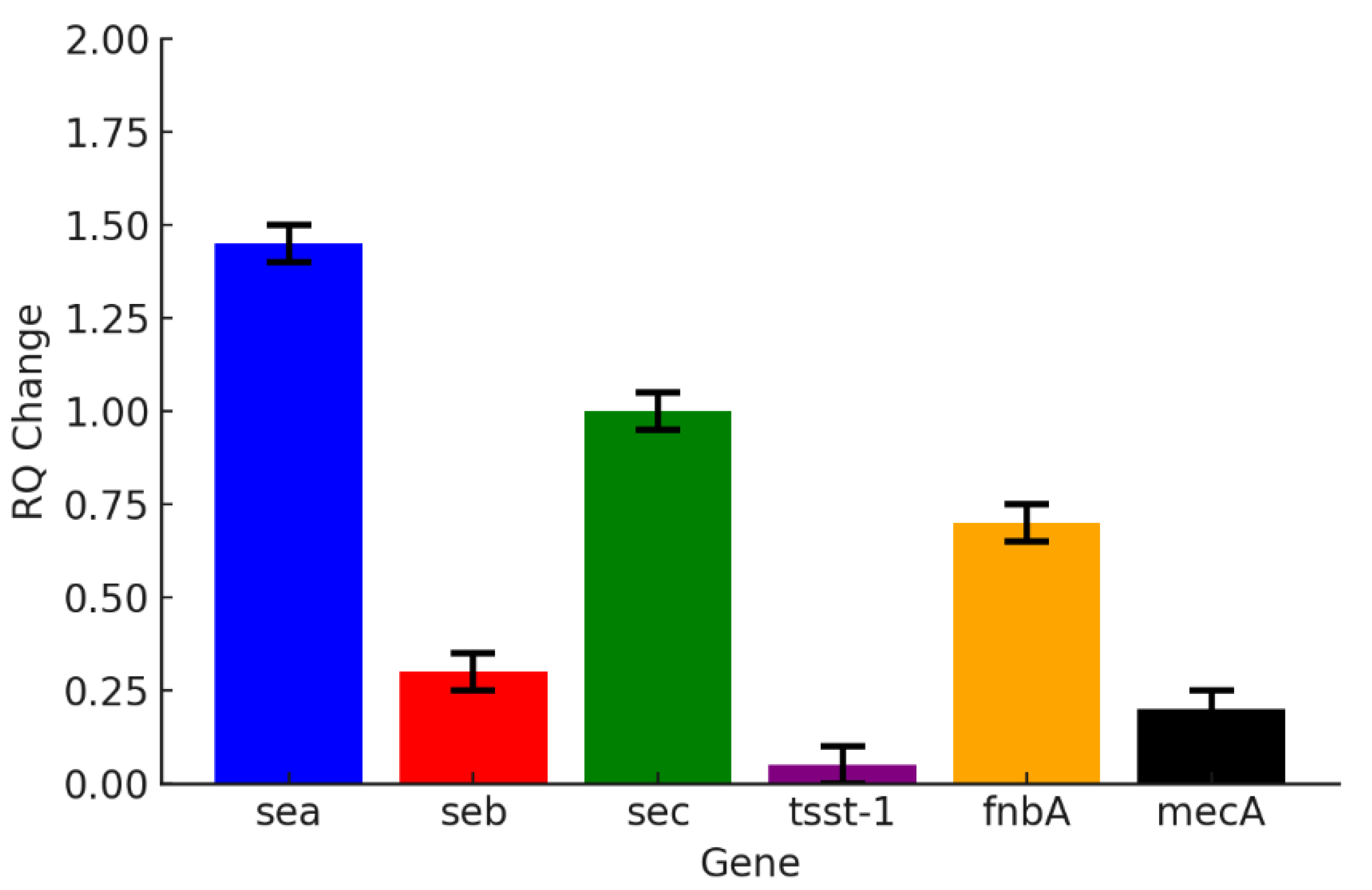

Unveiling the Antibacterial Potential of Melaleuca cajuputi Essential Oils Against MRSA: Integrating In Vitro Efficacy and In Silico Mechanistic Insights
 ,
,
Abstract
1. Introduction
2. Materials and Methods
2.1. Plant Collection
2.2. Essential Oils Extraction
2.3. GC-MS
2.4. In Silico Molecular Docking Analysis
2.5. Tested Bacteria
2.6. Antibacterial Assay
2.6.1. Disc Diffusion Method
2.6.2. Minimum Inhibitory Concentration (MIC) and Minimum Bactericidal Concentration (MBC)
2.7. Quantitative Real Time-Polymerase Chain Reaction (qRT-PCR)
2.8. Statistical Analysis
3. Results
3.1. Essential Oils
3.2. GC-MS Analysis
3.3. In Silico Molecular Docking
3.4. Antibacterial Assay
3.5. Gene Level Expression
4. Conclusions
Supplementary Materials
Author Contributions
Funding
Institutional Review Board Statement
Informed Consent Statement
Data Availability Statement
Acknowledgments
Conflicts of Interest
Abbreviations
| GC-MS | Gas Chromatography–Mass Spectrometry |
| MBC | Minimum Bactericidal Concentration |
| MIC | Minimum Inhibitory Concentration |
| MRSA | Methicillin-Resistant Staphylococcus aureus |
References
- Muteeb, G.; Rehman, M.T.; Shahwan, M.; Aatif, M. Origin of antibiotics and antibiotic resistance, and their impacts on drug development: A narrative review. Pharmaceuticals 2023, 16, 1615. [Google Scholar] [CrossRef] [Scilit]
- Elshobary, M.E.; Badawy, N.K.; Ashraf, Y.; Zatioun, A.A.; Masriya, H.H.; Ammar, M.M.; Mohamed, N.A.; Mourad, S.; Assy, A.M. Combating antibiotic resistance: Mechanisms, multidrug-resistant pathogens, and novel therapeutic approaches: An updated review. Pharmaceuticals 2025, 18, 402. [Google Scholar] [CrossRef] [Scilit]
- Angelini, P. Plant-derived antimicrobials and their crucial role in combating antimicrobial resistance. Antibiotics 2024, 13, 746. [Google Scholar] [CrossRef] [Scilit] [PubMed]
- Vaou, N.; Stavropoulou, E.; Voidarou, C.; Tsigalou, C.; Bezirtzoglou, E. Towards advances in medicinal plant antimicrobial activity: A review study on challenges and future perspectives. Microorganisms 2021, 9, 2041. [Google Scholar] [CrossRef] [Scilit] [PubMed]
- Turner, N.A.; Sharma-Kuinkel, B.K.; Maskarinec, S.A.; Eichenberger, E.M.; Shah, P.P.; Carugati, M.; Holland, T.L.; Fowler, V.G., Jr. Methicillin-resistant Staphylococcus aureus: An overview of basic and clinical research. Nat. Rev. Microbiol. 2019, 17, 203–218. [Google Scholar] [CrossRef] [Scilit] [PubMed]
- Lade, H.; Kim, J.S. Molecular determinants of β-lactam resistance in methicillin-resistant Staphylococcus aureus (MRSA): An updated review. Antibiotics 2023, 12, 1362. [Google Scholar] [CrossRef] [Scilit]
- Algammal, A.M.; Hetta, H.F.; Elkelish, A.; Alkhalifah, D.H.H.; Hozzein, W.N.; Batiha, G.E.; El Nahhas, N.; Mabrok, M.A. Methicillin-resistant Staphylococcus aureus (MRSA): One health perspective approach to the bacterium epidemiology, virulence factors, antibiotic-resistance, and zoonotic impact. Infect. Drug Resist. 2020, 13, 3255–3265. [Google Scholar] [CrossRef] [Scilit]
- Choo, E.J. Community-associated methicillin-resistant Staphylococcus aureus in nosocomial infections. Infect. Chemother. 2017, 49, 158–159. [Google Scholar] [CrossRef] [Scilit]
- Junaidi, N.S.S.A.; Shakrin, N.N.S.M.; Desa, M.N.M.; Yunus, W.M.Z.W. Dissemination pattern of hospital-acquired methicillin-resistant Staphylococcus aureus and community-acquired MRSA isolates from Malaysian hospitals: A review from a molecular perspective. Malays. J. Med. Sci. 2023, 30, 26–41. [Google Scholar] [CrossRef] [Scilit]
- Mohd Yuseri, N.A.N.; Abd Wahab, N.Z.; Asmara, H.S.; Wan Taib, W.R.; Abdul Manap, A.S. The anticancer potential of plant essential oils: Mechanisms, applications, and challenges. Trop. J. Nat. Prod. Res. 2025, 9, 2992–3006. [Google Scholar] [CrossRef] [Scilit]
- Yahya, A.K.; Abd Wahab, N.Z.; Ibrahim, N. Bioactive compounds of plant essential oils and their antiviral properties: A comprehensive review. Malays. J. Chem. 2024, 26, 123–136. [Google Scholar] [CrossRef] [Scilit]
- Pezantes-Orellana, C.; German Bermúdez, F.; Matías De la Cruz, C.; Montalvo, J.L.; Orellana-Manzano, A. Essential oils: A systematic review on revolutionizing health, nutrition, and omics for optimal well-being. Front. Med. 2024, 11, 1337785. [Google Scholar] [CrossRef] [Scilit] [PubMed]
- Mirković, S.; Martinović, M.; Tadić, V.M.; Nešić, I.; Jovanović, A.S.; Žugić, A. Antimicrobial and antioxidant activity of essential oils from selected Pinus species from Bosnia and Herzegovina. Antibiotics 2025, 14, 677. [Google Scholar] [CrossRef] [Scilit] [PubMed]
- Abdalla, M.S.; Salem, A.A.; Hussien, E.T.; Ramadan, S.S. Broad-spectrum bioactivities and therapeutic potential of essential oils of certain aromatic and medicinal plants and their combination. Sci. Rep. 2025, 15, 32463. [Google Scholar] [CrossRef] [Scilit]
- Ji, X.; Liu, N.; Huang, S.; Zhang, C. A comprehensive review of licorice: The preparation, chemical composition, bioactivities and its applications. Am. J. Chin. Med. 2024, 52, 667–716. [Google Scholar] [CrossRef] [Scilit]
- Angane, M.; Swift, S.; Huang, K.; Butts, C.A.; Quek, S.Y. Essential oils and their major components: An updated review on antimicrobial activities, mechanism of action and their potential application in the food industry. Foods 2022, 11, 464. [Google Scholar] [CrossRef] [Scilit]
- Falleh, H. Demystifying the power of essential oils: A review of their antibacterial properties and potential as natural food preservatives. EXCLI J. 2025, 24, 828–850. [Google Scholar] [CrossRef] [Scilit]
- Yaakub, S.Y.; Misman, M.A.; Rosli, S.N. Melaleuca cajuputi Powell. In Malaysia Biodiversity Information System (MyBIS); 2025. Available online: https://www.mybis.gov.my/art/886 (accessed on 23 September 2025).
- Govaerts, R.; Sobral, N.; Ashton, P.; Barrie, F.; Holst, B.K.; Landrum, L.L.; Matsumoto, K.; Mazine, F.F.; Nic Lughadha, E.; Proença, C.; et al. World Checklist of Myrtaceae; Royal Botanic Gardens: Kew, UK, 2008; pp. 1–455. [Google Scholar]
- Yamanoshita, T.; Nuyim, T.; Masumori, M.; Tange, T.; Kojima, K.; Yagi, H.; Sasaki, S. Growth response of Melaleuca cajuputi to flooding in a tropical peat swamp. J. For. Res. 2001, 6, 217–219. [Google Scholar] [CrossRef] [Scilit]
- Isah, M.; Rosdi, R.M.; Wahab, W.N.A.W.A.; Ishak, W.R.W.; Abdullah, H.; Sul’ain, M.S. Phytoconstituents and biological activities of Melaleuca cajuputi Powell: A scoping review. J. Appl. Pharm. Sci. 2023, 13, 10–23. [Google Scholar] [CrossRef] [Scilit]
- Desdiani, D.; Fadilah, F.; Sutarto, A.P. The effects of Melaleuca cajuputi oil herbal treatment on clinical, laboratory, and radiological improvement and length of hospital stay in COVID-19 patients. J. Appl. Pharm. Sci. 2022, 12, 122–127. [Google Scholar] [CrossRef] [Scilit]
- Abd Wahab, N.Z.; Noor Azam, S.N.A.; Sayed Abdul Kadir, S.M.S.F.; Abdullah, M.H. Evaluation of antimicrobial and sporicidal activities, and stability testing of herbal skin wash preparations from Melaleuca cajuputi subsp. cumingiana essential oils and Kyllinga nemoralis aqueous extract. Trop. J. Nat. Prod. Res. 2025, 9, 14–23. [Google Scholar] [CrossRef] [Scilit]
- Isah, M.; Sul’ain, M.D.; Wahab, W.W.A.; Abdullah, H.; Jamil, S.; Syamira, N.; Shabudin, M.; Shuid, A.N.; Ishak, W.R.W. Chemical profiling and mechanistic insights into the antibacterial efficacy of Melaleuca cajuputi leaf extract. BMC Complement. Med. Ther. 2025, 25, 121. [Google Scholar] [CrossRef] [Scilit] [PubMed]
- Abd Wahab, N.Z.; Ja’afar, N.S.A.; Ismail, S.B. Evaluation of antibacterial activity of essential oils of Melaleuca cajuputi Powell. J. Pure Appl. Microbiol. 2022, 16, 549–556. [Google Scholar] [CrossRef] [Scilit]
- Al-Abd, N.M.; Zurainee, M.N.; Mansor, M.; Hasan, M.S.; Kassim, M. Antifilarial and antibiotic activities of methanolic extracts of Melaleuca cajuputi flowers. Korean J. Parasitol. 2016, 54, 273–280. [Google Scholar] [CrossRef] [Scilit]
- Jubair, N.; Rajagopal, M.; Chinnappan, S.; Abdullah, N.B.; Fatima, A. Review on the antibacterial mechanism of plant-derived compounds against multidrug-resistant bacteria (MDR). Evid.-Based Complement. Altern. Med. 2021, 2021, 3663315. [Google Scholar] [CrossRef] [Scilit]
- Sati, H.; Carrara, E.; Savoldi, A.; Hansen, P.; Garlasco, J.; Campagnaro, E.; Boccia, S.; Castillo-Polo, J.A.; Magrini, E.; Garcia-Vello, P.; et al. The WHO bacterial priority pathogens list 2024: A prioritisation study to guide research, development, and public health strategies against antimicrobial resistance. Lancet Infect. Dis. 2025, 25, 1033–1043. [Google Scholar] [CrossRef] [Scilit]
- Institute for Medical Research (IMR). National Antibiotic Resistance Surveillance Report 2023; Ministry of Health Malaysia: Kuala Lumpur, Malaysia, 2023. Available online: https://library.nih.gov.my/e-doc/imr/nsar-2023.pdf (accessed on 23 September 2025).
- Che Hamzah, A.M.; Yeo, C.C.; Puah, S.M.; Chua, K.H.; Chew, C.H. Staphylococcus aureus infections in Malaysia: A review of antimicrobial resistance and characteristics of the clinical isolates, 1990–2017. Antibiotics 2019, 8, 128. [Google Scholar] [CrossRef] [Scilit]
- Vanamala, K.; Tatiparti, K.; Bhise, K.; Sau, S.; Scheetz, M.H.; Rybak, M.J.; Andes, D.; Iyer, A.K. Novel approaches for the treatment of methicillin-resistant Staphylococcus aureus: Using nanoparticles to overcome multidrug resistance. Drug Discov. Today 2021, 26, 31–43. [Google Scholar] [CrossRef] [Scilit]
- Amin, E.; Elwekeel, A.; Alshariedh, N.F.; Abdel-Bakky, M.S.; Hassan, M.H.A. GC–MS analysis and bioactivities of the essential oil of Suaeda aegyptiaca. Separations 2022, 9, 439. [Google Scholar] [CrossRef] [Scilit]
- Toan, T.Q.; Thao, L.P.P.; Chien, N.Q.; Van, N.T.H.; Phuong, Ð.L.; Huong, T.T.; Quan, P.M.; Inh, C.T.; Minh, P.T.H.; Bich, H.T.; et al. Determination of chemical composition and antimicrobial activity of Melaleuca cajuputi essential oil from Quang Tri Province, Vietnam. Asian J. Chem. 2020, 32, 2203–2207. [Google Scholar] [CrossRef] [Scilit]
- Fouzi, S.S.; Abd Wahab, N.Z.; Yan, L.C.; Ibrahim, N. Styrylpyrone derivative from Goniothalamus sp.: A powerful drug for fighting against herpes simplex virus type 1. Pharmacogn. J. 2021, 13, 1598–1606. [Google Scholar] [CrossRef] [Scilit]
- Kebede, T.; Gadisa, E.; Tufa, A. Antimicrobial activities evaluation and phytochemical screening of some selected medicinal plants: A possible alternative in the treatment of multidrug-resistant microbes. PLoS ONE 2021, 16, e0249253. [Google Scholar] [CrossRef] [Scilit] [PubMed]
- Shahzad, M.A.; Yousaf, A.; Ahsan, A.; Irshad, H.; Riaz, A.; Khan, A.; Ullah, I.; Sattar, S.; Bostan, N.; Javed, S. Virulence and resistance profiling of Staphylococcus aureus isolated from subclinical bovine mastitis in the Pakistani Pothohar region. Sci. Rep. 2024, 14, 14569. [Google Scholar] [CrossRef] [Scilit] [PubMed]
- Abd Wahab, N.Z.; Ibrahim, N. Styrylpyrone derivative (SPD), a Novel DENV-2 NS3, NS5 and prM inhibitor; inhibited RNA replication, protein transcription and production of DENV-2 progeny particles. Sains Malays. 2025, 54, 1405–1416. [Google Scholar] [CrossRef] [Scilit]
- Mohammed, I.O.; Alrasheid, A.A.; Hussein Ayoub, S.M. GC-MS analysis and study of the antimicrobial activity of Citrus paradisi, Citrus aurantifolia, and Citrus sinensis peel essential oils as hand sanitizer. Int. J. Microbiol. 2024, 2024, 4957712. [Google Scholar] [CrossRef] [Scilit]
- Lin, H.; Li, Z.; Sun, Y.; Zhang, Y.; Wang, S.; Zhang, Q.; Cai, T.; Xiang, W.; Zeng, C.; Tang, J. D-Limonene: Promising and sustainable natural bioactive compound. Appl. Sci. 2024, 14, 4605. [Google Scholar] [CrossRef] [Scilit]
- Rivas da Silva, A.C.; Lopes, P.M.; Barros de Azevedo, M.M.; Costa, D.C.; Alviano, C.S.; Alviano, D.S. Biological activities of α-pinene and β-pinene enantiomers. Molecules 2012, 17, 6305–6316. [Google Scholar] [CrossRef] [Scilit]
- Mahizan, N.A.; Yang, S.-K.; Moo, C.-L.; Song, A.A.-L.; Chong, C.-M.; Chong, C.-W.; Abushelaibi, A.; Lim, S.-H.E.; Lai, K.-S. Terpene derivatives as a potential agent against antimicrobial resistance (AMR) pathogens. Molecules 2019, 24, 2631. [Google Scholar] [CrossRef] [Scilit]
- Sathish Kumar, K.; Visnuvinayagam, S.; Teena, G.; Elavarasan, K.; Bindu, J.; Balange, A.K.; Sivaranjani, R.; Narasimhamurthy, L. Biochemical, antioxidant, and antimicrobial profiling of essential oils of indian origin for culinary applications. Int. J. Food Sci. 2024, 2024, 9326683. [Google Scholar] [CrossRef] [Scilit]
- Bassolé, I.H.N.; Juliani, H.R. Essential oils in combination and their antimicrobial properties. Molecules 2012, 17, 3989–4006. [Google Scholar] [CrossRef] [Scilit]
- Abd Ghani, M.F.; Othman, R.; Nordin, N. Molecular Docking Study of Naturally Derived Flavonoids with Antiapoptotic BCL-2 and BCL-XL Proteins toward Ovarian Cancer Treatment. J. Pharm. Bioallied Sci. 2020, 12, S676–S680. [Google Scholar] [CrossRef] [Scilit]
- Alsedfy, M.Y.; Ebnalwaled, A.A.; Moustafa, M.; Said, A.H. Investigating the binding affinity, molecular dynamics, and ADMET properties of curcumin-IONPs as a mucoadhesive bioavailable oral treatment for iron deficiency anemia. Sci. Rep. 2024, 14, 22027. [Google Scholar] [CrossRef] [Scilit]
- Regenthal, P.; Hansen, J.S.; André, I.; Lindkvist-Petersson, K. Correction: Thermal stability and structural changes in bacterial toxins responsible for food poisoning. PLoS ONE 2017, 12, e0175989. [Google Scholar] [CrossRef] [Scilit]
- Lillehoj, H.; Liu, Y.; Calsamiglia, S.; Fernandez-Miyakawa, M.E.; Chi, F.; Cravens, R.L.; Oh, S.; Gay, C.G. Phytochemicals as antibiotic alternatives to promote growth and enhance host health. Vet. Res. 2018, 49, 76. [Google Scholar] [CrossRef] [Scilit]
- Ramírez-Santos, J.; Calzada, F.; García-Hernández, N.; Barbosa, E.; Velázquez, C.; Valdes, M. Caryophyllene Oxide, a Bicyclic Terpenoid Isolated from Annona macroprophyllata with Antitumor Activity: In Vivo, In Vitro, and In Silico Studies. Int. J. Mol. Sci. 2024, 25, 13355. [Google Scholar] [CrossRef] [Scilit] [PubMed]
- Yoo, H.J.; Jwa, S.K. Inhibitory effects of β-caryophyllene on Streptococcus mutans biofilm. Arch. Oral Biol. 2018, 88, 42–46. [Google Scholar] [CrossRef] [Scilit] [PubMed]
- Farha, M.A.; Leung, A.; Sewell, E.W.; D’Elia, M.A.; Allison, S.E.; Ejim, L.; Pereira, P.M.; Pinho, M.G.; Wright, G.D.; Brown, E.D. Inhibition of WTA synthesis blocks the cooperative action of PBPs and sensitizes MRSA to β-lactams. ACS Chem. Biol. 2013, 8, 226–233. [Google Scholar] [CrossRef] [Scilit] [PubMed]
- Almeida-Bezerra, J.W.; da Costa Silva, J.T.; Morais-Braga, M.F.B.; da Cruz, R.P.; Alencar, G.G.; Alves, D.S.; de Sousa Rodrigues, E.Y.; de Sousa, S.G.; de Menezes, I.R.A.; Rocha, J.E.; et al. ADME/Tox study and the effect of β-Caryophyllene on the resistant strain of Staphylococcus aureus carrying the QacA/B efflux pump gene. Toxicol. Rep. 2025, 14, 101929. [Google Scholar] [CrossRef] [Scilit]
- Dam, N.A.L.; Bui, V.H.; Dat, T.T.H.; Nguyen, K.T.; Nguyen, T.T.; Duong, Q.Q.; Vo, T.L.; Titok, V.V. In Vitro and In Silico Antimicrobial Activity against Methicillin-Resistant Staphylococcus aureus of Essential Oils from Four Medicinal Plants in Xuan Thuy Mangrove Forest. J. Microbiol. Biotechnol. 2025, 35, e2409006. [Google Scholar] [CrossRef] [Scilit]
- Terefe, E.M.; Ghosh, A. Molecular Docking, Validation, Dynamics Simulations, and Pharmacokinetic Prediction of Phytochemicals Isolated from Croton dichogamus Against the HIV-1 Reverse Transcriptase. Bioinform. Biol. Insights 2022, 16, 11779322221125605. [Google Scholar] [CrossRef] [Scilit]
- Abd Wahab, N.Z.; Malza, N.M.N.M.; Rukayadi, Y. Phytochemical Screening and Evaluation of Antibacterial, Anticandidal, and Sporicidal Properties of Euphorbia tirucalli Extract in Terengganu, Malaysia. Hayati J. Biosci. 2024, 31, 903–915. [Google Scholar] [CrossRef] [Scilit]
- Ike, K.A.; Omaliko, P.C.; Fernander, M.C.; Sanders, B.M.; Enikuomehin, J.M.; Alabi, J.O.; Adelusi, O.O.; Kholif, A.E.; Thomas, M.D.; Anele, U.Y. Evaluating the Effect of an Essential Oil Blend on the Growth and Fitness of Gram-Positive and Gram-Negative Bacteria. Biology 2025, 14, 437. [Google Scholar] [CrossRef] [Scilit] [PubMed]
- Qasim, M.; Islam, W.; Rizwan, M.; Hussain, D.; Noman, A.; Khan, K.A.; Ghramh, H.A.; Han, X. Impact of Plant Monoterpenes on Insect Pest Management and Insect-Associated Microbes. Heliyon 2024, 10, e39120. [Google Scholar] [CrossRef] [Scilit] [PubMed]
- Juszczuk-Kubiak, E. Molecular Aspects of the Functioning of Pathogenic Bacteria Biofilm Based on Quorum Sensing (QS) Signal-Response System and Innovative Non-Antibiotic Strategies for Their Elimination. Int. J. Mol. Sci. 2024, 25, 2655. [Google Scholar] [CrossRef] [Scilit] [PubMed]
- Famuyide, I.M.; Aro, A.O.; Fasina, F.O.; Eloff, J.N.; McGaw, L.J. Antibacterial and Antibiofilm Activity of Acetone Leaf Extracts of Nine Under-Investigated South African Eugenia and Syzygium (Myrtaceae) Species and Their Selectivity Indices. BMC Complement. Altern. Med. 2019, 19, 141. [Google Scholar] [CrossRef] [Scilit]
- Carson, C.F.; Hammer, K.A.; Riley, T.V. Melaleuca alternifolia (Tea Tree) Oil: A Review of Antimicrobial and Other Medicinal Properties. Clin. Microbiol. Rev. 2006, 19, 50–62. [Google Scholar] [CrossRef] [Scilit]
- Mukarram, M.; Choudhary, S.; Khan, M.A.; Poltronieri, P.; Khan, M.M.A.; Ali, J.; Kurjak, D.; Shahid, M. Lemongrass Essential Oil Components with Antimicrobial and Anticancer Activities. Antioxidants 2021, 11, 20. [Google Scholar] [CrossRef] [Scilit]
- Liu, Y.; Han, N.; Meng, F. Magnolia Essential Oil: A Preliminary Exploration of Chemical Composition and Its Antimicrobial and Antioxidant Potential. Front. Microbiol. 2025, 16, 1509796. [Google Scholar] [CrossRef] [Scilit]
- Kovačević, Z.; Čabarkapa, I.; Šarić, L.; Pajić, M.; Tomanić, D.; Kokić, B.; Božić, D.D. Natural Solutions to Antimicrobial Resistance: The Role of Essential Oils in Poultry Meat Preservation with Focus on Gram-Negative Bacteria. Foods 2024, 13, 3905. [Google Scholar] [CrossRef] [Scilit]
- Cieza, M.Y.R.; Bonsaglia, E.C.R.; Rall, V.L.M.; Santos, M.V.D.; Silva, N.C.C. Staphylococcal Enterotoxins: Description and Importance in Food. Pathogens 2024, 13, 676. [Google Scholar] [CrossRef] [Scilit]
- Pinchuk, I.V.; Beswick, E.J.; Reyes, V.E. Staphylococcal Enterotoxins. Toxins 2010, 2, 2177–2197. [Google Scholar] [CrossRef] [Scilit]
- Paharik, A.E.; Horswill, A.R. The Staphylococcal Biofilm: Adhesins, Regulation, and Host Response. Microbiol. Spectr. 2016, 4, 529–566. [Google Scholar] [CrossRef] [Scilit]
- Fishovitz, J.; Hermoso, J.A.; Chang, M.; Mobashery, S. Penicillin-Binding Protein 2a of Methicillin-Resistant Staphylococcus aureus. IUBMB Life 2014, 66, 572–577. [Google Scholar] [CrossRef] [Scilit]
- Cheung, G.Y.C.; Bae, J.S.; Otto, M. Pathogenicity and Virulence of Staphylococcus aureus. Virulence 2021, 12, 547–569. [Google Scholar] [CrossRef] [Scilit]


| Target Gene | Primer Sequence | Function |
|---|---|---|
| sea | F 5′-AGC TTG TAT GTA TGG TGG TGT-3′ | Staphylococcal Enterotoxins |
| R 5′-ACG TCT TGC TTG AAG ATC CA-3′ | ||
| seb | F 5′-AGG ACA CTA AGT TAG GGA AT-3′ | Staphylococcal Enterotoxins |
| R 5′-CTC AGT TAC ACC ACC ATA CA-3′ | ||
| sec | F 5′-TGT AGG TAA AGT TAC AGG TGG T-3′ | Staphylococcal Enterotoxins |
| R 5′-TGT CTA GTT CTT GAG CTG TTA C-3′ | ||
| tsst-1 | F 5′-ATG GCA GCA TCA GCT TGA TA-3′ | Toxic Shock Syndrome-Toxin-1 |
| R 5′- TTT CCA ATA ACC ACC CGT TT-3′ | ||
| fnbA | F 5′-CAC AAC CAG CAA ATA TAG-3′ | Fibronectin Binding Protein A |
| R 5′-CTG TGT GGT AAT CAA TGT-3′ | ||
| mecA | F 5′-AAA ATC GAT GGT AAA GGT TGG C-3′ | Methicillin Resistance Gene |
| R 5′-AGT TCT GCA GTA CCG GAT TTG C-3′ | ||
| nuc | F 5′-GCG ATT GAT GGT GAT ACG GTI-3′ | Housekeeping Gene |
| R 5′-AGC CAA GCC TTG ACG AAC TAA AGC-3′ |
| Peak No. | RT (min) | Identified Compound (Best Hit) | CAS No. | Area % | Match Quality |
|---|---|---|---|---|---|
| 1 | 7.027 | Bicyclo[3.1.0]hexane, 4-methylene… | 003387-41-5 | 0.03 | 91 |
| 2 | 7.166 | Bicyclo[3.1.1]heptane, 6,6-dimethyl… | 018172-67-3 | 1.56 | 97 |
| 3 | 7.443 | β-pinene | 000127-91-3 | 0.45 | 92 |
| 4 | 7.891 | α-phellandrene | 000099-83-2 | 1.09 | 91 |
| 5 | 8.162 | 1,3-cyclohexadiene, 1-methyl-4-(…) | 000099-86-5 | 1.29 | 97 |
| 6 | 8.585 | D-limonene | 005989-27-5 | 8.59 | 98 |
| 7 | 8.926 | 1,3,7-octatriene, 3,7-dimethyl- | 000502-99-8 | 0.06 | 87 |
| 8 | 9.348 | γ-terpinene | 000099-85-4 | 6.98 | 97 |
| 9 | 10.074 | (+)-4-carene | 029050-33-7 | 8.87 | 98 |
| 10 | 10.124 | Benzene, 1-methyl-4-(1-methylethyl-) | 001195-32-0 | 0.23 | 97 |
| 11 | 10.370 | Bicyclo[4.3.0]nonane, 2-methylen… | 040954-37-8 | 0.02 | 70 |
| 12 | 10.572 | 3-carene | 013466-78-9 | 0.16 | 97 |
| 13 | 10.654 | 3-carene | 013466-78-9 | 0.45 | 94 |
| 14 | 10.938 | 3-carene | 013466-78-9 | 0.04 | 97 |
| 15 | 10.982 | 3-carene | 013466-78-9 | 0.06 | 95 |
| 16 | 11.058 | 3-carene | 013466-78-9 | 0.05 | 95 |
| 17 | 11.184 | 1,3,6-octatriene, 3,7-dimethyl-… | 003338-55-4 | 0.14 | 89 |
| 18 | 11.342 | 3-carene | 013466-78-9 | 0.03 | 93 |
| 19 | 11.399 | 3-carene | 013466-78-9 | 0.16 | 92 |
| 20 | 12.572 | Mandelic acid | 90-64-2 | 0.01 | 70 |
| 21 | 12.951 | γ-terpinene | 99-85-4 | 0.05 | 93 |
| 22 | 13.026 | γ-terpinene | 99-85-4 | 0.08 | 96 |
| 23 | 13.152 | γ-terpinene | 99-85-4 | 0.09 | 96 |
| 24 | 13.260 | γ-terpinene | 99-85-4 | 0.01 | 96 |
| 25 | 13.279 | γ-terpinene | 99-85-4 | 0.05 | 96 |
| 26 | 13.430 | γ-terpinene | 99-85-4 | 0.02 | 96 |
| 27 | 13.455 | γ-terpinene | 99-85-4 | 0.07 | 96 |
| 28 | 13.821 | 2,6-dimethyl-1,3,5,7-octatetraene | 460-01-5 | 0.01 | 83 |
| 29 | 13.872 | Benzene, tert-butyl- | 98-06-6 | 0.01 | 53 |
| 30 | 13.954 | 2,6-dimethyl-1,3,5,7-octatetraene | 460-01-5 | 0.01 | 89 |
| 31 | 14.357 | 3-carene | 13466-78-9 | 0.06 | 78 |
| 32 | 14.402 | 3-carene | 13466-78-9 | 0.02 | 70 |
| 33 | 14.452 | (+)-4-carene | 29050-33-7 | 0.03 | 64 |
| 34 | 14.534 | 3-carene | 13466-78-9 | 0.01 | 78 |
| 35 | 14.559 | 3-carene | 13466-78-9 | 0.01 | 83 |
| 36 | 14.641 | 3-carene | 13466-78-9 | 0.04 | 50 |
| 37 | 14.736 | (1R)-2,6,6-trimethylbicyclo[3.1… | 7785-70-8 | 0.09 | 87 |
| 38 | 15.234 | Cyclohexene, 5-methyl-3-(1-methyl… | 56816-08-1 | 0.00 | 52 |
| 39 | 15.442 | 6,7-dimethyl-1,2,3,5,8,8a-hexahy… | 107914-92-1 | 0.01 | 38 |
| 40 | 15.518 | 1,5,5-trimethyl-6-methylene-cycl… | 514-95-4 | 0.01 | 90 |
| 41 | 15.764 | 1,3,6-heptatriene, 2,5,5-trimethyl- | 29548-02-5 | 0.12 | 81 |
| 42 | 15.840 | Cyclohexene, 4-ethenyl-4-methyl- | 20307-84-0 | 0.26 | 99 |
| 43 | 16.099 | α-cubebene | 17699-14-8 | 0.01 | 97 |
| 44 | 16.263 | 3,7-dimethyloct-6-enyl ethyl car… | 1000373-78-1 | 0.01 | 74 |
| 45 | 16.288 | Tricyclo [4.4.0.0(2,8)]dec-3-en-5-ol | 1000193-38-7 | 0.01 | 50 |
| 46 | 16.464 | Cyclohexene, 1-methyl-4-(1-methyl… | 586-62-9 | 0.01 | 64 |
| 47 | 16.616 | α-ylangene | 1000374-19-0 | 0.02 | 99 |
| 48 | 16.767 | Copaene | 3856-25-5 | 0.14 | 99 |
| 49 | 16.912 | Cyclohexane, 1-ethenyl-1-methyl-… | 110823-68-2 | 0.19 | 94 |
| 50 | 17.171 | Cyclohexane, 1-ethenyl-1-methyl-… | 515-13-9 | 3.65 | 90 |
| 51 | 17.449 | Naphthalene, 1,2,3,4,4a,5,6,8a-o… | 473-13-2 | 0.04 | 64 |
| 52 | 17.537 | Methyleugenol | 93-15-2 | 0.06 | 95 |
| 53 | 17.922 | Caryophyllene | 87-44-5 | 7.65 | 99 |
| 54 | 17.960 | γ-elemene | 29873-99-2 | 0.33 | 95 |
| 55 | 18.067 | α-guaiene | 3691-12-1 | 0.08 | 99 |
| 56 | 18.168 | Aromandendrene | 489-39-4 | 0.07 | 99 |
| 57 | 18.319 | Naphthalene, decahydro-4a-methyl-… | 17066-67-0 | 0.04 | 70 |
| 58 | 18.452 | (E)-β-farnesene | 18794-84-8 | 0.10 | 96 |
| 59 | 18.647 | Humulene | 6753-98-6 | 4.33 | 97 |
| 60 | 18.887 | 2-isopropenyl-4a,8-dimethyl-1,2,… | 1000192-43-5 | 0.34 | 94 |
| 61 | 19.032 | α-muurolene | 31983-22-9 | 0.35 | 98 |
| 62 | 19.089 | 1,6-cyclodecadiene, 1-methyl-5-m… | 23986-74-5 | 0.34 | 98 |
| 63 | 19.291 | Naphthalene, 1,2,3,5,6,7,8,8a-o… | 10219-75-7 | 1.12 | 99 |
| 64 | 19.436 | Naphthalene, 1,2,3,4,4a,5,6,8a-o… | 473-13-2 | 1.15 | 91 |
| 65 | 19.505 | Naphthalene, 1,2,3,5,6,8a-hexahy… | 483-76-1 | 0.31 | 99 |
| 66 | 19.625 | Naphthalene, 1,2,3,5,6,7,8,8a-oc… | 10219-75-7 | 0.18 | 78 |
| 67 | 19.720 | γ-muurolene | 30021-74-0 | 0.07 | 98 |
| 68 | 19.808 | Naphthalene, 1,2,3,5,6,8a-hexahy… | 483-76-1 | 0.29 | 98 |
| 69 | 19.909 | Epizonarene | 41702-63-0 | 0.10 | 89 |
| 70 | 20.092 | Neoisolongifolene | 1000156-12-4 | 0.03 | 83 |
| 71 | 20.199 | Epizonarene | 41702-63-0 | 0.03 | 90 |
| 72 | 20.319 | α-calacorene | 21391-99-1 | 0.06 | 95 |
| 73 | 20.382 | Cadala-1(10),3,8-triene | 1000140-05-6 | 0.02 | 91 |
| 74 | 20.508 | cis-Z-α-bisabolene epoxide | 1000131-71-2 | 0.08 | 78 |
| 75 | 20.647 | Naphthalene, 1,2,3,4,4a,5,6,8a-o… | 473-13-2 | 0.16 | 93 |
| 76 | 20.836 | Cyclohexane, 1-ethenyl-1-methyl-… | 515-13-9 | 1.54 | 95 |
| 77 | 21.007 | α-guaiene | 3691-12-1 | 0.14 | 91 |
| 78 | 21.227 | Caryophyllene oxide | 1139-30-6 | 1.96 | 92 |
| 79 | 21.398 | Neoisolongifolene, 8,9-dehydro- | 67517-14-0 | 1.78 | 70 |
| 80 | 21.492 | Alloaromadendrene | 25246-27-9 | 0.83 | 52 |
| 81 | 21.921 | Guaiol | 489-86-1 | 12.92 | 70 |
| 82 | 21.978 | Benzoic acid, 4-[(trimethylsilyl)… | 27739-17-9 | 1.66 | 38 |
| 83 | 22.035 | 1H-indene, 1-ethylideneoctahydro… | 56362-87-9 | 0.49 | 51 |
| 84 | 22.142 | 1H-benzocycloheptene, 2,4a,5,6,7 | 3853-83-6 | 0.93 | 49 |
| 85 | 22.306 | Cyclohexene, 6-ethenyl-6-methyl-… | 5951-67-7 | 1.30 | 96 |
| 86 | 22.470 | 2-naphthalenemethanol, 1,2,3,4,4… | 1209-71-8 | 3.20 | 93 |
| 90 | 23.007 | β-panasinsene | 1000159-39-0 | 6.89 | 94 |
| 91 | 23.051 | Naphthalene, decahydro-4a-methyl- | 000515-17-3 | 3.67 | 90 |
| 92 | 23.757 | 2-acetyl-5-chloro-3-methylbenzo… | 51527-18-5 | 5.35 | 72 |
| 93 | 24.136 | Trans-farnesol | 106-28-5 | 0.35 | 64 |
| 94 | 24.357 | Naphthalene, 1,2,3,4,4a,5,6,8a-o… | 473-13-2 | 0.11 | 89 |
| 95 | 24.603 | Naphthalene, decahydro-4a-methyl… | 017066-67-0 | 0.05 | 97 |
| 96 | 24.678 | Naphthalene, decahydro-4a-methyl… | 017066-67-0 | 0.02 | 95 |
| 97 | 24.893 | Carbazole, 1,4-dimethyl- | 18028-55-2 | 0.09 | 55 |
| 98 | 24.912 | Carbazole, 1,4-dimethyl- | 18028-55-2 | 0.17 | 55 |
| 99 | 25.517 | 2′,3′,4′ trimethoxyacetophenone | 13909-73-4 | 0.02 | 42 |
| 100 | 25.555 | 3,5-pyridine-diamidoxime | 1000212-04-1 | 0.03 | 38 |
| 101 | 25.707 | cis-β-farnesene | 28973-97-9 | 0.07 | 70 |
| 102 | 26.237 | (Z,Z)-α-Farnesene | 1000293-03-1 | 0.01 | 38 |
| 103 | 26.767 | Ethylene, 1-nitro-2-[3-hydroxy-4… | 1000127-35-7 | 0.01 | 46 |
| 104 | 27.101 | Hexadecanoic acid, methyl ester (Methyl palmitate) | 112-39-0 | 0.09 | 98 |
| 105 | 27.246 | Pentadecanoic acid, 14-methyl-, methyl ester | 5129-60-2 | 0.01 | 97 |
| 106 | 27.871 | Benzenemethanol, 4-(1,1-dimethylethyl)- | 877-65-6 | 0.00 | 49 |
| 107 | 28.167 | n-hexadecanoic acid (palmitic acid) | 57-10-3 | 0.49 | 99 |
| 108 | 28.533 | n-hexadecanoic acid | 57-10-3 | 0.02 | 98 |
| 109 | 28.596 | n-hexadecanoic acid | 57-10-3 | 0.01 | 98 |
| 110 | 28.646 | n-hexadecanoic acid | 57-10-3 | 0.02 | 99 |
| 111 | 28.760 | n-hexadecanoic acid | 57-10-3 | 0.00 | 93 |
| 112 | 28.804 | n-hexadecanoic acid | 57-10-3 | 0.01 | 94 |
| 113 | 29.239 | n-hexadecanoic acid | 57-10-3 | 0.01 | 95 |
| 114 | 29.561 | Retene | 483-65-8 | 0.11 | 64 |
| 115 | 29.612 | Retene | 483-65-8 | 0.07 | 68 |
| 116 | 29.694 | Retene | 483-65-8 | 0.01 | 68 |
| 117 | 29.713 | Retene | 483-65-8 | 0.02 | 68 |
| 118 | 29.795 | Ethanone, 1-(4,6-dihydroxy-2,3,5-trihydroxyphenyl)- | 21987-07-5 | 0.07 | 64 |
| 119 | 29.883 | Ethanone, 1-(4,6-dihydroxy-2,3,5-trihydroxyphenyl)- | 21987-07-5 | 0.03 | 52 |
| 120 | 29.984 | 1H-3a,7-methanoazulene, octahydro- | 25491-20-7 | 0.12 | 93 |
| 121 | 30.135 | Cyclopentaneacetaldehyde, 2-formyl- | 5951-57-5 | 0.08 | 51 |
| 122 | 30.495 | 3,6-di(N-pyrrolidinyl)-1,2,4,5-tetrazine | 117040-53-6 | 0.04 | 38 |
| 123 | 30.571 | 1H-3a,7-methanoazulene, octahydro- | 25491-20-7 | 0.02 | 86 |
| 124 | 30.596 | 1H-3a,7-methanoazulene, octahydro- | 25491-20-7 | 0.02 | 92 |
| 125 | 30.646 | 1H-3a,7-methanoazulene, octahydro- | 25491-20-7 | 0.01 | 94 |
| 126 | 30.672 | 6-(1-hydroxymethylvinyl)-4,8a-dimethyl-1,2,3,4,4a,5,6,7-octahydronaphthalene | 1000190-51-4 | 0.02 | 42 |
| 127 | 30.741 | 1H-3a,7-methanoazulene, octahydro- | 025491-20-7 | 0.02 | 94 |
| 128 | 30.791 | 2H-2,4a-ethanonaphthalen-8(5H)-one | 032391-46-1 | 0.01 | 50 |
| 129 | 31.138 | Bicyclo[6.1.0]nonane, 9-(1-methylethenyl)- | 056666-90-1 | 0.04 | 70 |
| 130 | 31.189 | 1H-3a,7-methanoazulene, octahydro- | 025491-20-7 | 0.03 | 95 |
| 131 | 31.365 | 1H-3a,7-methanoazulene, octahydro- | 025491-20-7 | 0.01 | 95 |
| 132 | 31.448 | 1H-3a,7-methanoazulene, octahydro- | 025491-20-7 | 0.02 | 94 |
| 133 | 31.466 | Naphthalene, 1,2,4a,5,6,8a-hexahydro- | 483-75-0 | 0.02 | 66 |
| 134 | 32.337 | 1H-3a,7-methanoazulene, octahydro- | 025491-20-7 | 0.01 | 87 |
| 135 | 32.476 | 1H-3a,7-methanoazulene, octahydro- | 025491-20-7 | 0.01 | 95 |
| 136 | 32.652 | 1H-3a,7-methanoazulene, octahydro- | 025491-20-7 | 0.01 | 86 |
| 137 | 32.684 | 1H-3a,7-methanoazulene, octahydro- | 025491-20-7 | 0.01 | 95 |
| 138 | 32.766 | 1H-cycloprop[e]azulene, 1a,2,3,5,6,7,7a,7b-octahydro- | 021747-46-6 | 0.01 | 91 |
| 139 | 32.785 | 6-isopropenyl-4,8a-dimethyl-1,2,3,4,4a,5,6,7-octahydronaphthalene | 1000189-10-2 | 0.01 | 93 |
| 140 | 32.867 | 1H-3a,7-methanoazulene, octahydro- | 025491-20-7 | 0.01 | 89 |
| 141 | 32.924 | Bicyclo[7.2.0]undec-4-ene, 4,11-dimethyl- | 013877-93-5 | 0.01 | 91 |
| 142 | 32.943 | Isocaryophillene | 1000140-07-2 | 0.01 | 93 |
| 143 | 34.072 | 6-Isopropenyl-4,8a-dimethyl-1,2,3,4,4a,5,6,7-octahydronaphthalene | 1000189-10-2 | 0.00 | 90 |
| 144 | 34.129 | 1H-3a,7-methanoazulene, octahydro- | 025491-20-7 | 0.00 | 94 |
| 145 | 34.211 | 1H-3a,7-methanoazulene, octahydro- | 025491-20-7 | 0.00 | 89 |
| 146 | 34.255 | 1H-3a,7-methanoazulene, octahydro- | 025491-20-7 | 0.00 | 94 |
| 147 | 34.305 | Isocaryophillene | 1000140-07-2 | 0.00 | 66 |
| 148 | 40.027 | Zinc, dicyclopentyl- | 020525-74-0 | 0.00 | 38 |
| 149 | 40.273 | Anthracene, 9,10-dihydro-9,9,10-trimethyl- | 014923-29-6 | 0.00 | 55 |
| 150 | 40.437 | 9,10-methanoanthracen-11-ol, 9,10-dihydro- | 126615-74-5 | 0.00 | 62 |
| 151 | 47.087 | Methyl abietate | 000127-25-3 | 0.00 | 11 |
| 152 | 47.118 | Di-n-decylsulfone | 111530-37-1 | 0.01 | 12 |
| 153 | 49.383 | Acetic acid, [4-(1,1-dimethylethyl)phenyl] ester | 088530-52-3 | 0.01 | 38 |
| 154 | 49.497 | 2-ethylacridine | 055751-83-2 | 0.00 | 42 |
| Peak | Ligand | Docking Score/Binding Interaction | |||||
|---|---|---|---|---|---|---|---|
| Staphylococcal Enterotoxin A (SEA) | Staphylococcal Enterotoxin B (SEB) | Staphylococcal Enterotoxin C (SEC) | Toxic Shock Syndrome Toxin-1 (TSST-1) | Fibronectin-Binding Protein A (FnBPA) | Penicillin-Binding Protein 2a (PBP2a) | ||
| Alpha-guaiene | −5.942 kcal/mol Pi-Alkyl bond = A: PHE58, A: PHE57  | −6.759 kcal/mol Alkyl bond = B: LEU11. B: PRO6  | −6.022 kcal/mol Alkyl and Pi-Alkyl bond = A: LYS212, A: ALA203, A: TYR213, A: PHE208  | −7.578 kcal/mol Alkyl bond = A: LYS110  | −7.395 kcal/mol Alkyl bond = B: VAL256, B: PRO219, B: PRO309  | −6.827 kcal/mol Alkyl bond = A: LEU224, A: VAL217  |
| Alpha-muurolene | −5.716 kcal/mol Alkyl bond = A: LEU23  | −6.425 kcal/mol Pi-Sigma bond = A: PHE47 Pi-Alkyl bond = A: TYR46, B: TYR233 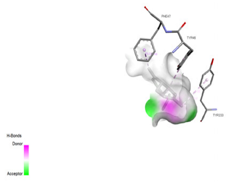 | −6.339 kcal/mol Alkyl and Pi-Alkyl bond = A: MET216, A: LYS212, A: TYR213, A: PHE208, A: ALA203  | −7.819 kcal/mol Alkyl bond = A: LYS111, A: LYS110  | −6.967 kcal/mol Alkyl and Pi-Alkyl bond = B: PHE306, B: PRO219, B: PRO309, B: VAL256  | −6.934 kcal/mol Alkyl and Pi-Alkyl bond = A: LYS604, A: TRP616, A: LEU603, A: ILE164, A: ALA601 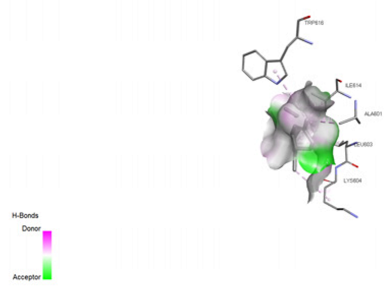 |
| Alpha-ylangene | −5.827 kcal/mol Alkyl bond = A: LEU23  | −6.309 kcal/mol Alkyl and Pi-Alkyl bond = B: ALA203, B: TYR213, B: LYS212  | −6.169 kcal/mol Alkyl bond = A: LYS162, A: VAL147  | −7.766 kcal/mol Alkyl bond = A: LYS111 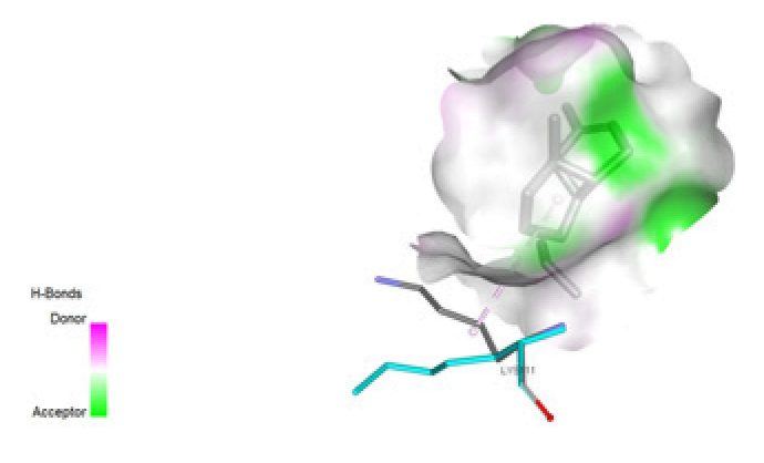 | −7.763 kcal/mol Alkyl and Pi-Alkyl bond = B: PHE306, B: VAL256  | −6.57 kcal/mol Pi-Sigma bond = A: TYR272 Alkyl bond = A: LYS289  |
| Alloaromandendrene | −5.974 kcal/mol Alkyl and Pi-Alkyl bond = A: PRO139, A: ARG161, A: TYR162  | −6.207 kcal/mol Pi-Alkyl bond = A: PHE47  | −6.177 kcal/mol Alkyl and Pi-Alkyl bond = A: LEU58, A: HIS31  | −7.948 kcal/mol Alkyl bond = A: LYS111  | −7.249 kcal/mol Alkyl bond = B: VAL256  | −6.828 kcal/mol Pi-Alkyl bond = A: TYR272  |
| Caryophyllene | −6.02 kcal/mol Pi-Alkyl bond = A: TYR162  | No interaction. | −5.897 kcal/mol Pi-Sigma bond = A: PHE196 Alkyl bond = A: LYS221  | No interaction. | −7.006 kcal/mol Alkyl bond = B: VAL256  | −7.001 kcal/mol Pi-Alkyl bond = A: TYR272 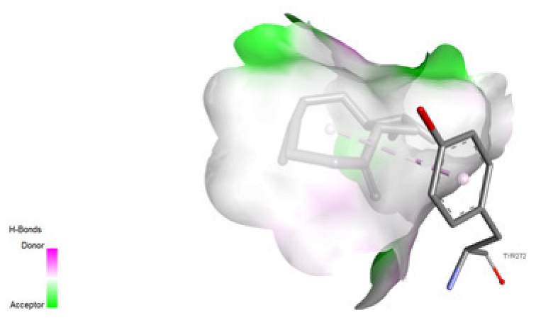 |
| Copaene | −5.685 kcal/mol Alkyl bond = A: ALA22, A: LEU23, A: LEU18  | −5.876 kcal/mol Pi-Alkyl bond = A: PHE47, A: TYR46, B: TYR233  | −6.312 kcal/mol Alkyl and Pi-Alkyl bond = A: MET216, A: TYR213, A: HIS12, A: LYS212  | −7.314 kcal/mol Pi-Alkyl bond = A: TRP52  | −7.915 kcal/mol Alkyl and Pi-Alkyl bond = B: PHE306, B: VAL256  | −6.303 kcal/mol Alkyl and Pi-Alkyl bond = A: LEU224, A: VAL217, A: PHE227, A: TYR223  |
| Cyclohexane | −3.532 kcal/mol Alkyl bond = A: LEU131, A: LYS178  | −3.582 kcal/mol Alkyl and Pi-Alkyl bond = A: TYR213, A: LYS212, A: ALA203  | −3.759 kcal/mol Alkyl and Pi-Alkyl bond = A: TYR213, A: LYS212, A: ALA203  | −4.74 kcal/mol Alkyl and Pi-Alkyl bond = A: ILE166, A: PHE212, A: LYS218  | −3.729 kcal/mol Alkyl and Pi-Alkyl bond = B: PHE367, B: LYS369, B: ILE409  | −4.063 kcal/mol Alkyl bond = A: LEU570, A: LEU455, A: LYS456  |
| D-limonene | −4.939 kcal/mol Alkyl and Pi-Alkyl bond = A: LEU23, A: LYS27, A: PHE175  | −4.999 kcal/mol Alkyl and Pi-Alkyl bond = A: ALA203, A: LYS212, A: LYS207, A: PHE208  | −5.097 kcal/mol Alkyl and Pi-Alkyl bond = A: ALA203, A: LYS212, A: TYR213, A; PHE208  | −6.163 kcal/mol Alkyl bond = A: LYS110, A: LYS111  | −6.004 kcal/mol Alkyl and Pi-Alkyl bond = A: VAL256, A: PRO309, A: PHE306, A: PRO219, C: ALA14  | −5.66 kcal/mol Alkyl and Pi-Alkyl bond = A: ALA276, A: TYR272, A: LYS273  |
| Napthalene | −5.187 kcal/mol Pi-Pi T-shaped and Amide-Pi Stacked bond = A: TYR162 Pi-Alkyl bond = A: PRO139, A: ARG161  | −5.195 kcal/mol Pi-Sigma bond = B: LEU11 Pi-Alkyl bond = B: PRO6 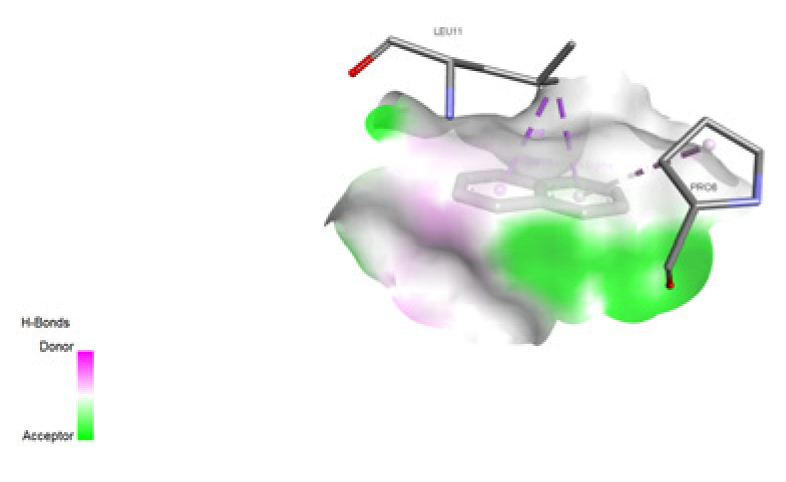 | −5.469 kcal/mol Pi-Donor Hydrogen bond = A: ASP209 Pi-Alkyl bond = A: LYS212 Pi-Sigma bond = A: ALA203  | −6.829 kcal/mol Pi-Cation bond = A: LYS211 Pi-Alkyl bond = A: LYS211 Pi-Pi Stacked bond = A: TRP52 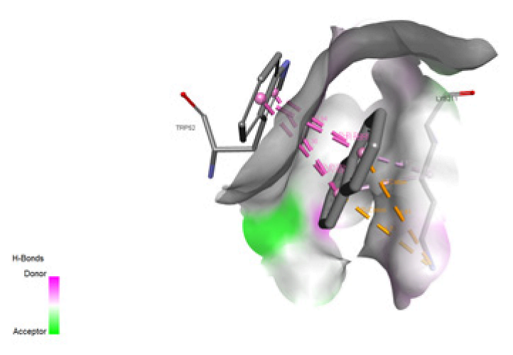 | −6.375 kcal/mol Pi Alkyl bond = A: VAL256, A: PRO309, C: ALA14  | −5.655 kcal/mol Pi-Donor Hydrogen bond = TYR344, A: THR399 Pi-Alkyl bond = A: LYS634, A: LYS394, A: ILE614 Pi-Pi T-shaped bond = A: TYR344  |
| 1, 6 Cyclodecadiene | −4.723 kcal/mol Pi-Alkyl bond = A: PHE58  | −4.82 kcal/mol Alkyl bond = B: ALA203, B: LYS212  | −5.097 kcal/mol Alkyl bond = A: ALA203, A: LYS212  | −6.428 kcal/mol Alkyl bond = A: ARG108  | −5.336 kcal/mol Alkyl bond = A: ILE409 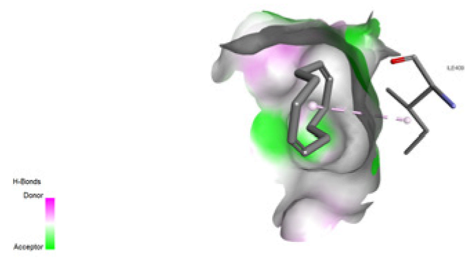 | −5.352 kcal/mol Alkyl bond = A: LEU570, A: LYS456  |
| Beta-pinene | −4.795 kcal/mol Alkyl bond = A: LEU23, A:LYS27  | −4.796 kcal/mol Alkyl and Pi-Alkyl bond = A: ALA203, A: LYS212, A: TYR213, A: PHE208 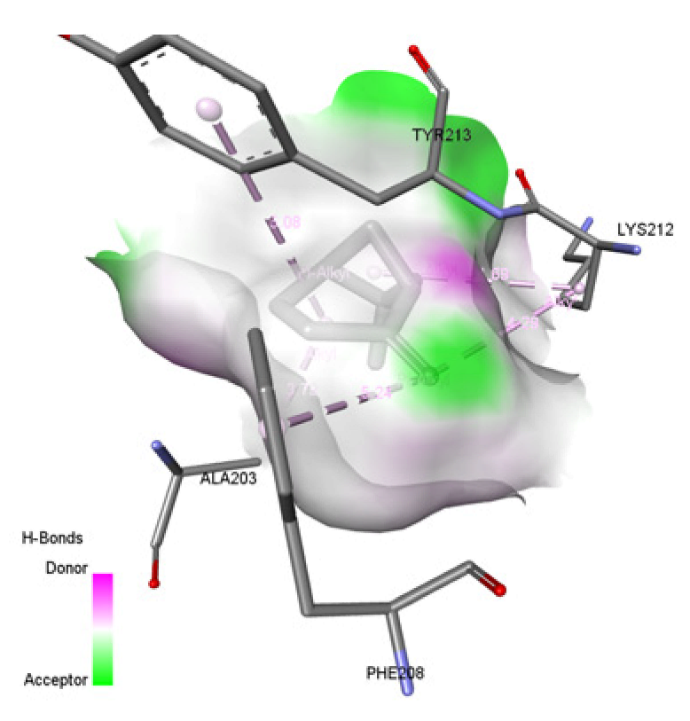 | −5.344 kcal/mol Alkyl and Pi-Alkyl bond = A:HIS31, A:LYS57, A:LEU58  | −6.201 kcal/mol Alkyl and Pi-Alkyl bond = A: ILE166, A: PHE212, A: LYS218  | −5.704 kcal/mol Alkyl bond = B:VAL256  | −5.265 kcal/mol Alkyl bond = A:VAL217, A:PRO370  |
| Alpha-phellandrene | −4.73 kcal/mol Alkyl & Pi-Alkyl bond = A: LEU23, A:LYS27, A:PHE175 Pi-Donor Hydrogen Bond = A:SER172 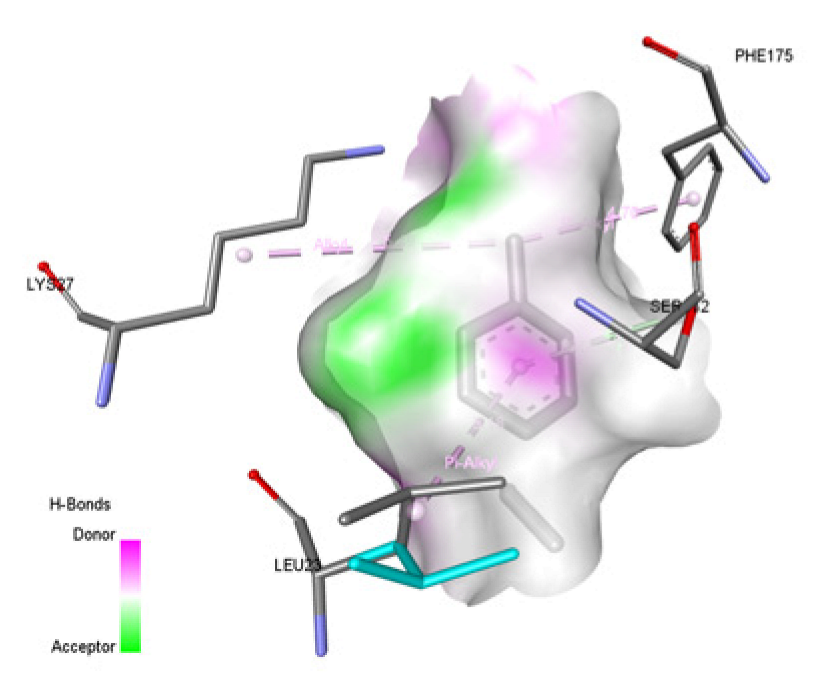 | −5.287 kcal/mol Pi-Alkyl bond = B: ALA203, B:HIS12, B:TYR213 Pi-Pi T-shaped & Amide-Pi Stacked = B:LYS212, B:TYR213  | −5.724 kcal/mol Pi-Alkyl bond = A:ALA203, A:HIS12, A:TYR213, A:LYS212 Pi-Pi T-shaped = A:TYR213  | −6.424 kcal/mol Alkyl and Pi-Alkyl bond = A:LEU49, A:TRP52, A:LYS211 Pi-Pi Stacked = A:TRP52 Pi-Cation = A:LYS211  | −5.98 kcal/mol Alkyl & Pi-Alkyl bond = B:LYS411 Pi-Sigma bond = B:ILE409  | −5.415 kcal/mol Pi-alkyl bond = A:ALA642 Pi-Pi Stacked = A:TYR446  |
| Gamma-terpinene | −4.706 kcal/mol Alkyl & Pi-Alkyl bond = A: TYR162, A:PRO139 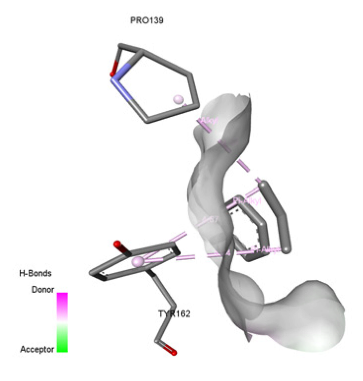 | −5.068 kcal/mol Alkyl and Pi-Alkyl bond = B:PRO6, B:TYR186, B:LEU11 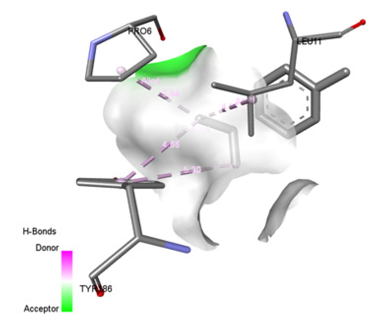 | −5.233 kcal/mol Alkyl and Pi-Alkyl bond = A: HIS12, A:MET216, A:TYR213  | −6.299 kcal/mol Alkyl and Pi-Alkyl bond = A:LEU49, A:TRP52, A:LYS211  | −5.817 kcal/mol Alkyl & Pi-Alkyl bond = B:LYS369, B:PHE367, B:ILE409  | −5.726 kcal/mol Alkyl & Pi-Alkyl bond = A:TYR272, A:ALA276, A:LYS273  |
| Methyleugenol | −4.726 kcal/mol Carbon hydrogen bond = A:GLU165  | −5.093 kcal/mol Conventional hydrogen bond = A:TYR46, B:TYR186  | −4.991 kcal/mol Conventional hydrogen bond = A:ASP209 Carbon hydrogen bond = A:ALA203, A:LYS207 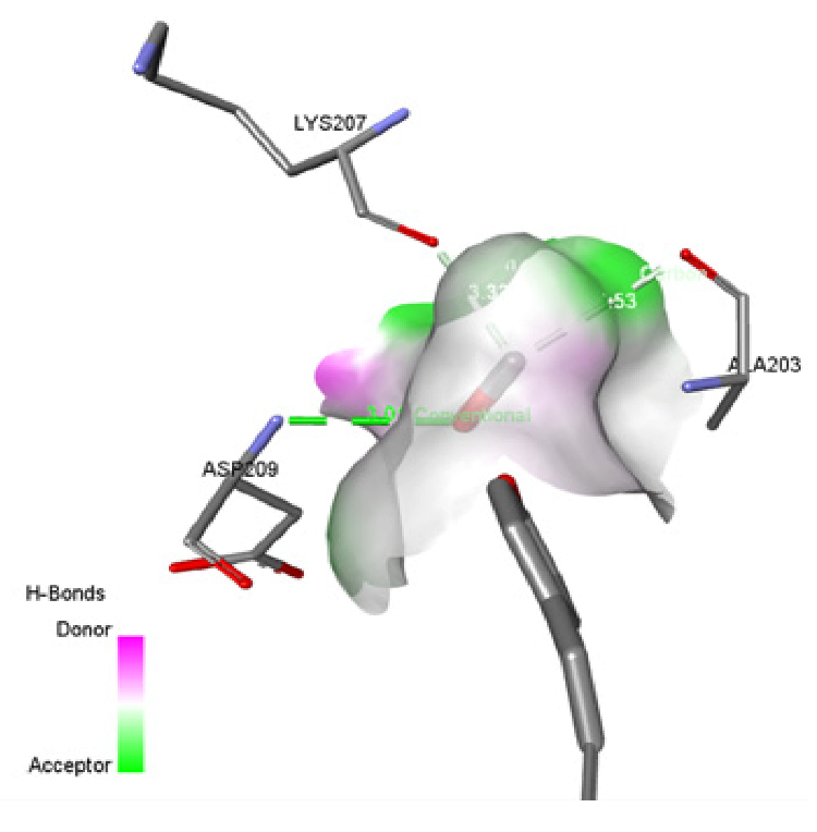 | −5.692 kcal/mol Conventional hydrogen bond = A:LYS111  | −5.269 kcal/mol Conventional hydrogen bond = B:SER351 Carbon hydrogen bond = B:His220  | −4.797 kcal/mol Conventional Hydrogen bond = A:HIS293  |
| Gamma-elemene | −4.431 kcal/mol Alkyl bond = A:ALA201, A:ILE212 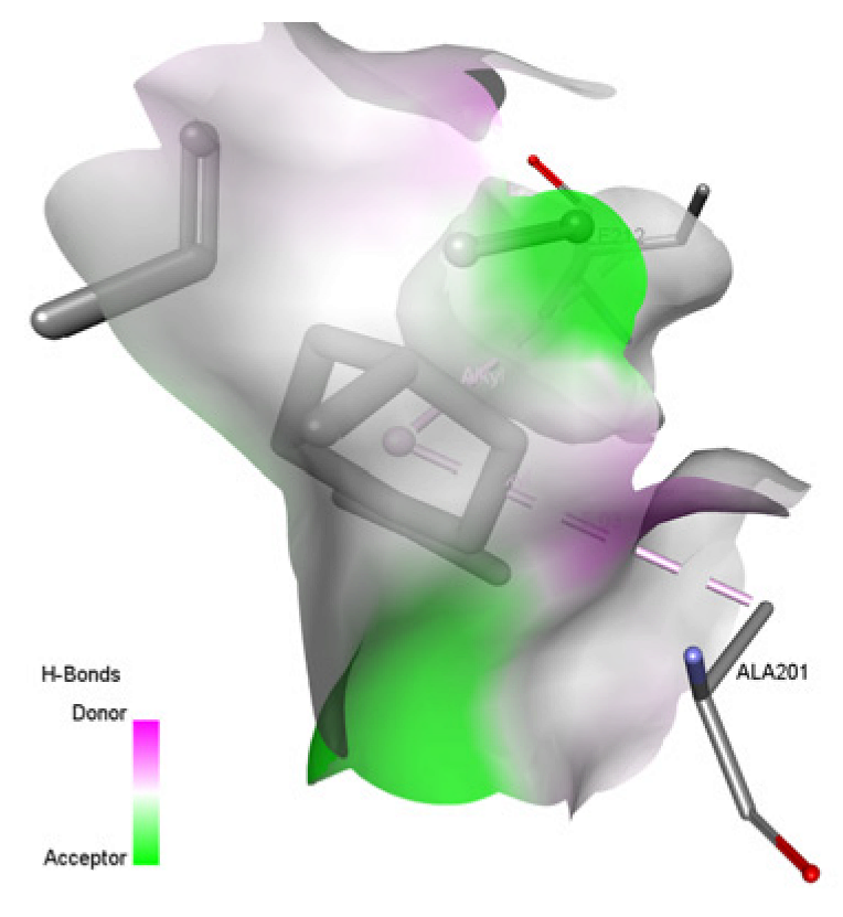 | −5.867 kcal/mol Pi-Alkyl bond = A:TYR46  | −5.558 kcal/mol Alkyl and Pi-Alkyl bond = A:PHE208, A:LYS212, A:TYR213  | −6.768 kcal/mol Alkyl bond = A:LYS110  | −6.516 kcal/mol Alkyl bond = B:LYS411, B:ILE409  | −5.961 kcal/mol Alkyl bond = A:VAL217  |
| Caryophyllene oxide | −5.813 kcal/mol Conventional hydrogen bond = A:THR59, A:ASP60  | −5.919 kcal/mol Carbon hydrogen bond = B:PRO8  | −5.909 kcal/mol Conventional hydrogen bond = A:ASN88 Pi-Alkyl bond = A:HIS31  | −6.933 kcal/mol Alkyl bond = A:LYS218 Conventional hydrogen & Carbon hydrogen bond = A:THR216  | −7.396 kcal/mol Alkyl bond = B:VAL256  | −6.577 kcal/mol Conventional Hydrogen bond = A:LYS218 Alkyl Bond = A:VAL217, A:PRO370  |
| α-calacorene | −5.501 kcal/mol Alkyl & Pi-Alkyl bond = A: TYR162, A:ALA210, A:ILE212 Pi-Sigma bond = A:ILE212  | −6.157 kcal/mol Pi-Alkyl bond = A:PHE47 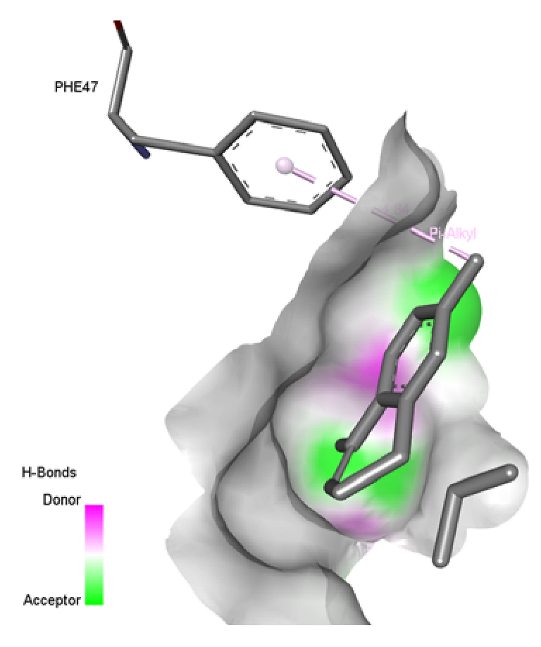 | −6.214 kcal/mol Alkyl and Pi-Alkyl bond = A: ALA203, A:PHE208, A:LYS212, A:TYR213 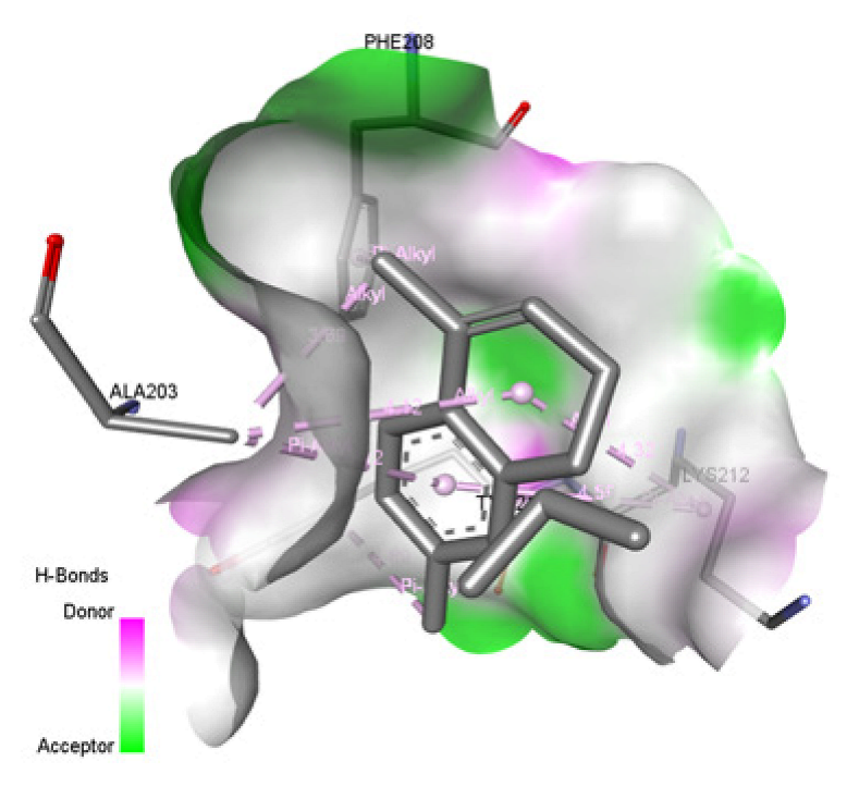 | −7.893 kcal/mol Alkyl and Pi-Alkyl bond = A:ILE125, A:TRP52, A:LYS211 Pi-Cation bond = A:LYS211 Pi-Pi Stacked = A:TRP52  | −7.038 kcal/mol Alkyl & Pi-alkyl bond = A:PRO309, A:VAL256, A:TYR 414, A:VAL365 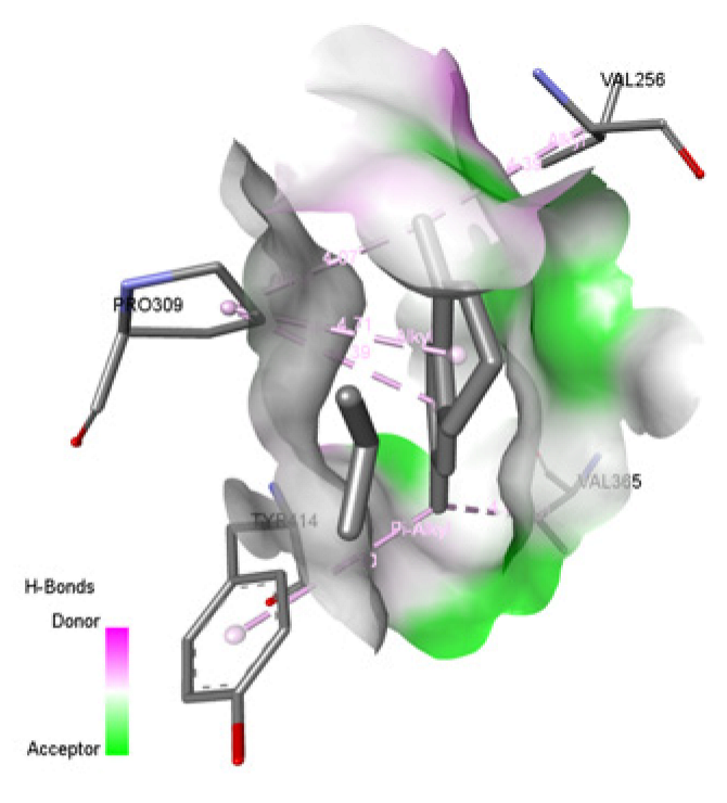 | −7.126 kcal/mol Alkyl & Pi-Alkyl bond = A:TYR272, A:ALA276, A:LYS273, A:LYS289 Pi-Pi T-shaped = A:TYR272 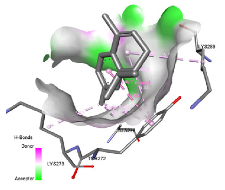 |
| Gamma-muurolene | −5.422 kcal/mol Alkyl bond = A:ARG161  | −6.363 kcal/mol Pi-Alkyl bond = A:PHE47, A:TYR46, B:TYR233  | −6.17 kcal/mol Alkyl bond = A:LYS162, A:VAL147  | −7.417 kcal/mol Alkyl bond = A:LYS111 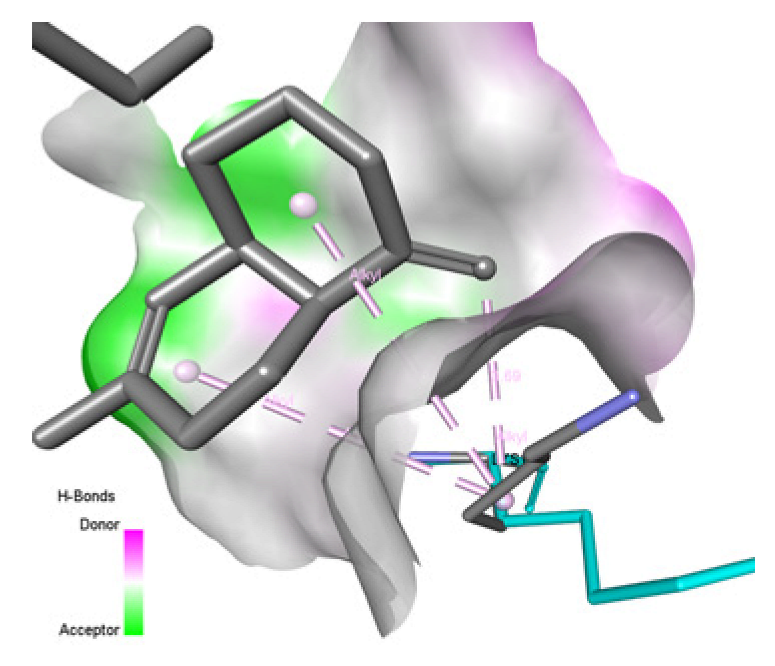 | −7.539 kcal/mol Alkyl & Pi-alkyl bond = B:PRO309, B:VAL256, B:LEU498, B:PRO219, B:PHE306  | −6.494 kcal/mol Alkyl & Pi-Alkyl bond = A:TYR272, A:ALA276, A:LYS273, A:LYS289 Pi-sigma = A:TYR272  |
| Guaiol | 8.944 kcal/mol Alkyl & Pi-Alkyl bond = A: TYR30, A:LEU26, A:ALA201, A:ILE212, A:LEU209 Unfavorable Bump = A:ILE212, A:ASP197, A:ARG160  | −6.481 kcal/mol Alkyl and Pi-Alkyl bond = A:PRO6, A:TYR186, A:LEU11, A:TYR233  | −4.821 kcal/mol Alkyl bond = A:MET215  | −7.179 kcal/mol Alkyl and Pi-Alkyl bond = A:TRP52, A:LYS111  | −7.274 kcal/mol Alkyl bond = B:PRO309, B:PRO219, B:VAL256  | −6.922 kcal/mol Alkyl & Pi-Alkyl bond = A:PHE227, A:VAL217, A:LEU190, A:ILE171, A:LEU224  |
| Humulene | −4.573 kcal/mol Alkyl bond = A:ILE212  | −5.846 kcal/mol Alkyl bond = B:ALA203, B:LYS212  | −5.601 kcal/mol Pi-Alkyl bond = A:PHE196  | −6.674 kcal/mol Alkyl bond = A:LYS111  | −6.149 kcal/mol Alkyl bond = B:LYS411  | −7.009 kcal/mol Pi-alkyl bond = A:TYR272  |
| Tested Bacteria | Zone of Inhibition (mm) | |||||||
|---|---|---|---|---|---|---|---|---|
| 100% | 50% | 25% | 12.50% | 6.25% | 3.13% | 10% DMSO (Negative Control) | Vancomycin 30 mcg (Positive Control) | |
| S. aureus ATCC25923 | 12.67 ± 0.67 | 9.67 ± 0.33 | 7.67 ± 0.33 | 7.00 ± 0.00 | 6.00 ± 0.00 | 6.00 ± 0.00 | 6.00 ± 0.00 | 17.67 ± 0.33 |
| MRSA 1 | 9.67 ± 0.33 | 7.67 ± 0.33 | 7.00 ± 0.00 | 6.67 ± 0.33 | 6.00 ± 0.00 | 6.00 ± 0.00 | 6.00 ± 0.00 | 16.33 ± 0.33 |
| MRSA 2 | 10.00 ± 0.00 | 7.33 ± 0.33 | 6.33 ± 0.33 | 6.00 ± 0.00 | 6.00 ± 0.00 | 6.00 ± 0.00 | 6.60 ± 0.00 | 18.00 ± 0.58 |
| MRSA 3 | 12.00 ± 0.00 | 8.67 ± 0.33 | 7.33 ± 0.33 | 6.00 ± 0.00 | 6.00 ± 0.00 | 6.00 ± 0.00 | 6.00 ± 0.00 | 18.00 ± 0.58 |
| MRSA 4 | 16.00 ± 0.58 | 7.67 ± 0.33 | 7.67 ± 0.33 | 6.67 ± 0.33 | 6.00 ± 0.00 | 6.00 ± 0.00 | 6.00 ± 0.00 | 17.33 ± 0.33 |
| MRSA 5 | 16.67 ± 0.88 | 8.67 ± 0.33 | 7.67 ± 0.33 | 6.00 ± 0.00 | 6.00 ± 0.00 | 6.00 ± 0.00 | 6.00 ± 0.00 | 16.00 ± 0.58 |
| MRSA 6 | 10.67 ± 0.33 | 8.33 ± 0.33 | 7.00 ± 0.00 | 6.33 ± 0.33 | 6.00 ± 0.00 | 6.00 ± 0.00 | 6.00 ± 0.00 | 16.00 ± 0.00 |
| MRSA 7 | 9.67 ± 0.33 | 8.0 ± 0.00 | 7.00 ± 0.00 | 6.00 ± 0.00 | 6.00 ± 0.00 | 6.00 ± 0.00 | 6.00 ± 0.00 | 18.00 ± 0.58 |
| MRSA 8 | 14.67 ± 0.67 | 9.67 ± 0.33 | 8.33 ± 0.33 | 6.33 ± 0.33 | 6.00 ± 0.00 | 6.00 ± 0.00 | 6.00 ± 0.00 | 15.33 ± 0.33 |
| Tested Bacteria | MIC (%) | MBC (%) | MBC/MIC |
|---|---|---|---|
| S. aureus ATCC25923 | 3.13 | 3.13 | 1 |
| MRSA 1 | 6.25 | 12.5 | 2 |
| MRSA 2 | 12.5 | 25 | 2 |
| MRSA 3 | 6.25 | 12.5 | 2 |
| MRSA 4 | 6.25 | 12.5 | 2 |
| MRSA 5 | 3.13 | 3.13 | 1 |
| MRSA 6 | 6.25 | 12.5 | 2 |
| MRSA 7 | 3.13 | 6.25 | 2 |
| MRSA 8 | 3.13 | 3.13 | 1 |
Disclaimer/Publisher’s Note: The statements, opinions and data contained in all publications are solely those of the individual author(s) and contributor(s) and not of MDPI and/or the editor(s). MDPI and/or the editor(s) disclaim responsibility for any injury to people or property resulting from any ideas, methods, instructions or products referred to in the content. |
© 2026 by the authors. Licensee MDPI, Basel, Switzerland. This article is an open access article distributed under the terms and conditions of the Creative Commons Attribution (CC BY) license.
Share and Cite
Abd Wahab, N.Z.; Kamal Rul Azrul, K.S.; Mohd Yuseri, N.A.N.; Yahya, A.K.; Si Wei, F.; Sayed Abdul Kadir, S.M.S.F.; Abdullah, M.H. Unveiling the Antibacterial Potential of Melaleuca cajuputi Essential Oils Against MRSA: Integrating In Vitro Efficacy and In Silico Mechanistic Insights. Bacteria 2026, 5, 13. https://doi.org/10.3390/bacteria5010013
Abd Wahab NZ, Kamal Rul Azrul KS, Mohd Yuseri NAN, Yahya AK, Si Wei F, Sayed Abdul Kadir SMSF, Abdullah MH. Unveiling the Antibacterial Potential of Melaleuca cajuputi Essential Oils Against MRSA: Integrating In Vitro Efficacy and In Silico Mechanistic Insights. Bacteria. 2026; 5(1):13. https://doi.org/10.3390/bacteria5010013
Chicago/Turabian StyleAbd Wahab, Noor Zarina, Kamal Saifullah Kamal Rul Azrul, Nur Ain Najwa Mohd Yuseri, Ahmad Khalis Yahya, Fong Si Wei, Sayed Mohd Saufi Fahmi Sayed Abdul Kadir, and Mohd Hanif Abdullah. 2026. "Unveiling the Antibacterial Potential of Melaleuca cajuputi Essential Oils Against MRSA: Integrating In Vitro Efficacy and In Silico Mechanistic Insights" Bacteria 5, no. 1: 13. https://doi.org/10.3390/bacteria5010013
APA StyleAbd Wahab, N. Z., Kamal Rul Azrul, K. S., Mohd Yuseri, N. A. N., Yahya, A. K., Si Wei, F., Sayed Abdul Kadir, S. M. S. F., & Abdullah, M. H. (2026). Unveiling the Antibacterial Potential of Melaleuca cajuputi Essential Oils Against MRSA: Integrating In Vitro Efficacy and In Silico Mechanistic Insights. Bacteria, 5(1), 13. https://doi.org/10.3390/bacteria5010013