Article

Osmopriming, Halopriming, and Chemopriming Differentially Modulate Stress Tolerance in Solanum lycopersicum Seeds Under Salinity and Iron Toxicity


by
Anny Nogueira Fraga
1,
Josinei Rodrigues Filho
1,
Marina Reis Pires
1,
Viviana Borges Corte
1 and
Hildegardo Seibert França
1,2,*
1
Department of Biological Sciences, Federal University of Espírito Santo (UFES), Vitória 29075-910, ES, Brazil
2
Bioproduct Development Laboratory, Federal Institute of Espírito Santo (IFES), campus Vila Velha, Vila Velha 29106-010, ES, Brazil
*
Author to whom correspondence should be addressed.
Seeds 2026, 5(4), 43; https://doi.org/10.3390/seeds5040043
Submission received: 28 April 2026 / Revised: 15 June 2026 / Accepted: 23 June 2026 / Published: 29 July 2026

Abstract

Solanum lycopersicum L. (tomato) is an economically and nutritionally important crop whose germination and early seedling growth are highly sensitive to abiotic stresses that disrupt physiological processes and intensify oxidative damage. Seed priming has emerged as a low-cost physiological preconditioning strategy to enhance stress tolerance by activating repair and defense mechanisms prior to stress exposure. This study evaluated the effects of osmopriming (PEG 6000), halopriming (KNO3), and chemopriming (SNP) on germination, early seedling growth, antioxidant enzyme activity, and lipid peroxidation in S. lycopersicum under salt stress and iron toxicity. Salt stress was induced with NaCl (100 mM) and iron toxicity with Fe-EDTA (4 mM). Priming treatments included osmopriming with PEG 6000 (−0.4 and −0.8 MPa), halopriming with KNO3 (25 and 50 mM), and chemopriming with sodium nitroprusside—SNP (0.1 and 0.2 mM). Under salt stress, NaCl (100 mM) completely inhibited germination and increased CAT activity by approximately 49% and POX activity by 4%; however, this antioxidant response was insufficient to reduce MDA content, which remained approximately 58% higher than the control. Under iron toxicity, Fe-EDTA (4 mM) reduced germination to 79% and caused a 425% increase in MDA content relative to the control, indicating severe oxidative damage despite a 59% increase in CAT and 47% increase in POX activities. Among the priming treatments evaluated, halopriming with KNO3 (50 mM) was the most effective strategy under salt stress, restoring germination to 81%, increasing CAT activity by 103%, and reducing MDA by 18% relative to non-primed salt-stressed seeds. Under iron toxicity, all priming treatments consistently increased antioxidant enzyme activities; however, none were capable of reducing lipid peroxidation or restoring seedling growth to control levels, likely due to continuous ROS generation via Fenton-type reactions that overwhelmed enzymatic detoxification capacity.

1. Introduction

S. lycopersicum (tomato), belonging to the Solanaceae family, is one of the most widely consumed vegetables worldwide, with high agricultural, economic, and nutritional relevance [1]. In Brazil, the crop occupies approximately 60,000 hectares, with national production estimated between 4.5 and 4.8 million tons and an average yield exceeding 74,000 kg ha−1. The states of Goiás, São Paulo, and Minas Gerais account for most of the national production, highlighting the importance of tomato cultivation for Brazilian agribusiness [2]. From a nutritional perspective, the tomato is widely incorporated into traditional meals, snacks, and fast-food products, occupying a central position in the agri-food chain [3,4]. Furthermore, its nutritional value is associated with the presence of bioactive compounds, especially carotenoids—liposoluble pigments widely distributed in plants—which are linked to protection against oxidative stress in the human body and the modulation of cellular signaling processes [1].
S. lycopersicum is classified as a species moderately sensitive to salt stress throughout its entire developmental cycle [5]. Soil salinity may originate from both natural processes and anthropogenic activities. Natural factors include rock weathering and the gradual accumulation of salts over time. Anthropogenic causes include inadequate irrigation practices, particularly the use of water with high salt concentrations, excessive fertilizer application, and inefficient drainage systems, which favor salt accumulation in the root zone [6,7]. The impacts of salinity include reduced osmotic potential and ionic toxicity, resulting from the accumulation of ions such as sodium (Na+) and chloride (Cl), compromising cellular homeostasis and inducing oxidative stress [8].
Iron is an essential micronutrient for plant metabolism, playing a fundamental role in key physiological processes such as electron transport in photosynthesis and cellular respiration. However, at high concentrations, this element can become toxic to plants, impairing their growth and development [9,10]. Iron toxicity may result from both anthropogenic activities and natural soil conditions, representing a relevant agronomic constraint, particularly in flooded, poorly drained, and naturally iron-rich soils. This phenomenon occurs more frequently in waterlogged areas and in soils such as ferruginous outcrops and acid sulfate soils [11]. S. lycopersicum plants exposed to excess iron (5 mM Fe-EDTA) showed severe reductions in shoot and root growth, along with increased electrolyte leakage and cell death, indicating compromised cellular membrane integrity. Excess iron also promoted redox imbalance, characterized by the accumulation of reactive oxygen species (ROS), resulting in oxidative damage associated with the inefficiency of the antioxidant system in sensitive plants [12].
Seed priming has been widely described as an effective strategy to enhance plant tolerance to abiotic stresses. This method consists of a physiological preconditioning process capable of preparing seeds to respond more efficiently to stress conditions. Priming promotes the early activation of physiological, biochemical, and molecular mechanisms, such as strengthening the antioxidant system, maintaining osmotic balance, and regulating stress-responsive genes, resulting in increased vigor and improved seedling establishment [13]. Different priming techniques have been employed to enhance seed tolerance to abiotic stresses, improving germination, early growth, and physiological responses associated with antioxidant defense. Among these techniques are osmopriming [14], halopriming [15], and chemopriming [16], which act through the early activation of metabolic processes and stress tolerance mechanisms.
Abiotic stresses, such as salinity, represent a growing threat to global agricultural production, compromising crop growth and productivity [17]. In S. lycopersicum, salinity affects physiological and biochemical processes and impairs germination, directly impacting early plant establishment [18]. In this context, seed priming stands out as a low-cost strategy to improve germination and early vigor by activating metabolic mechanisms and the antioxidant system [19,20,21]. However, despite recent advances, iron toxicity remains an emerging research area, with significant gaps in understanding, particularly regarding oxidative stress responses and the efficacy of priming strategies during germination and early seedling establishment [22].
Although Karim et al. [23] recently demonstrated the beneficial effects of KNO3 priming on tomato seedling physiology under salt stress, their study focused on established seedlings and did not address germination-phase responses or the comparative efficacy of multiple priming modalities. Furthermore, the effects of priming strategies on germination and early seedling establishment under iron toxicity—a stress of distinct mechanistic nature—remain largely unexplored. The present study addresses these gaps by evaluating three priming techniques (osmopriming, halopriming, and chemopriming) simultaneously, under both salinity and iron toxicity, with particular emphasis on the germination phase and associated oxidative stress responses.
This study aimed to evaluate the effects of osmopriming (PEG 6000), halopriming (KNO3), and chemopriming (SNP) on the tolerance of S. lycopersicum seeds, cultivar ‘Tomato salad’, to salt stress and iron (Fe) toxicity, by analyzing germination, early seedling growth, and physiological responses, including the activity of the antioxidant enzymes catalase (CAT) and peroxidase (POX), as well as lipid peroxidation, estimated by malondialdehyde (MDA) content.

2. Materials and Methods

2.1. Study Area

Germination tests and seedling growth measurements were conducted at the Plant Interactions Laboratory (LIVE), Federal University of Espírito Santo (UFES), Vitória, ES, Brazil. Physiological analyses were performed at the Bioproduct Development Laboratory, Federal Institute of Espírito Santo (IFES), Vila Velha, ES, Brazil.

2.2. Plant Material

Seeds of Solanum lycopersicum (salad tomato type, commercially designated as ‘Tomato salad’) were obtained from a commercial source (Topseed®, Agristar, Sao Paulo, Brazill). No formal cultivar designation was provided by the manufacturer, which represents a limitation for precise genotypic characterization and should be considered when interpreting and replicating the results. According to the manufacturer, the seed lot presented 99% purity.

2.3. Priming Treatments

Seeds of S. lycopersicum were subjected to osmopriming, halopriming, and chemopriming. Each priming technique was designed to allow seeds to progress through the initial phases of germination—imbibition (phase I) and metabolic activation (phase II)—without completing radicle protrusion (phase III), thereby promoting the pre-activation of defense mechanisms prior to stress exposure. During all treatments, Petri dishes were maintained in a Biochemical Oxygen Demand (BOD) incubator (Eletrolab, São Paulo, Brazil) at 25 °C under continuous white fluorescent light with a 24 h photoperiod. The temperature of 25 °C was selected because it falls within the optimal germination range for tomato seeds (20–25 °C), favoring metabolic activation during priming without inducing radicle protrusion when exposure time is properly controlled [24].
For osmopriming, seeds were placed on two layers of Germitest® paper (Prolab, Paraná, Brazil) moistened with polyethylene glycol (PEG 6000) (Dinâmica®, São Paulo, Brazil) solutions at osmotic potentials of −0.4 and −0.8 MPa for 24 h [25]. Osmotic potentials were adjusted according to established reference tables [26] at 25 °C, corresponding to 17.83 g and 30.27 g of PEG 6000 per 100 mL of solution, respectively. For halopriming, seeds were placed on Germitest® paper moistened with potassium nitrate (KNO3; MW 101.1 g mol−1, Dinâmica®, São Paulo, Brazil) solutions at 25 and 50 mM for 24 h [27].
The solutions were prepared by dissolving 0.2527 g and 0.5055 g of KNO3 in distilled water, adjusting the final volume to 100 mL. For chemopriming, seeds were placed on Germitest® paper moistened with sodium nitroprusside (SNP; MW 297.97 g mol−1, Neon®, São Paulo, Brazil) solutions at 0.1 and 0.2 mM for 12 h [28]. SNP solutions were prepared by dissolving 0.00297 g and 0.0059 g in distilled water, adjusting the final volume to 100 mL; due to the photosensitivity of SNP, solutions were prepared under low-light conditions and kept protected from light until use. The shorter priming duration used for SNP (12 h) was based on established protocols to avoid potential cytotoxicity associated with prolonged exposure to nitric oxide donors.
After priming treatments, seeds were removed from the Petri dishes, gently surface-dried on absorbent paper at room temperature for approximately 30 min to remove excess moisture, and immediately used in subsequent germination assays. This procedure of immediate sowing after surface drying has been reported to enhance germination responses by preserving the physiological effects induced during conditioning when seeds are not re-dried to their original moisture content [29].

2.4. Stress Conditions and Germination Test

After surface drying, seeds were distributed in 9 cm diameter Petri dishes containing two sheets of Germitest® paper. To induce salt stress, the substrate was moistened with a 100 mM NaCl (Dinâmica®, São Paulo, Brazil) solution [30], using a volume of approximately 17 mL, corresponding to 2.5 times the dry paper mass [31]. To induce iron toxicity, a 4 mM Fe-EDTA solution (Sigma-Aldrich, St. Louis, MO, USA) was used [32] at the same volume relative to the dry paper mass [31]. The absolute control group consisted of non-primed seeds germinated on Germitest® paper moistened with the same volume of distilled water.
Germination was conducted in a Biochemical Oxygen Demand (BOD) incubator at 25 °C under continuous white fluorescent light (24 h photoperiod) and monitored daily for 14 days. Seeds were considered germinated when radicle protrusion reached ≥ 2 mm, in accordance with the Brazilian Rules for Seed Testing [33].

2.5. Experimental Design

Two independent experiments were conducted sequentially under controlled conditions, one under salt stress (100 mM NaCl) and the other under iron toxicity (4 mM Fe-EDTA), both in a completely randomized design (CRD) with a single experimental factor (priming treatment). Each experiment comprised eight treatments—six priming treatments and two control groups—with four replicates of 25 seeds each (100 seeds per treatment in total), in accordance with the Brazilian Rules for Seed Testing [33].
The priming treatments consisted of osmopriming with PEG 6000 (−0.4 and −0.8 MPa), halopriming with KNO3 (25 and 50 mM), and chemopriming with SNP (0.1 and 0.2 mM), all subsequently exposed to their respective stress condition. The two control groups consisted of: (i) an absolute control, comprising non-primed seeds germinated on distilled water-moistened substrate; and (ii) a stress control, comprising non-primed seeds exposed to the respective stress condition (NaCl or Fe-EDTA).

2.6. Germination and Growth Parameters

Germination percentage (%G) was calculated at the end of the experiment as the proportion of germinated seeds relative to the total number of sown seeds, according to the Brazilian Rules for Seed Testing [33], using the formula: %G = (NG/NS) × 100, where NG is the number of germinated seeds and NS is the total number of sown seeds.
The germination speed index (GSI) was calculated according to Maguire [34] using the formula GSI = Σ (Gi/Ti), where Gi is the number of seeds newly germinated at each daily evaluation and Ti is the number of days elapsed from sowing to that respective evaluation.
At the end of the 14-day experimental period, primary root length and shoot length were measured in all germinated seedlings using a digital calliper (precision ±0.01 mm), and results were expressed in centimetres (cm). These measurements followed the recommendations of the Brazilian Rules for Seed Testing [33] for the assessment of initial seedling growth as a vigour parameter.

2.7. Biochemical Analyses

2.7.1. Soluble Proteins

Soluble protein content was determined by the colorimetric method of Bradford [35], using Coomassie Brilliant Blue G-250. Protein extracts were obtained by homogenising approximately 300 mg of fresh root tissue in extraction buffer, followed by centrifugation at 12,000× g for 20 min at 4 °C to remove cellular debris. The supernatant was used for protein quantification. Aliquots of the extract were reacted with Bradford reagent, and absorbance was measured at 595 nm using a spectrophotometer. Soluble protein content was determined by interpolating absorbance values against a standard curve constructed with bovine serum albumin (BSA; 0–0.9 mg mL−1), and results were expressed as mg of protein per gram of fresh mass.

2.7.2. Antioxidant Enzymes

The enzymatic extraction buffer consisted of 100 mM potassium phosphate (pH 6.8) containing 1 mM EDTA. Prior to use, phenylmethylsulfonyl fluoride (PMSF) was added as a protease inhibitor, and all procedures were carried out at 0–4 °C to preserve enzymatic activity.
Enzymatic extracts were obtained using a FastPrep-24 homogeniser (MP Biomedicals, Irvine, CA, USA). Fresh root samples (300 mg), collected from seedlings at 4 days after the onset of the germination test, were homogenised in extraction buffer containing polyvinylpolypyrrolidone (PVPP). The homogenate was centrifuged at 12,000× g for 20 min at 4 °C, and the resulting supernatant was collected and kept on ice until analysis.
Catalase (CAT) activity was determined by monitoring the decomposition of 10 mM hydrogen peroxide (H2O2), with absorbance measured at 240 nm. The initial absorbance was recorded immediately after insertion of the cuvette, and a second reading was taken after 2 min of reaction; enzyme activity was calculated based on the change in absorbance, as described by Aebi [36]. Peroxidase (POX) activity was evaluated using pyrogallol as substrate, with absorbance measured at 420 nm. The initial absorbance was recorded immediately after cuvette insertion and the final reading after 2 min of reaction; enzyme activity was calculated based on the change in absorbance over time, as described by Chance and Maehly [37]. CAT and POX activities were expressed on a protein basis, with soluble protein content determined by the Bradford method [35].

2.7.3. Lipid Peroxidation (MDA)

Oxidative stress was evaluated by measuring malondialdehyde (MDA) content, a product of lipid peroxidation, according to the method described by Cakmak and Horst [38]. Three biological replicates were used per treatment. For each replicate, 250 mg of fresh root tissue collected from seedlings at 4 days after the onset of the germination test were homogenised in 2.0 mL of 0.1% (w/v) trichloroacetic acid (TCA) using a FastPrep-24 homogeniser (MP Biomedicals) with Lysing Matrix D. The homogenate was centrifuged at 12,000× g for 15 min at 4 °C, and the supernatant was collected for analysis.
Aliquots of 1.0 mL of the supernatant were mixed with 1.0 mL of 0.5% (w/v) thiobarbituric acid (TBA) prepared in 20% (w/v) TCA. A blank was prepared by replacing the supernatant aliquot with 1.0 mL of 0.1% TCA. Samples were incubated in a sealed water bath at 90 °C for 35 min and then rapidly cooled in an ice bath. Absorbance was measured at 532 and 600 nm; the reading at 600 nm was used to correct for non-specific interferences. MDA concentration was calculated using the following equation:
MDA   ( nmol   mL 1 )   =   [ ( A 532 A 600 ) / 155 ] × 100
where 155 mM−1 cm−1 is the molar extinction coefficient of the MDA–TBA complex. Results were expressed as nmol mL−1.

2.8. Image Processing

Representative images (Figures 1 and 3) were processed using the PicWish application exclusively for image resolution enhancement and background editing for publication purposes. All processed images were carefully reviewed to ensure that no scientific content or morphological features were intentionally altered during image processing. The raw supplementary footage is provided in the Supplementary Materials.

2.9. Statistical Analysis

Data were subjected to an analysis of variance (ANOVA), and means were compared by Tukey’s test at a 5% probability level using the Sisvar software, version 5.6 (Universidade Federal de Lavras, Minas Gerais, Brazil) [39].

3. Results

3.1. Germination, Early Growth, Antioxidant Activity, and Lipid Peroxidation Under Salt Stress Conditions

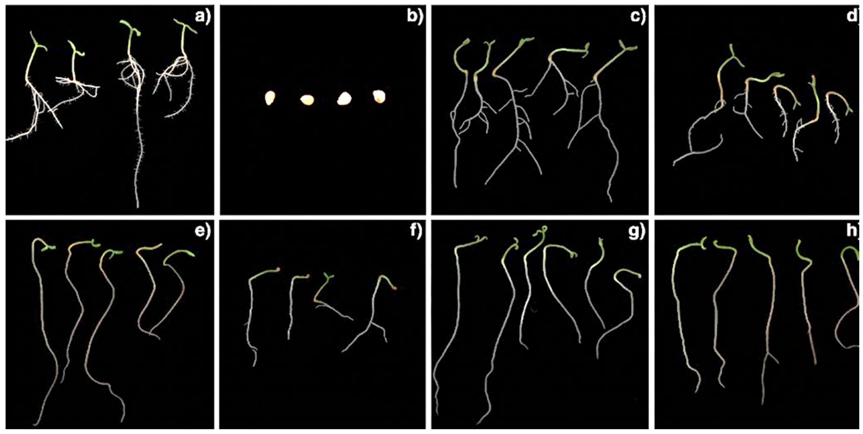
In the control treatment, 96% of S. lycopersicum seeds germinated, with a germination speed index (GSI) of 4.83, producing seedlings with an average primary root length of 1.76 cm, well-developed secondary roots, and a shoot length of 0.63 cm (Table 1; Figure 1a). In contrast, under salt stress conditions, S. lycopersicum seeds did not show germination throughout the experimental period (14 days), preventing seedling growth and, consequently, the measurement of morphological parameters (Table 1; Figure 1b). These results indicate that 100 mM NaCl imposed complete inhibition of germination, representing the most severe response observed across all treatments evaluated in this study.
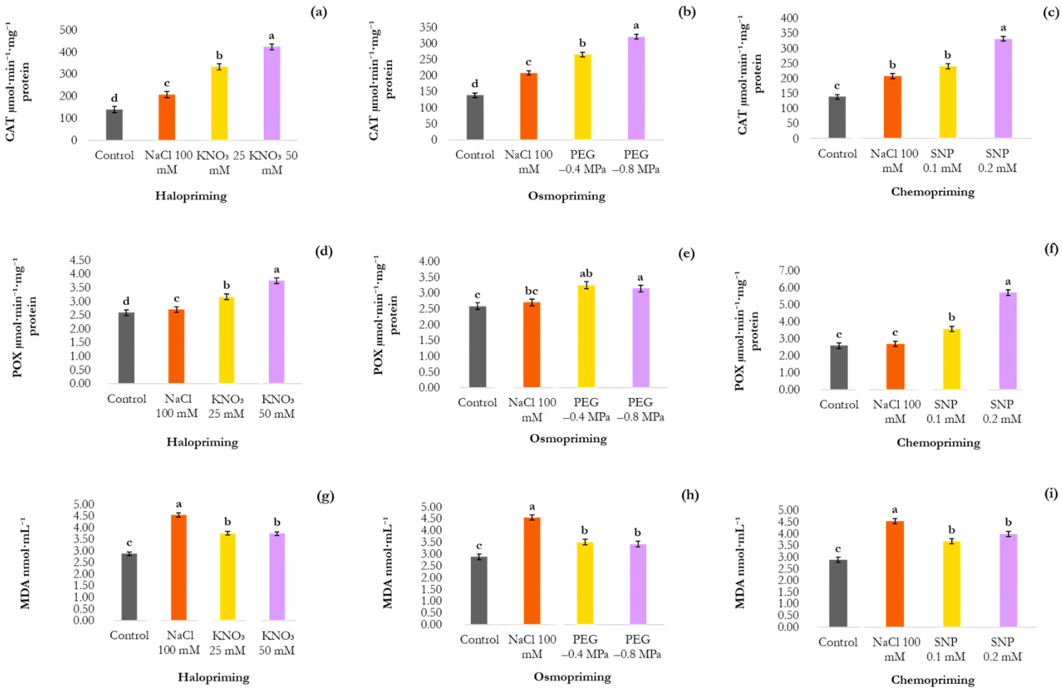
Under salt stress without priming, CAT activity increased by approximately 49% compared to the control, reaching 207.94 µmol·min−1·mg−1 protein, while POX activity showed a modest 4% increase, reaching 2.70 µmol·min−1·mg−1 protein. Despite this antioxidant response, the MDA content remained elevated, reaching 4.54 nmol·mL−1, a value approximately 58% higher than the control (2.87 nmol·mL−1), indicating that enzymatic antioxidant activity was insufficient to prevent lipid peroxidation under these conditions (Figure 2).

3.1.1. Germination, Initial Growth, Antioxidant Activity, and Lipid Peroxidation Under Halopriming (KNO3) in Salt Stress Conditions

In the treatment with halopriming using 25 mM KNO3, the seeds showed 56% germination and a GSI of 1.77, values significantly higher than those observed in seeds exposed to salt stress without treatment, in which no germination occurred. Halopriming with 50 mM KNO3 promoted germination responses closer to those observed in the control treatment, with 81% germination and a GSI of 3.75 (Table 1; Figure 1c,d). The seedlings resulting from seeds treated with 25 mM KNO3 showed an average root length of 4.86 cm and an average shoot length of 0.74 cm (Table 1; Figure 1c). Seedlings from seeds treated with 50 mM KNO3 exhibited an average root length of 4.57 cm and an average shoot length of 0.67 cm (Table 1; Figure 1d).
In the 25 mM treatment, CAT activity reached 332.87 µmol·min−1·mg−1 protein, corresponding to an increase of 60% compared to S. lycopersicum seeds under salt stress without treatment (207.94 µmol·min−1·mg−1 protein). However, at the concentration of 50 mM KNO3, an even more pronounced increase was observed, with activity reaching 423.24 µmol·min−1·mg−1 protein, representing an increase of 103% compared to salt stress without halopriming and 27% relative to the 25 mM treatment (Figure 2a). POX activity also increased following halopriming. At 25 mM KNO3, POX activity was 3.16 µmol·min−1·mg−1 protein, corresponding to an increase of 17% compared to salt stress without halopriming (2.70 µmol·min−1·mg−1 protein). At 50 mM, activity reached 3.75 µmol·min−1·mg−1 protein, representing increases of 38% and 18% compared to seeds without halopriming and those treated with 25 mM, respectively (Figure 2d).
MDA content showed a significant reduction in the KNO3 treatments, with reductions of 21% (3.75 nmol·mL−1) and 18% (3.74 nmol·mL−1) observed at 25 and 50 mM KNO3, respectively, compared to seeds under salt stress without halopriming (Figure 2g). These results indicate that halopriming with KNO3 effectively reduced lipid peroxidation and improved membrane integrity under salt stress conditions.

3.1.2. Germination, Initial Growth, Antioxidant Activity, and Lipid Peroxidation Under Osmopriming (PEG 6000) in Salt Stress Conditions

Osmopriming with PEG 6000 promoted partial recovery of germination under salt stress, with responses dependent on the osmotic potential applied. The treatment at −0.4 MPa resulted in 14% germination and a GSI of 0.50 (Table 1; Figure 1e), while −0.8 MPa yielded better performance, with 29% germination and a GSI of 1.00 (Table 1; Figure 1f). Regarding seedling growth, those from seeds treated at −0.4 MPa exhibited greater primary root length (6.61 cm), but without the formation of secondary roots, whereas −0.8 MPa resulted in a shorter root length (2.62 cm), with the emission of few secondary roots; shoot length was similar between treatments (Table 1; Figure 1e,f).
A significant increase in CAT activity was observed in seedlings derived from osmoprimed seeds. At −0.4 MPa, enzyme activity increased by 14% (265.01 µmol·min−1·mg−1 protein), and in the treatment with PEG at −0.8 MPa, the increase was even more pronounced, reaching 54% (320.79 µmol·min−1·mg−1 protein) compared to seeds exposed to salt stress without osmopriming; the latter value was 21% higher than the activity observed at −0.4 MPa (Figure 2b). POX activity was also stimulated by osmopriming. Seedlings from seeds treated with PEG at −0.4 MPa showed a 26% increase in POX activity (3.24 µmol·min−1·mg−1 protein) compared to salt stress without osmopriming. At −0.8 MPa, POX activity also increased, reaching 3.14 µmol·min−1·mg−1 protein, a 16% rise relative to the non-primed salt stress treatment (Figure 2e).
The MDA content decreased by 23% (3.50 nmol·mL−1) in seedlings derived from seeds subjected to osmopriming with PEG at −0.4 MPa. Similarly, treatment with PEG at −0.8 MPa promoted a reduction of approximately 25% (3.42 nmol·mL−1) (Figure 2h).

3.1.3. Germination, Initial Growth, Antioxidant Activity, and Lipid Peroxidation Under Chemopriming (SNP) in Salt Stress Conditions

Chemopriming with SNP promoted partial recovery of germination under salt stress, with 19% germination at 0.1 mM and 29% at 0.2 mM. Despite this recovery, germination values remained substantially below the control (96%), indicating that chemopriming partially mitigated, but did not fully reverse, the inhibitory effects of severe salt stress. The GSI showed values of 0.36 and 0.34 for 0.1 and 0.2 mM, respectively (Table 1).
Seedlings derived from SNP-primed seeds showed an average primary root length of 4.20 cm in the 0.1 mM treatment and 6.84 cm in the 0.2 mM treatment, with no formation of secondary roots. Average shoot length was 0.51 cm and 0.63 cm, respectively (Table 1; Figure 1g,h).
At 0.1 mM SNP, CAT activity increased by 15%, reaching 240.49 µmol·min−1·mg−1 protein compared to seeds under salt stress without chemopriming. POX activity was also stimulated, increasing by 32% to 3.57 µmol·min−1·mg−1 protein (Figure 2c,f). More pronounced physiological responses were observed at the concentration of 0.2 mM SNP. CAT activity increased to 332.15 µmol·min−1·mg−1 protein, corresponding to a 60% increase compared to the non-primed salt stress treatment and 38% higher than that observed at 0.1 mM. POX activity increased markedly by 111%, reaching 5.71 µmol·min−1·mg−1 protein, a value approximately 60% higher than that recorded for the lower concentration (Figure 2c,f).
MDA content decreased by 19% (3.68 nmol·mL−1) at 0.1 mM and by 12% (3.99 nmol·mL−1) at 0.2 mM SNP, with no statistical difference between concentrations, compared to seeds exposed to salt stress without treatment (4.54 nmol·mL−1) (Figure 2i).

3.2. Germination, Initial Growth, Antioxidant Activity, and Lipid Peroxidation Under Iron Toxicity

Under iron toxicity, the germination percentage of S. lycopersicum seeds was reduced to 79%, and the GSI to 3.78. These results contrast with the control treatment, in which 96% of the seeds germinated and the GSI reached 4.83 (Table 2).
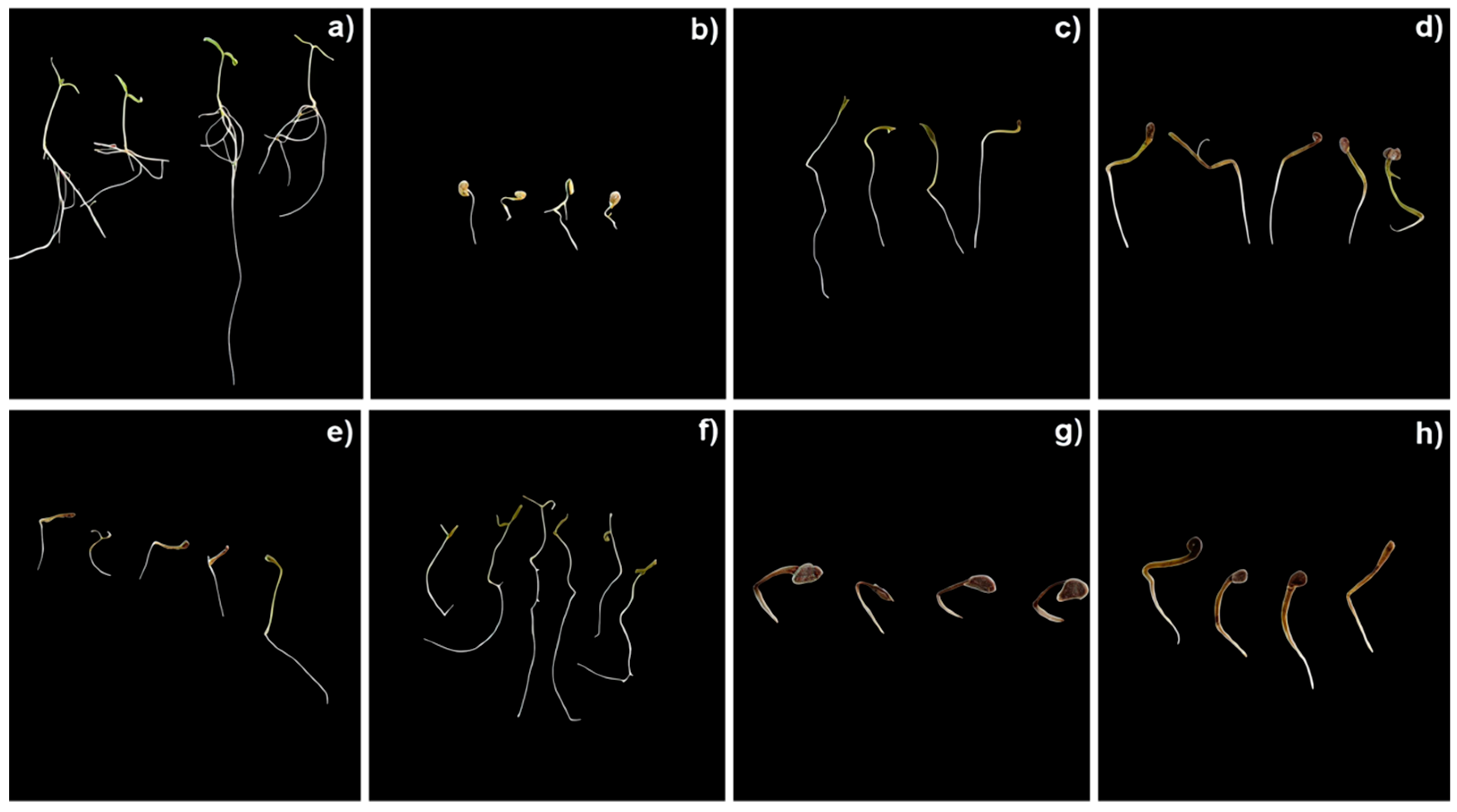
The initial development of the seedlings was severely impaired, as evidenced by the average primary root length of only 0.40 cm and the complete inhibition of shoot growth (Table 2). Furthermore, seedlings exposed to excess iron showed visible necrosis and root darkening (Figure 3b), indicative of iron deposition in root tissues and intensified oxidative reactions. In contrast, control seedlings developed a primary root of 1.76 cm, secondary roots, and a shoot averaging 0.63 cm in length (Table 2; Figure 3a).
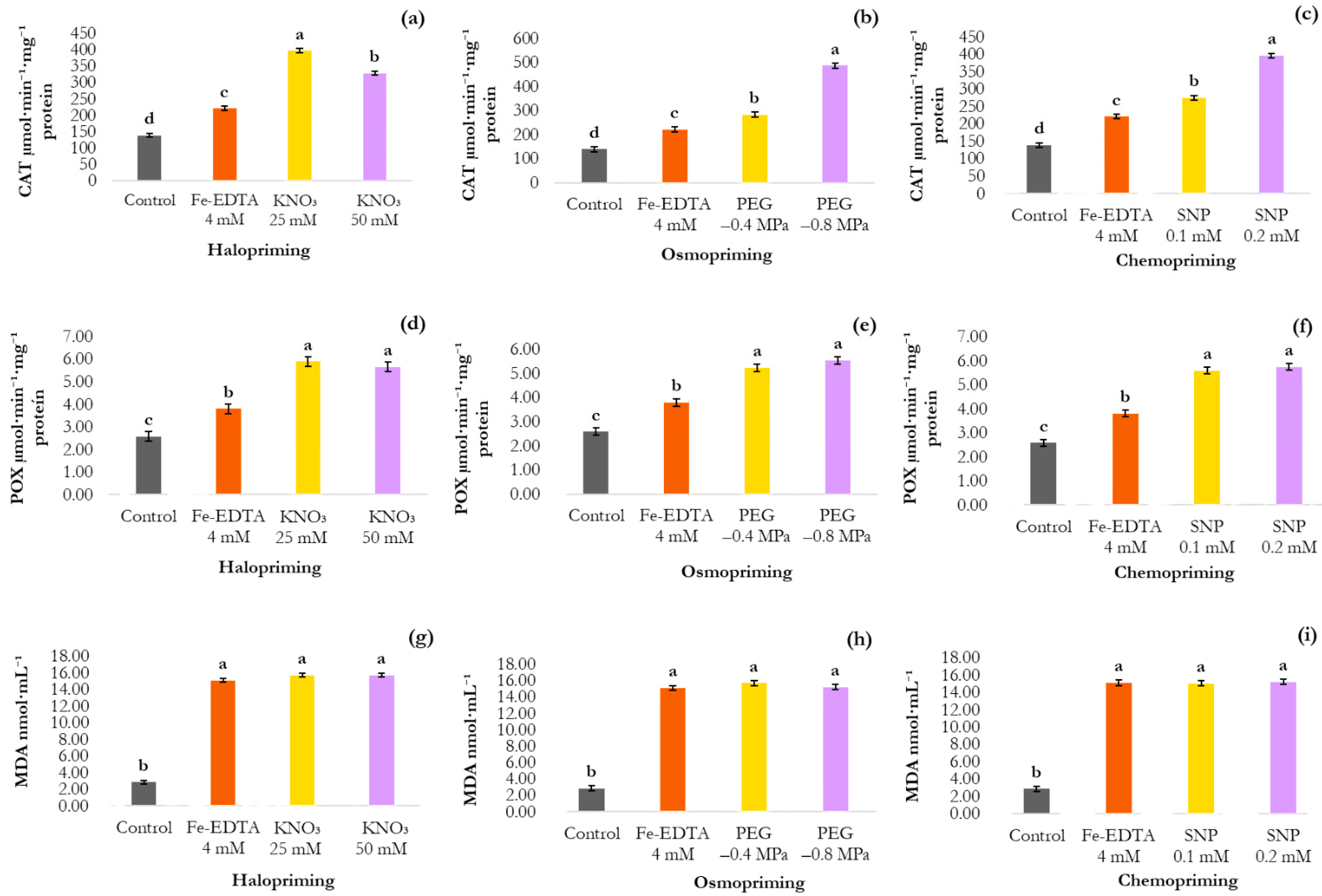
In seedlings exposed to iron toxicity, CAT activity increased by 59% compared to the control, reaching 221.40 µmol·min−1·mg−1 protein (138.87 µmol·min−1·mg−1 protein). POX also increased significantly by 47%, from 2.58 to 3.80 µmol·min−1·mg−1 protein (Figure 4). However, despite activation of both enzymes, MDA content increased by approximately 425% relative to the control (2.87 nmol·mL−1), reaching 15.10 nmol·mL−1, indicating that enzymatic antioxidant capacity was insufficient to prevent extensive lipid peroxidation under iron toxicity (Figure 4).

3.2.1. Germination, Initial Growth, Antioxidant Activity, and Lipid Peroxidation Under Osmopriming (PEG 6000) in Iron Toxicity Conditions

Osmopriming with PEG 6000 improved germination under iron toxicity at both osmotic potentials evaluated. At −0.4 MPa, germination reached 88%and the GSI 6.78, values exceeding those recorded under iron toxicity without treatment (79%; GSI 3.78) and the control (GSI 4.83). At −0.8 MPa, performance was even more pronounced, with 93% germination and a GSI of 7.03, corresponding to increases of 17.7% and 86%, respectively, relative to the non-primed iron toxicity treatment (Table 2).
Seedlings from seeds treated with PEG at −0.4 MPa showed an average root length of 1.27 cm and shoot recovery to 0.61 cm, a value close to the control (Table 2; Figure 3e). At −0.8 MPa, root length increased to 1.49 cm, with a shoot averaging 0.60 cm (Table 2; Figure 3f). However, neither treatment promoted the development of secondary roots.
CAT activity increased to 283.17 µmol·min−1·mg−1 protein at −0.4 MPa, representing a 27% increase compared to seedlings under iron toxicity without osmopriming (221.40 µmol·min−1·mg−1 protein). At −0.8 MPa, CAT activity reached 487.29 µmol·min−1·mg−1 protein, approximately 120% higher than in non-primed seedlings and 72% higher than at −0.4 MPa (Figure 4b). POX activity also increased significantly, reaching 5.23 µmol·min−1·mg−1 protein at −0.4 MPa (37% increase) and 5.54 µmol·min−1·mg−1 protein at −0.8 MPa (46% increase), compared to the non-primed iron toxicity treatment (3.80 µmol·min−1·mg−1 protein) (Figure 4e).
Despite the marked increase in antioxidant enzyme activities, MDA content remained elevated in both PEG treatments with values of 15.70 nmol·mL−1 at −0.4 MPa and 15.21 nmol·mL−1 at −0.8 MPa, neither differing statistically from the non-primed iron toxicity group (15.11 nmol·mL−1) (Figure 4h).

3.2.2. Germination, Initial Growth, Antioxidant Activity, and Lipid Peroxidation Under Halopriming (KNO3) in Iron Toxicity Conditions

Halopriming with KNO3, at both evaluated concentrations (25 and 50 mM), significantly enhanced the germination under iron toxicity, reaching 93% at 25 mM and 92% at 50 mM. The GSI reached 9.01 at 25 mM and 7.96 at 50 mM, both significantly higher than values recorded in seeds under iron toxicity without halopriming (3.52) and in the control treatment (4.83) (Table 2; Figure 3c,d).
The average root length reached 2.18 cm at 25 mM KNO3 and 0.99 cm at 50 mM, both significantly greater than in seeds under iron toxicity without halopriming (0.40 cm); however, no secondary roots were formed. Shoot development showed partial recovery, with averages of 0.64 cm at 25 mM and 0.55 cm at 50 mM, whereas seedlings under iron toxicity without halopriming showed no shoot development (Table 2; Figure 3c,d).
CAT activity increased significantly, reaching 398.04 µmol·min−1·mg−1 protein at 25 mM and 328.68 µmol·min−1·mg−1 protein at 50 mM, corresponding to increases of 80% and 48%, respectively, compared to seeds exposed to iron toxicity without treatment (221.40 µmol·min−1·mg−1 protein). CAT activity at 25 mM was approximately 21% higher than at 50 mM (Figure 4a). POX activity was also stimulated, reaching 5.90 µmol·min−1·mg−1 protein at 25 mM and 5.65 µmol·min−1·mg−1 protein at 50 mM, representing increases of 55% and 48%, respectively (Figure 4d).
Despite this antioxidant activation, MDA levels remained high at both KNO3 concentrations with values of 15.70 nmol·mL−1 at both 25 mM and 50 mM, not differing statistically from the non-primed iron toxicity group (15.11 nmol·mL−1) (Figure 4g). This result suggests that the activation of antioxidant defenses was not sufficient to contain the oxidative damage induced by iron stress.

3.2.3. Germination, Initial Growth, Antioxidant Activity, and Lipid Peroxidation Under Chemopriming (SNP) in Iron Toxicity Conditions

Chemopriming with SNP increased germination and GSI compared to seeds under iron toxicity without treatment. At 0.1 mM, germination reached 90% and GSI 6.90; at 0.2 mM, germination was 81% and GSI 6.43, both significantly higher than the non-primed iron toxicity group (79%; GSI 3.78) (Table 2).
All seedlings derived from seeds under iron toxicity, with or without chemopriming, showed a drastic reduction in root length (0.37 to 0.44 cm), representing an approximately 75% reduction compared to the control (1.76 cm), with no statistical differences among treatments. A complete absence of shoot development was observed, regardless of the chemopriming application (Table 2; Figure 3g,h).
Chemopriming with SNP increased CAT activity in seedlings exposed to iron toxicity, reaching 275.44 µmol·min−1·mg−1 protein at 0.1 mM and 396.17 µmol·min−1·mg−1 protein at 0.2 mM, corresponding to increases of 24% and 78%, respectively, compared to non-primed seedlings (221.40 µmol·min−1·mg−1 protein). The higher concentration promoted an additional 43% increase in CAT activity relative to 0.1 mM (Figure 4c). POX activity was also stimulated, reaching 5.59 µmol·min−1·mg−1 protein at 0.1 mM and 5.75 µmol·min−1·mg−1 protein at 0.2 mM, representing increases of 47% and 51%, respectively (Figure 4f).
Despite the activation of CAT and POX, MDA levels remained elevated and statistically similar to those of non-primed iron-stressed seedlings, with values of 15.05 nmol·mL−1 at 0.1 mM and 15.19 nmol·mL−1 at 0.2 mM (non-primed: 15.10 nmol·mL−1) (Figure 4i).

4. Discussion

4.1. Germination, Initial Growth, Antioxidant Activity, and Lipid Peroxidation Under Salt Stress and After Priming Treatments

Seed germination depends on the activation of hydrolytic enzymes, especially α-amylase, which is responsible for the degradation of starch stored in the endosperm into soluble sugars required for embryo energy supply and radicle protrusion [40]. The complete germination inhibition observed under 100 mM NaCl is consistent with ROS-mediated oxidative damage to cellular macromolecules, reducing enzyme activity and limiting reserve mobilization, a process essential for germination establishment [41,42].
The inhibition of germination under saline conditions also reflects the reduction in external osmotic potential, which restricts water uptake during imbibition [43,44], combined with the direct cytotoxic effects of Na+ and Cl accumulation in the embryo [45,46]. These findings are consistent with previous observations in Solanum melongena, in which NaCl exposure significantly reduced germination percentage and impaired early seedling growth in a cultivar-dependent manner [47]. The severity of the response observed in the present study underscores the particular susceptibility of the germination stage relative to subsequent developmental stages.
The co-occurrence of elevated CAT and POX activities alongside high MDA levels in salt-stressed non-primed seeds indicates that constitutive enzyme upregulation was insufficient to counteract ROS generation under severe ionic stress, allowing lipid peroxidation to persist and contributing to germination failure. Similar responses have been reported in tomato and quinoa under saline conditions [48,49]. This threshold effect underscores the rationale for seed priming as a strategy to pre-activate antioxidant networks before stress exposure.
Halopriming with KNO3 was the most effective priming strategy under salt stress, restoring germination most completely at 50 mM. This effect is likely mediated by NO production—stimulating hydrolytic enzyme activity and reserve mobilisation [50,51,52]—alongside K+-driven osmotic homeostasis, NO3 as a nitrogen source [53], and modulation of the ABA/GA ratio favouring radicle protrusion [54]. The dose-dependent superiority of 50 mM over 25 mM KNO3 suggests a concentration threshold for NO production and K+ availability sufficient to overcome salinity-imposed metabolic arrest.
These findings are consistent with previous observations in Zea mays (maize) and Oryza sativa (rice) and highlight the multiple mechanisms through which KNO3 enhances salt stress tolerance, including osmotic adjustment, reserve mobilization, hormonal signaling, and antioxidant defense. This integrated mode of action may explain its superiority over osmopriming and chemopriming under saline conditions in the present study [53,55].
The upregulation of CAT and POX in KNO3-primed seedlings is mechanistically linked to nitrate reduction by nitrate reductase, generating NO and thereby reinforcing ROS homeostasis [56,57]. Critically, the concomitant MDA reduction—absent in non-primed salt-stressed seeds despite comparable enzyme activation—confirms that halopriming renders antioxidant upregulation functionally effective, not merely quantitatively elevated. This behavior is consistent with findings in Hibiscus cannabinus after KNO3 priming [58].
Osmopriming with PEG 6000 promoted moderate germination recovery under salt stress, with responses dependent on osmotic potential. This osmotic-potential dependency suggests that the degree of metabolic pre-activation during priming directly influences subsequent stress tolerance capacity. PEG osmopriming has been associated with enhanced stress tolerance through osmolyte accumulation, maintenance of ionic homeostasis (reduced Na+ and increased K+ levels), and stimulation of the antioxidant system. Similar results have been observed in Solanum lycopersicum cv. ‘Micro-Tom’ subjected to osmopriming at −1.2 MPa [8,59].
The divergent secondary root formation patterns between PEG osmotic potentials—primary root elongation without lateral roots at −0.4 MPa versus shorter primary root with limited lateral roots at −0.8 MPa—indicate that salinity overrides priming-induced root organogenesis signals regardless of the potential applied. This reflects salinity-elevated ABA suppressing auxin-dependent lateral root initiation [60,61], compounded by SOS3–SOS2–SOS1 pathway dysregulation under excess Na+ [62,63]. These observations identify secondary root development as a salinity-sensitive parameter not fully rescued by priming, and point to auxin signalling and ionic homeostasis as priority targets for future research.
Osmopriming promoted significant reinforcement of the antioxidant system, accompanied by reductions in lipid peroxidation and electrolyte leakage, indicating improved membrane integrity [64]. Similar responses have been observed in Chenopodium quinoa (quinoa) and Solanum lycopersicum (tomato) cv. ‘Micro-Tom’ [8,49]. These findings suggest that membrane protection may represent a common mechanism associated with PEG-based osmopriming across different species.
Chemopriming with SNP produced the most limited germination recovery under salinity. Although SNP was the least effective among the three strategies, the partial recovery of germination demonstrates that exogenous NO can partially overcome salinity-induced inhibition through the stimulation of hydrolytic enzymes, reduction in ABA levels, and activation of GA signaling pathways [65]. These mechanisms have also been confirmed through the use of the NO scavenger cPTIO [66].
The inverse relationship between SNP concentration and primary root length is consistent with the known biphasic, concentration-dependent effects of NO on root growth [67], while the universal absence of secondary roots reflects salinity-mediated disruption of auxin-dependent root organogenesis, that NO signalling alone cannot overcome [68]. This decoupling between primary root promotion and lateral root organogenesis represents a key limitation of SNP chemopriming under salinity.
The concentration-dependent increase in CAT and POX activities promoted by SNP is mechanistically consistent with the role of NO in regulating redox homeostasis [65]. Similar responses have also been reported in Triticum aestivum (wheat) under saline conditions [28]. However, the modest germination recovery relative to KNO3 despite comparable enzyme activation suggests that antioxidant upregulation alone does not determine germination recovery under severe salinity—the additional osmotic and nitrogen-related benefits of KNO3 are critical for overcoming combined osmotic and ionic stress components.

4.2. Germination, Initial Growth, Antioxidant Activity, and Lipid Peroxidation Under Iron Toxicity and After Priming Treatments

Unlike salinity, which completely suppressed germination, iron toxicity induced a partial reduction in germination and GSI, consistent with a mechanistically distinct mode of stress imposition. Excess Fe2+ has been shown to reduce α-amylase activity, limit the energy supply required for germination, delay radicle emergence, and promote ROS overproduction, which in turn induces membrane lipid peroxidation and, under severe conditions, may lead to cell death. The partial rather than complete suppression of germination under iron toxicity likely reflects the fact that Fenton-driven oxidative damage, although severe, lacks the combined osmotic and ionic constraints characteristic of salinity, which together create a more restrictive environment for germination [10].
The severe impairment of primary root growth, complete absence of shoot development, and characteristic root necrosis and darkening in non-primed iron-stressed seedlings indicate that iron toxicity targets root meristematic tissues with particular intensity. Similar responses have been reported in Carica papaya (papaya) and Solanum lycopersicum (tomato) cv. ‘Marglobe’ [12,32]. Excess iron in the root apoplast generates ROS and promotes iron deposition, cellular necrosis, and increased oxidative reactions [69]. Apoplastic iron deposition may additionally create a physical diffusion barrier interfering with nutrient and water uptake, compounding metabolic dysfunction.
The paradox of elevated antioxidant enzyme activities alongside dramatically increased MDA in iron-stressed seedlings is central to understanding why all priming strategies failed to fully restore seedling growth. When ROS production exceeds antioxidant detoxification capacity, membrane integrity is severely compromised [12], and maintaining redox homeostasis becomes essential for cell survival [70]. This paradox reflects the self-sustaining nature of Fenton chemistry: Fe2+ catalyses ·OH production from H2O2, oxidises biological molecules, and regenerates Fe2+—a cycle that overwhelms enzymatic scavenging regardless of its magnitude. This mechanistic distinction has a critical practical implication: unlike salinity, iron toxicity creates an oxidative environment inherently resistant to enzyme-based mitigation, requiring strategies that directly limit iron bioavailability.
PEG osmopriming improved germination and GSI under iron toxicity, with the more negative potential (−0.8 MPa) yielding values that exceeded the control. This suggests that osmotic pre-activation accelerates germination-competent enzymatic pathways before toxic iron concentrations can fully suppress them—and that priming can positively accelerate germination kinetics, not merely mitigate inhibition, under metal stress. PEG osmopriming in Oryza sativa seeds subjected to ZnO stress increased α-amylase activity, soluble sugar content, and glutathione reductase activity, while reducing ROS production, providing a mechanistic parallel to the results observed in the present study [71].
The partial recovery of root and shoot development alongside the persistent absence of secondary roots in PEG-primed iron-stressed seedlings defines a clear boundary condition of osmopriming: primary organ development and germination kinetics can be partially rescued, but secondary root organogenesis cannot Cadmium stress inhibits lateral root emergence by disrupting OsPIN-mediated auxin distribution and root developmental signaling [72]. In addition, PEG treatment has been reported to be ineffective in preventing iron-induced ultrastructural damage in meristematic cells [71]. These findings suggest that the auxin transport machinery for lateral root initiation is particularly vulnerable to heavy metal disruption—a limitation not overcome by osmopriming alone. Future studies should investigate whether co-application of PEG priming with exogenous auxin supplementation restores secondary root formation under iron toxicity.
The marked upregulation of CAT and POX by PEG osmopriming under iron toxicity, without a corresponding MDA reduction, provides direct evidence for Fenton chemistry as the dominant ROS driver. Antioxidant upregulation in the absence of a corresponding reduction in MDA levels indicates continuous Fe-driven ROS generation that exceeds the detoxification capacity of the enzymatic antioxidant system [11]. This suggests that effective mitigation of iron toxicity requires not only reinforcement of antioxidant defenses but also strategies that directly target iron bioavailability, objectives that cannot be achieved through priming alone.
KNO3 halopriming significantly increased germination and the germination speed index (GSI) under iron toxicity, with values exceeding those of the control. This effect is consistent with responses observed under salt stress and is associated with nitrate metabolism, including its role as a nitrogen source, activation of germination-related enzymes, and improved physiological performance of seeds [50,53]. In addition, the literatures describe that priming processes may involve hormonal regulation associated with ABA and GA pathways, as well as NO-dependent enzymatic mechanisms in germination responses under stress conditions [50,53]. The generalised germination acceleration by KNO3 across both mechanistically distinct stresses reinforces that halopriming induces broadly competent germination-phase protection—however, this does not translate to equivalent seedling protection, revealing phase-specific vulnerabilities that priming cannot overcome. The concentration-dependent divergence in root length—greater recovery at 25 than 50 mM under iron toxicity—suggests that optimal KNO3 concentration may differ between stresses, possibly reflecting interactions between nitrate metabolism and iron-induced oxidative signalling that warrant further investigation.
The persistent absence of secondary roots at both concentrations indicates a strong limitation in lateral root establishment under metal stress [69,73,74]. This pattern has already been reported under iron and other metal toxicity conditions, in which lateral root growth and formation are severely impaired [73,74]. Moreover, seed priming studies indicate that processes related to early development and root establishment may exhibit a low capacity for recovery under stress conditions [75]. Even with the increase in enzymatic activity, MDA levels remained elevated [76], indicating that the antioxidant system was not sufficient to prevent lipid peroxidation under iron-induced oxidative stress, associated with the generation of reactive oxygen species via Fenton reactions [74].
SNP chemopriming improved germination and GSI under iron toxicity through the elevation of endogenous NO levels, enhanced reserve mobilization, and reinforcement of antioxidant defenses, consistent with observations reported for Triticum aestivum (trigo) under similar conditions [10].
However, the complete failure of SNP to rescue root growth and shoot development, despite antioxidant enzyme upregulation, reveals a specific vulnerability of root meristematic tissue to iron-mediated damage that transcends priming capacity. This outcome reflects the dual role of NO under heavy metal stress [77]: NO signalling activates antioxidant responses, but under iron-rich conditions, NO reacts with O2 to form peroxynitrite (ONOO) [78], intensifying lipid oxidation, promoting protein nitration, and inactivating enzymes.
This NO–peroxynitrite conversion is particularly relevant to SNP chemopriming: exogenous NO may paradoxically exacerbate oxidative damage under iron excess by providing substrate for peroxynitrite formation, partially antagonising its beneficial signalling effects—a mechanistic conflict that may explain why SNP was the least effective strategy for protecting seedling growth under iron toxicity. The persistent elevation of MDA across all priming strategies under iron toxicity, despite consistent antioxidant enzyme upregulation, collectively confirms that sustained Fenton-driven ROS generation and peroxynitrite-mediated cytotoxicity create an oxidative threshold that enzyme-based priming cannot overcome. Future strategies should therefore integrate priming with iron bioavailability-reducing approaches—seed coating with chelating agents, soil amendment with iron-immobilising compounds, or genotype selection for enhanced root-surface iron exclusion—to achieve meaningful protection under iron toxicity conditions.

5. Conclusions

Salinity stress and iron toxicity impaired seed germination, early seedling growth, and redox homeostasis in S. lycopersicum. Priming activated the antioxidant system under both conditions; however, its physiological efficacy was stress-type dependent. Under salinity stress, halopriming with KNO3 emerged as the most effective strategy. The 50 mM concentration showed superior performance in restoring germination, seed vigor, and redox balance, whereas the 25 mM concentration was more effective in promoting seedling growth.
Under iron toxicity, all priming treatments consistently increased antioxidant enzyme activity; however, none were capable of reducing lipid peroxidation or restoring seedling growth to control levels. These results suggest that conventional priming strategies are insufficient to counteract iron-mediated oxidative damage, likely due to the continuous generation of hydroxyl radicals via Fenton-type reactions and peroxynitrite formation, which collectively overwhelm enzymatic detoxification capacity. Future studies should investigate combined approaches—such as co-application of metal chelators or non-enzymatic antioxidants alongside conventional priming agents—to improve seedling protection under iron toxicity conditions. It should be noted that this study was conducted using a single commercial tomato accession, which limits the generalization of the findings to the species as a whole, given the broad genetic variability known to exist in germination responses to abiotic stresses within S. lycopersicum germplasm. Future studies should evaluate the differential responses of multiple genotypes—including contrasting cultivars for salt and iron tolerance—to establish more comprehensive recommendations for seed priming strategies under these stress conditions.

Supplementary Materials

The following supporting information can be downloaded at https://www.mdpi.com/article/10.3390/seeds5040043/s1, Supplementary File S1: Raw data and mean values of biochemical analyses. Supplementary File S2: Raw data and mean values of germination parameters and seedling growth traits. Supplementary File S3: Raw supplementary footage.

Author Contributions

A.N.F.: Conceptualization, methodology, investigation, data analysis, antioxidant enzyme and malondialdehyde (MDA) analyses, and writing—original draft. V.B.C.: Supervision, project administration, and writing—review and editing. H.S.F.: Contribution to antioxidant enzyme analyses and provision of laboratory facilities and equipment. J.R.F.: Supervision, methodology, project administration, and writing—review and editing. M.R.P.: Contribution to antioxidant enzyme, malondialdehyde (MDA), and protein analyses. All authors have read and agreed to the published version of the manuscript.

Funding

This work was funded by the National Council for Scientific and Technological Development (CNPq) (Grant No. 178270/2024-5) and Instituto Federal do Espírito Santo (IFES) by PRODIF founding 10/2026.

Institutional Review Board Statement

Not applicable.

Informed Consent Statement

Not applicable.

Data Availability Statement

The original data presented in this study are publicly available in the Figshare repository and can be accessed via the DOI: https://doi.org/10.6084/m9.figshare.32061051.

Acknowledgments

The authors acknowledge the Bioproduct Development Laboratory, Federal Institute of Espírito Santo, campus Vila Velha, for technical and infrastructure support. The authors also acknowledge the use of the PicWish application for image quality enhancement.

Conflicts of Interest

The authors declare no conflict of interest.

References

  1. Rawat, A.; Upadhyay, M.; Singh, O. Exploring the pharmacological potential and traditional use of Solanum lycopersicum L. (tomato): A review. J. Pharmacogn. Phytochem. 2024, 13, 768–771. [Google Scholar] [CrossRef] [Scilit]
  2. Brazilian Institute of Geography and Statistics (IBGE). Tomato: Agricultural Production; IBGE: Rio de Janeiro, Brazil, 2025. Available online: https://www.ibge.gov.br/explica/producao-agropecuaria/tomate/rj (accessed on 27 October 2025).
  3. Batista, T.B.L. Longevity of Primed Tomato Seeds: Physiological and Molecular Studies. Master’s Thesis, São Paulo State University (UNESP), Botucatu, Brazil, 2018. Available online: https://repositorio.unesp.br/server/api/core/bitstreams/895317f0-5f88-4ab9-be92-4f5341b9ca3b/content (accessed on 27 October 2025).
  4. CONAB. Tomato: Analysis of Production and Marketing Indicators in the Global, Brazilian, and Santa Catarina Markets; CONAB: Brasília, Brazil, 2019.
  5. Seth, R. An overview on impact of salinity stress in tomato under in vitro conditions. Int. J. Curr. Microbiol. Appl. Sci. 2024, 13, 191–201. [Google Scholar] [CrossRef] [Scilit]
  6. Stavi, I.; Thevs, N.; Priori, S. Soil salinity and sodicity in drylands: A review of causes, effects, monitoring, and restoration measures. Front. Environ. Sci. 2021, 9, 712831. [Google Scholar] [CrossRef] [Scilit]
  7. Kaushal, S.S.; Likens, G.E.; Mayer, P.M.; Shatka, R.R.; Shelton, S.A.; Grant, S.B.; Utz, R.M.; Yaculak, A.M.; Maas, C.M.; Reimer, J.E.; et al. The anthropogenic salt cycle. Nat. Rev. Earth Environ. 2023, 4, 770–784. [Google Scholar] [CrossRef] [Scilit] [PubMed]
  8. Habibi, N.; Aryan, S.; Sediqui, N.; Terada, N.; Sanada, A.; Kamata, A.; Koshio, K. Enhancing salt tolerance in tomato plants through PEG6000 seed priming. Plants 2025, 14, 1296. [Google Scholar] [CrossRef] [Scilit] [PubMed]
  9. Harish, V.; Aslam, S.; Chouhan, S.; Pratap, Y.; Lalotra, S. Iron toxicity in plants: A review. Int. J. Environ. Clim. Chang. 2023, 13, 1894–1900. [Google Scholar] [CrossRef] [Scilit]
  10. Shi, S.; Zhang, Z.; Ma, R.; Tao, Y.; Wang, Z.; Yang, Y. Exogenous nitric oxide relieves iron-inhibitory effect on wheat seed germination. Ecotoxicol. Environ. Saf. 2025, 291, 117875. [Google Scholar] [CrossRef] [Scilit] [PubMed]
  11. Lapaz, A.M.; Yoshida, C.H.P.; Gorni, P.H.; Freitas-Silva, L.; Araújo, T.O.; Ribeiro, C. Iron toxicity: Effects on plants and detoxi-fication strategies. Acta Bot. Bras. 2022, 36, e2021abb0131. [Google Scholar] [CrossRef] [Scilit]
  12. Das, U.; Rahman, M.M.; Roy, Z.R.; Kabir, A.H. Morpho-physiological retardations due to iron toxicity involve redox imbal-ance rather than photosynthetic damages in tomato. Plant Physiol. Biochem. 2020, 156, 55–63. [Google Scholar] [CrossRef] [Scilit] [PubMed]
  13. Sahoo, L.; Swain, B.; Yadav, D. A review on different priming strategies to mitigate abiotic stress in plants. Discov. Appl. Sci. 2024, 7, 618. [Google Scholar] [CrossRef] [Scilit]
  14. Fomekong, M.K.; Tetang, E.F.T.; Temegne, C.N.; Minlo, J.U.E.; Atabong, P.A.; Kameni, A.C.N.; Mir, B.A.; Youmbi, E.; Tonfack, L.B. Effect of osmopriming with PEG 6000 on seed germination performance in okra. Discov. Plants 2025, 2, 292. [Google Scholar] [CrossRef] [Scilit]
  15. Da Rocha, L.G.; Rau, B.A.; Silva, D.M.; Araújo, R.M.; Masetto, T.E. Potassium nitrate (KNO3) seed priming enhances soybean seed performance. Sci. Rep. 2025, 15, 33640. [Google Scholar] [CrossRef] [Scilit] [PubMed]
  16. Fejes, G.; Bodor, T.; Szőllősi, R.; Kondak, S.; Kutasi, K.; Fotopoulos, V.; Kolbert, Z. Nitric oxide as an integral element in prim-ing-induced tolerance. J. Exp. Bot. 2025, 76, 3669–3685. [Google Scholar] [CrossRef] [Scilit] [PubMed]
  17. Singh, N.; Maurya, V.; Gupta, K.; Sharma, I.; Sharma, A.; Kumar, R. Salt stress and its eco-friendly management in legumes. Discov. Agric. 2025, 3, 13. [Google Scholar] [CrossRef] [Scilit]
  18. Abbas, F.; Al-Naemi, S. Managing salinity stress through microclimate control to enhance tomato productivity in arid regions. Sci. Rep. 2026, 16, 13042. [Google Scholar] [CrossRef] [Scilit] [PubMed]
  19. Habibi, N.; Terada, N.; Sanada, A.; Koshio, K. Alleviating salt stress in tomatoes through seed priming with polyethylene glycol and sodium chloride combination. Stresses 2024, 4, 210–224. [Google Scholar] [CrossRef] [Scilit]
  20. Ellouzi, H.; Ben Slimene Debez, I.; Amraoui, S.; Rabhi, M.; Hanana, M.; Alyami, N.M.; Debez, A.; Abdelly, C.; Zorrig, W. Effect of seed priming with auxin on ROS detoxification and carbohydrate metabolism and their relationship with germination and early seedling establishment in salt stressed maize. BMC Plant Biol. 2024, 24, 704. [Google Scholar] [CrossRef] [Scilit] [PubMed]
  21. Karimi, M.R.; Sabokdast, M.; Korang Beheshti, H.; Abbasi, A.R.; Bihamta, M.R. Seed priming with salicylic acid enhances salt stress tolerance by boosting antioxidant defense in Phaseolus vulgaris genotypes. BMC Plant Biol. 2025, 25, 489. [Google Scholar] [CrossRef] [Scilit] [PubMed]
  22. Aung, M.S.; Masuda, H. How does rice defend against excess iron? Physiological and molecular mechanisms. Front. Plant Sci. 2020, 11, 1102. [Google Scholar] [CrossRef] [Scilit] [PubMed]
  23. Karim, M.R.; Sultana, S.; Altaf-Un-Nahar, M.; Islam, M.R.; Rahman, F.; Pretha, S.J.; Azam, M.G.; Hussain, S.; Yang, X.; Ibrahimova, U.; et al. Integrated Molecular Defense Mitigating Salt Stress in Tomatoes Using Synergistic Signaling Molecules. Physiol. Plant. 2025, 177, e70344. Available online: https://pubmed.ncbi.nlm.nih.gov/40536099/ (accessed on 15 September 2025). [CrossRef] [Scilit]
  24. Amooaghaie, R.; Nikzad, K. Role of nitric oxide in priming-induced low-temperature tolerance in two genotypes of tomato. Seed Sci. Res. 2013, 23, 123–131. [Google Scholar] [CrossRef] [Scilit]
  25. Pallaoro, D.S.; Camili, E.C.; Guimarães, S.C.; Albuquerque, M.C.D.F. Methods for priming maize seeds. J. Seed Sci. 2016, 38, 148–154. [Google Scholar] [CrossRef] [Scilit]
  26. Villela, F.A.; Doni-Filho, L.; Sequeira, E.L. Table of osmotic potential as a function of polyethylene glycol 6000 concentration and temperature. Pesqui. Agropecuária Bras. 1991, 26, 1957–1968. [Google Scholar]
  27. Ahmad, I.; Saleem, A.M.; Mustafa, G.; Ziaf, K.; Afzal, I.; Qasim, M. Seed halopriming enhances germination performance and seedling vigor of Gerbera jamesonii and Zinnia elegans. Sarhad J. Agric. 2017, 33, 199–205. [Google Scholar] [CrossRef] [Scilit]
  28. Ali, Q.; Daud, M.K.; Haider, M.Z.; Ali, S.; Rizwan, M.; Aslam, N.; Noman, A.; Iqbal, N.; Shahzad, F.; Deeba, F.; et al. Seed priming by sodium nitroprusside improves salt tolerance in wheat (Triticum aestivum L.) by enhancing physiological and biochemical parameters. Plant Physiol. Biochem. 2017, 119, 50–58. [Google Scholar] [CrossRef] [Scilit] [PubMed]
  29. Farooq, M.; Aziz, T.; ur Rehman, H.; ur Rehman, A.; Cheema, S.A. Evaluating surface drying and re-drying for wheat seed priming with polyamines: Effects on emergence, early seedling growth and starch metabolism. Acta Physiol. Plant. 2011, 33, 1707–1713. [Google Scholar] [CrossRef] [Scilit]
  30. Iradukunda, M.; van Iersel, M.W.; Seymour, L.; Lu, G.; Ferrarezi, R.S. Automated Imaging to Evaluate the Exogenous Gibberellin (Ga3) Impact on Seedlings from Salt-Stressed Lettuce Seeds. Sensors 2024, 24, 4228. [Google Scholar] [CrossRef] [Scilit] [PubMed]
  31. Ferreira, A.G.; Borghetti, F. Germination: From Basic to Applied; Artmed: Porto Alegre, Brazil, 2005; Available online: https://www.researchgate.net/publication/335078531_Germinacao_Do_basico_ao_aplicado (accessed on 13 November 2025).
  32. Rodrigues Filho, J.; Corte, V.B.; Perin, I.T.; Freitas, J.F.N.; Waichert, R.H.; dos Santos, C.R. Effects of Iron on oxidative stress of Cecropia hololeuca and Carica papaya plants. An. Acad. Bras. Cienc. 2022, 94, e20210154. [Google Scholar] [CrossRef] [Scilit] [PubMed]
  33. Ministry of Agriculture, Livestock and Food Supply (MAPA). Rules for Seed Testing; MAPA/ACS: Brasília, Brazil, 2009.
  34. Maguire, J.D. Speed of germination—Aid in selection and evaluation for seedling emergence and vigor. Crop Sci. 1962, 2, 176–177. [Google Scholar] [CrossRef] [Scilit]
  35. Bradford, M.M. A rapid and sensitive method for the quantitation of microgram quantities of protein utilizing the principle of protein-dye binding. Anal. Biochem. 1976, 72, 248–254. [Google Scholar] [CrossRef] [PubMed]
  36. Aebi, H. Catalase in vitro. Methods Enzymol. 1984, 105, 121–126. [Google Scholar] [CrossRef] [Scilit] [PubMed]
  37. Chance, B.; Maehly, A.C. Assay of catalases and peroxidases. Methods Enzymol. 1955, 2, 764–775. [Google Scholar] [CrossRef] [Scilit]
  38. Cakmak, I.; Horst, W.J. Effect of aluminium on lipid peroxidation, superoxide dismutase, catalase, and peroxidase activi-ties in root tips of soybean (Glycine max). Physiol. Plant. 1991, 83, 463–468. [Google Scholar] [CrossRef] [Scilit]
  39. Ferreira, D.F. Sisvar: A computer statistical analysis system. Cienc. Agrotec. 2011, 35, 1039–1042. [Google Scholar] [CrossRef] [Scilit]
  40. Weiss, D.; Ori, N. Mechanisms of cross talk between gibberellin and other hormones. Plant Physiol. 2007, 144, 1240–1246. [Google Scholar] [CrossRef] [Scilit] [PubMed]
  41. Hasanuzzaman, M.; Raihan, S.R.H.; Masud, A.A.C.; Rahman, K.; Nowroz, F.; Rahman, M.; Nahar, K.; Fujita, M. Regulation of reactive oxygen species under salinity stress. Int. J. Mol. Sci. 2021, 22, 9326. [Google Scholar] [CrossRef] [Scilit] [PubMed]
  42. Uçarlı, C. Effects of salinity on seed germination and early seedling stage. In Abiotic Stress in Plants; IntechOpen: London, UK, 2020. [Google Scholar] [CrossRef] [Scilit]
  43. Munns, R. Comparative physiology of salt and water stress. Plant Cell Environ. 2002, 25, 239–250. [Google Scholar] [CrossRef] [Scilit] [PubMed]
  44. Munns, R.; Passioura, J.B.; Colmer, T.D.; Byrt, C.S. Osmotic adjustment and energy limitations to plant growth in saline soil. New Phytol. 2020, 225, 1091–1096. [Google Scholar] [CrossRef] [Scilit] [PubMed]
  45. Daszkowska-Golec, A. Arabidopsis Seed Germination Under Abiotic Stress as a Concert of Action of Phytohormones. OMICS 2011, 15, 763–774. [Google Scholar] [CrossRef] [Scilit] [PubMed]
  46. El Sabagh, A.; Islam, M.S.; Skalicky, M.; Raza, M.A.; Singh, K.; Hossain, M.A.; Hossain, A.; Mahboob, W.; Iqbal, M.A.; Ratnasekera, D.; et al. Salinity stress in wheat (Triticum aestivum L.) in the changing climate: Adaptation and management strategies. Front. Agron. 2021, 3, 661932. [Google Scholar] [CrossRef] [Scilit]
  47. Hannachi, S.; Van Labeke, M.C. Salt stress affects germination, seedling growth and physiological responses differentially in eggplant cultivars (Solanum melongena L.). Sci. Hortic. 2018, 228, 56–65. [Google Scholar] [CrossRef] [Scilit]
  48. Metwally, R.A.; Soliman, S.A. Alleviation of the adverse effects of NaCl stress on tomato seedlings (Solanum lycopersicum L.) by Trichoderma viride through the antioxidative defense system. Bot. Stud. 2023, 64, 4. [Google Scholar] [CrossRef] [Scilit] [PubMed]
  49. Mamedi, A.; Hosseini, B.; Shabani, F. Seed osmopriming with Ca2+ and K+ improves salt tolerance in quinoa seeds and seedlings by amplifying antioxidant defense and ameliorating the osmotic adjustment process. Physiol. Mol. Biol. Plants 2022, 28, 1319–1333. [Google Scholar] [CrossRef] [Scilit] [PubMed]
  50. Lara, T.S.; Lira, J.M.S.; Rodrigues, A.C.; Rakocevic, M.; Alvarenga, A.A. Potassium Nitrate Priming Affects the Activity of Nitrate Reductase and Antioxidant Enzymes in Tomato Germination. Agric. Sci. 2014, 6, 72–81. [Google Scholar] [CrossRef] [Scilit]
  51. Charachimwe, R.R.; Chandiposha, M.; Manjeru, P. The Effect of Seed Priming to Improve Germination Parameters and Early Growth of Chickpea (Cicer arietnum L). Int. J. Agron. 2023, 2023, 1178679. [Google Scholar] [CrossRef] [Scilit]
  52. Rhaman, M.S. Seed Priming Before the Sprout: Revisiting an Established Technique for Stress-Resilient Germination. Seeds 2025, 4, 29. [Google Scholar] [CrossRef] [Scilit]
  53. Rehman, B.; Zulfiqar, A.; Attia, H.; Sardar, R.; Saleh, M.A.; Alamer, K.H.; Alsudays, I.M.; Mehmood, F.; uz Zaman, Q. Seed Priming with Potassium Nitrate Can Enhance Salt Stress Tolerance in Maize. Phyton 2024, 93, 1819–1838. [Google Scholar] [CrossRef] [Scilit]
  54. Hernández, J.A.; Díaz-Vivancos, P.; Acosta-Motos, J.R.; Barba-Espín, G. Potassium Nitrate Treatment Is Associated with Modulation of Seed Water Uptake, Antioxidative Metabolism and Phytohormone Levels of Pea Seedlings. Seeds 2021, 1, 5–15. [Google Scholar] [CrossRef] [Scilit]
  55. Dhillon, B.S.; Kumar, V.; Sagwal, P.; Kaur, N.; Mangat, G.S.; Singh, S. Seed Priming with Potassium Nitrate and Gibberellic Acid Enhances the Performance of Dry Direct Seeded Rice (Oryza sativa L.) in North-Western India. Agronomy 2021, 11, 849. [Google Scholar] [CrossRef] [Scilit]
  56. Gupta, P.; Saxena, G.; Gupta, R. Nitrate reductase-mediated nitric oxide synthesis in shaping stress resilience in plants. J. Exp. Bot. 2025, 76, 6634–6656. [Google Scholar] [CrossRef] [Scilit] [PubMed]
  57. Khator, K.; Parihar, S.; Jasik, J.; Shekhawat, G.S. Nitric oxide in plants: An insight on redox activity and responses toward abiotic stress signaling. Plant Signal. Behav. 2024, 19, e2298053. [Google Scholar] [CrossRef] [Scilit] [PubMed]
  58. Li, R.; Luo, D.; Rehman, M.; Li, X.; Wang, C.; Cao, S.; Xu, G.; Wang, M.; Chen, C.; Nie, J.; et al. Seed priming using different agents can alleviate salt stress in kenaf (Hibiscus cannabinus L.) by activating antioxidant system and related genes expression. Mol. Biol. Plants 2024, 30, 1741–1757. [Google Scholar] [CrossRef] [Scilit] [PubMed]
  59. Wang, R.; Li, C.; Zeng, L.; Liu, L.; Xi, J.; Li, J. Polyethylene Glycol Priming Enhances the Seed Germination and Seedling Growth of Scutellaria baicalensis Georgi under Salt Stress. Plants 2024, 13, 565. [Google Scholar] [CrossRef] [Scilit] [PubMed]
  60. Deak, K.I.; Malamy, J. Osmotic regulation of root system architecture. Plant J. 2005, 43, 17–28. [Google Scholar] [CrossRef] [Scilit] [PubMed]
  61. Shkolnik-Inbar, D.; Bar-Zvi, D. ABI4 mediates abscisic acid and cytokinin inhibition of lateral root formation by reducing polar auxin transport in Arabidopsis. Plant Cell 2010, 22, 3560–3573. [Google Scholar] [CrossRef] [Scilit] [PubMed]
  62. Tracy, F.E.; Gilliham, M.; Dodd, A.N.; Webb, A.A.R.; Tester, M. NaCl-induced changes in cytosolic free Ca2+ in Arabidopsis thaliana are heterogeneous and modified by external ionic composition. Plant Cell Environ. 2008, 31, 1063–1073. [Google Scholar] [CrossRef] [Scilit] [PubMed]
  63. Zhao, Y.; Wang, T.; Zhang, W.; Li, X. SOS3 mediates lateral root development under salt stress through regulation of auxin redistribution and maxima in Arabidopsis. New Phytol. 2011, 190, 1122–1134. [Google Scholar] [CrossRef] [Scilit] [PubMed]
  64. Wang, Y.; Zhou, E.; Yao, M.; Xue, D.; Zhao, N.; Zhou, Y.; Li, B.; Wang, K.H.; Miao, Y.; Wei, L. PEG-6000 Priming Improves Aged Soybean Seed Vigor via Carbon Metabolism, ROS Scavenging, Hormone Signaling, and Lignin Synthesis Regulation. Agronomy 2023, 13, 3021. [Google Scholar] [CrossRef] [Scilit]
  65. Zhang, Y.; Wang, R.; Wang, X.; Zhao, C.; Shen, H.; Yang, L. Nitric oxide regulates seed germination by integrating multiple signalling pathways. Int. J. Mol. Sci. 2023, 24, 9052. [Google Scholar] [CrossRef] [Scilit] [PubMed]
  66. Correa Aragunde, N.; Graziano, M.; Lamattina, L. Nitric oxide plays a central role in lateral root development in tomato. Planta 2004, 218, 900–905. [Google Scholar] [CrossRef] [Scilit] [PubMed]
  67. Alshareef, N.O.; Melino, V.J.; Saber, N.; De Rosa, A.; Rey, E.; Wang, J.V.; Albabili, S.; Byrt, C.; Tester, M.A.; Jukowska, M.M. Root remodeling mechanisms and salt tolerance trade-offs: The roles of HKT1, TMAC2, and TIP2;2 in Arabidopsis. PLoS Genet. 2025, 21, e1011713. [Google Scholar] [CrossRef] [Scilit] [PubMed]
  68. Das, A.; Pal, S.; Sarkar, A.K.; Addak, M.K. Mechanistic insights into nitric oxide signaling in shaping root architecture under challenging environments. Plant Sci. 2026, 364, 112903. [Google Scholar] [CrossRef] [Scilit] [PubMed]
  69. Fang, X.Z.; Xu, X.L.; Ye, Z.Q.; Liu, D.; Zhao, K.L.; Li, D.M.; Liu, X.X.; Jin, C.W. Excessive iron deposition in root apoplast is involved in growth arrest of roots in response to low pH. J. Exp. Bot. 2024, 75, 3188–3200. [Google Scholar] [CrossRef] [Scilit] [PubMed]
  70. Onyango, D.A.; Entila, F.; Dida, M.M.; Ismail, A.M.; Drame, K.N. Mechanistic understanding of iron toxicity tolerance in contrasting rice varieties from Africa: 1. Morpho-physiological and biochemical responses. Funct. Plant Biol. 2018, 46, 93–105. [Google Scholar] [CrossRef] [Scilit] [PubMed]
  71. Sheteiwy, M.S.; Fu, Y.; Hu, Q.; Nawaz, A.; Guan, Y.; Li, Z.; Huang, Y.; Hu, J. Seed priming with polyethylene glycol induces antioxidative defense and metabolic regulation of rice under nano-ZnO stress. Environ. Sci. Pollut. Res. 2016, 23, 19989–20002. [Google Scholar] [CrossRef] [Scilit] [PubMed]
  72. Wang, H.Q.; Xuan, W.; Huang, X.Y.; Mao, C.; Zhao, F.J. Cadmium inhibits lateral root emergence in rice by disrupting OsPIN-mediated auxin distribution and the protective effect of OsHMA3. Plant Cell Physiol. 2020, 62, 166–177. [Google Scholar] [CrossRef] [Scilit] [PubMed]
  73. Li, G.; Wu, J.; Kronzucker, H.J.; Li, B.; Shi, W. Physiological and molecular mechanisms of plant-root responses to iron toxicity. J. Plant Physiol. 2024, 297, 154257. [Google Scholar] [CrossRef] [Scilit] [PubMed]
  74. Aung, M.S.; Wairich, A.; Ricachenevsky, F.K. Editorial: Recent advances in iron excess toxicity and its interaction with metals in plants. Front. Plant Sci. 2024, 15, 1524947. [Google Scholar] [CrossRef] [Scilit] [PubMed]
  75. Akram, S.; Khan, A.R.; Junaid, J.A. A multivariate analysis of seed priming agents and dosage on germination perfor-mance, seedling growth and biochemical profiling in tomato. Sci. Rep. 2025, 15, 22991. [Google Scholar] [CrossRef] [Scilit] [PubMed]
  76. Wang, X.; Du, H.; Ma, M.; Rennenberg, H. The dual role of nitric oxide (NO) in plant responses to cadmium exposure. Sci. Total Environ. 2023, 892, 164597. [Google Scholar] [CrossRef] [Scilit] [PubMed]
  77. Radi, R. Oxygen radicals, nitric oxide, and peroxynitrite: Redox pathways in molecular medicine. Proc. Natl. Acad. Sci. USA 2018, 115, 5839–5848. [Google Scholar] [CrossRef] [Scilit] [PubMed]
  78. Corpas, F.J.; Barroso, J.B. Peroxynitrite (ONOO) is endogenously produced in Arabidopsis peroxisomes and is overpro-duced under cadmium stress. Ann. Bot. 2014, 113, 87–96. [Google Scholar] [CrossRef] [Scilit] [PubMed]
Figure 1. Seeds and seedlings of S. lycopersicum after 14 days of germination: (a) control; (b) seeds exposed to salt stress without priming (100 mM NaCl); and seedlings originating from seeds subjected to priming treatments followed by exposure to salt stress (100 mM NaCl): (c) 25 mM KNO3; (d) 50 mM KNO3; (e) PEG 6000 at −0.4 MPa; (f) PEG 6000 at −0.8 MPa; (g) 0.1 mM SNP; (h) 0.2 mM SNP.
Seeds 05 00043 g001
Figure 2. Activity of the antioxidant enzymes catalase (CAT) and peroxidase (POX), and malondialdehyde (MDA) content in roots of S. lycopersicum seedlings subjected to the following treatments: control; salt stress without priming (100 mM NaCl); and priming followed by exposure to salt stress. CAT activity is shown in (ac), POX activity in (df), and MDA content in (gi). Priming treatments consisted of KNO3 (25 and 50 mM), PEG 6000 (−0.4 and −0.8 MPa), and SNP (0.1 and 0.2 mM). Treatment means followed by the same letter do not differ significantly according to Tukey’s test at 5% probability level.
Seeds 05 00043 g002
Figure 3. Seedlings of S. lycopersicum after 14 days of germination. (a) control (b) seedlings derived from seeds exposed to iron without priming (4 mM Fe-EDTA); and seedlings derived from seeds subjected to priming treatments followed by exposure to iron (4 mM Fe-EDTA): (c) 25 mM KNO3 (d) 50 mM KNO3 (e) PEG 6000 at −0.4 MPa (f) PEG 6000 at −0.8 MPa (g) 0.1 mM SNP (h) 0.2 mM SNP.
Seeds 05 00043 g003
Figure 4. Activity of the antioxidant enzymes catalase (CAT) and peroxidase (POX), and malondialdehyde (MDA) content in roots of S. lycopersicum L. seedlings subjected to the following treatments: control; iron stress without priming (4 mM Fe-EDTA); and priming followed by exposure to iron. CAT activity is shown in (ac), POX activity in (df), and MDA content in (gi). Priming treatments consisted of KNO3 (25 and 50 mM), PEG 6000 (−0.4 and −0.8 MPa), and SNP (0.1 and 0.2 mM). Treatment means followed by the same letter do not differ significantly according to Tukey’s test at the 5% probability level.
Seeds 05 00043 g004
Table 1. Germination percentage (%G), germination speed index (GSI), and mean primary root length (RL) and shoot length (SL) of Solanum lycopersicum seedlings evaluated 14 days after sowing. The experiment included two non-primed controls (control: distilled water; and salinity stress control: 100 mM NaCl) and seeds subjected to different priming treatments (halopriming: 25 and 50 mM KNO3; osmopriming: PEG −0.4 and −0.8 MPa; and chemopriming: 0.1 and 0.2 mM SNP), all subsequently exposed to salinity stress (100 mM NaCl).
TreatmentsG (%)GSIRoot (cm)SL (cm)
Halopriming
Control96% a4.83 a1.76 b0.63 b
100 mM NaCl0% c0.00 d0.00 c0.00 c
25 mM KNO356% b1.77 c4.86 a0.74 a
50 mM KNO381% a3.75 b4.57 a0.67 b
Coefficient of variance (%)18.4314.2310.004.61
Osmopriming
Control96% a4.83 a1.76 c0.63 b
100 mM NaCl0% d0.00 d0.00 d0.00 c
PEG −0.4 MPa14% c0.50 c6.61 a0.65 a
PEG −0.8 MPa29% b1.00 b2.62 b0.60 a
Coefficient of variance (%)6.024.859.035.71
Chemopriming
Control96% a4.83 a1.76 c0.63 ab
100 mM NaCl0% d0.00 c0.00 d0.00 d
0.1 mM SNP19% c0.36 b4.20 b0.53 b
0.2 mM SNP29% b0.34 b6.84 a0.68 a
Coefficient of variance (%)11.509.5413.8010.66
Treatment means followed by the same letter in the column do not differ from each other according to the Tukey test at the 5% probability level.
Table 2. Germination percentage (%G), germination speed index (GSI), and mean primary root length (RL) and shoot length (SL) of Solanum lycopersicum seedlings evaluated 14 days after sowing. The experiment included two non-primed controls (control: distilled water; and iron toxicity control: 4 mM Fe-EDTA) and seeds subjected to different priming treatments (halopriming: 25 and 50 mM KNO3; osmopriming: PEG −0.4 and −0.8 MPa; and chemopriming: 0.1 and 0.2 mM SNP), all subsequently exposed to iron toxicity (4 mM Fe-EDTA).
TreatmentsG (%)GSIRoot (cm)SL (cm)
Halopriming
Control96% a4.83 c1.76 b0.63 a
4 mM Fe-EDTA79% b3.52 d0.40 d0.00 b
25 mM KNO393% a9.01 a2.18 a0.64 a
50 mM KNO392% a7.96 b0.99 c0.55 a
Coefficient of variance (%)5.675.8610.2412.64
Osmopriming
Control96% a4.83 b1.76 a0.63 a
4 mM Fe-EDTA79% b3.78 c0.40 c0.00 b
PEG −0.4 MPa88% ab6.78 a1.27 b0.61 a
PEG −0.8 MPa93% a7.03 a1.49 ab0.60 a
Coefficient of variance (%)5.758.8418.7212.22
Chemopriming
Control96% a4.83 b1.76 a0.63 a
4 mM Fe-EDTA79% c3.78 b0.40 b0.00 b
0.1 mM SNP90% ab6.90 a0.44 b0.00 b
0.2 mM SNP81% bc6.43 a0.44 b0.00 b
Coefficient of variance (%)5.7512.0814.185.16
Treatment means followed by the same letter in the column do not differ from each other according to the Tukey test at the 5% probability level.
Disclaimer/Publisher’s Note: The statements, opinions and data contained in all publications are solely those of the individual author(s) and contributor(s) and not of MDPI and/or the editor(s). MDPI and/or the editor(s) disclaim responsibility for any injury to people or property resulting from any ideas, methods, instructions or products referred to in the content.