


Abstract
Wounds, particularly chronic wounds, represent an increasing challenge for global health systems, affecting millions of people worldwide, and are often associated with persistent infections, biofilms, and multidrug-resistant microorganisms (MDRMs). In this context, the search for effective therapeutic alternatives has driven interest in photodynamic therapy (PDT), an approach in which light-excited photosensitizers promote the generation of reactive oxygen species (ROS) with antimicrobial and wound healing properties. Although first- and second-generation organic photosensitizers are widely used, they have significant limitations, including low aqueous solubility, self-aggregation, reduced photostability, and unsatisfactory ROS quantum yields. To overcome these drawbacks, various nanotechnology-based strategies have been explored. Among them, metallic nanoparticles stand out because they serve as carriers and exhibit intrinsic photosensitizing activity, high resistance to photobleaching, and remarkable extinction coefficients, which favor efficient singlet oxygen generation. Furthermore, metals such as gold and silver can enhance the performance of organic photosensitizers through a process known as metal-enhanced singlet oxygen generation, whereas others, such as copper, zinc, manganese, and magnesium, actively participate in biochemical events associated with the inflammatory and regenerative phases of wound healing. Considering these advances, this review compiles evidence published over the past five years regarding the use of metallic or metal-containing nanoparticles in PDT for acute and chronic wounds, with an emphasis on in vivo studies. In addition, we discuss the epidemiological and pathophysiological aspects of wounds and the intrinsic wound healing and antimicrobial properties of metallic compounds, thereby providing an integrated and up-to-date perspective.
1. Introduction
The global healthcare system faces persistent challenges related to wounds, which affect approximately 8 million people worldwide [1]. As a consequence of the disruption of the skin barrier and the discontinuity of its functions, cascading phenomena are triggered, leading to the development of acute or chronic wounds [2,3]. Within this general classification, acute wounds have a better prognosis, and patient recovery is usually achieved within a maximum period of 12 weeks. In contrast, chronic wounds are more concerning, as they result from the prolongation of the inflammatory phase of the wound healing process [4,5,6,7]. In terms of management, conventional therapy is often insufficient to reduce infectious conditions, especially in the presence of multidrug-resistant microorganisms and in cases of biofilm formation, which, although complex, are quite common in patients with chronic wounds [8,9]. In summary, the search for therapeutic strategies is a current reality, and photodynamic therapy (PDT) has emerged as a promising approach for optimizing topical wound healing.
In general, PDT is based on the ability of photosensitizing agents (PS) to promote photo-oxidation reactions in the presence of molecular oxygen when excited by light within their absorption band, resulting in the generation of reactive oxygen species (ROS). These products may be formed via type II mechanisms through direct energy transfer from PS to oxygen and/or type I mechanisms through electron transfer to biomolecules and consequent formation of free radicals [10,11]. Once formed, ROS can induce cell death through apoptotic, necrotic, or autophagic pathways [12]. This mechanism applies both in experimental models and in clinical practice and is used for different neoplasms, whereas when employed for the control of pathogens in infectious diseases, it receives the specific designation of antimicrobial photodynamic therapy (aPDT) [10].
In a more recent field of investigation, the mechanism of action of PDT has also proven to be promising for the healing of acute and chronic wounds, not only through the control of associated infections [13]. It is a fact that endogenous ROS production plays a fundamental role, when properly orchestrated, in each stage of tissue wound healing. During hemostasis, ROS reinforce vasoconstriction and platelet aggregation. During the inflammatory phase, they promote microbicidal activity and leukocyte recruitment. Finally, in the later stages of tissue regeneration, ROS are associated with the promotion of angiogenesis, keratinocyte migration, fibroblast proliferation, collagen deposition, and extracellular matrix formation [14]. Not surprisingly, several studies have demonstrated that both acute and chronic wounds may benefit from the use of low-power PDT, which provides exogenous ROS, as their results indicate immunomodulatory effects that can accelerate the regenerative process [13,15,16,17].
Among the clinically approved and most used photosensitizers are first-generation organic compounds, such as porphyrins, and second-generation compounds, including phenothiazines and phthalocyanines [18]. Although these molecules have demonstrated clinical utility, some organic photosensitizers present limitations, such as poor aqueous solubility and a tendency to self-aggregate in biological systems, which can compromise their photodynamic efficiency. In addition, certain compounds exhibit absorption profiles that are less favorable for deep tissue penetration, which may impact the efficiency of reactive oxygen species (ROS) generation [19,20]. Among the strategies that can be explored to overcome such limitations, the use of organic or inorganic nanoparticles for PS delivery has been widely investigated, resulting in significant improvements in the physicochemical and biological behaviors of these compounds and, consequently, increased PDT efficiency [21,22]. Despite the undeniable importance of organic nanoparticles, inorganic nanocarriers tend to exhibit superior stability, have easily scalable synthesis, and, in some cases, are less costly [23].
Moreover, these nanocarriers generally act only as delivery vehicles for photosensitizers, without possessing intrinsic therapeutic activity [23]. However, different metal-based or metallic nanoparticles have shown intrinsic photosensitizing activity. Thus, they can generate ROS under light irradiation with high quantum yields because of their high resistance to photobleaching and high extinction coefficients compared to those of organic PSs [19]. In addition, the association of plasmonic nanoparticles, such as gold and silver, with organic photosensitizers can further enhance singlet oxygen production in a process known as metal-enhanced singlet oxygen generation (MEO) [18,24]. In this context, noble metal nanoparticles have been applied as carriers for methylene blue [25,26,27], curcumin [28], indocyanine green [29], and chlorin e6 [30], with a focus on microbial eradication and wound healing through PDT. Finally, it is worth highlighting that metals such as copper, zinc, manganese, and magnesium also actively participate in biochemical events that are crucial to the inflammatory and regenerative phases of wound healing [31,32,33].
In light of the information presented, this review aims to compile research articles published over the last five years that have investigated the use of metallic or metal-containing nanoparticles in photodynamic therapy for wound treatment, with or without conventional photosensitizers. Although some recent reviews have contributed to the understanding of this topic, they have addressed it within specific systems. Yu et al. (2024) discussed nanoplatforms aimed at photothermal therapy (PTT) and photodynamic therapy (PDT) against resistant bacteria, emphasizing their synergistic effects with other therapeutic approaches such as sonodynamic therapy, gas therapy, nanoenzymes, and antibiotics [34]. Wang et al. (2021) [32] reviewed metallic biomaterials applied to wound healing, while other recent reviews have similarly addressed the use of nanomaterials in general [35] or inorganic nanocomposites [36] in the context of tissue regeneration, without focus on phototherapeutic applications. In this review, we provide a novel overview of nanoplatforms containing various metals applied to photodynamic therapy for acute and chronic wounds, with a focus on their in vivo outcomes. Additionally, analyses of wound epidemiology and pathophysiology are included, as well as an overview of the intrinsic wound healing and antimicrobial properties of metallic compounds, that is, properties not directly related to photodynamic activation.
2. Epidemiology, Pathophysiology, and Treatment of Wounds
2.1. Skin Structure and Functions
The complexity of human skin is attributed to a range of factors, from its high capacity for cellular renewal (in cycles of approximately 28 days) to protection against chemical agents (chemicals), physical agents (mechanical trauma, burns), and pathogenic agents (bacteria, fungi, and protozoa) [37]. In addition to its roles in sensory detection, substance production (vitamin D), excretion, thermoregulation, and metabolic homeostasis [3], the skin structure maintains full functionality and integrity through three fundamental layers: the epidermis, dermis, and subcutaneous tissue (hypodermis) [38].
The most superficial cutaneous layer is the epidermis, which consists of four to five successive sublayers (depending on the anatomical region): stratum corneum, stratum lucidum, stratum granulosum, stratum spinosum, and stratum basale [38,39]. The stratum corneum is predominantly composed of nonviable cells derived from the keratinization process of keratinocytes, which have completed their differentiation (cornification) to form the outermost protective structure of the organism [40,41]. In addition, the so-called appendages can be found, including hair follicles, sebaceous glands, sweat glands, and dermal adnexa. The dermis is a highly vascularized tissue that is responsible for nourishing and supporting the epidermis and is composed of extracellular matrix (ECM) proteins produced by fibroblastic cells, which synthesize essential components, such as collagen, elastin, proteoglycans, and glycosaminoglycans [37,39,42,43]. Below this layer, adipocytes constitute the subcutaneous tissue, which plays a role in vitamin D and triglyceride synthesis [44].
2.2. Acute and Chronic Wounds
Wounds or cutaneous lesions are characterized by impairment of the functional and anatomical integrity of the skin, triggering a series of physiological events in the organism. They may originate from surgical procedures, burns, punctures, cuts, abrasions, and other physical traumas, or even from pre-existing pathological conditions (e.g., diabetes or skin diseases) [2,3]. Wound classification can be correlated with wound depth. Superficial wounds involve damage restricted to the epidermal surface, whereas partial-thickness wounds affect both the epidermal and dermal layers. Full-thickness wounds extend into the subcutaneous fat and reach deeper and adjacent tissues [5,45]. However, in the literature, wounds are usually categorized as either acute or chronic. In this context, lesions are considered acute when healing is completed within 4–12 weeks, whereas in chronic lesions, healing occurs slowly and extends beyond three months [4].
It is worth emphasizing that both acute and chronic wounds have a significant impact on healthcare systems and the economy. This is evidenced by projections indicating annual expenditures exceeding 22 billion dollars globally by 2024, in addition to affecting approximately 8.0 million people who suffer from cutaneous lesions, whether infectious or non-infectious [1,5,6,46]. While acute wounds generally have better recovery outcomes, reports indicate that 1–2% of the global population suffers from chronic wounds [7,47,48]. In the case of chronic wounds, their prevalence increases in parallel with population aging and the rising incidence of conditions such as diabetes, obesity, autoimmune diseases, and cardiovascular disorders [4,49]. Older adults (>65 years of age) and groups with these comorbidities are usually the most affected [46,47,50]. Some authors equate the mortality rates associated with chronic wounds to those observed in cancer, as reported in studies indicating a mortality rate of 30.5% linked to chronic diabetic foot ulcers and 31% associated with cancer [4,32,49]. In locations such as the United States, it is estimated that approximately 6.7 million patients suffer from this type of wound, resulting in annual healthcare system expenditures of approximately US$25 billion [47].
2.3. Wound Healing Process
Skin disruption triggers a biological self-healing response known as wound healing. Healing is strongly influenced by intrinsic factors related to the individual (age, diet, and comorbidities) and factors inherent to the lesion itself (depth, location, and cause) [3,39,51]. The literature indicates that wound healing involves four stages that occur in an ordered and overlapping manner: hemostasis, inflammation, cell proliferation, and tissue remodeling (Figure 1) [2]. However, several factors can be critically detrimental to the repair stages, such as hypoxia, nutritional deficiency, infectious conditions, stress, advanced age of the affected individual, hormonal imbalance, use of medications (such as nonsteroidal anti-inflammatory drugs), smoking, alcohol consumption, the presence of chronic diseases related to the cardiovascular, respiratory, or immune systems, and genetic predisposition [39,47].
Figure 1.
Schematic representation of the phases of the wound healing process, the main events occurring in each stage, and the metallic compounds associated with beneficial effects. (1) Hemostasis: platelet activation, conversion of prothrombin to thrombin, and formation of a fibrin matrix with the participation of calcium and zinc. (2) Inflammation: increased vascular permeability, release of pro-inflammatory cytokines, and recruitment of neutrophils and macrophages to combat microorganisms, a phase in which ions such as copper, zinc, and manganese are physiologically involved. (3) Proliferation: formation of granulation tissue, angiogenesis, and re-epithelialization mainly supported by copper, zinc, and magnesium. (4) Remodeling: transition of fibroblasts into myofibroblasts, extracellular matrix reorganization, and replacement of type III collagen with type I collagen, a stage in which zinc and copper contribute. Gold (2) and titanium (4), although not physiologically involved in the natural wound healing cascade, may exert pharmacological effects when administered exogenously.
The regulation of wound healing may occur through the influence of several molecular mediators, whose expression changes dynamically throughout the process. Growth factors, such as PDGF and TGF-β1, are positive regulators at the beginning of healing, promoting the chemotaxis of various immune system cells, including neutrophils, macrophages and fibroblasts [52]. Pro-inflammatory cytokines (IL-1α, IL-1β, TNF-α, IL-6 and IL-8), released by neutrophils, increase with the initial activation of the inflammatory phase and decrease as healing progresses [39,53]. Once the cascade is activated, activated macrophages release PDGF and VEGF; the increase in the latter contributes to the local proliferation of fibroblasts, keratinocytes, and endothelial cells, as well as other growth factors (FGF, EGF, and TGF-β1) [39,52]. The inflammatory phase ceases with the release of anti-inflammatory interleukins (IL-10). Collagen synthesis may be stimulated by IGF-1 and TGF-β. During proliferation, an increase in growth factors is crucial for angiogenesis, particularly in the presence of TGF-β [53]. TGF-β also stimulates the production of type I and III collagen in fibroblasts during the remodeling phase, allowing them to differentiate into myofibroblasts. The conversion of type III collagen into type I occurs through matrix metalloproteinases (MMPs), capable of degrading provisional extracellular matrix proteins and assisting in the remodeling of a new matrix [39]. In addition, factors such as adequate oxygenation and the presence of essential nutrients (vitamin K and C, zinc) enable all these healing phases to occur properly [53].
In summary, the primary stage of wound healing is hemostasis, which consists of the formation of a temporary hemostatic ‘plug.’ In other words, to stop bleeding at the injury site, platelet aggregation is triggered, followed by activation of the coagulation cascade [2,53]. Thus, the conversion of prothrombin into thrombin results in the cleavage of fibrinogen into insoluble fibrin and clot formation. In parallel, the action of vasoactive components promotes vasoconstriction, thereby preventing excessive fluid loss, invasion by potentially pathogenic microorganisms, and prolonged exposure of adjacent tissues at the affected site. Activation of the coagulation cascade converts fibrinogen into fibrin, forming a fibrin matrix that temporarily maintains hemostatic balance [54].
Subsequently, platelet degranulation enables the onset of the inflammatory stage (lasting up to three days) through chemotactic effects induced by the release of inflammatory mediators, such as histamine and serotonin [2,53]. Consequently, vascular permeability and blood flow increase, triggering the characteristic signs of an inflammatory response. Simultaneously, certain pro-inflammatory cytokines (IL-1β, TNF-α, IL-6, and IL-8) and growth factors (platelet-derived growth factor, PDGF; transforming growth factor-β, TGF-β; epidermal growth factor, EGF; and fibroblast growth factor, FGF) mediate the recruitment of immune cells [2,39]. Among these cells, neutrophils are the first to be recruited and, together with monocytes, macrophages, mast cells, lymphocytes, and other cells, they act in an integrated manner to eliminate cellular debris and microorganisms that may occasionally trigger infections, thereby facilitating the progression to the subsequent healing stages [53,54].
Thus, following cell recruitment during the inflammatory stage, proliferation, or the granulation phase, begins after the third day post-injury and lasts for approximately 2–4 weeks [55]. In summary, this process consists of the formation of granulation tissue, angiogenesis, and re-epithelialization of the injured area [2]. Activated fibroblasts are key players in the production of granulation tissue [56]. In turn, they are capable of producing fibrillar components (collagen, proteoglycans, and fibronectin) that are essential for the formation of a temporary extracellular matrix, which replaces the previously established fibrin network [39,53].
In response to tissue hypoxia, vascular endothelial cells are activated and play a crucial role in tissue neoangiogenesis, which is associated with the expression of growth factors such as vascular endothelial growth factor (VEGF) and fibroblast growth factor (FGF), and the production of nitric oxide, thereby promoting vasodilation and neovascularization [52,57]. Thus, what begins as small capillary sprouts culminates in the formation of new vascular networks responsible for transporting oxygen and nutrients, which are essential for supporting the recovery of the tissue structure [55]. Finally, the differentiation and proliferation of keratinocytes in the region constitute re-epithelialization; thus, cell migration proceeds from the wound edge toward the central portion, restoring the skin barrier [4,32].
The final stage of wound healing is the remodeling process, in which the previous stages are terminated and the residual cells undergo apoptosis [3]. TGF-β induces resident fibroblasts to differentiate into myofibroblasts. Myofibroblasts exhibit α-SMA fibers, which provide greater contractile force at the wound site, promoting the restoration of tissue architecture and the formation of connective tissue [53]. In addition, accumulated collagen is converted from type III to a more mature form (type I collagen). This process occurs, on average, 6–12 months after injury through the action of matrix metalloproteinases (MMPs) and contributes to the acquisition of adequately resistant tissue, similar to the original [3,53]. In general, cutaneous appendages are re-established, and the outcome may range from the formation of a subtle scar to hypertrophic scars and keloids in cases of deeper trauma [54].
2.4. Causes, Diagnosis, and Management of Cutaneous Lesions
As previously mentioned, wounds are commonly classified as acute when healing occurs physiologically in an orderly manner within a short recovery period with minimal sequelae. These lesions are usually the result of trauma, surgical incisions, burns, electrical injuries, or chemical damage [3,45]. In contrast, chronic wounds have unpredictable recovery times and may persist for years [5]. Chronic wounds include diabetic ulcers, pressure ulcers, and vascular ulcers (venous or arterial) [3,38]. Dysregulation of the wound healing process occurs as a consequence of wound infection, an imbalance in the inflammatory phase, or nervous and vascular dysfunction [5,6,7]. Thus, pre-existing conditions, such as diabetes, are factors associated with the delay or interruption of the successive events that constitute the wound healing process [6,50].
Considering that the pathogenesis and etiology of these lesions are not uniform, this implies variability in diagnosis, management, and prevention of these lesions. In general, it is necessary to monitor the patient’s medical history and perform physical examination, imaging, and laboratory tests, and, in some cases, biopsy, to define the most appropriate therapeutic approach for each individual. Government authorities have proposed guidelines to support diagnosis by guiding the identification of clinical findings that favor optimized therapeutic management, such as the use of Doppler ultrasound in patients with diabetes, investigation of bacterial swabs in patients with clinical signs of infection, biomarker analysis, or simple assessment based on signs and symptoms (presence of exudate, foul odor, erythema, etc.). Further details can be found in the literature [58]. In addition, prevention strategies are directed toward the care of patients in risk groups, adopting approaches aimed at reducing the occurrence of ulcerations or worsening of existing wounds [58]. Moreover, the impact on the quality of life of these patients is significant; in addition to physical effects (chronic pain, amputation, and reduced mobility), there may also be psychosocial impacts (stress and social isolation) and treatment-related costs [48,59].
In this context, wound management requires the control of several factors that may be critical for therapeutic success. These include care of the injured tissue, which may undergo different types of debridement (autolytic, surgical, mechanical, biological, or enzymatic, when necessary) to remove devitalized material; adoption of strategies to prevent infectious conditions, such as the use of antimicrobial agents, including well-established antiseptics (iodine, chlorhexidine, etc.) or antibiotics, as well as other therapeutic devices (for example, absorbent dressings). Exudate management should also be considered, as maintaining moisture balance at the wound site can be a valuable determinant in the healing process, given that excessive fluid can compromise skin integrity, whereas insufficient moisture hinders cell migration required for tissue recovery. Finally, the evaluation of the wound edge is essential, as the detection of epithelial contraction is a favorable indicator of recovery; therefore, laser therapies may be adopted [3]. Thus, treatments are commonly classified as either conventional or regenerative [4,39,55].
Conventional treatments include traditional dressings, surgical interventions, and antimicrobial agents (topical and/or systemic). Conventional dressings, such as cotton gauze, polymeric films, and hydrocolloids, provide mechanical protection while maintaining moisture balance and gas exchange at the wound bed [4,49,60,61]. Although widely marketed, innovation in wound dressings is an emerging reality that seeks to transform these predominantly passive devices into platforms for delivering bioactive substances capable of accelerating wound healing [3]. Surgical treatment is performed through skin grafting, either in superficial lesions with epidermal damage (partial-thickness skin grafts) or in deeper and more severe lesions (full-thickness skin grafts), to address dermal involvement [56]. They are classified as autografts (the surgical gold standard) when healthy skin from the patient is used to restore the affected area, allografts, which are obtained from a secondary donor (living or deceased), and xenografts, which are derived from different species and transplanted into humans. In contrast to their clinical applications, these approaches have limitations related to cost, potential restrictions, and immunogenic responses in patients [4,49].
Infections are widely recognized as one of the main complications and barriers to the treatment of cutaneous lesions. To control infections through bactericidal or bacteriostatic effects, antibiotics are the agents most commonly employed in clinical practice [38]. Systemic antibiotics include aminoglycosides, beta-lactams, lincosamides, macrolides, and quinolones [62]. However, due to systemic toxicity, topical medications are preferred because of their reduced cytotoxicity at high concentrations, ease of application, avoidance of first-pass metabolism, and low risk of systemic side effects [38,39]. Topical medications include silver sulfadiazine, bacitracin, neomycin, polymyxin, mupirocin, and fusidic agents, which may be formulated in different dosage forms (gels, creams, ointments, lotions, pastes, suspensions, and foams) [38,39,56]. Compared with systemic antibiotics, which are strongly associated with bacterial resistance due to indiscriminate use, topical antibiotics may exhibit greater efficacy in disrupting biofilms. Moreover, local application is particularly beneficial during the inflammatory stage of the wound healing process; discontinuation is recommended to avoid the development of hypersensitivity and allergic responses [39,49,56].
Regenerative therapies have emerged as alternatives aimed at optimizing both the duration and effectiveness of tissue recovery, including scar formation reduction. The approaches employed include the use of innovative dressings, stem cell therapy, gene therapy, growth factor therapy, and strategies associated with tissue engineering [4,56]. In this context, photodynamic therapy can also be mentioned as a therapeutic strategy that is historically well established, while continuing to gain prominence owing to its technological modernization and expanding clinical applications.
3. Photodynamic Therapy
3.1. Brief History
Photodynamic therapy (PDT) is a specific type of treatment based on the interaction among three elements: light, oxygen, and a photosensitizing agent (PS) [63]. Thus, upon irradiation with an appropriate light source, a non-toxic photosensitizer is applied at the site and becomes capable of absorbing and transferring electrons to bacteria [64].
Photodynamic treatment consists of the application of a photosensitizer and light irradiation. The photosensitizer can be administered directly at the site of action or via a systemic route [65]. The basis of this treatment lies in the interaction between two main components: the photosensitizing agent, which must accumulate in the injured tissue, and light applied at a specific wavelength or a range of wavelengths. The photodynamic interaction between these two constituents activates photochemical and photobiological mechanisms responsible for the destruction of cells in damaged tissues [65,66].
History shows that PDT has been used as a treatment modality for many years in regions such as Egypt, India, and China [67]. In 1903, Niels Finsen received the Nobel Prize in Physiology or Medicine for his pioneering work in the application of photomediated therapy in the treatment of lupus vulgaris [65]; Oscar Raab elucidated the photodynamic action of acridine dye in paramecia, and together with Hermann von Tappeiner described the photodynamic effect [66]; Von Tappeiner and A. Jodlbauer, a dermatologist, applied PDT in patients with skin cancer [67].
3.2. Photochemical Mechanisms of PDT
There are two main photodynamic mechanisms, both of which are dependent on the presence of molecular oxygen within cells. Both mechanisms share the same initial event: after the photosensitizer enters the cellular cytoplasm, it is irradiated with light at an absorbable wavelength and converted from the ground singlet energy state to the excited singlet state. Part of the generated energy is released as fluorescence, whereas the remaining energy is transferred to the excited triplet energy state (T1) [68].
In the type I photodynamic reaction mechanism, the photosensitizer transfers energy from the excited triplet state to the surrounding biomolecules. The transfer of hydrogen or electrons results in the formation of free and anionic radicals. This process leads to the generation of reactive oxygen species (ROS), initially in the form of superoxide anion radicals (O2•−), triggering a cascade of reactions that culminate in oxidative stress and, consequently, cell death. In the type II photodynamic reaction mechanism, direct energy transfer occurs between the photosensitizer in the excited triplet state and the oxygen molecule in its ground state. In this manner, excited oxygen molecules, known as singlet oxygen, are generated, which are characterized by extremely strong oxidative properties [66,69], as illustrated in Figure 2.
Figure 2.
Light-mediated mechanisms of metallic and metal-based nanoparticles in photodynamic (PDT) and photothermal therapy (PTT), and the role of reactive oxygen species (ROS) in wound healing. 1. Metallic and metal-based nanoparticles can enhance PDT through multiple light-dependent pathways. Semiconductor metal oxides (e.g., TiO2 and ZnO) can promote UV-induced photocatalytic ROS generation. Plasmonic metals (e.g., Au, Ag, Cu, and Pt) exhibit localized surface plasmon resonance (LSPR), leading to hot-electron transfer and, in some cases, direct singlet oxygen (1O2) formation. These materials may also enhance ROS production through metal-enhanced singlet oxygen generation (MEO), improving the excitation and energy transfer efficiency of conventional photosensitizers. Additionally, light-to-heat conversion enables antimicrobial photothermal effects and can further potentiate photosensitizer–oxygen energy transfer. 2. The generated ROS contribute to different phases of wound healing, modulating inflammatory signaling, promoting angiogenesis and fibroblast migration during the proliferative phase, and supporting controlled extracellular matrix remodeling.
The role of ROS in wound healing can be viewed as a double-edged sword. Low to moderate concentrations are essential for host defense and for the signaling pathways responsible for regulating cell proliferation and tissue repair; conversely, excessive concentrations of these species can trigger oxidative stress, tissue damage, and impaired healing [70]. Thus, controlling ROS production is essential for hemostasis, cell migration, and tissue regeneration. Some of these regulatory controls are elucidated in the literature with a view toward topical treatment, such as the topical application of a low dose of H2O2, which induces angiogenesis and growth factors, as well as promoting keratinocyte and fibroblast function [14].
The selective inhibition of NOX enzymes, which are responsible for transporting electrons across biological membranes and reducing oxygen to superoxide, acts as a control of oxidative signaling during the stages of the healing and tissue remodeling process [71].
However, molecular control mechanisms linked to ROS production during wound healing processes can also be elucidated, such as: (i) inflammatory phase: generation of ROS via NADPH, which is essential for eliminating microorganisms and preventing infections; ROS act as second messengers in transcription factors, leading to the upregulation of pro-inflammatory cytokines and adhesion molecules [72]; (ii) proliferative phase: ROS, especially those generated by H2O2, promote the formation of endothelial cells and new blood vessels, processes that are crucial for the development of granulation tissue and the supply of nutrients to the wound bed; moderate concentrations of ROS stimulate the migration of fibroblasts and keratinocytes, thereby increasing collagen synthesis and extracellular matrix (ECM) deposition [73]; and (iii) the remodeling phase: physiological concentrations of ROS facilitate controlled collagen remodeling; in contrast, elevated levels cause pathological fibrosis or chronic ulceration. This control is mediated by the MMP/TIMP balance [74].
3.3. Biological Outcomes of PDT
Cell death induced by PDT depends on several microenvironmental factors, such as cell type, photosensitizer type or concentration, intracellular localization, light dose, and partial oxygen pressure. The generation of oxidative stress, which may lead to cellular apoptosis, may also occur when the photosensitizer is located not only at the cell membrane but also when it accumulates in organelles such as the endoplasmic reticulum, mitochondria, nucleus, lysosomes, and endosomes, and it may also be found in the cytoplasmic milieu. Photosensitization may also induce cellular autophagy, leading to the formation of autophagosomes and autolysosomes, ultimately resulting in the degradation of cytoplasmic components [65].
The high tolerability of photodynamic therapy by patients results from its selective mechanism of action, in addition to its ease of application [65]. Other advantages of this therapy include the control of treatment applications, such as irradiation parameters, treatment duration, type of light device used, and photosensitizer selectivity. PDT has been shown to be well tolerated by patients, with minimal invasiveness and low toxicity, and can be applied repeatedly at the same site. Compared with other therapies, PDT preserves fertility and does not affect pregnancy or childbirth [64].
However, some local reactions may occur because of the phototoxic mechanism of PDT. The following are notable: (i) discomfort and/or pain, which are almost invariably associated with therapies that use high irradiation doses. Although the response mechanism is not yet fully understood, it is known that an inflammatory reaction occurs with the release of histamine, nitric oxide, prostaglandins, and other cytokines, which may be related to pain during therapy; (ii) erythema accompanied by edema and crust formation, also related to the local inflammatory response; and (iii) pigmentary alterations, such as hyperpigmentation resulting from melanocyte activation, which usually normalizes a few weeks after treatment. Hypopigmentation may also occur and is associated with post-inflammatory effects [75].
Other limitations reported in the literature involve the chosen route of administration; for example, with the intravenous route, there are reports of problems with selective targeting, light penetration, and the development of resistance [76]. Erythematous skin reactions similar to sunburn, eye sensitivity, and photophobia may occur; therefore, patients undergoing this therapy should limit their exposure to sunlight or artificial light for a period of time [77].
In addition, most photosensitizers are activated by ultraviolet or visible light, which has a limited tissue penetration of approximately 1–3 mm. Another challenge is tissue hypoxia, which limits the oxygen concentration required for the mechanism of action of PDT [66].
3.4. Essential Components
The success of PDT is directly related to the interaction between its components, such as tissue oxygen, light sources for irradiation, and photosensitizers, which act together to activate its mechanisms of action.
3.4.1. Oxygen
Tissue oxygen is an important factor for the survival of endothelial cells and fibroblasts, which are directly involved in the wound healing process; however, the injury site is initially in a hypoxic state [78]. Thus, a deficiency in oxygen supply becomes a limiting factor for the activation of the photosensitizer used in PDT [79]. The hypoxic state can negatively interfere with the wound healing process by delaying tissue regeneration, promoting an environment favorable to an increased risk of infection, and impairing crucial stages of healing, such as cell proliferation, angiogenesis, and re-epithelialization [78].
3.4.2. Light Sources
The light source used in PDT must be appropriate for the target tissue, type of device employed (such as lasers, light-emitting diodes (LEDs), or lamps), and photosensitizer [63]. Historically, argon and metal vapor lasers have been used because of their high output power, dye laser pumping capability, and easy coupling to optical fibers. Dye-pumped argon lasers are among the most commonly used lasers in PDT treatments, as they can deliver high irradiance at the emission lines (up to 1 W/cm2). The main output lines of dye-pumped argon lasers are in the range of 10–500 mW/cm2, and their application requires a high level of technical support [80]. In light-emitting diodes (LEDs), light emission occurs through electroluminescence, and the LED structure is housed within a casing that directs the light toward the desired output direction. LED reflectors are compact, lightweight, and require less energy to emit light at the desired wavelengths. These emitters can generate output powers of up to 150 mW/cm2 over an area of 3 cm × 3 cm and can be manufactured to emit a wide range of wavelengths [81]. Another option for PDT is the use of high-power lamps with filtered output. Equipment maintenance is simpler and less expensive, and these devices have a broader spectral output. The use of narrow-band filter combinations is required, allowing the selection of the irradiation wavelength and helping to block high-power UV radiation as well as infrared emission, which is one of the main causes of heating in the treated area [80].
Intrinsic light parameters, such as (i) wavelength, (ii) irradiation energy, (iii) treatment time, (iv) fluence (the total amount of energy delivered to a given skin area), and (v) power density (the rate at which energy is delivered per unit area of the skin surface), may be related to the effectiveness of PDT treatment [67].
3.4.3. Photosensitizers
Photosensitizers are substances capable of absorbing light at a specific wavelength and initiating photodynamic reactions and desirable characteristics such as bioaccumulation at a specific site, maximum absorption between 600 and 800 nm, high capacity to generate reactive oxygen species in the excited state, and high light-dependent cytotoxicity [82]. For a substance to be considered an ideal photosensitizer, it should possess certain characteristics, such as a high degree of chemical purity to facilitate quality control, stability at room temperature, minimal cytotoxicity outside the therapeutic window, and high selectivity for target tissues [68]. Over the years, the most diverse types of photosensitizers have been classified into three generations.
The first commercial-scale use of photosensitizers for treatment occurred in the 1970s, when Dr. Thomas Dougherty and colleagues tested the ‘hematoporphyrin derivative’ (HpD); thus, first-generation photosensitizers are based on various forms of HpDs [64]. The first photosensitizer clinically approved by the Food and Drug Administration (FDA) in 1995 was marketed under the trade name Photofrin® (Axcan Pharma, Mont-Saint-Hilaire, QC, Canada) and can also be found under its generic name, sodium porfimer [66]. Despite their extensive clinical use, first-generation photosensitizers have several disadvantages, such as (i) low chemical purity, (ii) activation at wavelengths below 640 nm, which limits tissue penetration, and (iii) long half-life, rendering the application site extremely sensitive to light for several weeks. Consequently, a second-generation of photosensitizers was developed [64].
Second-generation photosensitizers are composed of pure synthetic compounds containing an aromatic macrocycle, such as porphyrins, benzoporphyrins, chlorins, bacteriochlorins, and phthalocyanines [83]. This generation began to be studied in the late 1980s, and when compared with the first generation, improvements were observed in characteristics such as (i) enhanced penetration into deeper tissues owing to the use of wavelengths between 650 and 800 nm and (ii) faster elimination from the body, resulting in fewer side effects. Conversely, their main drawback is their hydrophobic nature, which confers low water solubility and leads to aggregation under physiological conditions, limiting their intravenous administration [64].
An example of a second-generation photosensitizer is metallophthalocyanine. These compounds belong to the class of transition metal complexes known as N4 macrocyclic complexes, owing to their macrocyclic nature. These phthalocyanines can undergo rapid redox processes with minimal energy expenditure for reorganization, thus playing a role as mediators in electron transfer processes [84]. In this context, the literature reports the ability of these substances to inhibit the growth of S. aureus and E. coli in the presence of light [85], as well as their action in skin disinfection and wound healing when exposed to light in a mouse infection model inoculated with MRSA [86].
Third-generation photosensitizers consist of second-generation compounds modified with specific moieties, such as antibodies, carbohydrates, amino acids, or peptides, or encapsulated within nanocarriers, with the aim of increasing photosensitizer concentration and accumulation at target sites. With the rapid advancement of nanotechnology, various carriers have shown promise [87].
3.5. PDT in the Wound Healing Process
The healing potential of light, that is, its therapeutic use, has been recognized since approximately 3000 BC and has been explored over the years as a potential source of healing. In ancient times, it was believed that sunlight could strengthen the body, treat disorders related to mental health, and serve as an ally in the cure of virulent diseases such as plague [87,88].
However, Maiman reported the first use of laser radiation in the 1960s by harnessing light. This is considered a necessary milestone for the development of lasers aimed at modifying and controlling light production in the context of ideal therapeutic applications [88].
As previously reported, the wound remodeling process occurs through complex stages of reactions and interactions among biological and molecular events, including inflammation, proliferation, and remodeling [88]. However, in different types of wounds, these processes may not occur in an orchestrated manner, as in chronic wounds or in the presence of pre-existing physiological abnormalities [58]. In this context, PDT has been applied over the years for a wide variety of wound types. For example, in an investigation evaluating the influence of a 1047 nm neodymium laser (Nd:YLF) on the healing of nitrogen-induced burn wounds, the study was conducted using male Lewis rats treated with Nd:YLF irradiation. These results indicated an accelerated healing process in inflamed lesions [89].
Diabetic wounds are among the most common wounds arising from pathophysiological disorders. Accordingly, a study was conducted using a 980 nm gallium–aluminum–arsenide (GaAlAs) diode laser to treat wounds in genetically diabetic and non-diabetic C57BLKS/J mouse models. The results showed that in diabetic mice, laser application had a beneficial effect on wound repair, whereas in non-diabetic mice, it did not impair the healing process [90].
In addition to studies using animal wound models, clinical trials have investigated the use of light for the treatment of wounds caused by ulcers [90,91] and ulcers resulting from diabetes [92]. Therapies used to combat bacterial diseases consistently represent a major challenge, mainly because of the development of resistance to antimicrobial agents currently available on the market and the growing number of microbial agents being discovered. In this context, studies focusing on the prospecting of new antimicrobial agents and updating clinical protocols aimed at combating microbial resistance have gained increasing prominence [93].
Therapies used to combat bacterial diseases represent a major ongoing challenge, mainly due to the development of resistance to currently available antimicrobial agents and the increasing number of new antimicrobial agents being discovered. In this context, studies focused on prospecting for new antimicrobial agents and updating clinical protocols aimed at combating microbial resistance have gained increasing prominence. In this regard, formulated an assay for the development of a nanofibrous membrane combined with FDT and a phthalocyanine-derived photosensitizer, OBuPc (1,4,8,11,15,22,25-octabutyloxy-29H,31-phthalocyanine). The assayers demonstrated photodynamic antibacterial action against S. aureus and E. coli bacteria [85]. In another report, developed hybrid nanoparticles containing selenium tellurium and zinc oxide (SeTe-ZnO NP). The authors highlighted bacterial inactivation and prevention of biofilm formation, especially when irradiated with an 808 nm laser. They also reported efficacy in rapid wound healing using a BALB/c female mouse model [94].
As previously discussed, the wound healing process consists of multiple distinct yet overlapping and highly regulated stages, culminating in tissue repair and regeneration. Reactive oxygen species (ROS) play a crucial role in cellular signaling, immune activation, angiogenesis, and extracellular matrix remodeling. However, it is well established that excessive ROS production can delay wound closure [74].
Therefore, modulating reactive oxygen species (ROS) production by manipulating the antioxidant system is essential. This can be achieved through the following means: (i) N-acetylcysteine (NAC), which reduces ROS levels and promotes nitric oxide (NO) formation; (ii) controlled release of H2O2 by neutrophils and macrophages, inhibiting the growth of adjacent bacteria; and (iii) therapeutic application of ROS mediators in wound healing [73]. Metal nanoparticles may act as ROS mediators during wound healing, particularly through mechanisms that trigger ROS production at intermediate levels, block the production of proinflammatory cytokines, and prevent the intracellular accumulation of inflammatory factors. These processes contribute positively to the different phases of wound healing [95]. In light of the above, the role of metals, as well as their mechanisms of action in wound healing, will be discussed in detail in the following section.
4. Relevance of Metallic Compounds in Wound Healing
Among the strategies to overcome the limitations of conventional PSs, the use of organic or inorganic nanoparticles for PS delivery has been widely explored, leading to significant improvements in the physicochemical and biological behaviors of these compounds and, consequently, increased efficiency of PDT in wound treatment [21,96,97]. Despite the importance of organic nanoparticles, inorganic nanocarriers tend to exhibit superior stability, easily scalable synthesis, and, in some cases, lower cost. In addition, nanocarriers usually act solely as carriers of the loaded drug and are not expected to exhibit therapeutic activity on their own [23]. However, different metallic or metal-containing nanoparticles have demonstrated valuable intrinsic properties for the regeneration of infected and non-infected wounds. In the following subsections, we address the ability of metallic compounds to autonomously generate ROS, enhance singlet oxygen production by other photosensitizers, and contribute to the wound healing process through light-independent mechanisms [18,19,24,31].
4.1. Metals with Intrinsic Wound Healing and Antimicrobial Activity
When in contact with the wound microenvironment, metallic nanoparticles may release bioactive ions through redox transformations, surface catalysis, or cell-assisted dissolution, which may lead to the reproduction of the physiological effects described for the respective metals in their ionic forms [98,99,100]. Therefore, this subsection summarizes the documented roles of metals and metal ions in hemostasis, inflammation, and proliferation/re-epithelialization phases of wound healing, as schematically illustrated in Figure 1.
The onset of an injury is immediately followed by platelet aggregation, known as the hemostasis process, and certain metals play a central role in this stage. Within the platelet intracellular environment, zinc is present at concentrations approximately 60 times higher than in blood plasma and is secreted mainly by platelets, as well as by epithelial cells, into the wound microenvironment, where it acts as an agonist of platelet activation and aggregation [101,102]. Interestingly, the mechanism underlying its contribution to the blood coagulation cascade depends on second messengers, including intracellular calcium, within the protein kinase C signaling cascade [32,101]. As a result, with the increase in intracellular calcium levels, originating from the extracellular environment or intracellular stores, several key processes in the coagulation cascade are initiated, such as the conversion of prothrombin into thrombin, a fundamental event for thrombus formation [103]. Calcium also contributes to the reorganization of the actin cytoskeleton, enabling the platelet shape change from a circular to spiculated form, which precedes and facilitates platelet aggregate formation [104].
The hemostatic process precedes the inflammatory phase and is accompanied by the initiation of an immune system-orchestrated response. Following injury or infection, the first recruited granulocytes are neutrophils and macrophages [105]. In its free form, zinc directly affects the chemotactic performance, phagocytic capacity, and cytokine secretion of granulocytes, particularly macrophages [106,107]. Based on previous literature reviews compiling experimental evidence on zinc signaling, zinc modulates inflammatory responses in a biphasic, concentration-dependent manner. Relatively lower intracellular zinc levels have been associated with increased pro-inflammatory cytokine release, such as interleukins 1 and 6 and tumor necrosis factor-alpha (TNF-α), whereas higher levels could suppress their secretion [106]. This behavior is associated with the inhibitory effect of zinc on NF-κB, a major transcription factor involved in the immune response [31]. Another modulator of the inflammatory process produced by macrophages is platelet-derived growth factor (PDGF), which requires adequate intracellular copper levels to activate the Ras/Raf/MAPK, PI3K, and PLC-γ signaling pathways, which are mainly associated with cellular proliferation, migration, adhesion, and differentiation [108,109,110]. Neutrophils present at the lesion site during the inflammatory phase also produce reactive oxygen species (ROS), which are essential for combating infectious agents and digesting cellular debris [108]. Although essential, excessive ROS production is harmful to adjacent cells. For this reason, the antioxidant activity of copper, manganese, zinc, and gold ions is also valuable for controlling exacerbated inflammatory processes [31,35,36,103].
The final phase of wound healing is characterized by the intense occurrence of proliferative processes, such as angiogenesis and re-epithelialization, which are driven by the action of different metals [103,108]. Copper stimulates angiogenesis mainly through its interaction with vascular endothelial growth factor (VEGF) and angiogenin, a ribonuclease associated with intense neovascularization [109,111]. Copper stimulates VEGF expression while reducing the nuclear translocation of angiogenin, which is required for its function [109]. This metal is also intimately involved in the activity of the GHK–Cu2+ complex, formed by the peptides glycine, histidine, and lysine, to which antioxidant and anti-inflammatory activities have been attributed, and which is known to be fundamental for tissue remodeling [112,113]. GHK was first isolated in 1973 by Pickart, who, a few years later, demonstrated that this tripeptide not only has a high affinity for Cu2, but that the manifestation of its biological activity also depends on the formation of a complex between GHK and the metal [114,115]. Subsequent studies have correlated the presence of the GHK–Cu2 complex with cellular recruitment during the early post-injury phases [103,114] and with increased expression of collagen, metalloproteinases, and VEGF in subsequent stages [109]. During the final phase of wound healing, fibroblast migration and proliferation intensify, and magnesium and zinc are implicated as active ions in these stages. The presence of magnesium on titanium plates favored the adhesion and migration of cultured human gingival fibroblasts, in which an increased expression of the integrin receptor (ITGB) was identified. Conversely, titanium plates functionalized with zinc demonstrated a higher proliferation rate of the cell lineage, which was attributed to signaling cascades mediated by the TGF-β receptor and transcription factors of the SMAD family [33,103].
However, the contribution of metals and metal-based compounds to wound healing is not limited to their direct effects. Infection by pathogenic and/or opportunistic microorganisms causes impairments ranging from the establishment of the immune response to the re-epithelialization stage, preventing the resolution of the inflammatory process and delaying wound closure [116]. Therefore, the intrinsic antibacterial activities of certain metals are valuable in this context. In the case of copper, whose bactericidal activity has been recognized for thousands of years, the molecular targets, nature of bacterial modifications following contact, and sequence of subsequent events that culminate in the death of microorganisms exposed to the ion remain incompletely understood. Nevertheless, evidence points to two main mechanisms. First, the bacterial cell membrane is the primary target; once exposed to copper, it undergoes disruption, leading to cell death. Second, based on genotoxicity as the primary event, intracellular accumulation of ions leads to bacterial DNA damage, resulting in cell death and, ultimately, membrane rupture [117]. The mechanism underlying the bactericidal properties of gold is believed to involve adhesion to bacterial DNA, preventing the unwinding of the double helix that precedes replication and transcription of genetic material. It is also possible that metals interfere with bacterial energy metabolism by penetrating cells and inhibiting ATP synthase [35]. Similarly, once it crosses the cell wall of microorganisms, ionic silver binds to thiol, amide, carboxyl, and hydroxyl groups present in intracellular proteins and DNA, compromising basic metabolic activities, such as cell division and cellular respiration, ultimately leading to cell death [34,118].
4.2. Photophysical Properties of Metallic Nanoparticles
In addition to influencing key stages of wound healing and exerting antimicrobial activity independent of light stimulation, certain metallic nanoparticles can generate ROS under light irradiation with a high quantum yield owing to their high resistance to photobleaching and significant extinction coefficient compared to organic photosensitizers [19]. Semiconductor metal oxide nanoparticles, such as titanium dioxide (TiO2) and zinc oxide (ZnO), demonstrate the ability to act as catalysts for chemical reactions when exposed to light at an appropriate wavelength, typically in the ultraviolet (UV) region [119,120,121,122]. Specifically in antimicrobial photocatalysis, this property is exploited for the generation of reactive oxygen species aimed at the eradication of microorganisms [123,124]. TiO2 nanoparticles have been explored for the treatment of wounds infected with methicillin-resistant Staphylococcus aureus (MRSA) in vivo, significantly reducing the time required for wound healing compared to the other groups [125]. Similarly, Ag2O nanoparticles exhibited excellent in vitro antibacterial and antifungal activity against S. aureus and A. aureus, respectively, as well as superior in vivo wound healing activity compared to the other treatment groups [126]. However, it is necessary to note that light absorption in the ultraviolet (UV) spectrum (<400 nm) limits the biological application of these compounds, since in this range light exhibits low tissue penetration, and irradiation at these wavelengths may itself pose a risk of damage to the exposed tissue [127,128].
Among the strategies employed to extend the absorption spectra of these compounds into the visible and near-infrared (NIR) regions is their combination with noble metal nanoparticles, such as gold, silver, copper, platinum, and titanium, which exhibit the property of localized surface plasmon resonance (LSPR) [129,130]. In a highly simplified manner, LSPR occurs when metallic nanoparticles are exposed to light irradiation at a frequency similar to the oscillation frequency of the free electrons present on their surface [130,131]. Within this resonance frequency, free electrons act as optical antennas, concentrating and dissipating the accumulated light energy into their surroundings [131]. The non-radiative decay of plasmons on the metallic surface leads to the generation of hot electrons that can directly interact with molecular oxygen, resulting in the formation of singlet oxygen through a pathway in which the noble metal itself acts as a photosensitizer. For example, recently, aggregation-induced emission (AIE)-type copper nanostructures have been shown to generate reactive oxygen species for broad-spectrum antimicrobial activity, leading to bacterial eradication and improved wound healing outcomes in infected animal models [132]. In addition, when adsorbed onto or conjugated with semiconductor metals, hot electrons produced by plasmonic compounds can also be transferred to these materials. Consequently, the absorption spectra of semiconductor metals are extended into the visible or NIR regions, and their ROS-generating capacity is enhanced [129,133].
Interestingly, the effect of plasmonic metals on other metallic compounds has also been observed for conventional organic photosensitizers in a process known as metal-enhanced singlet oxygen generation (MEO) [18,24]. Plasmonic metals enhance the conversion rate of the photosensitizer from its ground state (S0) to its excited state (Sₙ) by behaving as optical antennas that capture and scatter incident light more efficiently. As a result, there is an enhancement of fluorescence and phosphorescence emissions and, ultimately, an improvement in the quantum yield of ROS [134]. Therefore, noble metal nanoparticles have been used as carriers for methylene blue [25,26,27], phthalocyanines [85,119], and indocyanine green [29], specifically for microbial eradication, inhibition of biofilm growth, and promotion of wound healing through photodynamic therapy.
Furthermore, light irradiation of two- and three-dimensional transition metal-based nanoparticles, plasmonic metals, carbon-based nanomaterials, and metal–organic frameworks (MOFs) also leads, albeit through different mechanisms, to heating of these materials and their surrounding environment as a result of light-to-heat conversion [120]. For this reason, the localized heating produced allows these compounds to be directed toward the eradication of microorganisms through antimicrobial photothermal therapy (aPTT) [121]. However, the mechanisms mediating this process and its applications within and outside the medical field in wound treatment have been extensively reviewed previously and, therefore, will not be the focus of this review [120]. Nevertheless, it is relevant to note that local hyperthermia enhances the energy transfer process from the photosensitizer in its excited state to molecular oxygen, which translates into greater efficiency in ROS generation [135].
In summary, metallic nanoparticles or metal-containing nanoparticles are noteworthy not only for their involvement in critical physiological stages of wound healing and their intrinsic antimicrobial activity but also for their photophysical properties, which qualify them as photosensitizers or adjuvants in photodynamic therapy. These characteristics confer multifunctionality to this group of nanomaterials, distinguishing them from conventional organic photosensitizers and justifying the growing interest in their application in wound treatment. In this context, the relevance of reviewing recently reported experimental results becomes evident, particularly those obtained from in vivo models, which allow the evaluation of platform efficacy in a manner more closely aligned with the clinical setting. Accordingly, the following section addresses the findings from the last five years for different classes of metallic nanoparticles.
4.3. Metal-Related Toxicity
The wound healing process occurs in a sequential and orderly manner, consisting of physiological stages within a complex network of hemostasis, inflammation, proliferation, and remodeling [136]. The indiscriminate use of antibiotics for the treatment of skin wounds has contributed to the emergence of drug-resistant strains, resulting in biofilm formation, the induction of localized purulent infections, and progression to chronic wounds. Consequently, wounds may act as a gateway for the development of sepsis and other systemic infections [137].
The development of nanomaterials containing metallic systems in their composition has been reported in the literature to be capable of exhibiting antimicrobial activity, primarily through mechanisms involving the induction of oxidative stress and membrane disruption capacity [136,137]. However, metallic-based substances such as gold, silver, silica, and quantum dots are well known to induce toxicity due to their ability to interact with multiple cellular systems, leading to adverse effects arising from mechanisms such as hemolysis, cellular cytotoxicity, and bioaccumulation, among others. Their biological effects and long-term safety profiles remain an important focus of ongoing investigations [138].
4.3.1. Hemolysis
The biocompatibility of inorganic nanoparticles and the delivery strategy can be evaluated using hemolysis testing and whole blood coagulation assays. The hemocompatibility of hydrogels is considered a prerequisite for their application in wound healing. Accordingly, the hydrogel used as a delivery platform for silver sulfide quantum dots (Ag2S) and mesoporous silica nanoparticles (mSiO2) was tested using murine erythrocytes and demonstrated a low hemolysis rate compared with the positive control (water) and the negative control (PBS buffer). Furthermore, the hydrogel showed the ability to absorb platelets and blood cells, thereby accelerating the coagulation process [137]. The hemolytic activity of a thermoresponsive hydrogel containing gold nanoparticles was compared with that of Triton X-100. Both samples were incubated with an erythrocyte suspension. A red coloration was observed in the Triton X-100 group, indicating hemolysis, whereas no statistically significant difference was observed in the group incubated with the hydrogel [139].
4.3.2. Cellular Cytotoxicity
The cytocompatibility of nanopreparations is commonly evaluated using MTT assays performed on healthy cells. Fibroblast cell lines are widely used for this purpose. In the development of a near-infrared (NIR)-activated nanoplatform composed of carbon quantum dots incorporating black phosphorus and tellurium (BP@CQDs), the formulation was incubated with L929 fibroblast cells at increasing concentrations of BP@CQDs. The results demonstrated that cell viability remained close to 100% even at the highest concentration tested [140]. For the incorporation of gold nanoparticles (Au NPs) into the zeolitic imidazolate framework-8 (ZIF-8) structure loaded within a hydrogel (denominated Au@ZIF@GCOA), cytocompatibility was evaluated for both the pure hydrogel and the Au@ZIF@GCOA nanocomposite. The NIH-3T3 cell line was used for the assay. The results demonstrated that the cell survival rate was similar for both the hydrogel and the nanocomposite. Live/Dead staining further revealed that viable cells exhibited morphology nearly identical to that of the control group. These findings confirmed the biocompatibility of the nanocomposite hydrogels [21].
Some human cell lines are also employed for biocompatibility assessments. In the development of self-assembled meso-tetra(4-carboxyphenyl) porphyrin nanoparticles functionalized with peptides and gold (TCPP-PG@Au), preliminary biocompatibility was investigated using the human L02 cell line. The results demonstrated negligible toxicity of TCPP-PG@Au at doses up to 128 μg/mL, which was significantly higher than the minimum inhibitory concentration (MIC) required against bacteria [141]. For triolein-based CDsome quantum dots, cytotoxicity was evaluated against human HaCaT skin cells under dark conditions and upon photoirradiation. Although 385 nm light is known to be harmful to human cells, cells treated with CDsomes and exposed to UV light maintained sufficient viability when irradiated at a power density of 25 mW/cm2 [136].
4.3.3. Bioaccumulation
The investigation of pathological damage in muscle tissue and vital organs is an important parameter in the safety assessment of inorganic nanomaterials. In a study involving the development of rose bengal adsorbed onto PCL nanofibers containing ZIF-8 (RB@ZIF-8), the biosafety of wound tissues was analyzed using the hematoxylin and eosin staining (H&E) technique. In the tissue treated with light, a low number of inflammatory cells was observed, indicating good biocompatibility [142]. Pathological examination was performed in BALB/c mice following administration of zinc oxide nanoparticles coated with the MOF ZIF-67 and loaded with a methylene blue precursor (ONP@ZnO2@ZIF-67). Histopathological analysis revealed no inflammatory findings or abnormalities in major organs, including the heart, liver, spleen, lungs, and kidneys. No in vivo toxicity was observed, demonstrating the high biocompatibility of the nanocomposite [143].
Based on the information discussed above, Table 1 provides a summary of the nanoparticles described in this review.
Table 1.
Studies which have predominantly evaluated the in vitro effects of metallic nanoparticles applied in photodynamic therapy (PDT) for wound healing.
5. Metallic and Metal-Related Nanomaterials for Wound Healing
5.1. Gold-Based Nanomaterials
As previously discussed, plasmonic metals have been prominent in phototherapy research because of their ability to interact with light through localized surface plasmon resonance (LSPR). This property allows them to absorb, scatter, and convert incident light energy in a manner not observed in conventional photosensitizers or other metals [129,133,152]. Among them, gold stands out in the field of biological applications because of its superior biocompatibility, resistance to photobleaching, and the possibility of conjugation to drugs and other ligands through direct bonding with thiol groups (Au–S chemistry) [152]. Chemical reduction methods, hydrothermal synthesis, co-precipitation, and so-called ‘green’ synthesis routes may be employed, in which a natural input is used as a reducing agent to obtain nanoparticulate metals [153]. Colloidal gold nanoparticles generally exhibit LSPR predominantly in the visible light spectrum (between 500 and 600 nm). However, this optical property is closely dependent on the size and shape of the nanoparticle, as well as on the physicochemical characteristics of the microenvironment in which they are excited, including the dielectric constant of the medium and temperature [154,155]. For example, spherical nanoparticles with a diameter of approximately 30 nm tend to exhibit maximum absorption and scattering near 500 and 700 nm, respectively, generating colloidal dispersions with an intense reddish color. As the particle size increases to approximately 100 nm, a red shift in the absorption spectrum is observed, along with predominant scattering in the blue/violet region [155,156]. In contrast to colloidal gold, gold nanoclusters (AuNCs) are characterized as ultrafine atomic aggregates (<2 nm) and consequently exhibit photoresponsive properties distinct from those of nanoparticulate gold, such as intrinsic luminescence and attenuation of LSPR. Typically, the therapeutic application of AuNCs is associated with surface modification using organic molecules capable of modulating their colloidal stability, optical properties, and physicochemical properties [157,158]. In the context of wound healing, this approach has emerged as a promising strategy to simultaneously enhance the antimicrobial and healing activities of AuNCs, as demonstrated in recent studies.
Arginine-conjugated AuNCs (Arg–Au22) combined with broad-spectrum light (visible–NIR; λ = 400–1100 nm, 100 mW·cm−2) showed superior performance in treating E. coli-infected wounds in mice than vancomycin. The final lesion area for wounds treated with the antibiotic or with Arg–Au22 plus light was 11.17 ± 0.17 mm2 and 6.50 ± 0.07 mm2, respectively, in a wound model created using an 8 mm diameter biopsy punch. This outcome was attributed to the synergism between the intrinsic antibacterial activity of AuNCs, photodynamic therapy (ROS generation), and the production of reactive nitrogen species (RNS) enabled by arginine [159]. By incorporating captopril-functionalized AuNCs into a carrageenan hydrogel, Zheng et al. (2023) developed a platform with combined photodynamic and photothermal activities (Au25Capt18). The association of the Au25Capt18 hydrogel with NIR irradiation (λ = 808 nm, 2 W·cm−2) promoted complete healing of S. aureus-infected wounds in C57BL/6 mice after 7 days, whereas in the other groups, more than 30% of the initial lesion area persisted [160].
In contrast to previous approaches, other studies have employed gold nanoparticles as nanocarriers or supporting agents for the action of classical photosensitizers, which remain the main drivers of photodynamic activity. In one specific case, the co-administration of gold ions and a porphyrin derivative produced in situ self-assembled nanocomplexes (TCPP-PG@Au) that were applicable to wound photodynamic therapy and lesion monitoring by fluorescence. When combined with white light irradiation (200 mW·cm−2), the metal complex reduced the area of methicillin-resistant Staphylococcus aureus (MRSA)-infected wounds in mice to 10.13% after 8 days of treatment, a percentage that remained approximately twice as high in the other groups [141]. Similarly, chlorin e6 (Ce6)-containing antimicrobial peptides were loaded onto gold nanoparticles (AP-AuNPs) to enhance the colloidal and biological stability of the photosensitizer. In BALB/c mice with wounds contaminated with S. aureus, the authors demonstrated the decisive effect of photodynamic therapy on lesion resolution, as the combination of AP-AuNPs with light (λ = 660 nm, 0.8 W·cm−2) led to near-complete wound healing, whereas treatment with AP-AuNPs alone reduced the lesion area by 60% after 10 days [30].
In addition, in a recent study, indocyanine green (ICG) was loaded onto gold nanoparticles, which were deposited on manganese dioxide (MnO2) nanoflakes, affording the APIM complex, for which photodynamic and photothermal potentials were demonstrated in wounded and S. aureus-infected mice. Upon irradiation (λ = 808 nm, 1 W·cm−2), lesions reached a maximum temperature of 45 °C in the presence of the nanocomplex and exhibited a 95.69% reduction in wound area after 9 days, outperforming APIM in the absence of light. The success of this hybrid platform reflects the synergism among the photosensitizer (responsible for the photodynamic effect), gold (which extends its excitability into the NIR and adds photothermal potential), and MnO2 (which acts as a catalyst and amplifier of ROS generation) [161].
The development of platforms that combine gold with metallic, metal–organic, or inorganic matrices, even in the absence of a classical photosensitizer, represents a prominent strategy for enhancing wound phototherapy. This is due to the possibility of combining the photophysical properties of both components and the fact that noble metals act as amplifiers of the optical and electronic properties of other compounds [129,133,152]. In this context, the combination of gold with ZIF-8 nanoparticles, a type of metal–organic framework (MOF), has been explored in a previous study, which reported increased ROS production and enhanced photothermal conversion of the hybrid carriers. The effect of incorporating gold into ZIF-8 nanoparticles loaded into a sodium alginate hydrogel was demonstrated in the healing of S. aureus-infected wounds in BALB/c mice. When combined with visible light irradiation (>400 nm, 100 mW·cm−2), hydrogel groups containing either only the MOF or the hybrid nanocarrier achieved wound closure rates of approximately 89% and 98%, respectively, after 14 days of treatment [21]. The superior performance of the hybrid nanoparticles combined with light was attributed to anti-inflammatory and angiogenic effects, as well as stimulation of fibroblast proliferation and increased collagen fiber deposition [21].
In addition, non-metallic inorganic materials, such as black phosphorus (BP), may benefit from their association with plasmonic metals, as previously demonstrated. Liu and colleagues (2021) developed a BP/Au nanocomposite aimed at a platform capable of exhibiting both photodynamic and photothermal activity under a single excitation wavelength (λ = 650 nm). Wound healing and bactericidal effects were investigated in BALB/c mice with S. aureus-infected wounds under low (0.5 W·cm−2) and high (1.5 W·cm−2) power irradiation. Only high-power irradiation generated sufficient hyperthermia to enable the combined manifestation of photothermal and photodynamic therapy, leading to approximately 88% wound healing after 14 days, which was significantly superior to that observed in the other groups [162].
5.2. Silver-Based Nanomaterials
Silver-based compounds have been highlighted for their antimicrobial properties since ancient times. Evidence suggests the use of silver in various medical applications, including solutions for the treatment of neonatal conjunctivitis [163], wound suture threads [164], and the prevention and treatment of infections in general [165]. From a modern perspective, silver nanoparticles (AgNPs) have emerged as a promising nanoscale system that can be obtained using both top-down and bottom-up approaches [165]. In the top-down approach, AgNPs are synthesized using physical methods such as laser ablation and sputtering, whereas bottom-up approaches include chemical techniques (using metal precursors, chemical reducing agents, and stabilizers) and biological methods (employing fungi, bacteria, plants, plant extracts, or biomolecules). The latter is responsible for so-called ‘green synthesis,’ which consists of a process that is more advantageous from an ecological, cost, and resource-availability perspective [166,167,168].
The application of silver nanoparticles is versatile, justifying their incorporation into medical devices (surgical instruments, catheters, and wound dressings), dental products, textiles, and the food industry [163,165,168]. Regarding their attractive properties for medicine, the antimicrobial activity of AgNPs is of the greatest relevance; however, they also exhibit antioxidant, antitumor, anti-angiogenic, and anti-inflammatory effects. In addition, AgNPs are of interest because of their unique optical and catalytic properties, small size (<100 nm), and ability to provide controlled release of Ag+ ions [164,167,169]. Indeed, the antimicrobial properties of AgNPs are correlated with their surface area; the smaller the size of the AgNPs (and at higher concentrations), the greater their impact on microorganisms, particularly multidrug-resistant organisms.
In this regard, multiple antimicrobial mechanisms have been proposed for these metallic nanosystems, including the following: (1) owing to their high reactivity, AgNPs can act at the level of the cell membrane, destabilizing it and increasing its permeability, which leads to membrane rupture, leakage of intracellular contents, and ultimately cell death; (2) upon crossing the cell membrane, their affinity for sulfur- or phosphorus-containing groups (present in DNA and proteins) can compromise molecular structure and functionality; (3) interaction with thiol groups in enzymes may result in the production of reactive oxygen species (ROS) and free radicals, inducing oxidative stress and potential cellular apoptosis; (4) the release of silver ions from the nanoparticles can interfere with multiple metabolic processes, including disruption of the respiratory chain; and (5) silver ions can impair antioxidant defenses through their ability to bind ROS scavengers such as glutathione, thereby inactivating and oxidizing this molecule, preventing its role in radical elimination and sustaining oxidative stress, which may be advantageous in cases of bacterial resistance [163,164,165].
In the field of wound healing, numerous studies have combined AgNPs with photosensitizers for application in photodynamic therapy (Table 1). In addition to the effects of photosensitizers, AgNPs help intensify the optical response of photodynamic therapy through plasmonic coupling, optimize photosensitizer delivery by improving bioavailability, and enable the synergistic formation of a system with enhanced antibacterial activity owing to their intrinsic actions [167]. AgNPs contribute to the reduction in cytokine levels and inflammatory responses, modulate innate immune responses, and influence scar formation, with keratinocyte differentiation and migration favoring the wound healing process [164].
Motivated by the potential of silver nanoparticles (AgNPs) combined with the photosensitizer chlorin e6–polyethylenimine (Ce6–PEI) and photodynamic therapy (PDT) to overcome the limitations of antibiotic therapy, researchers have used ICR mice as an animal model for cutaneous wound infection with Staphylococcus aureus. In vivo assays demonstrated that after 12 days, wounds treated with AgNPs–PEI–Ce6 followed by irradiation with 660 nm light (20 mW·cm−2, 20 min/day, daily) achieved complete healing. Compared with the positive control (PBS) and the other groups analyzed individually (AgNPs and Ce6–PEI, both with light), images of the wound area highlighted the exclusive success of the AgNPs–PEI–Ce6 formulation, and statistical analyses confirmed its effect on biofilm eradication. This group also exhibited significantly lower total scores in the clinical manifestation assessment (pus, crust formation, erythema, and pruritus) than the other groups. Histological analysis using hematoxylin and eosin (H&E) staining revealed that only the groups exposed to AgNPs combined with the photosensitizer exhibited an intact epidermis, healed interface, and preserved cutaneous structures (blood vessels and hair follicles), suggesting an efficient wound healing process. Newly formed continuous collagen fibers were highlighted by Masson’s trichrome staining, demonstrating that the AgNPs–PEI–Ce6 plus light combination was responsible for superior tissue regeneration compared to that in the other groups [170].
The incorporation of these nanosystems into suitable vehicles for topical application has also been reported. Xu et al. (2023) loaded silver nanoparticles into calcium alginate hydrogels (Ca/Ag) to optimize tissue adhesion, promote rapid hemostasis, and enhance wound healing, owing to their antibacterial and anti-inflammatory properties. The authors evaluated the in vivo wound healing capacity using male Sprague–Dawley rats, in which full-thickness cutaneous wounds were induced and subsequently treated with Ca hydrogel or Ca/Ag 2.5 hydrogel; all groups were irradiated with light at 410 nm for 6 min. The results demonstrated that after 3 days of treatment, the Ca/Ag 2.5 group exhibited the highest wound closure rate (60.9 ± 1.7%) compared to the other groups: 12.1 ± 3.6% (control + light), 23.8 ± 1.5% (Ca + light), and 23.4 ± 1.4% (Ca/Ag 2.5 without light). This trend persisted on days 6 and 10 (the final day of analysis), with closure rates of 85.4 ± 4.2% and 97.3 ± 3.6%, respectively, which were significantly higher than those of the other tested groups. Thus, accelerated wound healing promoted by calcium alginate hydrogels containing AgNPs was observed when associated with PDT. Consistently, histological analysis showed that the group treated with this formulation was distinguished by a considerable reduction in infiltrated inflammatory cells and the emergence of newly formed hair follicles and blood vessels, whereas the other groups exhibited a more persistent inflammatory profile. The researchers attributed the optimized healing process to exogenous reactive oxygen species (ROS) production by the system following light irradiation and the reduction in local inflammation. In the same study, the bioadhesive properties of Ca/Ag 2.5 were highlighted, making it suitable for wounds located at the joints. In addition, its hemostatic effect was investigated in an animal model of hepatic hemorrhage (male BALB/c mice), in which significantly reduced blood loss (0.23 ± 0.03 g) was observed compared to the control groups. As corroborated by histopathological analysis, the Ca/Ag 2.5 hydrogel exhibited considerable potential for preventing hemorrhage in severe wounds. Furthermore, in a model of burn wounds induced in mice and contaminated with MRSA, the in vivo anti-infective properties of the AgNP-based platform under light irradiation were demonstrated [171].
In another study, silver metallic nanoparticles were highlighted, and lipid nanobubbles were produced to encapsulate AgNPs, chlorin e6 (Ce6), and perfluorohexane (PFH) as an oxygen carrier, the latter intended to alleviate tissue hypoxia within biofilm-containing lesions. Thus, the resulting system (Mic(Ce6+PFH+AgNPs)) was combined with photodynamic therapy, yielding excellent in vitro antibiofilm performance, which was further confirmed in a trimodal treatment applied to a murine model of MRSA-contaminated subcutaneous abscesses in ICR mice. More specifically, LED light irradiation (660 nm, 20 mW·cm−2, 20 min/day, daily) combined with Mic(Ce6+PFH+AgNPs) demonstrated significant efficacy in bacterial elimination after six days, outperforming all other treatment groups. In parallel, wound healing performance was also evaluated by monitoring the wound area; by day five, virtually no abscess remained. Furthermore, histological analysis revealed the presence of blood vessels and newly formed hair follicles, as well as extensive and continuous collagen fibers and a connective epithelial layer, suggesting an excellent therapeutic effect of the combined therapy, which can effectively complete the wound healing process [172].
In addition to the hybrid systems described above, nanosheets based on graphene oxide and silver sulfide (GO/Ag2S) were tested in Kunming mice to treat infected subcutaneous abscesses in association with NIR irradiation (808 nm, 1.5 W·cm−2 for 10 min). The presence of light was a distinguishing factor, resulting in significantly smaller wounds in the GO/Ag2S-treated group than in the same formulations in the absence of irradiation. Similarly, light exposure was necessary to ensure a reduction in the number of colony-forming units at the wound site, corroborating the antimicrobial activity previously observed against E. coli and S. aureus in in vitro assays. Histological analysis revealed that the combination of GO/Ag2S and NIR light led to a relatively intact epithelial layer, reduced infiltration of inflammatory cells, and increased deposition of newly formed collagen fibers, supporting its potential for wound healing and inflammation control [173].
Photonic nanocomposites based on hollow cerium dioxide nanospheres and silver sulfide (Ag2S@H-CeO2) are potential multifunctional therapeutic platforms for the treatment of wounds infected with drug-resistant bacteria. BALB/c mice were used as a full-thickness wound model contaminated with MRSA. After 10 days of treatment, the group exposed to Ag2S@H-CeO2 combined with NIR irradiation (808 nm, 1.2 W·cm−2, 20 min) showed complete wound closure. Histologically, the therapeutic effect of light irradiation associated with the formulation was evidenced by the presence of well-defined epidermal and dermal layers, reduced lymphocyte infiltration, fibroblast hyperplasia, and the appearance of cutaneous appendages around the healed region, in contrast to the other tested groups. In addition, the presence of MRSA on the dermal surface of lesions was evaluated, and this promising combination demonstrated antibacterial activity, as evidenced by a considerable reduction in bacterial colonies. Thus, the tissue regeneration capacity and anti-infective potential of this therapeutic combination have been clearly demonstrated [174].
The authors explored the photodynamic and photothermal properties exhibited by the combination of selenium (Se), tellurium (Te), and silver (Ag)–based nanoparticles to produce a system (SeTe–Ag NPs) irradiated with NIR light (808 nm, 1 W·cm−2, 5 min). For the in vivo study, infected wound models (S. aureus and E. coli) were induced in BALB/c mice, and wound closure of approximately 52.8% and 68% was observed in the groups treated with SeTe NPs (without AgNPs) and SeTe–Ag NPs, respectively, in the absence of light. After 12 days of treatment, the group treated with SeTe–Ag NPs under light exposure showed superior performance, with a substantial lesion reduction of 96.3%. Histological analysis of this group revealed the presence of epithelial layers and fibroblasts, with more structured development of hair follicles and granulation tissue, as evidenced by Giemsa staining. The SeTe–Ag NPs plus light treatment also confirmed the antibacterial capability, as no signs of infection or bacteria were detected. Consistently, the antibacterial potential of this formulation was further confirmed by the standard agar plate dilution assay, which revealed only a small number of bacterial colonies compared to the other analyzed groups [175].
Studies featuring not only a therapeutic but also a phototheranostic approach have been reported in the literature. In this context, an innovative nanoplatform was developed using gold nanoparticles coated with chitosan, associated with silver, and conjugated with toluidine blue (TBO-chit-Au-AgNPs), which were subjected to light exposure (630 nm laser, 100 J·cm−2, 12 min and 50 s) to contribute to the treatment of diabetic foot ulcers. The efficacy of TBO-chit-Au-AgNP-mediated photodynamic therapy was evaluated in ulcers in Wistar rats with induced type 2 diabetes. The results showed a marked reduction in bacterial colonization (S. aureus and P. aeruginosa, isolated and/or combined) from the third day after infection, accompanied by near-complete wound healing. Histological analysis revealed the formation of an intact stratified squamous epithelium with pronounced neoangiogenesis and collagen deposition, suggesting remarkable tissue regeneration in the treated group. The effect of the proposed combination on growth factors and inflammatory cytokines has also been reported, with a reduction in inflammatory markers (IL-6 and TNF-α) and an increase in growth factors (EGF and VEGF), as well as the regulation of TGF-β-1 expression levels (a systemic marker of type 2 diabetes) and IGF-1 (a contributor to cellular granulation during wound healing). In summary, TBO-chit-Au-AgNPs associated with photodynamic therapy significantly improved ulcer healing in diabetic rat feet after 7 days of treatment, demonstrating their potential even in more critical clinical scenarios [176].
5.3. MOF-Based Nanomaterials
Metal–organic frameworks (MOFs) are hybrid materials composed of organic ligands and metal nodes (metal ions or clusters) linked by coordination bonds [177]. The first report on the synthesis of MOFs dates back to 1995, when Yaghi et al. developed a microporous structure composed of 1,3,5-benzenetricarboxylate (BTC) and cobalt cations. The central idea of the project was to construct a symmetric organic molecular structure capable of coordinating with metal ions to form layers of the resulting metal–organic compound [178].
Since then, MOFs have been extensively studied and employed in applications related to gas adsorption, biomedicine, catalysis, and as semiconductor materials [177]. The main properties of these structures that highlight their applicability in biomedicine are as follows: (i) their porous nature, which provides a large surface area, making them particularly suitable for applications involving adsorption, storage, or controlled release of substances; (ii) the possibility of selecting specific metal ions and organic ligands, allowing MOFs to be designed with unique properties responsive to stimuli such as changes in pH, temperature, or the presence of specific substances; and (iii) the functional performance of MOFs can be enhanced through modification of metal nodes and organic ligands, enabling responses with antimicrobial activity [179].
Another characteristic of these structures is the wide variety of synthesis methods available, such as (i) microwave-assisted synthesis, which is attractive because of its advantages, including the rapid synthesis of nanoporous materials, phase selectivity, uniform particle size distribution, easy morphology control, and fast crystallization; (ii) sonochemical method: this technique is simple, efficient, low-cost, and environmentally friendly; (iii) electrochemical method: this approach is effective and versatile, enabling continuous and salt-free synthesis. The principle of this methodology is based on supplying metal ions through anodic dissolution into a solution containing the organic ligand and a supporting electrolyte; and (iv) mechanochemical method: this method is based on milling with minimal or no solvent addition during MOF preparation [180].
Enhancing the wound healing process in skin infections is one of the main pillars of recent research studies that employ nanoscale metallic materials. To further investigate the effect of ZIF-8 on Staphylococcus aureus colonies, a nanocomposite encapsulating chlorin e6 was developed. Treatment with Ce6-doped ZIF-8 combined with 650 nm LED irradiation demonstrated accelerated recovery and complete, intact skin structure in BALB/c mouse wounds, as evidenced by H&E staining [181].
Methylene blue is a photosensitizer that has been widely explored for in vivo applications in wound healing. Methylene blue–loaded nanoparticles based on UiO-66(Ce), a cerium-based MOF [MB@UiO-66(Ce)], were used to develop a wound dressing containing a photocrosslinked silk fibroin hydrogel, in which a concentration-dependent increase in wound healing rate was observed against S. aureus strains [182]. A ZIF-8 nanocomposite containing ciprofloxacin (CIP) and methylene blue (MB) was applied in an in vivo model using female Kunming mice with MRSA-contaminated wounds. After 10 days of treatment with NIR irradiation at 660 nm and 10 mW·cm−2, an accelerated wound healing process was observed, along with a lower bacterial survival rate and a cure rate of 97.6% [183].
Rose bengal, a well-known type II photosensitizer suitable for antibacterial photodynamic therapy, has been explored as an alternative for accelerating wound healing. Polycaprolactone (PCL) nanofibers containing rose bengal and ZIF-8 (RB@ZIF-8) were developed and applied under laser irradiation (515 ± 5 nm, 1.8 mW·cm−2). Regarding the response to S. aureus strains, effective inhibition of bacterial infection was observed, and with respect to wound healing, complete wound recovery was achieved after 10 days of treatment with RB@ZIF-8 plus laser irradiation [142]. ZIF-8 encapsulating proteinase K (PK) and rose bengal was synthesized to evaluate its activity in a wound infection model in female BALB/c mice infected with S. aureus. Wounds and abscesses in animals treated with the nanocomposite in combination with light application were significantly reduced, and bacterial counts at the wound site also decreased, reaching 4.5 × 10⁷ CFU in the absence of light and 5.18 × 10⁸ CFU in the presence of light [184].
In the field of nanotechnology, combining different types of nanoparticles to improve their applications and properties has been explored. In this context, a hydrogel containing zirconium–porphyrinic MOFs functionalized with platinum and associated with laser irradiation was developed to enhance antimicrobial efficacy and accelerate wound healing in vivo. A wound recovery rate of nearly 100% was observed after 10 days of treatment compared to the other groups [11]. Similarly, copper–porphyrinic MOFs and MXene (a titanium-based compound) were synthesized, and their in vivo antibacterial performance was evaluated during wound recovery between days 6 and 14 after treatment. In addition, the animals’ body weight was monitored, with no changes observed in weight gain or growth, demonstrating the good biocompatibility of the material [185]. In a recent study, copper-based MOF cubes were developed to erradicate of multidrug-resistant bacteria through the synergistic effect of PDT and PTT therapies. Under dual irradiation at 420 and 808 nm for 15 min, the material, termed Cu-BN, achieved eradication rates of 99.94% and 99.83% against E. coli and S. aureus, respectively, reaching a maximum temperature of 58.6 °C and exhibiting a high singlet oxygen conversion efficiency [186].
Another approach highlighted in the literature is the use of metallic nanocomposites for both the diagnosis and treatment of health conditions, such as zeolitic imidazolate framework-67 (ZIF-67), which is a structure composed of cobalt(II) ions (Co2+) coordinated to nitrogen atoms of the 2-methylimidazole (2-Hmim) ligand. In this context, ZnO nanoparticles coated with the MOF ZIF-67 and loaded with a methylene blue precursor were developed. The in vivo wound-monitoring capability of this platform was elucidated in BALB/c mice using NIR irradiation at 660 nm, which enabled early infection detection through fluorescence and bioluminescence signal intensities. Regarding the nanocomposite’s effect on wound repair, accelerated wound closure and complete recovery were observed 15 days after injury, with an increase in the skin recovery area of up to 98% [143].
5.4. Titanium-Based Nanomaterials
Titanium nanoparticles exhibit strong absorption in the NIR region and can be used as promising photosensitizers and photothermal agents for antibacterial treatment. To this end, a multifunctional nanocarrier based on mesoporous TiO2 containing L-arginine (TiO2−x-LA) and encapsulated with polydopamine [TiO2−x-LA@PDA] was developed to improve antibacterial performance and wound recovery. A synergistic bactericidal effect was demonstrated using E. coli and S. aureus strains, achieving a bacterial inactivation rate of approximately 95%. In a wound model using S. aureus, excellent photothermal performance was observed, and by day 10 after treatment with TiO2−x-LA@PDA plus NIR irradiation (808 nm, 1.8 W·cm−2), wounds in mice tended toward complete healing [187]. Similarly, in another study, a heterostructure of semiconducting nanofibers composed of metallic compounds MoS2/TiO2 NFs was constructed and evaluated in a wound model using male Kunming mice infected with S. aureus. The group treated with dual-light irradiation in the visible (660 nm) and NIR (808 nm) regions at an intensity of 1 W·cm−2 achieved the highest wound healing rates [188].
An effective strategy for nanocarrier delivery is the incorporation of nanocarriers into a suitable vehicle. Curcumin, a natural photosensitizer, was encapsulated within a TiO2-based system and incorporated into a konjac glucomannan (KGM) biofilm. In vivo antimicrobial activity was evaluated using BALB/c mouse models of wounds infected with S. aureus and E. coli. After 10 days of treatment, the wound healing rate in the group treated with the nanocomposite combined with laser irradiation at 405 nm reached 80%, whereas its antimicrobial efficacy against S. aureus and E. coli was 97.83% and 97.95%, respectively, demonstrating that both activities were significantly enhanced after laser exposure [189].
Another delivery strategy involves the incorporation of the drug into a hydrogel. Accordingly, a catechol-modified methacrylated gelatin hydrogel (GelMAc) was developed, in which mesoporous polydopamine nanoparticles loaded with the photosensitizer chlorin e6 (Ce6) (MPDA NPs) were encapsulated and subsequently adhered to titanium for in vivo wound repair evaluation in female BALB/c mice. In groups treated with laser irradiation at 606 nm (100 mW·cm−2) in combination with the hydrogel, complete wound recovery was observed after 14 days of treatment [190].
Finally, several studies have described the use of titanium combined with MOFs, both metallic compounds, for application in wound recovery, such as the construction of platforms containing zinc-based MOFs incorporated with titanium within polylactic acid (PLA) nanofibrous membranes and titanium-added zirconium–porphyrinic MOFs in PLGA nanofibers. Both studies demonstrated antibacterial activity against E. coli and S. aureus, as well as efficacy in wound healing and recovery under irradiation, indicating promising potential for topical application in wound remodeling [191,192].
5.5. Quantum Dots
Quantum dots (QDs) are nanoscale semiconductor particles with unique optical properties and potential applications in photodynamic therapy. Among these nanomaterials, carbon-based QDs have a competitive advantage owing to their low cytotoxicity, ease of synthesis and modification, and highly uniform dispersibility in aqueous solutions [193]. Although metal-based nanomaterials exhibit proven antimicrobial activity, concerns regarding their aqueous stability and toxicity remain. In this context, carbon-based nanocomposites have emerged as alternatives to metals in photodynamic therapy.
A prominent group within this class of nanoparticles is carbon quantum dots (CQDs), which are characterized by their small size, facile and rapid synthesis, remarkable surface modification capability, and high biocompatibility. CQDs were conjugated onto the surface of copper-based nanosheets coordinated with tetrakis(4-carboxyphenyl)porphyrin (CuTCPP), a two-dimensional (2D) porphyrinic metal–organic framework (MOF), to achieve enhanced photodynamic performance through fluorescence resonance energy transfer (FRET), with CQDs acting as energy donors. The CQD-doped nanosheets were dispersed in gelatin methacrylate (GelMA) and polyacrylamide matrices. The resulting dressing was excited under mild illumination (xenon lamp, 500 W, λ > 420 nm) for 1 h and exhibited antibacterial activity against S. aureus (>99.99%) and E. coli (>99.99%). The structure also demonstrated good biocompatibility, as determined by in vitro hemolysis and cytotoxicity assays [194].
In another study, fluorescent CQDs extracted from ginger plants were conjugated into films produced from poly(vinyl alcohol)/chitosan (PCH-CDs), forming a hydrogel with photodynamic properties for treating infectious wounds. Wound healing studies revealed that after 11 days of treatment under UV light at 405 nm, wounds in mice in the PCH-CDs2 group were almost completely healed, with the wounds covered by a newly formed epidermis and surrounded by hair regrowth, whereas wounds in the two control groups were still healing, with wound areas of 15 ± 2% and 15 ± 3%, respectively. Importantly, the combined efficacy of the composite film in inhibiting infection and promoting wound repair exceeded that of streptomycin, the antibiotic used as a positive control, demonstrating the potential of the film as a therapeutic alternative to antibiotics [195].
In addition to CQDs, other carbon-based nanostructures, such as graphene quantum dots (GQDs) and black phosphorus quantum dots (BPQDs), have been used in wound treatment using photodynamic therapy. One strategy to overcome antimicrobial resistance involves the combination of GQDs with mesoporous silica nanoparticles loaded with erythromycin and evaluating the synergistic photodynamic effect with the antibiotic. Their activity was assessed in mice with wounds infected with S. aureus and E. coli. After a 14-day study, the enhanced effect of the combined platform compared to the control groups was evident, achieving a wound healing rate of 95.2%. In addition, wound inflammation levels, blood inflammatory factors, and biopsy analyses demonstrated greater therapeutic effectiveness than isolated GQDs and antibiotics alone. A hydrogel composed of poly(vinyl alcohol) (PVA) and sodium alginate containing BPQDs was developed and evaluated for its responsiveness to photodynamic therapy under NIR light irradiation. In vivo studies in rats with wounds infected by methicillin-resistant Staphylococcus aureus (MRSA) revealed a satisfactory wound healing rate of 95% after 12 days of treatment, accompanied by a reduced inflammatory response and regulation of vascular endothelial growth factor (VEGF) and basic fibroblast growth factor (FGF) expression. Furthermore, the nanoplatform demonstrated good biocompatibility and caused no apparent damage to the major organs of animals [196].
Quantum dot liposomes (CDsomes) are another interesting platform. The antimicrobial effect of amphiphilic carbon dots with liposome-like behavior, produced via simple pyrolysis of triolein, has been previously evaluated. These nanoparticles exhibit photosensitive properties under sequential ultraviolet (385 nm) and green (532 nm) light irradiation. When ‘activated’ by UV light, the CDsomes promote hydrogen peroxide (H2O2) formation through a two-electron reduction in molecular oxygen. Subsequent green light irradiation ‘deactivated’ the nanostructure by converting the generated H2O2 into hydroxyl radicals (•OH), triggering a cascade reaction. CDsomes were applied in the treatment of MRSA-contaminated wounds in diabetic mice and, by responding to the aforementioned mechanism, achieved a significant reduction in relative wound size to 18% by day 9 of the study compared with untreated groups, which was approximately twofold lower than that observed in groups not subjected to sequential photoirradiation [136].
5.6. Other Nanomaterials
In addition to the nanoparticles discussed above, there are other types of metallic systems that, although not yet extensively investigated, have been reported in some studies and are worth mentioning, such as silica nanoparticles. Among the subtypes of this group, mesoporous silica nanoparticles have been demonstrated to be excellent carriers for photosensitizer delivery because of their non-toxicity, chemical stability, and ease of surface modification [197]. However, this material has limitations owing to its low solubility in aqueous media and low bioavailability. To enhance the efficacy of chemophototherapy using such nanoparticles, hybrid mesoporous silica/zwitterionic polypeptide nanoparticles containing azithromycin (MSPNs-AZT/MB) were investigated to treat antibiotic-resistant bacterial infections and improve the wound healing process. To evaluate the in vivo antibacterial activity of the system, female Swiss mice were selected and divided into treatment groups: PBS control, AZT, MB, MSPNs-AZT/MB, and MSPNs-AZT/MB with 660 nm laser irradiation. The mice were treated with intravenous injections of the formulations, with or without NIR light exposure. After 10 days of treatment, the MSPNs-AZT/MB-NIR group exhibited a markedly smaller wound area than the other groups. Histological evaluation of this group also demonstrated near-complete regeneration of the dermal tissue and a thicker epidermis than that of the other animals. Finally, it is important to note that no significant changes in body weight were observed in the mice, highlighting the good biocompatibility of the system [198].
Zinc, in the form of zinc oxide (ZnO), is a semiconductor with multiple synthesis routes and unique characteristics that make it effective as a photosensitizer in photodynamic therapy. It is characterized by a wide band gap and high exciton binding energy, which confer superior optical and electronic performance, making it particularly efficient in the generation of reactive oxygen species (ROS) under light activation [199].
To illustrate the potential of Zn, a novel nanocomposite was designed based on ZnO, modified with reduced graphene oxide (rGO), and encapsulated with polydopamine (PDA), forming PDA–rGO–ZnO (PrZ). These modifications were intended to reduce the electrical resistance and narrow the band gap, thereby enhancing the optical and electrical responsiveness of conventional ZnO and enabling improved antibacterial effects through photoelectric synergy, with the goal of applying the nanocomposite in wound healing. The wound healing study was conducted in male Sprague–Dawley (SD) rats with infected wounds, divided into control groups and groups treated with ZnO or PrZ, with or without yellow light (YL) irradiation and/or electrical stimulation (ES). Wound closure was quantified in each group, and the YL + ES + PrZ group exhibited the best wound repair effect compared to the other groups. In addition, this treatment was effective in reducing the expression of pro-inflammatory factors (TNF-α and IL-6), promoting collagen formation and angiogenesis, and accelerating tissue healing [200].
In another study, a Z-scheme heterojunction composed of ZnO, graphitic carbon nitride (g-C3N4), and carbon dots (CDs), termed ZCCN, was investigated. In this system, ZnO and g-C3N4 act as photocatalytic materials, while the CDs serve as electron transport facilitators, thereby increasing the photocatalytic efficiency and consequently enhancing ROS production in photodynamic therapy, with the potential to accelerate wound healing. In vivo experiments were conducted in male Wistar rats, which were divided into two groups and observed for 14 days: a control group and a group treated with ZCCN under visible-light excitation (0.6 W·cm−2). On the final day of treatment, the ZCCN plus light group achieved near-complete wound healing with only a small number of inflammatory cells present at the wound site, a result superior to that of the control group, which still exhibited skin trauma, epidermal cell necrosis, and epidermal fissures [201]. The association between ZnO and the photosensitizer chlorin e6 (Ce6) was evaluated by incorporating these compounds into chitosan sponges, forming a CS–ZnO/Ce6 dressing for the treatment of cutaneous abscesses. Upon light irradiation (660 nm), Ce6 within the sponge generates abundant ROS, leading to an immediate photodynamic bactericidal effect against multidrug-resistant strains. Concurrently, the presence of ZnO ensured continuous inhibition of bacterial growth, preventing reinfection and promoting wound healing. This effect was investigated in male BALB/c mice with MRSA-inoculated wounds. After 14 days, the wound area treated with CS–ZnO/Ce6 plus light showed 89% recovery, with a smooth wound surface and the formation of new epidermal tissue. In contrast, 57% of wounds in the PBS control group remained open with irregular scarring, demonstrating the antibacterial effect of the sponge and its potential to accelerate wound healing [202].
Magnesium is also a promising metal for wound treatment, primarily because of its ability to promote cell proliferation, differentiation, and tissue regeneration. In the literature, the biological effects of nanoparticles based on carboxymethyl chitosan and chlorin e6 (CMCS/Ce6 NPs) linked to magnesium ions (Mg2+) have already been demonstrated using a hybrid acrylamide hydrogel as a delivery vehicle. In vivo results indicated a significantly accelerated wound healing process promoted by the hydrogel, attributed to the antibacterial effects of CMCS/Ce6 nanoparticles, cell proliferation induced by Mg2, and good bioadhesion and moisture retention of the polyacrylamide (PAM) hydrogel, which together favored collagen deposition and blood vessel maturation [203].
Iron is another material used to facilitate wound healing, both alone and as a supplementary therapeutic carrier. In nanotechnology, iron oxide stands out because of its antibacterial and magnetic properties, making it a nanomaterial with potential for association with photodynamic therapy [204]. Iron oxide nanoparticles (IONPs) were coated with soluble chitosan to exploit their cationic nature to facilitate bacterial interactions and the penetration of the structure into antibiotic-resistant biofilms. In combination, the anionic photosensitizer chlorin e6 (Ce6) was encapsulated to achieve a synergistic effect under near-infrared (NIR) laser irradiation. The resulting nanocomposite, termed Ce6@WCS-IONP, was evaluated in vivo for the treatment of MRSA-infected wounds in a murine model using BALB/c mice. Mice in the Ce6@WCS-IONP plus light group demonstrated accelerated regeneration compared to the other evaluated groups. After 14 days of treatment, only mice in the Ce6@WCS-IONP plus light group showed no apparent abscess formation, and wound area recovery reached 74.1%, in contrast to 38.1% in the PBS group. The study also revealed a significantly improved therapeutic efficacy of the nanocomposite compared to vancomycin treatment, with an enhancement of 28.1% [205].
Another potential application of iron lies in its ability to deplete reduced glutathione (GSH), an antioxidant that protects bacteria against oxidative stress and reduces the efficacy of photodynamic therapy. In light of this, nanoplatforms were produced through the formation of metal chelate complexes (FeP) between ferric ions and pyrophosphate, followed by surface adsorption of the photosensitizer ZnPc(COOH)8 (octa-carboxylated zinc phthalocyanine) mediated by polylysine (PL), with the aim of reducing GSH levels and enhancing photodynamic therapy in wound treatment. A study evaluating wound area reduction in mice showed that the FeP@PL:ZnPc(COOH)8 group achieved an average regeneration of 83.1% in S. aureus-infected wounds by day 9, representing the most effective outcome compared to the other treatment groups [206].
Finally, manganese can be identified as a metal with potential applications in wound regeneration using photodynamic therapy. Manganese pentacarbonyl bromide (MnBr(CO)5), together with indocyanine green (ICG), was encapsulated in a nanogel formed from a peptide dendrimer (G3KBPY) modified with 2,2′-bipyridine-4-carboxylic acid (BPY). This platform (ICG&CO@G3KBPY) enables the generation of carbon monoxide (CO) upon photothermal and photodynamic stimulation, thereby enhancing the effectiveness of bacterial biofilm eradication. The in vivo effects of ICG&CO@G3KBPY were evaluated in female BALB/c mice infected with S. aureus biofilms implanted subcutaneously via catheters. Two groups stood out in the experiments: the animals treated with ICG&CO@G3KBPY and ICG@G3KBPY, both combined with NIR light irradiation, which revealed effective elimination of biofilms in the wounds. Further studies are required to fully assess the potential of Mn as a photosensitizing agent [207].
5.7. Hydrogel-, Superhydrophobic- and Nanoemulgel-Based Metallic Nanomaterials
Metallic nanoparticles (MNPs) have been extensively investigated owing to their functional versatility and unique physicochemical properties, particularly their high catalytic activity. However, the limited colloidal stability of these nanomaterials remains a critical challenge, as their tendency toward aggregation and oxidation may compromise their efficacy and biological applicability. In this context, several strategies have been proposed to overcome these limitations, notably the use of polymeric matrices or structural supports capable of promoting stabilization, homogeneous dispersion, and preservation of functional properties [208].
Among these approaches, hydrogels have emerged as promising platforms for the incorporation and controlled release of MNPs. Their amphiphilic properties enhance biocompatibility and interactions with biomolecules, thereby expanding their potential as drug delivery systems and consolidating their application in the treatment of chronic wounds Structurally, hydrogels consist of three-dimensional hydrophilic polymeric networks with a high swelling capacity, capable of retaining large volumes of water while maintaining structural integrity. Combined with their biocompatibility, biodegradability, and mechanical properties similar to those of native tissues, these features make them highly suitable for tissue engineering and wound healing therapies. In addition to maintaining a moist microenvironment favorable for regeneration, these systems absorb exudates, allow gas exchange, act as a barrier against microorganisms, and stimulate essential cellular processes such as migration and proliferation. Their low adhesiveness also enables atraumatic removal, thereby minimizing damage to newly formed tissues [209].
The incorporation of nanomaterials into the polymeric matrix may further enhance the mechanical performance and structural stability of hydrogels, while imparting additional functionalities, such as antimicrobial, antioxidant, and stimuli-responsive properties. In particular, metallic and metal oxide nanoparticles, including silver, copper, and zinc, exhibit broad-spectrum antibacterial activity mediated by disruption of bacterial membranes, induction of reactive oxygen species (ROS), and release of antimicrobial ions [209].
As an example of the translational potential of these systems, an agarose- and chitosan-based hydrogel incorporating palladium nanoparticles (PdNPs) and doxorubicin was developed as an adjuvant therapeutic platform for postoperative treatment of solid tumors. The system functioned simultaneously as a wound dressing and as a reservoir for sustained drug release. Upon irradiation with near-infrared light (808 nm), PdNPs enabled efficient photothermal conversion, inducing localized temperature elevation, controlled doxorubicin release, and ROS generation, resulting in combined photothermal, photodynamic, and chemotherapeutic effects. In vitro and in vivo assays demonstrated inhibition of tumor growth, reduction in recurrence, prevention of surgical site infection, and stimulation of angiogenesis and tissue regeneration, indicating its clinical potential as an adjuvant therapy [210]. Similarly, crosslinked chitosan/polyvinyl alcohol (CS/PVA) hydrogels functionalized with a bismuth-based hybrid photosensitizer (BC@Bi2O3) led to the development of the CP/BC@Bi system, which exhibited high ROS generation under light irradiation. The material demonstrated significant antibacterial activity against E. coli and S. aureus, including biofilm inhibition. In a murine model of full-thickness cutaneous wounds infected with S. aureus, treatment with CP/BC@Bi combined with light irradiation resulted in a marked reduction in bacterial load and accelerated wound healing, achieving a wound contraction rate of approximately 64.9% by day five, superior to control groups. Additionally, inflammatory modulation was observed, evidenced by decreased IL-6 and TNF-α levels, along with organized dermal regeneration characterized by neovascularization and formation of skin appendages. These findings confirm that the CP/BC@Bi hydrogel promotes healing of infected wounds through an ROS-mediated photodynamic mechanism, integrating antimicrobial activity, inflammatory control, and tissue regeneration [211].
In parallel, superhydrophobic structures have been investigated as promising platforms for the loading and stabilization of metals and metallic nanoparticles, particularly in functional systems with biomedical applications. These surfaces significantly reduce the contact area between water droplets and the solid substrate, promoting a state of nearly complete non-wettability. This behavior is typically achieved through the combination of micro- and nanostructured surface features with low surface-energy materials, favoring the formation of nearly spherical droplets with high surface mobility [212].
In this context, a multifunctional waterborne polyurethane (WPU)-based wound dressing incorporating a hybrid nano-sensitizer ZnO@PDA/Ag was developed, designed to promote synergistic photothermal and photodynamic therapy. The nano-sensitizer was synthesized by coating zinc oxide (ZnO) nanoparticles with polydopamine (PDA), followed by the in situ deposition of silver nanoparticles (Ag), aiming to improve the biocompatibility and photoinduced efficiency of the system. The effectiveness of the dressings was evaluated in vivo using a Staphylococcus aureus-infected wound model in Sprague–Dawley rats. The photoactive dressings exhibited enhanced antibacterial activity and improved wound closure compared to the control group. The groups treated with WZPA (waterborne polyurethane containing ZnO@PDA/Ag), particularly under near-infrared (NIR) irradiation, showed a significant reduction in wound area and accelerated healing over a 10-day period, findings that were further supported by histological analysis. Overall, the WPU/ZnO@PDA/Ag films demonstrated significant potential as photoactive wound dressings, exhibiting enhanced antibacterial and wound healing properties, thereby supporting their applicability in the development of advanced wound treatment systems [213].
Another promising strategy to enhance the therapeutic efficacy of metallic nanoparticles involves their incorporation into nanoemulgels. Nanoemulgels are hybrid colloidal systems formed by dispersing a nanoemulsion within a gel matrix, thereby integrating the advantages of both delivery platforms. The nanoemulsion phase protects the encapsulated drug from enzymatic degradation and hydrolytic processes, while improving its solubilization and skin permeation, similar to other lipid-based nanocarriers. However, in addition to enhancing transdermal penetration, it is essential to maintain therapeutic drug concentrations at the target site for a prolonged period. In this regard, the gel matrix plays a crucial role by increasing viscosity, improving spreadability, and prolonging residence time at the application site. Additionally, it contributes to reducing surface and interfacial tension, thereby enhancing the thermodynamic stability of the system. Compared to other nanoformulations, nanoemulgels offer high drug-loading capacity, improved tissue diffusion, and reduced skin irritation potential, making them particularly suitable for topical administration [214].
Although direct reports on metal-associated nanoemulgels applied to photodynamic therapy for wound healing remain limited, related approaches have shown promising outcomes. Photosensitizer-loaded nanoemulsions and PDT-activated hydrogels have demonstrated significant antimicrobial activity and the ability to promote tissue regeneration. These findings support the potential development of nanoemulgel platforms that combine photosensitizer-loaded nanoemulsions with gel matrices for effective topical photodynamic therapy. As supporting evidence, a study investigated antimicrobial photodynamic therapy using zinc phthalocyanine (ZnPc) encapsulated in a nanoemulsion against Staphylococcus aureus (Gram-positive) and Escherichia coli (Gram-negative). ZnPc was selected because of its high encapsulation efficiency in nanoemulsion systems, which enhances its physicochemical stability and therapeutic performance, given that its hydrophobic nature promotes aggregation and reduces activity when inadequately delivered. A bactericidal effect was observed exclusively when nanoencapsulated ZnPc (1000 µL) was combined with red diode light irradiation, achieving photoinactivation rates of 85% for S. aureus and 75% for E. coli. The greater susceptibility of the Gram-positive strain was attributed to the lower structural complexity of its cell wall. The optimal dark incubation time was determined to be 30 min, allowing adequate photosensitizer internalization, whereas longer incubation periods increased light-independent toxicity. Overall, the findings indicate that nanoemulsion-based delivery enhances ZnPc transport, potentiates reactive oxygen species generation upon light activation, and improves the efficacy of photodynamic therapy against multidrug-resistant microorganisms, reinforcing its potential as a promising topical antimicrobial strategy [215].
6. Clinical Trials and Patents
Within the landscape of clinical trials, photodynamic therapy has been identified as a widely explored alternative for wound treatment, although it is predominantly associated with classical photosensitizers, such as methylene blue [216] and 5-aminolevulinic acid (ALA) [217,218], and other related compounds [219]. International databases report the application of PDT in the treatment of diabetic foot ulcers, infected chronic wounds, and even in the prevention of postoperative infections [220,221]. Similarly, the antimicrobial properties of metallic nanoparticles have been applied to infection control, as reported in several clinical trials [222,223]. Although these studies were not specifically designed to evaluate cutaneous wound treatment, a number of ongoing clinical trials are investigating a wide range of applications for metallic nanoparticles, including those based on silver [224,225], gold [226], zinc [227], copper [222], iron [228], and titanium [229] (Table 2).
Table 2.
Overview of clinical trials and patent applications involving photodynamic therapy and/or metallic nanoparticles for infection control and wound-related applications.
Silver nanoparticles have been widely investigated owing to their broad-spectrum potential against resistant microorganisms and may be directed toward the treatment and control of various infectious diseases, including in the dental field [224,225]. Gold-based systems [226] have demonstrated antimicrobial efficacy, particularly in the prevention of dental caries, reinforcing the predominance of localized anti-infective approaches in current clinical investigations. Antibacterial and antifungal properties have also been highlighted in studies involving copper [222,223] and zinc oxide [227] nanoparticles, the latter being further advantageous because of its effect against heat- and pressure-resistant spores [225], with records mentioning applications in the treatment of fungal foot infections [227]. Clinical trials involving iron oxide nanoparticles [228] have evaluated their antibacterial and antibiofilm properties. In addition to direct therapeutic applications, researchers are interested in verifying the application of these systems in the optimization of biomaterials, such as biomedical or dental devices [222], as well as evaluating their safety and biocompatibility aspects for use in patients, as in studies conducted with titanium-based nanoparticles [229].
In contrast, no registered clinical trials have investigated the association of PDT with metallic nanoparticles specifically for wound treatment, despite numerous preclinical studies reporting favorable outcomes for this therapeutic combination. This scenario highlights a translational gap, as patent filings and registrations contrast with the limited number of clinical trials addressing wound applications for these promising therapeutic platforms. This discrepancy is likely related to aspects of metallic nanoparticles that remain insufficiently explored, such as environmental impact, cost, biocompatibility and safety, process standardization (e.g., synthesis and characterization), biodistribution, long-term effects, scalability, ethical challenges, and regulatory hurdles [236,237].
A composite hydrogel dressing for photodynamic therapy, which was patented in China in 2021, can be used to treat wound infections, along with a method for its preparation [230]. By incorporating carbon quantum dots into the system, the photoactivation efficiency of ZnO was enhanced, and the wound healing process was promoted, including through the release of zinc ions, which are known to favor fibroblast proliferation and differentiation. The invention also aimed to overcome the technical limitations inherent to similar products by proposing simpler processes with lower equipment and resource demands, focusing on a biocompatible platform with pronounced antimicrobial activity [230]. Similarly, researchers have developed a bacterial cellulose hydrogel membrane capable of carrying photoactivated nanoparticles upon light exposure, particularly titanium dioxide (TiO2) nanoparticles [231].
In another study, photodynamic therapy was mediated by gold nanoparticles coated with chitosan, associated with silver, and conjugated with toluidine blue (TBO-chit-Au-AgNPs), developed to combat diabetic foot ulcers caused by multidrug-resistant strains [232]. Associated studies demonstrated strong antibiofilm activity, highlighting its potential for infection control. Similarly, an antibacterial material based on silver–polyethylenimine–chlorin e6 nanoparticles for PDT, as well as the method for the preparation and application of the innovative product, was registered [233]. The objective of this study was to propose a system with a simple preparation method, good biocompatibility, and positive effects on the elimination of both planktonic bacteria and biofilms. More recently, a methodology for a complex formed by copper porphyrin (CuTCPP) nanosheets of titanium carbide incorporated into a recombinant collagen nanocomposite hydrogel was registered [234]. This approach resulted in a system that, when combined with PDT, exhibited antibacterial properties and could be applied to treat wounds infected with multidrug-resistant bacteria while promoting wound healing. In addition, a patent application was filed for hydroxyapatite nanoparticles proposed to assist in the treatment of septic wounds, optimizing the clinical efficacy of conventional treatment when combined with photodynamic therapy [235]. Finally, an increasing number of innovative products are seeking consolidation through patent registration, reinforcing the need to encourage clinical trials that would enable further progress along this translational pathway.
7. Future Perspectives
Despite the promising preclinical evidence, a clear gap remains before clinical consolidation, as discussed throughout this work, indicating that several obstacles continue to hinder the substantial advancement of this innovative therapeutic modality. The combination of metal-containing nanoparticles with the advantages of photodynamic therapy (PDT) demonstrates considerable potential, as supported by numerous in vitro and in vivo studies cited herein. However, this potential has not yet been reflected in the clinical landscape. An analysis of registered clinical trials reveals studies investigating PDT specifically for wound healing, as well as separate investigations exploring metallic nanoparticles, predominantly within antimicrobial strategies. Nevertheless, integrated clinical approaches combining both technologies remain limited. This aspect is particularly interesting considering that microbial resistance and biofilm formation are critical factors that may impair lesion healing, rendering even the most conventional treatments ineffective [8].
In contrast, clinical studies investigating PDT combined with metal-containing nanoparticles applied to cutaneous lesions are not easily found, demonstrating the need for more in-depth investigations that contribute to the development of refined clinical trials specifically directed toward the wound healing process. To this end, other initial limitations must be addressed, such as factors inherent to the nanoformulations themselves: (I) standardization of production techniques, prioritizing more environmentally sustainable approaches (e.g., green synthesis methods), in addition to ensuring reproducibility of critical physicochemical parameters (such as morphology and size, polydispersity index, and zeta potential); (II) cost optimization; (III) definition of delivery platforms (e.g., hydrogels and patches); and (IV) compliance with regulatory requirements (safety, biocompatibility, and toxicological aspects), among others [238,239]. Likewise, factors related to PDT should also be considered, and the establishment of more specific treatment protocols would enable better comparison among studies; therefore, parameters such as light irradiation settings, exposure time, and whether or not photosensitizers are used must be considered and standardized, so that in the future it will be easier to adapt and personalize the treatment according to the patient’s clinical condition [20,240].
Furthermore, it is possible to envision a broader perspective of this emerging combined therapy, which has been progressively overcoming limitations present in conventional treatments, such as microbial resistance, recurrent infections, and delayed wound healing. Through its multimodal mechanism of action, PDT combined with metal-containing nanoparticles acts on multiple fronts of the wound healing process, with mechanisms that include antimicrobial activity, modulation of the inflammatory phase, stimulation of angiogenesis, and other effects that contribute to tissue repair. The incorporation of systems such as hydrogels and nanoemulgels may also be regarded as future strategies capable of enhancing the therapeutic performance of these tools, as well as other smart delivery platforms or stimuli-responsive materials. With such technological refinement, the advancement of these therapeutic interventions may signal new directions for the pharmaceutical field.
Finally, it is important to emphasize the urgency of studies that further investigate aspects of biocompatibility, biodistribution, toxicity risks, and possible adverse effects of these hybrid systems. Although the topical application of these therapeutic alternatives is relatively regarded as a low-risk option on intact skin, disruption of the skin barrier represents a new scenario that must be better explored through appropriate experimental models [241]. This would enable the safe and effective delivery of pharmaceutical products to patients, with better understanding of short- and long-term effects. Once these initial challenges are overcome and more robust clinical studies are conducted, the next advancement may be the large-scale and commercially applicable development of a promising technology that can be strategically used to treat different clinical wound conditions.
8. Conclusions
The evidence presented herein demonstrates the evolution and valuable mechanisms of metallic or metal-containing nanostructures associated with photodynamic therapy (PDT) for wound healing. Overall, there is a wide diversity of metal-based nanoparticles that, acting synergistically with PDT, enable plasmonic effects and enhanced ROS generation, as well as potentiate immunomodulation and antimicrobial activity at the target sites, contributing to successful tissue repair. In vivo studies have validated this, showing excellent therapeutic outcomes in accelerating wound closure, particularly when compared with conventional approaches. However, despite the considerable number of patents and deposits reported, the integration of these therapeutic modalities still faces barriers to effective clinical translation. Contributing factors include variability in treatment protocols (e.g., different light irradiation parameters, synthesis techniques, physicochemical characteristics of nanocarriers, or types of photosensitizers employed) and the lack of systematic and robust safety evaluations for these multifunctional systems. Comprehensive studies addressing biodistribution, biocompatibility, and toxicity are particularly needed to allow meaningful comparisons in the literature. Therefore, advancing research on this therapeutic combination (metal-based nanoparticles + PDT) in the context of wound healing is highly relevant and points to a promising avenue for the development of hybrid systems. By addressing the identified gaps, the current limitations of preclinical-stage investigations can be overcome, enabling the full potential of these tools to be optimized and strategically applied in clinical wound management.
Author Contributions
Conceptualization, R.P. and J.O.E.; literature review, G.S.G.C., L.C.C.F.C., A.V.F.R. and A.B.M.d.F.; Formal Analysis, G.S.G.C. and L.C.C.F.C., writing—original draft preparation, G.S.G.C., L.C.C.F.C., A.V.F.R. and A.B.M.d.F.; writing—review and editing, R.P. and J.O.E.; visualization, G.S.G.C., L.C.C.F.C. and A.V.F.R.; project administration, R.P. and J.O.E. All authors have read and agreed to the published version of the manuscript.
Funding
Supported by “Conselho Nacional de Desenvolvimento Científico e Tecnológico” (CNPq, Brazil) grants # 444288/2024-4, 404042/2025-2, 306325/2025-0 and 404565/2023-9. A.V.F.R. is supported by “Coordenação de Aperfeiçoamento de Pessoal de Nível Superior” (CAPES, Brazil) fellowship (grant code 001). L.CC.F.C and G.S.G.C are supported by “Fundação Cearense de Apoio ao Desenvolvimento Científico e Tecnológico” (FUNCAP, Brazil) fellowship. This work has been developed in the framework of National Institute of Science and Technology (INCT), through the INCT T-Bio2–Translational Biodiscovery and Biomodels (grant no. 408566/2024-8).
Institutional Review Board Statement
Not applicable.
Informed Consent Statement
Not applicable.
Data Availability Statement
No new data were created or analysed in this study. Data sharing is not applicable to this article.
Acknowledgments
The Graphical Abstract and Figures were created in the Mind the Graph Platform, available at www.mindthegraph.com. During the preparation of this manuscript, the author(s) used https://www.chatgpt.com/ and PaperPal for language improvement, specifically to improve spelling, grammar, word choice, readability, and clarity. After using these tools/services, the author(s) reviewed and edited the content as needed and take(s) full responsibility for the content of the publication.
Conflicts of Interest
The authors declare no conflicts of interest.
References
- Falcone, M.; De Angelis, B.; Pea, F.; Scalise, A.; Stefani, S.; Tasinato, R.; Zanetti, O.; Paola, L.D. Challenges in the management of chronic wound infections. J. Glob. Antimicrob. Resist. 2021, 26, 140–147. [Google Scholar] [CrossRef] [Scilit]
- Kushwaha, A.; Goswami, L.; Kim, B.S. Nanomaterial-Based Therapy for Wound Healing. Nanomaterials 2022, 12, 618. [Google Scholar] [CrossRef] [Scilit] [PubMed]
- Tottoli, E.M.; Dorati, R.; Genta, I.; Chiesa, E.; Pisani, S.; Conti, B. Skin wound healing process and new emerging technologies for skin wound care and regeneration. Pharmaceutics 2020, 12, 735. [Google Scholar] [CrossRef] [Scilit]
- Kolimi, P.; Narala, S.; Nyavanandi, D.; Youssef, A.A.A.; Dudhipala, N. Innovative Treatment Strategies to Accelerate Wound Healing: Trajectory and Recent Advancements. Cells 2022, 11, 2439. [Google Scholar] [CrossRef] [Scilit]
- Liu, T.; Lu, Y.; Zhan, R.; Qian, W.; Luo, G. Nanomaterials and nanomaterials-based drug delivery to promote cutaneous wound healing. Adv. Drug Deliv. Rev. 2023, 193, 114670. [Google Scholar] [CrossRef] [Scilit]
- Raziyeva, K.; Kim, Y.; Zharkinbekov, Z.; Kassymbek, K.; Jimi, S.; Saparov, A. Immunology of acute and chronic wound healing. Biomolecules 2021, 11, 700. [Google Scholar] [CrossRef] [Scilit]
- Singer, A.J. Healing Mechanisms in Cutaneous Wounds: Tipping the Balance. Tissue Eng. Part B Rev. 2022, 28, 1151–1167. [Google Scholar] [CrossRef] [Scilit]
- Kaiser, P.; Wächter, J.; Windbergs, M. Therapy of infected wounds: Overcoming clinical challenges by advanced drug delivery systems. Drug Deliv. Transl. Res. 2021, 11, 1545–1567. [Google Scholar] [CrossRef] [Scilit] [PubMed]
- Thaarup, I.C.; Iversen, A.K.S.; Lichtenberg, M.; Bjarnsholt, T.; Jakobsen, T.H. Biofilm Survival Strategies in Chronic Wounds. Microorganisms 2022, 10, 775. [Google Scholar] [CrossRef] [Scilit] [PubMed]
- Youf, R.; Müller, M.; Balasini, A.; Thétiot, F.; Müller, M.; Hascoët, A.; Jonas, U.; Schönherr, H.; Lemercier, G.; Montier, T.; et al. Antimicrobial photodynamic therapy: Latest developments with a focus on combinatory strategies. Pharmaceutics 2021, 13, 1995. [Google Scholar] [CrossRef] [Scilit]
- Chen, Y.; Li, D.; Zhong, Y.; Lu, Z.; Wang, D. NIR regulated upconversion nanoparticles@metal-organic framework composite hydrogel dressing with catalase-like performance and enhanced antibacterial efficacy for accelerating wound healing. Int. J. Biol. Macromol. 2023, 235, 123683. [Google Scholar] [CrossRef] [Scilit]
- Swamy, P.C.A.; Sivaraman, G.; Priyanka, R.N.; Raja, S.O.; Ponnuvel, K.; Shanmugpriya, J.; Gulyani, A. Near Infrared (NIR) absorbing dyes as promising photosensitizer for photo dynamic therapy. Coord. Chem. Rev. 2020, 411, 213233. [Google Scholar] [CrossRef] [Scilit]
- Nesi-Reis, V.; Lera-Nonose, D.S.S.L.; Oyama, J.; Silva-Lalucci, M.P.P.; Demarchi, I.G.; Aristides, S.M.A.; Teixeira, J.J.V.; Silveira, T.G.V.; Lonardoni, M.V.C. Contribution of photodynamic therapy in wound healing: A systematic review. Photodiagnosis Photodyn. Ther. 2018, 21, 294–305. [Google Scholar] [CrossRef] [Scilit]
- Khorsandi, K.; Hosseinzadeh, R.; Esfahani, H.S.; Zandsalimi, K.; Shahidi, F.K.; Abrahamse, H. Accelerating skin regeneration and wound healing by controlled ROS from photodynamic treatment. Inflamm. Regen. 2022, 42, 40. [Google Scholar] [CrossRef] [Scilit] [PubMed]
- Yang, Z.; Hu, X.; Zhou, L.; He, Y.; Zhang, X.; Yang, J.; Ju, Z.; Liou, Y.C.; Shen, H.M.; Luo, G.; et al. Photodynamic therapy accelerates skin wound healing through promoting re-epithelialization. Burn. Trauma 2021, 9, tkab008. [Google Scholar] [CrossRef] [Scilit] [PubMed]
- Evangelou, G.; Krasagakis, K.; Giannikaki, E.; Kruger-Krasagakis, S.; Tosca, A. Successful treatment of cutaneous leishmaniasis with intralesional aminolevulinic acid photodynamic therapy. Photodermatol. Photoimmunol. Photomed. 2011, 27, 254–256. [Google Scholar] [CrossRef] [Scilit] [PubMed]
- Mills, S.J.; Farrar, M.D.; Ashcroft, G.S.; Griffiths, C.E.M.; Hardman, M.J.; Rhodes, L.E. Topical photodynamic therapy following excisional wounding of human skin increases production of transforming growth factor-β3 and matrix metalloproteinases 1 and 9, with associated improvement in dermal matrix organization. Br. J. Dermatol. 2014, 171, 55–62. [Google Scholar] [CrossRef] [Scilit]
- Farooq, S.; de Araujo, R.E. Identifying high performance gold nanoshells for singlet oxygen generation enhancement. Photodiagnosis Photodyn. Ther. 2021, 35, 102466. [Google Scholar] [CrossRef] [Scilit]
- Younis, M.R.; He, G.; Qu, J.; Lin, J.; Huang, P.; Xia, X.H. Inorganic Nanomaterials with Intrinsic Singlet Oxygen Generation for Photodynamic Therapy. Adv. Sci. 2021, 8, e2102587. [Google Scholar] [CrossRef] [Scilit]
- Correia, J.H.; Rodrigues, J.A.; Pimenta, S.; Dong, T.; Yang, Z. Photodynamic therapy review: Principles, photosensitizers, applications, and future directions. Pharmaceutics 2021, 13, 1332. [Google Scholar] [CrossRef] [Scilit]
- Deng, Z.; Li, M.; Hu, Y.; He, Y.; Tao, B.; Yuan, Z.; Wang, R.; Chen, M.; Luo, Z.; Cai, K. Injectable biomimetic hydrogels encapsulating Gold/metal–organic frameworks nanocomposites for enhanced antibacterial and wound healing activity under visible light actuation. Chem. Eng. J. 2021, 420, 129668. [Google Scholar] [CrossRef] [Scilit]
- Nivedita; Sharma, S.; Krisnawati, D.I.; Cheng, T.M.; Kuo, T.R. Recent Advances in Nanoparticle-Mediated Antibacterial Photodynamic Therapy. Int. J. Mol. Sci. 2025, 26, 10949. [Google Scholar] [CrossRef] [Scilit] [PubMed]
- Esposti, L.D.; Carella, F.; Iafisco, M. Inorganic nanoparticles for theranostic use. In Electrofluidodynamic Technologies (EFDTs) for Biomaterials and Medical Devices: Prinviples and Advances; Woodhead Publishing: Londong, UK, 2018; pp. 351–376. [Google Scholar] [CrossRef] [Scilit]
- Pashootan, P.; Saadati, F.; Fahimi, H.; Rahmati, M.; Strippoli, R.; Zarrabi, A.; Cordani, M.; Moosavi, M.A. Metal-based nanoparticles in cancer therapy: Exploring photodynamic therapy and its interplay with regulated cell death pathways. Int. J. Pharm. 2024, 649, 123622. [Google Scholar] [CrossRef] [Scilit]
- Belekov, E.; Kholikov, K.; Cooper, L.; Banga, S.; Er, A.O. Improved antimicrobial properties of methylene blue attached to silver nanoparticles. Photodiagnosis Photodyn. Ther. 2020, 32, 102012. [Google Scholar] [CrossRef] [Scilit]
- Hakimov, S.; Kylychbekov, S.; Harness, B.; Neupane, S.; Hurley, J.; Brooks, A.; Banga, S.; Er, A.O. Evaluation of silver nanoparticles attached to methylene blue as an antimicrobial agent and its cytotoxicity. Photodiagnosis Photodyn. Ther. 2022, 39, 102904. [Google Scholar] [CrossRef] [Scilit]
- Darabpour, E.; Kashef, N.; Amini, S.M.; Kharrazi, S.; Djavid, G.E. Fast and effective photodynamic inactivation of 4-day-old biofilm of methicillin-resistant Staphylococcus aureus using methylene blue-conjugated gold nanoparticles. J. Drug Deliv. Sci. Technol. 2017, 37, 134–140. [Google Scholar] [CrossRef] [Scilit]
- Azimzadeh, M.; Greco, G.; Farmani, A.; Pourhajibagher, M.; Taherkhani, A.; Alikhani, M.Y.; Bahador, A. Synergistic effects of nano curcumin mediated photodynamic inactivation and nano-silver@colistin against Pseudomonas aeruginosa biofilms. Photodiagnosis Photodyn. Ther. 2024, 45, 103971. [Google Scholar] [CrossRef] [Scilit]
- Li, Y.; Wen, T.; Zhao, R.; Liu, X.; Ji, T.; Wang, H.; Shi, X.; Shi, J.; Wei, J.; Zhao, Y.; et al. Localized electric field of plasmonic nanoplatform enhanced photodynamic tumor therapy. ACS Nano 2014, 8, 11529–11542. [Google Scholar] [CrossRef] [Scilit]
- Qiu, L.; Wang, C.; Lan, M.; Guo, Q.; Du, X.; Zhou, S.; Cui, P.; Hong, T.; Jiang, P.; Wang, J.; et al. Antibacterial Photodynamic Gold Nanoparticles for Skin Infection. ACS Appl. Bio Mater. 2021, 4, 3124–3132. [Google Scholar] [CrossRef] [Scilit]
- Chasapis, C.T.; Ntoupa, P.S.A.; Spiliopoulou, C.A.; Stefanidou, M.E. Recent aspects of the effects of zinc on human health. Arch. Toxicol. 2020, 94, 1443–1460. [Google Scholar] [CrossRef] [Scilit] [PubMed]
- Wang, M.; Huang, X.; Zheng, H.; Tang, Y.; Zeng, K.; Shao, L.; Li, L. Nanomaterials applied in wound healing: Mechanisms, limitations and perspectives. J. Control. Release 2021, 337, 236–247. [Google Scholar] [CrossRef] [Scilit]
- Lin, P.H.; Sermersheim, M.; Li, H.; Lee, P.H.U.; Steinberg, S.M.; Ma, J. Zinc in wound healing modulation. Nutrients 2018, 10, 16. [Google Scholar] [CrossRef] [Scilit]
- Yu, B.; Liu, Q.; Sun, J.; Fu, X.; Zhang, Y.; Sun, X. Phototherapy-based multifunctional nanoplatform for synergistic therapy against drug resistance bacteria: Progress, advances and challenges. Chem. Eng. J. 2024, 487, 150705. [Google Scholar] [CrossRef] [Scilit]
- Nandhini, J.; Karthikeyan, E.; Rajeshkumar, S. Nanomaterials for wound healing: Current status and futuristic frontier. Biomed. Technol. 2024, 6, 26–45. [Google Scholar] [CrossRef] [Scilit]
- Nethi, S.K.; Das, S.; Patra, C.R.; Mukherjee, S. Recent advances in inorganic nanomaterials for wound-healing applications. Biomater. Sci. 2019, 7, 2652–2674. [Google Scholar] [CrossRef] [Scilit] [PubMed]
- Aleemardani, M.; Trikić, M.Z.; Green, N.H.; Claeyssens, F. The importance of mimicking dermal-epidermal junction for skin tissue engineering: A review. Bioengineering 2021, 8, 148. [Google Scholar] [CrossRef] [Scilit]
- Liang, Y.; Liang, Y.; Zhang, H.; Guo, B. Antibacterial biomaterials for skin wound dressing. Asian J. Pharm. Sci. 2022, 17, 353–384. [Google Scholar] [CrossRef] [Scilit]
- Gushiken, L.F.S.; Beserra, F.P.; Bastos, J.K.; Jackson, C.J.; Pellizzon, C.H. Cutaneous wound healing: An update from physiopathology to current therapies. Life 2021, 11, 665. [Google Scholar] [CrossRef] [Scilit] [PubMed]
- Abdo, J.M.; Sopko, N.A.; Milner, S.M. The applied anatomy of human skin: A model for regeneration. Wound Med. 2020, 28, 100179. [Google Scholar] [CrossRef] [Scilit]
- Lefèvre-Utile, A.; Braun, C.; Haftek, M.; Aubin, F. Five functional aspects of the epidermal barrier. Int. J. Mol. Sci. 2021, 22, 11676. [Google Scholar] [CrossRef] [Scilit]
- Quan, T. Molecular insights of human skin epidermal and dermal aging. J. Dermatol. Sci. 2023, 112, 48–53. [Google Scholar] [CrossRef] [Scilit]
- Rahma, A.; Lane, M.E. Skin Barrier Function in Infants: Update and Outlook. Pharmaceutics 2022, 14, 433. [Google Scholar] [CrossRef] [Scilit]
- Konop, M.; Rybka, M.; Drapała, A. Keratin biomaterials in skin wound healing, an old player in modern medicine: A mini review. Pharmaceutics 2021, 13, 2029. [Google Scholar] [CrossRef] [Scilit] [PubMed]
- Deng, X.; Gould, M.; Ali, M.A. A review of current advancements for wound healing: Biomaterial applications and medical devices. J. Biomed. Mater. Res.-Part B Appl. Biomater. 2022, 110, 2542–2573. [Google Scholar] [CrossRef] [Scilit]
- Nguyen, H.M.; Le, T.T.N.; Nguyen, A.T.; Le, H.N.T.; Pham, T.T. Biomedical materials for wound dressing: Recent advances and applications. RSC Adv. 2023, 13, 5509–5528. [Google Scholar] [CrossRef] [Scilit] [PubMed]
- Hossain, M.I.; Zahid, M.S.; Chowdhury, M.A.; Hossain, M.M.M.; Hossain, N.; Islam, M.A.; Mobarak, M.H. Smart bandage: A device for wound monitoring and targeted treatment. Results Chem. 2024, 7, 101292. [Google Scholar] [CrossRef] [Scilit]
- Man, E.; Hoskins, C. Towards advanced wound regeneration. Eur. J. Pharm. Sci. 2020, 149, 105360. [Google Scholar] [CrossRef] [Scilit]
- Las Heras, K.; Igartua, M.; Santos-Vizcaino, E.; Hernandez, R.M. Chronic wounds: Current status, available strategies and emerging therapeutic solutions. J. Control. Release 2020, 328, 532–550. [Google Scholar] [CrossRef] [Scilit] [PubMed]
- Schilrreff, P.; Alexiev, U. Chronic Inflammation in Non-Healing Skin Wounds and Promising Natural Bioactive Compounds Treatment. Int. J. Mol. Sci. 2022, 23, 4928. [Google Scholar] [CrossRef] [Scilit]
- Bragazzi, N.L.; Sellami, M.; Salem, I.; Conic, R.; Kimak, M.; Pigatto, P.D.M.; Damiani, G. Fasting and its impact on skin anatomy, physiology, and physiopathology: A comprehensive review of the literature. Nutrients 2019, 11, 249. [Google Scholar] [CrossRef] [Scilit]
- Diller, R.B.; Tabor, A.J. The Role of the Extracellular Matrix (ECM) in Wound Healing: A Review. Biomimetics 2022, 7, 87. [Google Scholar] [CrossRef] [Scilit]
- Tiwari, R.; Pathak, K. Local Drug Delivery Strategies towards Wound Healing. Pharmaceutics 2023, 15, 634. [Google Scholar] [CrossRef] [Scilit]
- Potekaev, N.N.; Borzykh, O.B.; Medvedev, G.V.; Pushkin, D.V.; Petrova, M.M.; Petrov, A.V.; Dmitrenko, D.V.; Karpova, E.I.; Demina, O.M.; Shnayder, N.A. The role of extracellular matrix in skin wound healing. J. Clin. Med. 2021, 10, 5947. [Google Scholar] [CrossRef] [Scilit] [PubMed]
- Negut, I.; Dorcioman, G.; Grumezescu, V. Scaffolds for wound healing applications. Polymers 2020, 12, 2010. [Google Scholar] [CrossRef] [Scilit]
- Al Mamun, A.; Shao, C.; Geng, P.; Wang, S.; Xiao, J. Recent advances in molecular mechanisms of skin wound healing and its treatments. Front. Immunol. 2024, 15, 1395479. [Google Scholar] [CrossRef] [Scilit] [PubMed]
- Narauskaitė, D.; Vydmantaitė, G.; Rusteikaitė, J.; Sampath, R.; Rudaitytė, A.; Stašytė, G.; Calvente, M.I.A.; Jekabsone, A. Extracellular vesicles in skin wound healing. Pharmaceuticals 2021, 14, 811. [Google Scholar] [CrossRef] [Scilit] [PubMed]
- Falanga, V.; Isseroff, R.R.; Soulika, A.M.; Romanelli, M.; Margolis, D.; Kapp, S.; Granick, M.; Harding, K. Chronic wounds. Nat. Rev. Dis. Primers 2022, 8, 50. [Google Scholar] [CrossRef] [Scilit] [PubMed]
- Gupta, S.; Sagar, S.; Maheshwari, G.; Kisaka, T.; Tripathi, S. Chronic wounds: Magnitude, socioeconomic burden and consequences. Wounds Asia 2021, 4, 8–14. [Google Scholar]
- Dong, R.; Guo, B. Smart wound dressings for wound healing. Nano Today 2021, 41, 101290. [Google Scholar] [CrossRef] [Scilit]
- Zhu, J.; Zhou, H.; Gerhard, E.M.; Zhang, S.; Rodríguez, F.I.P.; Pan, T.; Yang, H.; Lin, Y.; Yang, J.; Cheng, H. Smart bioadhesives for wound healing and closure. Bioact. Mater. 2023, 19, 360–375. [Google Scholar] [CrossRef] [Scilit]
- Smith, R.; Russo, J.; Fiegel, J.; Brogden, N. Antibiotic delivery strategies to treat skin infections when innate antimicrobial defense fails. Antibiotics 2020, 9, 56. [Google Scholar] [CrossRef] [Scilit]
- Kim, M.M.; Darafsheh, A. Light Sources and Dosimetry Techniques for Photodynamic Therapy. Photochem. Photobiol. 2020, 96, 280–294. [Google Scholar] [CrossRef] [Scilit]
- Niculescu, A.G.; Grumezescu, A.M. Photodynamic therapy—An up-to-date review. Appl. Sci. 2021, 11, 3626. [Google Scholar] [CrossRef] [Scilit]
- Singh, P.P.; Sinha, S.; Gahtori, P.; Mishra, D.N.; Pandey, G.; Srivastava, V. Recent advancement in photosensitizers for photodynamic therapy. Dye. Pigment. 2024, 229, 112262. [Google Scholar] [CrossRef] [Scilit]
- Oluwajembola, A.M.; Cleanclay, W.D.; Onyia, A.F.; Chikere, B.N.; Zakari, S.; Ndifreke, E.; De Campos, O.C. Photosensitizers in photodynamic therapy: An advancement in cancer treatment. Results Chem. 2024, 10, 101715. [Google Scholar] [CrossRef] [Scilit]
- Austin, E.; Wang, J.Y.; Ozog, D.M.; Zeitouni, N.; Lim, H.W.; Jagdeo, J. Photodynamic Therapy: Overview and Mechanism of Action. J. Am. Acad. Dermatol. 2025. [Google Scholar] [CrossRef] [Scilit]
- Kwiatkowski, S.; Knap, B.; Przystupski, D.; Saczko, J.; Kędzierska, E.; Knap-Czop, K.; Kotlińska, J.; Michel, O.; Kotowski, K.; Kulbacka, J. Photodynamic therapy—Mechanisms, photosensitizers and combinations. Biomed. Pharmacother. 2018, 106, 1098–1107. [Google Scholar] [CrossRef] [Scilit]
- Wang, X.; Peng, J.; Meng, C.; Feng, F. Recent advances for enhanced photodynamic therapy: From new mechanisms to innovative strategies. Chem. Sci. 2024, 15, 12234–12257. [Google Scholar] [CrossRef] [Scilit]
- Ramzan, R.; Muthugala, N.M.; Karunaratne, V.; Amsadeen, S.; De Silva, N.S.M.; Mannapperuma, S.Y. Reactive Oxygen Species in Wound Healing, Balancing Damage and Repair: A Literature Review. Int. J. Health Sci. Res. 2025, 15, 255–263. [Google Scholar] [CrossRef] [Scilit]
- André-Lévigne, D.; Modarressi, A.; Pepper, M.S.; Pittet-Cuénod, B. Reactive oxygen species and NOX enzymes are emerging as key players in cutaneous wound repair. Int. J. Mol. Sci. 2017, 18, 2149. [Google Scholar] [CrossRef] [Scilit]
- Wang, G.; Yang, F.; Zhou, W.; Xiao, N.; Luo, M.; Tang, Z. The initiation of oxidative stress and therapeutic strategies in wound healing. Biomed. Pharmacother. 2023, 157, 114004. [Google Scholar] [CrossRef] [Scilit] [PubMed]
- Dunnill, C.; Patton, T.; Brennan, J.; Barrett, J.; Dryden, M.; Cooke, J.; Leaper, D.; Georgopoulos, N.T. Reactive oxygen species (ROS) and wound healing: The functional role of ROS and emerging ROS-modulating technologies for augmentation of the healing process. Int. Wound J. 2017, 14, 89–96. [Google Scholar] [CrossRef] [Scilit]
- Ukaegbu, K.; Allen, E.; Svoboda, K.K.H. Reactive Oxygen Species and Antioxidants in Wound Healing: Mechanisms and Therapeutic Potential. Int. Wound J. 2025, 22, e70330. [Google Scholar] [CrossRef] [Scilit]
- Ibbotson, S.H.; Wong, T.H.; Morton, C.A.; Collier, N.J.; Haylett, A.; McKenna, K.E.; Mallipeddi, R.; Moseley, H.; Rhodes, L.E.; Seukeran, D.C.; et al. Adverse effects of topical photodynamic therapy: A consensus review and approach to management. Br. J. Dermatol. 2019, 180, 715–729. [Google Scholar] [CrossRef] [Scilit]
- Allamyradov, Y.; Yosef, J.B.; Annamuradov, B.; Ateyeh, M.; Street, C.; Whipple, H.; Er, A.O. Photodynamic Therapy Review: Past, Present, Future, Opportunities and Challenges. Photochem 2024, 4, 434–461. [Google Scholar] [CrossRef] [Scilit]
- Valic, M.S.; Zheng, M.; Husby, T.; Guidolin, K.; Sahovaler, A.; Tzelnick, S.; Jiang, W.; Halim, M.; Schimmer, P.; El-Sayes, A.; et al. Cutaneous photosensitivity of phototheranostic porphyrin–lipid nanoparticles. Photochem. Photobiol. 2025, 101, 1541–1558. [Google Scholar] [CrossRef] [Scilit]
- Han, R.; Xiao, Y.; Bai, Q.; Choi, C.H.J. Self-therapeutic metal-based nanoparticles for treating inflammatory diseases. Acta Pharm. Sin. B. 2023, 13, 1847–1865. [Google Scholar] [CrossRef] [Scilit]
- Li, G.; Wu, M.; Xu, Y.; Wang, Q.; Liu, J.; Zhou, X.; Ji, H.; Tang, Q.; Gu, X.; Liu, S.; et al. Recent progress in the development of singlet oxygen carriers for enhanced photodynamic therapy. Coord. Chem. Rev. 2023, 478, 214979. [Google Scholar] [CrossRef] [Scilit]
- Brancaleon, L.; Moseley, H. Laser and non-laser light sources for photodynamic therapy. Lasers Med. Sci. 2002, 17, 173–186. [Google Scholar] [CrossRef] [Scilit]
- Mang, T.S. Lasers and light sources for PDT: Past, present and future. Photodiagnosis Photodyn. Ther. 2004, 1, 43–48. [Google Scholar] [CrossRef] [Scilit] [PubMed]
- Mfouo-Tynga, I.S.; Dias, L.D.; Inada, N.M.; Kurachi, C. Features of third generation photosensitizers used in anticancer photodynamic therapy: Review. Photodiagnosis Photodyn. Ther. 2021, 34, 102091. [Google Scholar] [CrossRef] [Scilit]
- Dhanalekshmi, K.I.; Sangeetha, K.; Magesan, P.; Johnson, J.; Zhang, X.; Jayamoorthy, K. Photodynamic cancer therapy: Role of Ag- and Au-based hybrid nano-photosensitizers. J. Biomol. Struct. Dyn. 2022, 40, 4766–4773. [Google Scholar] [CrossRef] [Scilit] [PubMed]
- Zagal, J.H.; Griveau, S.; Silva, J.F.; Nyokong, T.; Bedioui, F. Metallophthalocyanine-based molecular materials as catalysts for electrochemical reactions. Coord. Chem. Rev. 2010, 254, 2755–2791. [Google Scholar] [CrossRef] [Scilit]
- El-Aziz, A.M.A.; Serag, E.; El-Khouly, M.E. NIR light-triggered photodynamic antibacterial nanofiber membrane based on polycaprolactone and phthalocyanine derivative for biomedical applications. RSC Adv. 2024, 14, 24424–24437. [Google Scholar] [CrossRef] [Scilit]
- Mai, Y.; Wang, Z.; Zhou, Y.; Wang, G.; Chen, J.; Lin, Y.; Ji, P.; Zhang, W.; Jing, Q.; Chen, L.; et al. From disinfectants to antibiotics: Enhanced biosafety of quaternary ammonium compounds by chemical modification. J. Hazard. Mater. 2023, 460, 132454. [Google Scholar] [CrossRef] [Scilit] [PubMed]
- Aires-Fernandes, M.; Costa, R.B.; Amaral, S.R.D.; Mussagy, C.U.; Santos-Ebinuma, V.C.; Primo, F.L. Development of Biotechnological Photosensitizers for Photodynamic Therapy: Cancer Research and Treatment—From Benchtop to Clinical Practice. Molecules 2022, 27, 6848. [Google Scholar] [CrossRef] [Scilit]
- Whinfield, A.L.; Aitkenhead, I. The light revival: Does phototherapy promote wound healing? A review. Foot 2009, 19, 117–124. [Google Scholar] [CrossRef] [Scilit]
- Ribeiro, M.S.; De, D.; Teixeira, F.; Silva, D.A.; Maldonado, E.P.; Rossi, D.E.; Zezell, D.M. Effects of 1047-nm Neodymium Laser Radiation on Skin Wound Healing. J. Clin. Laser Med. Surg. 2002, 20, 37–40. [Google Scholar] [CrossRef] [Scilit]
- Kawalec, J.S.; Hetherington, V.J.; Pfennigwerth, T.C.; Dockery, D.S.; Dolce, M. Effect of a diode laser on wound healing by using diabetic and nondiabetic mice. J. Foot Ankle Surg. 2004, 43, 214–220. [Google Scholar] [CrossRef] [Scilit]
- Landau, Z.; Sommer, A.; Miller, E.B. Topical hyperbaric oxygen and low-energy laser for the treatment of chronic ulcers. Eur. J. Intern. Med. 2006, 17, 272–275. [Google Scholar] [CrossRef] [Scilit] [PubMed]
- Kazemi-Khoo, N. Successful treatment of diabetic foot ulcers with low-level laser therapy. Foot 2006, 16, 184–187. [Google Scholar] [CrossRef] [Scilit]
- Shi, X.; Zhang, C.Y.; Gao, J.; Wang, Z. Recent advances in photodynamic therapy for cancer and infectious diseases. WIREs Nanomed. Nanobiotechnol. 2019, 11, e1560. [Google Scholar] [CrossRef] [Scilit]
- Wang, Y.; Khan, S.S.; Ullah, I.; Rady, A.; Aldahmash, B.; Yu, Y.; Liu, L.; Zhu, X. One pot synthesis of SeTe–ZnO nanoparticles for antibacterial and wound healing applications. RSC Adv. 2025, 15, 3439–3447. [Google Scholar] [CrossRef] [Scilit] [PubMed]
- Zheng, Q.; Chen, C.; Liu, Y.; Gao, J.; Li, L.; Yin, C.; Yuan, X. Metal Nanoparticles: Advanced and Promising Technology in Diabetic Wound Therapy. Int. J. Nanomed. 2024, 19, 965–992. [Google Scholar] [CrossRef] [Scilit] [PubMed]
- Krishan, J.; Gutierrez, T.; Zanatta, G.C.; Ortega, A.L.M.; Cla, A.; Isabella, M.; Sanita, P.V.; Barbugli, A.; Garcia, E.; Mima, D.O. Encapsulation of curcumin in polymeric nanoparticles for antimicrobial Photodynamic Therapy. PLoS ONE 2017, 12, e0187418. [Google Scholar] [CrossRef] [Scilit]
- Sakima, V.T.; Barbugli, P.A.; Cerri, P.S.; Chorilli, M.; Carmello, J.C.; Pavarina, A.C.; De Oliveira Mima, E.G. Antimicrobial photodynamic therapy mediated by curcumin-loaded polymeric nanoparticles in a murine model of oral candidiasis. Molecules 2018, 23, 2075. [Google Scholar] [CrossRef] [Scilit]
- Gilbert, B.; Fakra, S.C.; Xia, T.; Pokhrel, S.; Mädler, L.; Nel, A.E. The fate of ZnO nanoparticles administered to human bronchial epithelial cells. ACS Nano 2012, 6, 4921–4930. [Google Scholar] [CrossRef] [Scilit] [PubMed]
- Carlander, U.; Midander, K.; Hedberg, Y.S.; Johanson, G.; Bottai, M.; Karlsson, H.L. Macrophage-Assisted Dissolution of Gold Nanoparticles. ACS Appl. Bio Mater. 2019, 2, 1006–1016. [Google Scholar] [CrossRef] [Scilit]
- Liu, J.; Hurt, R.H. Ion release kinetics and particle persistence in aqueous nano-silver colloids. Environ. Sci. Technol. 2010, 44, 2169–2175. [Google Scholar] [CrossRef] [Scilit]
- Watson, B.R.; White, N.A.; Taylor, K.A.; Howes, J.M.; Malcor, J.D.M.; Bihan, D.; Sage, S.O.; Farndale, R.W.; Pugh, N. Zinc is a transmembrane agonist that induces platelet activation in a tyrosine phosphorylation-dependent manner. Metallomics 2016, 8, 91–100. [Google Scholar] [CrossRef] [Scilit]
- Taylor, K.A.; Pugh, N. The contribution of zinc to platelet behaviour during haemostasis and thrombosis. Metallomics 2016, 8, 144–155. [Google Scholar] [CrossRef] [Scilit]
- Wang, H.; Xu, Z.; Li, Q.; Wu, J. Application of metal-based biomaterials in wound repair. Eng. Regen. 2021, 2, 137–153. [Google Scholar] [CrossRef] [Scilit]
- Varga-Szabo, D.; Braun, A.; Nieswandt, B. Calcium signaling in platelets. J. Thromb. Haemost. 2009, 7, 1057–1066. [Google Scholar] [CrossRef] [Scilit]
- LeBert, D.C.; Huttenlocher, A. Inflammation and wound repair. Semin. Immunol. 2014, 26, 315–320. [Google Scholar] [CrossRef] [Scilit]
- Haase, H.; Rink, L. Signal transduction in monocytes: The role of zinc ions. BioMetals 2007, 20, 579–585. [Google Scholar] [CrossRef] [Scilit]
- Hasan, R.; Rink, L.; Haase, H. Chelation of Free Zn2+ Impairs Chemotaxis, Phagocytosis, Oxidative Burst, Degranulation, and Cytokine Production by Neutrophil Granulocytes. Biol. Trace Elem. Res. 2016, 171, 79–88. [Google Scholar] [CrossRef] [Scilit] [PubMed]
- Hosgood, G. Stages of Wound Healing and Their Clinical Relevance. Vet. Clin. N. Am.-Small Anim. Pract. 2006, 36, 667–685. [Google Scholar] [CrossRef] [Scilit] [PubMed]
- Kornblatt, A.P.; Nicoletti, V.G.; Travaglia, A. The neglected role of copper ions in wound healing. J. Inorg. Biochem. 2016, 161, 1–8. [Google Scholar] [CrossRef] [Scilit] [PubMed]
- da Silva Santos, E.; Nogueira, K.A.B.; Fernandes, L.C.C.; Martins, J.R.P.; Reis, A.V.F.; de Brito Vieira Neto, J.; da Silva, I.J.; Pessoa, C.; Petrilli, R.; Eloy, J.O. EGFR targeting for cancer therapy: Pharmacology and immunoconjugates with drugs and nanoparticles. Int. J. Pharm. 2021, 592, 120082. [Google Scholar] [CrossRef] [Scilit]
- Sheng, J.; Xu, Z. Three decades of research on angiogenin: A review and perspective. Acta Biochim. Biophys. Sin. 2016, 48, 399–410. [Google Scholar] [CrossRef] [Scilit]
- Xu, X.; Pan, Y.; King, F. Extending the applicability of pulsed glow discharge mass spectrometry to GHK-Cu determination. Int. J. Mass Spectrom. 2020, 449, 116274. [Google Scholar] [CrossRef] [Scilit]
- Kang, Y.A.; Choi, H.R.; Na, J.I.; Huh, C.H.; Kim, M.J.; Youn, S.W.; Kim, K.H.; Park, K.C. Copper-GHK increases integrin expression and p63 positivity by keratinocytes. Arch. Dermatol. Res. 2009, 301, 301–306. [Google Scholar] [CrossRef] [Scilit] [PubMed]
- Pickart, L.; Vasquez-Soltero, J.M.; Margolina, A. GHK Peptide as a Natural Modulator of Multiple Cellular Pathways in Skin Regeneration. Biomed Res. Int. 2015, 2015, 648108. [Google Scholar] [CrossRef] [Scilit]
- Pickart, L.; Freedman, J.H.; Loker, W.J.; Peisach, J.; Perkins, C.M.; Stenkamp, R.E.; Weinstein, B. Growth-modulation plasma tripeptide may function by facilitating copper uptake into cells. Nature 1980, 288, 715–717. [Google Scholar] [CrossRef] [Scilit]
- Ahmadian, Z.; Gheybi, H.; Adeli, M. Efficient wound healing by antibacterial property: Advances and trends of hydrogels, hydrogel-metal NP composites and photothermal therapy platforms. J. Drug Deliv. Sci. Technol. 2022, 73, 103458. [Google Scholar] [CrossRef] [Scilit]
- Mahmoudi, P.; Akbarpour, M.R.; Lakeh, H.B.; Jing, F.; Hadidi, M.R.; Akhavan, B. Antibacterial Ti–Cu implants: A critical review on mechanisms of action. Mater. Today Bio 2022, 17, 100447. [Google Scholar] [CrossRef] [Scilit] [PubMed]
- Singh, H.; Du, J.; Singh, P.; Yi, T.H. Ecofriendly synthesis of silver and gold nanoparticles by Euphrasia officinalis leaf extract and its biomedical applications. Artif. Cells Nanomed. Biotechnol. 2018, 46, 1163–1170. [Google Scholar] [CrossRef] [Scilit]
- Chen, J.; Yang, L.; Chen, J.; Liu, W.; Zhang, D.; Xu, P.; Dai, T.; Shang, L.; Yang, Y.; Tang, S.; et al. Composite of silver nanoparticles and photosensitizer leads to mutual enhancement of antimicrobial efficacy and promotes wound healing. Chem. Eng. J. 2019, 374, 1373–1381. [Google Scholar] [CrossRef] [Scilit]
- Cui, X.; Ruan, Q.; Zhuo, X.; Xia, X.; Hu, J.; Fu, R.; Li, Y.; Wang, J.; Xu, H. Photothermal Nanomaterials: A Powerful Light-to-Heat Converter. Chem. Rev. 2023, 123, 6891–6952. [Google Scholar] [CrossRef] [Scilit]
- Doveri, L.; Fernandez, Y.A.D.; Dacarro, G. Nanomaterials for Photothermal Antimicrobial Surfaces. ACS Omega 2024, 9, 25575–25590. [Google Scholar] [CrossRef] [Scilit]
- Pinzari, F. Synthesis, Photocatalytic and Bio Activity of ZnO-TiO2 Nanocomposites: A Review Study. Reactions 2024, 5, 680–739. [Google Scholar] [CrossRef] [Scilit]
- Boluki, E.; Pourhajibagher, M.; Bahador, A. The combination of antimicrobial photocatalysis and antimicrobial photodynamic therapy to eradicate the extensively drug-resistant colistin resistant Acinetobacter baumannii. Photodiagnosis Photodyn. Ther. 2020, 31, 101816. [Google Scholar] [CrossRef] [Scilit]
- Wu, X.; Huang, Y.Y.; Kushida, Y.; Bhayana, B.; Hamblin, M.R. Broad-spectrum antimicrobial photocatalysis mediated by titanium dioxide and UVA is potentiated by addition of bromide ion via formation of hypobromite. Free Radic. Biol. Med. 2016, 95, 74–81. [Google Scholar] [CrossRef] [Scilit]
- Nikpasand, A.; Parvizi, M.R. Evaluation of the Effect of Titanium Dioxide Nanoparticles/Gelatin Composite on Infected Skin Wound Healing; An Animal Model Study. Bull. Emerg. Trauma 2019, 7, 366–372. [Google Scholar] [CrossRef] [Scilit]
- Li, R.; Chen, Z.; Ren, N.; Wang, Y.; Wang, Y.; Yu, F. Biosynthesis of silver oxide nanoparticles and their photocatalytic and antimicrobial activity evaluation for wound healing applications in nursing care. J. Photochem. Photobiol. B Biol. 2019, 199, 111593. [Google Scholar] [CrossRef] [Scilit]
- Hu, J.J.; Lei, Q.; Zhang, X.Z. Recent advances in photonanomedicines for enhanced cancer photodynamic therapy. Prog. Mater. Sci. 2020, 114, 100685. [Google Scholar] [CrossRef] [Scilit]
- Liou, J.W.; Chang, H.H. Bactericidal effects and mechanisms of visible light-responsive titanium dioxide photocatalysts on pathogenic bacteria. Arch. Immunol. Ther. Exp. 2012, 60, 267–275. [Google Scholar] [CrossRef] [Scilit] [PubMed]
- Zhou, N.; López-Puente, V.; Wang, Q.; Polavarapu, L.; Pastoriza-Santos, I.; Xu, Q.H. Plasmon-enhanced light harvesting: Applications in enhanced photocatalysis, photodynamic therapy and photovoltaics. RSC Adv. 2015, 5, 29076–29097. [Google Scholar] [CrossRef] [Scilit]
- Kong, T.; Liao, A.; Xu, Y.; Qiao, X.; Zhang, H.; Zhang, L.; Zhang, C. Recent advances and mechanism of plasmonic metal-semiconductor photocatalysis. RSC Adv. 2024, 14, 17041–17050. [Google Scholar] [CrossRef] [Scilit] [PubMed]
- Luo, J.; Wu, Q.; Zhou, L.; Lu, W.; Yang, W.; Zhu, J. Plasmon-induced hot carrier dynamics and utilization. Photonics Insights 2023, 2, R08. [Google Scholar] [CrossRef] [Scilit]
- Jiang, Z.; Wei, Y.; Wang, Y.; Han, S.; Li, Z.; Liu, S.; Wang, Z.; Li, Z.; Feng, T.; Zhu, H.; et al. Copper nanoclusters with aggregation-induced emission: An effective photodynamic antibacterial agent for treating bacteria-infected wounds. Nanoscale 2025, 17, 5778–5787. [Google Scholar] [CrossRef] [Scilit]
- George, B.P.; Chota, A.; Sarbadhikary, P.; Abrahamse, H. Fundamentals and applications of metal nanoparticle- enhanced singlet oxygen generation for improved cancer photodynamic therapy. Front. Chem. 2022, 10, 964674. [Google Scholar] [CrossRef] [Scilit] [PubMed]
- Zhang, Y.; Aslan, K.; Previte, M.J.R.; Geddes, C.D. Metal-enhanced singlet oxygen generation: A consequence of plasmon enhanced triplet yields. J. Fluoresc. 2007, 17, 345–349. [Google Scholar] [CrossRef] [Scilit] [PubMed]
- Chen, X.; Lin, Z.; Cheng, N.; Mo, Y.; Lu, L.; Hou, J.; Li, Z.; Nie, X.; Gao, S.; Hua, Q. Recent advances in NIR-II photothermal and photodynamic therapies for drug-resistant wound infections. Mater. Today Bio 2025, 32, 101871. [Google Scholar] [CrossRef] [Scilit]
- Wei, S.C.; Nain, A.; Lin, Y.F.; Wu, R.S.; Srivastava, P.; Chang, L.; Huang, Y.F.; Chang, H.T.; Chuang, K.T.; Huang, C.C. Light triggered programmable states of carbon dot liposomes accelerate chronic wound healing via photocatalytic cascade reaction. Carbon 2023, 201, 952–961. [Google Scholar] [CrossRef] [Scilit]
- Du, T.; Xiao, Z.; Cao, J.; Wei, L.; Li, C.; Jiao, J.; Song, Z.; Liu, J.; Du, X.; Wang, S. NIR-activated multi-hit therapeutic Ag2S quantum dot-based hydrogel for healing of bacteria-infected wounds. Acta Biomater. 2022, 145, 88–105. [Google Scholar] [CrossRef] [Scilit] [PubMed]
- Wang, Y.L.; Lee, Y.H.; Chou, C.L.; Chang, Y.S.; Liu, W.C.; Chiu, H.W. Oxidative stress and potential effects of metal nanoparticles: A review of biocompatibility and toxicity concerns. Environ. Pollut. 2024, 346, 123617. [Google Scholar] [CrossRef] [Scilit]
- Huang, X.; Xu, L.; Yu, X.; Li, Y.; Huang, Z.; Xu, R.; Zeng, W.; Zhang, Z.; Li, W.; Deng, F. Near-infrared light-responsive multifunctional hydrogel releasing peptide-functionalized gold nanorods sequentially for diabetic wound healing. J. Colloid Interface Sci. 2023, 639, 369–384. [Google Scholar] [CrossRef] [Scilit]
- Liu, B.; Su, Y.; Wu, S.; Shen, J. Local photothermal/photodynamic synergistic antibacterial therapy based on two-dimensional BP@CQDs triggered by single NIR light source. Photodiagnosis Photodyn. Ther. 2022, 39, 102905. [Google Scholar] [CrossRef] [Scilit]
- Guo, Z.; Xiao, J.; Liu, W.; Wang, Y.; Liu, T.; Jiang, H.; Liu, X.; Wang, X. Cationic porphyrin-based nanoparticles assisted with bio-assembly imaging-guided strategy for efficient inactivation of bacteria and promote wound healing. Chem. Eng. J. 2023, 457, 141218. [Google Scholar] [CrossRef] [Scilit]
- Qian, S.; Song, L.; Sun, L.; Zhang, X.; Xin, Z.; Yin, J.; Luan, S. Metal-organic framework/poly (ε-caprolactone) hybrid electrospun nanofibrous membranes with effective photodynamic antibacterial activities. J. Photochem. Photobiol. A Chem. 2020, 400, 112626. [Google Scholar] [CrossRef] [Scilit]
- Huang, K.; Li, F.; Yuan, K.; Yang, Y.; Chang, H.; Liang, Y.; Yan, X.; Zhao, J.; Tang, T.; Yang, S. A MOF-armored zinc-peroxide nanotheranostic platform for eradicating drug resistant bacteria via image-guided and in situ activated photodynamic therapy. Appl. Mater. Today 2022, 28, 101513. [Google Scholar] [CrossRef] [Scilit]
- Monteiro, J.S.C.; Rangel, E.E.; de Oliveira, S.C.P.S.; Crugeira, P.J.L.; Nunes, I.P.F.; Sandra, S.R.C.; Sampaio, F.J.P.; de Almeida, P.F.; Pinheiro, A.L.B. Enhancement of photodynamic inactivation of planktonic cultures of Staphylococcus aureus by DMMB-AuNPs. Photodiagnosis Photodyn. Ther. 2020, 31, 101930. [Google Scholar] [CrossRef] [Scilit]
- Xie, Y.; Zheng, W.; Jiang, X. Near-Infrared Light-Activated Phototherapy by Gold Nanoclusters for Dispersing Biofilms. ACS Appl. Mater. Interfaces 2020, 12, 9041–9049. [Google Scholar] [CrossRef] [Scilit]
- Okkeh, M.; De Vita, L.; Bruni, G.; Doveri, L.; Minzioni, P.; Restivo, E.; Patrini, M.; Pallavicini, P.; Visai, L. Photodynamic toluidine blue-gold nanoconjugates as a novel therapeutic for Staphylococcal biofilms. RSC Adv. 2023, 13, 33887–33904. [Google Scholar] [CrossRef] [Scilit]
- Braga, T.L.; de Souza Rossin, A.R.; Fernandes, J.A.; de Souza Bonfim de Mendonça, P.; de Castro-Hoshino, L.V.; Baesso, M.L.; de Freitas, C.F.; Radovanovic, E.; Caetano, W. Electrospun silver nanoparticles/curcumin-loaded nanohybrids for Photodynamic Therapy. Appl. Mater. Today 2024, 36, 102073. [Google Scholar] [CrossRef] [Scilit]
- Caruso, G.R.; Tonani, L.; Marcato, P.D.; von Zeska Kress, M.R. Phenothiazinium photosensitizers associated with silver nanoparticles in enhancement of antimicrobial photodynamic therapy. Antibiotics 2021, 10, 569. [Google Scholar] [CrossRef] [Scilit] [PubMed]
- Parasuraman, P.; Thamanna, R.Y.; Shaji, C.; Sharan, A.; Bahkali, A.H.; Al-Harthi, H.F.; Syed, A.; Anju, V.T.; Dyavaiah, M.; Siddhardha, B. Biogenic silver nanoparticles decorated with methylene blue potentiated the photodynamic inactivation of Pseudomonas aeruginosa and Staphylococcus aureus. Pharmaceutics 2020, 12, 709. [Google Scholar] [CrossRef] [Scilit] [PubMed]
- Ghosal, K.; Kováčová, M.; Humpolíček, P.; Vajďák, J.; Bodík, M.; Špitalský, Z. Antibacterial photodynamic activity of hydrophobic carbon quantum dots and polycaprolactone based nanocomposite processed via both electrospinning and solvent casting method. Photodiagnosis Photodyn. Ther. 2021, 35, 102455. [Google Scholar] [CrossRef] [Scilit] [PubMed]
- Huang, H.H.; Anand, A.; Lin, C.J.; Lin, H.J.; Lin, Y.W.; Harroun, S.G.; Huang, C.C. LED irradiation of halogen/nitrogen-doped polymeric graphene quantum dots triggers the photodynamic inactivation of bacteria in infected wounds. Carbon 2021, 174, 710–722. [Google Scholar] [CrossRef] [Scilit]
- Koc, S.N.T.; Benam, S.R.; Aral, I.P.; Shahbazi, R.; Ulubayram, K. Gold nanoparticles-mediated photothermal and photodynamic therapies for cancer. Int. J. Pharm. 2024, 655, 124057. [Google Scholar] [CrossRef] [Scilit] [PubMed]
- Gul, M.; Kashif, M.; Muhammad, S.; Azizi, S.; Sun, H. Various Methods of Synthesis and Applications of Gold-Based Nanomaterials: A Detailed Review. Cryst. Growth Des. 2025, 25, 2227–2266. [Google Scholar] [CrossRef] [Scilit]
- Daniel, M.-C.; Astruc, D. Gold Nanoparticles: Assembly, Supramolecular Chemistry, Quantum-Size-Related Properties, and Applications toward Biology, Catalysis, and Nanotechnology. Chem. Rev. 2004, 104, 293–346. [Google Scholar] [CrossRef] [Scilit]
- Mingos, D.M.P. Historical introduction to gold colloids, clusters and nanoparticles. In Gold Clusters, Colloids and Nanoparticles I; Structure and Bonding; Springer: Cham, Switzerland, 2014; Volume 161, pp. 1–47. [Google Scholar] [CrossRef] [Scilit]
- Link, S.; El-Sayed, M.A. Shape and size dependence of radiative, non-radiative and photothermal properties of gold nanocrystals. Int. Rev. Phys. Chem. 2000, 19, 409–453. [Google Scholar] [CrossRef] [Scilit]
- van de Looij, S.M.; Hebels, E.R.; Viola, M.; Hembury, M.; Oliveira, S.; Vermonden, T. Gold Nanoclusters: Imaging, Therapy, and Theranostic Roles in Biomedical Applications. Bioconjugate Chem. 2022, 33, 4–23. [Google Scholar] [CrossRef] [Scilit]
- Wu, Z.; Jin, R. On the ligand’s role in the fluorescence of gold nanoclusters. Nano Lett. 2010, 10, 2568–2573. [Google Scholar] [CrossRef] [Scilit]
- Dou, X.; Saalah, S.; Chiam, C.K.; Xie, J.; Sipaut, C.S. Modulating the photodynamic modality of Au22 nanoclusters through surface conjugation of arginine for promoted healing of bacteria-infected wounds. Nanoscale 2024, 16, 20089–20099. [Google Scholar] [CrossRef] [Scilit]
- Zheng, Y.; Zhu, Y.; Dai, J.; Lei, J.; You, J.; Chen, N.; Wang, L.; Luo, M.; Wu, J. Atomically precise Au nanocluster-embedded carrageenan for single near-infrared light-triggered photothermal and photodynamic antibacterial therapy. Int. J. Biol. Macromol. 2023, 230, 123452. [Google Scholar] [CrossRef] [Scilit]
- Yin, S.; Linklater, D.P.; Li, Z.; He, S.; Li, Z.; Xiang, S.; Zong, Z.; Li, D.; Wen, L.; Shi, X.; et al. Antibiotic-Free Phototherapy via an Indocyanine Green-Decorated Gold Nanoparticle/Manganese Dioxide Nanocomposite. ACS Appl. Nano Mater. 2025, 8, 3545–3557. [Google Scholar] [CrossRef] [Scilit]
- Liu, B.; Su, Y.; Wu, S.; Shen, J. Two dimensional BP@AuNP nanocomposites for photothermal/photodynamic therapy mediated wound disinfection and infected wound healing under a single light source. New J. Chem. 2021, 45, 18124–18130. [Google Scholar] [CrossRef] [Scilit]
- Rodrigues, A.S.; Batista, J.G.S.; Rodrigues, M.Á.V.; Thipe, V.C.; Minarini, L.A.R.; Lopes, P.S.; Lugão, A.B. Advances in silver nanoparticles: A comprehensive review on their potential as antimicrobial agents and their mechanisms of action elucidated by proteomics. Front. Microbiol. 2024, 15, 1440065. [Google Scholar] [CrossRef] [Scilit]
- Rybka, M.; Mazurek, Ł.; Konop, M. Beneficial Effect of Wound Dressings Containing Silver and Silver Nanoparticles in Wound Healing—From Experimental Studies to Clinical Practice. Life 2023, 13, 69. [Google Scholar] [CrossRef] [Scilit]
- Bruna, T.; Maldonado-Bravo, F.; Jara, P.; Caro, N. Silver nanoparticles and their antibacterial applications. Int. J. Mol. Sci. 2021, 22, 7202. [Google Scholar] [CrossRef] [Scilit]
- Dawadi, S.; Katuwal, S.; Gupta, A.; Lamichhane, U.; Thapa, R.; Jaisi, S.; Lamichhane, G.; Bhattarai, D.P.; Parajuli, N. Current Research on Silver Nanoparticles: Synthesis, Characterization, and Applications. J. Nanomater. 2021, 2021, 6687290. [Google Scholar] [CrossRef] [Scilit]
- Godakhindi, V.; Kravitz, E.; Vivero-Escoto, J.L. Light-Activable Silver Nanoparticles for Combatting Antibiotic-Resistant Bacteria and Biofilms. Molecules 2025, 30, 626. [Google Scholar] [CrossRef] [Scilit] [PubMed]
- Jaswal, T.; Gupta, J. A review on the toxicity of silver nanoparticles on human health. Mater. Today Proc. 2021, 81, 859–863. [Google Scholar] [CrossRef] [Scilit]
- Jangid, H.; Singh, S.; Kashyap, P.; Singh, A.; Kumar, G. Advancing biomedical applications: An in-depth analysis of silver nanoparticles in antimicrobial, anticancer, and wound healing roles. Front. Pharmacol. 2024, 15, 1438227. [Google Scholar] [CrossRef] [Scilit] [PubMed]
- Sun, P.; Ye, L.; Tan, X.; Peng, J.; Zhao, L.; Zhou, Y. Silver Nanoparticle-Assisted Photodynamic Therapy for Biofilm Eradication. ACS Appl. Nano Mater. 2022, 5, 8251–8259. [Google Scholar] [CrossRef] [Scilit]
- Xu, M.; Ji, X.; Huo, J.; Chen, J.; Liu, N.; Li, Z.; Jia, Q.; Sun, B.; Zhu, M.; Li, P. Nonreleasing AgNP Colloids Composite Hydrogel with Potent Hemostatic, Photodynamic Bactericidal and Wound Healing-Promoting Properties. ACS Appl. Mater. Interfaces 2023, 15, 17742–17756. [Google Scholar] [CrossRef] [Scilit]
- Ye, L.; Yang, L.; Tan, X.; Yang, P.; Liu, Y.; Peng, J.; Zhao, L.; Zhou, Y. Oxygen-Loaded Lipid Nanobubbles for Biofilm Eradication by Combined Trimodal Treatment of Oxygen, Silver, and Photodynamic Therapy. ACS Appl. Nano Mater. 2023, 6, 11715–11724. [Google Scholar] [CrossRef] [Scilit]
- Wu, J.; Deng, T.; Wu, Q.; Qiu, Y.; Wei, Y.; Zhao, Q.; Zhang, Y. NIR-responsive GO/Ag2S heterostructure for rapid bacteria-killing and wound healing. Appl. Mater. Today 2023, 32, 101826. [Google Scholar] [CrossRef] [Scilit]
- Hu, X.; Shang, J.; Mu, R.X.; Qi, Q.; Quan, C.S.; Li, J.; Zhang, Y.M. Rough Ag2S@H-CeO2 photonic nanocomposites for effective eradication of drug-resistant bacteria and improved healing of infected cutaneous wounds. Colloids Surf. B Biointerfaces 2024, 243, 114119. [Google Scholar] [CrossRef] [Scilit]
- Ullah, I.; Khan, S.S.; Ahmad, W.; Liu, L.; Rady, A.; Aldahmash, B.; Yu, C.; Wang, Y. Silver incorporated SeTe nanoparticles with enhanced photothermal and photodynamic properties for synergistic effects on anti-bacterial activity and wound healing. RSC Adv. 2024, 14, 18871–18878. [Google Scholar] [CrossRef] [Scilit] [PubMed]
- Akhtar, F.; Khan, A.U.; Qazi, B.; Kulanthaivel, S.; Mishra, P.; Akhtar, K.; Ali, A. A nano phototheranostic approach of toluidine blue conjugated gold silver core shells mediated photodynamic therapy to treat diabetic foot ulcer. Sci. Rep. 2021, 11, 24464. [Google Scholar] [CrossRef] [Scilit]
- Luo, L.; Jin, R. Atomically precise metal nanoclusters meet metal-organic frameworks. iScience 2021, 24, 103206. [Google Scholar] [CrossRef] [Scilit]
- Yaghi, O.M.; Li, G.; Li, H. Selective binding and removal of guests in a microporous metal-organic framework. Nature 1995, 378, 703–706. [Google Scholar] [CrossRef] [Scilit]
- Davoodian, D.; Rashkhar, S.K.; Es-Haghi, A. Harnessing the power of copper-based metal-organic framework (HKUST-1) nanostructures for advanced wound healing. Mater. Adv. 2025, 6, 2477–2502. [Google Scholar] [CrossRef] [Scilit]
- Dutt, S.; Kumar, A.; Singh, S. Synthesis of Metal Organic Frameworks (MOFs) and Their Derived Materials for Energy Storage Applications. Clean Technol. 2023, 5, 140–166. [Google Scholar] [CrossRef] [Scilit]
- Li, J.; Gopal, A.; Karaosmanoglu, S.; Lin, J.; Munshi, T.; Zhang, W.; Chen, X.; Yan, L. Photosensitizer doped zeolitic imidazolate framework-8 nanocomposites for combined antibacterial therapy to overcome methicillin-resistant Staphylococcus aureus (MRSA). Colloids Surf. B Biointerfaces 2020, 190, 110900. [Google Scholar] [CrossRef] [Scilit] [PubMed]
- Li, Z.; Zheng, A.; Mao, Z.; Li, F.; Su, T.; Cao, L.; Wang, W.; Liu, Y.; Wang, C. Silk fibroin–gelatin photo-crosslinked 3D-bioprinted hydrogel with MOF-methylene blue nanoparticles for infected wound healing. Int. J. Bioprinting 2023, 9, 773. [Google Scholar] [CrossRef] [Scilit]
- Xiang, Y.L.; Huang, S.H.; Tang, D.Y.; Zhang, P.C.; Yong, Y.; Zhou, Q.H. MOF/polypeptide hybrid nanocomposites with pH/lipase dual responsive drug release and photodynamic effect for combination treatment of drug-resistant bacterial infection. Mater. Des. 2023, 232, 112180. [Google Scholar] [CrossRef] [Scilit]
- Ding, M.; Zhao, W.; Zhang, X.; Song, L.; Luan, S. Charge-switchable MOF nanocomplex for enhanced biofilm penetration and eradication. J. Hazard. Mater. 2022, 439, 129594. [Google Scholar] [CrossRef] [Scilit]
- Guan, S.X.; Xu, T.; Zhang, J.Y.; Luo, Y.G.; Zhai, X.; Zhang, N.; Fang, Y.Z.; Ke, Q.F. Cu-MOFs based photocatalyst triggered antibacterial platform for wound healing: 2D/2D Schottky junction and DFT calculation. J. Hazard. Mater. 2023, 454, 131531. [Google Scholar] [CrossRef] [Scilit] [PubMed]
- Wang, X.; Zou, T.; Wang, W.; Xu, K.; Zhang, H. Uniform Cu-Based Metal–Organic Framework Micrometer Cubes with Synergistically Enhanced Photodynamic/Photothermal Properties for Rapid Eradication of Multidrug-Resistant Bacteria. Pharmaceutics 2025, 17, 1018. [Google Scholar] [CrossRef] [Scilit] [PubMed]
- Xiang, S.; Wang, M.; Li, L.; Shen, J. Synergistic antibacterial effect of multifunctional TiO2−X-based nanoplatform loading arginine and polydopamine for promoting infected wounds healing. Colloids Surf. B Biointerfaces 2023, 226, 113332. [Google Scholar] [CrossRef] [Scilit] [PubMed]
- Dong, M.; Sun, X.; Bu, T.; Zhang, H.; Wang, J.; He, K.; Li, L.; Li, Z.; Wang, L. 3D/2D TMSs/TiO2 nanofibers heterojunctions for photodynamic-photothermal and oxidase-like synergistic antibacterial therapy co-driven by VIS and NIR biowindows. Compos. Part B Eng. 2022, 230, 109498. [Google Scholar] [CrossRef] [Scilit]
- Su, R.; Su, W.; Cai, J.; Cen, L.; Huang, S.; Wang, Y.; Li, P. Photodynamic antibacterial application of TiO2/curcumin/hydroxypropyl-cyclodextrin and its konjac glucomannan composite films. Int. J. Biol. Macromol. 2024, 254, 127716. [Google Scholar] [CrossRef] [Scilit]
- He, Y.; Leng, J.; Li, K.; Xu, K.; Lin, C.; Yuan, Z.; Zhang, R.; Wang, D.; Tao, B.; Huang, T.J.; et al. A multifunctional hydrogel coating to direct fibroblast activation and infected wound healing via simultaneously controllable photobiomodulation and photodynamic therapies. Biomaterials 2021, 278, 121164. [Google Scholar] [CrossRef] [Scilit]
- Chen, M.; Long, Z.; Dong, R.; Wang, L.; Zhang, J.; Li, S.; Zhao, X.; Hou, X.; Shao, H.; Jiang, X. Titanium Incorporation into Zr-Porphyrinic Metal–Organic Frameworks with Enhanced Antibacterial Activity against Multidrug-Resistant Pathogens. Small 2020, 16, 1906240. [Google Scholar] [CrossRef] [Scilit]
- Zhang, S.; Ye, J.; Liu, X.; Wang, Y.; Li, C.; Fang, J.; Chang, B.; Qi, Y.; Li, Y.; Ning, G. Titanium carbide/zeolite imidazole framework-8/polylactic acid electrospun membrane for near-infrared regulated photothermal/photodynamic therapy of drug-resistant bacterial infections. J. Colloid Interface Sci. 2021, 599, 390–403. [Google Scholar] [CrossRef] [Scilit]
- Zare, I.; Nasab, S.Z.; Rahi, A.; Ghaee, A.; Koohkhezri, M.; Farani, M.R.; Gholipour, H.M.; Atabaki, A.H.; Hamblin, M.R.; Mostafavi, E.; et al. Antimicrobial carbon materials-based quantum dots: From synthesis strategies to antibacterial properties for diagnostic and therapeutic applications in wound healing. Coord. Chem. Rev. 2025, 522, 216211. [Google Scholar] [CrossRef] [Scilit]
- Wang, Y.; Nie, X.; Lv, Z.; Hao, Y.; Wang, Q.; Wei, Q. A fast hemostatic and enhanced photodynamic 2-dimensional metal-organic framework loaded aerogel patch for wound management. J. Colloid Interface Sci. 2024, 656, 376–388. [Google Scholar] [CrossRef] [Scilit]
- Wen, F.; Su, W.; Cen, L.; Chen, Y.; Huo, L.; Zhong, H.; Li, P. Natural fluorescent carbon quantum dots embedded polyvinyl alcohol/chitosan film with photoregulation and high antibacterial efficiency for infected wound healing. Int. J. Biol. Macromol. 2025, 306, 141716. [Google Scholar] [CrossRef] [Scilit]
- Huang, S.; Xu, S.; Hu, Y.; Zhao, X.; Chang, L.; Chen, Z.; Mei, X. Preparation of NIR-responsive, ROS-generating and antibacterial black phosphorus quantum dots for promoting the MRSA-infected wound healing in diabetic rats. Acta Biomater. 2022, 137, 199–217. [Google Scholar] [CrossRef] [Scilit]
- Su, G.; Zhong, X.; Qiu, S.; Fan, J.; Zhou, H.; Zhou, X. Preparation of mesoporous silica-based nanocomposites with synergistically antibacterial performance from nano-metal (oxide) and polydopamine. Nanotechnology 2022, 33, 155702. [Google Scholar] [CrossRef] [Scilit] [PubMed]
- Xiang, Y.L.; Huang, S.H.; Hu, Q.H.; Wang, Q.Y.; Zhao, M.Q.; Jiang, Y.C.; Chen, X.; Lin, J.; Zhou, Q.H. Zwitterionic meso-silica/polypeptide hybrid nanoparticles for efficient azithromycin delivery and photodynamic therapy for synergistic treatment of drug-resistant bacterial infection. Int. J. Biol. Macromol. 2022, 219, 597–610. [Google Scholar] [CrossRef] [Scilit] [PubMed]
- Zaparoli, H.H.; de Oliveira, M.; Pontes, F.M.L.; Ranzeti, M.T.F.; Rinaldo, D.; Piacenti-Silva, M. Green Synthesis of Zinc Oxide Nanoparticles: A Possible Photosensitizing Agent in Photodynamic Therapy. Mater. Res. 2024, 27, e20240156. [Google Scholar] [CrossRef] [Scilit]
- Weng, Z.; Yu, F.; Leng, Q.; Zhao, S.; Xu, Y.; Zhang, W.; Zhu, Z.; Ye, J.; Wei, Q.; Wang, X. Electrical and visible light dual-responsive ZnO nanocomposite with multiple wound healing capability. Mater. Sci. Eng. C 2021, 124, 112066. [Google Scholar] [CrossRef] [Scilit]
- Xiang, Y.; Zhou, Q.; Li, Z.; Cui, Z.; Liu, X.; Liang, Y.; Zhu, S.; Zheng, Y.; Yeung, K.W.K.; Wu, S. A Z-scheme heterojunction of ZnO/CDots/C3N4 for strengthened photoresponsive bacteria-killing and acceleration of wound healing. J. Mater. Sci. Technol. 2020, 57, 1–11. [Google Scholar] [CrossRef] [Scilit]
- Jin, Y.; Wang, C.; Xia, Z.; Niu, P.; Li, Y.; Miao, W. Photodynamic chitosan sponges with dual instant and enduring bactericidal potency for treating skin abscesses. Carbohydr. Polym. 2023, 306, 120589. [Google Scholar] [CrossRef] [Scilit]
- Kang, T.; Guo, Z.; Lu, Y.; Cun, J.; Gao, W.; Pu, Y.; He, B. Hybrid Hydrogels of Polyacrylamide and Self-assembly Photodynamic Nanoparticles with Diverse Adhesion for Infected Chronic Wound Healing. Biomacromolecules 2024, 25, 7475–7484. [Google Scholar] [CrossRef] [Scilit] [PubMed]
- Lu, Z.; Yu, D.; Nie, F.; Wang, Y.; Chong, Y. Iron Nanoparticles Open Up New Directions for Promoting Healing in Chronic Wounds in the Context of Bacterial Infection. Pharmaceutics 2023, 15, 2327. [Google Scholar] [CrossRef] [Scilit]
- Jin, Y.; Zhao, B.; Guo, W.; Li, Y.; Min, J.; Miao, W. Penetration and photodynamic ablation of drug-resistant biofilm by cationic Iron oxide nanoparticles. J. Control. Release 2022, 348, 911–923. [Google Scholar] [CrossRef] [Scilit]
- Zheng, K.; Wang, J.; Zhou, S.; Li, M.; Zhang, P.; Ding, C. A three-in-one nanoplatform with photo/chemodynamic activities combined with glutathione depletion for treating bacterial infections. J. Colloid Interface Sci. 2023, 651, 448–463. [Google Scholar] [CrossRef] [Scilit]
- Cai, X.; Tian, J.; Zhu, J.; Chen, J.; Li, L.; Yang, C.; Chen, J.; Chen, D. Photodynamic and photothermal co-driven CO-enhanced multi-mode synergistic antibacterial nanoplatform to effectively fight against biofilm infections. Chem. Eng. J. 2021, 426, 131919. [Google Scholar] [CrossRef] [Scilit]
- Khan, M.S.J.; Alkhadher, S.A.A.; Sidek, L.M.; Kamal, T.; Asiri, A.M.; Khan, S.B.; Zawawi, M.H.; Basri, H.; Ahmed, A.N. Metal nanoparticles entrapment in chitosan-carbon black composite hydrogel towards sustainable environmental solutions. Int. J. Biol. Macromol. 2025, 296, 139717. [Google Scholar] [CrossRef] [Scilit] [PubMed]
- Norahan, M.H.; Pedroza-González, S.C.; Sánchez-Salazar, M.G.; Álvarez, M.M.; de Santiago, G.T. Structural and biological engineering of 3D hydrogels for wound healing. Bioact. Mater. 2023, 24, 197–235. [Google Scholar] [CrossRef] [Scilit]
- Yin, X.; Fan, T.; Zheng, N.; Yang, J.; Yan, L.; He, S.; Ai, F.; Hu, J. Palladium nanoparticle based smart hydrogels for NIR light-triggered photothermal/photodynamic therapy and drug release with wound healing capability. Nanoscale Adv. 2023, 5, 1729–1739. [Google Scholar] [CrossRef] [Scilit]
- Ding, X.; Bai, M.; Yang, J.; Li, J.; Wang, M.; Wang, X.; Li, P.; Wang, J.; Liu, M.; Huang, G.; et al. Metal-carbon dual network reinforced bismuth-based chitosan hydrogels for improved photoresponsiveness, antibacterial activity, and wound healing. Int. J. Biol. Macromol. 2025, 319, 145376. [Google Scholar] [CrossRef] [Scilit]
- Wang, J.; Yu, Z.; Yu, J.; Feng, Y.; Ye, C.; Tong, X.; Huang, Y.; Wang, T.; Zhu, L.; Li, Y.; et al. Myrrh-loaded superhydrophobic gauze effectively treats wound infections and promotes healing: In vitro and in vivo efficacy. Biomater. Adv. 2026, 180, 214538. [Google Scholar] [CrossRef] [Scilit] [PubMed]
- Salehi, N.; Mohammadi, A.; Alinezhad, V.; Bochani, S.; Kalantari-Hesari, A.; Haghi, F.; Valdez, F.J.S.; Buenfil-Chi, T.J.; Maleki, A.; Beigi-Boroujeni, S. Synergistic photothermal and photodynamic therapy to promote bacteria-infected wound healing using ZnO@PDA/Ag-integrated waterborne polyurethane films. J. Mater. Chem. B 2025, 13, 6177–6198. [Google Scholar] [CrossRef] [Scilit]
- Donthi, M.R.; Munnangi, S.R.; Krishna, K.V.; Saha, R.N.; Singhvi, G.; Dubey, S.K. Nanoemulgel: A Novel Nano Carrier as a Tool for Topical Drug Delivery. Pharmaceutics 2023, 15, 164. [Google Scholar] [CrossRef] [Scilit]
- Felifel, N.T.; Sliem, M.A.; Kamel, Z.; Bojarska, J.; Seadawy, M.G.; Amin, R.M.; Elnagdy, S.M. Antimicrobial Photodynamic Therapy against Escherichia coli and Staphylococcus aureus Using Nanoemulsion-Encapsulated Zinc Phthalocyanine. Microorganisms 2023, 11, 1143. [Google Scholar] [CrossRef] [Scilit]
- Action of Photodynamic Therapy on Wound Quality and Tissue Repair in the Diabetic Foot. ClinicalTrials.gov. NCT06416462. Available online: https://clinicaltrials.gov/study/NCT06416462#study-plan (accessed on 15 September 2025).
- Low Concentration ALA-PDT in Treatment of Skin Ulcer. ClinicalTrials.gov. NCT04689243. Available online: https://clinicaltrials.gov/study/NCT04689243 (accessed on 15 September 2025).
- Photodynamic Therapy with Hydrogel Dressing for Chronic Wounds. ClinicalTrials.gov. NCT06445699. Available online: https://clinicaltrials.gov/study/NCT06445699#study-plan (accessed on 15 September 2025).
- Clinical Trial to Investigate Treatment with Photodynamic Therapy to Reduce Levels of Bacteria in Leg Ulcers. ClinicalTrials.gov. NCT00825760. Available online: https://clinicaltrials.gov/study/NCT00825760 (accessed on 15 September 2025).
- Effect of Photodynamic Treatment on Skin Microbiome. Single Center Study (PHOMIC). ClinicalTrials.gov. NCT04067843. Available online: https://clinicaltrials.gov/study/NCT04067843 (accessed on 15 September 2025).
- Effect of Photodynamic Therapy on Skin Microbiome. Single Center Study (PHOMIC-II). ClinicalTrials.gov. NCT04618276. Available online: https://clinicaltrials.gov/study/NCT04618276 (accessed on 15 September 2025).
- Effect of the Incoportation of Copper and Zinc Nanoparticles Into Dental Adhesives (1170575). ClinicalTrials.gov. NCT03635138. Available online: https://clinicaltrials.gov/study/NCT03635138?intr=metallic%20nanoparticles&rank=2 (accessed on 17 September 2025).
- Effect of Metallic Nanoparticles on Nosocomial Bacteria. ClinicalTrials.gov. NCT04775238. Available online: https://clinicaltrials.gov/study/NCT04775238?intr=metallic%20nanoparticles&rank=1 (accessed on 17 September 2025).
- Silver Nanoparticles in Multidrug Resistant Bacteria. ClinicalTrials.gov. NCT04431440. Available online: https://clinicaltrials.gov/study/NCT04431440?intr=silver%20nanoparticles&rank=1 (accessed on 17 September 2025).
- Evaluation of Antimicrobial Efficacy and Postoperative Pain After Using Silver Nanoparticles and Chitosan Nanoparticles Against Enterococcus Faecalis and Candida Albicans Biofilm. ClinicalTrials.gov. NCT06172023. Available online: https://clinicaltrials.gov/study/NCT06172023?intr=silver%20nanoparticles&rank=5 (accessed on 17 September 2025).
- Antimicrobial Efficacy of Biogenic Gold Nano Particle from Pelargonium Graveolens Leaves Extract Mouthwash for Children (GNPG). ClinicalTrials.gov. NCT05816512. Available online: https://clinicaltrials.gov/study/NCT05816512?intr=gold%20nanoparticles&cond=antimicrobial&rank=1 (accessed on 17 September 2025).
- Effectiveness and Safety of Whitfield’s Solution, Zinc Oxide Nanoparticles Solution or Combination for the Treatment of Fungal Feet Infection (WhitfieldZinc). ClinicalTrials.gov. NCT05901961. Available online: https://clinicaltrials.gov/study/NCT05901961?intr=zinc%20nanoparticles&rank=1 (accessed on 20 September 2025).
- A New Clinical Use of Ferumoxytol Nanoparticles: An Antibiofilm Treatment. ClinicalTrials.gov. NCT06110494. Available online: https://clinicaltrials.gov/study/NCT06110494?intr=iron%20nanoparticles&rank=7 (accessed on 20 September 2025).
- Pilot Study–Putative Penetration of Nanoparticles in Sunscreen in Intact or Sunburned Skin. ClinicalTrials.gov. NCT01552135. Available online: https://clinicaltrials.gov/study/NCT01552135?intr=titanium%20nanoparticles&page=2&rank=11 (accessed on 20 September 2025).
- A Composite Hydrogel Dressing for Photodynamic Therapy of Wound Infection and Preparation Method Thereof. European Patent Office. CN109200334B. Available online: https://worldwide.espacenet.com/patent/search/family/64981484/publication/CN109200334B?q=CN109200334B (accessed on 20 September 2025).
- Photoactivated Antimicrobial Wound Dressing and Method Relating Thereto. European Patent Office US2009209897A1. Available online: https://worldwide.espacenet.com/patent/search?q=pn%3DUS2009209897A1 (accessed on 21 September 2025).
- TBO Conjugated Chit-AU-AGNPS Mediated Photodynamic Therapy Against Diabetic Foot Ulcer Caused by Multi-Drug Resistant Bacteria. World Intellectual Property Organization. IN202011046527. Available online: https://patentscope.wipo.int/search/pt/detail.jsf?docId=IN366092478&_cid=P20-MFKF8I-72540-1 (accessed on 21 September 2025).
- Antibacterial Material Based on Nano-Silver and Photodynamic Therapy and Preparation Method and Application of Antibacterial Material. World Intellectual Property Organization. CN113577274. Available online: https://patentscope.wipo.int/search/pt/detail.jsf?docId=CN341355306&_cid=P20-MFKF8I-72540-1 (accessed on 21 September 2025).
- Preparation Method of Recombinant Collagen Nano-Composite Hydrogel for Repairing Wound Infected by Multiple Drug-resistant Bactéria. European Patent Office. CN119303152. Available online: https://worldwide.espacenet.com/publicationDetails/biblio?II=0&ND=3&adjacent=true&locale=en_EP&FT=D&date=20250114&CC=CN&NR=119303152A&KC=A (accessed on 21 September 2025).
- Agent for Treating Septic Wounds, Method for Preparing It and Method of Treating Septic Wounds. European Patent Office. RU2530589. Available online: https://worldwide.espacenet.com/publicationDetails/biblio?II=17&ND=3&adjacent=true&locale=en_EP&FT=D&date=20141010&CC=RU&NR=2530589C1&KC=C1 (accessed on 21 September 2025).
- Rodríguez-Gómez, F.D.; Monferrer, D.; Penon, O.; Rivera-Gil, P. Regulatory pathways and guidelines for nanotechnology-enabled health products: A comparative review of EU and US frameworks. Front. Med. 2025, 12, 1544393. [Google Scholar] [CrossRef] [Scilit] [PubMed]
- Đorđević, S.; Gonzalez, M.M.; Conejos-Sánchez, I.; Carreira, B.; Pozzi, S.; Acúrcio, R.C.; Satchi-Fainaro, R.; Florindo, H.F.; Vicent, M.J. Current hurdles to the translation of nanomedicines from bench to the clinic. Drug Deliv. Transl. Res. 2022, 12, 500–525. [Google Scholar] [CrossRef] [Scilit] [PubMed]
- Nandhini, S.N.; Sisubalan, N.; Vijayan, A.; Karthikeyan, C.; Gnanaraj, M.; Gideon, D.A.M.; Jebastin, T.; Varaprasad, K.; Sadiku, R. Recent advances in green synthesized nanoparticles for bactericidal and wound healing applications. Heliyon 2023, 9, e13128. [Google Scholar] [CrossRef] [Scilit]
- Mendes, C.; Thirupathi, A.; Corrêa, M.E.A.B.; Gu, Y.; Silveira, P.C.L. The Use of Metallic Nanoparticles in Wound Healing: New Perspectives. Int. J. Mol. Sci. 2022, 23, 15376. [Google Scholar] [CrossRef] [Scilit]
- de Oliveira, A.B.; Ferrisse, T.M.; Fontana, C.R.; Basso, F.G.; Brighenti, F.L. Photodynamic therapy for treating infected skin wounds: A systematic review and meta-analysis from randomized clinical trials. Photodiagnosis Photodyn. Ther. 2022, 40, 103118. [Google Scholar] [CrossRef] [Scilit]
- Mascarenhas-Melo, F.; Mathur, A.; Murugappan, S.; Sharma, A.; Tanwar, K.; Dua, K.; Singh, S.K.; Mazzola, P.G.; Yadav, D.N.; Rengan, A.K.; et al. Inorganic nanoparticles in dermopharmaceutical and cosmetic products: Properties, formulation development, toxicity, and regulatory issues. Eur. J. Pharm. Biopharm. 2023, 192, 25–40. [Google Scholar] [CrossRef] [Scilit] [PubMed]
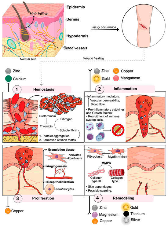
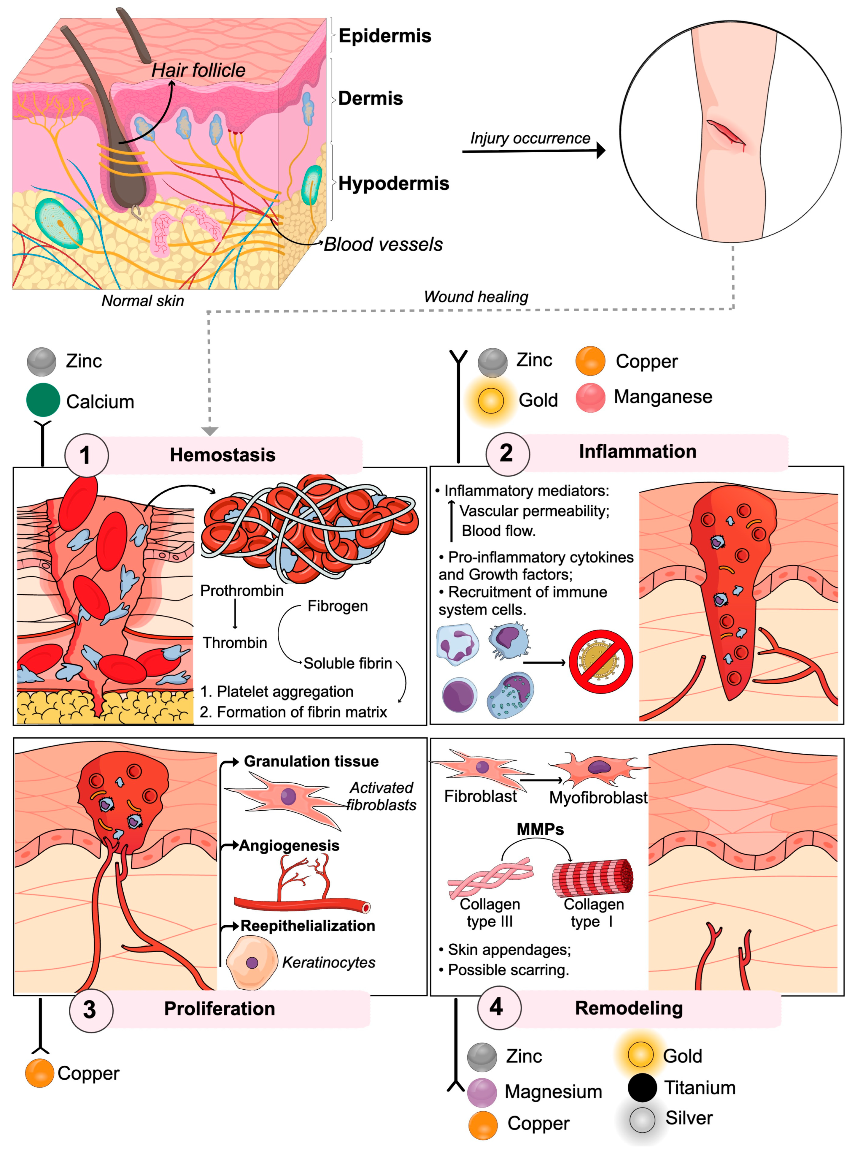
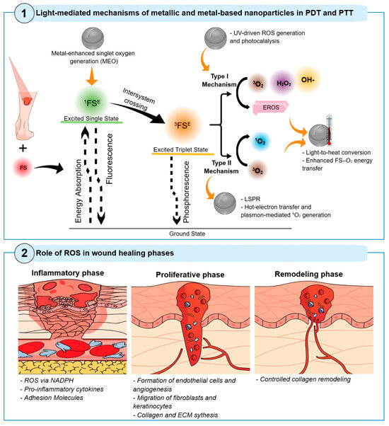
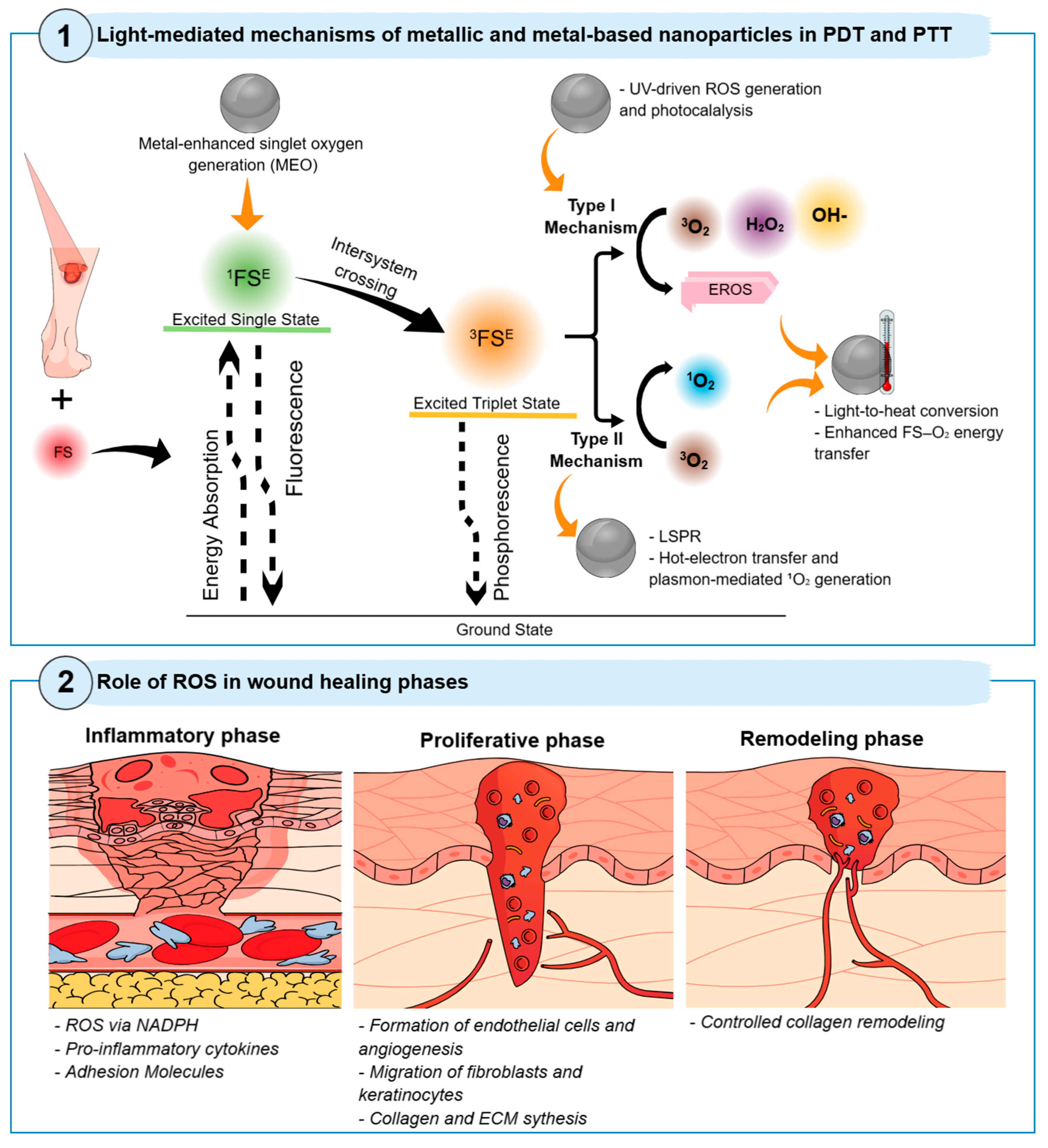

Disclaimer/Publisher’s Note: The statements, opinions and data contained in all publications are solely those of the individual author(s) and contributor(s) and not of MDPI and/or the editor(s). MDPI and/or the editor(s) disclaim responsibility for any injury to people or property resulting from any ideas, methods, instructions or products referred to in the content. |
© 2026 by the authors. Licensee MDPI, Basel, Switzerland. This article is an open access article distributed under the terms and conditions of the Creative Commons Attribution (CC BY) license.