


Abstract
Foodborne diseases represent a major public health concern, particularly those associated with dairy products contaminated with Staphylococcus aureus, a pathogen capable of producing heat-stable enterotoxins. This study evaluated the potential of native lactic acid bacteria (LAB) isolated from artisanal kefir grains as natural biocontrol agents in fermented dairy foods. Kefir grains obtained from three artisanal producers were microbiologically characterized, revealing LAB as the dominant group and the absence of Enterobacteriaceae. Strains belonging mainly to the genera Lactobacillus sensu lato, Leuconostoc, and Pediococcus were isolated and exhibited differentiated metabolic profiles. Safety assessment showed no hemolytic activity and an overall susceptibility to clinically relevant antibiotics, although genus-dependent intrinsic resistance patterns were observed. Several strains displayed enzymatic activities related to carbohydrate digestion and high tolerance to simulated gastrointestinal conditions, with survival rates exceeding 90% during both gastric and intestinal phases. Neutralized cell-free supernatant (CFS) demonstrated differential inhibitory activity, with significant antagonism of S. aureus and E. coli, comparable to those of commercial reference strains. In a yogurt model system stored at 4 °C, selected Lactobacillus and Pediococcus strains induced a progressive and significant reduction in S. aureus populations, achieving complete elimination to undetectable levels in shorter times than commercial probiotic strains. Overall, these results demonstrate that native LAB from artisanal kefir grains exhibit an adequate safety and functional profile, together with strong antagonistic activity, supporting their potential application as natural protective cultures to improve the food hygiene of fermented dairy products.
1. Introduction
Foodborne diseases (FBD) constitute a major global public health problem, affecting both developed and developing countries [1]. These diseases occur after the ingestion of food or beverages contaminated with biological agents, including bacteria, viruses, and parasites, as well as toxins or chemical contaminants [2]. FBD encompass a wide spectrum of clinical manifestations, ranging from mild gastrointestinal disorders to severe conditions that may require hospitalization or result in death [3]. According to estimates from the World Health Organization (WHO), approximately 600 million people fall ill each year due to the consumption of contaminated food, and around 420,000 deaths annually are attributed to FBD worldwide [1], particularly among vulnerable groups such as children under five years of age, pregnant women, older adults, and individuals with compromised immune systems [4]. The globalization of food markets, together with changes in food production, processing, distribution, and consumption systems, has increased the complexity of controlling microbiological risks.
Among foodborne pathogens, Staphylococcus aureus stands out due to its high prevalence and particular relevance in dairy products [5]. This bacterium is widely distributed in humans, animals, and the environment, which facilitates food contamination during production, processing, and handling stages [6]. Its importance in food safety is mainly associated with its ability to produce staphylococcal enterotoxins, including enterotoxin A (SEA), followed by SEB, SEC, SED, and SEE [7]. These toxins act as superantigens capable of inducing massive T-cell activation and the release of proinflammatory cytokines, leading to acute symptoms such as nausea, vomiting, abdominal pain, and diarrhea within a few hours of ingestion of the contaminated food [8,9]. A critical feature of these enterotoxins is their high physicochemical stability, as they are resistant to digestive enzymes and can retain biological activity even after conventional thermal treatments [7]. In dairy matrices, whose nutritional composition favors microbial growth, S. aureus can reach concentrations sufficient to produce enterotoxins that remain active even when bacterial cells are inactivated during processing or storage [10,11], representing a major challenge for the hygienic and microbiological safety of both fermented and non-fermented dairy foods.
Kefir is a traditional fermented dairy product obtained by inoculating milk with kefir grains [12]. These grains consist of a polysaccharide matrix, known as kefiran, associated with proteins that harbor a complex microbial community composed of lactic acid bacteria, acetic acid bacteria, and yeasts, which act symbiotically during the fermentation process [13]. As a result, kefir is a beverage with distinctive sensory characteristics and a nutritional and functional profile of increasing interest [14,15]. In recent years, kefir consumption has increased steadily, driven by the growing demand for functional foods and consumer preference for products perceived as natural and beneficial to health [16,17]. At the same time, artisanal or homemade kefir production has become popular due to its low cost and ease of preparation; however, this type of production is characterized by high variability in fermentation conditions, grain handling, and hygienic practices [18]. These factors can directly influence the microbial composition of the final product, its microbiological quality, safety, and shelf life.
Conventional strategies for controlling microbial contamination in foods are mainly based on the use of chemical preservatives and thermal treatments; however, these approaches present limitations related to consumer acceptance, potential losses in sensory and nutritional quality, and the persistence of heat-stable toxins [19]. In this context, lactic acid bacteria (LAB) have gained relevance as innovative and natural tools to improve food safety due to their probiotic properties. Probiotics are defined as live microorganisms that, when administered in adequate amounts, survive gastrointestinal transit, interact with the host microbiota and/or host cells, and exert beneficial physiological effects through metabolic, immunological, or ecological mechanisms [20]. In addition, LAB contribute to pathogen control through mechanisms of exclusion and antagonism against foodborne microorganisms, primarily mediated by the production of organic acids, bacteriocins, and volatile organic compounds [21,22]. Numerous studies have demonstrated the antagonistic activity of LAB against foodborne pathogens such as Staphylococcus aureus, Salmonella spp., Escherichia coli, Listeria monocytogenes, Bacillus cereus, and Campylobacter jejuni, highlighting their potential application in fermented foods as native biocontrol agents and their contribution to food safety and hygiene [22,23].
In this study, we hypothesized that LAB derived from kefir grains used in artisanal production exhibit probiotic characteristics, lack relevant virulence factors, and possess the ability to antagonize foodborne pathogens through the production of antimicrobial compounds, including bacteriocins. Therefore, the objective of this work was to isolate and identify native LAB strains from kefir grains and to evaluate their safety, probiotic potential, and antagonistic activity against S. aureus in order to explore their application as a natural strategy to improve the hygiene, safety, and microbiological quality of fermented dairy foods, thereby contributing to the protection of human health.
2. Materials and Methods
2.1. The Microbiological Analysis of Kefir Grains
Kefir samples were collected from three local dairy producers located in Chilpancingo de los Bravo, Guerrero (southeastern Mexico). The selected producers prepare kefir artisanally using pasteurized cow’s milk and traditional kefir grains as starter cultures. The fermentation process is carried out by inoculating milk with kefir grains and incubating it at room temperature (20–25 °C) for 18–24 h. After fermentation, the grains are separated by filtration and reused for subsequent fermentation cycles to maintain active cultures. Samples were transported to the laboratory under refrigerated conditions. Under sterile conditions, kefir grains were separated from the fermented product using a sterile strainer and gently rinsed with sterile distilled water. We homogenized five grams of each sample using a sterile mortar and pestle with 50 mL of sterile saline solution (0.89% NaCl, w/v). Serial dilutions were prepared up to 10−8, and aliquots were plated on selective culture media for the enumeration of the main microbial groups, as shown in Table 1 [24]. Colony differentiation was primarily based on macroscopic characteristics, Gram staining, and microscopic examination. Results were expressed as colony-forming units per gram of kefir grains (CFU g−1).
Table 1.
Culture media used for the microbiological analysis of kefir grains.
2.2. Isolation and Identification of Lactic Acid Bacteria
Lactic acid bacteria (LAB) were isolated from kefir grains using serial dilutions plated on MRS agar, as described above. Distinct colony morphotypes were selected and subcultured under the same conditions until pure cultures were obtained. Isolates were initially characterized by Gram staining and catalase and cytochrome oxidase activity tests [25]. Carbohydrate fermentation assays were performed in test tubes (13 × 100 mm) containing 5 mL of phenol red basal broth (BD BBL™), individually supplemented with 0.5% (w/v) glucose, lactose, mannose, sucrose, fructose, trehalose, maltose, galactose, cellobiose, or rhamnose (Sigma-Aldrich, St. Louis, MO, USA). To generate microaerophilic conditions, 250 µL (3–4 mm layer) of sterile mineral oil was added to each tube, followed by incubation at 30 °C for 72 h. Fermentation was considered positive when a color change in the pH indicator from red to yellow was observed. Taxonomic assignment was performed based on biochemical profiles according to the criteria described by [26].
2.3. Antibiotic Susceptibility and Hemolytic Activity
Antibiotic susceptibility profiles were evaluated using the disk diffusion (Kirby–Bauer) method. LAB suspensions were adjusted to a turbidity equivalent to the 0.5 McFarland standard (≈1 × 10⁸ CFU mL−1) and evenly spread on the surface of MRS agar plates. Antibiotic disks (Oxoid™, Thermo Fisher Scientific, Waltham, MA, USA) were placed on the plates: VAN: vancomycin (30 µg); ERY: erythromycin (15 µg); CHL: chloramphenicol (30 µg); AMP: ampicillin (10 µg); PEN: penicillin G (10 µg); TET: tetracycline (30 µg); GEN: gentamicin (10 µg); KAN: kanamycin (30 µg). Hemolytic activity (α, β, or γ hemolysis) was evaluated by streaking LAB on blood agar base (NutriSelect® Plus) supplemented with 5% (v/v) sterile defibrinated sheep blood. The presence of clear zones around colonies was interpreted as positive hemolytic activity. Plates for both assays were incubated at 30 °C for 48 h under microaerophilic conditions (5% CO2) using BD GasPak™ EZ container systems.
2.4. Detection of Digestive Enzyme Activities
The production of enzymes involved in macromolecule digestion was evaluated using plate assays by inoculating 20 µL of LAB suspensions (≈1 × 10⁸ CFU mL−1) onto modified MRS agar containing specific substrates, as described by [27]. Amylase activity was assessed using starch (10 g L−1); after incubation at 30 °C for 96 h, plates were flooded with Lugol’s iodine solution (5%) for 15 min, and clear halos around colonies were recorded as positive results. Lipase activity was evaluated using egg yolk lecithin (10%, v/v), with clear halo formation after incubation at 30 °C for 72 h considered positive. Protease activity was assessed using skim milk agar (1% casein), incubated at 30 °C for 72 h, and positive reactions were indicated by clear zones. Cellulase activity was evaluated using carboxymethylcellulose (10 g L−1), incubated at 30 °C for 96 h; plates were then stained with Congo red (0.1% w/v) for 2 min, and the presence of clear hydrolysis halos was considered positive. Bile salt hydrolase activity was assessed on MRS agar supplemented with taurodeoxycholic acid (5 g L−1) and calcium chloride (0.5 g L−1), incubated at 30 °C for 96 h; opaque halos or precipitates around colonies were recorded as positive results [28].
2.5. Simulation of Gastrointestinal Transit Survival
Survival during simulated gastrointestinal transit, a key attribute associated with probiotic potential, was evaluated following the method described by [29]. The gastric phase was simulated using a solution containing 0.23% NaCl, 2.3% HCl, and 5% (w/v) pepsin, adjusted to pH 3 to mimic postprandial gastric conditions. The intestinal phase was simulated using a solution containing 0.3% (w/v) bile salts and 0.1% (w/v) pancreatin dissolved in phosphate-buffered saline (137 mM NaCl, 2.7 mM KCl, 10 mM Na2HPO4, and 1.8 mM KH2PO4, pH 7.2). Bacterial suspensions (1 × 10⁸ CFU mL−1) were incubated in the gastric solution at 37 °C for 90 min, followed by centrifugation and resuspension in the intestinal solution and incubation at 37 °C for an additional 90 min, resulting in a total simulated transit time of 180 min. Survival rate (%) was calculated using the equation: survival rate = (B/A) × 100, where A and B represent CFU mL−1 counts before and after treatment, respectively.
2.6. Inhibitory Activity of LAB
The inhibitory activity of LAB isolates, potentially associated with antimicrobial compounds, was evaluated using a disk diffusion assay following the method described by [30]. Cell-free supernatants were obtained from LAB cultures (48 h at 30 °C), filtered through 0.22 µm membranes, and neutralized to pH 7.0 to eliminate the inhibitory effect of organic acids. Sterile Whatman® filter paper disks (6 mm diameter) were impregnated with 50 µL of neutralized cell-free supernatant (CFS) and placed on Mueller–Hinton agar plates (g L−1: casein hydrolysate peptone 17.5, beef extract 2.0, starch 1.5, agar 17.0) previously inoculated by surface swabbing with fresh suspensions of Staphylococcus aureus subsp. aureus ATCC 33862 or Escherichia coli ATCC 25922 (≈1 × 10⁸ CFU mL−1). Plates were incubated at 37 °C for 24 h. Commercial strains Lactobacillus casei strain Shirota, isolated from the fermented milk beverage Yakult® (Yakult, S.A. de C.V., Ixtapaluca, Mexico), and Lactobacillus acidophilus, obtained from Simibacilos Forte® (Farmacias de Similares, Ciudad de México, Mexico), were used as reference LAB strains. Inhibitory activity was considered positive based on the presence of inhibition halos around the disks. LAB strains with the highest inhibitory activity were selected for subsequent experiments.
2.7. Antagonistic Effect of LAB Against Staphylococcus aureus in Yogurt
To evaluate the antagonistic activity of LAB isolates in a standardized dairy matrix, yogurt was used as a model fermented milk system. Under aseptic conditions, 100 mL of plain natural yogurt (Alpura®, Ganaderos Productores de Leche Pura, S.A.P.I. de C.V.), without added sugars or flavors, were dispensed into sterile 125 mL flasks and subjected to the following treatments: T1, Pediococcus sp. C5K1 + S. aureus ATCC 33862; T2, Lactobacillus sp. C6K2 + S. aureus ATCC 33862; T3, Lactobacillus sp. C5K3 + S. aureus ATCC 33862; T4, Lactobacillus casei + S. aureus ATCC 33862; T5, Lactobacillus acidophilus + S. aureus ATCC 33862; T6, S. aureus ATCC 33862 (positive control); and T7, saline solution (negative control). LAB strains were selected based on their high inhibitory activity in vitro. Cultures were grown for 48 h, harvested by centrifugation, washed twice, and resuspended in sterile saline solution (0.89% NaCl, w/v). Both LAB and/or S. aureus were individually inoculated into each yogurt matrix at a final concentration of 1 × 10⁸ CFU per flask according to each treatment. The use of a high S. aureus inoculum was intentional to simulate an extreme contamination scenario. Flasks were stored at 4 °C for 10 days. Every 48 h, 100 µL samples were collected, plated on BD Baird–Parker agar, and incubated at 37 °C for 48 h. S. aureus concentrations were determined by colony counting and expressed as log10 CFU mL−1.
2.8. Statistical Analysis
All experiments were performed in triplicate, and results are expressed as mean ± standard deviation. Microbiological counts were log10-transformed prior to statistical analysis. Differences between two treatments were evaluated using Student’s t-test, while multiple comparisons were analyzed by one-way analysis of variance (ANOVA) followed by Tukey’s post hoc test. Differences were considered statistically significant at p < 0.05. All statistical analyses were performed using GraphPad Prism version 10 (GraphPad Software, San Diego, CA, USA).
3. Results
3.1. Microbiological Composition of Kefir Grains
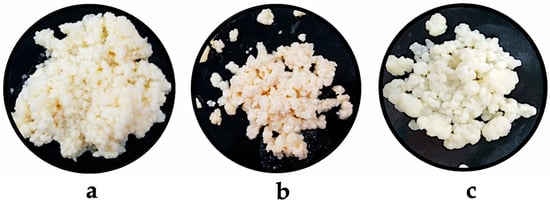
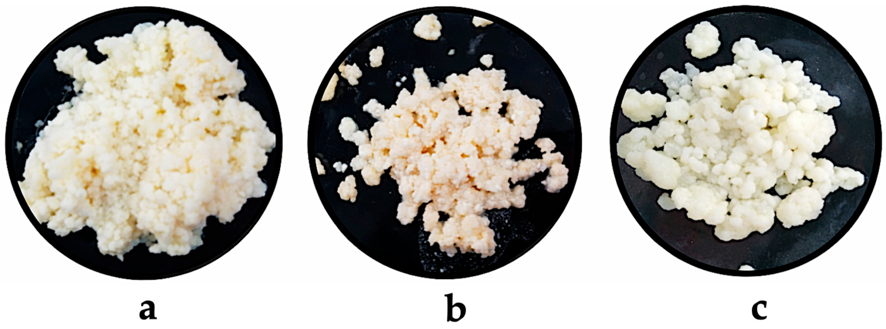
The macroscopic analysis of kefir grains obtained from three artisanal producers revealed clear differences in color and texture (Figure 1). Kefir 1 grains exhibited the characteristic cauliflower-like shape, with a whitish-yellow coloration and a viscous consistency (Figure 1a), whereas kefir 2 grains showed a more yellowish tone and a semi-solid consistency (Figure 1b). In contrast, kefir 3 grains displayed a whitish-yellow coloration similar to kefir 1, but with a more compact structure and a firm consistency (Figure 1c).
Figure 1.
Macroscopic morphology of kefir grains obtained from three artisanal producers (a–c). Variations in compactness, grain size, and color are observed among samples.
Microbiological analysis confirmed significant differences in the microbial composition of the grains (Table 2). LAB were the dominant microbial group in all samples, with counts ranging from 1.79 × 10⁶ to 4.24 × 10⁶ CFU g−1. Kefir 1 and kefir 2 grains exhibited significantly higher LAB concentrations compared to kefir 3 grains (p < 0.0001), with no statistically significant differences observed between the former two. Marked differences were observed in yeast populations among samples. Kefir 2 grains showed the highest yeast abundance (4.88 × 10⁶ CFU g−1), followed by kefir 3 (2.68 × 10⁶ CFU g−1), whereas kefir 1 presented significantly lower counts (1.63 × 10⁵ CFU g−1) (p < 0.0001).
Table 2.
Microbiological composition of kefir grains obtained from three artisanal producers.
Halotolerant Gram-positive cocci were detected in all samples, with kefir 2 grains showing the highest abundance (1.50 × 10⁵ CFU g−1). No significant differences were observed between kefir 1 and kefir 3 grains. The absence of mannitol-fermenting colonies suggests that the halotolerant cocci detected do not correspond to mannitol-positive staphylococci of sanitary relevance. Furthermore, no growth of Enterobacteriaceae was detected in any of the analyzed samples, indicating adequate microbiological quality and the absence of fecal contamination during kefir fermentation.
3.2. Diversity of Lactic Acid Bacteria from Kefir Grains
A total of 18 strains were isolated from the kefir grains, corresponding to six isolates per sample. Based on microscopic morphology, Gram staining, and catalase and oxidase tests, 10 strains were classified as LAB, whereas the remaining eight isolates corresponded to yeasts and were excluded from this study. The distribution of LAB among kefir grains was heterogeneous: kefir 1 contributed 50% of the isolates, followed by kefir 2 with 40%, while kefir 3 contributed only one strain (10%). This distribution pattern is consistent with the previously observed microbiological counts and suggests a higher cultivable LAB diversity in kefir grains 1 and 2 compared with kefir 3. Biochemical identification revealed the presence of isolates belonging to the genera Lactobacillus sensu lato, Leuconostoc, and Pediococcus, groups commonly associated with complex dairy fermentations. Leuconostoc spp. predominated in kefir 1 grains, whereas Lactobacillus spp. were more frequently isolated from kefir 2. Kefir 3 was represented by a single isolate exhibiting a metabolic profile consistent with heterofermentative LAB within the Lactobacillus sensu lato group. Carbohydrate fermentation profiles revealed distinct metabolic patterns among genera and strains while preserving core characteristics typical of LAB (Table 3). Glucose, lactose, sucrose, and fructose were fermented by 100% of the strains, reflecting efficient metabolic adaptation to monosaccharides and disaccharides commonly present in dairy matrices. Maltose was utilized by 90% of the isolates, with the exception of Lactobacillus sp. C4K2. Trehalose fermentation was observed in most Lactobacillus and Pediococcus strains, whereas some Leuconostoc isolates lacked this capability. Mannose and cellobiose fermentation showed high inter-strain variability and allowed clear metabolic discrimination among genera. None of the evaluated strains were able to ferment rhamnose.
Table 3.
Carbohydrate fermentation profile of lactic acid bacteria isolated from artisanal kefir grains.
3.3. Safety Assessment Based on Antimicrobial Susceptibility and Hemolytic Activity
The antimicrobial susceptibility profiles of LAB were evaluated against antibiotics of clinical and regulatory relevance. The results revealed marked differences among strains and genera. Overall, Lactobacillus sp. and Pediococcus sp. isolates exhibited susceptibility to ampicillin, penicillin, and chloramphenicol. In particular, Lactobacillus sp. C2K1, C4K2, C5K2, C6K2, and C5K3, as well as Pediococcus sp. C5K1, showed no growth in the presence of these antibiotics. In contrast, Leuconostoc sp. C4K1, C6K1, and C1K2 displayed broad resistance to vancomycin. Susceptibility to erythromycin varied among strains, with Lactobacillus and Pediococcus strains showing sensitivity and Leuconostoc isolates exhibiting predominant resistance. Most isolates were resistant to tetracycline and kanamycin, while variable responses were observed for gentamicin. The antibiotic susceptibility results are presented in Table S1. Strains exhibiting resistance to clinically relevant antibiotics would not be considered suitable candidates for starter culture development. Importantly, none of the isolates exhibited hemolytic activity on blood agar, supporting their safety profile from a probiotic standpoint.
3.4. In Vitro Evaluation of LAB Adaptation to Gastrointestinal Transit
In vitro adaptation to gastrointestinal transit was assessed by determining enzymatic activities associated with digestion, as well as survival under simulated gastric and intestinal conditions. The results revealed a selective, strain-dependent enzymatic profile, with a predominance of activities related to carbohydrate degradation (Table 4). Cellulolytic activity was detected in five isolates (Lactobacillus sp. C2K1, Leuconostoc sp. C6K1, Leuconostoc sp. C1K2, Lactobacillus sp. C4K2, and Lactobacillus sp. C5K2). Amylolytic activity was the most prevalent trait, detected in 70% of the isolates across all LAB genera. In contrast, bile salt hydrolase activity was limited to two Lactobacillus strains (C2K1 and C4K2), while lipolytic and proteolytic activities were not detected in any isolate.
Table 4.
Enzymatic profile of lactic acid bacteria isolated from artisanal kefir grains.
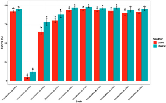
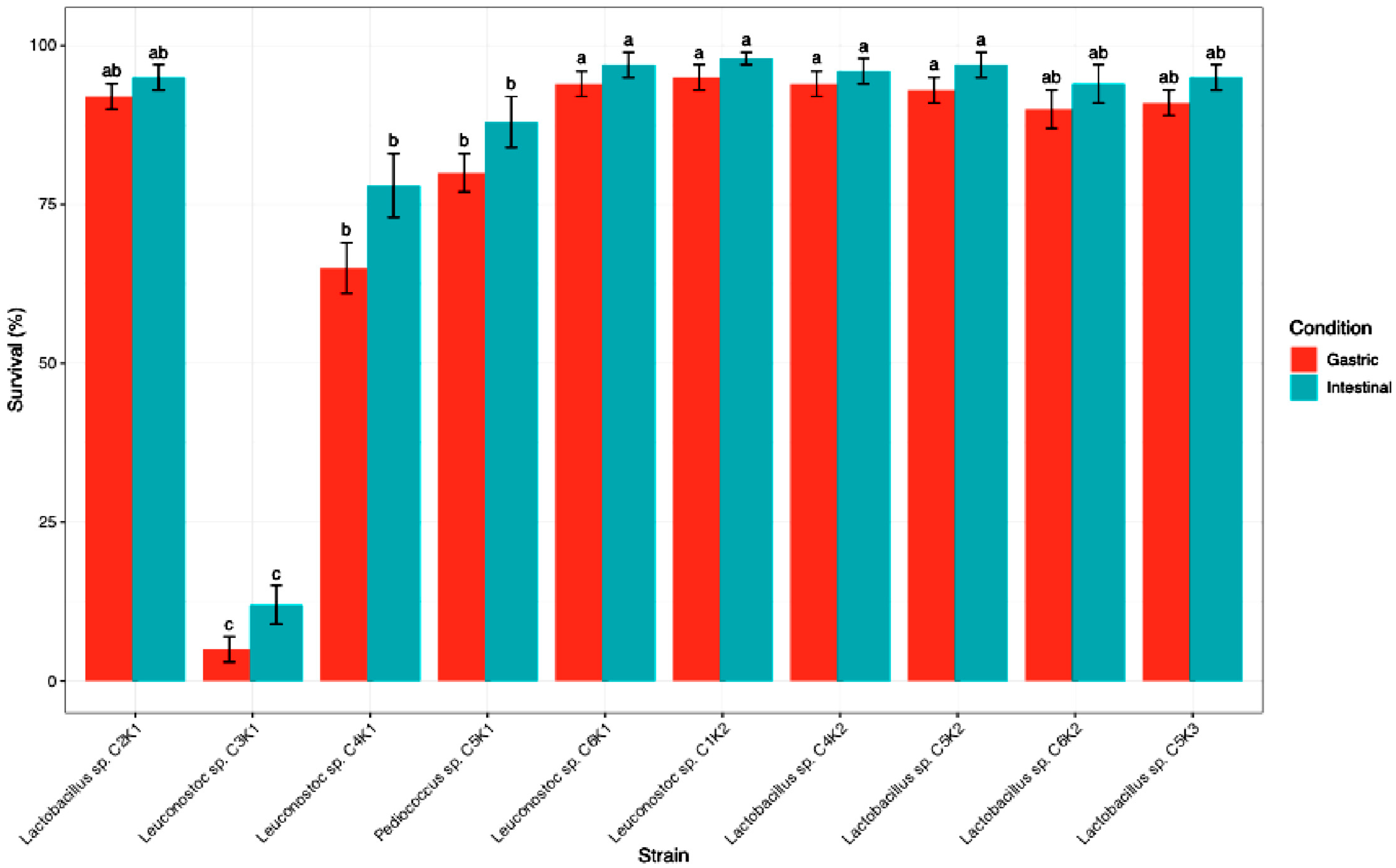
Significant differences in survival under simulated gastrointestinal conditions were observed among strains and between phases (p < 0.05; Figure 2). During the gastric phase, most isolates maintained high survival rates (>90%), with no significant differences among Lactobacillus sp. C2K1, Leuconostoc sp. C6K1, Leuconostoc sp. C1K2, Lactobacillus sp. C4K2, Lactobacillus sp. C5K2, and Lactobacillus sp. C6K2. In contrast, Leuconostoc sp. C4K1 and Pediococcus sp. C5K1 showed intermediate survival, whereas Leuconostoc sp. C3K1 exhibited markedly reduced survival (5 ± 2%). Under simulated intestinal conditions, survival increased overall, with Leuconostoc sp. C1K2, Leuconostoc sp. C6K1, and all Lactobacillus strains showing the highest survival levels (≥94%), without significant differences among them. Leuconostoc sp. C4K1 and Pediococcus sp. C5K1 again displayed intermediate survival, while Leuconostoc sp. C3K1 remained significantly less tolerant (12 ± 3%; p < 0.05). However, studies including the oral phase would be required to more accurately simulate the complete gastrointestinal digestion process.
Figure 2.
Survival (%) of lactic acid bacteria during simulated gastrointestinal transit. Data are expressed as mean ± standard deviation (n = 3). Different lowercase letters indicate statistically significant differences (one-way ANOVA, Tukey’s HSD post hoc test, p < 0.05).
3.5. Evaluation of Inhibitory Activity
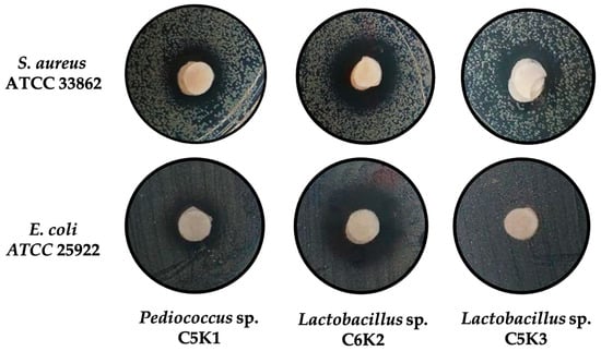
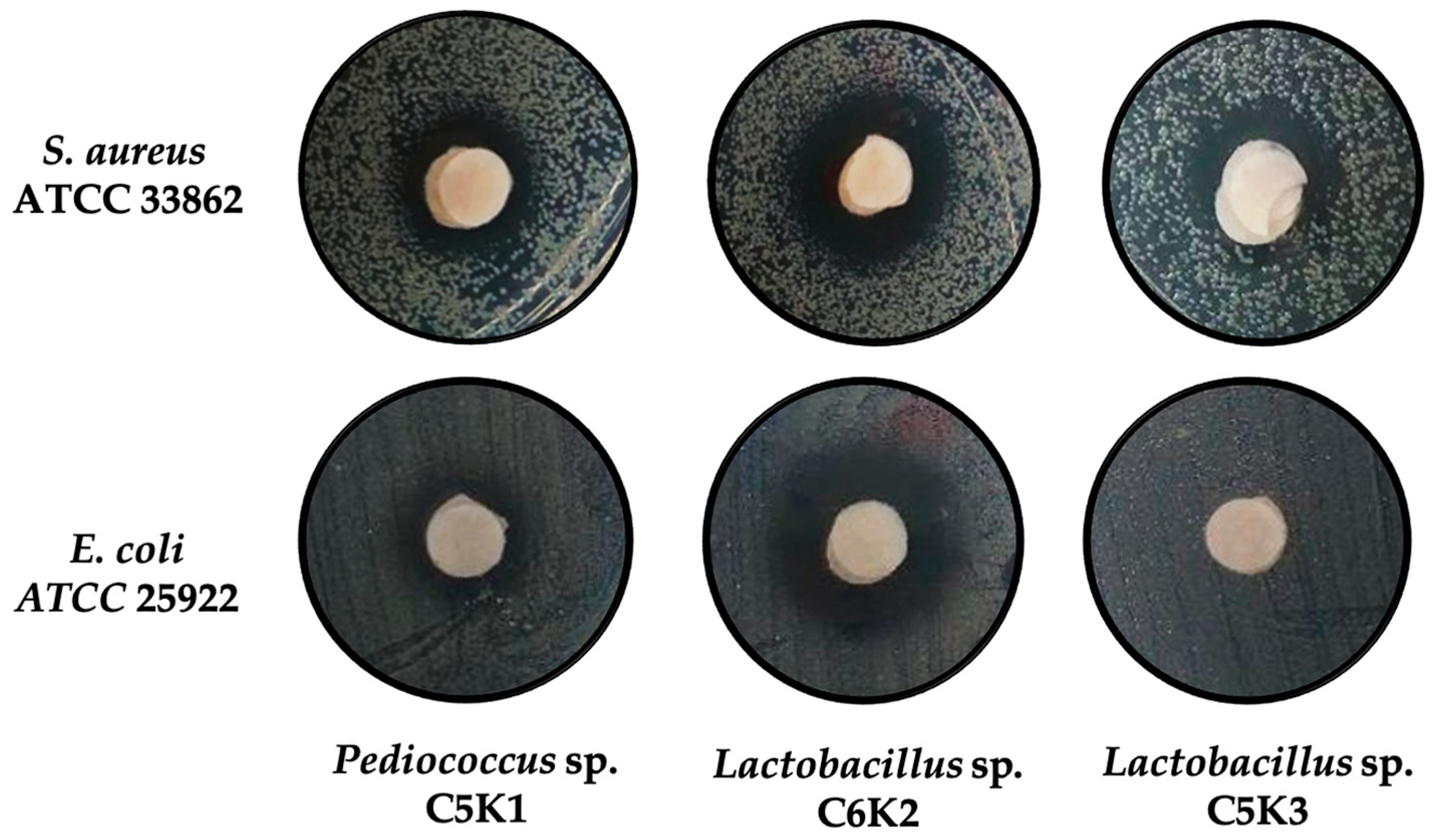
The antagonistic activity of LAB was evaluated using cell-free supernatants against S. aureus ATCC 33862 and E. coli ATCC 25922. The results revealed a differential inhibitory activity, with significant variations at both the intra- and intergeneric levels (Figure 3, Table 5). Against S. aureus, several strains exhibited inhibitory activity, with inhibition halo diameters ranging from 6.33 ± 5.50 mm to 18.00 ± 2.00 mm. The highest activity was observed in the supernatants of Leuconostoc sp. C1K2, followed by Lactobacillus sp. C6K2 and the commercial reference strain L. acidophilus. In contrast, Lactobacillus sp. C2K1 and Leuconostoc sp. C3K1 did not show detectable inhibition against this pathogen.
Figure 3.
In vitro evaluation of inhibitory activity by lactic acid bacteria using cell-free supernatants against Staphylococcus aureus and Escherichia coli.
Table 5.
Antagonistic activity of lactic acid bacteria against foodborne pathogens.
In the case of the Gram-negative bacterium E. coli, inhibitory activity was also strain-dependent. Pediococcus sp. C5K1 exhibited the strongest antagonistic effect (20.33 ± 0.57 mm). Similarly, Leuconostoc sp. C1K2 and Lactobacillus sp. C6K2 displayed consistent inhibitory activity, with halo diameters of 16.33 ± 1.52 mm and 16.00 ± 1.72 mm, respectively, values statistically similar to those produced by L. casei and L. acidophilus. A second group of isolates, including Lactobacillus sp. C2K1, Leuconostoc sp. C3K1, Lactobacillus sp. C4K2, and Lactobacillus sp. C5K2, showed moderate inhibitory activity, with halo diameters ranging between 9.00 and 12.00 mm. In contrast, no detectable inhibition against E. coli was observed for Leuconostoc sp. C6K1 or Lactobacillus sp. C5K3 under the experimental conditions evaluated, highlighting marked differences in antagonistic potential even among phylogenetically related isolates.
3.6. LAB as Biocontrol of S. aureus in Yogurt
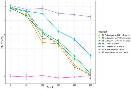
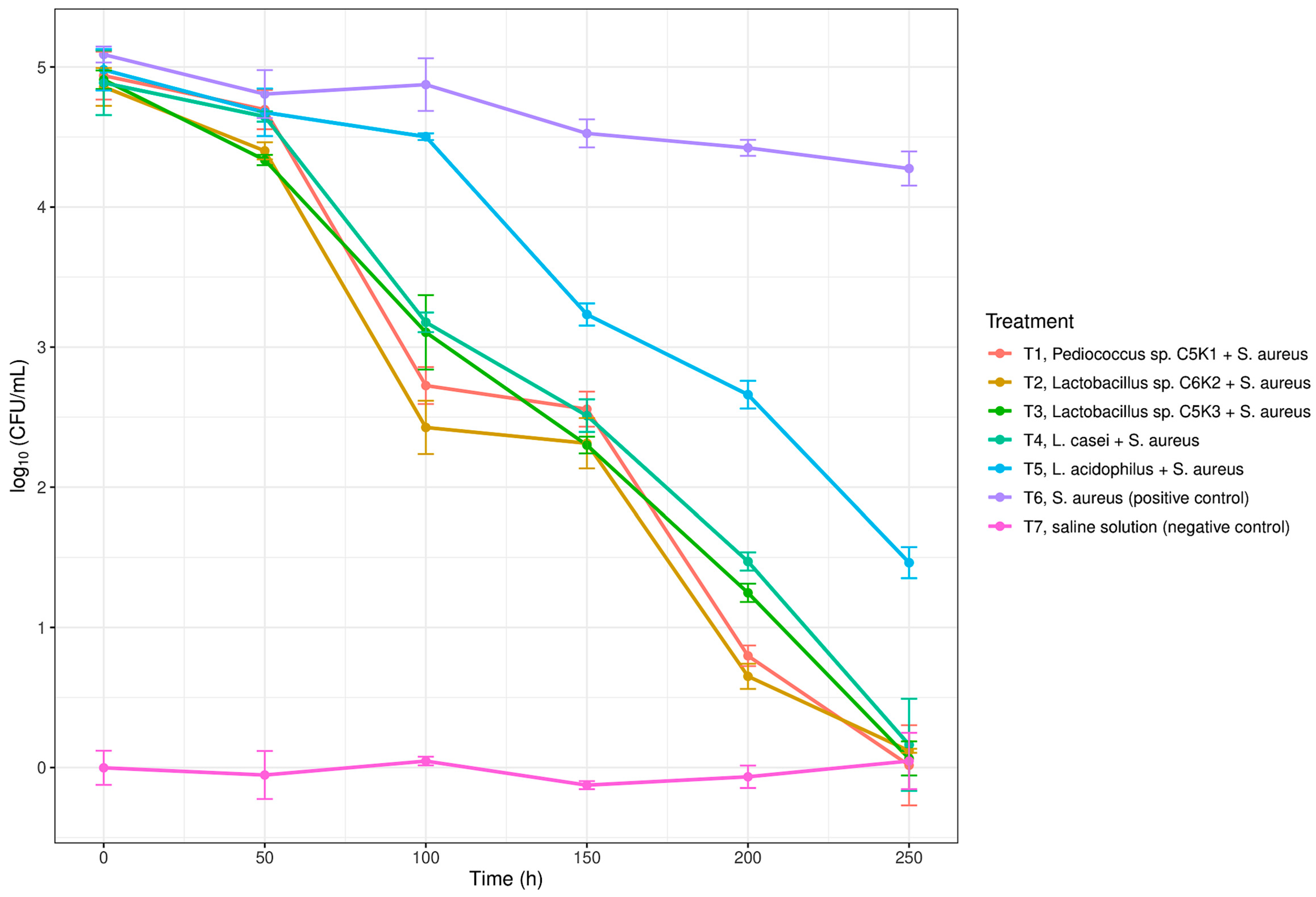
Using LAB with inhibitory activity, antagonistic effects against the pathogen S. aureus were evaluated in a real yogurt matrix stored at 4 °C (Figure 4). The results showed a progressive and time-dependent reduction in S. aureus counts in all treatments containing LAB, compared with the positive control without antagonists (T6). From the first 96 h of storage, treatments T1–T5 exhibited statistically significant reductions (p < 0.05) in S. aureus concentration relative to the positive control (T6).
Figure 4.
Antagonistic activity of lactic acid bacteria against S. aureus ATCC 33862 in a yogurt matrix. Pathogen counts are expressed as log10(CFU/mL) over the storage period at 4 °C. Data are presented as mean ± standard deviation.
In particular, Lactobacillus sp. C5K3 (T3) and Pediococcus sp. C5K1 (T1) showed the strongest inhibitory effect, reaching values close to 2 log10 CFU mL−1 between 96 and 144 h. These reductions were significantly greater than those observed for the other treatments (p < 0.05). Subsequently, both treatments achieved complete elimination of the pathogen, with undetectable counts from 192 h onward. Similarly, treatments with the commercial strains L. casei (T4) and L. acidophilus (T5) induced a significant decrease in S. aureus throughout the storage period (p < 0.05), reaching undetectable levels by the end of the assay. However, their reduction kinetics were slightly slower compared with LAB isolated from kefir grains. In contrast, Lactobacillus sp. C6K2 (T2) exhibited a moderate antagonistic effect, with counts significantly lower than those of the positive control (p < 0.05), but higher than those observed for T1 and T3 at intermediate time points. The positive control (T6) only showed a gradual decrease attributable to refrigeration conditions and the yogurt matrix itself, whereas the negative control (T7) remained free of microbial growth throughout the experimental period. Overall, these findings support the potential application of the evaluated LAB as a natural biocontrol strategy to improve hygiene and microbiological safety in fermented dairy products.
4. Discussion
Effective control of foodborne diseases (FBDs) requires an integrated public health approach that encompasses the entire food chain from primary production to final consumption. This approach should incorporate appropriate hygiene practices, epidemiological surveillance, and efficient regulatory frameworks. In this context, the development of innovative strategies based on biocontrol and the exploitation of the beneficial microbiota of fermented foods has emerged as a promising complement to conventional food safety practices. In the present study, the potential of lactic acid bacteria (LAB) isolated from artisanal kefir grains as natural biocontrol agents against S. aureus in yogurt was evaluated from a food hygiene and safety perspective.
Our results confirm that milk kefir grains constitute complex, structurally organized, and metabolically active microbial ecosystems, dominated primarily by lactic acid bacteria and yeasts. Studies based on both culture-dependent and culture-independent approaches [31,32] have demonstrated that kefir grains harbor stable microbial communities in which species of the genus Lactobacillus sensu lato—such as L. kefiranofaciens, L. kefiri, L. parakefiri, and L. kefirgranum—predominate, accompanied by Leuconostoc, Lactococcus, Pediococcus, Streptococcus, and even acetic acid bacteria [33,34]. In addition, the presence of diverse yeasts belonging to the genera Saccharomyces, Kluyveromyces, Pichia, and Candida has been reported [35,36].
LAB represented the dominant microbial group in all analyzed samples, with counts on the order of 106 CFU g−1, values comparable to those reported for kefir grains from other regions of Mexico [37] and from various countries worldwide [38,39,40]. During milk kefir fermentation, this bacterial group plays a central role in medium acidification, production of antimicrobial metabolites, and maintenance of microbial stability within the system [41]. In the present study, differences were observed in the abundance of LAB, yeasts, and halotolerant Gram-positive cocci among kefir grains obtained from different artisanal producers. Such variability has been attributed to factors including the geographic origin of the grains, the type and chemical composition of the milk used, the grain-to-milk ratio, fermentation temperature, and hygienic handling conditions [37,42]. In artisanal systems, where standardized processes are lacking, these variables—together with subculturing frequency—may modify the structure of the microbial consortium without necessarily compromising its fermentative functionality.
From the analyzed grains, LAB belonging mainly to the genera Lactobacillus sensu lato, Leuconostoc, and Pediococcus were recovered. These genera are considered safe for food use and hold the status of “generally recognized as safe” (GRAS). The Lactobacillus sensu lato group comprises a diverse set of phylogenetically related species that, following recent taxonomic reclassification, include genera such as Lactobacillus (sensu stricto), Lactiplantibacillus, Lacticaseibacillus, Levilactobacillus, and Limosilactobacillus, among others [43]. These bacteria are characterized as Gram-positive, non-spore-forming, catalase-negative, and predominantly acidogenic, with homo- or heterofermentative metabolisms depending on the species [44]. The genus Leuconostoc, in turn, includes Gram-positive, catalase-negative, obligately heterofermentative bacteria capable of producing lactic acid along with CO2, ethanol, and aromatic compounds such as diacetyl and acetoin [45]. Some species also possess the ability to synthesize exopolysaccharides, such as dextrans and levans, which contribute to the texture and stability of the fermented matrix [46]. In contrast, the genus Pediococcus comprises Gram-positive LAB with a coccoid morphology arranged in tetrads, exhibiting homofermentative metabolism and recognized for their high tolerance to low pH and elevated salt concentrations [47].
Tolerance to gastrointestinal transit is a key criterion for evaluating the probiotic potential of LAB. In this study, the adaptation of the isolated strains was evidenced by their enzymatic profiles and high survival rates under simulated gastric and intestinal pH conditions. Amylolytic activity was the most frequently detected and was observed in most strains regardless of genus, indicating a generalized adaptation for the utilization of complex carbohydrates in the intestine. Amylolytic LAB have been reported to convert starch directly into lactic acid through enzymes such as amylases and pullulanases, as observed in species of Lactobacillus, Lactococcus, and Leuconostoc [48]. Secondly, cellulolytic activity was detected in 50% of the strains, mainly in Lactobacillus and Leuconostoc, suggesting an enhanced capacity to utilize complex polysaccharides. This trait may confer metabolic advantages during intestinal transit and be beneficial for digestion in animals and humans consuming plant-based diets [49]. The absence of lipolytic and proteolytic activities suggests that gastrointestinal adaptation of these LAB relies primarily on carbohydrate metabolism.
Most LAB exhibited high tolerance to the gastric phase, whereas some Leuconostoc isolates showed greater sensitivity to acid stress. During the intestinal phase, Lactobacillus strains and some Leuconostoc isolates achieved the highest survival rates (≥94%), indicating better adaptation to neutral conditions in the presence of bile salts. Bile salts act as antimicrobial agents capable of damaging DNA and disrupting cell membrane integrity. Some Lactobacillus species produce the enzyme bile salt hydrolase (BSH) as a protective mechanism against this stress [50,51]. This activity was limited and exclusive to two Lactobacillus isolates, consistent with the well-documented bile tolerance of this genus [52]. Other tolerance mechanisms described in LAB include modifications in cell membrane composition, activation of ABC-type efflux pumps, and induction of stress response proteins, which may explain the high survival observed even in strains lacking detectable BSH activity [53].
Bacteriocin production by LAB represents one of the most promising mechanisms for the biological control of pathogens in food matrices. These molecules are ribosomally synthesized antimicrobial peptides that exhibit high specificity and effectiveness, primarily against Gram-positive bacteria [54]. Bacteriocins are classified into two main groups based on their biochemical characteristics and mechanisms of action: Group I, which includes small, heat-stable lantibiotics with modified amino acids, and Group II, which comprises non-lantibiotic bacteriocins such as pediocins, two-peptide bacteriocins, circular bacteriocins, and linear bacteriocins [55,56]. In this study, several strains exhibited notable inhibitory activity against S. aureus and E. coli, particularly isolates belonging to Leuconostoc sp. and Pediococcus sp., reaching levels comparable to or exceeding those of commercial reference strains such as Lactobacillus acidophilus [57]. Although this activity could be associated with the production of antimicrobial compounds such as bacteriocins, the specific mechanism was not determined in this study. Therefore, further analyses, including enzymatic treatments and compound characterization, would be required to confirm the presence and identity of bacteriocins responsible for the observed inhibitory effects.
Staphylococcus aureus is one of the main etiological agents of foodborne intoxications associated with dairy products [58]. In fermented products such as yogurt, although acidic pH and fermentative microbiota limit its growth, S. aureus has been shown to survive during early stages of fermentation or storage and to produce enterotoxins before being fully inhibited, posing a relevant health risk [59]. In this study, the addition of LAB isolated from artisanal kefir grains significantly reduced S. aureus counts in yogurt stored at 4 °C, reaching non-detectable levels in some treatments. This effect was greater than that observed in the control without LAB, highlighting the added value of microbial biocontrol as a complementary food safety strategy. The observed reduction kinetics suggest that certain autochthonous strains, particularly Lactobacillus sp. and Pediococcus sp., can exert an antagonistic effect comparable to or even superior to that of commercial reference strains such as Lactobacillus acidophilus [57].
The inhibition of S. aureus in the dairy matrix is likely attributable to the combined action of multiple LAB mechanisms, including lactic acid production, which rapidly lowers substrate pH, as well as the synthesis of additional antimicrobial metabolites such as hydrogen peroxide, volatile organic compounds, and bacteriocins, which directly contribute to pathogen antagonism [22,60]. Sustained efficacy during refrigerated storage further suggests adequate functional stability of these LAB under realistic preservation conditions. Numerous studies have demonstrated that the incorporation of LAB and/or their metabolites is effective for food preservation, shelf-life extension, and prevention of microbial spoilage in various production contexts, particularly in European and Asian countries [61]. In this regard, their implementation is especially relevant in Latin American countries such as Mexico, where artisanal and semi-industrial production systems face greater challenges in standardization and sanitary control. Moreover, the application of antagonistic LAB has been extended to other food systems, such as meat products, for the control of Pseudomonas spp. [62], underscoring the broad potential of LAB to reduce the incidence of foodborne diseases and associated public health problems.
Despite these promising findings, some limitations of the present study should be acknowledged. The identification of the isolates was based on phenotypic and biochemical characterization, and molecular approaches would provide a more precise taxonomic resolution. In addition, the antimicrobial activity observed was evaluated as inhibitory activity using neutralized cell-free supernatants, and the specific compounds responsible for this effect were not characterized. Future studies should therefore include molecular identification of the isolates, characterization of the antimicrobial metabolites involved, and evaluation of their technological performance under controlled fermentation conditions. Such approaches would contribute to a better understanding of the mechanisms underlying the observed antagonistic activity and to the potential application of these LAB strains in food safety strategies.
5. Conclusions
Overall, the results suggest that LAB native to artisanal kefir grains may represent a potential resource for improving the hygiene and microbiological safety of fermented dairy products, particularly in matrices such as yogurt. The combination of an adequate safety profile, tolerance to simulated gastrointestinal conditions, and inhibitory activity against relevant pathogens indicates that these strains could be considered candidates for future biocontrol applications and the development of protective cultures. From a food safety perspective, the incorporation of LAB with antagonistic activity may contribute to limiting the growth of foodborne pathogens in dairy products, especially in artisanal or small-scale production systems. Furthermore, the exploration of the autochthonous microbiota of traditional fermented foods aligns with current trends toward more sustainable, natural, and culturally relevant food systems.
Supplementary Materials
The following supporting information can be downloaded at: https://www.mdpi.com/article/10.3390/hygiene6020021/s1, Table S1: Antibiotic susceptibility profiles of lactic acid bacteria isolated from kefir grains.
Author Contributions
Conceptualization, B.M.-D.M. and J.L.A.-N.; Methodology, J.R.D.-G. and J.T.-J.; Software, G.C.-F. and J.L.A.-N.; Validation, J.L.A.-N.; Formal analysis, J.T.-J. and G.C.-F.; Investigation, J.R.D.-G., J.T.-J., B.M.-D.M., L.J.G.-G., G.C.-F., J.R.-G., and J.L.A.-N.; Resources, J.T.-J. and J.L.A.-N.; Data curation, L.J.G.-G., G.C.-F., and J.R.-G.; Writing—original draft preparation, J.T.-J., L.J.G.-G., J.R.-G. and J.L.A.-N.; Writing—review and editing, J.L.A.-N.; Supervision, B.M.-D.M. and J.L.A.-N.; Project administration, J.L.A.-N. All authors have read and agreed to the published version of the manuscript.
Funding
This research received no external funding.
Institutional Review Board Statement
Not applicable.
Informed Consent Statement
Not applicable.
Data Availability Statement
The original contributions presented in this study are included in the article. Further inquiries can be directed to the corresponding author.
Acknowledgments
The authors thank Kenia Itzel Sollano-Peralta for technical support.
Conflicts of Interest
The authors declare no conflicts of interest.
References
- Bhaskar, S.V. Chapter 1—Foodborne diseases—Disease burden. In Food Safety in the 21st Century; Gupta, R.K., Dudeja, Singh, M., Eds.; Academic Press: Cambridge, MA, USA, 2017; pp. 1–10. [Google Scholar] [CrossRef] [Scilit]
- Todd, E.C.D. Foodborne Diseases: Overview of Biological Hazards and Foodborne Diseases. In Encyclopedia of Food Safety; Motarjemi, Y., Ed.; Academic Press: Cambridge, MA, USA, 2014; pp. 221–242. [Google Scholar] [CrossRef] [Scilit]
- Keddy, K.H.; Hoffmann, S.; Founou, L.L.; Estrada-Garcia, T.; Gobena, T.; Havelaar, A.H.; Jakobsen, L.S.; Kubota, K.; Law, C.; Lake, R.; et al. Quantifying national burdens of foodborne disease—Four imperatives for global impact. PLoS Glob. Public Health 2025, 5, e0004309. [Google Scholar] [CrossRef] [Scilit]
- Saeed, B.Q.; Osaili, T.M.; Taha, S. Foodborne diseases risk factors associated with food safety knowledge and practices of women in Sharjah-United Arab Emirate. Food Control 2021, 125, 108024. [Google Scholar] [CrossRef] [Scilit]
- Dai, J.; Wu, S.; Huang, J.; Wu, Q.; Zhang, F.; Zhang, J.; Wang, J.; Ding, Y.; Zhang, S.; Yang, X.; et al. Prevalence and Characterization of Staphylococcus aureus Isolated From Pasteurized Milk in China. Front. Microbiol. 2019, 10, 641. [Google Scholar] [CrossRef] [Scilit] [PubMed]
- Léguillier, V.; Pinamonti, D.; Chang, C.-M.; Gunjan; Mukherjee, R.; Himanshu; Cossetini, A.; Manzano, M.; Anba-Mondoloni, J.; Malet-Villemagne, J.; et al. A review and meta-analysis of Staphylococcus aureus prevalence in foods. Microbe 2024, 4, 100131. [Google Scholar] [CrossRef] [Scilit]
- Cieza, M.Y.; Bonsaglia, E.C.; Rall, V.L.; Santos, M.V.; Silva, N.C. Staphylococcal Enterotoxins: Description and Importance in Food. Pathogens 2024, 13, 676. [Google Scholar] [CrossRef] [Scilit] [PubMed]
- Krakauer, T. Staphylococcal Superantigens: Pyrogenic Toxins Induce Toxic Shock. Toxins 2019, 11, 178. [Google Scholar] [CrossRef] [Scilit]
- Pinchuk, I.V.; Beswick, E.J.; Reyes, V.E. Staphylococcal Enterotoxins. Toxins 2010, 2, 2177–2197. [Google Scholar] [CrossRef] [Scilit] [PubMed]
- Berger, T.F.H.; Brasca, M.; Buchner, M.; Bütikofer, U.; Castiglioni, B.; Cremonesi, P.; Eliskases-Lechner, F.; Fritsch, L.; Morandi, S.; Schwendimann, L. Critical Factors Affecting the Prevalence of Staphylococcus aureus and Staphylococcal Enterotoxins in Raw Milk Cheese in the Alpine Region of Austria, Italy, and Switzerland. Foods 2025, 14, 2176. [Google Scholar] [CrossRef] [Scilit]
- Kadariya, J.; Smith, T.C.; Thapaliya, D. Staphylococcus aureus and Staphylococcal Food-Borne Disease: An Ongoing Challenge in Public Health. BioMed Res. Int. 2014, 2014, 827965. [Google Scholar] [CrossRef] [Scilit]
- Mudoor Sooresh, M.; Jayawickrama, A.; Silva, A.; Nguyen, S.; Schmidt, S.; Sebastian, J.; Carey, S.; Harynuk, J.; Willing, B.P.; Bourrie, B.C.T. Fermentation of kefir with traditional freeze-dried starter cultures successfully recreates fresh culture fermented kefir. Front. Microbiol. 2025, 16, 1655390. [Google Scholar] [CrossRef] [Scilit]
- Moretti, A.F.; Moure, M.C.; Quiñoy, F.; Esposito, F.; Simonelli, N.; Medrano, M.; León-Peláez, Á. Water kefir, a fermented beverage containing probiotic microorganisms: From ancient and artisanal manufacture to industrialized and regulated commercialization. Future Foods 2022, 5, 100123. [Google Scholar] [CrossRef] [Scilit]
- Azizi, N.F.; Kumar, M.R.; Yeap, S.K.; Abdullah, J.O.; Khalid, M.; Omar, A.R.; Osman, M.A.; Mortadza, S.A.; Alitheen, N.B. Kefir and Its Biological Activities. Foods 2021, 10, 1210. [Google Scholar] [CrossRef] [Scilit] [PubMed]
- Chomphoosee, T.; Seesuriyachan, P.; Wattanutchariya, W.; Tipbunjong, C.; Therdtatha, P.; techapun, C.; Insomphun, C.; Panti, N.; Moukamnerd, C. A novel beverage of coffee cherry (cascara) water kefir rich in antioxidants, bioactive compounds, and exhibiting promising antibacterial and sensory qualities. LWT 2025, 219, 117539. [Google Scholar] [CrossRef] [Scilit]
- Justel, M.A.; Outeiriño, E.B.; Guerra, N.P. Production of Kefir and Kefir-like Beverages: Fundamental Aspects, Advances, and Future Challenges. Processes 2026, 14, 73. [Google Scholar] [CrossRef] [Scilit]
- Kurniawan; Milanda, T.; Kusuma, S.A.F. Kefir as a functional probiotic: Microbial composition and health effects. Front. Food Sci. Technol. 2026, 5, 1725280. [Google Scholar] [CrossRef] [Scilit]
- Alves, E.; Ntungwe, E.N.; Gregório, J.; Rodrigues, L.M.; Pereira-Leite, C.; Caleja, C.; Pereira, E.; Barros, L.; Aguilar-Vilas, M.V.; Rosado, C.; et al. Characterization of Kefir Produced in Household Conditions: Physicochemical and Nutritional Profile, and Storage Stability. Foods 2021, 10, 1057. [Google Scholar] [CrossRef] [Scilit] [PubMed]
- Urban-Chmiel, R.; Osek, J.; Wieczorek, K. Methods of Controlling Microbial Contamination of Food. Pathogens 2025, 14, 492. [Google Scholar] [CrossRef] [Scilit]
- Chakravarty, K.; Gaur, S.; Kumar, R.; Jha, N.K.; Gupta, P.K. Exploring the multifaceted therapeutic potential of probiotics: A review of current insights and applications. Probiotics Antimicrob. Proteins 2025, 17, 341–363. [Google Scholar] [CrossRef] [Scilit]
- Parada Fabián, J.C.; Álvarez Contreras, A.K.; Natividad Bonifacio, I.; Hernández Robles, M.F.; Vázquez Quiñones, C.R.; Quiñones Ramírez, E.I.; Vázquez Salinas, C. Toward safer and sustainable food preservation: A comprehensive review of bacteriocins in the food industry. Biosci. Rep. 2025, 45, 277–302. [Google Scholar] [CrossRef] [Scilit]
- Zhang, Y.; Li, L.; Pang, X.; Zhang, S.; Liu, Y.; Wang, Y.; Xie, N.; Li, X. Lactic Acid Bacteria as the Green and Safe Food Preservatives: Their Mechanisms, Applications and Prospects. Foods 2026, 15, 241. [Google Scholar] [CrossRef] [Scilit]
- Pujato, S.A.; Mercanti, D.J.; Briggiler Marcó, M.; Capra, M.L.; Quiberoni, A.; Guglielmotti, D.M. Bacteriocins from lactic acid bacteria: Strategies for the bioprotection of dairy foods. Front. Food Sci. Technol. 2024, 4, 1439891. [Google Scholar] [CrossRef] [Scilit]
- Irigoyen, A.; Arana, I.; Castiella, M.; Torre, P.; Ibáñez, F.C. Microbiological, physicochemical, and sensory characteristics of kefir during storage. Food Chem. 2005, 90, 613–620. [Google Scholar] [CrossRef] [Scilit]
- Winn, W.C.; Allen, S.D.; Janda, W.M. Koneman: Diagnóstico Microbiológico: Texto y Atlas en Color; Médica Panamericana Buenos Aires: Buenos Aires, Argentina, 2008. [Google Scholar]
- Axelsson, L. Lactic acid bacteria: Classification and physiology. Food Sci. Technol. 2004, 139, 1–66. [Google Scholar]
- Herrera-Quiterio, A.; Toledo-Hernández, E.; Aguirre-Noyola, J.L.; Romero, Y.; Ramos, J.; Palemón-Alberto, F.; Toribio-Jiménez, J. Antagonic and plant growth-promoting effects of bacteria isolated from mine tailings at El Fraile, Mexico. Rev. Argent. Microbiol. 2020, 52, 231–239. [Google Scholar] [CrossRef] [Scilit]
- Wang, Z.; Zeng, X.; Mo, Y.; Smith, K.; Guo, Y.; Lin, J. Identification and Characterization of a Bile Salt Hydrolase from Lactobacillus salivarius for Development of Novel Alternatives to Antibiotic Growth Promoters. Appl. Environ. Microbiol. 2012, 78, 8795–8802. [Google Scholar] [CrossRef] [Scilit] [PubMed]
- Haghshenas, B.; Nami, Y.; Haghshenas, M.; Abdullah, N.; Rosli, R.; Radiah, D.; Yari Khosroushahi, A. Bioactivity characterization of Lactobacillus strains isolated from dairy products. MicrobiologyOpen 2015, 4, 803–813. [Google Scholar] [CrossRef] [Scilit] [PubMed]
- Baick, S.-C.; Kim, C.-H. Assessment of Characteristics and Functional Properties of Lactobacillus Species Isolated from Kimchi for Dairy Use. Korean J. Food Sci. Anim. Resour. 2015, 35, 339–349. [Google Scholar] [CrossRef] [Scilit]
- Vardjan, T.; Lorbeg, P.M.; Rogelj, I.; Majhenič, A.Č. Characterization and stability of lactobacilli and yeast microbiota in kefir grains. J. Dairy Sci. 2013, 96, 2729–2736. [Google Scholar] [CrossRef] [Scilit]
- Korsak, N.; Taminiau, B.; Leclercq, M.; Nezer, C.; Crevecoeur, S.; Ferauche, C.; Detry, E.; Delcenserie, V.; Daube, G. Evaluation of the microbiota of kefir samples using metagenetic analysis targeting the 16S and 26S ribosomal DNA fragments. J. Dairy Sci. 2015, 98, 3684–3689. [Google Scholar] [CrossRef] [Scilit]
- Biçer, Y.; Telli, A.E.; Sönmez, G.; Turkal, G.; Telli, N.; Uçar, G. Comparison of commercial and traditional kefir microbiota using metagenomic analysis. Int. J. Dairy Technol. 2021, 74, 528–534. [Google Scholar] [CrossRef] [Scilit]
- Diosma, G.; Romanin, D.E.; Rey-Burusco, M.F.; Londero, A.; Garrote, G.L. Yeasts from kefir grains: Isolation, identification, and probiotic characterization. World J. Microbiol. Biotechnol. 2014, 30, 43–53. [Google Scholar] [CrossRef] [Scilit] [PubMed]
- Plessas, S.; Nouska, C.; Mantzourani, I.; Kourkoutas, Y.; Alexopoulos, A.; Bezirtzoglou, E. Microbiological exploration of different types of kefir grains. Fermentation 2017, 3, 1. [Google Scholar] [CrossRef] [Scilit]
- Gut, A.M.; Vasiljevic, T.; Yeager, T.; Donkor, O.N. Characterization of yeasts isolated from traditional kefir grains for potential probiotic properties. J. Funct. Foods 2019, 58, 56–66. [Google Scholar] [CrossRef] [Scilit]
- Avila-Reyes, S.V.; Márquez-Morales, C.E.; Moreno-León, G.R.; Jiménez-Aparicio, A.R.; Arenas-Ocampo, M.L.; Solorza-Feria, J.; García-Armenta, E.; Villalobos-Espinosa, J.C. Comparative analysis of fermentation conditions on the increase of biomass and morphology of milk kefir grains. Appl. Sci. 2022, 12, 2459. [Google Scholar] [CrossRef] [Scilit]
- Kalamaki, M.S.; Angelidis, A.S. High-throughput, sequence-based analysis of the microbiota of Greek kefir grains from two geographic regions. Food Technol. Biotechnol. 2020, 58, 138. [Google Scholar] [CrossRef] [Scilit]
- Garofalo, C.; Osimani, A.; Milanović, V.; Aquilanti, L.; De Filippis, F.; Stellato, G.; Di Mauro, S.; Turchetti, B.; Buzzini, P.; Ercolini, D.; et al. Bacteria and yeast microbiota in milk kefir grains from different Italian regions. Food Microbiol. 2015, 49, 123–133. [Google Scholar] [CrossRef] [Scilit] [PubMed]
- Legowo, A.M.; Rizqiati, H.; Priambodo, A.S.; Megananda, R.C.; Yudha, W.A. Physical and microbiological characteristics grain kefir tablets in different tablet manufacturing methods. Int. J. Agric. Biol. 2023, 29, 387–392. [Google Scholar] [CrossRef] [Scilit]
- Mora-Villalobos, J.A.; Montero-Zamora, J.; Barboza, N.; Rojas-Garbanzo, C.; Usaga, J.; Redondo-Solano, M.; Schroedter, L.; Olszewska-Widdrat, A.; López-Gómez, J.P. Multi-product lactic acid bacteria fermentations: A review. Fermentation 2020, 6, 23. [Google Scholar] [CrossRef] [Scilit]
- Hecer, C.; Ulusoy, B.; Kaynarca, D. Effect of different fermentation conditions on composition of kefir microbiota. Int. Food Res. J. 2019, 26, 401–409. [Google Scholar]
- Ibrahim, F.; Lebeer, S.; Salvetti, E.; Felis, G.E. The genus Lactobacillus—Across the past and future. In Lactic Acid Bacteria; CRC Press: Boca Raton, FL, USA, 2024; pp. 28–39. [Google Scholar]
- Mejía-Caballero, A.; Marco, M.L. Lactobacilli biology, applications and host interactions. Nat. Rev. Microbiol. 2025, 24, 111–126. [Google Scholar] [CrossRef] [Scilit] [PubMed]
- Raimondi, S.; Candeliere, F.; Amaretti, A.; Costa, S.; Vertuani, S.; Spampinato, G.; Rossi, M. Phylogenomic analysis of the genus Leuconostoc. Front. Microbiol. 2022, 13, 897656. [Google Scholar] [CrossRef] [Scilit] [PubMed]
- Zikmanis, P.; Brants, K.; Kolesovs, S.; Semjonovs, P. Extracellular polysaccharides produced by bacteria of the Leuconostoc genus. World J. Microbiol. Biotechnol. 2020, 36, 161. [Google Scholar] [CrossRef] [Scilit] [PubMed]
- Todorov, S.D.; Dioso, C.M.; Liong, M.T.; Nero, L.A.; Khosravi-Darani, K.; Ivanova, I.V. Beneficial features of pediococcus: From starter cultures and inhibitory activities to probiotic benefits. World J. Microbiol. Biotechnol. 2023, 39, 4. [Google Scholar] [CrossRef] [Scilit]
- Reddy, G.; Altaf, M.D.; Naveena, B.J.; Venkateshwar, M.; Kumar, E.V. Amylolytic bacterial lactic acid fermentation—A review. Biotechnol. Adv. 2008, 26, 22–34. [Google Scholar] [CrossRef] [Scilit] [PubMed]
- Martínez-Romero, E.; Aguirre-Noyola, J.L.; Bustamante-Brito, R.; González-Román, P.; Hernández-Oaxaca, D.; Higareda-Alvear, V.; Montes-Carreto, L.M.; Martínez-Romero, J.C.; Rosenblueth, M.; Servín-Garcidueñas, L.E. We and herbivores eat endophytes. Microb. Biotechnol. 2021, 14, 1282–1299. [Google Scholar] [CrossRef] [Scilit] [PubMed]
- De Boever, P.; Wouters, R.; Verschaeve, L.; Berckmans, P.; Schoeters, G.; Verstraete, W. Protective effect of the bile salt hydrolase-active Lactobacillus reuteri against bile salt cytotoxicity. Appl. Microbiol. Biotechnol. 2000, 53, 709–714. [Google Scholar] [CrossRef] [Scilit]
- Patel, A.K.; Singhania, R.R.; Pandey, A.; Chincholkar, S.B. Probiotic bile salt hydrolase: Current developments and perspectives. Appl. Biochem. Biotechnol. 2010, 162, 166–180. [Google Scholar] [CrossRef] [Scilit]
- Pan, X.; Chen, F.; Wu, T.; Tang, H.; Zhao, Z. The acid, bile tolerance and antimicrobial property of Lactobacillus acidophilus NIT. Food Control 2009, 20, 598–602. [Google Scholar] [CrossRef] [Scilit]
- Ruiz, L.; Margolles, A.; Sánchez, B. Bile resistance mechanisms in Lactobacillus and Bifidobacterium. Front. Microbiol. 2013, 4, 396. [Google Scholar] [CrossRef] [Scilit]
- Daba, G.M.; Elkhateeb, W.A. Ribosomally synthesized bacteriocins of lactic acid bacteria: Simplicity yet having wide potentials–A review. Int. J. Biol. Macromol. 2024, 256, 128325. [Google Scholar] [CrossRef] [Scilit]
- Kumar, A.; Ruhal, R.; Kataria, R. Bacteriocins of lactic acid bacteria as a potential antimicrobial peptide. In Biomimicry Materials and Applications; Wliey: Hoboken, NJ, USA, 2023; pp. 83–103. [Google Scholar] [CrossRef] [Scilit]
- Yi, Y.; Li, P.; Zhao, F.; Zhang, T.; Shan, Y.; Wang, X.; Liu, B.; Chen, Y.; Zhao, X.; Lü, X. Current status and potentiality of class II bacteriocins from lactic acid bacteria: Structure, mode of action and applications in the food industry. Trends Food Sci. Technol. 2022, 120, 387–401. [Google Scholar] [CrossRef] [Scilit]
- Fagheei Aghmiyuni, Z.; Saderi, H.; Owlia, P.; Saidi, N. Evaluation of the Effect of Lactobacillus acidophilus ATCC 4356 Bacteriocin against Staphylococcus aureus. BioMed Res. Int. 2024, 2024, 4119960. [Google Scholar] [CrossRef] [Scilit] [PubMed]
- Szczuka, E.; Porada, K.; Wesołowska, M.; Łęska, B. Occurrence and characteristics of Staphylococcus aureus isolated from dairy products. Molecules 2022, 27, 4649. [Google Scholar] [CrossRef] [Scilit] [PubMed]
- Zhang, J.; Wang, J.; Jin, J.; Li, X.; Zhang, H.; Shi, X.; Zhao, C. Prevalence, antibiotic resistance, and enterotoxin genes of Staphylococcus aureus isolated from milk and dairy products worldwide: A systematic review and meta-analysis. Food Res. Int. 2022, 162, 111969. [Google Scholar] [CrossRef] [Scilit]
- Zapaśnik, A.; Sokołowska, B.; Bryła, M. Role of lactic acid bacteria in food preservation and safety. Foods 2022, 11, 1283. [Google Scholar] [CrossRef] [Scilit] [PubMed]
- Anumudu, C.K.; Omoregbe, O.; Hart, A.; Miri, T.; Eze, U.A.; Onyeaka, H. Applications of bacteriocins of lactic acid bacteria in biotechnology and food preservation: A bibliometric review. Open Microbiol. J. 2022, 16, e187428582206300. [Google Scholar] [CrossRef] [Scilit]
- Marcelli, V.; Osimani, A.; Aquilanti, L. Research progress in the use of lactic acid bacteria as natural biopreservatives against Pseudomonas spp. in meat and meat products: A review. Food Res. Int. 2024, 196, 115129. [Google Scholar] [CrossRef] [Scilit]

Disclaimer/Publisher’s Note: The statements, opinions and data contained in all publications are solely those of the individual author(s) and contributor(s) and not of MDPI and/or the editor(s). MDPI and/or the editor(s) disclaim responsibility for any injury to people or property resulting from any ideas, methods, instructions or products referred to in the content. |
© 2026 by the authors. Licensee MDPI, Basel, Switzerland. This article is an open access article distributed under the terms and conditions of the Creative Commons Attribution (CC BY) license.