Article

Inhibitory Effect of Verapamil in the Treatment of Mixed Biofilm of Candida albicans and Staphylococcus aureus


by
Jaroslava Dekkerová
* and
Lucia Černáková
Department of Microbiology and Virology, Faculty of Natural Sciences, Comenius University in Bratislava, Ilkovičova 6, 84215 Bratislava, Slovakia
*
Author to whom correspondence should be addressed.
Submission received: 5 January 2026 / Revised: 31 January 2026 / Accepted: 2 February 2026 / Published: 5 February 2026

Abstract

Verapamil (VER) is a calcium channel blocker used to treat cardiovascular diseases. However, some studies also suggest its antimicrobial potential. Changes in calcium abundance in yeasts can lead to decreased expression of transcription factors for genes related to morphology, resistance, and biofilm. Hyphal growth in Candida albicans is necessary for biofilm formation, especially in mixed biofilms with Staphylococcus aureus. This research studied the antibiofilm activity of VER in mixed biofilms of C. albicans SC5314 and S. aureus CCM3953. First, the minimal inhibitory concentration of VER was determined for single-species biofilms. Subsequently, a subinhibitory concentration of VER (1 mM) was tested on mixed biofilms. Biomass was reduced by 20% for C. albicans and 30% for S. aureus. The morphology of C. albicans was altered, and a decrease in S. aureus cells was also observed. qPCR was used to determine changes in HWP1 and ALS3 gene expression in biofilms formed w/wo VER. A decrease in the expression of both genes was observed. In vivo experiments with Galleria mellonella confirmed the antibiofilm activity of VER against mixed infections of C. albicans and S. aureus. These results suggest that VER regulates the morphology of C. albicans, resulting in changes in biofilm composition and the adhesion of S. aureus.

1. Introduction

Candida albicans leads in fungal biofilm-associated nosocomial infections worldwide [1,2]. Fungal biofilm is composed of a mix of yeast cells, hyphae, pseudohyphae, extracellular matrix, and other substances that strengthen the composition of biofilm’s structure and make it more resistant to antimicrobial therapy [3]. The abundance of hyphae is crucial for C. albicans biofilm. It was observed that in the biofilm of C. albicans, more than 50% of total cells are in filamentous form [4]. There are also some studies indicating a higher invasiveness of biofilms of the Candida sp. composed mainly of hyphae, compared to biofilm only formed by yeast cells [4,5,6,7]. The presence of hyphae is not only essential for biofilm stability but also contributes to the adherence of microbial counterparts of bacteria in polymicrobial biofilms [8,9]. It is unusual to find biofilm composed only of one species. Staphylococcus aureus is a Gram-positive bacterium, very often found together in mixed biofilm with C. albicans [9,10,11]. S. aureus uses hyphae for better adhesion to fungi, and this interaction stabilizes biofilms and makes them more complex [11,12]. It is also known that this bacterium uses fungal protein ALS3 to attach to the fungal cells in polymicrobial biofilm. According to some studies, the abundance of ALS3 is higher in the mycelial form of C. albicans [12,13,14]. Biofilms are associated with antimicrobial resistance, especially in the nosocomial environment which has remained a serious medical problem worldwide [15,16]. This phenomenon significantly complicates the treatment of many infectious diseases. It is known that the annual cost of antimicrobial resistance in the European Union (EU) and European Economic Area (EEA) countries is nearly 11.7 billion EUR, and more than 35,000 people die from antimicrobial-resistant infections in the EU/EEA each year [17,18]. Despite recent progress in antimicrobial therapy, biofilms formed by pathogenic microorganisms on different medical devices and tissues still represent a serious medical problem. Once biofilms are formed, conventional therapy fails due to the fact that biofilms display high levels of resistance to the most common antibiotics or antifungals [16]. Nowadays, antimicrobial strategies try to use alternative options, like repurposing the drugs that are not primarily targeted against microbial cells or their use in combination with standard therapeuticals [19,20]. Verapamil (VER) is a calcium L-type channel inhibitor used in the treatment of cardiovascular diseases or high blood pressure [21]. Calcium channel blockers (CCBs) can inhibit the intracellular calcium ion flow by blocking the calcium ion channel on the membrane of the myocardium and vascular smooth muscles, thereby reducing the intracellular calcium ion level and inducing changes in the function of tissues and organs such as the cardiovascular system [22]. CCBs are clinically available drugs that are widely used in the treatment of cardiovascular diseases, such as hypertension and ischemic heart disease [23]. VER has shown in vivo antimicrobial activity against C. albicans, primarily by inhibiting its growth, adhesion, and colonization. It works by blocking calcium channels, which disrupts hyphal development, the more invasive form of the fungus [24]. It also inhibits efflux pumps and has shown a synergistic effect when combined with established antifungals, such as fluconazole and caspofungin, in animal models [24,25]. The use of VER in antibacterial therapy has not been well described; however, some studies suggest its antibacterial potential, as well as its inhibitory effect on efflux pumps responsible for the mechanism of antibacterial resistance [26,27,28]. The hypothesis of the presented study was that VER could block calcium channels in C. albicans, resulting in a decrease in the calcium abundance level in yeast which could influence the composition of biofilms and genetic regulation of important genes during invasion and the biofilm development of C. albicans as well. Those processes could block the yeast-to-hyphae transition in C albicans and subsequently block the adhesion of S. aureus in mixed fungal–bacterial biofilm. Additionally, this study describes for the first time the antimicrobial potential of VER against the biofilm of S. aureus. Overall, the main aim of this research was to study the antibiofilm activity of VER in mixed biofilm of C. albicans SC5314 and S. aureus CCM3953 and to highlight the antimicrobial impact of this drug in vitro and in vivo.

2. Materials and Methods

2.1. Characterization of Microorganisms

The strains of yeast C. albicans SC 5314 [29] and the bacterium S. aureus CCM 3953 (corresponding to ATCC 25923, Czech Collection of Microorganisms, Brno, Czech Republic) were used in the experiments. Both strains were preserved at −80 °C in 1 mL of yeast extract peptone dextrose broth (YPD, Biolife, Milan, Italy) for C. albicans SC 5314 and Mueller–Hinton broth (MHB, Biolife, Milan, Italy) for S. aureus CCM 3953 supplemented with 30% glycerol (Centralchem, Bratislava, Slovakia). Before use, 20 μL of stock from microorganisms was inoculated into 20 mL of MHB or YPD and cultivated overnight on a shaker (Orbital Shaker–Incubator ES-20, BioSan, Riga, Latvia) at 150 rpm, 37 °C for 16 h. The cells were then washed twice with phosphate-buffered saline buffer (PBS, 137 mM NaCl, 2.7 mM KCl, 10 mM Na2HPO4, 2 mM KH2PO4, pH 7.4, all chemicals from Centralchem, Bratislava, Slovakia) and adjusted to the appropriate density for each experiment.

2.2. Inhibitory Effect of VER on the Mixed Biofilm of C. albicans and S. aureus

Before the main experiments, the susceptibility of single biofilms of C. albicans and S. aureus to Verapamil (VER; Sigma, Steinheim, Germany) was tested in vitro following the 96-well microtiter plate method first described by Ramage et al. with some modifications [30]. Briefly, for the yeast biofilm the initial C. albicans inoculum was adjusted to 2 × 10⁶ cells/mL in RPMI 1640 medium containing 2% D-glucose (Centralchem, Bratislava, Slovakia) and buffered with 165 mM morpholine propanesulfonic acid (MOPS; SigmaAldrich, St. Louis, MO, USA) to pH 7.0. For the bacterial biofilm, the initial S. aureus inoculum was prepared in RPMI 1640 medium using spectrophotometer OD560 0.5, which represents 1 × 10⁸ cells/mL. Afterwards, 100 µL of suspension was seeded into the wells in 96-well microtiter plate (Sardstedt AG & Co., Nümbrecht, Germany). The effectiveness of VER was determined in terms of SMIC50 (minimal inhibitory concentration of VER against sessile biofilm). Briefly, the stock solution of 50 mM VER was prepared in water and diluted to the following concentrations in RPMI: 16 mM, 8 mM, 4 mM, 2 mM, 1 mM, 0.5 mM, 0.25 mM, and 0.125 mM in RPMI 1640 medium. Afterwards, 100 µL of a 2-fold serial dilution of VER were added to the wells and plates with yeast or bacteria, and the plates were incubated at 37 °C for 24 h. The biofilm was quantified by crystal violet staining assay, and the metabolic activity of the biofilm was determined according to the protocol described by Merrit et al. [31]. The quantity of biofilms was measured at OD570 using a microplate reader Glomax, (Promega Corp., Madison, WI, USA). Results were calculated as a mean value ± standard deviation (SD) from at least five parallel wells and from two independent experiments. Each experiment contained a positive control (biofilm without drug) and a negative control (no cells, to monitor contamination and to be able to calculate the percent inhibition). The extent of biofilm inhibition was calculated as a percentage of colorimetric readings of biofilm cells in treated wells and compared to the control sample without agents, which was set to 100%. From these values, the “sessile minimum inhibitory concentrations” (SMICs) were calculated at 50% inhibition as per Ramage et al. [30]. Finally, the antibiofilm activity of the VER was tested against mixed biofilms of C. albicans and S. aureus. The concentration of 8 mM VER was selected for further experiments, representing SMIC50. VER was added at the beginning of biofilm formation. The preparation of a mixed biofilm has already been described in the protocol of Kendra et al. [32]. First, the biofilm of C. albicans was prepared. The initial inoculum was prepared as mentioned previously. A total of 100 µL of inoculum was added to the wells of the 96-well plate and filled with 100 µL of fresh RPMI with VER. The final concentration of VER was 8 mM. The RPMI medium without VER was added as a control. Plates were then incubated at 37 °C for 24 h. After incubation time, the dispersal cells and medium were removed and 100 µL of S. aureus suspension (OD = 0.5) was added to the biofilms of C. albicans. Plates were incubated at 37 °C for an additional 24 h to form mixed biofilm. Biofilms were quantified by staining with crystal violet, as mentioned, and by counting colony-forming units (CFUs) to detect the proportion of C. albicans and S. aureus in biomass. Briefly, the biofilm layers were scraped off and serially diluted in PBS. Dilutions were plated onto MHA plates with fluconazole (4 µg/mL, Zentiva, Prague, Czech Republic) and YPD with gentamicin (4 µg/mL, Applichem, Darmstadt, Germany) and then cultivated at 37 °C (Thermostatic Cabinet, Lovibond, Dortmund, Germany) for 24 h. The results were evaluated by counting the CFUs. Experiments were performed in three parallel wells and were repeated in at least three independent experiments.

2.3. Microscopy of Biofilms

For microscopy, mixed biofilms of C. albicans and S. aureus were prepared using a similar protocol as above but instead using 24-well microtiter plates (Sardstedt AG & Co., Nümbrecht, Germany) with a final volume of 500 μL in each well. VER was added at the beginning of biofilm formation, and the final concentration was 1 mM (subinhibitory concentration). After biofilm development, the medium was aspirated, the biofilms were gently washed with PBS and stained with crystal violet solution (0.6 g crystal violet, SigmaAldrich, St. Louis, MO, USA, prepared in 10 mL of isopropanol, 10 mL of methanol, and 180 mL of Millipore water). After 5 min, the crystal violet was removed, and the plates were washed twice with 500 μL of distilled water. Samples were directly observed using a 40× objective in a microscope using brightfield spectrum (Zeiss, Jena, Germany).

2.4. Quantification of Gene Expression of ALS3 and HWP1 Genes Responsible for Hyphae Development of C. albicans in Mixed Biofilms of C. albicans and S. aureus Formed in the Presence of VER

RNA was isolated from the mixed biofilms of C. albicans and S. aureus in the presence or absence of 1 mM VER (subinhibitory concentration), as was described in the previous paragraph. Briefly, RNA was extracted from scraped biofilm cells developed on a 24-well plate (Sarstedt, Nümbrecht, Germany) according to the manufacturer’s recommendations of the GeneJET RNA Purification Kit (Thermo Scientific, Waltham, MA, USA). The extracted RNA was treated with DNase (DNase1; Sigma-Aldrich, Darmstadt, Germany). The cDNA synthesis was performed using a Maxima First Strand cDNA Synthesis Kit for RT-qPCR (Thermo Scientific, Waltham, MA, USA) according to the manufacturer’s instructions. Synthesized cDNA was used directly for quantitative real-time PCR (qPCR). The gene ACT1 was used as a housekeeping gene, and the primer sets for the ALS3 and HWP1 genes were used as target genes to quantify their expression in biofilms w/wo VER. All oligonucleotide sequences within the used primers are listed in Table S1 (Supplementary Data). The specificity of primers was tested by PCR and melting curve analyses of primers after qPCR was performed as well (all data are available in the Supplementary Materials; Figures S1 and S2). All primers were synthesized by Metabion International AG (Planegg/Steinkirchen, Germany). The HOT FIREPol® EvaGreen® qPCR Mix Plus (Solis Biodyne, EU, Tartu, Estonia) and Lightcycler® (Roche Diagnostics, Indianapolis, IN, USA) qPCR system were used for qPCR. A thermal protocol for qPCR was established according to the manufacturer’s instructions. Data was analyzed using the LightCycler® PRO Development Software, version 1.0 (Roche Diagnostics, Indianapolis, IN, USA). Relative gene expression was calculated according to the 2ΔΔCT method [33]. The values for the biofilm without Verapamil were set as controls and normalized to 1. Data was collected from at least three qPCR assays with three parallel samples at each measurement.

2.5. Inhibitory Effect of VER on Co-Infection of C. albicans and S. aureus In Vivo Using the G. mellonella Model

Sixth instar G. mellonella larvae (own breeding) were used for experimental work. Larvae were stored in wood shavings in the dark at 18 °C until the experiment and used within 2 weeks of being inactivated by heat shock. Sixteen randomly selected G. mellonella larvae (weight range 200–300 mg) were used per experimental group in all assays. Larvae were inoculated through the last left proleg using a Hamilton syringe (Hamilton Company, Reno, NV, USA). Multiple injections were performed through different prolegs. The killing assay was performed as previously described [5]. The experimental groups were as follows: larvae inoculated with co-infection of C. albicans and S. aureus (inoculum was prepared as follows: 10 µL of inoculum prepared as mixture of 5 µL of 2 × 10⁶ cells/mL for C. albicans and 5 µL of S. aureus in OD = 0.6); larvae inoculated with co-infection of C. albicans and S. aureus and afterward treated with VER (10 µL of VER 8 mM in PBS); larvae inoculated with PBS (to monitor the injection trauma); untouched larvae (negative control); larvae inoculated with VER alone (to monitor toxicity of VER in live organism). Caterpillars were then incubated at 37 °C in plastic Petri dishes (Sarstedt, Nümbrecht, Germany) for 4 days and scored daily for survival. Larvae were considered dead when they displayed no movement in response to touch. Killing experiments were performed twice. The antimicrobial potential of VER in co-infection of C. albicans and S. aureus was also evaluated by the counting of CFUs from larval body after 24 h after initial infection and treatment from a parallel experiment according to the protocol described by Genç et al. with some modification [34]. Briefly, hemolymph (approximately 20 μL/larva) was collected and resuspended in PBS. The hemolymph mixture was serially diluted with PBS and plated onto MHA plates with fluconazole (4 µg/mL, Zentiva, Prague, Czech Republic) for determination of bacterial load, and YPD with gentamicin (4 µg/mL, Applichem, Darmstadt, Germany) for determination of fungal load, and then cultivated at 37 °C (Thermostatic Cabinet, Lovibond, Dortmund, Germany) for 24 h. The results were evaluated by counting the CFUs. Microbial load was calculated as CFUs/larva. In vivo experiments were repeated twice.

2.6. Statistical Analysis

The normality of the data was determined by the Shapiro–Wilk test. For further analysis of the effectiveness of Verapamil in the treatment of mixed biofilm, Dunn’s comparison test was used to determine significant differences between the groups. Analysis of relative gene expression was completed with Welch’s ANOVA in GraphPad Prism 10, software version 10.6.1.(892) (GraphPad Software Inc., San Diego, CA, USA). Differences were considered significant at various p-values: p < 0.05 (*), p < 0.01 (**), p < 0.001 (***), and p < 0.0001 (****). For in vivo experiments, the survival of G. mellonella was determined by killing curves. The Kaplan–Meier method was used, and the comparison between curves was done with the log-rank test using GraphPad Prism 10, software version 10.6.1.(892) (GraphPad Software Inc., San Diego, CA, USA).

3. Results and Discussion

3.1. Antimicrobial Effectiveness of VER Against Single Biofilms of C. albicans and S. aureus

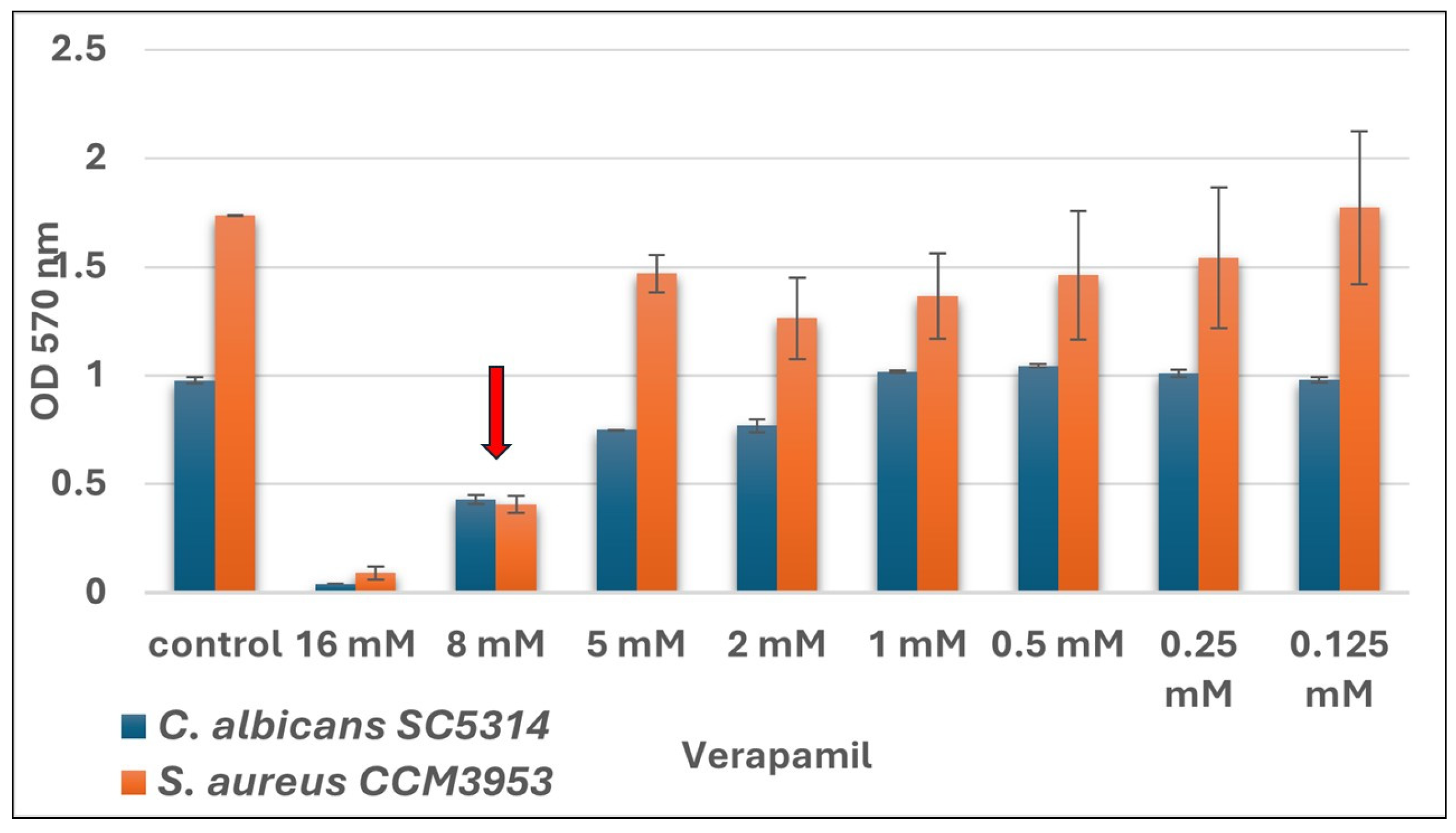
VER is a calcium L-type channel inhibitor, primarily used in human medicine in the therapy of cardiovascular diseases or high blood pressure [35]. However, some studies suggest its antimicrobial potential [25,36,37,38,39]. C. albicans is the most common fungal pathogen associated with nosocomial infections and biofilm [40]. Due to similarity with the L-type channel, VER can block the level of intracellular calcium in this yeast as well [41,42]. Calcium is important for cell homeostasis, yeast-to-hyphae transition, or for influencing the activation of important transcription factors like CRZ1 associated with the triggering of gene expression of the virulence- or resistance-associated genes in yeasts [42,43]. Hyphae are the most invasive morphological form of C. albicans and play a key role in polymicrobial biofilms, especially in mixed biofilms of C. albicans and S. aureus [44]. Bacteria use hyphae as a “skeleton” for attachment and to strengthen adhesion during biofilm development [45]. This study was focused on determining the antimicrobial activity of VER against biofilms of C. albicans and S. aureus in vitro and in vivo using the G. mellonella model as well as to determine its role in the regulation of the expression of the ALS3 and HWP1 genes that are important in species interactions within mixed biofilm of C. albicans and S. aureus. Prior to the main experiments, we tested the antimicrobial activity of VER by the determination of SMIC50 of VER to single biofilms of C. albicans and S. aureus. The susceptibility of biofilms was tested for eight different concentrations of VER. Results showed the same rate (8 mM) for SMIC50 for biofilms of C. albicans SC5314 and S. aureus CCM3953 as well. Results are illustrated in Figure 1.
There are a few studies about the antibiofilm activity of VER against biofilms of Candida sp. [38,39,42]. Vega-Chacón et al. suggested SMIC50 of VER against single biofilms as 8 and 16 mg/mL, respectively, which is slightly more than our results. According to the molecular weight of VER, our SMIC50 = 8 mM is equal to 4 mg/mL. However, the study of Yu et al. reported SMIC50 of Ver as 5 mg/mL, which is in correlation with our results [24,42]. The activity of VER was tested against biofilms of non-albicans Candida as well. Scorzioni et al. reported SMIC50 of VER as 7.25 mM for Candida krusei, Candida parapsilosis and Candida glabrata [38]. In conclusion, the antibiofilm activity varies among each Candida species. On the other hand, the effectiveness of VER against biofilms of S. aureus has not been tested yet. However, few studies about the antimicrobial potential and inhibition of efflux pumps in bacteria leading to increased susceptibility of bacteria to antibiotics, or the use of VER in combination with antibiotics to enhance its activity, have been published [28,36,46,47]. Our results showed SMIC50 = 8 mM against biofilm of S. aureus; however, a slight inhibition (below 50%) has been observed as well in concentrations of 2 mM and 4 mM of VER (Figure 1). Recently, Yan et al. tested the inhibitory effect of calcium channel blockers, including VER, against the bacterium Pseudomonas aeruginosa. They confirmed the antibacterial potential of VER, depending on the applied concentration. VER inhibited the growth of P. aeruginosa at a concentration of 512 ug/mL, which is equal to 1 mM of VER [47]. Furthermore, another study used VER as an inhibitor of the efflux pump of resistant S. aureus, and the authors reported MIC50 as 1 mg/mL (2 mM) [37]. Both data are slightly lower than our results; the authors have not tested the activity of VER against biofilms and, moreover, in the first mentioned study the authors used P. aeruginosa, the Gram-negative bacteria with a different composition of the cell wall than the Gram-positive S. aureus used in our study. However, the use of calcium channel blockers seems to be a very promising strategy to combat bacteria in general [36,47].

3.2. Effectiveness of VER Against Polymicrobial Biofilm of C. albicans and S. aureus

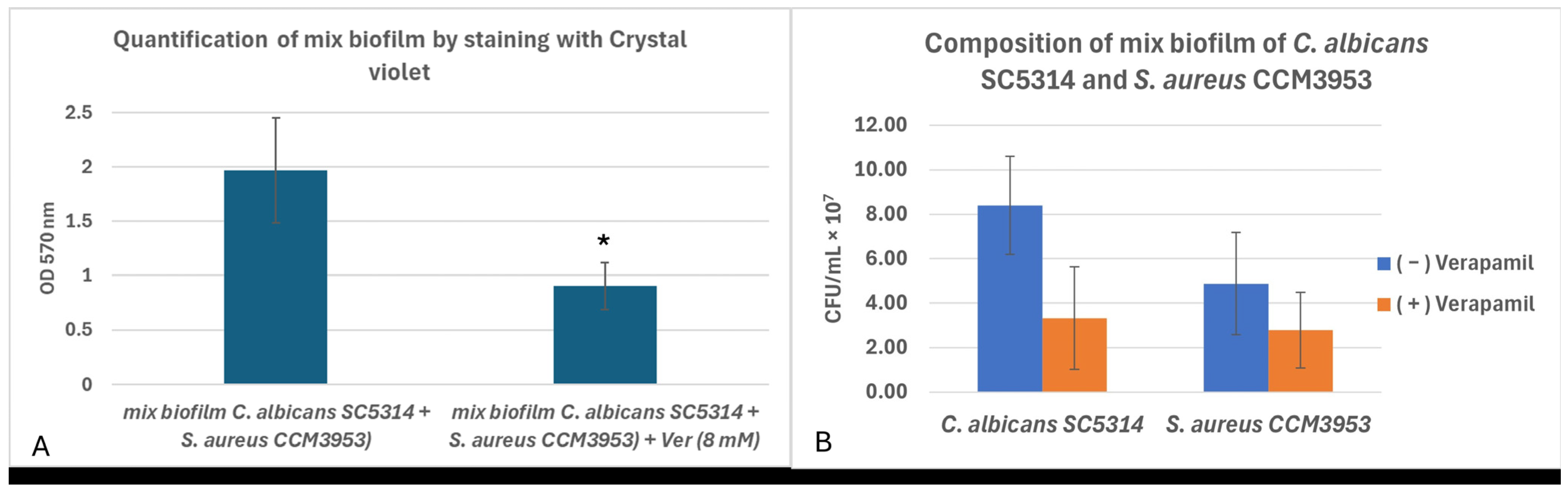
For the next experiments, SMIC50 8 mM of VER was tested against the polymicrobial biofilm of C. albicans and S. aureus. The significant inhibition activity of VER was observed against mixed biofilm (p = 0.048). The reduction in biomass in mixed biofilm was 60% after treatment with VER (Figure 2A). Additionally, the composition of mixed biofilm w/wo VER was examined by counting CFU/mL from 48 h biofilms as well. The amount of C. albicans SC 5314 decreased by 0.6 log and the amount of S. aureus CCM 3953 decreased by 0.2 log in the presence of VER. (Figure 2B).
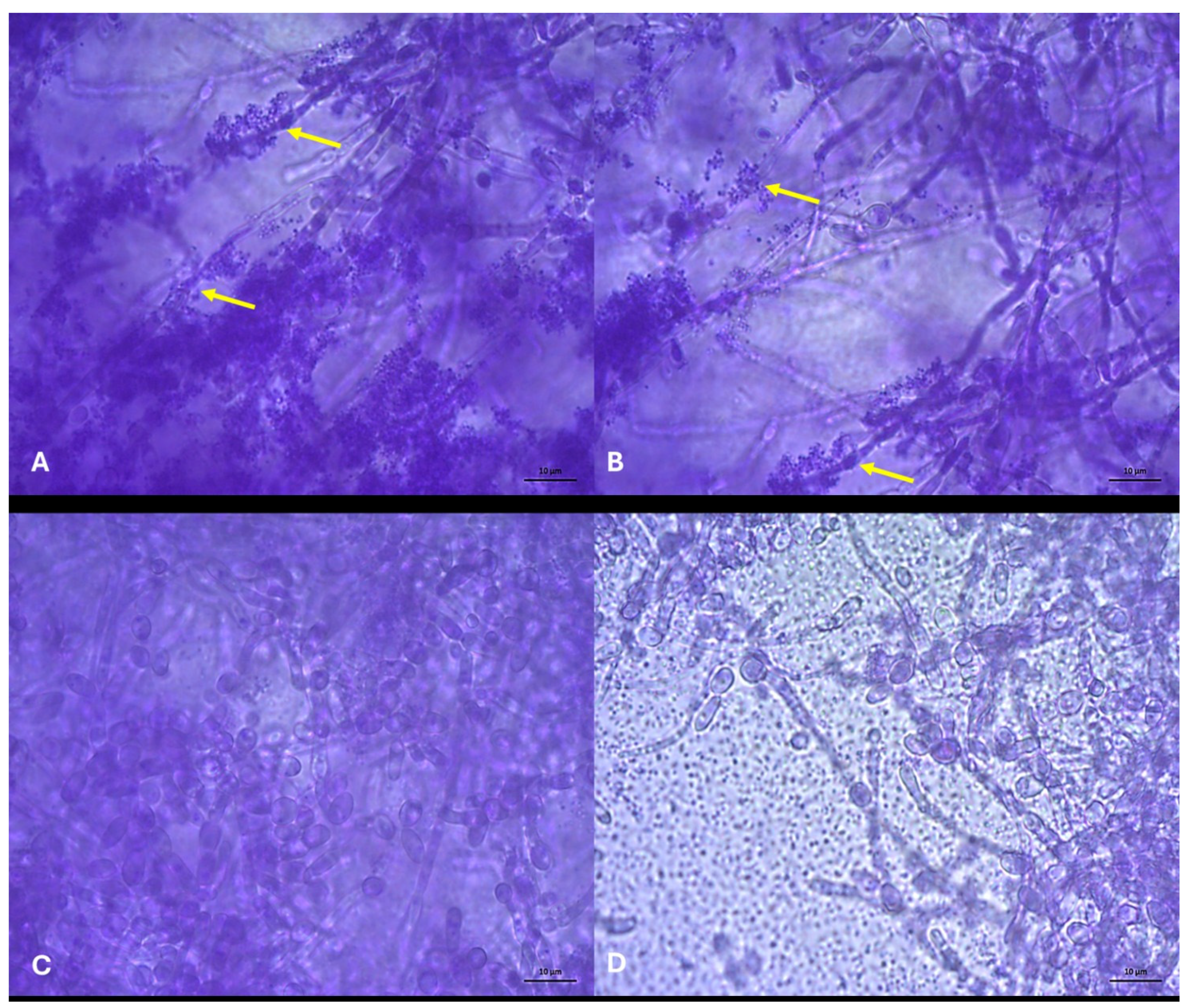
Additionally, the results were supported by microscopical analyses of polymicrobial biofilm w/wo VER. In the mixed biofilms of C. albicans and S. aureus without any treatment, huge hyphae forms of Candida and bunches of bacterial aggregates bound across hyphae were observed. Interestingly, in the biofilm formed in the presence of VER, the morphology of C. albicans was changed, and a decrease in the adhesion of S. aureus cells was observed (Figure 3).
The presence of hyphae is very typical for C. albicans biofilms, and this morphological form is considered to be more invasive [48]. In the mixed biofilms of C. albicans and S. aureus the presence of hyphae is essential for bacterial adhesion [12,44]. S. aureus uses the ALS3 protein of C. albicans to adhere to the surface hyphae. This interaction facilitates the synergistic growth of biofilms, enhancing virulence and allowing S. aureus to “hitchhike” on C. albicans filaments to invade and disseminate within host tissues [12,13]. The level of intracellular calcium in yeasts is important to cell homeostasis as well as fungal growth and hyphal development [43]. Hyphae of the dimorphic fungus C. albicans exhibit directional tip responses when grown in contact with surfaces. On hard surfaces or in liquid media, the trajectory of hyphal growth is typically linear, with tip re-orientation events limited to encounters with topographical features (thigmotropism). In contrast, when grown on semisolid surfaces, the tips of C. albicans hyphae grow in an oscillatory manner to form regular two-dimensional sinusoidal curves and three-dimensional helices [49]. Brand et al. confirmed that calcium signaling and homeostasis are required for normal sinusoidal growth of hyphae and its production in C. albicans is attenuated when Ca2+ homeostasis is perturbed [49]. The yeast thigmotropism is calcium-dependent and is attenuated by a blockade of calcium signaling pathways; for example, by the chelation of extracellular calcium or by the deletion of the stretch-activated calcium channel, Mid1. This channel is thought to act as a mechanosensor for external, contact-mediated interactions and may also serve to mark the site of new tip expansion via localized calcium influx. Mid1 is a putative regulator of the voltage-gated calcium channel, Cch1, and together they control calcium influx into the cell [50]. Calcium ions are an important second messenger in developmental and stress signaling pathways, where a rise in cytosolic calcium activates the calcium-dependent signaling pathway via the phosphatase, calcineurin [51,52] and the calcineurin-dependent transcription factor, Crz1 [43,51,53]. Fungal cells maintain a low cytosolic calcium concentration of ∼100 nM by expelling ions from the cytosol into intracellular organelles, such as the Golgi or the vacuole, and may potentially also expel calcium ions from the cytosol across the plasma membrane to the exterior. This ensures that relatively low calcium fluxes can result in significant changes in cytoplasmic Ca2+ concentration and hence a high responsiveness of the signaling pathway [49]. Calcium channel inhibitors, such as VER, can block the proper influx of calcium into the cells and change fungal homeostasis, resulting in improper Ca2+ concentration that is critical for yeast-to-hyphae transition and growth, thus the production and orientation of fungal hyphae are blocked [47,49]. Overall, S. aureus could not attach to the fungus properly, resulting in decreased adhesion to the fungus and a reduction in the ability of bacteria to form biofilm. Our results confirmed for the first time that VER regulates the morphology of C. albicans, resulting in changes in the composition of biofilm and adhesion of S. aureus in mixed biofilm.

3.3. Verapamil Regulates Relative Changes in Expression of Biofilm-Associated Genes ALS3 and HWP1 Within Mixed Biofilm of C. albicans and S. aureus

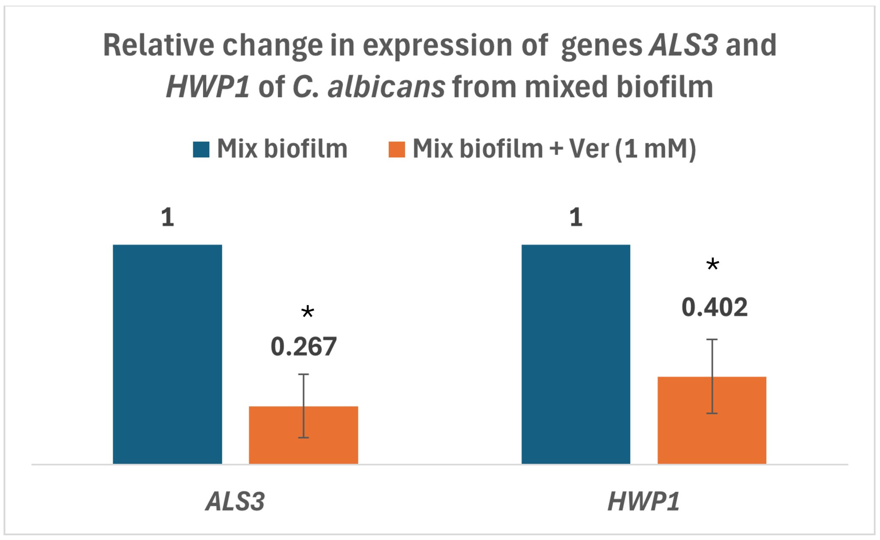
Quantitative real-time PCR was used to determine changes in the HWP1 and ALS3 gene expression in mixed biofilms formed w/wo subinhibitory concentration (1 mM) of VER. Results showed a significant decrease in expression of both genes (2× for ALS3; p = 0.018 and 3× for HWP1; p = 0.037) (Figure 4).
The concentration of intracellular calcium is important in yeast virulence [49]. Calcium is important to the activation of the yeast transcriptional factor CRZ1 and its translocation to the nucleus and further regulation of virulence or resistance-associated gene expression [52]. Some of those genes, such as HWP1 or ALS3, are associated with biofilm development of C. albicans [54,55]. The HWP1 gene genetically regulates the yeast-to-hyphae transition in C. albicans. Downregulation of this gene may be a result of the inhibition of calcium levels by VER. Similarly, Yu et al. described downregulation of the HWP1 gene under treatment of C. albicans biofilm by VER [24]. The ALS3 gene is important in the first stage of biofilm development of C. albicans as well [55]. Downregulation of this gene was observed in the presented study, which could lead to a decrease in ALS3 protein abundance in the biofilm of C. albicans. In mixed biofilm of C. albicans and S. aureus, bacteria use the ALS3 protein to attach to the fungus [12]. Our results proved that in the presence of VER, the attachment and adhesion of S. aureus to C. albicans was reduced, resulting in the incomplexity of mixed biofilm. Targeting antibiofilm strategies to the ALS3 protein is promising in polymicrobial biofilms. Interestingly, the study of Schmidt et al. revealed the inhibition activity of a fungal vaccine, NDV-3, contains the N-terminal portion of the C. albicans agglutinin-like sequence 3 protein (Als3p), and demonstrated that the Als3p vaccine antigen protects mice from neither oropharyngeal, vaginal and intravenous challenges with C. albicans and other selected species of Candida as well as both intravenous challenges and skin and soft tissue infection with S. aureus [56]. Targeting antimicrobial fight by blocking adhesins ALS3 against adhesins of Candida can be very promising as well to combat S. aureus due to the sequence and structural homology of ALS3 with cell surface proteins on S. aureus [57]. In our study, the calcium channel inhibitor, VER, effectively inhibited the yeast-to-hyphae transition and downregulated the expression of ALS3 and HWP1 genes as well as subsequently blocking the adhesion and biofilm development of S. aureus in mixed biofilm with C. albicans.

3.4. Antimicrobial Effectiveness of VER to Co-Infection of C. albicans and S. aureus In Vivo Using G. mellonella

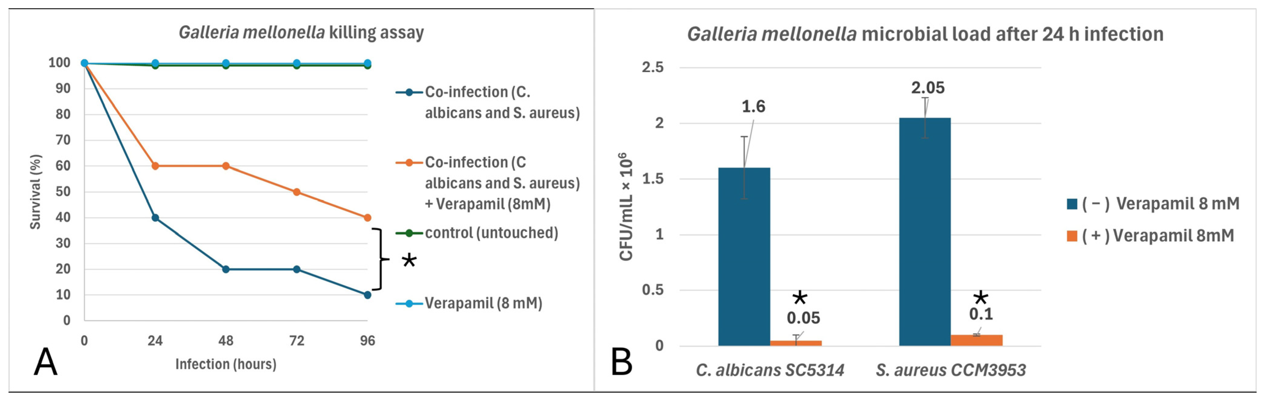
G. mellonella (the greater wax moth) larvae are used extensively across biological sciences as infection models for microbial species, and for toxicity testing of novel drug compounds [58]. Studies have shown a strong correlation between toxicity data obtained in G. mellonella and those from mammalian models, including mice and rats [59,60,61]. In our study, in vivo experiments with G. mellonella confirmed the antibiofilm activity of VER against mixed infection of C. albicans and S. aureus as well. First, the toxicity of VER (8 mM) was excluded in the G. mellonella model. Afterwards, this concentration of VER was used in the treatment of co-infection of C. albicans and S. aureus. Survival of larvae significantly increased by 30% (Figure 5A). The differences between both killing curves were statistically evaluated according to the Mantel–Cox log-rank test (p = 0.0336, HR 2.545, 95% CI 0.8787–7.370). Microbial load after 24 h from infection treated with VER was significantly reduced as well; p = 0.05 for C. albicans SC5314 and p = 0.049 for S. aureus CCM3953 (Figure 5B). The reduction in C. albicans was observed in 2 log and the reduction in S. aureus was about 1 log in the presence of VER.
The antimicrobial potential of VER in vivo was tested in some other studies as well [24,39], but none of them performed a treatment of co-infection with different microbial species as was done in the presented study. In the research of Vega-Chacón et al., the authors tested the inhibition of VER alone and in combination with fluconazole to the Candida sp. in vivo using G. mellonella. Similarly to our results, VER increased the survival of larvae infected with the Candida sp. as well as reduced fungal load after infection. Interestingly, the effectiveness of VER was better in the case of fluconazole-resistant C. albicans. The azole-resistant Candida sp. has increased activity of the efflux pump system. The main hypothesis was that, due to an increased efflux system, VER could be effectively transported to the cells in case of a resistant Candida sp. using efflux pumps as a substrate, and furthermore could inhibit calcium channels inside the cells and trigger reactions leading to the blocking of the yeast-to-hyphae transition and downregulation of hyphal-associated genes in the Candida sp. However, in the study presented, no resistant clinical isolates were used. Another in vivo study revealed that VER decreased invasion and the adhesion of hyphae of C. albicans in the mouse model [24]. The authors investigated the in vivo effect of Verapamil on C. albicans morphogenesis using gastrointestinal colonization models. In the antibiotic-free mouse model, VER 10 mg (kg day) was exposed to mice with C. albicans infection, and then the CFUs of C. albicans were monitored daily in the fecal pellets of the mice. The amount of CFUs of C. albicans in the group treated with VER decreased 2-fold on the 7th day post-inoculation and decreased more than 15-fold on the 14th day compared with the control group. Verapamil displayed an inhibitory effect on C. albicans gastrointestinal colonization, which may be associated with its effect on hyphal development and the adhesion of this pathogen [24]. The obtained results agree with the study mentioned, but in this work the G. mellonella model was used, and the results of the experiment were collected after 24 h post-infection. G. mellonella larvae are widely used as an infection model to assess microbial virulence, conduct drug toxicity testing, and serve as a preliminary means of evaluating the in vivo efficacy of novel antimicrobial compounds by reducing the use of mammalian models, in line with the principles of reduction, refinement, and replacement that govern the ethical use of mammalian species in research [58]. In conclusion, using live-model organisms is necessary in testing the potential of drugs for repurposing. A potential limitation of repurposing VER, and of this study as well, lies in the strict dosage of VER, due to its potential toxicity relating to the overdose [62]. The daily dose of VER for humans is 480 mg (in two or three doses) [63]. Overall, in clinical repurposing of VER the max daily dose must be taken into consideration, thus the potential application of VER for the treatment of biofilms must be done accurately and very carefully. For future experiments we would like to focus on the preparation of nanomaterial or hydrogels with a slow release of lower concentrations of VER for the treatment of superficial infections or biofilms that would be effective against pathogens but still safe for humans. Additionally, we would also like to perform testing on ex vivo models (for topical application), like the ex vivo mouse tongue biofilm model, RHE (Reconstituted Human Epithelium) model or porcine skin model, and in vivo testing on mice should be done before any other clinical repurposing of VER.

4. Conclusions

For the first time, this study used VER, a calcium channel inhibitor, in the treatment of polymicrobial biofilm of C. albicans and S. aureus. The obtained results suggested that VER not only inhibited the mixed C. albicans and S. aureus biofilm, but also regulated yeast morphogenesis, thereby reducing the adhesion of S. aureus in the mixed biofilm. The regulation of the expression of the genes responsible for hyphal formation, ALS3 and HWP1, was reduced, thus clearly reducing hyphal formation of C. albicans, which also affected bacterial adherence in vitro and in vivo. The repurposing of drugs is a promising strategy to combat biofilm-associated infections. Overall, VER is an existing drug for other conditions, making it a potential candidate for drug repurposing to treat fungal or bacterial infections. Its ability to act synergistically with other drugs and combat resistance makes it a promising option to consider for treating biofilm-associated infections, especially in high-risk patients.

Supplementary Materials

The following supporting information can be downloaded at: https://www.mdpi.com/article/10.3390/hygiene6010007/s1, Table S1: List of oligonucleotide sequences used in this study; Figure S1: Visualization of PCR products by gel electrophoresis in 2% agarose gel; Figure S2: Melting curves/peaks of fungal ACT1 (A), ALS3 (B) and HWP1(C) genes qPCR products.

Author Contributions

Conceptualization, J.D.; methodology, J.D.; validation, J.D. and L.Č.; formal analysis, L.Č.; investigation, J.D.; writing—original draft preparation, J.D.; writing—review and editing, J.D. and L.Č.; All authors have read and agreed to the published version of the manuscript.

Funding

This research was funded by the Slovak Research and Development Agency under contracts of no. APVV-21-0302; project VEGA 1/0240/23 supported by the Ministry of Education, Research, Development and Youth of the Slovak Republic, and by EU Next Generation EU through the Recovery and Resilience Plan Slovakia under the project no. 09I01-03-V04-00022.

Institutional Review Board Statement

Not applicable.

Informed Consent Statement

Not applicable.

Data Availability Statement

The original contributions presented in this study are included in the article and Supplementary Materials. Further inquiries can be directed to the corresponding author.

Conflicts of Interest

The authors declare no conflicts of interest.

References

  1. Kovács, F.; Balla, N.; Bozó, A.; Harmath, A.; Jakab, Á.; Tóth, Z.; Nagy, F.; Majoros, L.; Kovács, R. Epidemiology, clinical characteristics, outcome and biofilm forming properties in candidaemia: A single-centre retrospective 4-year analysis from Hungary. Mycoses 2024, 67, e13727. [Google Scholar] [CrossRef]
  2. Hoenigl, M.; Salmanton-García, J.; Egger, M.; Gangneux, J.-P.; Bicanic, T.; Arikan-Akdagli, S.; Alastruey-Izquierdo, A.; Klimko, N.; Barac, A.; Özenci, V.; et al. ECMM Candida III Study Group. Guideline adherence and survival of patients with candidaemia in Europe: Results from the ECMM Candida III multinational European observational cohort study. Lancet Infect. Dis. 2023, 23, 751–761. [Google Scholar] [CrossRef]
  3. Atriwal, T.; Azeem, K.; Husain, F.M.; Hussain, A.; Khan, M.N.; Alajmi, M.F.; Abid, M. Mechanistic Understanding of Candida albicans Biofilm Formation and Approaches for Its Inhibition. Front. Microbiol. 2021, 12, 638609. [Google Scholar] [CrossRef] [PubMed]
  4. Paramonova, E.; Krom, B.P.; van der Mei, H.C.; Busscher, H.J.; Sharma, P.K. Hyphal content determines the compression strength of Candida albicans biofilms. Microbiology 2009, 155, 1997–2003. [Google Scholar] [CrossRef] [PubMed]
  5. Dekkerová-Chupáčová, J.; Borghi, E.; Morace, G.; Bujdáková, H. Up-Regulation of Antimicrobial Peptides Gallerimycin and Galiomicin in Galleria mellonella Infected with Candida Yeasts Displaying Different Virulence Traits. Mycopathologia 2018, 183, 935–940. [Google Scholar] [CrossRef] [PubMed]
  6. Jacobsen, I.D.; Wilson, D.; Wächtler, B.; Brunke, S.; Naglik, J.R.; Hube, B. Candida albicans dimorphism as a therapeutic target. Expert Rev. Anti-Infect. Ther. 2012, 10, 85–93. [Google Scholar] [CrossRef]
  7. Hawser, S.P.; Douglas, L.J. Biofilm formation by Candida species on the surface of catheter materials in vitro. Infect. Immun. 1994, 62, 915–921. [Google Scholar] [CrossRef]
  8. Ponde, N.O.; Lortal, L.; Ramage, G.; Naglik, J.R.; Richardson, J.P. Candida albicans biofilms and polymicrobial interactions. Crit. Rev. Microbiol. 2021, 47, 91–111. [Google Scholar] [CrossRef]
  9. O’Donnell, L.E.; Millhouse, E.; Leighann Sherry, L.; Kean, R.; Malcolm, J.; Nile, C.J.; Ramage, G. Polymicrobial Candida biofilms: Friends and foe in the oral cavity. FEMS Yeast Res. 2015, 15, fov077. [Google Scholar] [CrossRef]
  10. Harriott, M.M.; Noverr, M.C. Candida albicans and Staphylococcus aureus form polymicrobial biofilms: Effects on antimicrobial resistance. Antimicrob. Agents Chemother. 2009, 53, 3914–3922. [Google Scholar] [CrossRef]
  11. Kean, R.; Rajendran, R.; Haggarty, J.; Townsend, E.M.; Short, B.; Burgess, K.E.; Lang, S.; Millington, O.; Mackay, W.G.; Williams, C.; et al. Candida albicans Mycofilms Support Staphylococcus aureus Colonization and Enhances Miconazole Resistance in Dual-Species Interactions. Front. Microbiol. 2017, 8, 258. [Google Scholar] [CrossRef]
  12. Peters, B.M.; Ovchinnikova, E.S.; Krom, B.P.; Schlecht, L.M.; Zhou, H.; Hoyer, L.L.; Busscher, H.J.; van der Mei, H.C.; Jabra-Rizk, M.A.; Shirtliff, M.E. Staphylococcus aureus adherence to Candida albicans hyphae is mediated by the hyphal adhesin Als3p. Microbiology 2012, 158, 2975–2986. [Google Scholar] [CrossRef]
  13. Van Dyck, K.; Viela, F.; Mathelié-Guinlet, M.; Demuyser, L.; Hauben, E.; Jabra-Rizk, M.A.; Velde, G.V.; Dufrêne, Y.F.; Krom, B.P.; Van Dijck, P. Adhesion of Staphylococcus aureus to Candida albicans During Co-Infection Promotes Bacterial Dissemination Through the Host Immune Response. Front. Cell. Infect. Microbiol. 2021, 10, 624839. [Google Scholar] [CrossRef] [PubMed]
  14. Liu, Y.; Filler, S.G. Candida albicans Als3, a multifunctional adhesin and invasin. Eukaryot. Cell 2011, 10, 168–173. [Google Scholar] [CrossRef] [PubMed]
  15. Simões, M.; Simões, L.C.; Vieira, M.J. A review of current and emergent biofilm control strategies. LWT—Food Sci. Technol. 2010, 43, 573–583. [Google Scholar] [CrossRef]
  16. Sharma, S.; Mohler, J.; Mahajan, S.D.; Schwartz, S.A.; Bruggemann, L.; Aalinkeel, R. Microbial Biofilm: A Review on Formation, Infection, Antibiotic Resistance, Control Measures, and Innovative Treatment. Microorganisms 2023, 11, 1614. [Google Scholar] [CrossRef]
  17. European Centre for Disease Prevention and Control. Antimicrobial Resistance in the EU/EEA (EARS-Net)—Annual Epidemiological Report 2024; ECDC: Stockholm, Sweden, 2025. [Google Scholar]
  18. Organisation for Economic Co-operation and Development (OECD). Fighting Antimicrobial Resistance in the EU/EEA, Embracing a One Health Approach; OECD: Paris, France, 2023; Available online: http://oe.cd/amr-eaad2023 (accessed on 30 December 2025).
  19. Aloni-Grinstein, R.; Mamroud, E.; Gal, Y. New Frontiers for Old Medications: Repurposing Approved Drugs Against Gram-Negative Bacterial Infections. Microorganisms 2025, 13, 2115. [Google Scholar] [CrossRef]
  20. Roudbary, M.; Branquinha, M.H.; Santos, A.L.S. Editorial: Drug repurposing to fight resistant fungal species: Recent developments as novel therapeutic strategies. Front. Cell. Infect. Microbiol. 2025, 15, 1633037. [Google Scholar] [CrossRef]
  21. Simpson, W.G. The calcium channel blocker verapamil and cancer chemotherapy. Cell Calcium 1985, 6, 449–467. [Google Scholar] [CrossRef]
  22. Sueta, D.; Tabata, N.; Hokimoto, S. Clinical roles of calcium channel blockers in ischemic heart diseases. Hypertens. Res. 2017, 40, 423–428. [Google Scholar] [CrossRef]
  23. McKeever, R.G.; Patel, P.; Hamilton, R.J. Calcium Channel Blockers. In StatPearls [Internet]; StatPearls Publishing: Treasure Island, FL, USA, 2025. [Google Scholar]
  24. Yu, Q.; Ding, X.; Zhang, B.; Xu, N.; Jia, C.; Mao, J.; Zhang, B.; Xing, L.; Li, M. Inhibitory effect of verapamil on Candida albicans hyphal development, adhesion and gastrointestinal colonization. FEMS Yeast Res. 2014, 14, 633–641. [Google Scholar] [CrossRef] [PubMed]
  25. Feng, W.; Yang, J.; Ma, Y.; Xi, Z.; Ren, Q.; Wang, S.; Ning, H. Aspirin and verapamil increase the sensitivity of Candida albicans to caspofungin under planktonic and biofilm conditions. J. Glob. Antimicrob. Resist. 2021, 24, 32–39. [Google Scholar] [CrossRef] [PubMed]
  26. Adams, K.N.; Szumowski, J.D.; Ramakrishnan, L. Verapamil, and its metabolite norverapamil, inhibit macrophage-induced, bacterial efflux pump-mediated tolerance to multiple anti-tubercular drugs. J. Infect. Dis. 2014, 210, 456–466. [Google Scholar] [CrossRef] [PubMed]
  27. Fountain, A.J.; Waller, N.J.E.; Cheung, C.Y.; Jowsey, W.; Chrisp, M.T.; Troll, M.; Edelstein, P.H.; Cook, G.M.; McNeil, M.B.; Ramakrishnan, L. Verapamil and its metabolite norverapamil inhibit the Mycobacterium tuberculosis MmpS5L5 efflux pump to increase bedaquiline activity. Proc. Natl. Acad. Sci. USA 2025, 122, e2426827122. [Google Scholar] [CrossRef]
  28. Ahmed, N.; Biswas, P.; Mogal, R.; Sarker, R.; Tareq, M.I.; Ahmed, S.; Akter, M.; Miah, T.; Kundo, N.K.; Hasan, N.; et al. Breaking down resistance: Verapamil analogues augment the efficacy of antibiotics against Streptococcus pneumoniae via MATE transporter interference. Inform. Med. Unlocked 2024, 47, 101493. [Google Scholar] [CrossRef]
  29. Gillum, A.M.; Tsay, E.Y.; Kirsch, D.R. Isolation of the Candida albicans Gene for Orotidine-5′-Phosphate Decarboxylase by Complementation of S. cerevisiae Ura3 and E. coli pyrF Mutations. Mol. Gen. Genet. 1984, 198, 179–182. [Google Scholar] [CrossRef]
  30. Ramage, G.; Van de Walle, K.; Wickes, B.L.; Lopez-Ribot, J.L. Standardized method for in vitro antifungal susceptibility testing of Candida albicans. Antimicrob. Agents Chemother. 2001, 45, 2475–2479. [Google Scholar] [CrossRef]
  31. Merritt, J.H.; Kadouri, D.E.; O’Toole, G.A. Growing and analyzing static biofilms. Curr. Protoc. Microbiol. 2005, 1, 1B.1.1–1B.1.17. [Google Scholar] [CrossRef]
  32. Kendra, S.; Czucz Varga, J.; Gaálová-Radochová, B.; Bujdáková, H. Practical application of PMA-qPCR assay for determination of viable cells of inter-species biofilm of Candida albicans-Staphylococcus aureus. Biol. Methods Protoc. 2024, 9, bpae081. [Google Scholar] [CrossRef]
  33. Livak, K.J.; Schmittgen, T.D. Analysis of relative gene expression data using real-time quantitative PCR and the 2(-Delta Delta C(T)) Method. Methods 2001, 4, 402–408. [Google Scholar] [CrossRef] [PubMed]
  34. Genç, T.T.; Kaya, S.; Günay, M.; Çakaloğlu, Ç. Humoral immune response of Galleria mellonella after mono- and co-injection with Hypericum perforatum extract and Candida albicans. APMIS 2024, 132, 358–370. [Google Scholar] [CrossRef] [PubMed]
  35. Brogden, R.N.; Benfield, P. Verapamil: A review of its pharmacological properties and therapeutic use in coronary artery disease. Drugs 1996, 51, 792–819. [Google Scholar] [PubMed]
  36. Tiwana, G.; Cock, I.E.; Taylor, S.M.; Cheesman, M.J. Beyond Antibiotics: Repurposing Non-Antibiotic Drugs as Novel Antibacterial Agents to Combat Resistance. Int. J. Mol. Sci. 2025, 26, 9880. [Google Scholar] [CrossRef] [PubMed]
  37. Baiomy, A.A.; Shaker, G.H.; Abbas, H.A. Sensitizing multi drug resistant Staphylococcus aureus isolated from surgical site infections to antimicrobials by efflux pump inhibitors. Afr. Health Sci. 2020, 20, 1632–1645. [Google Scholar] [CrossRef]
  38. Scorzoni, L.; Menezes, R.T.; Pereira, T.C.; Oliveira, P.S.; Ribeiro, F.D.C.; Santos, E.L.D.S.; Fugisaki, L.R.; DE Oliveira, L.D.; Amorim, J.B.O. Antifungal and anti-biofilm effect of the calcium channel blocker verapamil on non-albicans Candida species. An. Acad. Bras. Cienc. 2020, 92, e20200703. [Google Scholar] [CrossRef]
  39. Vega-Chacón, Y.; de Albuquerque, M.C.; Pavarina, A.C.; Goldman, G.H.; Mima, E.G.d.O. Verapamil inhibits efflux pumps in Candida albicans, exhibits synergism with fluconazole, and increases survival of Galleria mellonella. Virulence 2021, 12, 231–243. [Google Scholar] [CrossRef]
  40. Amann, V.; Kissmann, A.K.; Firacative, C.; Rosenau, F. Biofilm-Associated Candidiasis: Pathogenesis, Prevalence, Challenges and Therapeutic Options. Pharmaceuticals 2025, 18, 460. [Google Scholar] [CrossRef]
  41. Teng, J.; Goto, R.; Iida, K.; Kojima, I.; Iida, H. Ion-channel blocker sensitivity of voltage-gated calcium-channel homologue Cch1 in Saccharomyces cerevisiae. Microbiology 2008, 154, 3775–3781. [Google Scholar]
  42. Yu, Q.; Ding, X.; Xu, N.; Cheng, X.; Qian, K.; Zhang, B.; Xing, L.; Li, M. In vitro activity of verapamil alone and in combination with fluconazole or tunicamycin against Candida albicans biofilms. Int. J. Antimicrob. Agents 2013, 41, 179–182. [Google Scholar] [CrossRef]
  43. Li, W.; Shrivastava, M.; Lu, H.; Jiang, Y. Calcium-calcineurin signaling pathway in Candida albicans: A potential drug target. Microbiol. Res. 2021, 249, 126786. [Google Scholar] [CrossRef]
  44. Dühring, S.; Schuster, S. Studying mixed-species biofilms of Candida albicans and Staphylococcus aureus using evolutionary game theory. PLoS ONE 2024, 19, e0297307. [Google Scholar] [CrossRef]
  45. Jing, Q.; Liu, R.; Jiang, Q.; Liu, Y.; He, J.; Zhou, X.; Yu, O.Y.; Chu, C.-H.; Cheng, L.; Ren, B.; et al. Staphylococcus aureus wraps around Candida albicans and synergistically escapes from Neutrophil extracellular traps. Front. Immunol. 2024, 15, 1422440. [Google Scholar] [CrossRef]
  46. Liu, K.; Buitenhek, E.; Kuijl, C.P.; Mulla, Y.; Luirink, J.; Bald, D. Verapamil Suppresses the Development of Resistance Against Anti-Tuberculosis Drugs in Mycobacteria. Int. J. Mol. Sci. 2025, 26, 11124. [Google Scholar] [CrossRef]
  47. Yan, Z.; Xia, L.; Xu, X.; Ma, B.; Yuan, X.; Yang, K.; Li, K.; Ye, X.; Zhang, L.; Chen, T. Exploring calcium channel blocker as a candidate drug for Pseudomonas aeruginosa through network pharmacology and experimental validation. Chem. Biol. Drug Des. 2023, 102, 1353–1366. [Google Scholar] [CrossRef]
  48. Desai, J.V. Candida albicans Hyphae: From Growth Initiation to Invasion. J. Fungi 2018, 4, 10. [Google Scholar] [CrossRef] [PubMed]
  49. Brand, A.; Lee, K.; Veses, V.; Gow, N.A. Calcium homeostasis is required for contact-dependent helical and sinusoidal tip growth in Candida albicans hyphae. Mol. Microbiol. 2009, 71, 1155–1164. [Google Scholar] [CrossRef] [PubMed]
  50. Fischer, M.; Schnell, N.; Chattaway, J.; Davies, P.; Dixon, G.; Sanders, D. The Saccharomyces cerevisiae CCH1 gene is involved in calcium influx and mating. FEBS Lett. 1997, 419, 259–262. [Google Scholar] [CrossRef] [PubMed]
  51. Yu, Q.; Xu, N.; Li, M. Calcium homeostasis systems and calcium signaling pathways in Candida albicans—A review. Wei Sheng Wu Xue Bao 2012, 52, 422–428. (In Chinese) [Google Scholar]
  52. Onyewu, C.; Wormley, F.L., Jr.; Perfect, J.R.; Heitman, J. The calcineurin target, Crz1, functions in azole tolerance but is not required for virulence of Candida albicans. Infect. Immun. 2004, 72, 7330–7333. [Google Scholar] [CrossRef]
  53. Karababa, M.; Valentino, E.; Pardini, G.; Coste, A.T.; Bille, J.; Sanglard, D. CRZ1, a target of the calcineurin pathway in Candida albicans. Mol. Microbiol. 2006, 59, 1429–1451. [Google Scholar] [CrossRef]
  54. Nobile, C.J.; Nett, J.E.; Andes, D.R.; Mitchell, A.P. Function of Candida albicans Adhesin Hwp1 in Biofilm Formation. Eukaryot. Cell 2006, 5, 1604–1610. [Google Scholar] [CrossRef] [PubMed]
  55. Deng, K.; Jiang, W.; Jiang, Y.; Deng, Q.; Cao, J.; Yang, W.; Zhao, X. ALS3 Expression as an Indicator for Candida albicans Biofilm Formation and Drug Resistance. Front. Microbiol. 2021, 29, 655242. [Google Scholar] [CrossRef] [PubMed]
  56. Schmidt, C.S.; White, C.J.; Ibrahim, A.S.; Filler, S.G.; Fu, Y.; Yeaman, M.R.; Edwards, J.E., Jr.; Hennessey, J.P., Jr. NDV-3, a recombinant alum-adjuvanted vaccine for Candida and Staphylococcus aureus, is safe and immunogenic in healthy adults. Vaccine 2012, 30, 7594–7600. [Google Scholar] [CrossRef] [PubMed]
  57. Sheppard, D.C.; Yeaman, M.R.; Welch, W.H.; Phan, Q.T.; Fu, Y.; Ibrahim, A.S.; Filler, S.G.; Zhang, M.; Waring, A.J.; Edwards, J.E., Jr. Functional and structural diversity in the Als protein family of Candida albicans. J. Biol. Chem. 2004, 279, 30480–30489. [Google Scholar] [CrossRef]
  58. Barton, T.E.; Duignan, L.; Kadioglu, A.; Fothergill, J.L.; Neill, D.R. Galleria mellonella as an Antimicrobial Screening Model. J. Vis. Exp. 2024, 212, e67210. [Google Scholar] [CrossRef]
  59. Menard, G.; Rouillon, A.; Cattoir, V.; Donnio, P.Y. Galleria mellonella as a suitable model of bacterial infection: Past, present and future. Front. Cell Infect. Microbiol. 2021, 11, 782733. [Google Scholar]
  60. Piatek, M.; Sheehan, G.; Kavanagh, K. Galleria mellonella: The versatile host for drug discovery, in vivo toxicity testing and characterizing host-pathogen interactions. Antibiotics 2021, 10, 1545. [Google Scholar]
  61. Brennan, M.; Thomas, D.Y.; Whiteway, M.; Kavanagh, K. Correlation between virulence of Candida albicans mutants in mice and Galleria mellonella larvae. FEMS Immunol. Med. Microbiol. 2002, 34, 153–157. [Google Scholar] [CrossRef]
  62. Isbister, G.K.; Jenkins, S.; Harris, K.; Downes, M.A.; Isoardi, K.Z. Calcium channel blocker overdose: Not all the same toxicity. Br. J. Clin. Pharmacol. 2025, 91, 740–747. [Google Scholar] [CrossRef]
  63. Fahie, S.; Cassagnol, M. Verapamil. In StatPearls [Internet]; StatPearls Publishing: Treasure Island, FL, USA, 2025. Available online: https://www.ncbi.nlm.nih.gov/books/NBK538495/ (accessed on 30 December 2025).
Figure 1. SMIC50 (minimal inhibitory concentration) (marked with arrow) of VER to biofilms of C. albicans SC 5314 (blue) and S. aureus CCM3953 (orange).
Hygiene 06 00007 g001
Figure 2. (A) Crystal violet staining of mixed biofilms of C. albicans SC5314 and S. aureus CCM3953 w/wo VER (8 mM). (B) Counting of CFU/mL of C. albicans SC5413 and S. aureus CCM3953 from mixed biofilm w/wo Verapamil (8 mM) after 48 h of biofilm development. * p < 0.05.
Hygiene 06 00007 g002
Figure 3. Light microscopy of mixed biofilms of C. albicans SC5314 and S. aureus CCM3953 stained with crystal violet; (A,B) control native mixed biofilm, huge bunches of bacteria bound around hyphae of yeast are marked with an arrow; (C,D) mixed biofilm with VER (subinhibitory concentration, 1 mM). Reduction in hyphae and a decrease in bacterial adhesion to yeasts were observed.
Hygiene 06 00007 g003
Figure 4. Relative change in the gene expression of ALS3 and HWP1 genes associated with adhesion and hyphae development of C. albicans in mixed biofilms w/wo subinhibitory concentration of Verapamil (1 mM). * p < 0.05.
Hygiene 06 00007 g004
Figure 5. In vivo experiments with G. mellonella. (A) Survival of larvae after co-infection with C. albicans and S. aureus and treatment with VER (8 mM); (B) Analysis of microbial load after 24 h from co-infection and treatment with VER (8 mM). * p < 0.05.
Hygiene 06 00007 g005
Disclaimer/Publisher’s Note: The statements, opinions and data contained in all publications are solely those of the individual author(s) and contributor(s) and not of MDPI and/or the editor(s). MDPI and/or the editor(s) disclaim responsibility for any injury to people or property resulting from any ideas, methods, instructions or products referred to in the content.