


Abstract
The diversity of cyanobacteria from the semi-arid region of Rajasthan, India, remains vastly unexplored and warrants systematic investigation. We isolated two cyanobacterial strains (SN2022/33 & AT2016/25) of non-heterocytous, filamentous cyanobacterium from samples of sandy soil biological crusts and investigated them using a polyphasic approach. Based on 16S rRNA gene sequence identity, both strains formed a distinct lineage, with 16S sequence identity (p-distance) < 95% to the closest sister genera Trichocoleus, Venetifunis, Trichothermofontia, and Pinocchia. Analyses of 16S-23S Internal Transcribed Spacer (ITS) secondary structures (D1-D1′, BoxB, and V3 helixes) yielded substantial differences from phylogenetically associated taxa. Morphologically, both strains corresponded to members of the family Trichocoleusaceae (Leptolyngbyales), with tapered filaments and conical-pointed end cells. Most significantly, this taxon exhibited a form of true branching, with prolific unilateral or bilateral extrusions, something that had previously been the exclusive purview of members of the Nostocaceae. The combined evidence from conventional and molecular studies supports the recognition of the isolates as a novel taxon hereby described as Edaphifilum ginni gen. et sp. nov., in accordance with the International Code of Nomenclature (ICN) for Algae, Fungi, and Plants.
1. Introduction
Cyanobacteria are ancient, phototrophic microorganisms that emerged ca. 3.5 BYA and have played a vital role in creating an oxygen-rich atmosphere [1,2,3]. They are known for their vast ecological tolerance [4,5,6], including extreme environments (e.g., saline–alkaline lakes, hot springs, deserts, volcanic soils, and polar regions) [7]. The European and North American cyanobacterial diversity has been the object of much attention, although many regions have only had cursory attention paid thus far [8]. For example, numerous cyanobacterial lineages have been reported throughout the Indian subcontinent [9,10,11,12], with a recent surge in newly described cyanobacterial taxa from terrestrial habitats [13,14,15,16]. Nonetheless, cyanobacteria have been extensively studied within tropical regions of Indian subcontinent [17,18]; however, their occurrence in Northwest India remains poorly documented.
Cyanobacterial taxonomy has undergone significant revisions, shifting from traditional morphology-based classifications to advanced genetic and genomic phylogenetic frameworks [19,20]. This more comprehensive approach has led to the establishment of ten new orders and fifteen new families based primarily on genomic assessments [21]. However, the ability to exhibit “true-branching” has long been considered one of the stable morphological features of cyanobacterial taxonomy (sensu [22]). Exclusively the purview of members of the Nostocaceae, the ability to undergo cell division in multiple planes, coupled with the ability to produce “specialized cells” (i.e., heterocytes and akinetes), has set this lineage apart from all other cyanobacteria. Whether these features are obligatory or not led to the synonymizing the traditional orders Nostocales (inducible, non-obligatory) with the Stigonematales (obligatory), but no other cyanobacterial lineage has the “true-branching” feature.
The family Trichocoleusaceae was created based on comparisons of the 16S rRNA gene phylogenies and analyses of the secondary structures, particularly the nucleotide pattern in helices 23 and 27, which were used to split apart the polyphyletic branches of Leptolyngbya-like genera [23]. Furthermore, in the updated classification of cyanobacterial orders proposed using phylogenomic and polyphasic approaches, this family was retained as valid and monophyletic within the order Leptolyngbyales [21]. Currently, this family contains three validly described genera: Trichocoleus [24], Trichothermofontia [25] and Venetifunis [26]. The present study contributes to the α-level taxonomy of Trichocoleusaceae by describing an edaphic cyanobacterium from a semi-arid region of India. Two strains were isolated from biological crusts found in sandy soils of Rajasthan, which is characterized by poor water retention capacity and nutrient scarcity. The strains were investigated using a polyphasic approach integrating morphological data with 16S rRNA gene sequences, secondary structures of ITS regions, and ecology. We observed that the studied strains exhibit true branching, marking them as the first non-nostocalean taxa capable of such a feat. The 16S rRNA gene sequencing results confirmed the separate phylogenetic position of this morphotype in comparison to non-heterocytous cyanobacteria, and, hence, the studied strains are described as a new genus (Edaphifilum), with the type species E. ginni in accordance with the International Code of Nomenclature for Algae, Fungi, and Plants (ICN) employing the phylogenetic species concept [27].
2. Materials and Methods
2.1. Sampling and Cultivation
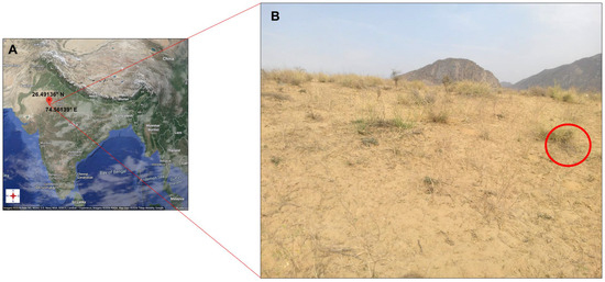
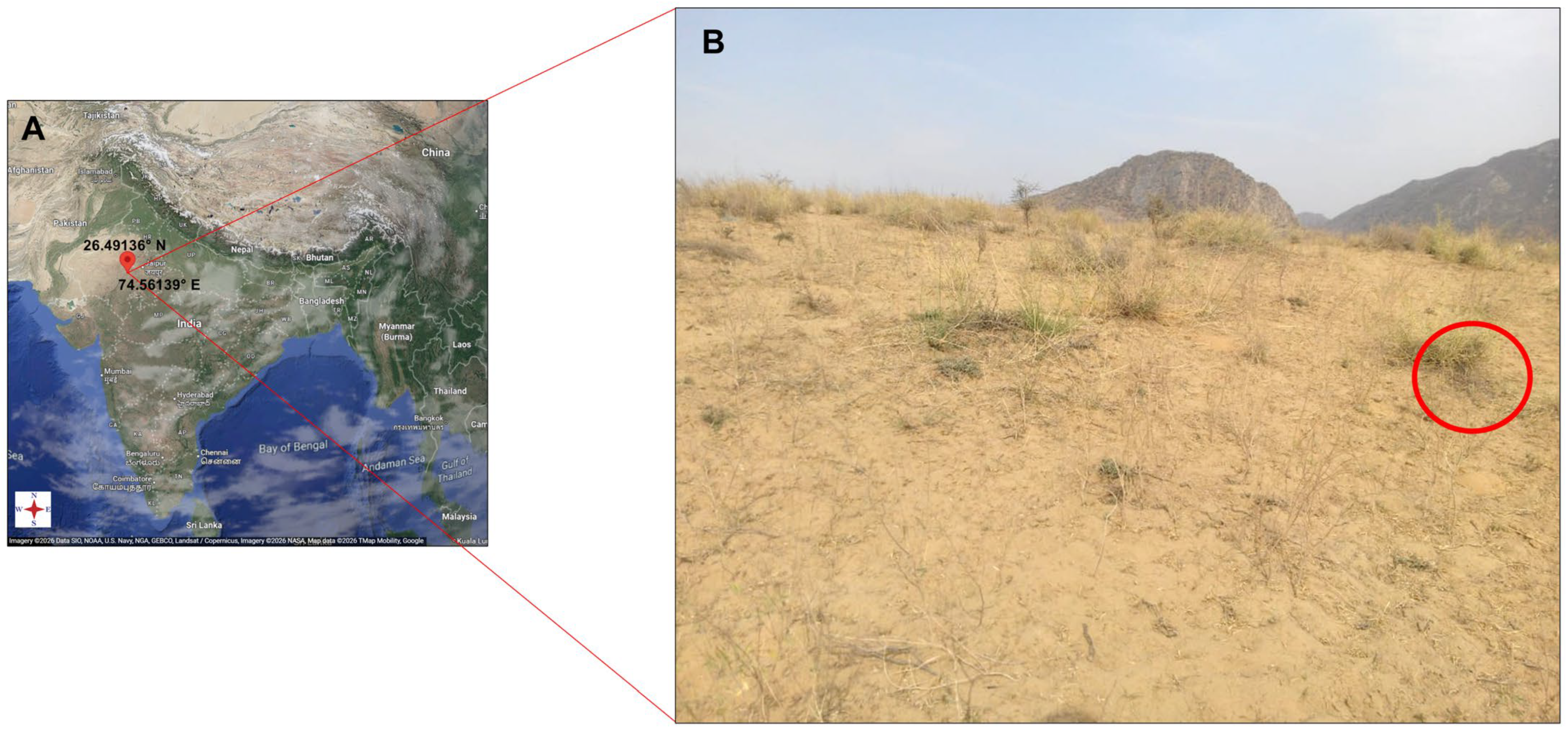
The sampling sites were two different locations in the Pushkar region of Rajasthan, India (Figure 1A). Biological crust samples from arid, sandy soils were collected in August 2015 (26°29′38.4″ N, 74°33′40.8″ E) and September 2022 (26°29′28.90″ N, 74°33′41.0″ E) (Figure 1B). Physico-chemical characteristics were recorded via a multi-parameter PCSTestrTM35 (Eutech instrument, Oakton, Singapore) (Table S1). Two strains (SN2022/33 & AT2016/25) were isolated using a Pasteur pipette with an optical microscope (Axio Lab Al, Carl Zeiss, Goettingen, Germany) until uni-algal cultures were established. The strains were grown in BG-11 medium [22] and kept under white, fluorescent lights (35 mE m−2 s−1), at 26 ± 2 °C, and a 12 h light/dark cycle. Strains were additionally cultured in nitrogen-free BG-11 medium to assess their capacity for heterocyte formation.
Figure 1.
(A) Location of a sampling site (Pushkar, Rajasthan, India); (B) sampling habitat (circle denotes the exact spot of sample collection). Map source: Google maps.
2.2. Morphological Analysis
The morphological characteristics of both strains (SN2022/33 & AT2016/25) were documented using an Axio Lab Al Light Microscope (Carl Zeiss; Goettingen, Germany). Differential Interference Contrast (DIC) greyscale microphotographs were obtained with a Leica DMi8 Confocal Inverted Microscope (Wetzlar, Germany), and bright-field DIC images were captured with a Nikon Y-TV55 camera (Tokyo, Japan). In addition, for septum visualization, fluorescence images were captured by applying the eGFP setting available in the confocal inverted microscope. Microphotographs were processed using Adobe Photoshop v11 to improve clarity and add annotations. A preliminary identification of studied strains was carried out using standard taxonomic keys [28].
2.3. Developmental Frequency of Branching
The frequency statistics of extrusions and both T- and V-type branching was inferred by observing 1000 filaments in a cumulative batch of 100 filaments. Afterwards, the variance-to-mean ratio (VMR) was calculated to test Poisson distribution fit for randomness. Chi-square goodness-of-fit test and paired-sample t-test were employed to assess the proportional dominance between the branching types with a null hypothesis of 1:1 expected ratio. Lastly, Pearson’s correlation analysis and simple linear regression were used to check the independence of occurrence in branching and extrusions. All statistical modeling was executed in Jyputor Notebook 7.2.2 using pandas and scipy.stats libraries.
2.4. Molecular Characterization
The DynabeadTM DNA DirectTM Universal DNA isolation kit (Vilnius, Lithuania) was used to extract genomic DNA from a freshly grown culture following the manufacturer’s protocol. The amplification of 16S rRNA gene and 16S-23S ITS region was performed using primers 8F [29] and 340R [30]. The PCR reaction was prepared in a total volume of 25 μL, consisting of 2.5 μL of 10× PCR buffer (Qiagen, Hilden, Germany), 2 μL of template DNA, 0.5 μL of 10 mM primers, 0.625 μL of 20 mM dinucleoside triphosphates (Qiagen) in equimolar proportions, and 0.15 μL of Taq DNA polymerase (5 U/mL, Qiagen). A total of 35 cycles were performed using a C1000 Touch™ Thermal Cycler (Bio-Rad, Jaipur, India). PCR amplification of the 16S rRNA gene and the 16S-23S ITS region was performed with the conditions mentioned in previous research [31]. The amplified fragments were visualized on a 0.8% agarose gel stained with SYBR-Safe dye. The amplified 16S rRNA gene and 16S-23S ITS region products were purified using the HiPurA® PCR Product Purification Kit (Himedia, Thane, India) according to the manufacturer’s protocol. The 16S rRNA gene was sequenced commercially (Eurofins Genomics, Bangalore, India) with 8F and 1495R primers [29] along with internal primers [32], and the 16S-23S ITS gene was sequenced with 322F and 340R primers [30].
2.5. Phylogenetic Analysis
The 16S rRNA gene sequences for each strain were assembled into a single consensus sequence using SeqAssem ver. 07/2008. The resulting sequence was queried against the NCBI GenBank database using the BLASTn (https://blast.ncbi.nlm.nih.gov/Blast.cgi?PROGRAM=blastn&PAGE_TYPE=BlastSearch&LINK_LOC=blasthome, accessed on 15 January 2026) algorithm and sequences with the highest similarity scores were downloaded for further analysis. 16S rRNA gene sequences of reference strains belonging to the filamentous non-heterocytous orders (Leptolyngbyales, Pseudanabaenales, Oculatellales, Desertifilales, Oscillatoriales, Spirulinales, Nodosilineales, and Coleofasciculales) were retrieved in order to determine potential sister genera, as well as some representative strains exhibiting true branching. All sequences were aligned using MAFFT v7.5, and aligned sequences were inspected in Mesquite v3.81 to check for misaligned regions. The final dataset consisted of 119 sequences with an average length of 1325 bp. The Maximum Likelihood (ML) phylogenetic tree was constructed in IQ-TREE v2.1.3 [33] by giving the command iqtree2 -s alignment.nex -m MFP -b 1000. Furthermore, the best-fit substitution model, TPM3+I+G4 was determined automatically with ModelFinder built into IQ-TREE using the Bayesian Information Criterion (BIC) scores. The branch support values were assessed with 1000 standard bootstrap replicates [34]. A Bayesian Inference (BI) tree was constructed to check the accuracy and reliability of the ML tree using the same MAFFT aligned file in MrBayes v3.2.7a [35]. Two independent runs with four Markov chains each were executed for 10 million generations, with 25% burn-in frequency. Additionally, the print frequency, sampling frequency, and diagnostic frequency were set to 1000 until the standard deviation of split frequency reached below 0.01. Gloeobacter violaceus PCC 7421 (AF132791.1) was used as an outgroup. The tree files generated from IQ-TREE and MrBayes were visualized and edited in FigTree v1.4.4 [36].
p-distances among studied strains and related genera were computed in MEGA 11 [37]. The secondary structure of the D1-D1′, BoxB, and V3 helix was folded using MFold [38], redrawn in RNAstructure draw v6.4 [39] and edited with Inkscape v1.3.2 software [40]. The sequences of the 16S rRNA gene and 16S-23S ITS gene were deposited in GenBank under the accession numbers PX572938, PX698345 (SN2022/33), and PX572939, PX698344 (AT2016/25).
3. Results
3.1. Taxonomic Description
Phylum: Cyanobacteriophyta.
Order: Leptolyngbyales.
Family: Trichocoleusaceae.
Edaphifilum A. Tomer, S. Sonam, N. Pareek, P. Singh, D.A. Casamatta & P.K. Dadheech gen. nov.
Diagnosis: Morphologically similar to Trichocoleus but differs in distinct true branching, rarely possessing multiple trichomes within a common sheath, displaying motile filaments, and presenting smaller cell dimensions. Edaphifilum forms a distinct phylogenetic clade and shares low 16S rRNA gene similarity with phylogenetically related taxa such as Trichocoleus, Venetifunis, Trichothermofontia, and Pinocchia. Additionally, significant differences are observed in the 16-23S ITS secondary structures of the D1-D1′, Box-B, and V3 helices, distinguishing it from other members of the Trichocoleusaceae.
Description: Filaments mostly solitary, rarely forming mats, straight, heteropolar. Sheath present in older cultures, rarely containing 2–3 trichomes. Trichomes 1.5–2.5 (3) µm wide, forming true branches in mature cultures, immotile, constricted at the cross-walls, and tapered at ends. Cells more or less isodiametric, non-heterocytous with homogenous cell content. Apical cells acute-conical, attenuated, without calyptra. Reproduction via hormogonia disintegration by means of necridic cells.
Type species: Edaphifilum ginni A. Tomer, S. Sonam, N. Pareek, P. Singh, D.A. Casamatta & P.K. Dadheech.
Etymology: Edaphifilum, an N.L. n. Edaphic = soil, La. filum = thread, n.; Edaphifilum soil-dwelling filament. The name of the genus “Edaphifilum” was chosen because it was isolated from sandy crust found in the semi-arid region of the Pushkar region.
Edaphifilum ginni A. Tomer, S. Sonam, N. Pareek, P. Singh, D.A. Casamatta & P.K. Dadheech sp. nov. (Figure 2, Figure 3 and Figure 4 and Figure S1; Table S2).
Figure 2.
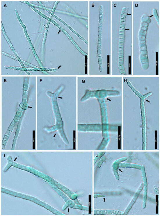
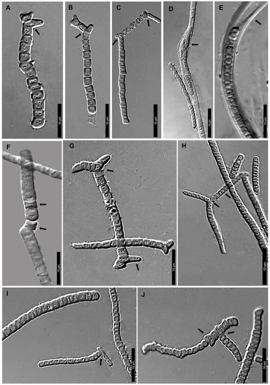
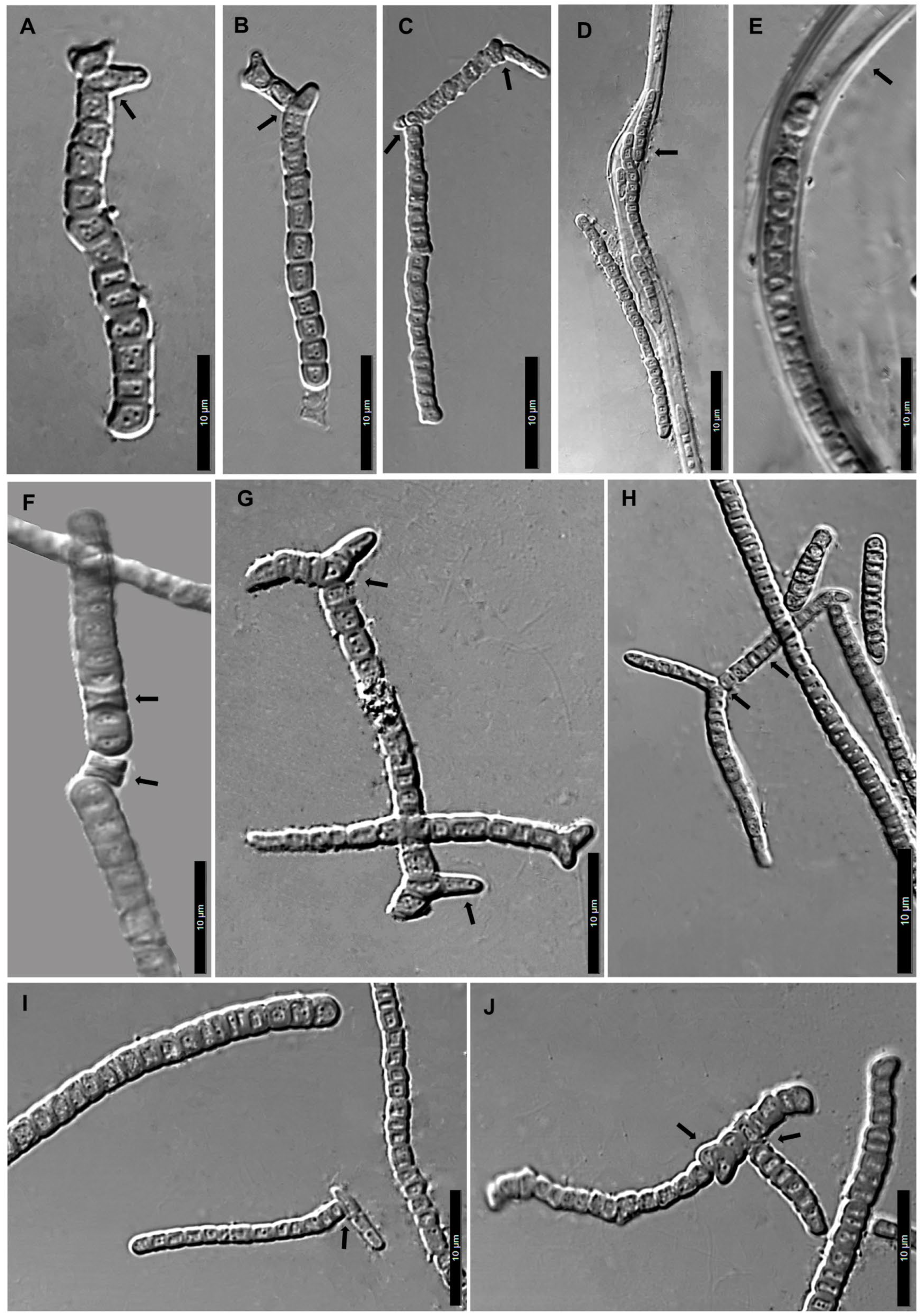
Bright-field differential interference contrast (DIC) microphotographs of E. ginni (SN2022/33 & AT2016/25). (A) Straight-to-slightly wavy filament with arrow pointing towards constriction at cross-wall and apical cell; (B) isopolar filament with conical ends; (C) straight filament showing horizontal and diagonal cell division; (D) horizontal division in apical cell; (E) trichome with vertically dividing cells; (F) short trichome with multiaxial arrangement; (G,J) trichome with apical T-branch; (H) true dichotomous V-branch; (I) trichome with apical V-branch and lateral T-branch. Scale bar 10 µm.
Figure 3.
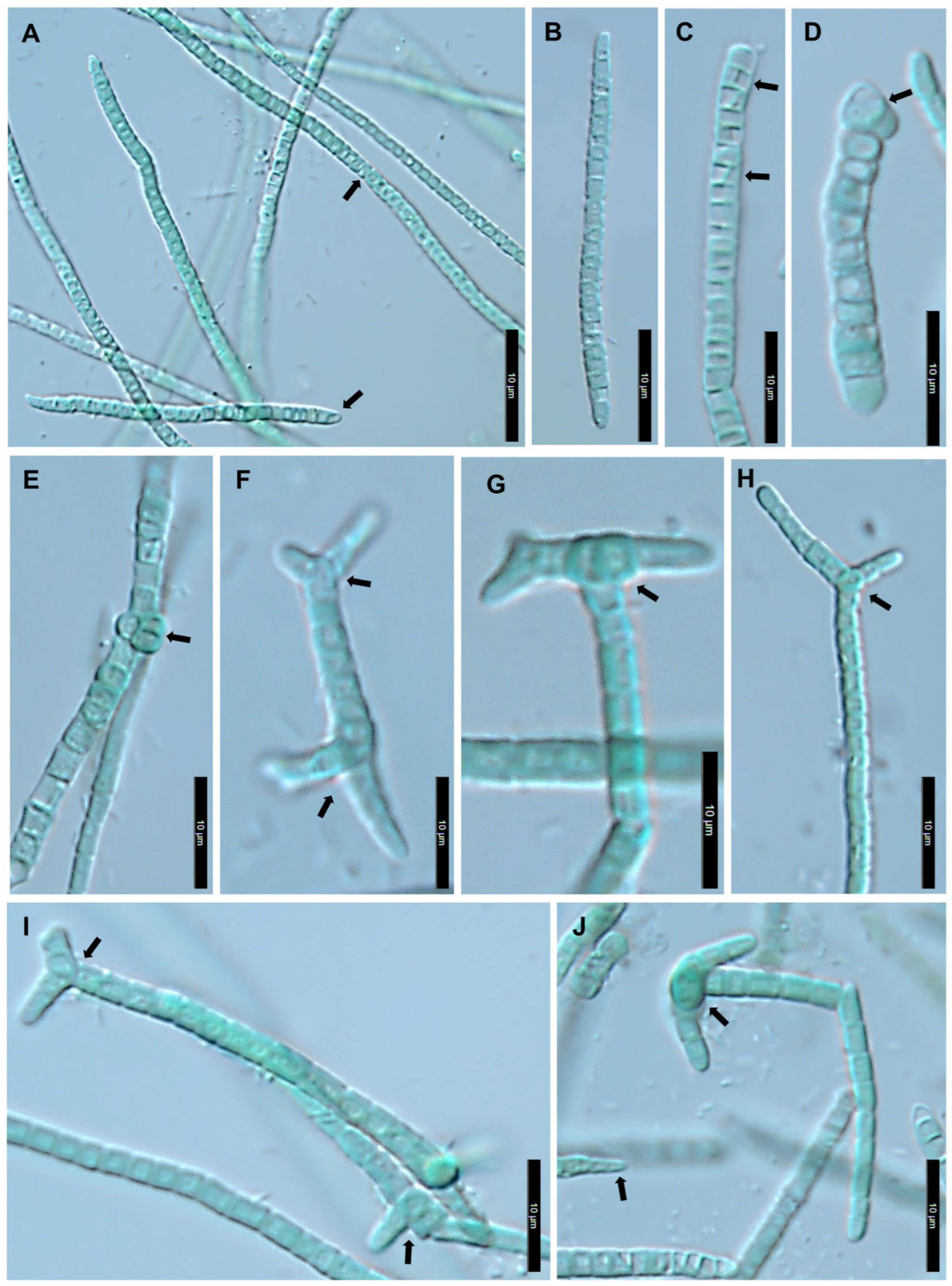
Differential interference contrast (DIC) microphotographs of true branching observed in E. ginni (SN2022/33 and AT2016/25). (A) Initiation of lateral branch by protuberance; (B) initiation of V-branch; (C) mature T-branch formed by protuberance; (D) trichomes enveloped in common sheath (older culture); (E) trichome encapsulated within thick sheath (older culture); (F) trichome disintegration by means of necridic cells; (G) bilateral V-branch with bottom arrowhead indicating protuberance; (H) mature true dichotomous V-branch with arrowhead pointing to branch point & main trichome; (I) apical T-branch; (J) lateral T-branch formed by lens-shaped septation with arrowhead pointing to branch point and oblique cell division. Scale bar 10 µm.
Figure 4.
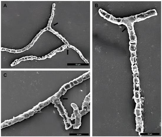
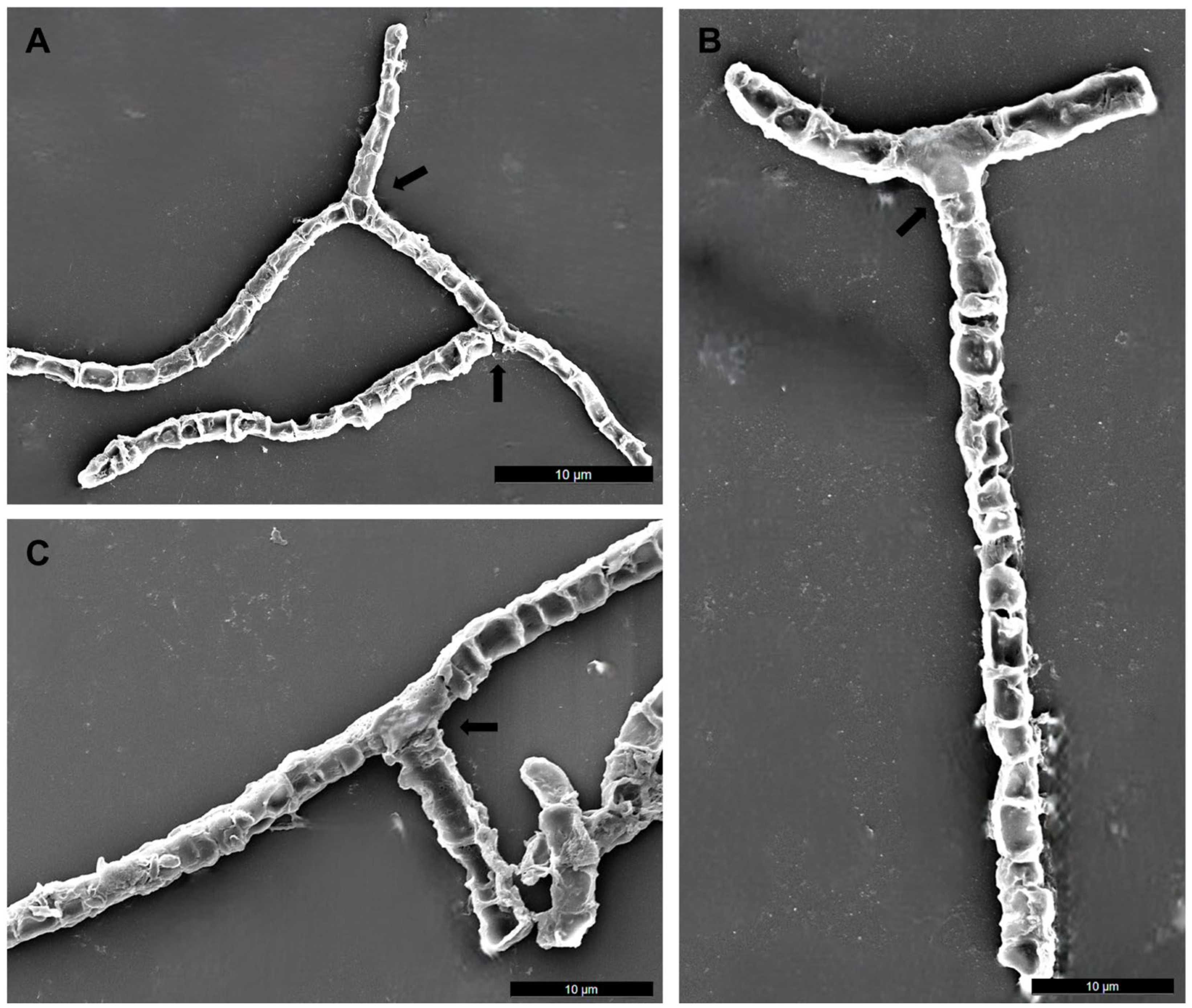
Field emission scanning electron microscopic (FE–SEM) images of E. ginni (SN2022/33 & AT2016/25). (A) Slightly wavy trichome with arrow indicating bilateral V-branch; (B) trichome with unilateral V-branch (see arrow), and (C) trichome with lateral T-branch (see arrow). Scale bar 10 µm.
Description: Blue-green filaments, mostly straight or slightly wavy, solitary, heteropolar or much or less isopolar in the actively growing phase. Sheaths thick in old cultures, widened, diffluent, overpassing filament, rarely encompassing 2 trichomes. Trichomes are multiseriate in old cultures with distinctly constricted cells at cross-walls, and mostly attenuated at terminal ends. Filaments may exhibit true branching: V-type (dichotomous) or T-type (apical or lateral branch initiated by lateral protuberance or vertical division or lens-shaped septation). Hormogonia are short, uniseriate, and without sheaths. Cells 1.5–2.5 (3) µm wide and 2.0–3.5 µm long that are rarely isodiametric or shorter or longer than wide, non-heterocytous and contain 2 to 4 polyphosphate granules per cell. Apical cells are 3.0–6.0 µm long, obtuse–round or conical to sharply pointed in shape, straight or rarely slightly bent, sometimes longer than other cells, and narrowed. Extrusions observed at terminal apices, either from both ends of the filament or from the cells located in the middle of filament. Reproduction by immotile hormogonia via necridic cells.
Holotype: A portion of the culture of Edaphifilum ginni is preserved in metabolically inactive form in the Global Collection of Cyanobacteria (GCC) (https://ccinfo.wdcm.org/details?regnum=1165, accessed on 7 January 2026), Varanasi, India, and is available under the accession number GCC 202581.
Isotype: Dry specimens of both strains have been deposited in University of Rajasthan Herbarium (RUBL), Jaipur, Rajasthan, India, with accession numbers RUBL 21933 (SN2022/33) & RUBL 21932 (AT2016/25).
Type locality: Biological crust on sandy soil, Pushkar, Ajmer district, Rajasthan, India.
Etymology: The specific epithet “ginni” is derived from a gold coin name in the Hindi language of India, which means yellow in color, and the studied strains were isolated from sandy soil that is similarly golden-yellow in color.
Type strain: SN2022/33.
3.2. Developmental Frequency of Branching
The variance-to-mean ratio revealed that extrusions (VMR = 8.23) were irregularly distributed along the filament and were typically observed at apices. In contrast, both T-type branching (VMR = 1.45) and V-type branching (VMR = 0.78) followed a random but uniform Poisson distribution (Table S3). Chi-square test (c2 = 30.86; p < 0.001) and paired-sample t-tests (t(9) = 4.469; p = 0.0015) confirmed that T-type branching (M = 3.90; sd = 2.38) is significantly more prevalent that V-type branching (mean = 0.30; sd = 0.48). Lastly, Pearson’s correlation analysis demonstrated that the occurrence of extrusions, T-type branching, and V-type branching are completely independent of one another (all |r| < 0.3; p > 0.05).
3.3. Molecular and Phylogenetic Analysis
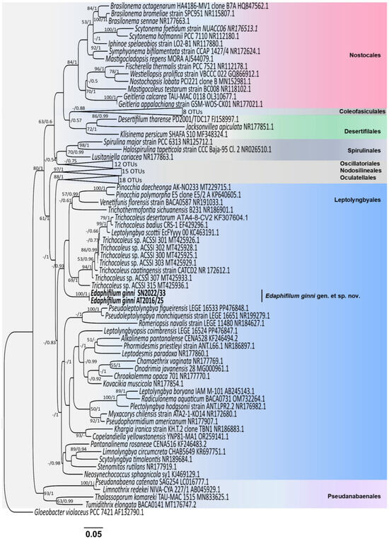
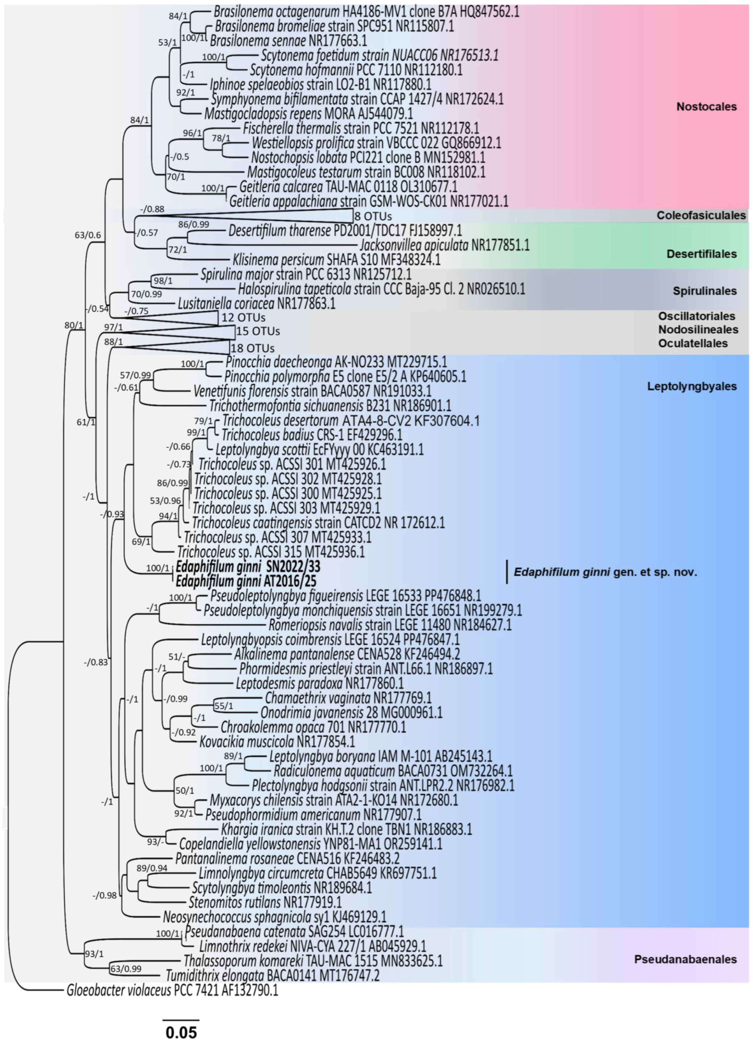
BLASTn indicated the highest 16S rRNA gene identity with an unclassified strain of Trichocoleus sp. (ACSSI 315) and T. desertorum. In both ML and BI phylogenies, E. ginni formed a distinct clade within the Leptolyngbyales (Figure 5). The sister clade to E. ginni includes morphologically similar genera such as Pinocchia, Venetifunis, Trichothermofontia, and Trichocoleus. The uncorrected p-distance indicates that E. ginni shares 94.81% 16S sequence similarity with T. caatingensis, while its similarity drops to 91.57% with T. sichuanensis (Table 1).
Figure 5.
Maximum likelihood phylogenetic tree constructed using 16S rRNA gene sequences of 119 genera from different orders showing the position of E. ginni (Leptolyngbyales). Numbers at the node represent statistical values in the following order: standard bootstrap/posterior probability. Values ≥ 50% (ML) and 0.50 (BI) are shown, and the full un-collapsed tree is available in Figure S2. Scale bar = 0.05 nucleotide substitutions per site.
Table 1.
16S rRNA gene identity (p-distance) between Edaphifilum ginni and other related taxa.
3.4. Analysis of 16S-23S ITS Region
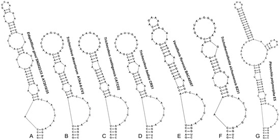
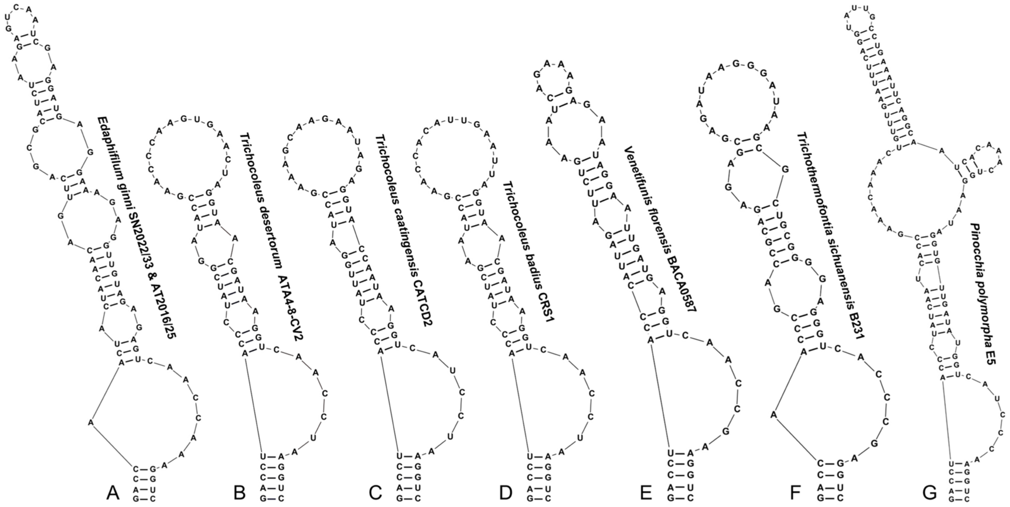
Both strains had identical sequences in the 16S-23S ITS region (522 bp), containing both tRNAIle and tRNAAla, genes. The analysis of ITS secondary structures of E. ginni exhibited unique features from closely related strains (Figure 6 and Figure 7). The D1-D1′ helix was 81 bp long, consisting of a small terminal loop with 5′-GUCAA-3′ nucleotides, subtended by a purine-rich symmetric bilateral bulge (2:2 nt). It was further preceded by two asymmetric bilateral bulges formed by the same set of bases (6 nt), but situated in reverse symmetry from 5′ to 3′ side, barring one nucleotide (A) on the 3′ side of the structure in the second bulge. Moreover, the basal stem was 4 base pairs long (5′-GACC:GGUC-3′), and the bilateral bulge above the basal stem consisted of 8 nt (AAACCAAC) on the 3′ side and 1 nt (A) on the 5′ side, which was further proceeded by a small asymmetric bulge of 3 nt. When compared to related genera Trichocoleus, Venetifunis, Trichothermofontia, and Pinocchia, the basal stem of E. ginni was found to be shorter & similar to T. sichuanensis B231 in having only 4 bp, while others had 5 bp (5′-GACCU:AGGUC-3′) in their stem sequence. Additionally, E. ginni had a great number of bulges and a smaller terminal loop, which separates it from phylogenetically related genera (Figure 6A–G).
Figure 6.
Secondary structures of D1-D1′ helices of E. ginni (A) and representatives from phylogenetically close genera (B–G).
Figure 7.
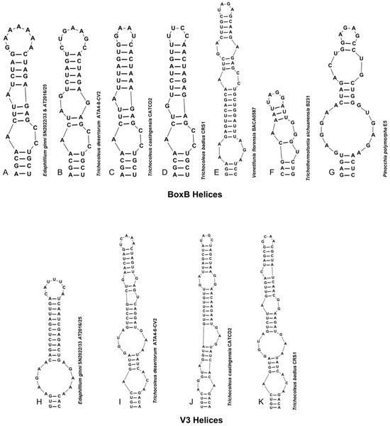
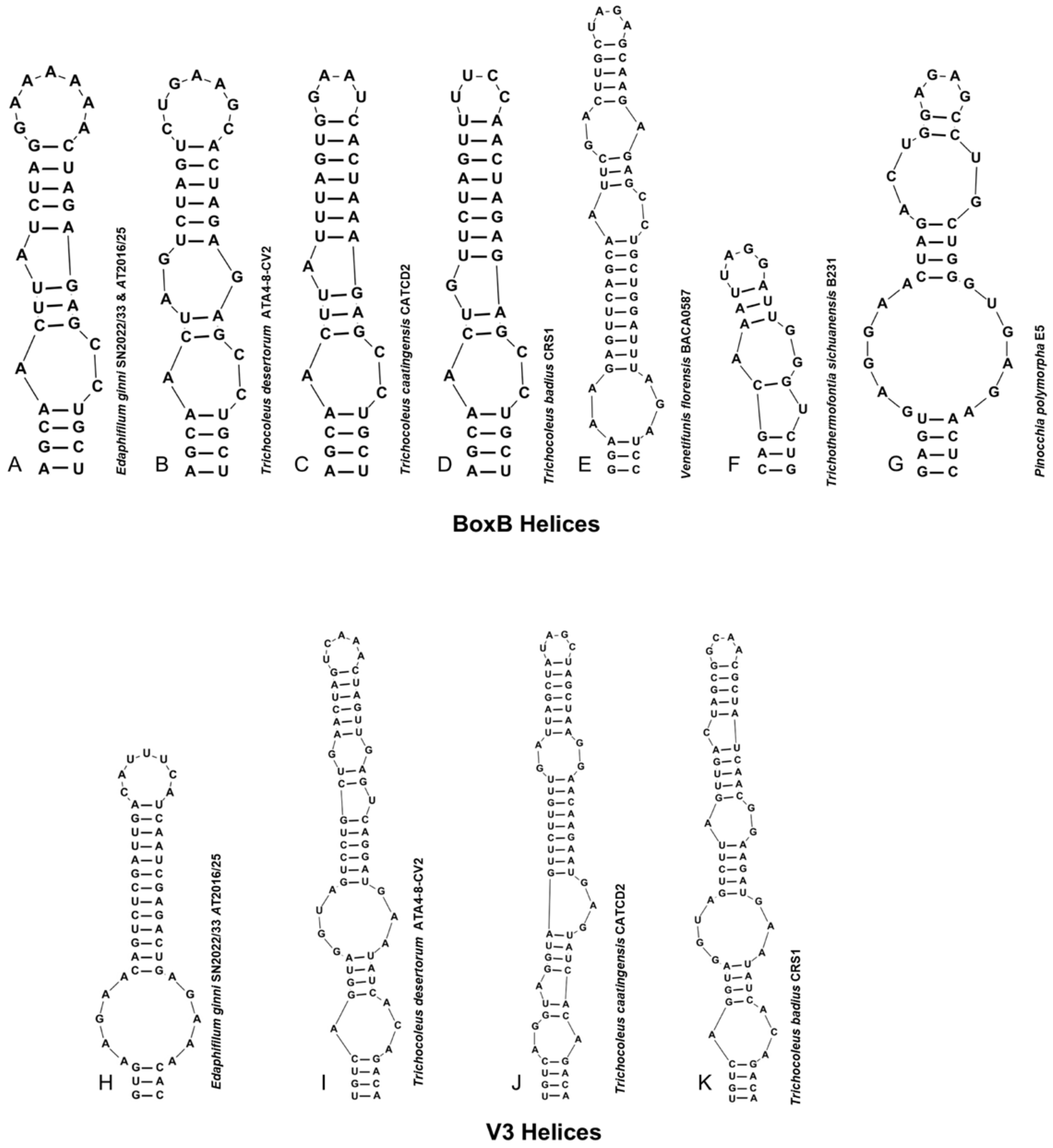
Secondary structures of BoxB (A–G) and V3 helices (H–K) of E. ginni and representatives from phylogenetically close genera.
The sequence of the BoxB helix of E. ginni was 35 nt long, comprising two asymmetric bulges and a terminal loop of seven purine-rich bases (5′-GAAAAAA-3′). The basal stem contained a 4 nt (5′-AGCA:UGCU-3′) sequence, followed by a bilateral bulge with non-uniform distribution of unpaired bases (1:2 nt) on the 5′ and 3′ sides. Moreover, a unilateral bulge in the middle of the helix with only an adenine nucleotide base was observed on the 5′ side. The related genera had two or three bulges in the BoxB helix with a terminal loop of 4 or 7 nt and a basal stem of 3 or 4 nt with a variable stem sequence (Figure 7A–G).
The V3 helix of E. ginni was 49 nt long, consisting of one symmetric bilateral bulge (5:5 nt) above the basal stem and a terminal loop of 7 nt (5′-CAUUUCA-3′). The basal stem was short and formed of 3 nt (5′-GUG:CAC-3′), which differs from other strains such as T. desertorum ATA4-8-CV2, T. badius CRS1, and T. caatingensis CATCD2, as they had 4 nt (5′-UGUC:GACA-3′)-long stem sequence. The helical structure of other taxa such as T. desertorum, T. caatingensis, and T. badius was long with more bulges and a smaller terminal loop in contrast to E. ginni. Lastly, the V3 helix of V. florensis, T. sichuanensis, and P. polymorpha could not be predicted due to partial sequences (Figure 7H–K).
4. Discussion
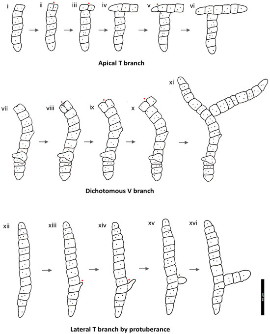
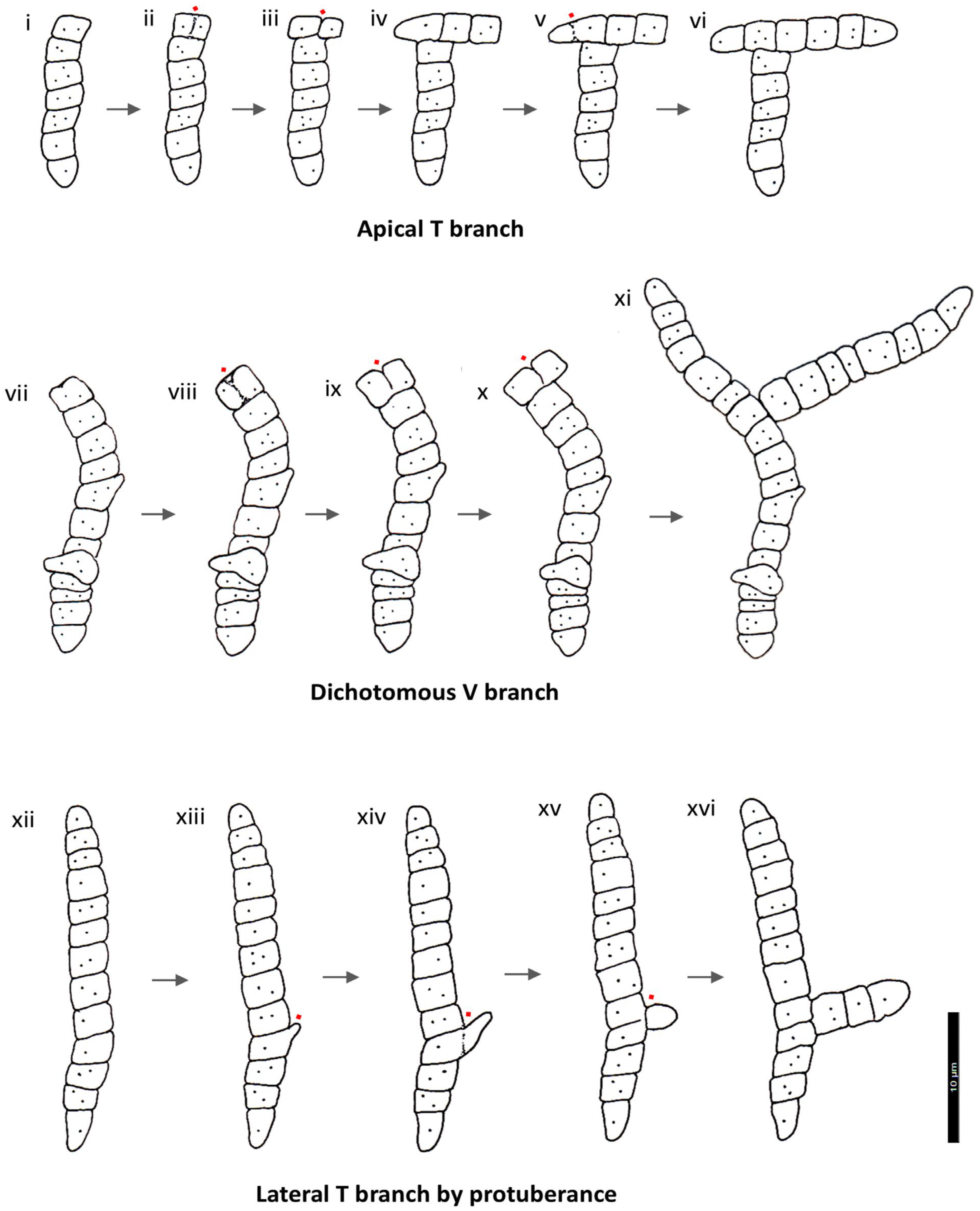
True branching in cyanobacteria is characterized by branch-point cells that contact three different adjoining cells [41]. Previously thought to be unique to heterocytous lineages [21], three types of morphologies have been identified: ‘T’ where lateral branches create a T-shape, ‘Y’ involving apical bifurcation resulting in a Y-shape, and ‘V’ showing either dichotomous or pseudo-dichotomous bifurcation that forms a V-shape) [42,43]. This trait has been restricted to Nostocales, a monophyletic order capable of heterocyte formation [44,45,46]. The occurrence of true branching in Edaphifilum represents the first instance of this feature being observed outside the Nostocales. Intriguingly, the ontogeny of some Edaphifilum branches (Figure 3B,I and Figure S1D,E) seems to deviate from the previously described division planes [42], yet the mature branches appear T- or V-shaped (sequential steps of branch development are illustrated in Figure 8). Furthermore, some branches were initiated through a protuberance (Figure 3A,C and Figure S1), which superficially resembled the branches reported in Stigonema sp. [47] and Mastigocladus sp. [48,49].
Figure 8.
Line drawings of E. ginni (SN2022/33 & AT2016/25) illustrating putative developmental stages of true branches. (i–vi): apical T-branch initiated by vertical division in cell; (vii–xi): dichotomous V-branch; (xii–xvi): lateral T-branch formed by protuberance. Red dots showing division points. Scale bar 10 µm.
Morphologically, SN2022/33 & AT2016/25 strains were identical, with true branching and thick sheathes in older cultures, cells nearly isodiametric or slightly longer or shorter than wide, and reproduction via hormogonia disintegration by means of necridic cells. Edaphifilum was somewhat similar in morphology to Trichocoleus delicatulus [24] and of T. desertorum [50], though they could be separated from each other via light microscopy (Table 2). For instance, filaments of Trichocoleus possessed multiple trichomes and were wider (2.3–6.3 µm) within thick sheaths, whereas filaments of Edaphifilum were narrower (<3 µm wide), mostly solitary, or only rarely encompassing two or three trichomes in older cultures (Figure 3D).
Table 2.
Morphological and habitat comparisons among Edaphifilum ginni and the phylogenetically closest taxa.
The novel branching pattern of Edaphifilum is characterized by the formation and proliferation of unilateral or bilateral protuberances and extrusions from both apical (Figure 2C, Figure 3A–C,G and Figure S1E) and intercalary cells (Figure 2E,F,I and Figure S1B), resulting in a multiaxial filament architecture. Notably, the branch-point cells maintain the continuity of the cytoplasm with the parental trichome, confirming this as a true-branching event [41]. This form of true branching, not previously reported outside the heterocytous Nostocales, indicates that our knowledge of cyanobacterial diversity remains fragmentary, and extensive sampling is needed to uncover more cyanobacteria like Edaphifilum. Furthermore, in the pre-genomic era, cyanobacteria were phenotypically categorized into five quasi-taxonomic ‘Subsections’ [22]. The Subsections I & II encompassed unicellular cyanobacteria that had spherical, cylindrical or oval morphology, whereas the remaining three subsections represented global filamentous morphotypes (long chain of interconnected cells resulted by repeated intercalary cell divisions). The salient features of cyanobacteria from Subsections III–V were as stated: Subsection III (trichomes not truly branched, cell division in only plane and unable to produce heterocytes), Subsection IV (trichomes not truly branched, cell division in only plane and able to produce heterocytes), and Subsection V (trichomes truly branched, cell division in more than one plane and able to produce heterocytes) [22]. This set of global morphotypes logically and biologically lacked ‘Subsection VI’ (trichomes truly branched, cell division occur in more than one plane and unable to produce heterocytes) and we believe that strains of Edaphifilum may belong to the postulated Subsection VI. It is also undeniable that the modern cyanobacterial classification system relies on the polyphasic approach [19,21] and the morphotype-based system has become a historical phenomenon. However, systematics is an inherently dynamic field with the descriptions of new morphotypes, and the true perception of cyanobacterial diversity is not feasible without the knowledge of global morphotypes.
The 16S rRNA analyses revealed that the studied strains represent a distinct evolutionary lineage within the Trichocoleusaceae. Both strains of Edaphifilum were found to be phylogenetically distinct from Trichocoleus, Trichothermofontia, Venetifunis, and Pinocchia (Figure 5). Pinocchia has consistently clustered in the Trichocoleusaceae with strong statistical support [25,26], and, thus, the current placement of Pinocchia in the Leptolyngbyaceae is taxonomically incongruent and could be shifted to Trichocoleusaceae [23,26]. Furthermore, the 16S rRNA gene identity matrix (p-distance) further substantiated the delineation of the studied strains as a novel genus, with p-distance values between E. ginni and the closest available genera < 95% (Table 1). In bacteriology, a minimum threshold of ≤94.5% was suggested by researchers [51,52,53] to establish a new genus; however, this value is not absolute, and the taxa exceeding this limit can be demarcated as novel genera if reinforced by preeminent morphological features, or environmental data [53]. It is recommended that a minimum threshold of ±95%, coupled with at least one diacritical autapomorphic character, be employed for establishing new genera [54]. In this case, the diacritical feature is the unique form of branching. Both strains shared ca. 94.81% 16S rRNA gene identity with T. caatingensis, 94.71% with T. badius, and 94.62% with T. desertorum ATA4-8-CV2.
The secondary structure of the 16S-23S rRNA ITS region is considered an important criterion while describing novel cyanobacteria [31,55,56]. The comparison of the D1-D1′, BoxB, and V3 helices of E. ginni displayed variations in sequence, length, base arrangement, number of bulges, and stem-loop structure compared to sister taxa (Table S4).
Ecology plays a crucial role in classification, as genetic divergence and evolutionary adaptations can be elucidated by considering environmental factors [28,57]. Strains of Edaphifilum were isolated from a biological crust in sandy soils of Rajasthan, which is characterized by poor water retention capacity and nutrient scarcity. Sister clades to Edaphifilum are found in diverse habitats, including freshwater thermal springs, rocks, desert soils, and quartzite seep walls [24,25,26,50,58]. Edaphifilum was isolated from soils that are slightly alkaline with low salinity, typical of the microhabitats from semi-arid regions [5,59]. However, such measurements represent only the conditions at the moment of sampling and do not fully reflect the long-term environmental stresses. The climate of Pushkar and its surrounding semi-arid region is characterized by pronounced seasonal temperature fluctuations where average temperatures reach approximately 40–45 °C in summer [60,61]. These high temperatures, combined with low rainfall and intense solar radiation, create conditions of periodic desiccation, thermal stress, and osmotic imbalance typical of semi-arid ecosystems [5,59,60,61]. We observed that the studied strains possessed thicker sheaths and pointed apical cells similar to Microcoleus vaginatus (sensu lato) thriving in Colorado Plateau crusts [62], and these characters could be an adaptive strategy to cope up with intense solar radiation. Due to climatic shifts and anthropogenic activities, drylands are expanding worldwide at a fast pace [63,64,65]. Thus, isolation of Edaphifilum strains from xeric environmental conditions could pave the way for novel studies aimed at controlling desertification and soil reclamation for agricultural activities in arid zones.
5. Conclusions
Edaphifilum ginni formed an independent lineage within the Trichocoleusaceae, sharing less than 95% 16S rRNA gene sequence similarity with the closest related genera. The strains also exhibited true branching, characterized by unilateral or bilateral extrusions from both apical and intercalary cells. Additionally, unique folded secondary structures in the D1-D1′, Box B and V3 helices of the 16S-23S ITS region were observed. Collectively, these morphological, molecular, and structural evidences support the proposal of a novel cyanobacterial taxon, Edaphifilum ginni gen. et sp. nov., within the Leptolyngbyales (Cyanobacteriophyta).
Supplementary Materials
The following supporting information can be downloaded at: https://www.mdpi.com/article/10.3390/phycology6020056/s1, Figure S1: Fluorescence images of E. ginni (SN2022/33 & AT2016/25). (A) Trichome with vertically dividing cells; (B) initiation of T-branch via protuberance & middle arrow pointing towards initiation of intercalary branch; (C) trichome with true dichotomous V-branch; (D) lateral T-branch; (E) slightly wavy trichomes with arrowhead indicating apical T-branch, initiation of apical branch; (F) cells with vertical division and T-branches. Scale bar 10 µm; Figure S2: Uncompressed maximum likelihood phylogenetic tree of E. ginni along with 118 cyanobacterial taxa. Numbers at the node represent statistical values in the following order: standard bootstrap/posterior probability. Values ≥ 50% (ML) and 0.50 (BI) are shown. Scale bar = 0.05 nucleotide substitutions per site; Table S1: Physiochemical characteristics of the sampling site located in Pushkar, Ajmer, Rajasthan; Table S2: Morphological characteristics of E. ginni observed during different growth stages; Table S3: Statistical summary of morphological features E. ginni and Table S4: Comparison of ITS region lengths (nt) of D1-D1′, BoxB and V3 helix folded structures of E. ginni and related taxa.
Author Contributions
Conceptualization, S.S., P.S., D.A.C. and P.K.D.; funding acquisition, N.P. and P.K.D.; investigation, A.K.T., S.S. and S.A.; Supervision, P.K.D.; writing—original draft, S.S.; writing—review and editing, S.S., P.S., D.A.C. and P.K.D. All authors have read and agreed to the published version of the manuscript.
Funding
This research received no external funding.
Institutional Review Board Statement
Not applicable.
Data Availability Statement
The data presented in this study are openly available in GenBank [https://www.ncbi.nlm.nih.gov/genbank/ accessed on 20 November 2025] under accession no PX572938, PX698345 (SN2022/33), and PX572939, PX698344 (AT2016/25); Global Collection of Cyanobacteria (GCC) [https://ccinfo.wdcm.org/details?regnum=1165 accessed on 16 February 2026] under accession number GCC 202581 (SN2022/33) and University of Rajasthan Herbarium (RUBL), with accession number RUBL 21933 (SN2022/33) & RUBL 21932 (AT2016/25).
Acknowledgments
We are thankful to the Department of Microbiology, Central University of Rajasthan, for providing the necessary facilities for this research work. Special thanks to Hansa for technical assistance while taking photos with the confocal microscope available in the Central Instrumentation Facility (CIF) of the Central University of Rajasthan. We also thank the Department of Bioscience and Biotechnology, Banasthali Vidyapith, Rajasthan, for helping with bright-field DIC images. Sonam is grateful to Central University of Rajasthan for providing financial assistance through University non-NET fellowship.
Conflicts of Interest
The authors declare no conflicts of interest.
References
- Olson, J.M. ‘Evolution of photosynthesis’ (1970), re-examined thirty years later. Photosynth. Res. 2001, 68, 95–112. [Google Scholar] [CrossRef] [Scilit]
- Kulasooriya, S. Cyanobacteria: Pioneers of planet earth. Ceylon J. Sci. Biol. Sci. 2012, 40, 71–88. [Google Scholar] [CrossRef] [Scilit]
- Sonam, S.; Dadheech, P.K. Cyanobacteria—The pioneering photoautotrophs. In Cyanobacteria; Elsevier: Amsterdam, The Netherlands, 2024; pp. 1–18. [Google Scholar] [CrossRef] [Scilit]
- Castenholz, R.W. Oxygenic photosynthetic bacteria (Sect. 19). Group l. Cyanobacteria. Bergey’s Man. Syst. Bacteriol. 1989, 3, 1710–1799. [Google Scholar]
- Whitton, B.A.; Potts, M. Introduction to the cyanobacteria. In Ecology of Cyanobacteria II; Springer: Dordrecht, The Netherlands, 2012; pp. 1–13. [Google Scholar]
- Waterbury, J.B. The cyanobacteria—Isolation, purification and identification. In The Prokaryotes; Springer: New York, NY, USA, 2006; pp. 1053–1073. [Google Scholar]
- Seckbach, J. Genesis—In the Beginning; Seckbach, J., Ed.; Cellular Origin, Life in Extreme Habitats and Astrobiology; Springer: Dordrecht, The Netherlands, 1999; Volume 22, ISBN 978-94-007-2940-7. [Google Scholar]
- Dvořák, P.; Hašler, P.; Casamatta, D.A.; Poulíčková, A. Underestimated cyanobacterial diversity: Trends and perspectives of research in tropical environments. Fottea 2021, 21, 110–127. [Google Scholar] [CrossRef] [Scilit]
- Chakraborty, S.; Maruthanayagam, V.; Achari, A.; Pramanik, A.; Jaisankar, P.; Mukherjee, J. Euryhalinema mangrovii gen. nov., sp. nov. and Leptoelongatus litoralis gen. nov., sp. nov. (Leptolyngbyaceae) isolated from an Indian mangrove forest. Phytotaxa 2019, 422, 58–74. [Google Scholar] [CrossRef] [Scilit]
- Chavadar, M.; Saraf, A.; Suradkar, A.; Mishra, D.; Kumar, N.; Singh, P. Constrictifilum karadense gen. et sp. nov., a new nostocalean genus from Maharashtra, India. FEMS Microbiol. Lett. 2021, 368, fnab057. [Google Scholar] [CrossRef] [Scilit]
- Kumar, N.; Saraf, A.; Pal, S.; Mishra, D.; Singh, P. Insights into the phylogenetic inconsistencies of the genus Amazonocrinis and description of epilithic Amazonocrinis malviyae sp. nov. (Cyanobacteria, Nostocales) from Jammu and Kashmir, India. Int. J. Syst. Evol. Microbiol. 2022, 72, 005658. [Google Scholar] [CrossRef] [Scilit]
- Sonam, S.; Anand, S.; Pareek, N.; Singh, P.; Casamatta, D.A.; Dadheech, P.K. The novel halotolerant, filamentous cyanobacterium Krienitziella sambharensis gen. et sp. nov. (Nodosilineales, Cyanobacteriophyta) isolated from an Indian wetland (Sambhar Salt Lake, India). Diversity 2026, 18, 181. [Google Scholar] [CrossRef] [Scilit]
- Akagha, M.U.; Pietrasiak, N.; Bustos, D.F.; Vondrášková, A.; Lamb, S.C.; Johansen, J.R. Albertania and Egbenema gen. nov. from Nigeria and the United States, expanding biodiversity in the oculatellaceae (Cyanobacteria). J. Phycol. 2023, 59, 1217–1236. [Google Scholar] [CrossRef] [Scilit] [PubMed]
- Cordeiro, R.; Luz, R.; Lage, S.; Menezes, C.; Dias, E.; Flores, C.; Fonseca, A.; Gonçalves, V. Metabolomic and taxonomic characterization of Haloleptolyngbya lusitanica sp. nov. (Cyanobacteria, Synechococcales). Phycologia 2024, 63, 225–234. [Google Scholar] [CrossRef] [Scilit]
- Jusko, B.M.; Johansen, J.R. Description of six new cyanobacterial species from soil biocrusts on san nicolas island, california, in three genera previously restricted to Brazil. J. Phycol. 2024, 60, 133–151. [Google Scholar] [CrossRef] [Scilit]
- Pal, S.; Saraf, A.; Kumar, N.; Singh, P. Igniting taxonomic curiosity: The amazing story of Amazonocrinis with the description of a new genus Ahomia gen. nov. and novel species of Ahomia, Amazonocrinis, and Dendronalium from the biodiversity-rich northeast region of India. J. Phycol. 2024, 60, 387–408. [Google Scholar] [CrossRef] [Scilit]
- Pal, S.; Saraf, A.; Kumar, N.; Pant, H.; Badruddin, S.S.A.; Sajibulla, S.M.A.; Nijamuddin, S.K.; Prajapati, A.; Talukdar, U.; Kohar, N.; et al. Polyphasic characterization of 15 heterocytous cyanobacterial isolates from different habitats of India and description of 9 novel species belonging to the genera Desikacharya, Aliinostoc, and Desmonostoc. Algal Res. 2025, 86, 103873. [Google Scholar] [CrossRef] [Scilit]
- Pant, H.; Kumar, N.; Pal, S.; Singh, P. Exploring cyanobacteria from diverse habitats of the Konkan region of India, unveiling novel species of the genera Desikacharya, Pseudoaliinostoc, and Chlorogloeopsis using a polyphasic approach. J. Phycol. 2025, 61, 194–217. [Google Scholar] [CrossRef] [Scilit] [PubMed]
- Komárek, J.; Kaštovský, J.; Mareš, J.; Johansen, J.R. Taxonomic classification of cyanoprokaryotes (cyanobacterial genera) 2014, using a polyphasic approach. Preslia 2014, 86, 295–335. [Google Scholar]
- Komárek, J. A polyphasic approach for the taxonomy of cyanobacteria: Principles and applications. Eur. J. Phycol. 2016, 51, 346–353. [Google Scholar] [CrossRef] [Scilit]
- Strunecký, O.; Ivanova, A.P.; Mareš, J. An updated classification of cyanobacterial orders and families based on phylogenomic and polyphasic analysis. J. Phycol. 2023, 59, 12–51. [Google Scholar] [CrossRef] [Scilit]
- Rippka, R.; Deruelles, J.; Waterbury, J.B.; Herdman, M.; Stanier, R.Y. Generic assignments, strain histories and properties of pure cultures of cyanobacteria. Microbiology 1979, 111, 1–61. [Google Scholar] [CrossRef] [Scilit]
- Mai, T.; Johansen, J.R.; Pietrasiak, N.; Bohunická, M.; Martin, M.P. Revision of the synechococcales (cyanobacteria) through recognition of four families including Oculatellaceae fam. nov. and Trichocoleaceae fam. nov. and six new genera containing 14 species. Phytotaxa 2018, 365, 1–59. [Google Scholar] [CrossRef] [Scilit]
- Anagnostidis, K. Nomenclatural changes in cyanoprokaryotic order oscillatoriales. Preslia 2001, 73, 359–375. [Google Scholar]
- Tang, J.; Zhou, H.; Jiang, Y.; Yao, D.; Waleron, K.F.; Du, L.-M.; Daroch, M. Characterization of a novel thermophilic cyanobacterium within Trichocoleusaceae, Trichothermofontia sichuanensis gen. et sp. nov., and Its CO2-concentrating mechanism. Front. Microbiol. 2023, 14, 1111809. [Google Scholar] [CrossRef] [Scilit] [PubMed]
- Luz, R.; Cordeiro, R.; Kaštovský, J.; Johansen, J.R.; Dias, E.; Fonseca, A.; Urbatzka, R.; Vasconcelos, V.; Gonçalves, V. New terrestrial cyanobacteria from the Azores Islands: Description of Venetifunis gen. nov. and new species of Albertania, Kovacikia and Pegethrix. Phycologia 2023, 62, 483–498. [Google Scholar] [CrossRef] [Scilit]
- Johansen, J.R.; Casamatta, D.A. Recognizing cyanobacterial diversity through adoption of a new species paradigm. Algol. Stud. 2005, 117, 71–93. [Google Scholar] [CrossRef] [Scilit]
- Komárek, J. Cyanoprokaryota 2. Teil/2nd Part: Oscillatoriales; Büdel, B., Krienitz, L., Gartner, G., Schagerl, M., Eds.; Süsswasserflora von Mitteleuropa; Elsevier GmbH: Amsterdam, The Netherlands, 2005; Volume 19. [Google Scholar]
- Lane, D.J. 16S/23S rRNA Sequencing. In Nucleic Acid Techniques in Bacterial Systematics; Stackebrandt, E., Goodfellow, M., Eds.; John Wiley and Sons: New York, NY, USA, 1991; pp. 115–175. [Google Scholar]
- Iteman, I.; Rippka, R.; Tandeau de Marsac, N.; Herdman, M. Comparison of conserved structural and regulatory domains within divergent 16S rRNA–23S rRNA spacer sequences of cyanobacteria. Microbiology 2000, 146, 1275–1286. [Google Scholar] [CrossRef] [Scilit]
- Pal, S.; Saraf, A.; Kumar, N.; Singh, P. Phycological exploration of the global biodiversity hotspots of northeast India: Discovery of a new species of soil-dwelling cyanobacteria, Desikacharya kailashaharensis sp. nov. FEMS Microbiol. Lett. 2022, 369, fnac099. [Google Scholar] [CrossRef] [Scilit]
- Edwards, U.; Rogall, T.; Blöcker, H.; Emde, M.; Böttger, E.C. Isolation and direct complete nucleotide determination of entire genes. Characterization of a gene coding for 16S Ribosomal RNA. Nucleic Acids Res. 1989, 17, 7843–7853. [Google Scholar] [CrossRef] [Scilit] [PubMed]
- Minh, B.Q.; Schmidt, H.A.; Chernomor, O.; Schrempf, D.; Woodhams, M.D.; von Haeseler, A.; Lanfear, R. IQ-TREE 2: New models and efficient methods for phylogenetic inference in the genomic era. Mol. Biol. Evol. 2020, 37, 1530–1534. [Google Scholar] [CrossRef] [Scilit] [PubMed]
- Hoang, D.T.; Chernomor, O.; von Haeseler, A.; Minh, B.Q.; Vinh, L.S. UFBoot2: Improving the ultrafast bootstrap approximation. Mol. Biol. Evol. 2018, 35, 518–522. [Google Scholar] [CrossRef] [Scilit]
- Ronquist, F.; Teslenko, M.; van der Mark, P.; Ayres, D.L.; Darling, A.; Höhna, S.; Larget, B.; Liu, L.; Suchard, M.A.; Huelsenbeck, J.P. MrBayes 3.2: Efficient bayesian phylogenetic inference and model choice across a large model space. Syst. Biol. 2012, 61, 539–542. [Google Scholar] [CrossRef] [Scilit]
- Rambaut, A. Figtree, Version 1.4.4; Institute of Evolutionary Biology, University of Edinburgh: Edinburgh, UK, 2018. Available online: https://tree.bio.ed.ac.uk/software/figtree/ (accessed on 12 February 2026).
- Tamura, K.; Stecher, G.; Kumar, S. MEGA11: Molecular evolutionary genetics analysis Version 11. Mol. Biol. Evol. 2021, 38, 3022–3027. [Google Scholar] [CrossRef] [Scilit]
- Zuker, M. Mfold web server for nucleic acid folding and hybridization prediction. Nucleic Acids Res. 2003, 31, 3406–3415. [Google Scholar] [CrossRef] [Scilit]
- Reuter, J.S.; Mathews, D.H. RNAstructure: Software for RNA secondary structure prediction and analysis. BMC Bioinform. 2010, 11, 129. [Google Scholar] [CrossRef] [Scilit] [PubMed]
- Inkscape Project Inkscape, Version 1.2.1; Inkscape: Boston, MA, USA, 2022. Available online: https://Inkscape.Org (accessed on 12 February 2026).
- Stanier, R.Y.; Kunisawa, R.; Mandel, M.; Cohen-Bazire, G. Purification and properties of unicellular Blue-Green Algae (Order Chroococcales). Bacteriol. Rev. 1971, 35, 171–205. [Google Scholar] [CrossRef]
- Golubić, S.; Hernández-Mariné, M.; Hoffmann, L. Developmental aspects of branching in filamentous cyanophyta/cyanobacteria. Algol. Stud. 1996, 83, 303–329. [Google Scholar] [CrossRef] [Scilit]
- Gugger, M.F.; Hoffmann, L. Polyphyly of true branching cyanobacteria (Stigonematales). Int. J. Syst. Evol. Microbiol. 2004, 54, 349–357. [Google Scholar] [CrossRef] [Scilit]
- Kabirnataj, S.; Nematzadeh, G.A.; Talebi, A.F.; Tabatabaei, M.; Singh, P. Neowestiellopsis gen. nov., a new genus of true branched cyanobacteria with the description of Neowestiellopsis persica sp. nov. and Neowestiellopsis bilateralis sp. nov., isolated from Iran. Plant Syst. Evol. 2018, 304, 501–510. [Google Scholar] [CrossRef] [Scilit]
- Kilgore, C.; Johansen, J.R.; Mai, T.; Hauer, T.; Casamatta, D.A.; Sheil, C. Molecular characterization of Geitleria appalachiana sp. nov. (Nostocales, Cyanobacteria) and formation of Geitleriaceae fam. nov. Fottea 2018, 18, 150–163. [Google Scholar] [CrossRef] [Scilit]
- Casamatta, D.A.; Villanueva, C.D.; Garvey, A.D.; Stocks, H.S.; Vaccarino, M.; Dvořák, P.; Hašler, P.; Johansen, J.R. Reptodigitus chapmanii (Nostocales, Hapalosiphonaceae) gen. nov.: A unique nostocalean (cyanobacteria) genus based on a polyphasic approach. J. Phycol. 2020, 56, 425–436. [Google Scholar] [CrossRef] [Scilit]
- Song, G.; Jiang, Y.; Li, R. Scytolyngbya timoleontis, gen. et sp. nov. (Leptolyngbyaceae, Cyanobacteria): A novel false branching cyanobacteria from China. Phytotaxa 2015, 224, 72–84. [Google Scholar] [CrossRef] [Scilit]
- Jaiswal, T.P.; Chakraborty, S.; Singh, P.; Mishra, A.K.; Singh, S.S. Description of hot spring dwelling Mastigocladus ambikapurensis sp. nov., using a polyphasic approach. Plant Syst. Evol. 2021, 307, 33. [Google Scholar] [CrossRef] [Scilit]
- Pokorný, J.; Vondrášková, A.; Wipplingerová, M.; Kaštovský, J. Four novel taxa of cyanobacteria from a unique thermal cave habitat in Vromoner Canyon, Albania. J. Phycol. 2025, 61, 1394–1422. [Google Scholar] [CrossRef] [Scilit] [PubMed]
- Muhlsteinova, R.; Johansen, J.R.; Pietrasiak, N.; Martin, M.P.; Osorio-Santos, K.; Warren, S.D. Polyphasic characterization of Trichocoleus desertorum sp. nov. (Pseudanabaenales, Cyanobacteria) from desert soils and phylogenetic placement of the genus Trichocoleus. Phytotaxa 2014, 163, 241–261. [Google Scholar] [CrossRef] [Scilit]
- Stackebrandt, E.; Goebel, B.M. Taxonomic Note: A place for DNA-DNA reassociation and 16S rRNA sequence analysis in the present species definition in bacteriology. Int. J. Syst. Evol. Microbiol. 1994, 44, 846–849. [Google Scholar] [CrossRef] [Scilit]
- Stackebrandt, E.; Ebers, J. Taxonomic parameters revisited: Tarnished gold standards. Microbiol. Today 2006, 33, 152. [Google Scholar]
- Yarza, P.; Yilmaz, P.; Pruesse, E.; Glöckner, F.O.; Ludwig, W.; Schleifer, K.-H.; Whitman, W.B.; Euzéby, J.; Amann, R.; Rosselló-Móra, R. Uniting the classification of cultured and uncultured bacteria and archaea using 16S rRNA gene sequences. Nat. Rev. Microbiol. 2014, 12, 635–645. [Google Scholar] [CrossRef] [Scilit]
- Komárek, J. Recent changes (2008) in cyanobacteria taxonomy based on a combination of molecular background with phenotype and ecological consequences (Genus and Species Concept). Hydrobiologia 2010, 639, 245–259. [Google Scholar] [CrossRef] [Scilit]
- Boyer, S.L.; Flechtner, V.R.; Johansen, J.R. Is the 16S-23S rRNA internal transcribed spacer region a good tool for use in molecular systematics and population genetics? A case study in cyanobacteria. Mol. Biol. Evol. 2001, 18, 1057–1069. [Google Scholar] [CrossRef] [Scilit]
- Johansen, J.R.; Kovacik, L.; Casamatta, D.A.; Iková, K.F.; Kaštovský, J. Utility of 16S-23S ITS sequence and secondary structure for recognition of intrageneric and intergeneric limits within cyanobacterial taxa: Leptolyngbya corticola sp. nov. (Pseudanabaenaceae, Cyanobacteria). Nova Hedwig. 2011, 92, 283–302. [Google Scholar] [CrossRef] [Scilit]
- Strunecký, O.; Komárek, J.; Elster, J. Biogeography of Phormidium Autumnale (Oscillatoriales, Cyanobacteria) in western and central spitsbergen. Pol. Polar Res. 2012, 33, 369–382. [Google Scholar] [CrossRef] [Scilit]
- Dvořák, P.; Jahodářová, E.; Hašler, P.; Gusev, E.; Poulíčková, A. A New Tropical Cyanobacterium Pinocchia polymorpha gen. et sp. nov. derived from the genus Pseudanabaena. Fottea 2015, 15, 113–120. [Google Scholar] [CrossRef] [Scilit]
- Singh, J.S.; Kumar, A.; Rai, A.N.; Singh, D.P. Cyanobacteria: A precious bio-resource in agriculture, ecosystem, and environmental sustainability. Front. Microbiol. 2016, 7, 529. [Google Scholar] [CrossRef] [Scilit]
- Mehta, D.J.; Yadav, S.M. Long-Term trend analysis of climate variables for arid and semi-arid regions of an Indian State Rajasthan. Int. J. Hydrol. Sci. Technol. 2022, 13, 191. [Google Scholar] [CrossRef] [Scilit]
- Singh, R.B.; Kumar, A. Climate variability and water resource scarcity in drylands of Rajasthan, India. Geoenviron. Disasters 2015, 2, 7. [Google Scholar] [CrossRef] [Scilit]
- Dabravolski, S.A.; Isayenkov, S.V. Metabolites facilitating adaptation of desert cyanobacteria to extremely arid environments. Plants 2022, 11, 3225. [Google Scholar] [CrossRef] [Scilit]
- Feng, S.; Fu, Q. Expansion of global drylands under a warming climate. Atmos. Chem. Phys. 2013, 13, 10081–10094. [Google Scholar] [CrossRef] [Scilit]
- Huang, J.; Yu, H.; Guan, X.; Wang, G.; Guo, R. Accelerated dryland expansion under climate change. Nat. Clim. Change 2016, 6, 166–171. [Google Scholar] [CrossRef] [Scilit]
- Burrell, A.L.; Evans, J.P.; De Kauwe, M.G. Anthropogenic climate change has driven over 5 million km2 of drylands towards desertification. Nat. Commun. 2020, 11, 3853. [Google Scholar] [CrossRef] [Scilit]

Disclaimer/Publisher’s Note: The statements, opinions and data contained in all publications are solely those of the individual author(s) and contributor(s) and not of MDPI and/or the editor(s). MDPI and/or the editor(s) disclaim responsibility for any injury to people or property resulting from any ideas, methods, instructions or products referred to in the content. |
© 2026 by the authors. Licensee MDPI, Basel, Switzerland. This article is an open access article distributed under the terms and conditions of the Creative Commons Attribution (CC BY) license.