Article

New Insights into the Taxonomy and Ecological Diversity of the Genus Poterioochromonas (Chrysophyceae)


1
Institute of Hydrobiology, Chinese Academy of Sciences, Wuhan 430072, China
2
College of Advanced Agricultural Sciences, University of Chinese Academy of Sciences, Beijing 100049, China
3
Freshwater Algae Culture Collection at the Institute of Hydrobiology, National Aquatic Biological Resource Center, Wuhan 430072, China
4
Papanin Institute for Biology of Inland Waters, Russian Academy of Sciences, Borok 152742, Russia
*
Authors to whom correspondence should be addressed.
Phycology 2026, 6(2), 52; https://doi.org/10.3390/phycology6020052
Submission received: 26 February 2026 / Revised: 4 May 2026 / Accepted: 5 May 2026 / Published: 13 May 2026

Abstract

Poterioochromonas is a typical mixotrophic chrysophyte that plays an important ecological role in natural aquatic environments and has received particular attention from morphological and ecological perspectives over the last few decades because of its peculiar mode of feeding and relevance for practical applications. However, the taxonomic classification of this genus remains controversial, and the true extent of its diversity remains largely unknown. Here, we use a complementary approach of culturing, morphological and phylogenetic analyses, and sequence database mining to address this issue. We collected 16 cultures of Poterioochromonas to determine the essential morphological characteristics and clarify the taxonomy and phylogeny of the genus. Our results confirmed that all Poterioochromonas strains produce lorica, which is the diagnostic character for the genus. We suggest that the shape of the lorica cup and the morphology of the cyst could be used as diagnostic characteristics to differentiate different species within the genus. Molecular phylogenetic analysis based on the SSU rDNA and rbcL gene sequences confirmed the monophyly of Poterioochromonas, which is subdivided into heterotrophic and mixotrophic clades. Comparative analysis of six molecular markers revealed that the COI gene is the most sensitive for distinguishing both inter- and intraspecific relationships. An exhaustive screening of the NCBI GenBank database and publicly available amplicon sequencing datasets revealed 100 SSU rDNA gene sequences for Poterioochromonas. The results showed that many soil-derived environmental sequences grouped with heterotrophic Poterioochromonas and indicated that the heterotrophic representatives of the genus are abundant in the soil environment. The results also revealed that many environmental sequences did not group with any reference sequences of known species, indicating that the genus Poterioochromonas is much more diverse than previously thought. This study contributes to a clearer taxonomic and distributional framework for Poterioochromonas, thereby facilitating future basic and applied research on this genus and similar mixotrophic chrysophytes.

1. Introduction

Poterioochromonas Scherffe [1] is a single-celled, free-swimming biflagellate chrysophycean inhabiting freshwater and marine environments that combines both phototrophic and phagotrophic modes of nutrition (mixotrophy) [2,3]. Poterioochromonas is considered a model organism that is widely used to study the evolution and nutrition strategies of mixotrophic protists [2]. This nutritional flexibility makes Poterioochromonas a key player in microbial food webs, particularly in systems where light or dissolved nutrients are limited [4,5]. Poterioochromonas spp. are considered to be the major grazers of bacteria in aquatic systems [6], and they also consume a large spectrum of microalgae, including Chlorella spp., Scenedesmus spp., Nanochloropsis oceanica, and Synechocystis spp. [7,8], causing heavy economic losses in commercial cultures of microalgae [9]. These have also been reported to play an important role in the grazing of toxic cyanobacteria and in the biodegradation of microcystins [10,11,12]. Furthermore, it is feasible to culture P. malhamensis under heterotrophic conditions to produce the highly water-soluble bioactive β-1,3-glucans for food and pharmaceutical applications [13].
Correct identification is highly important for basic studies of Poterioochromonas and practical applications. Since the first description of Poterioochromonas in 1901 [1], seven valid species have been reported on the basis of both morphological and molecular analyses [14]. Due to their small size and simple morphology, Poterioochromonas cells are very difficult to distinguish from other chrysophytes with similar morphology, which makes the identification of both Poterioochromonas and Spumella-like or Ochromonas-like flagellates extremely difficult [15,16]. The presence of lorica is generally regarded as an important structure for identifying Poterioochromonas [15], especially in distinguishing these from the genus Ochromonas Vysotskii, which has nearly the same cell morphology but lacks a lorica. Since Poterioochromonas do not have a lorica at some stages of life, it is possible that Poterioochromonas and Ochromonas representatives have been misidentified, which is likely the reason why recent molecular phylogenetic results have shown that the genus Poterioochromonas may contain some taxa without a lorica [15]. The presence of yellow-brown chloroplast is another important characteristic of Poterioochromonas; however, recently, Jeong et al. reported four new heterotrophic Poterioochromonas species whose plastids had been lost [14]. Their research provided new knowledge about the definition of Poterioochromonas, as all the previously reported Poterioochromonas species were mixotrophic. Therefore, it is necessary to evaluate valid morphological characteristics for the identification of Poterioochromonas.
Given the complex morphological variability, the taxonomy of Poterioochromonas on the basis of molecular data is more reliable [15]. Andersen et al. first discussed the molecular phylogeny of Poterioochromonas by using SSU rRNA gene sequences and showed that Poterioochromonas are probably an intermediate form between Ochromonas and Dinobryon Ehrenberg [15]. Chen et al. conducted a detailed redescription of P. malhamensis by providing exhaustive morphological and molecular data [17], which is a very valuable reference for the taxonomic study of Poterioochromonas. Moreover, different regions of rDNA genes can resolve different levels of phylogenetic relationships [18]. However, for the identification of Poterioochromonas, most studies have focused only on the SSU rDNA gene [7]. More recently, Jeong et al. conducted a phylogenetic analysis using a combined dataset of five gene sequences and revealed that the phylogenetic tree of Poterioochromonas is distinctly divided according to nutritional mode [14]. This finding is new and needs to be supported; so, it would be very important to find more heterotrophic species of Poterioochromonas in the environment. Additional knowledge about the diversity and distribution of Poterioochromonas will be useful for understanding the ecological function of this genus, as well as for developing control methods in mass algal cultivation.
Here, we report a comprehensive investigation of sixteen cultured strains of Poterioochromonas by using morphological and molecular phylogenetic analysis to validate the diagnostic morphological and molecular features of the genus. Moreover, we perform exhaustive sequence screening from public databases to determine the distribution of Poterioochromonas in different habits. Thus, our study provides a comprehensive reference for the taxonomy and diversity of Poterioochromonas, which will be helpful for both basic and applied research on this chrysophycean.

2. Materials and Methods

2.1. Strains Origin and Cultivation

Sixteen strains of Poterioochromonas, including 14 strains of Poterioochromonas malhamensis (Pringsheim) Péterfi ex Andersen, one strain of Poterioochromonas andersenii Jeong, Kim & Shin strain CCMP1862, and one strain of Poterioochromonas longicaulis Jeong, Kim & Shin strain CCMP3181, were investigated (Table S1). Among these strains, nine strains were obtained from public collection centers, including the NIES collection (Japan; NIES 2144, a subculture of SAG933-1a), the SAG Culture Collection of Algae (Germany; SAG933-1a, SAG933-1c, SAG933-1d, SAG933-8, SAG933-9), and the National Center for Marine Algae and Microbiota (USA; CCMP2740, CCMP1862, CCMP3181), and the other seven strains were collected by the authors from China (FACHB-3603, P01 77-JZ, P03 78-JZ, CMBB-1, DO2004) and the USA (CMBB008, CMBB010). These strains were cultured in AF-6 medium [19] and maintained at 23 ± 1 °C with continuous light of 50 μmol photons·m–2·s–1 in a clean room [7].

2.2. Light Microscopy

Observations and photomicrography were performed with differential interference contrast (DIC) using a Zeiss Axio Imager 2 upright microscope (Zeiss, Oberkochen, Germany). The cells in the stationary phase were used for observation, and at least 30 cells were observed for each strain. For observations of lorica, 1 mL of Calcofluor White-Evans blue Stain (CW-Eb, Sigma-Aldrich, St. Louis, MO, USA) was added to 1 mL of cell suspension, which was then incubated in the dark for 30 min before observation under fluorescence microscopy using blue light excitation and a 435 nm barrier filter [20]. The number and morphology of chloroplasts in the cells were examined using a fluorescence microscope equipped with filter sets for blue and green excitation (Olympus, Tokyo, Japan, BX53). To measure lorica length and cell size, 30 cells were randomly selected for microphotography, and, then, measurements were made directly from the screen using cellSens Standard software version 1.7.1 (Olympus, Tokyo, Japan, BX53). The standard deviation was calculated according to standard statistical methods [21].
Cyst formation was induced based on the protocol described by [17], with modifications. A high density of cells (>106 cells/mL) cultivated in AF-6 medium containing wheat (1 grain/10 mL) was maintained under static conditions: low light intensity (10–20 μmol photons·m–2·s–1) under a 12:12 day/night light cycle. Cysts could usually be induced after two weeks of cultivation.

2.3. Electron Microscopy

Only the cysts of P. longicaulis strain CCMP3181 were used for scanning electron microscopy (SEM) analysis. The cyst suspension was centrifuged (3000× g for 10 min) with a refrigerated centrifuge (model Fersco 17, Thermo Fisher Scientific, Waltham, MA, USA). The pellet was collected, thoroughly dried over an alcohol lamp, and then subjected to acid digestion with HCl, H2SO4, and HNO3 sequentially to remove organic matter. The purified cysts were desiccated in a vacuum desiccator containing self-indicating silica gel for three days [22]. Subsequently, samples were prepared for scanning electron microscopy (SEM) and imaged using a Hitachi S-4800 field-emission SEM (Hitachi, Tokyo, Japan).
For transmission electron microscopy (TEM), the cells were aggregated into a pellet by gentle centrifugation (3000× g for 10 min) and fixed overnight at 4 °C with an equal volume of fixative containing 4% glutaraldehyde in phosphate-buffered saline (PBS) buffer (pH 7.4). After being washed in PBS, the samples were fixed again with 1% OsO4 in PBS for 2 h at room temperature. After acetone dehydration, the samples were infiltrated with Spurr’s epoxy resin and then embedded and polymerized in Spurr’s epoxy resin at 60 °C for 16 h. Ultrathin sections were cut on an Ultracut-R microtome (Leica, Wetzlar, Germany) and double stained with 2% uranyl acetate and Sato’s lead citrate [23]. The preparations were examined with a Hitachi HT7700 transmission electron microscope (Hitachi, Tokyo, Japan) operated at 80 kV.

2.4. DNA Extraction, Amplification and Sequencing

Total DNA was extracted from the cells using a DNeasy Blood & Tissue Kit (Qiagen, Hilden, Germany) according to the manufacturer’s instructions. The nuclear-encoded small subunit rDNA gene (18S rDNA) and the internal transcribed spacer of nuclear ribosomal DNA (ITS, including ITS1, 5.8S rDNA and ITS2), the mitochondria-encoded cytochrome oxidase subunit I (COI) gene, and the plastid-encoded RuBisCo large subunit (rbcL) gene of these strains were amplified by polymerase chain reaction (PCR) with universal eukaryotic SSU rDNA primers [24], ITS1–ITS4 primers [25], invertebrate LCO1490–LCO2198 primers [26], and rbcF–rbcR primers [17], respectively. The primer sequences and PCR amplification conditions are listed in the electronic Supplementary Materials (Table S2). Amplified DNA fragments were purified after gel-electrophoresis with an E.Z.N.A.® Gel Extraction Kit (OMEGA Bio-tek, Norcross, GA, USA) and then cloned into the pGEM-T® T-Easy vector (Promega, Tokyo, Japan) following the manufacturer’s instructions. Finally, 5 to 10 positive clones were selected for sequencing for each PCR product.

2.5. Phylogenetic Analysis

We generated a 1850 bp complete sequence of the nuclear 18S rDNA gene and a 700 bp partial sequence of the plastid rbcL gene for 16 strains of Poterioochromonas (Table S1). The 18S rDNA and rbcL gene sequences were used for phylogenetic analysis to determine the phylogenetic position of Poterioochromonas within the class Chrysophyceae. Sequences from representative species of the class Eustigmatophyceae were used as an outgroup. The maximum likelihood phylogeny was inferred using IQ-TREE ver. 1.6.12 [27], with 1000 nonparametric bootstrap pseudoreplicates under the best fit model determined by the in-built ModelFinder [28]. For the 18S rDNA dataset, the optimal model for the Maximum Likelihood (ML) phylogenetic tree was TIM2+F+R4, whereas for the rbcL dataset, the best model was GTR+F+I+G4. Bayesian Inference phylogenies were conducted using MrBayes version 3.2.7a [29] with the TIMef model. The analysis was run with two parallel chains for a total of 10,000,000 generations. To ensure convergence and proper sampling, the initial 25% of the sampled data was discarded as burn-in. Convergence of the chains was assessed using the standard diagnostics provided by MrBayes.
For all strains of Poterioochromonas, we also generated a 725 bp complete sequence of the ITS region, including 267 bp for ITS1, 180 bp for the 5.8S rDNA gene, and 278 bp for ITS2, and partial 714 bp sequences of the mitochondrial COI gene (Table S1). The combined gene sequences of SSU rDNA, COI, rbcL, ITS1, ITS2 and 5.8S rDNA were used for phylogenetic analysis among different strains of Poterioochromonas malhamensis. The sequences of Poterioochromonas andersenii were used as an outgroup. Maximum Likelihood analysis was performed in IQ-TREE with 1000 bootstrap replicates under the best-fit model selected by ModelFinder. Bayesian Inference was conducted in MrBayes under the best-fit model, running for 10 million generations with a 25% burn-in. Convergence was assessed using standard diagnostics. Pairwise genetic distance matrices were constructed for each of the six DNA markers (18S rDNA, ITS1, 5.8S rDNA, ITS2, COI, and rbcL) using MEGA11 [30,31].

2.6. Meta-Analysis of Public Amplicon Datasets

To investigate the distribution of Poterioochromonas, we collected publicly available amplicon sequencing datasets which were amplified for eukaryotic V4 SSU rDNA region from the European Bioinformatics Institute (EBI; https://www.ebi.ac.uk/, accessed on 1 May 2026) and conducted a meta-analysis. We also attempted to use V9 SSU rDNA region databases for distributional analysis but found that it was virtually impossible to retrieve any sequences of Poterioochromonas.
Samples with fewer than 10,000 sequencing reads were excluded, and samples with storage or metadata errors were removed. In total, 2020 samples from 11 independent projects were included in this study, comprising 17 freshwater samples from 6 projects, 1645 soil samples from 2 projects, and 202 marine samples from 3 projects [32,33,34,35,36,37,38,39,40] (Supplementary Table S3). Raw sequencing reads were quality-filtered using fastp (v0.23.2), followed by amplicon sequence variant (ASV) inference and relative abundance estimation using QIIME 2 (v2024.2). ASVs supported by at least 5 reads were retained. For paired-end sequencing data, the SampleData [PairedEndSequencesWithQuality] format was used with a minimum read length threshold of 200 bp, whereas single-end sequencing data were processed using the SampleData [SequencesWithQuality] format with a minimum length threshold of 300 bp. DADA2 was applied for denoising and error correction. Taxonomic assignment was performed using a custom reference database that integrated PR2 (v5.1.1) with the Poterioochromonas sequences generated in this study.
A total of 165,914 ASVs were detected across all samples. These ASV sequences were subsequently compared against the Poterioochromonas reference database using BLASTn (v2.13.0), and hits with ≥97% sequence similarity were retained. This procedure identified 52 Poterioochromonas-associated ASVs. Custom scripts were then developed to extract information on abundance and assess the distribution of chrysophytes across different habitats. In total, 7 chrysophyte ASVs were detected in freshwater samples, 3 in marine samples, and 42 in soil samples.
Finally, after filtering out poorly aligned sequences, a phylogenetic tree was constructed with Maximum Likelihood analysis and Bayesian Inference (as mentioned in “2.5. Phylogenetic analysis”) by using 43 high-quality Poterioochromonas ASVs along with 57 reference Poterioochromonas sequences (≥97% similarity) obtained from NCBI. This phylogenetic framework subsequently enabled the analysis of Poterioochromonas distributional patterns across diverse ecosystems.

3. Results

3.1. Light Microscopy

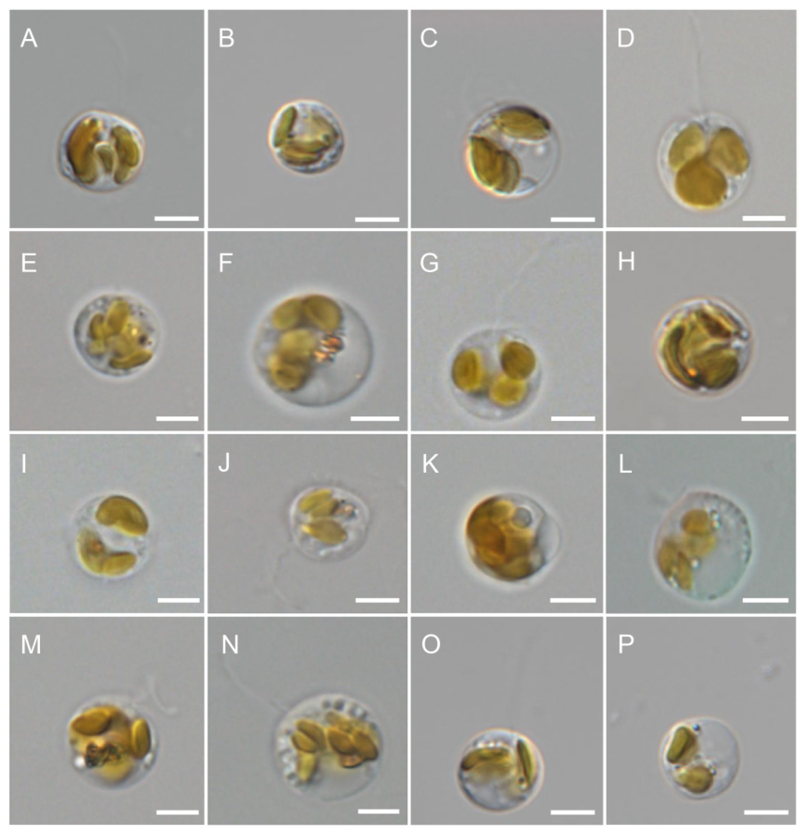
The morphology of all the studied Poterioochromonas strains was very similar. They were nearly spherical or slightly oval, and possessed brown chloroplasts and two unequal flagella (Figure 1). There was no pyrenoid or eyespot. Overall, the average size of the cells varied from 7 to 15 μm in length and from 6 to 12 μm in width (usually 6 to 12 μm in diameter), with P. andersenii (Figure 1P) being relatively smaller than P. malhamensis (Figure 1A–N) and P. longicaulis (Figure 1O) (Table 1).
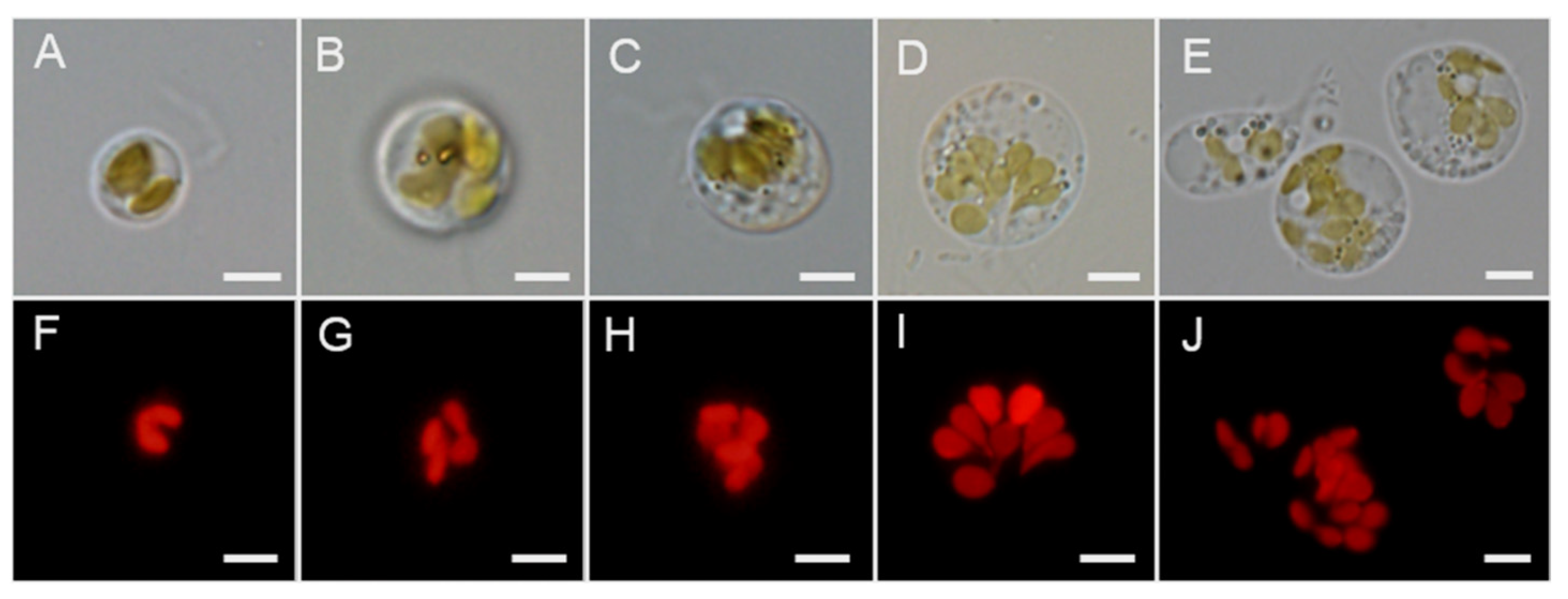
The morphology of the chloroplasts of all the studied strains was carefully examined by fluorescence microscopy (Figure 2). The shape of the chloroplasts was similar across all the examined strains, as all the chloroplasts were bilobed and connected by a very narrow bridge (Figure 2A,F). The number of chloroplasts varied both within one strain and among different strains of P. malhamensis (Table 1), including two (Figure 2B,G), three (Figure 2C,H), four (Figure 2D,I) and even several sets (Figure 2E,J).
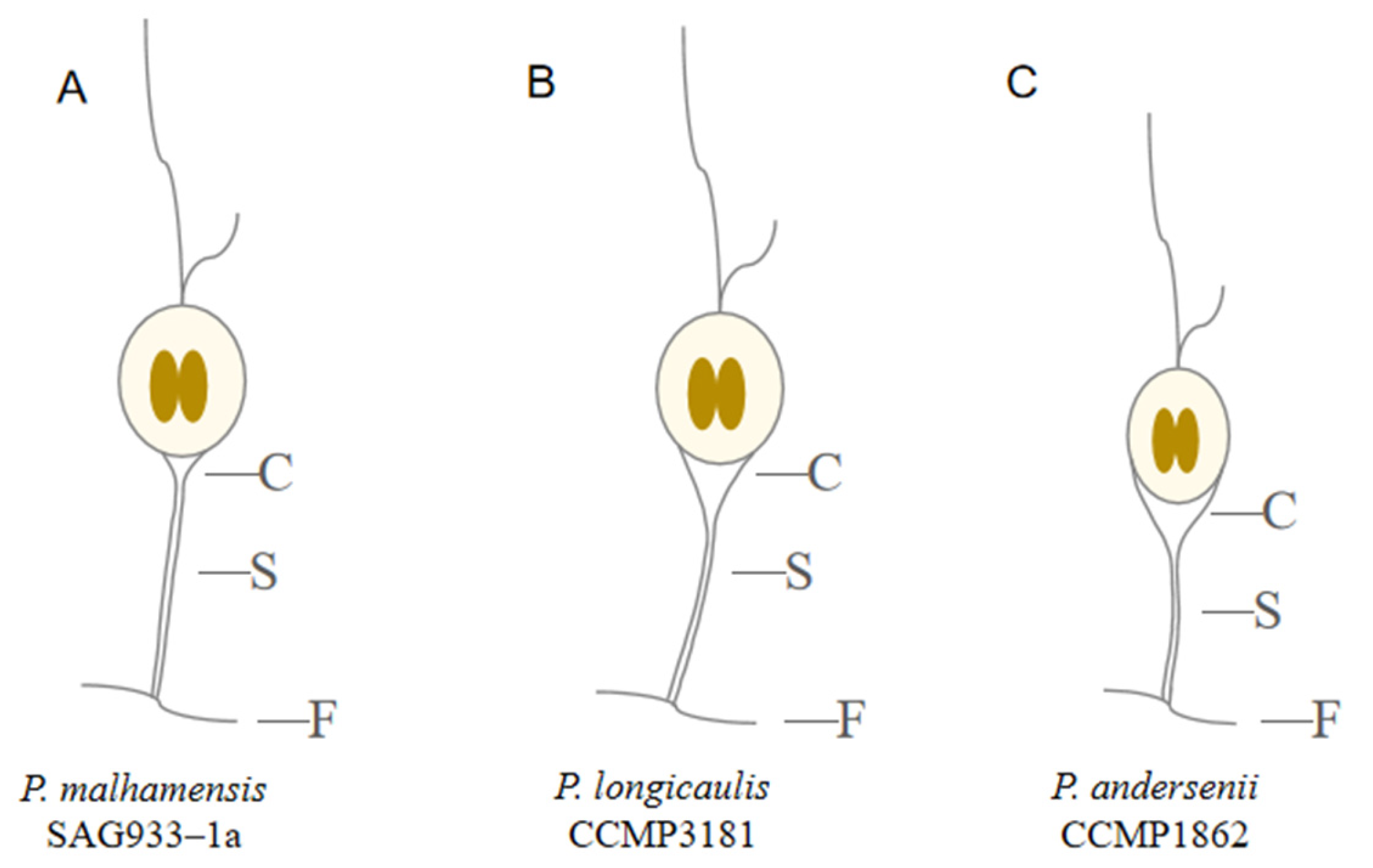
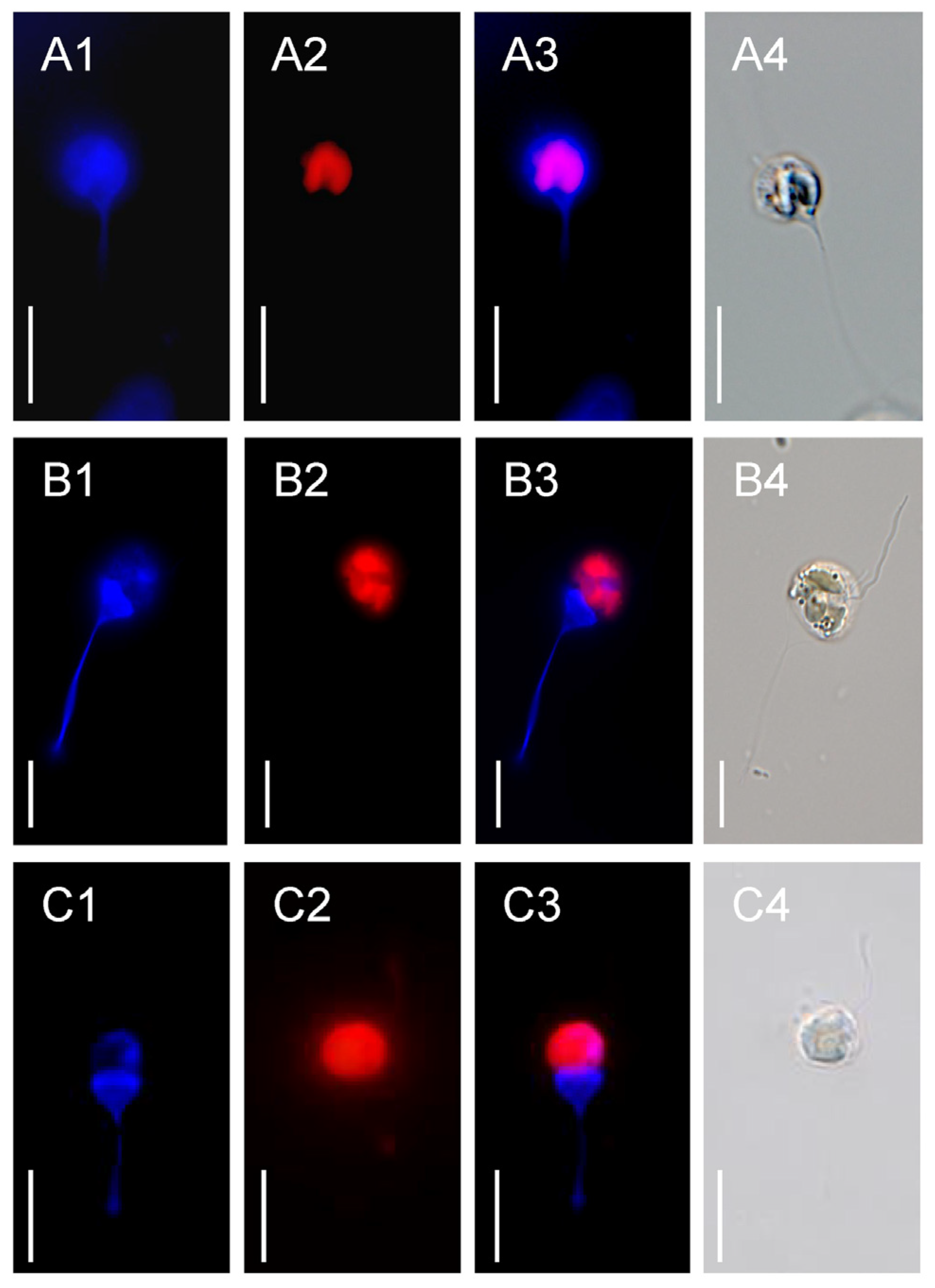
All the studied Poterioochromonas strains were observed to have lorica after CW-Eb staining, which is difficult to distinguish under a light microscope. All the lorica had similar general morphology, including a cup, a long stalk, and feet (Figure 3), whereas the shape and location of the lorica differed among the species (Figure 4), especially the lengths of the stalks, which differed significantly (Figure 5; Table 1). For P. malhamensis, represented by the SAG933-1a strain, the cup of the lorica was small and shallow, bordering only the very bottom of the cell (Figure 3A and Figure 4A); for P. longicaulis, represented by the CCMP3181 strain, the cup reached one fourth of the cell size (Figure 3B and Figure 4B); for P. andersenii, represented by the CCMP1862 strain, the cup of the lorica was broad and deep, with the upper edge of the cup at the level of the lower third of the cell (Figure 3C and Figure 4C).
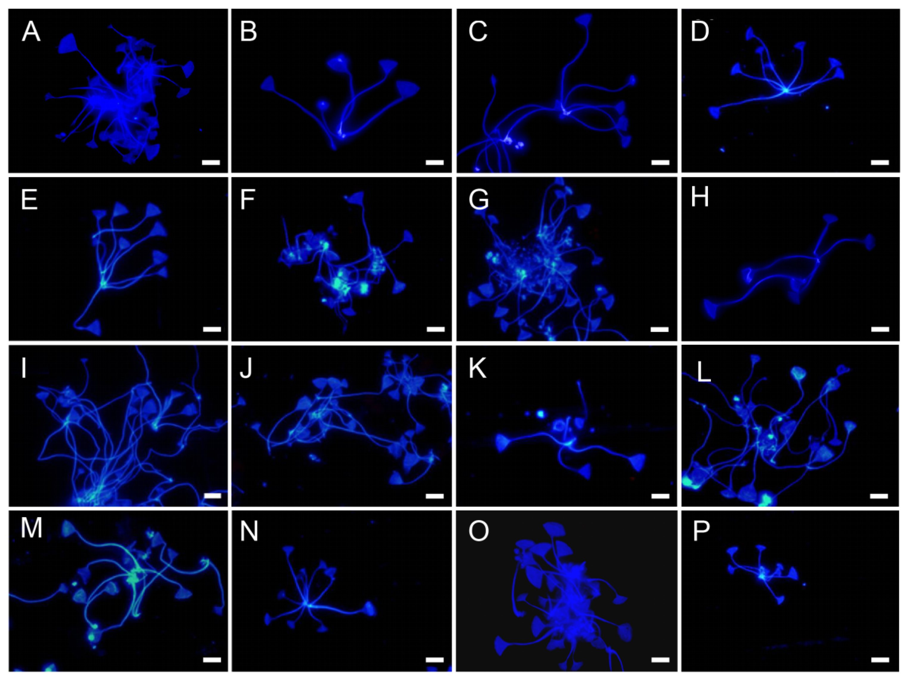
For all strains of Poterioochromonas, a tendency toward clustering of lorica stalk bases and pseudocolony formation was observed (Figure 5). The size of the lorica cup of Poterioochromonas ranged from 2 to 7 μm in depth and 2 to 10 μm in width, and the size of the lorica stalk ranged from 13 to 71 μm in length and 0.2 to 0.7 μm in width, with the lorica stalk of strain SAG933-1a obviously longer (66–71 μm) than those of the other strains (Figure 5I, Table 1). P. andersenii strain CCMP1862 was characterized by the shortest lorica stalk length, measuring 13–21 μm (Figure 5P, Table 1).

3.2. Electron Microscopy

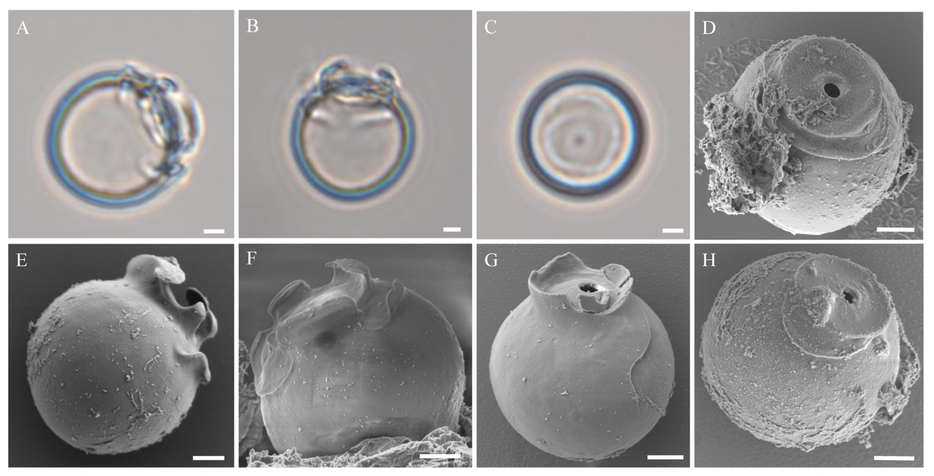
Five of the studied Poterioochromonas strains (P01 77-JZ, P03 78-JZ, CMBB-1, CMBB010, and CCMP3181; Table 1) exhibited the formation of stomatocysts. Among them, cysts were most frequently observed in the P. longicaulis CCMP3181 culture, and they were collected for SEM observation. The cysts of strain CCMP3181 are spherical (Figure 6), with a pore at the top of the cyst and a three-winged collar and a flange that is sometimes segmented (Figure 6A,B,E,F). The immature stage of the cyst only has a raised collar (Figure 6C,G), around which a flange structure begins to form (Figure 6D,H).
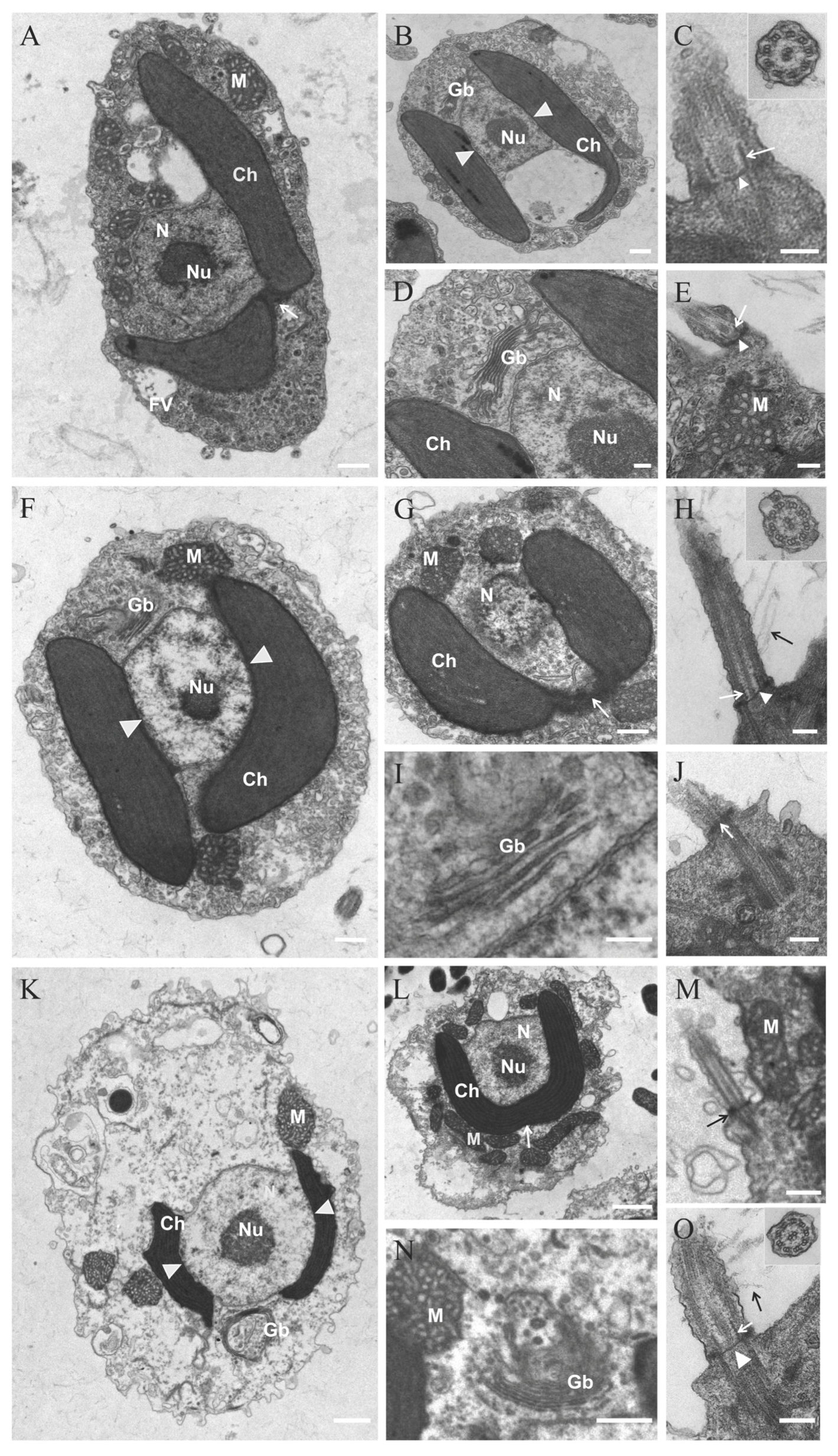
Three species of Poterioochromonas, including P. malhamensis SAG933-1a, P. andersenii CCMP1862, and P. longicaulis CCMP3181, were selected for TEM observation. All three species exhibited similar cellular ultrastructure. All of them possessed a large nucleus with a central nucleolus positioned near the cell center, featuring prominently stained chromatin (Figure 7A,B,F,K,L). The bilobed chloroplast, lacking a pyrenoid, was connected by a slender bridge and encircled the nucleus (Figure 7A,G,L), with its outermost membrane continuous with the nuclear envelope (Figure 7B,F,K). A single Golgi apparatus, appearing as stacked semicircular rings, was observed adjacent to the nucleus alongside associated vesicles (Figure 7D,I,N). The mitochondria with tubular cristae were dispersed throughout the cytoplasm, particularly near the cell periphery (Figure 7A,E–G,K,N). The flagellar ultrastructure was consistent across strains, displaying the classic ‘9+2’ microtubule axoneme (Figure 7C inset, Figure 7H inset, Figure 7O inset) and mastigonemes (Figure 7H,O). The transition zone between the basal bodies and axonemes contained a transitional helix (4–5 gyres) and a transitional plate (Figure 7C,E,H,J,M,O).

3.3. Molecular Phylogeny

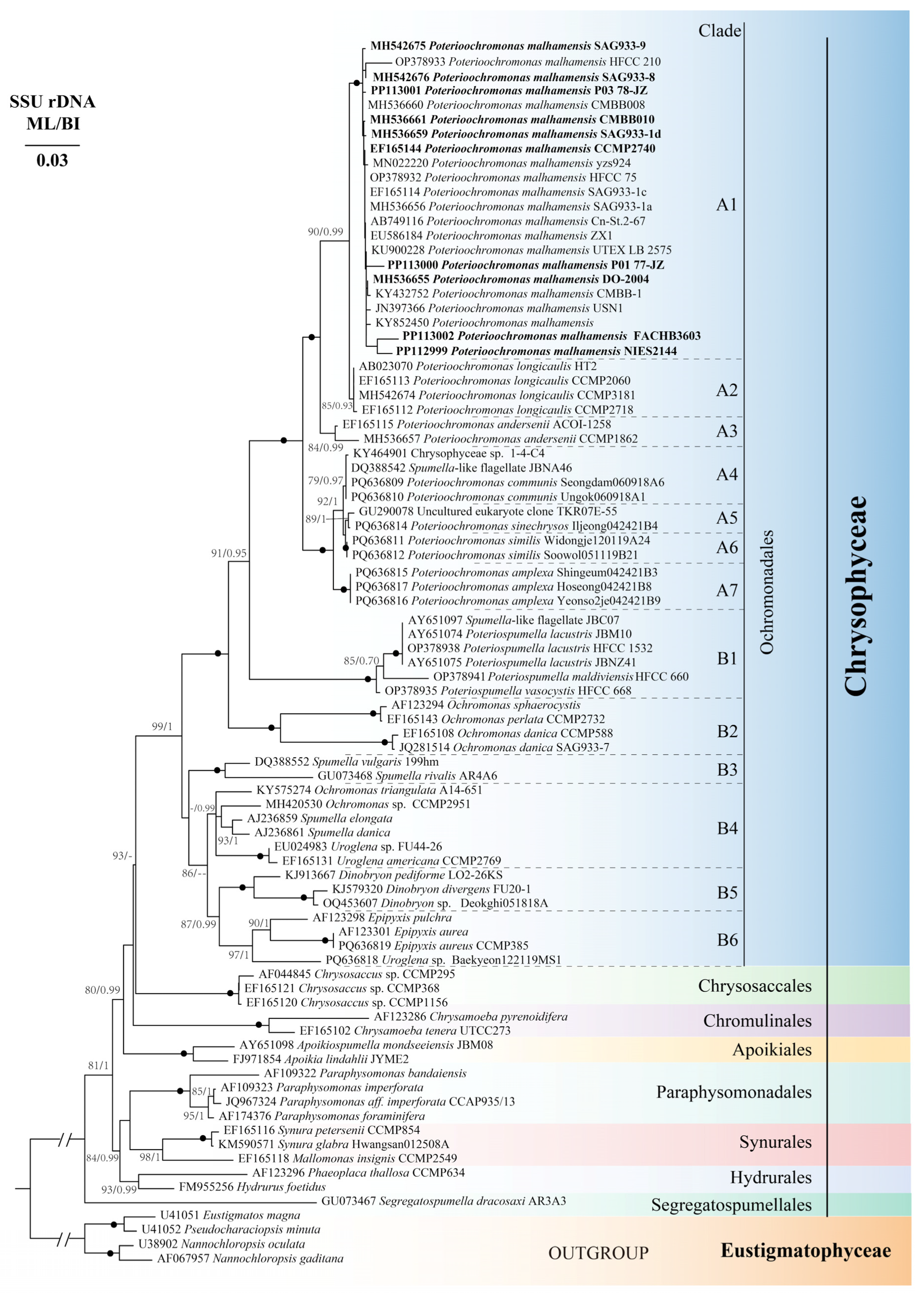
The phylogenetic analyses of Chrysophyceae based on 18S rDNA sequence data included eight orders, Ochromonadales, Chrysosaccales, Chromulinales, Apoikiales, Paraphysomonadales, Synurales, Hydrurales and Segregatospumellales, all of which were well resolved as monophyletic (Figure 8). Ochromonadales was divided into major monophyletic clade A (MLBS = 100, PP = 1.00) and several lineages (B1-B6). All the Poterioochromonas strains grouped together and were located in clade A, which consisted of two major groups: a mixotrophic group composed of subclades A1, A2 and A3 (MLBS = 100, PP = 1.00) and a heterotrophic group composed of subclades A4, A5, A6, and A7 (MLBS = 100, PP = 1.00). The authentic type strain SAG933-1a for P. malhamensis was located in subclade A1 (MLBS = 100, PP = 1.00), which included almost all studied strains of P. malhamensis. The P. longicaulis type strain CCMP3181 was located in subclade A2 and clustered with two other strains, CCMP2060 and CCMP2718, and HT2 strain, previously identified as P. malhamensis. The authentic type strain CCMP1862 for P. andersenii was located in subclade A3. Subclade A4 included species P. communis with two undefined strains 1-4-C4 and JBNA46. Subclade A5 included species P. sinechrysos with one undefined strain, TKR07E-55. Subclades A6 and A7 included species P. similis and P. amplexa, respectively. Subclade B1 (MLBS = 100, PP = 1.00) included three species of Poteriospumella and Spumella-like flagellate. Subclade B2 included three species of Ochromonas. Subclade B3 contained two species of Spumella. Weakly supported subclade B4 included three genera of Ochromonas, Spumella and Uroglena, and was grouped with low support with weakly supported subclade B5; the latter consisted of species of Dinobryon and Ochromonas. Subclade B6 included two species of Epipyxis and one undefined strain of Uroglena.
The rbcL gene sequences were also used to construct ML and BI phylogenetic trees (Figure S1), which were generally consistent with the 18S rDNA gene phylogeny (Figure 8).
The pairwise genetic distances for Poterioochromonas are shown in Table 2. Among the 16 Poterioochromonas strains examined, 14 strains were assigned to P. malhamensis, one strain, CCMP1862, belonged to P. andersenii, and one strain, CCMP3181, belonged to P. longicaulis. The COI gene exhibited the greatest intraspecific differences among the 14 strains of P. malhamensis (0.0–4.7%), followed by rbcL (0.0–2.4%), 18S rDNA (0.0–1.3%), ITS1 (0.0–1.2%), and 5.8S rDNA (0.0–1.1%), while the ITS2 presented the smallest intraspecific differences, with a range of 0.0–0.7%. In terms of interspecific differences among the three species of Poterioochromonas, the COI gene also presented the greatest interspecific variation (11.5–17.6%), followed by ITS1 (6.1–16.4%), rbcL (9.1–11.2%), ITS2 (7.3–10.2%), and 5.8S rDNA (1.5–2.9%), whereas the 18S rDNA gene presented the smallest interspecific variation, with a range of 0.5–2.9%.
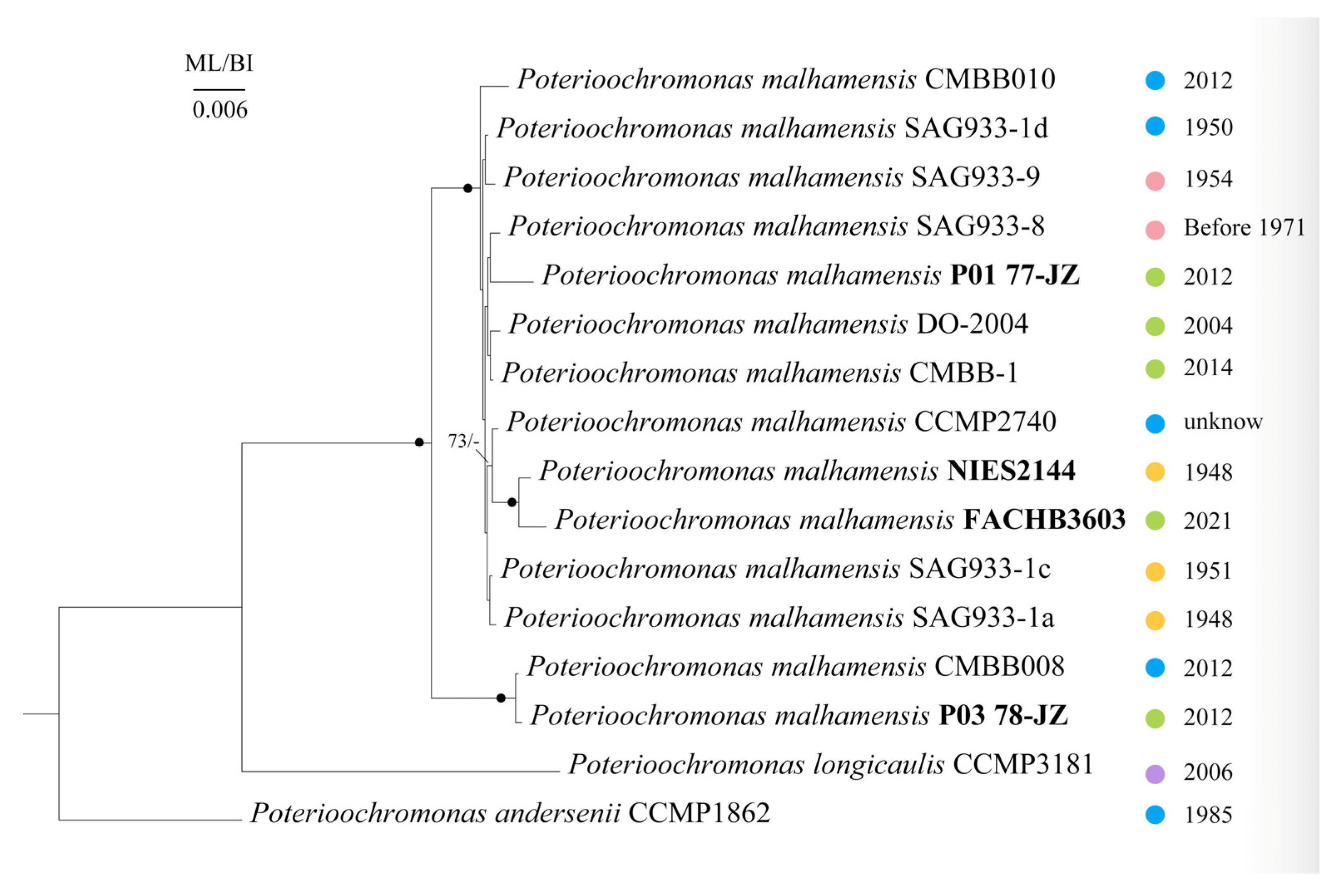
To further differentiate the phylogenetic relationships among different strains of Poterioochromonas, we constructed phylogenetic trees on the basis of combined gene sequences of the 18S rDNA, COI, rbcL, ITS1, ITS2 and 5.8S rDNA, with P. andersenii CCMP1862 serving as an outgroup (Figure 9). The results revealed that all 14 strains of P. malhamensis grouped together with full support and showed a sister relationship with P. longicaulis CCMP3181. The aforementioned P. malhamensis strains were collected from five countries of different regions, namely, China, the USA, the UK, Germany, and Australia (Table S1). However, the clustering of the strains seemed to be independent of geographic location, since strain NIES 2144 from the UK formed a well-supported subgroup with strain FACHB 3603 (from China), and strain CMBB008 from the USA and strain P03 78-JZ from China also formed a subgroup (Figure 9). Notably, the strain NIES2144 did not group with strain SAG933-1a, but the former was the subculture of the latter. It is possible that genetic differences accumulated over more than 70 years of parallel cultivation of these strains since 1948.

3.4. Diversity of Poterioochromonas in Different Habitats Predicted by Environmental Sequences

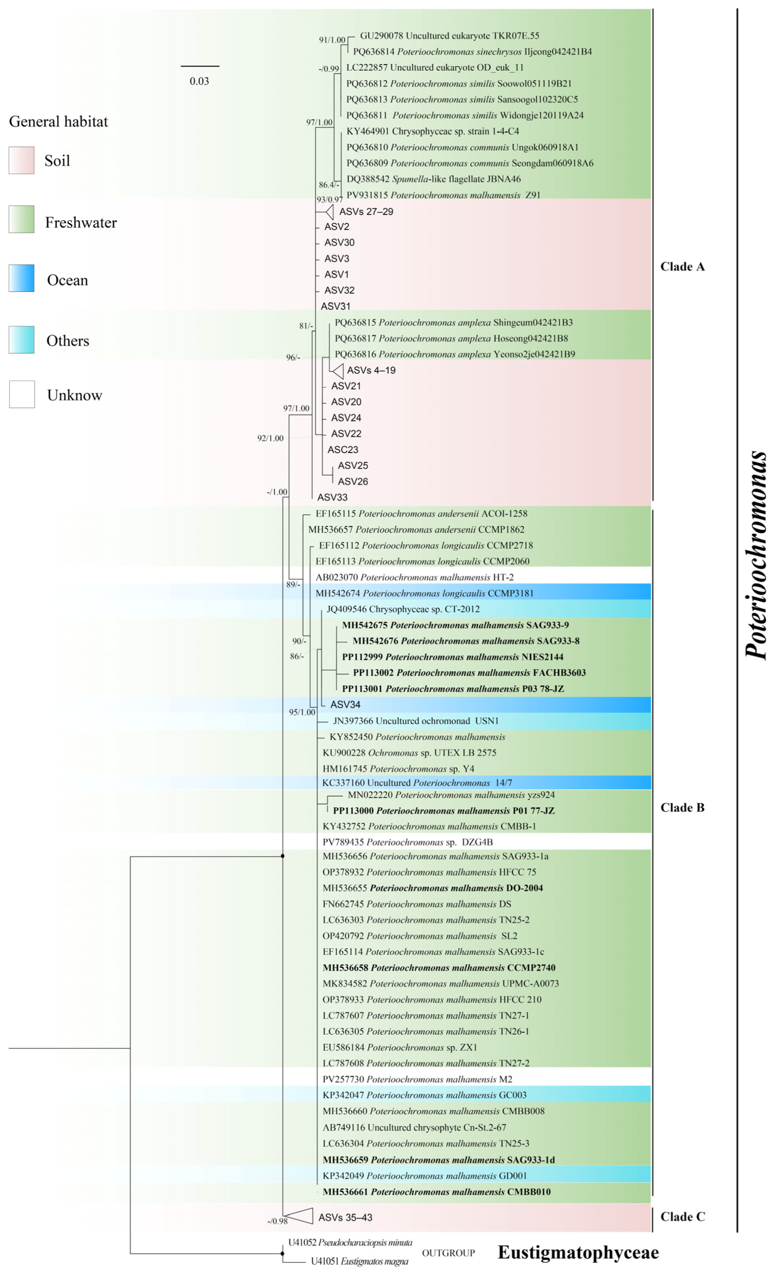
A comprehensive search in GenBank revealed 57 SSU rDNA sequences unambiguously attributable to Poterioochromonas (≥97% similarity to P. malhamensis SAG933-1a; as of 3 February 2026), and an exhaustive screening of 454 sequence data from EBI database revealed 43 distinct environmental Poterioochromonas amplicon sequence variants (ASVs) (Table S3). The phylogenetic tree based on V4 regions of the 100 SSU rDNA sequences comprises two major monophyletic clades (A and B), along with clade C, which is sister to both (Figure 10). Clade A is predominantly composed of four heterotrophic Poterioochromonas species (P. communis, P. similis, P. sinechrysos, and P. amplexa) and some unidentified taxa from freshwaters, as well as several soil-derived ASVs. Clade B is predominantly composed of P. malhamensis as well as two other mixotrophic species, P. andersenii and P. longicaulis, which primarily originated from freshwater environments. Also, several sequences from other habitat types were grouped in this clade, such as P. longicaulis CCMP3181, the uncultured Poterioochromonas 14/7, and ASV34 from marine habitats, as well as several other environmental isolates, including Chrysophyceae sp. CT-2012 from the gut of Reticulitermes santonensis, P. malhamensis strains GC003 and GD001 from mosses, and an uncultured ochromonad USN1 from the lichen Usnea longissima. Clade C was composed exclusively of soil-derived ASVs without any reference sequences of cultured representatives.

4. Discussion

4.1. Morphological Diagnostic Features of the Genus Poterioochromonas

Poterioochromonas is traditionally considered to be characterized by a small cell size (6–10 µm in diameter), one long flagellum and one short flagellum, possession of a chloroplast in addition to digestive vacuoles in the cytoplasm, and the presence of a cup-shaped lorica [15,17,41,42]. However, as the presence of lorica has not been detected in some strains, many taxonomists have determined that the structure of lorica could be a critical diagnostic morphological feature for the genus Poterioochromonas [15]. This study is the first to comprehensively examine the taxonomy of Poterioochromonas by conducting unified morphological observations on 16 strains, including almost all reported Poterioochromonas strains from all over the world, to correct the morphological diagnosis of this genus. Our observations in the current study revealed that all strains possessed lorica with variable lorica cup shapes and lorica stalk lengths (Figure 3, Figure 4 and Figure 5). Therefore, we confirmed that all strains of Poterioochromonas produce lorica. The reason that previous researchers have not observed this structure may be due in large part to the difficulty of reliably identifying lorica. Since our study, as well as several other studies [15,17,20], revealed that the lorica structure clearly became visible after CW-Eb staining, we strongly recommend using CW-Eb staining as the standard method for identifying Poterioochromonas. In addition, in the process of observation, more attention should be given to collecting samples from the bottom of the culture vessel, as more cells with lorica can be found there.
Cyst morphology is a characteristic feature of Chrysophyceae and is considered more reliable than vegetative cell morphology for distinguishing the species in some groups of chrysophytes [43,44]. On the basis of our observations of strain CCMP3181 (Figure 6) and previous studies on other strains of Poterioochromonas [15,17], it seems that the species of Poterioochromonas have a similar general cyst appearance, as all of them have a pore at the top of the cyst and a three-winged collar, which is quite different from the cyst structures of Ochromonas [15,43] and Spumella [44]. Therefore, we believe that the general morphology of a cyst can be used as a generic diagnostic feature to distinguish Poterioochromonas from other related genera. However, we checked 16 strains of Poterioochromonas, and only four strains of P. malhamensis and the CCMP3181 strain of P. longicaulis were observed to form cysts, with the CCMP3181 strain forming stomatocysts more frequently (Table 1). Similarly, Findenig et al. investigated 90 strains of Spumella-like flagellates and observed encystment in only six strains despite the broad range of temperature regimes and chemical factors tested [44]. Therefore, it is possible that other strains of Poterioochromonas can also produce cysts, although this has not been observed; so, it would be very important to develop reliable methods for controlling the formation and germination of cysts [45].

4.2. Morphological Features of Species Diagnostics Within the Genus Poterioochromonas

As reported by the latest research [14,46], Poterioochromonas includes both mixotrophic and heterotrophic species, which forms a functional continuum of nutritional modes between autotrophy and heterotrophy [4,47]. Mixotrophic species, including Poterioochromonas stipitata Scherffel 1901 [1], Poterioochromonas nutans Jane 1944 [48] and Poterioochromonas malhamensis (Pringsheim) Péterfi ex Andersen 2017 [15,42], were reported in early studies. Jeong et al. reported six new species, including two other mixotrophs, P. andersenii and P. longicaulis, and four heterotrophic species, P. amplexa, P. communis, P. similis, and P. sinechrysos [14], which expands our knowledge of the species diversity and the diversity of trophic strategies within the genus. However, the validity of the three long-described species has been questioned until recently [15], and for newly established species [14], some unclear features still need to be confirmed. In the present study, we applied unified methods to conduct a comprehensive analysis of currently available Poterioochromonas cultures and our newly isolated strains.
P. stipitata Scherffel 1901 is the generic type of Poterioochromonas whose type of material was collected from Csorba Lake, Tàtra, Hungary (Scherffel 1901) [1]. However, the designated holotype and authentic culture had not been deposited. The CCMP1862 strain was isolated and initially identified as P. stipitata by Andersen et al. [49] and was renamed as P. andersenii because of the presence of both solitary and colonial forms and distinct molecular phylogenetic position [14]. The formation of colonies was observed for strain CCMP1862 in our study as well; however, it seems that the stalk of one cell was attached to another cell’s lorica cup, with no genuine connections among individuals. The examination after CW-Eb staining revealed that the CCMP1862 strain has a special cup morphology, as its upper edge of the lorica cup is located at the lower third of the cell (Figure 3C and Figure 4C), whereas for P. malhamensis and P. longicaulis, their lorica cup edges are located near the bottom of the cell or reached one fourth of the cell size, respectively (Figure 3 and Figure 4). Since the three Poterioochromonas species can be clearly differentiated from each other on the basis of the location of the lorica cup, we recommend that the shape of the lorica cup could be a diagnostic characteristic for the species differentiation of Poterioochromonas.
P. malhamensis was initially identified as Ochromonas malhamensis by Pringsheim [41] on the basis of a strain now known as SAG933-1a. Péterfi [42] reclassified the species as P. malhamensis after demonstrating the presence of a lorica using light and electron microscopy. Andersen et al. validated and proposed a new combination, Poterioochromonas malhamensis (E.G. Pringsheim) L.Sx. Péterfi ex R.A. Andersen [15]. P. malhamensis appears to be the most common species in the genus Poterioochromonas, accounting for more than 90% of all reports in the literature [7,8,9,10,11,12,13]. In this study, we clarified the current confusion in the identification of P. malhamensis by collecting 14 strains of P. malhamensis (Figure 8). Our phylogenetic reconstructions confirmed that all the P. malhamensis strains clustered with the authentic type strain SAG933-1a, and all strains of Poterioochromonas formed a clade (Figure 8), supporting the monophyly of Poterioochromonas. Notably, lorica was not detected for the DO2004 strain previously [50]. Our study reexamined the DO2004 strain and confirmed the presence of the lorica structure (Figure 5G) and its grouping with the type strain SAG933-1a in the phylogenetic tree (Figure 8), which proves that it belongs to P. malhamensis. Similarly, the other three undefined species, Poterioochromonas sp. strain ZX1, Poterioochromonas sp. strain Cn-St-2-67, and Poterioochromonas sp. strain UTEX-LB-2575, are probably P. malhamensis, as they phylogenetically clustered with the type strain SAG933-1a (Figure 8). Our examination also revealed that the number of chloroplasts in P. malhamensis varied greatly (Table 1; Figure 2). Pringsheim’s analysis of P. malhamensis SAG933-1a revealed that the strain had one chloroplast consisting of two connected parts [41]. For the same strain, Péterfi [42] observed 1–3 chloroplasts or more, and we found that it had one or two chloroplasts, whereas some other strains, such as strain SAG933-1d, sometimes had more than four chloroplasts (Table 1; Figure 2). We even noticed that a single chloroplast changed to two chloroplasts during the observation of the same cell of P. malhamensis. These findings indicate that the number of chloroplasts varies greatly between different strains and even within the same strain of P. malhamensis and is not a suitable diagnostic marker. In addition, the length of the lorica stalk varied significantly among strains of P. malhamensis, from 18–25 µm in strain P03 78-JZ to 66–71 µm in strain SAG933-1a, which significantly exceeds the length of the stalk in P. andersenii strain CCMP1862 (13–21 µm; Table 1, Figure 5). It seems that the lorica stalks of the strains cultured in the laboratory for a long period of time appear to be longer than those of the newly isolated strains.
P. longicaulis was described as a new species because of the separate phylogenetic position, as well as the hemispherical appearance of lorica cup and significantly longer lorica stalk on the basis of the previously established strain CCMP2060 [14]. In this study, although strains CCMP3181 and CCMP2718 were not examined, they were also described as P. longicaulis since they clustered with strain CCMP2060 in the phylogenetic tree [14]. In our study, with more strains and more molecular markers, we reexamined strain CCMP3181, which was isolated by R.A. Andersen in 2006 from Australia (Table S1), and confirmed that this strain branched separately from the clades of other Poterioochromonas species but always grouped with strains CCMP 2060 and CCMP 2718 and P. malhamensis strain HT2 (Figure 8). Moreover, we found that the lorica cup morphology of strain CCMP 3181 differed from those of the other species (Figure 3, Figure 4). Our results support the establishment of strain CCMP 3181 as a novel species P. longicaulis [14] and indicate that the taxonomic assignment of P. malhamensis strain HT2 should be P. longicaulis. In addition, SEM observation revealed that the CCMP3181 strain possessed a three-winged collar structure (Figure 6), which was similar to the described cyst structures of strains CCMP2060 and CCMP2718 [15]. Our previous study documented a unique apical plug in cysts of P. malhamensis CMBB008 [17], which should be a siliceous projection based on the description in “Atlas of Chrysophycean Cysts” [51], a feature absent in strains CCMP3181, CCMP2060 and CCMP2718, indicating that the structural variation in the cysts may represent a diagnostic characteristic for interspecific differentiation of Poterioochromonas. Moreover, P. longicaulis CCMP3181 had a higher cyst formation rate than any other Poterioochromonas strain tested in our study (Table 1), while many strains of Poterioochromonas did not form cysts at all. As mentioned by Findenig et al. [44], cyst formation in Spumella-like flagellates may be related to habitat characteristics and independent of their taxonomic affiliation. We think that the occurrence of cyst formation is unsuitable as a taxonomic criterion for species differentiation in Poterioochromonas.
Overall, we suggest that the shape of the lorica cup and the structural variations in the stomatocyst could be diagnostic characteristics for species differentiation within the genus Poterioochromonas. Other investigated morphological characteristics of Poterioochromonas, such as the number of chloroplasts, the lorica stalk length, and the occurrence of cyst formation, overlap significantly among different species or not stable, and we believe that they are not suitable features for species diagnostics.

4.3. Molecular Features and Phylogeny of Poterioochromonas

Molecular features can overcome the limitations of species delineation solely based on morphological characteristics. In the latest study, the 10 helices of the V2 region and the E23-5 helix of the V4 region from SSU rDNA gene sequences have been efficiently used for the delimitation of Poterioochromonas [14]. In our study, we obtained SSU rDNA, ITS, COI and rbcL genetic markers to evaluate the intraspecific and interspecific differences in Poterioochromonas. Our results revealed that in both the intra- and interspecific groups, the COI gene presented the greatest divergence, with pairwise distances of 0–4.7% and 11.5–17.6%, respectively (Table 2), which indicates that the COI gene is suitable for distinguishing both intra- and interspecific differences in Poterioochromonas. Our previous study developed a qPCR method based on the COI gene sequences to rapidly detect and quantify P. malhamensis from the environment, indicating that the COI gene contained enough specific regions to distinguish P. malhamensis from other species [52]. Several other studies have also shown that the COI gene is indeed a highly variable marker and is suitable for distinguishing closely related lineages of chrysophytes [18].
A recent study reported for the first time that there is a heterotrophic clade within the genus Poterioochromonas, and the phylogenetic tree of Poterioochromonas is clearly divided according to nutritional modes [14]. Our phylogenetic analyses using SSU rDNA and rbcL gene sequences and a larger taxonomic sampling confirmed the monophyly of Poterioochromonas, which consisted of mixotrophic and heterotrophic clades (Figure 8). The loss of photosynthesis in many chrysophyte lineages is an excellent example of parallel evolution of heterotrophy from phototrophic or mixotrophic ancestors [53]. Thus, the relationships between Poterioochromonas and other chrysophytes with similar nutritional modes require further study with increased taxon sampling and genomic analyses.

4.4. Ecological Diversity of Poterioochromonas

Poterioochromonas has been found in more than 20 countries from aquatic and soil environments, and can coexist across both plants and animals regardless of longitude or latitude [9,54]. Most of the reported Poterioochromonas are from freshwater habitats [7,14,17,55], but their actual distribution in natural environments is poorly understood. Notably, our phylogenetic analysis revealed no clear geographic clustering among strains of the dominant species P. malhamensis (Figure 9). Moreover, our research indicates that Poterioochromonas is predominantly found in freshwaters and soil, with limited distribution in marine environments (Figure 10). The reference sequences of four heterotrophic Poterioochromonas species (P. communis, P. similis, P. sinechrysos, and P. amplexa) collected from freshwater habitats clustered with numerous soil-derived environmental sequences (Clade A; Figure 10). The result indicated that heterotrophic plastid-lacking Poterioochromonas representatives could be abundant in the soil environment. Moreover, more unreported heterotrophic Poterioochromonas taxa may exist in soil environments since Clade A contains numerous environmental ASVs that do not cluster with known species. Because Poterioochromonas can form cysts, we think that the greater abundance of Poterioochromonas sequences in soil may be related to this. Our previous study even detected P. malhamensis from air where it probably lives as a cyst [52], suggesting that Poterioochromonas may possess strong surviving adaptability.
The majority of the isolates of the three mixotrophic species (P. malhamensis, P. longicaulis and P. andersenii) were freshwater-derived, with only a small proportion distributed in marine environments (Clade B; Figure 10). Among those mixotrophic species, P. malhamensis has been repeatedly detected from various freshwater habitats, including ponds or lakes [42,43,55], algal bloom-affected water systems [8,10], and microalgae culture systems [7,17,50], as well as from lichen [56], mosses [57], and the guts of termites [58]. P. malhamensis was rarely detected in the environmental eDNA samples in our study, and only one marine ASV (ASV34, revealed in the Tara Ocean database, Table S3) clustered within the clade of P. malhamensis. The other two mixotrophic species, P. longicaulis and P. andersenii, have seldom been reported and were identified predominantly in freshwaters. The results suggest that mixotrophic Poterioochromonas species are distributed mainly in freshwater habitats.
Our distributional analysis (Figure 10) also showed that Clade C is composed exclusively of ASVs from soil environments. The sequences in this clade did not cluster with any known species reference sequences of Poterioochromonas, indicating that the diversity of Poterioochromonas far exceeds the current understanding. Since Clade A species are heterotrophic, and Clade B species exhibit mixotrophy, future isolation and investigation of the Clade C species from the natural environments would be crucial for elucidating the nutritional evolution within Poterioochromonas and similar mixotrophic protists.

4.5. Implications for Understanding the Ecological Functions of Poterioochromonas

Recent studies revealed considerable morphological and nutritional diversity among species within the genus Poterioochromonas, reflecting their multifaceted functions in ecosystems [3]. Ecologically, mixotrophic organisms serve as critical links between the microbial loop and classical food webs [59]. Mixotrophic species of Poterioochromonas engage in both photosynthesis and phagotrophy, directly regulating carbon and nutrient fluxes between autotrophic and heterotrophic pathways. In contrast, heterotrophic species actively consume bacteria and organic detritus, thereby driving organic matter remineralization [60]. From an applied perspective, their efficient phagocytic activity offers promising avenues for biocontrol-based water remediation strategies, including the targeted suppression of harmful bacteria or excessive plankton blooms [12]. The transformation of nutritional modes can alter the biochemical composition of the mixotrophic flagellate P. malhamensis and affect the grazing ability of its predator [61,62,63]. Therefore, shifts in Poterioochromonas population dynamics and trophic composition can substantially influence aquatic microbial community structure and ecosystem functioning. In the future, integrating in situ observational approaches, functional genomics, and metabolomics will be helpful to elucidate the ecological functions, competitive strategies, and biogeochemical contributions of different Poterioochromonas species in natural environments.
The importance of mixotrophic chrysophytes as grazers of bacteria in microbial food webs has therefore been widely recognized [60]. However, the role of mixotrophic chrysophytes as grazers of planktonic algae has been overlooked in spite of considerable data for such chrysomonads [7,8,9,10,11,12,13]. Pringsheim first reported that P. malhamensis (as Ochromonas malhamensis) ingested small algae [41]. Recently, P. malhamensis was reported to be the main grazer in the commercial cultivation of microalgae, and caused heavy economic losses [7,9]. It is notable that only P. malhamensis among all species of Poterioochromonas was reported to graze algae, it is unclear whether the algivorous ability was universal to Poterioochromonas. Furthermore, as mentioned above, Poterioochromonas has wide distribution and strong surviving adaptability, and rapid detection and efficient control of Poterioochromonas is very challenging. In this study, we proved that the COI gene is variable enough to differentiate Poterioochromonas intra- and interspecifically, and in our previous study, we developed a qPCR method with COI gene to detect P. malhamensis from microalgal cultures [52]. In the future, more sensitive indirect screening measures need to be developed based on unique features of microalgal prey and predator interactions [64]. To date, the physical method of ultrasonication [65], and the chemical methods of adding NH4HCO3 [66] and maintaining CO2-mediated low culture pH [67] have been developed to control P. malhamensis contamination in mass cultivation of Chlorella. In the future, more methods should be developed to control Poterioochromonas efficiently based on its specific morphological (such as having a lorica structure and not having cell wall) or physiological characters (such as being mixotrophic). For example, some specific chemical could be used to inhibit the formation of lorica, thereby hindering the growth of Poterioochromonas, or some chemical which is sensitive on the organism without cell wall could be used so that Poterioochromonas could be treated specifically. Moreover, some organic compounds such as glucose could be added to enrich the medium to induce Poterioochromonas live with chemoheterotrophic mode but not phagotrophic mode [62], thereby reducing the grazing of Poterioochromonas on microalgae.

5. Conclusions

In this study, we offer new insights into the taxonomy and diversity of Poterioochromonas on the basis of expanded taxon sampling, diverse microscopic techniques, and multiple genetic markers, as well as exhaustive sequence data screening. Our study confirmed that the presence of lorica should be the criterion for the definition of Poterioochromonas, along with the general morphology of a stomatocyst. Lorica cup morphology and stomatocyst structural features show promise as diagnostic traits for distinguishing Poterioochromonas species. In contrast, characteristics such as chloroplast number, stalk length, and cyst formation exhibit considerable overlap or instability across species, making them unreliable for species identification. The COI gene could serve as a reliable molecular marker to distinguish both inter- and intraspecific relationships in Poterioochromonas. The results from the meta-analysis of available environmental sequences indicated that the diversity of Poterioochromonas far exceeds the current understanding and that more unreported heterotrophic Poterioochromonas taxa may exist in soil environments.

Supplementary Materials

The following supporting information can be downloaded at: https://www.mdpi.com/article/10.3390/phycology6020052/s1, Figure S1: Phylogenetic tree of Chrysophyceae based on the rbcL gene sequences; Table S1: The sources of 16 strains of Poterioochromonas and the GenBank registration numbers for different DNA markers; Table S2: Primers and procedures for amplifying four different DNA markers; Table S3: Background information for the public amplicon datasets used in this study.

Author Contributions

M.J. writing, visualization, validation, methodology, investigation, formal analysis and data curation. M.C. writing—review and editing, and resources. K.C. investigation, formal analysis and data curation. H.W. writing—review and editing, resources. T.L. resources. X.Z. investigation. L.S. writing—review and editing, and resources. D.V.T. writing—review, validation, funding acquisition. Y.G. writing—review and editing, supervision, funding acquisition. All authors have read and agreed to the published version of the manuscript.

Funding

This study was financially supported by the National Natural Science Foundation of China (Nos. 32361133561, 32300355, 31772419 and 31872201). The work of D.V.T. was supported by the Russian Science Foundation (grant No. 24-44-00093, https://rscf.ru/project/24-44-00093/, assessed on 1 May 2026).

Institutional Review Board Statement

Not applicable.

Informed Consent Statement

Not applicable.

Data Availability Statement

Data will be made available on request.

Acknowledgments

The authors would like to thank Robert A. Andersen for providing the culture of Poterioochromonas malhamensis CMBB010, and to Xiaoming Zhang of the Institute of Hydrobiology, Chinese Academy of Sciences, for generously providing the algal strains.

Conflicts of Interest

The authors declare that they have no known competing financial interests or personal relationships that could have appeared to influence the work reported in this paper.

References

  1. Scherffel, A. Kleiner beitrag zur phylogenie einiger gruppen niederer organismen. Bot. Ztg. 1901, 59, 143–158. [Google Scholar]
  2. Majda, S.; Beisser, D.; Boenigk, J. Nutrient-driven genome evolution revealed by comparative genomics of chrysomonad flagellates. Commun. Biol. 2021, 4, 328. [Google Scholar] [CrossRef] [Scilit]
  3. Bock, C.; Olefeld, J.L.; Vogt, J.C.; Albach, D.C.; Boenigk, J. Phylogenetic and functional diversity of Chrysophyceae in inland waters. Org. Divers. Evol. 2022, 22, 327–341. [Google Scholar] [CrossRef] [Scilit]
  4. Schenone, L.; Aarons, Z.S.; García-Martínez, M.; Happe, A.; Redoglio, A. Mixotrophic protists and ecological stoichiometry: Connecting homeostasis and nutrient limitation from organisms to communities. Front. Ecol. Evol. 2024, 12, 1505037. [Google Scholar] [CrossRef] [Scilit]
  5. Chang, C.M.; Grattepanche, J.D.; Sanders, R.W. Influences of temperature and nutrient concentration on the balance of phototrophy and phagotrophy in a freshwater mixotrophic chrysophyte. J. Plankton Res. 2025, 7, fbaf038. [Google Scholar] [CrossRef] [Scilit]
  6. Sanders, R.W.; Porter, K.G.; Caron, D.A. Relationship between phototrophy and phagotrophy in the mixotrophic chrysophyte Poterioochromonas malhamensis. Microb. Ecol. 1990, 19, 97–109. [Google Scholar] [CrossRef] [Scilit]
  7. Ma, M.Y.; Gong, Y.C.; Hu, Q. Identification and feeding characteristics of the mixotrophic flagellate Poterioochromonas malhamensis, a microalgal predator isolated from outdoor massive Chlorella culture. Algal Res. 2018, 29, 142–153. [Google Scholar] [CrossRef] [Scilit]
  8. Long, B.; Ramos, A.; Yuan, J.S.; Dai, S.Y. Cell aggregation and temperature regulation mitigate Poterioochromonas predation on fast-growing cyanobacteria. Algal Res. 2025, 91, 104285. [Google Scholar] [CrossRef] [Scilit]
  9. Ma, M.Y.; Wei, C.J.; Huang, W.J.; He, Y.; Gong, Y.C.; Hu, Q. A systematic review of the predatory contaminant Poterioochromonas in microalgal culture. J. Appl. Phycol. 2023, 35, 1103–1114. [Google Scholar] [CrossRef] [Scilit]
  10. Yan, H.; Li, Q.; Liu, X.; Jin, A.; Yao, Y. Octanoic acid helps Poterioochromonas malhamensis control toxic Microcystis sp. J. Appl. Phycol. 2024, 36, 2531–2543. [Google Scholar] [CrossRef] [Scilit]
  11. Chen, Y.; Zhang, X.; Bai, X.; Xu, Y.; Xu, H.; Li, L. Molecular mechanisms underlying the bloom-forming cyanobacterium Microcystis defence against predation by flagellates. J. Water Process Eng. 2025, 77, 108611. [Google Scholar] [CrossRef] [Scilit]
  12. Ma, M.Y.; Wang, F.C.; Wei, C.J.; Chen, J.P.; Jin, H.; Wang, H.X.; Song, L.R.; Hu, Q.; Gong, Y.C. Establishment of high-cell-density heterotrophic cultivation of Poterioochromonas malhamensis contributes to achieving biological control of Microcystis. J. Appl. Phycol. 2022, 34, 423–434. [Google Scholar] [CrossRef] [Scilit]
  13. Ma, M.Y.; Li, Y.H.; Chen, J.P.; Wang, F.C.; Yuan, L.; Li, Y.; Zhang, B.C.; Ye, D.; Han, D.X.; Jin, H.; et al. High-cell-density cultivation of the flagellate alga Poterioochromonas malhamensis for biomanufacturing the water-soluble β-1,3-glucan with multiple biological activities. Bioresour. Technol. 2021, 337, 125447. [Google Scholar] [CrossRef] [Scilit]
  14. Jeong, M.; Kim, J.I.; Shin, W. Taxonomy of the genus Poterioochromonas (Chrysophyceae) based on morphological and molecular evidence. J. Phycol. 2025, 61, 607–622. [Google Scholar] [CrossRef] [Scilit] [PubMed]
  15. Andersen, R.A.; Graf, L.; Malakhov, Y.; Yoon, H.S. Rediscovery of the Ochromonas type species Ochromonas triangulata (Chrysophyceae) from its type locality (Lake Veysove, Donetsk region, Ukraine). Phycologia 2017, 56, 591–604. [Google Scholar] [CrossRef] [Scilit]
  16. Jeong, M.; Kim, J.I.; Nam, S.W.; Shin, W. Molecular phylogeny and taxonomy of the genus Spumella (chrysophyceae) based on morphological and molecular evidence. Front. Plant Sci. 2021, 12, 758067. [Google Scholar] [CrossRef] [Scilit]
  17. Chen, M.; Ma, M.Y.; Wang, H.X.; Hu, Q.; Gong, Y.C. Redescription of mixotrophic Poterioochromonas malhamensis and its phylogenetic analysis. Acta. Hydrobiol. Sin. 2020, 44, 901–911. [Google Scholar]
  18. Bock, C.; Chatzinotas, A.; Boenigk, J. Genetic diversity in chrysophytes: Comparison of different gene markers. Fottea 2017, 17, 209–221. [Google Scholar] [CrossRef] [Scilit]
  19. Kato, S. Laboratory culture and morphology of Colaclum vesiculosum Ehrb. (Euglenophyceae). Jpn. J. Phycol. 1982, 30, 63–67. [Google Scholar]
  20. Peck, R.K. Structure of loricae and stalks of several bacterivorous chrysomonads (chrysophyceae): Taxonomical importance and possible ecological significance. Protist 2010, 161, 148–159. [Google Scholar] [CrossRef] [Scilit]
  21. Sokal, R.R.; Rohlf, F.J. Biometry: The Principles and Practice of Statistics in Biological Research, 4th ed.; W.H. Freeman and Company: New York, NY, USA, 2012. [Google Scholar]
  22. Gong, Y.; Patterson, D.J.; Li, Y.; Hu, Z.; Sommerfeld, M.; Chen, Y.; Hu, Q. Vernalophrys algivore gen. nov., sp. nov. (Rhizaria: Cercozoa: Vampyrellida), a new algal predator isolated from outdoor mass culture of Scenedesmus dimorphus. Appl. Environ. Microbiol. 2015, 81, 3900–3913. [Google Scholar] [CrossRef] [Scilit] [PubMed]
  23. Hanaichi, T.; Sato, T.; Iwamoto, T.; Malavasi-Yamashiro, J.; Hoshino, M.; Mizuno, N. A stable lead by modification of Sato’s method. Microscopy 1986, 35, 304–306. [Google Scholar] [CrossRef] [Scilit]
  24. Fokam, Z.; Ngassam, P.; Strüder-Kypke, M.C.; Lynn, D.H. Genetic diversity and phylogenetic position of the subclass Astomatia (Ciliophora) based on a sampling of six genera from West African oligochaetes (Glossoscolecidae, Megascolecidae), including description of the new genus Paraclausilocola n. gen. Eur. J. Protistol. 2011, 47, 161–171. [Google Scholar] [CrossRef] [Scilit]
  25. Qu, L.Y.; Lv, S.; Gao, C.L.; Sun, P.; Sun, X.Q. Structure and sequence analysis of 18s rdna and its gene of phaeocystis isolate from the bohai sea. Adv. Mar. Sci. 2008, 26, 200–206. [Google Scholar]
  26. Folmer, O.; Black, M.; Hoeh, W.; Lutz, R.; Vrijenhoek, R. DNA primers for amplification of mitochondrial cytochrome c oxidase subunit I from diverse metazoan invertebrates. Mol. Mar. Biol. Biotechnol. 1994, 3, 294–299. [Google Scholar]
  27. Nguyen, L.T.; Schmidt, H.A.; von Haeseler, A.; Minh, B.Q. IQTREE: A fast and effective stochastic algorithm for estimating maximum likelihood phylogenies. Mol. Biol. Evol. 2015, 32, 268–274. [Google Scholar] [CrossRef] [Scilit] [PubMed]
  28. Posada, D.; Crandall, K.A. MODELTEST: Testing the model of DNA substitution. Bioinformatics 1998, 14, 817–818. [Google Scholar] [CrossRef] [Scilit]
  29. Ronquist, F.; Teslenko, M.; van der Mark, P.; Ayres, D.L.; Darling, A.; Höhna, S.; Larget, B.; Liu, L.; Suchard, M.A.; Huelsenbeck, J.P. MrBayes 3.2: Efficient Bayesian phylogenetic inference and model choice across a large model space. Syst. Biol. 2012, 61, 539–542. [Google Scholar] [CrossRef] [Scilit] [PubMed]
  30. Saitou, N.; Nei, M. The neighbor-joining method: A new method for reconstructing phylogenetic trees. Mol. Biol. Evol. 1987, 4, 406–425. [Google Scholar] [CrossRef] [Scilit]
  31. Tamura, K.; Stecher, G.; Kumar, S. MEGA11: Molecular evolutionary genetics analysis version 11.0. Mol. Biol. Evol. 2021, 38, 3022–3027. [Google Scholar] [CrossRef] [Scilit]
  32. Guidi, L.; Chaffron, S.; Bittner, L.; Eveillard, D.; Larhlimi, A.; Roux, S.; Darzi, Y.; Audic, S.; Berline, L.; Brum, J.; et al. Plankton networks driving carbon export in the oligotrophic ocean. Nature 2016, 532, 465–470. [Google Scholar] [CrossRef] [Scilit]
  33. Logares, R.; Deutschmann, I.M.; Junger, P.C.; Giner, C.R.; Krabberød, A.K.; Schmidt, T.S.B.; Rubinat-Ripoll, L.; Mestre, M.; Salazar, G.; Ruiz-González, C.; et al. Disentangling the mechanisms shaping the surface ocean microbiota. Microbiome 2020, 8, 55. [Google Scholar] [CrossRef] [Scilit]
  34. Mahé, F.; Mayor, J.; Bunge, J.; Chi, J.; Siemensmeyer, T.; Stoeck, T.; Wahl, B.; Paprotka, T.; Filker, S.; Dunthorn, M. Comparing high-throughput platforms for sequencing the V4 Region of SSU-rDNA in environmental microbial eukaryotic diversity surveys. J. Eukaryot. Microbiol. 2015, 62, 338–345. [Google Scholar] [CrossRef] [Scilit] [PubMed]
  35. Seppey, C.V.; Broennimann, O.; Buri, A.; Yashiro, E.; Pinto-Figueroa, E.A.; Singer, D.; Blandenier, Q.; Mitchell, E.A.; Niculita-Hirzel, H.; Guisan, A.; et al. Soil protist diversity in the Swiss western Alps is better predicted by topo-climatic than by edaphic variables. J. Biogeogr. 2020, 47, 866–878. [Google Scholar] [CrossRef] [Scilit]
  36. Shi, X.; Li, S.; Li, H.; Chen, F.; Wu, Q. The community structure of picophytoplankton in Lake Fuxian, a deep and oligotrophic mountain lake. Front. Microbiol. 2019, 10, 2016. [Google Scholar] [CrossRef] [Scilit]
  37. Shi, X.; Li, S.; Zhang, M.; Liu, C.; Wu, Q. Temperature mainly determines the temporal succession of the photosynthetic picoeukaryote community in Lake Chaohu, a highly eutrophic shallow lake. Sci. Total Environ. 2020, 702, 134803. [Google Scholar] [CrossRef] [Scilit]
  38. Annenkova, N.V.; Giner, C.R.; Logares, R. Tracing the origin of planktonic protists in an ancient lake. Microorganisms 2020, 8, 543. [Google Scholar] [CrossRef] [Scilit] [PubMed]
  39. Simon, M.; Jardillier, L.; Deschamps, P.; Moreira, D.; Restoux, G.; Bertolino, P.; López-García, P. Complex communities of small protists and unexpected occurrence of typical marine lineages in shallow freshwater systems. Environ. Microbiol. 2015, 17, 3610–3627. [Google Scholar] [CrossRef] [Scilit]
  40. Salmaso, N.; Boscaini, A.; Pindo, M. Unraveling the diversity of eukaryotic microplankton in a large and deep Perialpine lake using a high throughput sequencing approach. Front. Microbiol. 2020, 11, 789. [Google Scholar] [CrossRef] [Scilit] [PubMed]
  41. Pringsheim, E.G. On the nutrition of Ochromonas. Q. J. Cell. Sci. 1952, 93, 71–96. [Google Scholar] [CrossRef] [Scilit]
  42. Péterfi, L.S. Fine structure of Poterioochromonas malhamensis (Pringsheim) comb. nov. with special reference to the lorica. Nova. Hedwigia. 1969, 17, 93–103. [Google Scholar]
  43. Holen, D.A. Chrysophyte stomatocyst production in laboratory culture and descriptions of seven cyst morphotypes. Phycologia 2014, 53, 426–432. [Google Scholar] [CrossRef] [Scilit]
  44. Findenig, B.M.; Chatzinotas, A.; Boenigk, J. Taxonomic and ecological characterization of stomatocysts of Spumella-like flagellates (chrysophyceae). J. Phycol. 2010, 46, 868–881. [Google Scholar] [CrossRef] [Scilit]
  45. Holen, D.A.; Princiotta, S. Encystment by the mixotrophic flagellate Ochromonas pinguis Conrad (Chrysophyceae) under manipulated laboratory conditions. Fottea 2023, 23, 208–216. [Google Scholar] [CrossRef] [Scilit]
  46. Kapustin, D.A.; Iurmanov, A.A.; Kulikovskiy, M.S. A nomenclator of the genus Ochromonas sensu lato (Chrysophyceae). Phytotaxa 2026, 737, 17–33. [Google Scholar] [CrossRef] [Scilit]
  47. Millette, N.C.; Gast, R.J.; Luo, J.Y.; Moeller, H.V.; Stamieszkin, K.; Andersen, K.H.; Brownlee, E.F.; Cohen, N.R.; Duhamel, S.; Dutkiewicz, S.; et al. Mixoplankton and mixotrophy: Future research priorities. J. Plankton Res. 2023, 45, 576–596. [Google Scholar] [CrossRef] [Scilit] [PubMed]
  48. Jane, F.W. XXXVI.—Observations on some British Chrysomonads—I. Poterioochromonas nutans, sp. n. J. Nat. Hist. 1944, 11, 340–344. [Google Scholar] [CrossRef] [Scilit]
  49. Andersen, R.A.; Van de Peer, Y.; Potter, D.; Potter, D.; Sexton, J.P.; Kawachi, M.; Lajeunesse, T. Phylogenetic analysis of the SSU rRNA from members of the Chrysophyceae. Protist 1999, 150, 71–84. [Google Scholar] [CrossRef] [Scilit]
  50. Guo, S.J.; Song, L.R. Observation on Poterioochromonas sp. (Chrysophyte). J. Water Resour. Prot. 2010, 2, 592–596. [Google Scholar] [CrossRef]
  51. Wilkinson, A.N.; Zeeb, B.A.; Smol, J.P. Atlas of Chrysophycean Cysts; Springer Nature: Dordrecht, The Netherlands, 2001. [Google Scholar]
  52. Wang, X.H.; Li, H.N.; Zhan, X.L.; Ma, M.Y.; Yuan, D.N.; Hu, Q.; Gong, Y.C. Development and application of quantitative real-time PCR based on the mitochondrial cytochrome oxidase subunit I gene for early detection of the grazer Poterioochromonas malhamensis contaminating Chlorella culture. Algal Res. 2021, 53, 102133. [Google Scholar] [CrossRef] [Scilit]
  53. Gastineau, R.; Yilmaz, E.; Solak, C.N.; Lemieux, C.; Turmel, M.; Witkowski, A. Complete chloroplast genome of the mixotrophic chrysophyte Poterioochromonas malhamensis (Ochromonadales, Synurophyceae) from Van Lake in Eastern Anatolia. Mitochondrial DNA B 2021, 6, 2719–2721. [Google Scholar] [CrossRef] [Scilit]
  54. Zhang, F.; Tian, Y.; He, J. Occurrence of the freshwater chrysophyte Poterioochromonas malhamensis in a high arctic marine ecosystem. Water 2021, 13, 2129. [Google Scholar] [CrossRef] [Scilit]
  55. Yan, H.; Li, Q.; Chen, B.; Shi, M.; Zhang, T. Identification and feeding characteristics of the mixotrophic flagellate Poterioochromonas malhamensis, a microalgal predator isolated from planting water of Pontederia cordata. Environ. Sci. Pollut. Res. 2022, 29, 40599–40611. [Google Scholar] [CrossRef] [Scilit]
  56. He, Y.; Zhang, Z. Diversity of organism in the Usnea longissima lichen. Afr. J. Microbiol. Res. 2012, 6, 4797–4804. [Google Scholar] [CrossRef] [Scilit]
  57. Feng, J.; Zhang, X.; Wang, G.; Xie, S. Morphology and phylogenetic relationships of the chrysophytes associated with two mosses. Symbiosis 2016, 69, 151–159. [Google Scholar] [CrossRef] [Scilit]
  58. Tarayre, C.; Bauwens, J.; Brasseur, C.; Mattéotti, C.; Destain, J.; Vandenbol, M.; Portetelle, D.; Pauw, E.D.; Haubruge, E.; Francis, F.; et al. Isolation of an amylolytic chrysophyte, Poterioochromonas sp., from the digestive tract of the termite Reticulitermes santonensis. Biotechnol. Agron. Soc. Environ. 2014, 18, 19–31. [Google Scholar]
  59. Mitra, A.; Caron, D.A.; Faure, E.; Flynn, K.J.; Leles, S.G.; Hansen, P.J.; McManus, G.B.; Not, F.; Hallegraeff, G.M.; Pitta, P.; et al. The mixoplankton database: Diversity of photo-phago-trophic plankton in form, function, and distribution across the global ocean. J. Eukaryot. Microbiol. 2023, 70, e12972. [Google Scholar] [CrossRef] [Scilit]
  60. Pietsch, T.; Nitsche, F.; Arndt, H. High molecular diversity in the functional group of small bacterivorous non-scaled chrysomonad flagellates. Eur. J. Protistol. 2022, 86, e125915. [Google Scholar] [CrossRef] [Scilit]
  61. Ma, M.Y.; Yang, W.T.; Chen, H.; Ke, W.W.; Gong, Y.C.; Hu, Q. Transcriptional profile reveals the physiological responses to prey availability in the mixotrophic chrysophyte Poterioochromonas malhamensis. Front. Microbiol. 2023, 14, 1173541. [Google Scholar] [CrossRef] [Scilit]
  62. Ma, M.Y.; Wei, C.J.; Chen, M.; Wang, H.Y.; Gong, Y.C.; Hu, Q. Effects of nutritional mode on the physiological and biochemical characteristics of the mixotrophic flagellate Poterioochromonas malhamensis and the potential ecological implications. Microorganisms 2022, 10, 852. [Google Scholar] [CrossRef] [Scilit]
  63. Boenigk, J.; Beisser, D.; Franke, L.; Klar, L.; Ilić, M.; Fink, P. Differences in pigment composition and concentration between phototrophic, mixotrophic, and heterotrophic Chrysophyceae. Fottea 2023, 23, 223–234. [Google Scholar] [CrossRef] [Scilit]
  64. Deore, P.; Beardall, J.; Noronha, S. A perspective on the current status of approaches for early detection of microalgal grazing. J. Appl. Phycol. 2020, 32, 3723–3733. [Google Scholar] [CrossRef] [Scilit]
  65. Wang, Y.; Gong, Y.C.; Dai, L.L.; Sommerfeld, M.; Zhang, C.W.; Hu, Q. Identification of harmful protozoa in outdoor cultivation of Chlorella and the use of ultrasonication to control contamination. Algal Res. 2018, 31, 298–310. [Google Scholar] [CrossRef] [Scilit]
  66. He, Y.; Ma, M.Y.; Hu, Q.; Gong, Y.C. Assessment of NH4HCO3 for the control of the predator flagellate Poterioochromonas malhamensis in pilot-scale culture of Chlorella sorokiniana. Algal Res. 2021, 60, 102481. [Google Scholar] [CrossRef] [Scilit]
  67. Ma, M.Y.; Yuan, D.N.; He, Y.; Park, M.; Gong, Y.C.; Hu, Q. Effective control of Poterioochromonas malhamensis in pilot-scale culture of Chlorella sorokiniana GT-1 by maintaining CO2—Mediated low culture pH. Algal Res. 2017, 26, 436–444. [Google Scholar] [CrossRef] [Scilit]
Figure 1. Light micrographs of 16 strains from 3 species of Poterioochromonas. P. malhamensis (AN): (A)—strain FACHB-3603; (B)—strain P01 77-JZ; (C)—strain P03 78-JZ; (D)—strain CMBB-1; (E)—strain CMBB008; (F)—strain CMBB010; (G)—strain DO2004; (H)—strain NIES 2144; (I)—strain SAG933-1a; (J)—strain SAG933-1c; (K)—strain SAG933-1d; (L)—strain SAG933-8; (M)—strain SAG933-9; (N)—strain CCMP2740. P. longicaulis (O)—strain CCMP3181. P. andersenii (P)—strain CCMP1862. Scale bars = 5 μm.
Phycology 06 00052 g001
Figure 2. Various numbers of chloroplasts in Poterioochromonas malhamensis (strain SAG933-1d). (AE)—differential interference contrast (DIC) microscopy; (FJ)—epifluorescence microscopy (autofluorescence.) (A,F) One bilobed chloroplast. (B,G) Two sets of chloroplasts. (C,H) Three sets of chloroplasts. (D,I) Four sets of chloroplasts. (E,J) Two, three and several sets of chloroplasts. Scale bars = 5 μm.
Phycology 06 00052 g002
Figure 3. Diagram of Poterioochromonas showing variability in the size and shape of the lorica cup and the relative position of the cell within the cup. (A) P. malhamensis SAG933-1a, showing that the cup of the lorica was small and shallow, bordering only the very bottom of the cell; (B) P. longicaulis CCMP3181, showing that the cup reached one fourth of the cell size; (C) P. andersenii CCMP1862, showing that the cup of the lorica was broad and deep, with the upper edge of the cup at the level of the lower third of the cell. C—lorica cup; S—lorica stalk; F—lorica foot.
Phycology 06 00052 g003
Figure 4. Light and fluorescence micrographs of the lorica of Poterioochromonas species. The red autofluorescence originated from the chloroplasts and blue fluorescence originated from the lorica. (A1A3,B1B3,C1C3)—fluorescence microscopy; (A4,B4,C4)—differential interference contrast images. (A) P. malhamensis SAG933-1a; (B) P. longicaulis CCMP3181; (C) P. andersenii CCMP1862. Scale bar = 10 µm.
Phycology 06 00052 g004
Figure 5. Morphology of the lorica of 16 strains from 3 species of Poterioochromonas. The blue fluorescence originated from the lorica. P. malhamensis (AN): (A)—strain FACHB-3603; (B)—strain P01 77-JZ; (C)—strain P03 78-JZ; (D)—strain CMBB-1; (E)—strain CMBB008; (F)—strain CMBB010; (G)—strain DO2004; (H)—strain NIES 2144; (I)—strain SAG933-1a; (J)—strain SAG933-1c; (K)—strain SAG933-1d; (L)—strain SAG933-8; (M)—strain SAG933-9; (N)—strain CCMP2740; P. longicaulis (O)—strain CCMP3181; P. andersenii (P)—strain CCMP1862. Scale bars = 5 μm.
Phycology 06 00052 g005
Figure 6. Morphology of the stomatocyst of Poterioochromonas longicaulis CCMP3181. (AC)—differential interference contrast images; (DH)—scanning electron microscope image. (A,B,E,F)—cysts with a three-winged collar and a flange; (C,D,G,H)—immature cysts with a raised collar. Scale bars = 2 μm.
Phycology 06 00052 g006
Figure 7. Transmission electron microscopy images of Poterioochromonas. P. malhamensis SAG933-1a (AE): (A) two lobes of the bilobed chloroplast connected with a slim joint (arrow); (B) the outermost membrane of the chloroplast is continuous with the outer nuclear envelope (arrowheads); (C) transitional helix (white arrow) and transitional plate (arrowhead) of the flagellum—the inset shows a 9+2-type flagellar axoneme; (D) Golgi body; (E) transitional helix (white arrow) and transitional plate (arrowhead) of the flagellum. P. longicaulis CCMP3181 (FJ): (F) the outermost membrane of the chloroplast is continuous with the outer nuclear envelope (arrowheads); (G) two lobes of the bilobed chloroplast connected with a slim joint (arrow); (H) mastigonemes of the flagellum (black arrow) and transitional helix (white arrow) and transitional plate (arrowhead) of the flagellum, the inset shows a 9+2-type flagellar axoneme; (I) Golgi body; (J) transitional plate of the flagellum (arrow). P. andersenii CCMP1862 (KO): (K) the outermost membrane of the chloroplast continuous with the outer nuclear envelope (arrowheads); (L) two lobes of the bilobed chloroplast connected with a slim joint (arrow); (M) transition region of the flagellum with a transitional plate (black arrow); (N) Golgi body and mitochondrion with tubular cristae; (O) mastigonemes of the flagellum (black arrow) and transitional helix (white arrow) and transitional plate (arrowhead) of the flagellum; the inset shows a 9+2-type flagellar axoneme. Ch. Chloroplast; Fv. Food vacuole; Gb. Golgi body; M. Mitochondrion; N. Nucleus; Nu. Nucleolus. Scale bars: 0.5 μm for (AC,FH); 0.2 μm for (D,E,I,J,MO); 1 μm for (K,L).
Phycology 06 00052 g007
Figure 8. Phylogenetic tree of Chrysophyceae based on 18S rDNA gene sequences. A total of 85 sequences with 1850 aligned positions were used to construct the phylogenetic tree. The maximum-likelihood bootstrap values (MLBS values, left) and Bayesian posterior probabilities (PP, right) are shown at the nodes. The black circles indicate full support (100% MLBS and 1.00 PP), and (–) denotes values < 50% for MLBS or <0.70 for PP. Four species of Eustigmatophyceae were used as outgroups. The scale bar indicates a sequence divergence of 3%. Bold black font indicates the sequences obtained in this study.
Phycology 06 00052 g008
Figure 9. Phylogenetic tree of Poterioochromonas based on the combined gene sequences of SSU rDNA, COI, rbcL, ITS1, ITS2 and 5.8S rDNA. A total of 16 sequences with 3260 aligned positions were used to construct the phylogenetic tree. The maximum-likelihood bootstrap values (MLBS values, left) and Bayesian posterior probabilities (PP, right) are shown at each node. The black circles indicate full support (100% MLBS and 1.00 PP), and (–) denotes values <50% for MLBS or <0.70 for PP. The scale bar indicates a sequence divergence of 0.6%. The color of the circle indicates the country of origin of the strain. Blue: the USA; pink: Germany; green: China; yellow: the UK; purple: Australia. The year marked on the figure indicates the year the strain was collected. Bold black font indicates that all genes of corresponding strains were sequenced in this study, whereas only some genes were sequenced for other strains in our study. Detailed information on gene sequences is listed in Table S1.
Phycology 06 00052 g009
Figure 10. Diversity of Poterioochromonas amplicon sequence variants (ASVs) found in the publicly available datasets in relation to cultured isolates and their habitats. Phylogenetic tree was constructed using the V4 region of the SSU rDNA. The maximum-likelihood bootstrap values (MLBS values, left) and Bayesian posterior probabilities (PP, right) are shown at the nodes. The black circles indicate full support (100% MLBS and 1.00 PP), and (–) denotes values <50% for MLBS or <0.70 for PP. The scale bar indicates a sequence divergence of 0.1%. Bold black font indicates sequences that were either newly obtained or taxonomically corrected in this study. The ASV sequences were derived from environmental sources (see Table S3). Strain CT-2012 was isolated from the gut of Reticulitermes santonensis; strains GC003 and GD001 were moss-associated; strain USN1 was lichen-associated. Clade A includes hetertrophic Poterioochromonas; Clade B includes mixotrophic Poterioochromonas; Clade C includes unknown Poterioochromonas.
Phycology 06 00052 g010
Table 1. Summary of the cell morphology of the 16 strains of Poterioochromonas in the present study. (“—”: Not found; “+”: Found; “+ +”: Frequently found).
NumberTaxonStrainCell Size (μm)Chloroplast NumberLorica (μm)Cyst
LengthWidthDiameterCupStalk
DepthWidthLengthWidth
1P. malhamensisFACHB-36039—137—108—101, 2, 3 or more3—75—1020—290.2—0.7
2P. malhamensisP01 77-JZ6—106—87—91, 22—53—714—260.2—0.5+
3P. malhamensisP03 78-JZ7—137—118—121, 23—65—918—250.4—0.6+
4P. malhamensisCMBB-18—116—98—101, 2, 32—43—623—470.3—0.6+
5P. malhamensisCMBB0087—106—87—101, 22—74—927—420.2—0.5+
6P. malhamensisCMBB0109—128—119—101, 2 or more2—34—625—360.3—0.5
7P. malhamensisDO20049—149—128—121, 2, 3 or more1—34—625—380.3—0.5
8P. malhamensisNIES 21447—127—107—101, 2, 32—34—616—300.3—0.5
9P. malhamensisSAG933-1a7—116—98—101, 23—64—866—710.2—0.6
10P. malhamensisSAG933-1c7—116—109—121, 2, 31—42—615—470.3—0.5
11P. malhamensisSAG933-1d9—128—118—111, 2, 3, 4 or more2—44—725—380.4—0.5
12P. malhamensisSAG933-89—148—117—101, 22—43—637—620.3—0.7
13P. malhamensisSAG933-98—107—97—111, 2, 31—33—630—480.2—0.6
14P. malhamensisCCMP27407—116—107—92, 3, 41—43—625—440.3—0.5
15P.longicaulisCCMP31819—159—118—111, 22—54—924—340.4—0.7+ +
16P. anderseniiCCMP18627—96—86—91, 22—64—813—210.2—0.6
Table 2. Comparison of the intra- and interspecies genetic distances of the 16 strains of Poterioochromonas in the present study.
Molecular Marker
18S rDNACOIrbcLITS1ITS25.8S rDNA
Intra-species differences
(P. malhamensis)
Sequence
length
(nt)
1760714700267278180
Pairwise
distance
0–1.3%0–4.7%0–2.4%0–1.2%0–0.7%0–1.1%
Inter-species differencesSequence
length
(nt)
1760714700267–286276–278180
Pairwise
distance
0.5–2.9%11.5–17.6%9.1–11.2%6.1–16.4%7.3–10.2%1.5–2.9%
Disclaimer/Publisher’s Note: The statements, opinions and data contained in all publications are solely those of the individual author(s) and contributor(s) and not of MDPI and/or the editor(s). MDPI and/or the editor(s) disclaim responsibility for any injury to people or property resulting from any ideas, methods, instructions or products referred to in the content.