Article

Biological and Physico-Chemical Properties of Lobosphaera sp. Packed in Metallized Polyethylene Terephthalate/Polyethylene (PETmet/PE)

by
Valter F. R. Martins
,
Ana J. Alves
,
Fátima Poças
,
Manuela Pintado
,
Rui M. S. C. Morais
* and
Alcina M. M. B. Morais
*
CBQF—Centro de Biotecnologia e Química Fina—Laboratório Associado, Escola Superior de Biotecnologia, Universidade Católica Portuguesa, Rua Diogo Botelho, 1327, 4169-005 Porto, Portugal
*
Authors to whom correspondence should be addressed.
Phycology 2025, 5(3), 35; https://doi.org/10.3390/phycology5030035
Submission received: 15 July 2025 / Revised: 30 July 2025 / Accepted: 4 August 2025 / Published: 6 August 2025

Abstract

This study evaluated the effects of different storage conditions, varying in light exposure, relative humidity (RH), and packaging materials, on the physicochemical stability of Lobosphaera sp. biomass, the retention of bioactive compounds, and the bioactivity of its extracts. Under light and 75% RH, the biomass absorbed moisture over time, reaching 0.779 ± 0.003 g/g dry weight (DW) after three months. This was accompanied by a decline in luminosity, chroma, and hue values. In contrast, samples stored under other conditions showed minimal changes, indicating that high humidity, combined with light exposure, compromises biomass stability. Packaging in metalized polyethylene terephthalate (PETmet/PE) effectively preserved the water content, color, and carotenoid levels during a two-month storage period. Bioactive compounds extracted via hydroethanolic ultrasound-assisted extraction yielded 15.48 ± 1.35% DW. Total phenolic content (TPC) of the extracts declined over time in both PETmet/PE and low-density polyethylene (LDPE) packaging, though the decrease was less pronounced in PETmet/PE. Antioxidant activity, assessed via the ABTS assay, remained stable, regardless of storage duration or packaging. Antimicrobial activity of the extract decreased over time but remained more effective against Gram-positive bacteria (Staphylococcus aureus, Bacillus cereus, and Listeria monocytogenes), with PETmet/PE packaging better preserving antimicrobial efficacy than LDPE. These findings underscore the importance of optimized storage conditions and packaging for maintaining the quality and bioactivity of Lobosphaera sp. biomass and its extracts.

1. Introduction

Lobosphaera sp. is an oleoginous green microalga well known for its high content of bioactive compounds, such as carotenoids and polyunsaturated fatty acids (PUFAs). More specifically, its ability to produce and accumulate substantial amounts of fatty acids, such as eicosapentaenoic acid (EPA) and docosahexaenoic acid (DHA), has attracted increasing attention in recent years as a sustainable alternative to fish and plant-based oils in food applications [1]. These compounds have shown cardioprotective effects [2]. In dietary studies, feeding Lobosphaera incisa to zebrafish (Danio rerio) has been shown to increase fatty acid content in the gut, particularly arachidonic acid (ARA), thereby enhancing immunomodulatory functions, such as improved resistance to streptococcal infection [3]. Lobosphaera sp. is also rich in protein with 39.2 ± 0.7 g/100 g DW [1], which is nutritionally valuable.
Microalgae preservation faces several challenges, as the preservation of all quality attributes requires optimized storage conditions suitable for the type of film packaging used. Packaging with conventional materials (e.g., polyethylene, polypropylene) primarily focuses on barrier properties (oxygen, water vapor, and light), whereas other types of packaging, such as functional packaging, incorporate mechanisms like antioxidant, antimicrobial, anti-enzymatic, anti-browning, or pH control effects, and may include the addition of bioactive compounds to enhance packaging functionality. In conventional packaging, permeability to water vapor and oxygen can lead to lipid oxidation, resulting in oxidative rancidity caused by reactions between oxygen and unsaturated fatty acids in fats and oils or hydrolytic rancidity, which involves the breakdown of triglycerides into free fatty acids, leading to undesirable flavors [4]. Light exposure can also cause photodegradation, including the degradation of micronutrients (e.g., light-sensitive vitamins), photo-oxidation of lipids, pigments discoloration (as chlorophylls and carotenoids degrade under UV exposure), and deterioration of sensory qualities (e.g., degradation of flavor and aromatic compounds, negatively impacting taste and smell) [5].
Although studies have been performed on the applications of microalgae in food packaging [6], there is little research on packaging for microalgae in powder [7]. Petroleum is a raw material that can be manufactured into fibers, films, and objects with simple or more complex shapes [8]. Films can be produced to obtain plastic packaging for food products, including microalgae biomass in powder (freeze-dried). Polyethylene (PE) is an aliphatic hydrocarbon polymer classified as a polyolefin. Polyethylene terephthalate (PET) is classified as a partly aromatic polyester [8]. Metallized polyethylene terephthalate (PETmet) offers significant advantages over standard PET for packaging powders, particularly in terms of barrier properties. The addition of a thin metal layer (typically aluminum) on PET films enhances their oxygen and moisture barrier capabilities, which is critical for preserving sensitive components like pigments, antioxidants, and fatty acids in microalgae powders. Additionally, PETmet provides light shielding, which is especially important for preventing photo-oxidation [9,10].
The biodegradability of PET has been explored from an environmental perspective. Aboeingna et al. [11] investigated the enzymatic degradation of PET using hydrolases, identifying it as a promising strategy for recycling. In particular, enzymes from the PETase and cutinase families have demonstrated significant PET-degrading activity. Gao et al. [12] conducted a review focused on the characterization and enzymatic mechanisms of PET hydrolases. They also discussed future directions in PET biodegradation and its subsequent biotransformation into value-added compounds.
The present study focuses on the effects of using PETmet/PE packaging on the preservation of freeze-dried Lobosphaera sp. microalga, with a focus on the antioxidant and antimicrobial activities of ultrasound-assisted ethanolic extracts from this biomass. It also examines changes in total carotenoid and chlorophyll contents, as well as moisture contents. Additionally, the effects of the storage conditions, including light exposure (presence or absence) and relative humidity (50% or 75%) at 23 °C, on the water content and color of Lobosphaera sp. were evaluated.

2. Materials and Methods

2.1. Microalga Biomass

Lobosphaera sp. biomass was supplied frozen by A4F—Algae for Future, Portugal. It was stored at −20 °C until it was necessary for the packaging studies and extraction assays of bioactive compounds and then it was freeze-dried.

2.2. Storage Studies at Different Conditions of Light and Relative Humidity

Biomass (20 g) was accurately weighed and placed in a Petri dish, which was then positioned inside a designated container corresponding to each experimental condition. Containers A and B were maintained at 50% relative humidity (RH), achieved using a saturated magnesium nitrate solution. Containers C and D were exposed to 75% RH, maintained with a saturated sodium chloride solution. Each RH condition was further subdivided based on the light exposure, with containers either wrapped in aluminum foil (dark conditions) or left uncovered (light conditions). All containers were stored in a temperature-controlled room set at 23 °C. After one month of storage, three replicate samples of microalgal biomass (2 g each) were collected for color and water content analysis. This sampling procedure was repeated monthly for a period of three months, with biomass color assessed visually at each interval.

2.3. Packaging Studies

2.3.1. Packaging Materials

Two packaging materials were used: low-density polyethylene (LDPE) and metalized polyethylene terephthalate with polyethylene (PETmet/PE), with thicknesses of 0.043 ± 0.002 mm and 0.045 ± 0.001 mm, respectively (measured with a micrometer, TMI-MI20, New Castle, DE, USA). The water vapor permeability of each material was determined at 23 °C and 50% RH, by monitoring the weight change of calcium chloride (initially 5 g) enclosed in bags with an effective surface of 48 cm2 area and stored for two months. Five replicates were performed for each material.

2.3.2. Packaging of Biomass and Quality Analysis During Storage

Previous research [7] demonstrated that packaging Porphyridium cruentum biomass in PETmet/PE resulted in better quality preservation compared to LDPE. Consequently, the quality assessments of Lobosphaera biomass during storage were conducted exclusively using PETmet/PE packaging. Biomass (3 g) was placed in bags made of PETmet/PE with a usable surface area of 48 cm2. The bags were sealed and stored in an air-conditioned room at 23 °C and 50% RH. Four replicates were performed for each sampling time. Each sample was analyzed for color, water content, total carotenoids and chlorophylls.
Subsequently, the same procedure was followed with 4 g of biomass for the extraction of bioactive compounds. Both PETmet/PE and LDPE bags were used for packaging, and analyses included total phenolic content, as well as antioxidant and antimicrobial activities. Four replicates were performed for each packaging material and sampling time.

2.4. Physico-Chemical Characterization of Lobosphaera sp. Biomass

2.4.1. Color Analysis

The color analyses were performed on approximately 2 g of biomass of each replicate, by using a Minolta CR-300 colorimeter (Konica-Minolta, Osaka, Japan) in the CIE L*a*b* mode color [13]. For each sample, ten measurements were taken on each of the three replicates. The evaluated color parameters included L* (lightness), a* (green–red coordinate) and b* (blue–yellow coordinate). The hue angle and the saturation index (chroma), and total color difference (ΔE) were calculated using the following equations:
H u e = a r c t a n b * a *
C h r o m a = a * 2 + b * 2
Δ E = L 0 * L * 2 + a 0 * a * 2 + b 0 * b * 2
L0*, a0*, and b0* are the values of the control (samples not packed) at time = 0 day.

2.4.2. Moisture Content Determination

Moisture content was determined using 2 g of biomass previously analyzed for color. The sample was placed in a Petri dish and dried in an oven (FP115, Binder, Tuttlingen, Germany) at 105 °C until constant weight was achieved, following a method adapted from AOAC [14]. The determinations were performed in triplicate.

2.4.3. Carotenoid and Chlorophyll Content Determination

Total carotenoid and chlorophyll contents were determined using 0.5 g of biomass from each sampling bag, transferred to pre-weighed 15 mL Falcon tubes. To each tube, 5 mL of acetone was added. The mixtures were subjected to extraction using an Ultra-Turrax probe (IKA T18 Digital Ultra Turrax, Staufen, Germany). Following extraction, a centrifugation was carried out at 5000 rpm for 10 min. The supernatant was collected and appropriately diluted for absorbance measurements at 661, 644, and 470 nm in a UV-Vis spectrophotometer (Shimadzu, 1240, Kyoto, Japan). Chlorophyll a, chlorophyll b, and total carotenoids contents were determined using the following equations, respectively [15]:
Chlorophyll a (μg mL−1) = Ca = 11.24A661 − 2.04A644
Chlorophyll b (μg mL−1) = Cb = 20.13A644 − 4.19A661
Carotenoids (μg mL−1) = (1000 × A470 − 1.90Ca − 63.14Cb)/214

2.5. Extraction of Bioactive Compounds from Lobosphaera sp.

The extraction of bioactive compounds from Lobosphaera sp. was performed according to the method described by Martins et al. [1], employing a conventional solvent extraction using a hydroalcoholic solution composed of water and ethanol in a 1:9 (v/v) ratio. One gram of Lobosphaera sp. powder was suspended in 30 mL of the extraction solvent and incubated at 50 °C with constant agitation at 120 rpm in an orbital shaker (MaxQ6000, Thermo Scientific, Waltham, MA, USA) for 120 min. This extraction step was repeated twice to maximize yield. Following incubation, the mixture was subjected to ultrasonic homogenization using a probe sonicator (Vibra-Cell, Sonics, Newtown, CT, USA) operated at 20 kHz, with 30-s pulses applied over a total duration of 10 min. The homogenized solution was then filtered, and ethanol was removed via rotary evaporation (Rotavapor R-210, Büchi Labortechnik AG, Flawil, Switzerland). The resulting aqueous extract was subsequently lyophilized to obtain a dry extract powder [1].

2.6. Characterization of the Bioactivity of Lobosphaera sp. Extracts

2.6.1. Determination of Total Phenolic Content (TPC)

The extract obtained as described in Section 2.4 was used for total phenolic content (TPC) analysis, with a total of 40 mg of the dried extract being reconstituted in 2 mL of distilled water (20 mg/mL). All experiments were conducted in triplicate.
TPC was quantified using the Folin–Ciocalteu colorimetric assay, following Martins et al.’s methodology [1]. Briefly, 100 µL of diluted Folin–Ciocalteu reagent (20% v/v) was mixed with 30 µL of extract solution, followed by the addition of 100 µL sodium carbonate (7.4% w/v). The reaction mixture was incubated for 30 min at room temperature in the dark. Absorbance was then measured at 765 nm using a microplate reader (Synergy H1, Biotek, Winooski, VT, USA). Gallic acid was used for calibration, and results were expressed as mg gallic acid equivalents per 100 mg of dry extract (mg GAE/100 mg DW).

2.6.2. Antioxidant Activity Assays

The antioxidant capacity of the extracts (20 mg/mL) was evaluated using ABTS. Each assay was performed in triplicate across three independent extractions.
The ABTS (2,2′-azinobis(3-ethylbenzothiazoline-6-sulphonic acid)) assay was performed according to Martins et al.′s method [1]. The ABTS•⁺ radical was generated by reacting ABTS (0.0384 g in 10 mL water) with potassium persulfate (0.0066 g in 10 mL water) and adjusted to an absorbance of 0.700 ± 0.020 at 734 nm. An aliquot of extract (20 µL) was reacted with 180 µL of the ABTS solution in the dark at room temperature for 5 min. Absorbance was read at 734 nm. Trolox was used as the standard, and results were expressed as µmol Trolox equivalents per 100 mg of dry extract (µmol TE/100 mg DW). The percentage of inhibition was calculated using the following equation:
I (%) = [(Abs A0 − Abs sample) ÷ Abs A0] × 100

2.6.3. Antimicrobial Activity Assays

The antimicrobial activity assays were conducted following the methodology described by Martins et al. [16]. The extract obtained in Section 2.4. was used to prepare solutions at concentrations of 1%, 2%, and 3% (w/v) in distilled water.
An overnight bacterial culture was grown in Mueller–Hinton broth (MHB) and adjusted to an optical density of 0.2 at 610 nm (approximately 108 CFU/mL), then diluted to an inoculum of 105–106 CFU/mL. The bacterial suspension and extract solutions were loaded into a 96-well plate and incubated at 37 °C. Optical density (OD) readings at 600 nm were taken hourly over 24 h using a microplate reader (Multiskan GO, Thermo Scientific, Vantaa, Finland). MHB with no extract served as a positive control; sterile MHB was used as a negative control.
Bacterial growth inhibition was calculated using the following equation:
Inhibition (%) = ((OD bacteria control − OD bacteria)/OD bacteria control) × 100
Tested bacterial strains included Gram-negative species Escherichia coli ATCC 25922, Yersinia enterocolitica NCTC 10406, and Salmonella enterica serovar Enteritidis ATCC 13076, as well as Gram-positive species Staphylococcus aureus ATCC 6538, Bacillus cereus NCTC 2599, and Listeria monocytogenes NCTC 10357.

2.7. Statistical Analysis

All data are expressed as mean ± standard deviation (n = 3), with calculations and interpretations based on standard principles of statistical inference. Statistical analysis was performed using one-way repeated measures ANOVA with three conditions for the ABTS assay and four conditions for the antimicrobial activity assay, considering a significance level of p < 0.05. Analyses were conducted using IBM SPSS Statistics v22. Assumptions of normality and homogeneity of variance were evaluated using the Shapiro–Wilk and Levene′s tests, respectively. Sphericity was assessed with Mauchly′s test. If the sphericity assumption was violated, the Greenhouse–Geisser correction was applied; otherwise, the standard F-test was used. Post hoc comparisons were conducted using Fisher′s LSD test for within-subject comparisons and the Bonferroni correction for between-subject comparisons.

3. Results and Discussion

3.1. Effects of the Storage of Lobosphaera sp. Biomass Under Different Conditions of Light and Relative Humidity

The water content of Lobosphaera biomass remained approximately constant during storage under all conditions, except under light exposure and 75% relative humidity (RH). Under these conditions, the water content increased over time (Figure 1), reaching 0.779 ± 0.003 g/g dry weight (DW) after three months of storage. This is a considerably high value, corresponding to nearly 44% of the total weight. The RH level seems to be the determining factor in this observation, as storage under light and 50% RH did not produce the same effect. Similar results have been reported for Porphyridium cruentum [7].
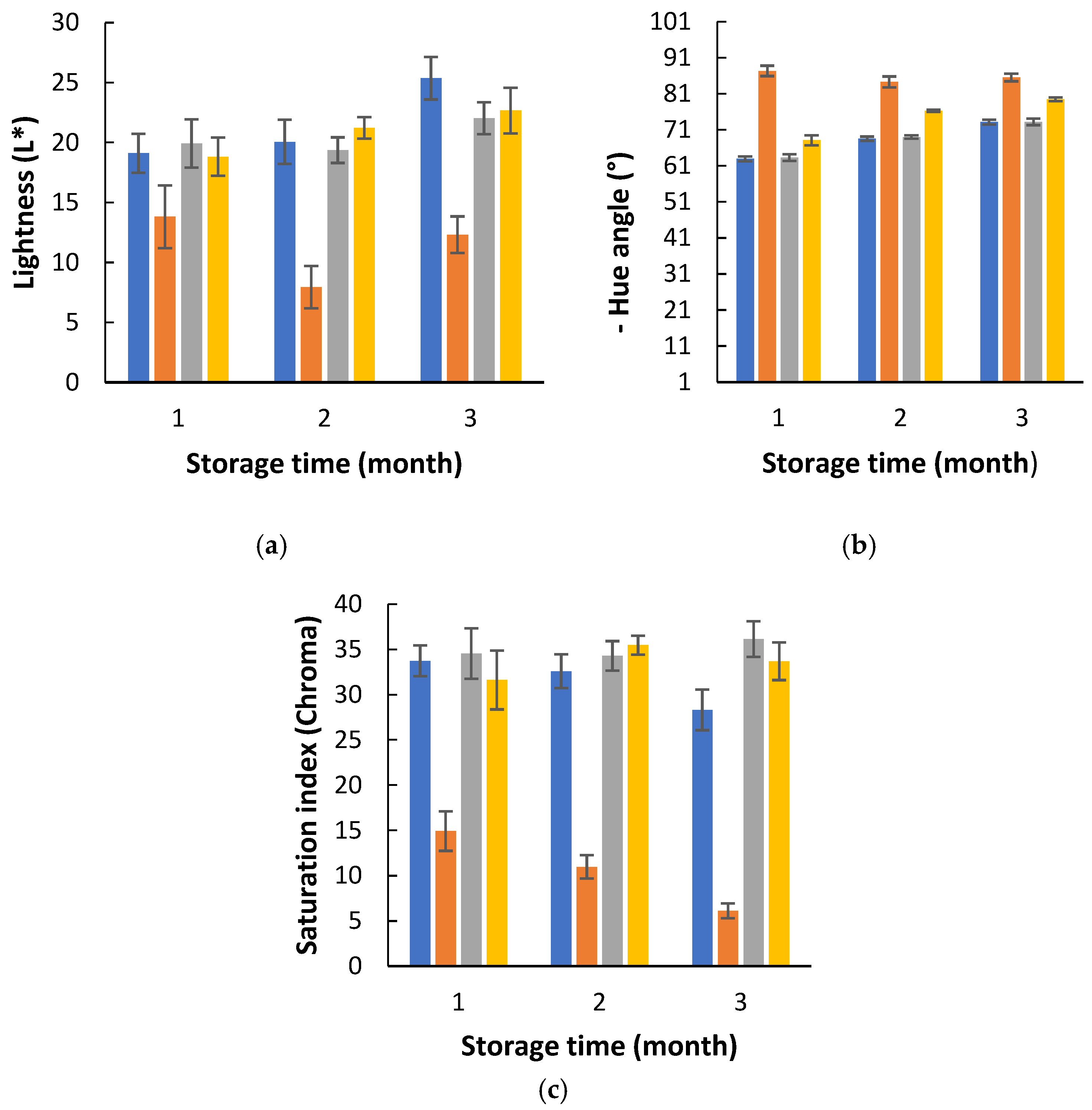
The biomass luminosity was relatively low (below 26.00) and was most affected by the condition of light exposure and 75% RH, exhibiting reduced L* values compared to the other conditions, with a decrease observed during the first two months of storage (Figure 2a), indicating that the samples became darker. Hue values were also more negative under this condition than under others, although all samples showed negative hue values throughout storage (Figure 2b). Samples stored under light and 75% RH presented lower chroma values than those stored under other conditions, and these values decreased progressively during storage (Figure 2c). Nevertheless, the observed changes in color parameters were not visually perceptible. Similar results were reported by Morais et al. [7] for P. cruentum.
Therefore, the condition of light exposure and 75% RH does not seem adequate for the preservation of Lobosphaera during storage.

3.2. Effects of the PETmet/PE Packaging on Biomass Quality

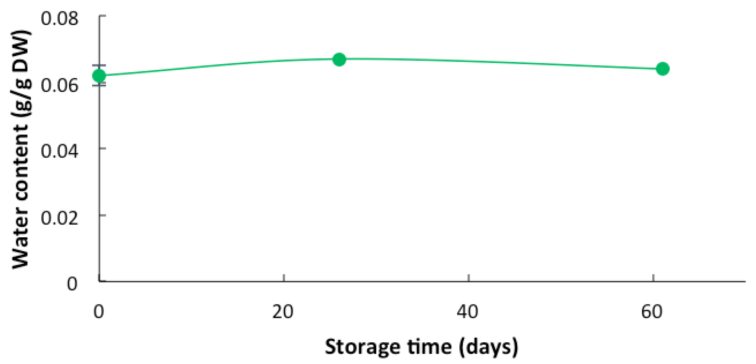
Figure 3 shows that the water content in biomass packed in PETmet/PE remained approximately constant throughout the storage period. These results are consistent with those reported in previous studies on Porphyridium cruentum [7] and suggest PETmet/PE is an effective material to protect Lobosphaera powder from water absorption and light, thereby allowing long-term storage.
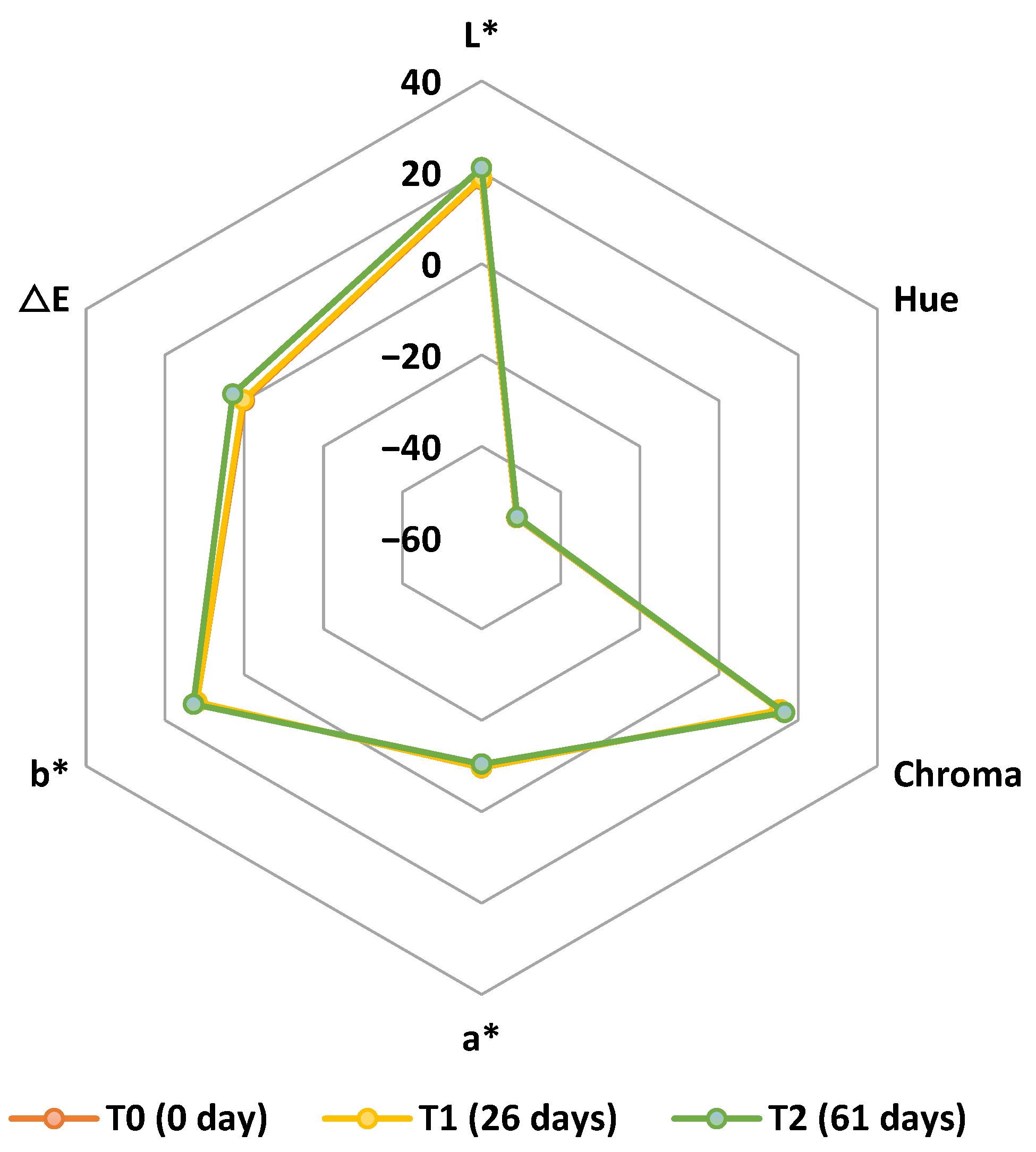
Concerning color, it was observed that the color parameters remained relatively stable during two months of storage. There was no significant difference in the values of all color parameters between T0 (0 day) and T1 (26 days) (Figure 4). The chroma values were similar to the b* values, with the a* values being notably low (Figure 4).
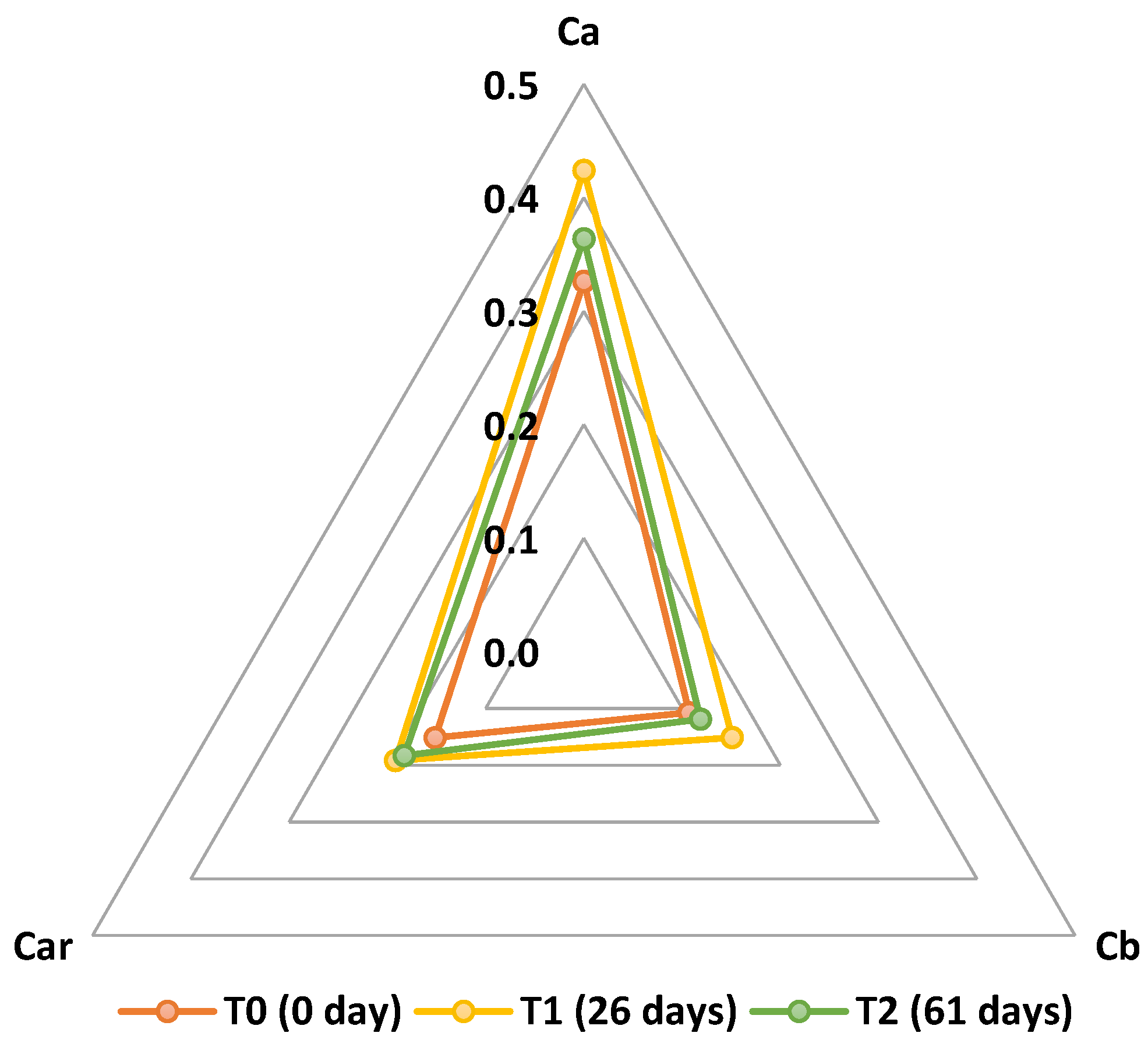
A slight decrease in carotenoid content was observed between one and two months of storage (Figure 5). This finding is favorable, as carotenoids are bioactive compounds that contribute to the antioxidant activity of the Lobosphaera biomass. Preserving carotenoids is crucial for maintaining the quality of this microalga powder, as these compounds have significant nutritional and health-related applications. Carotenoids are important antioxidants and play a vital role in human health by contributing to the prevention of various diseases, including certain cancers and eye disorders [17].

3.3. Yield of Extraction of Bioactive Compounds from Lobosphaera sp.

The hydroethanolic ultrasound-assisted extraction of bioactive compounds from Lobosphaera sp. yielded 15.48 ± 1.35% dry weight (DW). Similarly, Martins et al. [18] reported lower extraction efficiencies for Arthrospira sp., 12.99 ± 0.90% DW. Monteiro et al. [19] applied ultrasound-assisted extraction using hydroalcoholic solutions at 50% and 80% ethanol to Nannochloropsis sp., obtaining yields of 17.33 ± 1.00% and 24.44 ± 1.88%, respectively. The same method applied to Chlorella sp. resulted in higher yields of 26.72 ± 1.74% and 28.05 ± 2.09%, respectively.

3.4. Effects of PETmet/PE Packaging on the Bioactivity of Lobosphaera sp. Bioactive-Rich Extracts

3.4.1. Total Phenolic Content (TPC) and Antioxidant Activity (ABTS)

Total phenolic content (TPC) reflects the total amount of phenolic compounds present, which are well known for their antioxidant properties. Bioactive compounds from Lobosphaera sp. have scarcely been studied in literature, although some studies are available [1,20]. The variability of TPC in microalgae is largely influenced by several factors, including the extraction methods employed as well as environmental conditions during cultivation and storage [21].
In the present study, the TPC of the Lobosphaera sp. hydroethanolic extract was initially 2.83 ± 0.09 mg GAE/100 mg DW and decreased after one and two months of storage for both packaging materials (Table 1). A significant effect of time was observed, indicating that TPC changed across the three time points (0, 1, and 2 months). Pairwise comparisons showed significant differences between all time points, except between 1 and 2 months, which were statistically similar. Additionally, a significant effect of packaging material was also found, confirming that the type of packaging influenced the phenolic content. A significant interaction between time and packaging material was also observed, indicating that the change in TPC over time depended on the type of packaging used.
Morais et al. [7] observed no significant changes in TPC of Porphyridium cruentum hydroethanolic extract during two months of storage using the same packaging materials. Notably, after two months of storage, Lobosphaera sp. extract presented higher TPC values (2.45 ± 0.14 mg GAE/100 mg DW for PETmet/PE and 2.19 ± 0.16 mg GAE/100 mg DW for LDPE) compared to P. cruentum extracts, which were around 0.6 mg GAE/100 mg DW. Martins et al. [1] reported a TPC of 1.07 ± 0.05 mg GAE/100 mg DW for Lobosphaera sp., lower than the initial value found here, possibly due to biomass variability. The same authors reported TPC values ranging between 1.07 ± 0.05 and 3.18 ± 0.53 mg GAE/100 mg DW across seven microalgae extracts obtained via the same methodology. Cuong et al. [22] reported a TPC of 0.894 ± 0.20 mg GAE/g DW for a 70% hydroethanolic extract of Odontella sp., a lower value likely reflecting a less efficient extraction method.
Results for antioxidant activity (ABTS) are presented in Table 1. No significant changes in ABTS activity were observed during storage, nor were there significant differences between packaging materials.
Similarly, Morais et al. [7] found no significant differences in antioxidant activity (measured by ABTS, DPPH, and ORAC) in P. cruentrum ethanolic extracts stored for two months in the same packaging materials, although ABTS and ORAC values decreased considerably. Martins et al. [1] reported ABTS values between 1.35 ± 0.27 and 2.85 ± 0.45 μmol TE/100 mg DW for seven microalgae extracts obtained by the same methodology, the highest value corresponding to Limnospira sp.

3.4.2. Antimicrobial Activity

The hydroethanolic extract of Lobosphaera sp., tested at concentrations of 1%, 2%, and 3%, exhibited antimicrobial activity against several bacteria. Bacterial growth inhibition decreased over the two-month storage period for all bacteria tested. Comparisons among the three extract concentrations showed statistically significant differences for most bacteria, except for Listeria monocytogenes, which showed no significant difference between 1 and 2% concentrations (Table 2). Overall, growth inhibition increased with extract concentration. Additionally, results were generally significantly different between packaging materials for all bacteria, with better inhibition observed in samples stored in PETmet/PE packaging.
The strongest inhibition (100%) was achieved against Gram-positive bacteria (Staphylococcus aureus, Bacillus cereus, and Listeria monocytogenes) at the start of storage (0 months) across all extract concentrations. Similarly, Listeria monocytogenes showed 100% inhibition after 1 month of storage and 3% extract concentration in PETmet/PE packaging. Lower inhibition levels were observed for Gram-negative bacteria, likely due to their lipopolysaccharide-rich outer membranes, which restrict the penetration of foreign molecules [23]. After two months of storage, the highest inhibition was seen for Yersinia enterocolitica and Bacillus cereus with 3% extract concentration and PETmet/PE packaging, with values of 52.45 ± 1.18% and 50.37 ± 1.02%, respectively (Table 2). Inhibition of the other bacteria remained below 50% for both packaging materials.
Martins et al. [24] also reported notable antimicrobial properties for Lobosphaera sp. extract obtained via hydroethanolic ultrasound-assisted extraction. The minimal inhibitory concentrations (MICs) were 2.5 mg/100 µL for Escherichia coli and Yersinia enterocolitica, and 1.25 mg/100 µL for Salmonella enterica serovar Enteritidis (Gram-negative bacteria). MICs for Gram-positive bacteria were 5 mg/100 µL for Staphylococcus aureus and Bacillus cereus, and 1.25 mg/100 µL for Listeria monocytogenes. Minimal bactericidal concentrations (MBCs) exceeded 5 mg/100 µL for all tested bacteria.

4. Conclusions

The quality of Lobosphaera sp. biomass was significantly influenced by storage conditions, particularly relative humidity and light exposure. Storage under light and 75% RH led to increased moisture uptake and darker coloration, rendering it unsuitable for biomass preservation. In contrast, PETmet/PE packaging effectively maintained moisture stability, pigment retention, and the integrity of bioactive compounds over two months. The hydroethanolic ultrasound-assisted extraction method yielded a substantial quantity of bioactive compounds, with initially high total phenolic content (TPC) and strong antimicrobial activity, particularly against Gram-positive bacteria. Although both TPC and antimicrobial efficacy declined during storage, PETmet/PE packaging offered better preservation compared to LDPE. These findings highlight the critical role of controlled storage environments and advanced packaging materials in preserving the functional and commercial value of freeze-dried Lobosphaera sp. powder.
The environmental footprint and biodegradability of PETmet/PE warrant evaluation in future studies. Microstructural or cell integrity assessments could also help link visual and physical traits to chemical stability. In addition, expanding antimicrobial tests to include foodborne or aquaculture-relevant pathogens would enhance the applicability of the results.

Author Contributions

Conceptualization, R.M.S.C.M. and A.M.M.B.M.; methodology, V.F.R.M., A.M.M.B.M. and F.P.; software, V.F.R.M.; validation, R.M.S.C.M. and A.M.M.B.M.; formal analysis, A.M.M.B.M.; investigation, V.F.R.M. and A.J.A.; resources, R.M.S.C.M., M.P., and F.P.; data curation, V.F.R.M. and A.J.A.; writing—original draft preparation, A.M.M.B.M. and V.F.R.M.; writing—review and editing, A.M.M.B.M. and R.M.S.C.M.; visualization, A.M.M.B.M.; supervision, R.M.S.C.M., A.M.M.B.M. and M.P.; project administration, R.M.S.C.M.; funding acquisition, R.M.S.C.M. and M.P. All authors have read and agreed to the published version of the manuscript.

Funding

This work was supported by National Funds from FCT—Fundação para a Ciência e a Tecnologia through Project UIDB/50016/2020 and by project EXTRATOTECA—COMPETE2020 POCI-01-0247-FEDER-033784. Valter Martins acknowledges FCT for the PhD research grant with reference UI/BD/152825/2022.

Institutional Review Board Statement

Not applicable.

Informed Consent Statement

Not applicable.

Data Availability Statement

Data are contained within the article. Further inquiries can be directed to the corresponding authors.

Conflicts of Interest

The authors declare no conflicts of interest. The funders had no role in the design of the study; in the collection, analyses, or interpretation of data; in the writing of the manuscript; or in the decision to publish the results.

References

  1. Martins, V.F.R.; Lopes, A.I.; Machado, M.; Costa, E.M.; Ribeiro, T.B.; Poças, F.; Pintado, M.E.; Morais, R.M.S.C.; Morais, A.M.M.B. Biodegradable Films with Polysaccharides, Proteins, and Bioactive Compounds from Lobosphaera sp.: Antioxidant and Antimicrobial Activities. Foods 2025, 14, 1327. [Google Scholar] [CrossRef] [PubMed]
  2. Komprda, T. Eicosapentanoic and docosahexaenoicacids as inflammation-modulating and lipid homeostasis influencing nutraceuticals: A review. J. Funct. Foods 2012, 4, 25–38. [Google Scholar] [CrossRef]
  3. Lazado, C.C.; Nayak, S.; Khozin-Goldberg, I.; Zilberg, D. The Gut Mucosal Barrier of Zebrafish (Danio rerio) Responds to the Time-Restricted Delivery of Lobosphaera incisa-Enriched Diets. Fish Shellfish Immunol. 2019, 89, 368–377. [Google Scholar] [CrossRef] [PubMed]
  4. Maté, J.I.; Saltveit, M.E.; Krochta, J.M. Peanut and Walnut Rancidity: Effects of Oxygen Concentration and Relative Humidity. J. Food Sci. 1996, 61, 465–469. [Google Scholar] [CrossRef]
  5. Wang, A.; Catherine, H.D.; Rachel, M.H.; Sean, F.O.; J’Nai, B.P.; Kemia, A.A.; Susan, E.D. Efficacy of light-protective additive packaging in protecting milk freshness in a retail dairy case with LED lighting at different light intensities. Food Res. Int. 2018, 114, 1–9. [Google Scholar] [CrossRef] [PubMed]
  6. Dakhili, S.; Hashami, Z.; Rostami, N.; Moradi, S.; Moslehishad, M.; Shojaee-Aliabadi, S. Innovative applications of microalgae in biodegradable food packaging: A review. J. Agric. Food Res. 2025, 19, 101723. [Google Scholar] [CrossRef]
  7. Morais, A.M.M.B.; Martins, V.F.; Alves, A.J.; Poças, F.; Morais, R.M.S.C. Packaging and storage of Porphyridium cruentum: Metallised polyethylene terephthalate/polyethylene (PETmet/PE) versus polyethylene (PE). Acta Aliment. 2024, 53, 419–431. [Google Scholar] [CrossRef]
  8. Benedicte, L.; Philippe, R. Poly (ethylene terephthalate). In Handbook of Engineering and Speciality Thermoplastics, Polyethers and Polyesters; Thomas, S., Visakh, P.M., Eds.; Scrivener LLC: Beverly, MA, USA, 2011; Volume 3, p. 97. [Google Scholar]
  9. Garnier, G.; Yrieix, B.; Brechet, Y.; Flandin, L. Influence of structural feature of aluminum coatings on mechanical and water barrier properties of metallized PET films. J. App. Polym. Sci. 2010, 115, 3110–3119. [Google Scholar] [CrossRef]
  10. Anonymous. Metallized PET Film: What Can PET Met be Used for? AlFiPa. Available online: https://alfipa.com/applications/metallized-pet-met-foil/?utm_source=chatgpt.com (accessed on 10 July 2025).
  11. Aboeinga, M.M.; Kalyaanamoorthy, S. QM/MM investigation to identify the Hallmarks of superior PET biodegradation activity of PETases over cutinase. ACS Sustain. Chem. Eng. 2022, 10, 15618–16069. [Google Scholar] [CrossRef]
  12. Gao, R.; Pan, H.; Lian, J. Recent advances in the discovery, characterization, and engineering of poly(ethylene terephthalate) (PET) hydrolases. Enzym. Microb. Technol. 2021, 150, 109868. [Google Scholar] [CrossRef] [PubMed]
  13. Commission Internationale de L’Eclairage (C.I.E.). Recommendations on Uniform Color Spaces, Color-Difference Equations, Psychometric Color Terms. Supplement No. 2 to C.I.E. Publication No. 15, (E.-1.3. 1) 1971; C.I.E.: Paris, France, 1978; Volume TC1. [Google Scholar]
  14. A.O.A.C. Method, A.O.A.C. 935.29. In Official Methods of Analysis, 18th ed.; Association of the Official Analytical Chemists: Arlington, VA, USA, 2005. [Google Scholar]
  15. Lichtenthaler, H.K.; Abuslima, E.; Nick, P. Strong increase of photosynthetic pigments and leaf size in a pruned Ginkgo biloba tree. Photosynthetica 2023, 61, 297–307. [Google Scholar] [CrossRef] [PubMed]
  16. Martins, V.F.R.; Ribeiro, T.B.; Lopes, A.I.; Pintado, M.E.; Morais, R.M.S.C.; Morais, A.M.M.B. Comparison among Different Green Extraction Methods of Polyphenolic Compounds from Exhausted Olive Oil Pomace and the Bioactivity of the Extracts. Molecules 2024, 29, 1935. [Google Scholar] [CrossRef]
  17. Mostafa, H.S.; Hashem, M.M. Microalgae as a source of carotenoids in foods, obstacles and solutions. Phytochem. Rev. 2024, 1–43. [Google Scholar] [CrossRef]
  18. Martins, V.F.R.; Poças, F.; Pintado, M.; Morais, R.M.S.C.; Morais, A.M.M.B. Edible Films with Protein and Bioactive Compounds from Arthrospira sp. Biol. Life Sci. Forum 2024, 40, 6. [Google Scholar]
  19. Monteiro, M.; Santos, R.A.; Iglesias, P.; Couto, A.; Serra, C.R.; Gouvinhas, I.; Barros, A.; Oliva-Teles, A.; Enes, P.; Díaz-Rosales, P. Effect of Extraction Method and Solvent System on the Phenolic Content and Antioxidant Activity of Selected Macro- and Microalgae Extracts. J. Appl. Phycol. 2020, 32, 349–362. [Google Scholar] [CrossRef]
  20. Corrêa, P.S.; Coimbra, R.S.T.; Caetano, N.S. Exploring the Antioxidant Potential of Lobosphaera Sp. and Odontella Sp. Biomasses under Different Extraction and Preservation Conditions. In Proceedings of the Seagriculture EU 2023 12th International Seaweed Conference, Trondheim, Norway, 21–22 June 2023. [Google Scholar]
  21. Torres, P.; Osaki, S.; Silveira, E.; dos Santos, D.Y.A.C.; Chow, F. Comprehensive evaluation of Folin-Ciocalteu Assay for Total Phenolic Quantification in Algae (Chlorophyta, Phaeophyceae and Rhodophyta). Algal Res. 2024, 80, 103503. [Google Scholar] [CrossRef]
  22. Cuong, D.M.; Yang, S.H.; Kim, J.S.; Moon, J.Y.; Choi, J.; Go, G.M.; Cho, S.K. Evaluation of Antioxidant and Anti-Inflammatory Activity and Identification of Bioactive Compound from the Marine Diatom, Odontella aurita Extract. Appl. Biol. Chem. 2024, 67, 46. [Google Scholar] [CrossRef]
  23. Gómez-Cruz, I.; Cara, C.; Romero, I.; Castro, E.; Gullón, B. Valorisation of Exhausted Olive Pomace by an Ecofriendly Solvent Extraction Process of Natural Antioxidants. Antioxidants 2020, 9, 1010. [Google Scholar] [CrossRef] [PubMed]
  24. Martins, V.F.R.; Lopes, A.I.; Gomes, D.; Parreira, C.; Badenes, S.M.; Costa, L.; Pintado, M.; Morais, A.M.M.B.; Morais, R.M.S.C. Unravelling the Potential of Seven Microalgae Species: Nutritional, Antioxidant, and Antimicrobial Properties and Application. Appl. Sci. 2025, 15, 6691. [Google Scholar] [CrossRef]
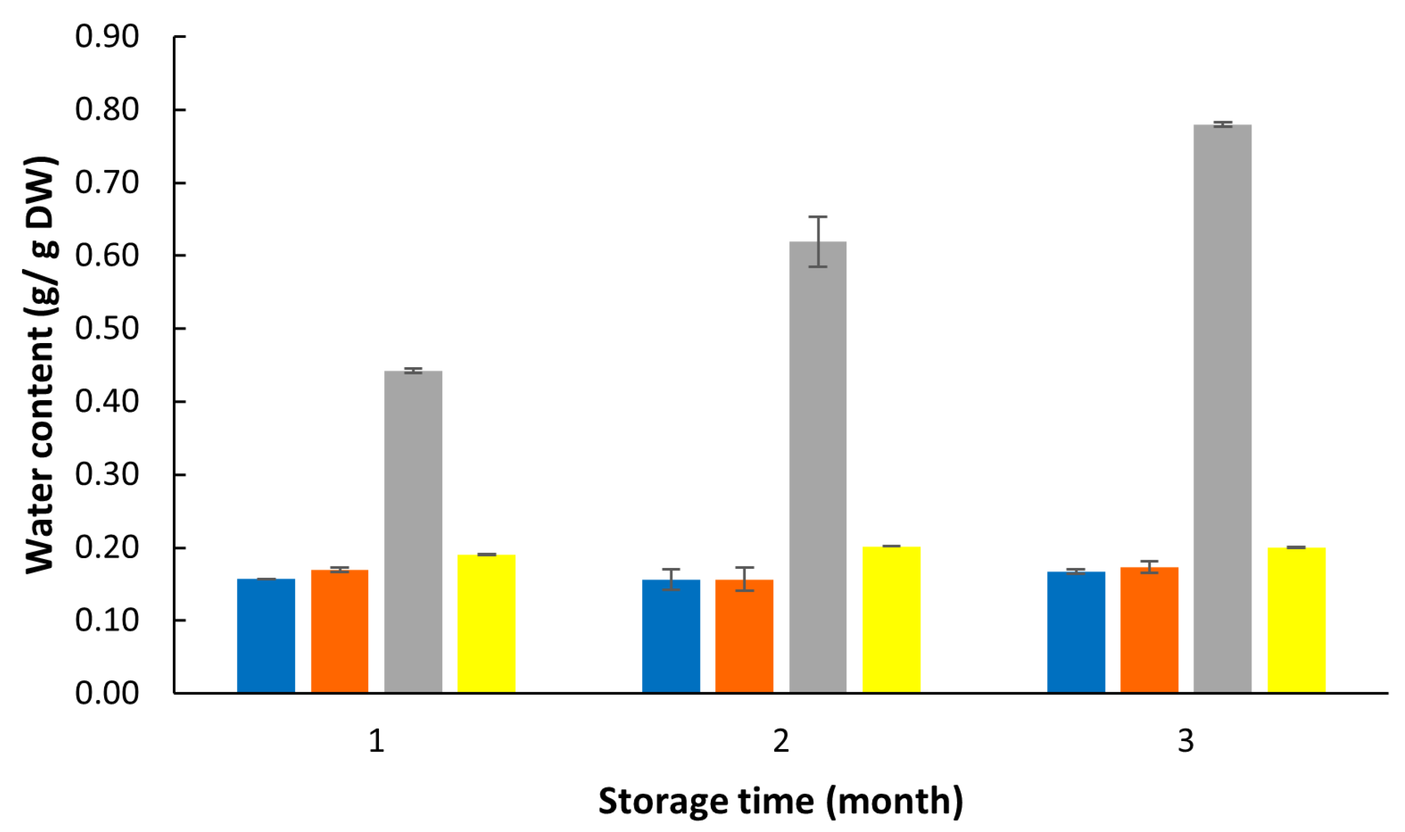
Figure 1. Water content of Lobosphaera exposed to light and 50% HR (blue); no light and 50% HR (orange); light and 75% HR (grey); no light and 75% HR (yellow).
Phycology 05 00035 g001
Figure 2. Color of Lobosphaera exposed to light and 50% HR (blue); light and 75% HR (orange); no light and 50% HR (grey); no light and 75% HR (yellow). Lightness, L* (a); Hue angle (b); Saturation index, Chroma (c).
Phycology 05 00035 g002
Figure 3. Water content of Lobosphaera packed in PETmet/PE and stored for two months.
Phycology 05 00035 g003
Figure 4. Color parameters of Lobosphaera packed in PETmet/PE and stored for two months.
Phycology 05 00035 g004
Figure 5. Carotenoids (Car), chlorophyll a (Ca), and chlorophyll b (Cb) contents of Lobosphaera packed in PETmet/PE and stored for two months.
Phycology 05 00035 g005
Table 1. Total phenolic content (TPC) and antioxidant activity (ABTS) of Lobosphaera sp. extract after storage in PETmet/PE and LDPE for two months.
Packaging MaterialStorage Time
(Month)
TPC
(mg GAE/100 mg Sample DW)
ABTS
(µmol of TE/100 mg Sample DW)
-02.83 ± 0.093.61 ± 0.39
PETmet/PE12.53 ± 0.333.65 ± 0.32
LDPE 2.52 ± 0.083.45 ± 0.47
PETmet/PE22.45 ± 0.143.47 ± 0.26
LDPE 2.19 ± 0.163.28 ± 0.46
Table 2. Bacterial growth inhibition of Lobosphaera sp. extract at 1%, 2%, and 3% concentration after storage of the microalga in PETmet/PE and LDPE for two months.
Packaging MaterialStorage Time
(Month)
BacteriaInhibition (%)
1% Extract2% Extract3% Extract
-0Escherichia coli34.18 ± 4.0539.02 ± 4.4956.44 ± 2.85
PETmet/PE124.56 ± 5.9930.03 ± 7.0152.60 ± 4.45
LDPE16.06 ± 1.5029.69 ± 5.5848.93 ± 2.27
PETmet/PE214.86 ± 5.2513.80 ± 2.7431.73 ± 2.91
LDPE20.28 ± 1.110.72 ± 2.265.66 ± 1.82
-0Yersinia enterocolitica41.25 ± 1.0655.13 ± 1.2671.15 ± 2.51
PETmet/PE133.32 ± 1.0637.43 ± 1.6358.54 ± 1.96
LDPE110.13 ± 3.1719.14 ± 1.6152.08 ± 2.02
PETmet/PE214.69 ± 2.1228.16 ± 1.9252.45 ± 1.18
LDPE21.28 ±0.724.16 ± 1.9312.57 ± 1.39
-0Salmonella enterica serovar Enteritidis41.37 ± 1.1759.57 ± 1.0469.35 ± 2.84
PETmet/PE130.16 ± 1.9939.65 ± 1.2761.61 ± 1.61
LDPE121.23 ± 2.3924.41 ± 1.3856.73 ± 1.36
PETmet/PE227.33 ± 1.5736.80 ± 1.5346.78 ± 1.43
LDPE28.03 ± 4.7418.10 ± 0.9823.99 ± 1.04
-0Staphylococus aureus100.00 ± 0.00100.00 ± 0.00100.00 ± 0.00
PETmet/PE185.92 ± 2.7491.94 ± 1.9298.33 ± 2.23
LDPE118.13 ± 2.7623.66 ± 2.7251.29 ± 3.98
PETmet/PE225.14 ± 1.9731.45 ± 1.5144.94 ± 1.37
LDPE21.68 ± 1.052.04 ± 1.5716.94 ± 1.60
-0Bacillus cereus100.00 ± 0.00100.00 ± 0.00100.00 ± 0.00
PETmet/PE191.04 ± 1.7094.65 ± 1.44100.00 ± 0.00
LDPE116.85 ± 2.8219.68 ± 2.7242.33 ± 2.89
PETmet/PE236.42 ± 1.6640.81 ± 1.5050.37 ± 1.02
LDPE29.60 ± 0.9813.41 ± 2.6916.43 ± 1.55
-0Listeria monocytogenes100.00 ± 0.00100.00 ± 0.00100.00 ± 0.00
PETmet/PE196.44 ± 3.0999.62 ± 2.43100.00 ± 0.00
LDPE15.95 ± 2.958.00 ± 1.8953.99 ± 1.31
PETmet/PE27.52 ± 1.7212.84 ± 3.0233.76 ± 1.83
LDPE21.59 ± 3.803.32 ± 1.309.81 ± 1.47
Disclaimer/Publisher’s Note: The statements, opinions and data contained in all publications are solely those of the individual author(s) and contributor(s) and not of MDPI and/or the editor(s). MDPI and/or the editor(s) disclaim responsibility for any injury to people or property resulting from any ideas, methods, instructions or products referred to in the content.