Article

Fibrinolytic Enzyme from Green Microalgae: A New Potential Drug for Thrombolytic Therapy?

by
Yanara Alessandra Santana Moura
1,
Ariadne Tennyle Vieira De Souza
2,
Páblo Eugênio Da Costa e Silva
3,
Marllyn Marques Da Silva
1,
Ana Lúcia Figueiredo Porto
1 and
Raquel Pedrosa Bezerra
1,*
1
Department of Morphology and Animal Physiology, Federal Rural University of Pernambuco-UFRPE, Dom Manoel de Medeiros Ave., Recife 52171-900, PE, Brazil
2
Biological Sciences Postgraduate Program, Federal University of Pernambuco-UFPE, Professor Moraes Rego Ave., Recife 50670-901, PE, Brazil
3
Keizo Asami Immunology Lab (LIKA), Federal University of Pernambuco-UFPE, Professor Moraes Rego Ave., Recife 50670-901, PE, Brazil
*
Author to whom correspondence should be addressed.
Phycology 2023, 3(4), 436-446; https://doi.org/10.3390/phycology3040029
Submission received: 31 July 2023 / Revised: 20 September 2023 / Accepted: 22 September 2023 / Published: 5 October 2023

Abstract

:
Thrombosis is characterized by the pathological formation of fibrin clots within a blood vessel, leading to the obstruction of blood flow. Fibrinolytic enzymes from microorganisms have been shown to be more efficient and safer in dissolving clots. Then, this study aimed to evaluate the cell growth and fibrinolytic enzyme production of Tetradesmus obliquus under different cultivation conditions. T. obliquus grew under autotrophic and mixotrophic conditions using different concentrations of corn steep liquor (0.25 ≤ CSL ≤ 4.00%). The cells were concentrated and lysed via two different methods (sonication or homogenization) to trigger the release of the enzyme. It was precipitated via acetone or ammonium sulfate additions and purified using ion exchange chromatography. The highest biomass productivity (Px = 130 ± 12.8 mg∙L−1day−1), specific growth rate (µmax = 0.17 ± 0.00 day−1), and fibrinolytic activity (391 ± 40.0 U∙mg−1) was achieved on a mixotrophic cultivation at a 0.25% CSL concentration. The results showed that the homogenizing method had better performance in the release of enzyme, and the precipitation with acetone obtained the highest fibrinolytic activity (567 ± 49.3 U∙mg−1). The purified enzyme showed a specific activity of 1221 ± 31 U∙mg−1 and a molecular mass of 97 kDa. So, the fibrinolytic enzyme from T. obliquus had higher activity when compared to the other fibrinolytic enzymes, being a potential source for the development of therapeutic agents in thrombosis treatment. Additional studies are needed to investigate the biochemical properties and biological profile of this enzyme.

1. Introduction

Cardiovascular diseases (CVDs) are the main cause of death worldwide and are responsible for about 32% of all global deaths [1]. Thrombosis is a severe CVD complication characterized by the pathological formation of fibrin clots that obstruct blood flow, leading to intense clinical manifestations such as acute ischemic stroke, myocardial infarction, and venous thromboembolism [2,3].
Currently, thrombosis treatment is based on antiplatelet or anticoagulant agents, which may lead to severe bleeding problems, such as hemorrhage [4,5]. Alternatively, fibrinolytic agents such as the tissue plasminogen activator (t-PA), urokinase (u-PA), and streptokinase have been widely used for thrombosis therapy. However, these drugs have some limitations including a short half-life, low specificity to fibrin, high cost, and excessive bleeding [6]. Thus, the search for more effective and safe fibrinolytic enzymes has become the key to thrombosis treatment.
In this sense, fibrinolytic enzymes from photosynthetic microorganisms such as Chlorella vulgaris, Arthrospira platensis, and Dunaliella tertiolecta have shown promising antithrombotic effects [7,8,9]. However, up to now, there is no report on the fibrinolytic potential of the Tetradesmus genus, although some bioactive compounds such as lectin, linoleic acid, and flavonoids with anticancer and antimicrobial activities have already been reported in the literature [10,11,12].
The production of microalgae has tripled in the last 5 years [13]. The interest in world production is due to some characteristics such as high photosynthetic efficiency, fast growth rate, resistance to various contaminants, and the capacity to grow on non-arable lands and be cultured using different growth conditions (autotrophic, heterotrophic, and mixotrophic growth modes) [14,15]. Specifically, previous studies have shown that mixotrophic conditions using different organic carbon substrates improve enzyme production and the T. obliquus biomass yields [16,17].
Organic wastes and by-products are frequently used as substrates for mixotrophic growth and are advantageous for sustainable resource recycling and the cost reduction in microalgal production [18]. Corn steep liquor (CSL) is a by-product from the corn wet-milling industries and has high amounts of carbohydrates, amino acids, vitamins, organic acids, and minerals, being a nitrogen-rich source used for the microalgal cultivation [19]. By the way, this by-product has been successful in the production of fibrinolytic enzymes from C. vulgaris, A. platensis, and D. tertiolecta [7,8,9]. So, this study aims to evaluate and compare the biomass and fibrinolytic enzyme productions from T. obliquus cultivated under autotrophic and mixotrophic (using CSL) growth conditions.

2. Materials and Methods

2.1. Culture Media and Growth Conditions

T. obliquus (SISGEN A5F5402) was isolated from Açude of Apipucos (Recife, Pernambuco, Brazil, coordinates 8°1′13.08″ S; 34°55′56.51″ W) and cultivated under autotrophic condition in 1000 mL Erlenmeyer flasks containing 400 mL of the BG-11 medium [20] with an initial concentration of 50 mg∙L−1, temperature of 30 ± 1 °C, continuous light intensity of 40 μmol photons m−2∙s−1, and under constant aeration [21]. The mixotrophic condition was defined by the addition of different concentrations of corn steep liquor (0.25, 0.50, 0.75, 1.00, 2.00, and 4.00% (v/v)) into the BG-11 medium. The corn steep liquor (CSL) (Corn Products Brazil, Cabo de Santo Agostinho, PE, Brazil) was previously treated via autoclaving and precipitation according to Liggett and Koffler [22].
Cell growth was measured daily until the end of the exponential growth phase. The cell biomass was harvested via centrifugation (5000 rpm for 5 min), washed three times with distilled water, freeze-dried, and stored at 4 °C.
The biomass concentration was determined by measuring the optical density (OD) at λ665 nm via a UV/Visible spectrophotometer using an appropriate calibration curve correlating OD665 to the biomass concentration (Equation (1), R2 = 0.99).
B i o m a s s   c o n c e n t r a t i o n = 0.0041 O D 665 + 0.0486

2.2. Kinetic Parameters

The biomass productivity (Px) at the end of cultivation was calculated via Equation (2):
P x = ( X t X 0 ) t c
where Xt is the final cell concentration (mg∙L−1), X0 is the initial cell concentration (mg∙L−1), and tc time is the culture’s final cell concentration (days).
The maximum specific growth rate (μmax), expressed in day−1, was calculated via the following equation:
μ m a x = 1 t ln X j x j 1
where Xj and Xj−1 are the cell concentrations at the end and the beginning of each time interval (Δt = 1 day).

2.3. Fibrinolytic Enzyme Extraction

The cell biomass (100 mg∙mL−1) was resuspended in 0.02 M Tris-HCl buffer (pH 7.4) and submitted to two different extraction methods: (1) homogenization by constant stirring for 30 min in ice bath [23]; and (2) sonication using a sonicator (Bandelin Sonoplus HD 2070, Microtip MS 72, Berlin, Germany) with 20 pulses for 1 min with intervals of 1 min between each pulse in ice bath [10]. Both the homogenized and sonicated biomasses were centrifuged at 15,000 rpm for 10 min at 4 °C and the cell extract was used for further analysis.

2.4. Precipitation Methods

Cell extract was precipitated using two different solvents: (1) acetone (80%); and (2) ammonium sulfate at 0–40% and 40–70% (w/v). The protein precipitated by using ammonium sulfate was dialyzed against 0.02 M Tris-HCl buffer for 6 h at 25 °C.

2.5. Protein Purification

The redissolved protein was loaded onto ion-exchange chromatography using DEAE Sephadex column (1.6 × 50 cm) pre-equilibrated with 0.02 M Tris-HCl buffer and eluted with the same buffer at a flow rate of 0.5 mL∙min−1, and fractions of 0.5 mL were collected. The absorbance was measured at λ280 nm.

2.6. Sodium Dodecyl Sulfate-Polyacrylamide Gel Electrophoresis

Sodium dodecyl sulfate-polyacrylamide gel electrophoresis (SDS-PAGE) was carried out using a 12% polyacrylamide gel as described by Laemmli [24]. The molecular mass was calibrated using a Molecular Mass Marker Kit (14,000–200,000 Da, Sigma-Aldrich, St. Louis, MO, USA). Protein bands were detected by staining with silver.

2.7. Protein Concentration Analysis

Protein concentration was obtained using the BCA Protein Assay Reagent Kit (BCATM Protein Assay Kit, Thermo Fisher Scientific, Waltham, MA, USA). Bovine serum albumin was used as the standard.

2.8. Protease Activity Assay

Protease activity was assayed using azocasein as a substrate. The reaction mixture contained 30 µL of 0.08 mM azocasein, 140 µL of 0.02 M Tris-HCl, and 30 µL of the T. obliquus cell extracts. After 15 min, the reaction was stopped by the trichloroacetic acid (TCA) addition and the absorbance of liquid fraction was measured at λ450 nm using a microplate reader. One unit of azocasein activity was defined as the amount of enzyme required to increase the absorbance by 0.001 per minute, and the protease activity was expressed as activity units [25].

2.9. Determination of Fibrinolytic Enzyme

2.9.1. Fibrinolytic Plate Assay

The fibrinolytic activity was determined on a fibrin plate [26] with adaptations. Typically, the fibrin plate was prepared by mixing 0.45% bovine fibrinogen and 0.02 M Tris-HCl buffer with 2% agarose dissolved in 0.02 M Tris-HCl buffer and 200 μL of CaCl2. The prepared solution was poured into a Petri plate (90 × 15 mm) containing 200 µL of a thrombin suspension. The fibrinolytic activity of the cell extracts was obtained by creating wells of 5 mm, which were impregnated with 20 µL of the T. obliquus extracts and incubated at 37 °C for 20 h. The zone of clearance was defined as the fibrinolytic activity of the cell extracts.

2.9.2. Fibrinolytic Assay Using Spectrophotometry

The fibrinolytic activity was evaluated according to Wang [27] using the spectrophotometric method. Fibrinogen (0.72%) and 0.02 M Tris-HCl buffer was placed in a test tube and incubated at 37 °C for 5 min. After the addition of the thrombin (20 U∙mL−1) solution, the resulting mixture was incubated at 37 °C for 10 min, the enzyme solution was added, and incubation continued at 37 °C. At 60 min, the reaction was stopped by adding 0.2 M TCA. Finally, the solution was centrifugated (8000 rpm for 10 min) and the supernatant was measured at λ275 nm. One unit (U) of fibrinolytic activity was defined as the amount of enzyme required to increase 0.01 units of absorbance per minute.

2.10. Statistical Analysis

All the experiments were performed in duplicates and the data are presented as the mean ± standard deviation (SD). The statistical analyses were performed using one-way analysis of variance (ANOVA), followed by Tukey’s test as post hoc. p values < 0.05 were considered statistically significant.

3. Results and Discussion

3.1. Cell Growth Profile and Kinetic Parameters of T. obliquus Cultivation under Different Growth Conditions

Cell growth profiles of T. obliquus in autotrophic and mixotrophic growth conditions using different CSL concentrations are shown in Figure 1. A short lag phase of two days was observed in autotrophic growth (Figure 1) due to the previous adaptation of T. obliquus cells in a culture medium constituted by only inorganic nitrogen sources such as NaNO3 and (NH4)5[Fe(C6H4O7)2]. In this condition, the exponential phase was of 16 days reaching the highest maximum biomass concentration (Xm) values (1970 ± 231 mg∙L−1). In the mixotrophic cultivation using 0.25% CSL, the exponential growth phase began after 8 days of cultivation and obtained a Xm value of 1625 ± 207 mg∙L−1 (Figure 1), and when the CLS was increased to 0.50%, slow cell growth was observed during 15 days, reaching the lowest Xm of 936 ± 82.8 mg∙L−1. In the CSL concentration higher than 0.50%, no T. obliquus cell growth was observed. Similar results were observed in the mixotrophic cultivation of A. platensis and D. tertiolecta using CSL concentrations above 0.6% and 1.0%, which inhibited cell growth [8,9].
CSL concentration also influenced the cell growth kinetic parameters. As shown in Table 1, the biomass productivity (Px = 130 ± 12.8 mg∙L−1day−1) and specific growth rate (µmax = 0.17 ± 0.00 day−1) in the mixotrophic cultivation using 0.25% CSL was higher than those cultivated with 0.50% CSL (Px = 93.4 ± 10.9 mg∙L−1day−1; µmax = 0.12 ± 0.00 day−1). These results showed that higher CSL concentrations (>0.50%) in the mixotrophic cultivation of T. obliquus decrease the Px and µmax values probably due to stress provoked by the excess of nitrogen [28,29]. CSL is rich in protein content (420 mg∙g−1) and the main amino acids available are arginine (44.30 mg∙g−1), alanine (35.70 mg∙g−1), and glutamic acid (42.00 mg∙g−1), showing that CSL is a potential organic N-source [30,31]. By the way, CSL has been considered as a low-cost material for the microbial production of enzymes [18,32,33], and its effects on the fibrinolytic enzyme production of T. obliquus has not yet been studied. The highest biomass productivity (130 ± 12.8 mg∙L−1day−1) was obtained in the cultivation using 0.25% CSL, which was selected for further steps.

3.2. Protease and Fibrinolytic Productions

The cell growth condition has an important influence on the production of microbial enzymes. As shown in Table 1, T. obliquus produced a high amount of protease enzyme when cultivated under mixotrophic conditions using 0.25% CSL (84.7 U∙mg−1), followed by the autotrophic (12.5 U∙mg−1) and mixotrophic 0.50% CSL (5.85 U∙mg−1) conditions. Moreover, the protease activity of T. obliquus cultivated on 0.25% CSL is higher than those produced by different marine algae, such as Ulva lactuca (6.55–7.33 U∙mg−1), Ulva fasciata (8.00 U∙mg−1), Enteromorpha sp. (6.74–9.60 U∙mg−1), and Chaetomorpha antenna (9.40 U∙mg−1) [34].
No significant difference in fibrinolytic activities was observed between autotrophic (430 ± 40.2 U∙mg−1) and mixotrophic using the 0.25% CSL (391 ± 40.0 U∙mg−1) cultivations, which were higher than those using 0.50% CSL (135 ± 7.0 U∙mg−1). The results showed that high CSL concentrations inhibit protease and fibrinolytic enzyme productions. Fibrinolytic enzyme production from the autotrophic and 0.25% CSL mixotrophic were higher than those obtained from the other photosynthetic microorganisms, such as A. platensis (268 ± 10.7 U∙mg−1) and C. vulgaris (302 ± 37.5 U∙mg−1) [7,8]. It is well known that the biochemical composition of the microalgae biomass, e.g., enzyme production, depends on the culture conditions such as the medium composition [35]. Then, the highest enzyme activities were obtained using 0.25% CSL, which also enhanced the enzyme production by A. platensis [8]. On the other hand, the cultivation with a higher CSL concentration (≥0.50%) decreases enzyme production, since a high concentration of some nutrients, such as nitrogen, might affect the biomass [36].

3.3. Effect of Extraction Methods on the Enzymatic Activities

The extraction methods influence enzyme activity. The extraction of enzymes was evaluated using the homogenization and sonication methods. Homogenization was the most efficient method to extract protease (12.5 ± 1.35 U∙mg−1) and fibrinolytic enzymes (430 ± 40.2 U∙mg−1) from the autotrophic cultivation, while the sonication method decreased the protease and fibrinolytic activities to 4.50 ± 0.40 and 149 ± 3.8 U∙mg−1, respectively (Table 1). Similar results were observed in the T. obliquus extracts from the mixotrophic cultures using 0.25% CSL, which also showed higher protease (12.5 ± 2.94 U∙mg−1) and fibrinolytic (391 ± 40.0 U∙mg−1) activities using the homogenization method when compared to the sonication methods. These data were similar to those obtained by Silva et al. [9] which showed that the fibrinolytic activity from D. tertiolecta was 819 and 422 U∙mg−1 using homogenization and sonication, respectively. This can be explained by the possible enzyme denaturation caused by the prolonged sonication time, high temperature, or elevated frequency, as reported by Sukor et al. [37] and Ranjha et al. [38]. Then, these results showed that homogenization is more effective in the extraction of the protease and fibrinolytic enzymes from T. obliquus.
Therefore, according to Table 1, the mixotrophic cultivation using 0.25% CSL was selected for further steps due to its higher biomass productivity and significantly high enzyme production. Moreover, the extract from the 0.25% CSL cultivation obtained via the homogenization method showed higher fibrinolytic activity compared to the sonication method; thus, this extract was selected for the purification steps.

3.4. Effect of Precipitation Methods on the Enzymatic Activities

The T. obliquus extract-rich protein was precipitated using acetone or ammonium sulfate in two fractions of saturation (0–40% and 40–70%). Both 0–40% and 40–70% ammonium sulfate fractions showed similar protease activity (655 ± 69.3 and 623 ± 192 U∙mg−1, respectively), which was higher compared to the acetone precipitation (206 ± 17.5 U∙mg−1). Then, the ammonium sulfate fractions are more advantageous for the protease activity applications. However, although the 0–40% ammonium sulfate fraction exhibited the highest fibrinolytic activity (625 ± 20.2 U∙mg−1), the acetone fraction showed better performance due to its potential fibrinolytic activity (567 ± 49.3 U∙mg−1) and the highest enzyme yield of 80.3%. These results are similar to those reported by Barros et al. [8] and Silva et al. [9] for the fibrinolytic enzymes from A. platensis and D. tertiolecta, which showed fibrinolytic activity of 256 U∙mg−1 (yield = 53.8%) and 435 U∙mg−1 (yield = 37%), respectively.
Taking into account that the fibrinolytic activity measures the enzyme capacity of degrading fibrin specifically, and the acetone fraction showed the highest recovery yield, this fraction was considered more advantageous to be studied for thrombosis therapy purposes. Acetone is listed among the Generally Recognized as Safe (GRAS) by the Food and Drug Administration (FDA) since toxicological and medical studies show no adverse effects on human health [39]. Additionally, the use of acetone for precipitation includes some advantages such as simple-step extraction, less cost, and less time consumed [40,41]. Therefore, acetone was selected as the most advantageous precipitating agent to obtain the fibrinolytic enzyme from T. obliquus.

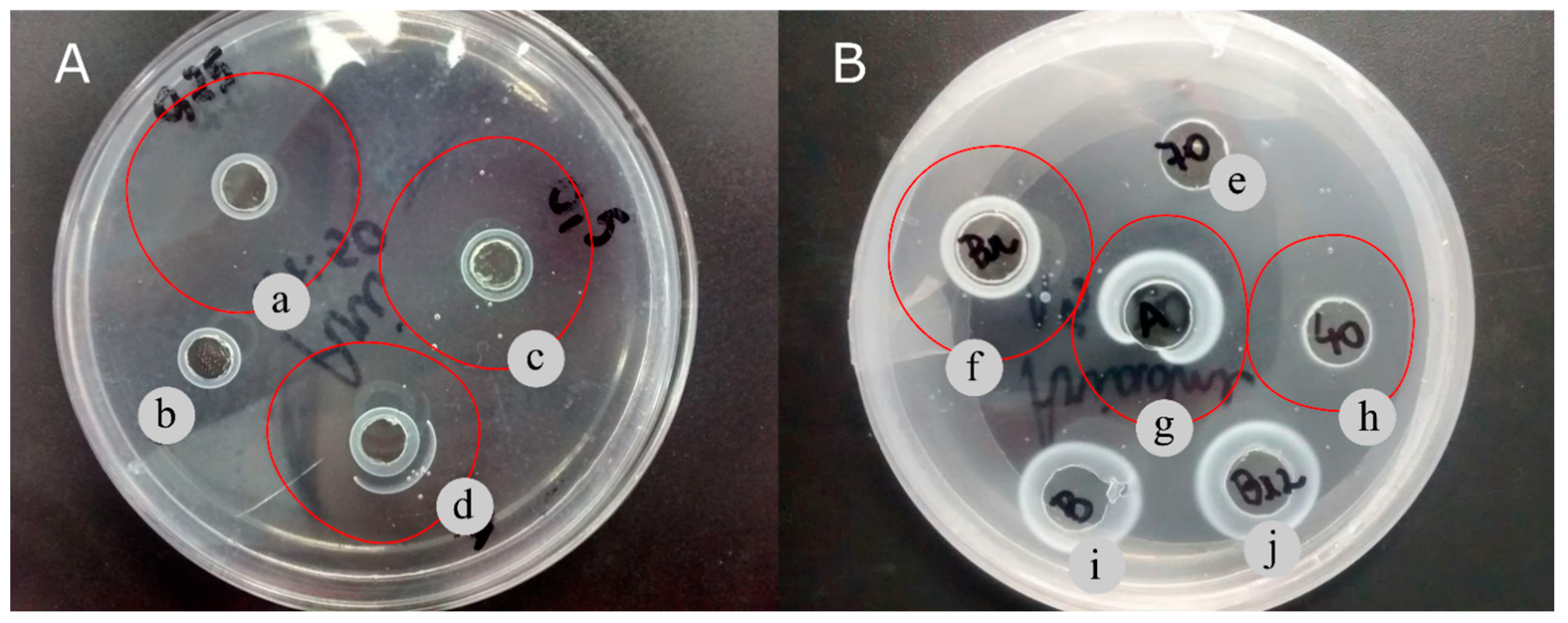
3.5. Fibrinolytic Activity in Fibrin Plate

Figure 2 shows a qualitative assessment of the fibrinolytic activity from T. obliquus by the fibrin plate method. The cell extract from T. obliquus cultivated in 0.25% CSL showed a high clear zone (82 mm2) when compared to the cell extract obtained from the cell extracts cultivated autotrophically (69 mm2) or mixotrophically with 0.50% CSL (69 mm2) (Figure 2A), confirming that the 0.25% CSL cultivation has higher fibrinolytic production by this qualitative analysis. These values are higher than those of the fibrinolytic enzymes from Bionectria sp. strains, which ranged from 21.9 to 66.7 mm2 [42].
The fibrinolytic activities of protein precipitated by different precipitating agents are shown in Figure 2B. Both the 0–40% ammonium sulfate and acetone fractions exhibited a clear zone of fibrin degradation around the well after 48 h, as shown in Figure 2B (letters g and h) and Table 2. On the other hand, the 40–70% ammonium sulfate fraction did not show a clear zone area of hydrolysis (Figure 2B; letter e).

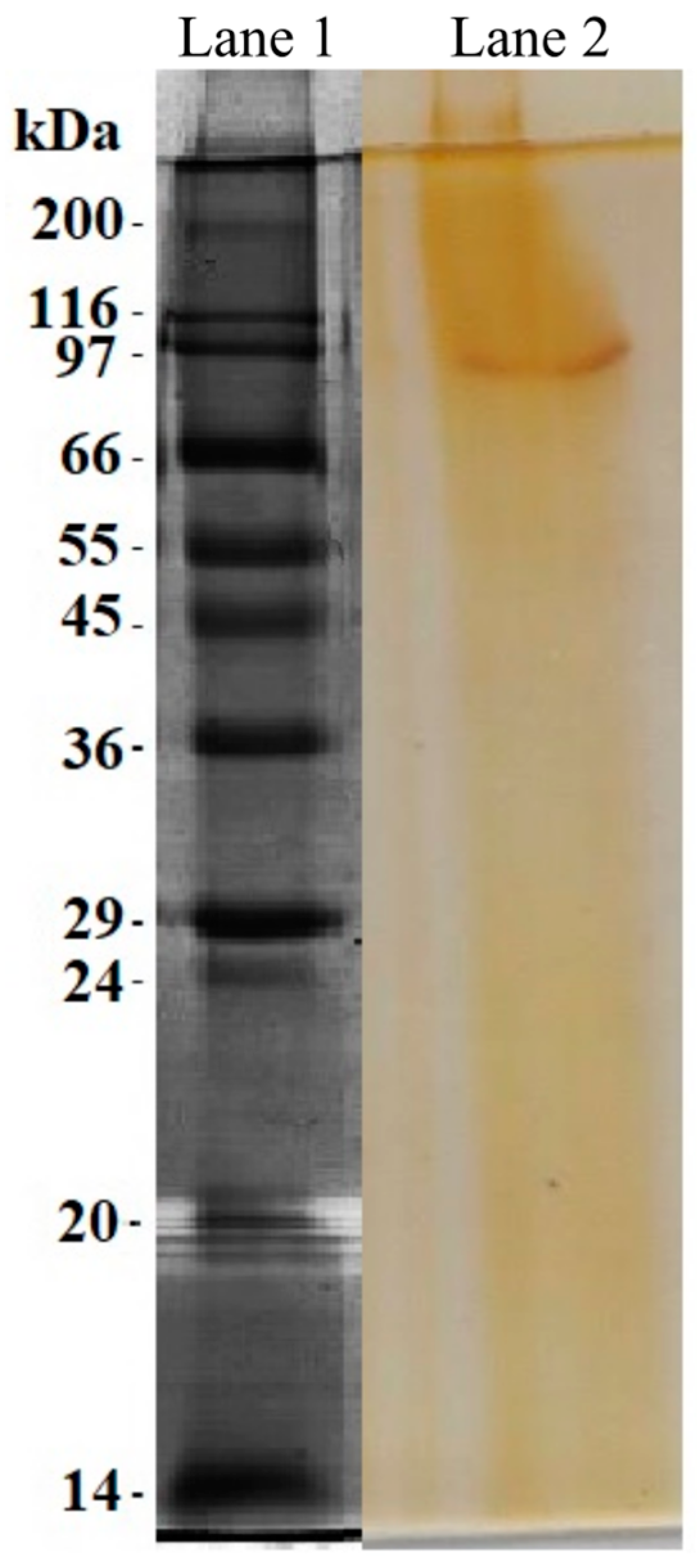
3.6. Enzyme Purification

The fibrinolytic enzyme from T. obliquus was purified using a combination of acetone precipitation and the DEAE-Sephadex ion exchange column. The chromatogram shown in Figure 3 exhibits a single peak (fractions 4–12). These protein fractions were pooled and concentrated, showing a fibrinolytic activity of 1221 ± 31 U∙mg−1. After purification via DEAE-Sephadex, the fibrinolytic enzyme was 2.46-fold purified with a yield of 8.90%, which is relative to that of the cell extract (Table 3). In general, the activity of the purified fibrinolytic enzyme from T. obliquus was significantly higher than those obtained by Silva et al. [9] for the fibrinolytic enzyme from D. tertiolecta, which showed fibrinolytic activity of 670 U∙mg−1. Similarly, the fibrinolytic activity of the fibrinolytic enzyme from T. obliquus is also higher compared to diverse macroalgal species, such as Costaria costata (915 U∙mg−1), Codium divaricatum (6.3 U∙mg−1), Codium fragile (61.5 U∙mg−1), and Ulva pertusa (295 U∙mg−1), and also various bacterial species such as Bacillus flexus (315 U∙mg−1), Bacillus velezensis BS2 (131 U∙mg−1), Bacillus subtilis HQS-3 (30.0 U∙mg−1), and Bacillus subtilis ICTF-1 (280 U∙mg−1) [23,43,44,45,46].
Briefly, the extracts obtained by the homogenization method were purified using acetone precipitation and the DEAE-Sephadex chromatography, showing potential fibrinolytic activity of 1221 ± 31 U∙mg−1 (Table 3). The same sample was also submitted to the SDS-PAGE analysis.
SDS-PAGE showed one protein band with a molecular mass of probably 97 kDa (Figure 4). This is higher than that exhibited by the other T. obliquus proteins reported by Silva et al. [21] and Heide et al. [47] that have a molecular mass of 78 and 12 kDa, respectively. Additionally, the molecular mass of the fibrinolytic enzymes obtained from the other microalgae species, including A. platensis (72 kDa), C. vulgaris (45 kDa), and D. tertiolecta (10 kDa), is also lower than the fibrinolytic enzyme from T. obliquus [7,8,9]. These results show that this is a different protein from those reported previously.

4. Conclusions

In the present study, it was possible to extract and purify an enzyme from T. obliquus microalgae with a specific activity of 1221 ± 31 U∙mg−1. The mixotrophic cultivation using an inexpensive and advantageous agro-industrial by-product (0.25% CSL) showed higher growth kinetic parameters and fibrinolytic production. Additionally, cell extraction via homogenization had the highest fibrinolytic activity, while the protein precipitation with acetone exhibited the highest recovery yield. In general, these methods are considered simple, efficient, less costly, less time-consuming, and are recognized as safe for human health, which can facilitate this enzyme production as well as its purification. Future research may continue to investigate the enzyme biochemical and biological profile as well as its mechanism of action for thrombolytic activity. Finally, these results can conclude that the fibrinolytic enzyme from T. obliquus has wide potential for industrial application besides its promising effects as an alternative to thrombolytic therapy.

Author Contributions

Conceptualization, A.T.V.D.S., P.E.D.C.e.S. and R.P.B.; methodology, A.T.V.D.S., P.E.D.C.e.S. and Y.A.S.M.; software, Y.A.S.M.; validation, R.P.B.; formal analysis, R.P.B., M.M.D.S., A.T.V.D.S. and Y.A.S.M.; investigation, A.T.V.D.S. and R.P.B.; resources, A.L.F.P.; data curation, Y.A.S.M., A.T.V.D.S. and R.P.B.; writing—original draft preparation, Y.A.S.M.; writing—review and editing, R.P.B.; visualization, Y.A.S.M., M.M.D.S. and R.P.B.; supervision, R.P.B.; project administration, R.P.B. and A.L.F.P.; funding acquisition, A.L.F.P. All authors have read and agreed to the published version of the manuscript.

Funding

This work was financed by the Coordination for the Improvement of Higher Education Personnel—Brazil (CAPES)—Finance Code 001, by the Foundation for Science and Technology of the State of Pernambuco (FACEPE, APQ-0252-5.07/14; APQ-1480-2.08/22) and the National Council for Scientific and Technological Development (CNPq).

Data Availability Statement

Data not available.

Acknowledgments

The authors acknowledge the Research Support Center (CENAPESQ, Recife, Brazil) and the Laboratory of Technology of Bioactive (LABTECBIO, Recife, Brazil).

Conflicts of Interest

The authors declare no conflict of interest.

References

  1. World Health Organization, Cardiovascular Diseases (CVDs). 2021. Available online: Https://Www.Who.Int/News-Room/Fact-Sheets/Detail/Cardiovascular-Diseases-(Cvds) (accessed on 15 November 2022).
  2. Zhao, L.; Lin, X.; Fu, J.; Zhang, J.; Tang, W.; He, Z. A Novel Bi-Functional Fibrinolytic Enzyme with Anticoagulant and Thrombolytic Activities from a Marine-Derived Fungus Aspergillus versicolor ZLH-1. Mar. Drugs 2022, 20, 356. [Google Scholar] [CrossRef] [PubMed]
  3. Wendelboe, A.M.; Raskob, G.E. Global Burden of Thrombosis. Circ. Res. 2016, 118, 1340–1347. [Google Scholar] [CrossRef] [PubMed]
  4. Metharom, P.; Berndt, M.C.; Baker, R.I.; Andrews, R.K. Current State and Novel Approaches of Antiplatelet Therapy. Arter. Thromb. Vasc. Biol. 2015, 35, 1327–1338. [Google Scholar] [CrossRef]
  5. Rashid, Q.; Singh, P.; Abid, M.; Jairajpuri, M.A. Limitations of conventional anticoagulant therapy and the promises of non-heparin based conformational activators of antithrombin. J. Thromb. Thrombolysis 2012, 34, 251–259. [Google Scholar] [CrossRef] [PubMed]
  6. Zhou, Y.; Chen, H.; Yu, B.; Chen, G.; Liang, Z. Purification and Characterization of a Fibrinolytic Enzyme from Marine Bacillus velezensis Z01 and Assessment of Its Therapeutic Efficacy In Vivo. Microorganisms 2022, 10, 843. [Google Scholar] [CrossRef]
  7. da Costa e Silva, P.E.; de Barros, R.C.; Albuquerque, W.W.C.; Brandão, R.M.P.; Bezerra, R.P.; Porto, A.L.F. In vitro thrombolytic activity of a purified fibrinolytic enzyme from Chlorella vulgaris. J. Chromatogr. B 2018, 1092, 524–529. [Google Scholar] [CrossRef] [PubMed]
  8. de Barros, P.D.S.; da Costa e Silva, P.E.; Nascimento, T.P.; Costa, R.M.P.B.; Bezerra, R.P.; Porto, A.L.F. Fibrinolytic enzyme from Arthrospira platensis cultivated in medium culture supplemented with corn steep liquor. Int. J. Biol. Macromol. 2020, 164, 3446–3453. [Google Scholar] [CrossRef]
  9. DA Silva, T.A.; Silva, P.E.D.C.E.; Nascimento, T.P.; Costa, R.M.; Converti, A.; Porto, A.L.F.; Bezerra, R.P. Cost-effective fibrinolytic enzyme production by microalga Dunaliella tertiolecta using medium supplemented with corn steep liquor. Anais da Academia Brasileira de Ciências 2023, 95, e20220552. [Google Scholar] [CrossRef]
  10. Silva-Júnior, J.N.; De Aguiar, E.M.; Mota, R.A.; Bezerra, R.P.; Porto, A.L.F.; Herculano, P.N.; Marques, D.A.V. Antimicrobial Activity of Photosynthetic Microorganisms Biomass Extract against Bacterial Isolates Causing Mastitis. J. Dairy Vet. Sci. 2019, 10, 555788. [Google Scholar]
  11. Marrez, D.A.; Naguib, M.M.; Sultan, Y.Y.; Higazy, A.M. Antimicrobial and anticancer activities of Scenedesmus obliquus metabolites. Heliyon 2019, 5, e01404. [Google Scholar] [CrossRef]
  12. Sharif, A.P.; Habibi, K.; Bijarpas, Z.K.; Tolami, H.F.; Alkinani, T.A.; Jameh, M.; Dehkaei, A.A.; Monhaser, S.K.; Daemi, H.B.; Mahmoudi, A.; et al. Cytotoxic Effect of a Novel GaFe2O4@Ag Nanocomposite Synthesized by Scenedesmus obliquus on Gastric Cancer Cell Line and Evaluation of BAX, Bcl-2 and CASP8 Genes Expression. J. Clust. Sci. 2023, 34, 1065–1075. [Google Scholar] [CrossRef]
  13. Barbosa, M.J.; Janssen, M.; Südfeld, C.; D’adamo, S.; Wijffels, R.H. Hypes, hopes, and the way forward for microalgal biotechnology. Trends Biotechnol. 2023, 41, 452–471. [Google Scholar] [CrossRef] [PubMed]
  14. Morillas-España, A.; Villaró, S.; Ciardi, M.; Acién, G.; Lafarga, T. Production of Scenedesmus almeriensis Using Pilot-Scale Raceway Reactors Located inside a Greenhouse. Phycology 2022, 2, 76–85. [Google Scholar] [CrossRef]
  15. Silva, G.; Cerqueira, K.; Rodrigues, J.; Silva, K.; Coelho, D.; Souza, R. Cultivation of Microalgae Chlorella vulgaris in Open Reactor for Bioethanol Production. Phycology 2023, 3, 325–336. [Google Scholar] [CrossRef]
  16. Bentahar, J.; Doyen, A.; Beaulieu, L.; Deschênes, J.-S. Investigation of β-galactosidase production by microalga Tetradesmus obliquus in determined growth conditions. J. Appl. Phycol. 2019, 31, 301–308. [Google Scholar] [CrossRef]
  17. Piasecka, A.; Nawrocka, A.; Wiącek, D.; Krzemińska, I. Agro-industrial by-product in photoheterotrophic and mixotrophic culture of Tetradesmus obliquus: Production of ω3 and ω6 essential fatty acids with biotechnological importance. Sci. Rep. 2020, 10, 6411. [Google Scholar] [CrossRef]
  18. Kim, S.; Lim, D.; Lee, D.; Yu, J.; Lee, T. Valorization of corn steep liquor for efficient paramylon production using Euglena gracilis: The impact of precultivation and light-dark cycle. Algal Res. 2022, 61, 102587. [Google Scholar] [CrossRef]
  19. Chiani, M.; Akbarzadeh, A.; Farhangi, A.; Mehrabi, M.R. Production of Desferrioxamine B (Desferal) using Corn Steep Liquor in Streptomyces pilosus. Pak. J. Biol. Sci. 2010, 13, 1151–1155. [Google Scholar] [CrossRef]
  20. Allen, M.M.; Stanier, R.Y. Growth and division of some unicellular blue-greenalgae. J. Gen. Microbiol. 1968, 51, 199–202. [Google Scholar] [CrossRef]
  21. Silva, A.J.; Cavalcanti, V.L.R.; Porto, A.L.F.; Gama, W.A.; Brandão-Costa, R.M.P.; Bezerra, R.P. The green microalgae Tetradesmus obliquus (Scenedesmus acutus) as lectin source in the recognition of ABO blood type: Purification and characterization. J. Appl. Phycol. 2020, 32, 103–110. [Google Scholar] [CrossRef]
  22. Liggett, R.W.; Koffler, H. Corn steep liquor in microbiology. Bacteriol. Rev. 1948, 12, 297–311. [Google Scholar] [CrossRef] [PubMed]
  23. Matsubara, K.; Hori, K.; Matsuura, Y.; Miyazawa, K. Purification and characterization of a fibrinolytic enzyme and identification of fibrinogen clotting enzyme in a marine green alga, Codium divaricatum. Comp. Biochem. Physiol. Part B Biochem. Mol. Biol. 2000, 125, 137–143. [Google Scholar] [CrossRef] [PubMed]
  24. Laemmli, U.K. Cleavage of Structural Proteins during the Assembly of the Head of Bacteriophage T4. Nature 1970, 227, 680–685. [Google Scholar] [CrossRef] [PubMed]
  25. de Alencar, R.B.; Biondi, M.M.; Paiva, P.M.G.; Vieira, V.L.A.; Carvalho, L.B.; Bezerra, R.S. Alkaline proteases from digestive tract of four tropical fishes. Braz. J. Food Technol. 2003, 6, 279–284. [Google Scholar]
  26. Astrup, T.; Müllertz, S. The fibrin plate method for estimating fibrinolytic activity. Arch. Biochem. Biophys. 1952, 40, 346–351. [Google Scholar] [CrossRef]
  27. Wang, S.-L.; Wu, Y.-Y.; Liang, T.-W. Purification and biochemical characterization of a nattokinase by conversion of shrimp shell with Bacillus subtilis TKU007. New Biotechnol. 2011, 28, 196–202. [Google Scholar] [CrossRef]
  28. Mohammad Mirzaie, M.A.; Kalbasi, M.; Mousavi, S.M.; Ghobadian, B. Statistical evaluation and modeling of cheap substrate-based cultivation medium of Chlorella vulgaris to enhance microalgae lipid as new potential feedstock for biolubricant. Prep. Biochem. Biotechnol. 2016, 46, 368–375. [Google Scholar] [CrossRef]
  29. Arbib, Z.; Ruiz, J.; Álvarez-Díaz, P.; Garrido-Pérez, C.; Barragan, J.; Perales, J.A. Photobiotreatment: Influence of nitrogen and phosphorus ratio in wastewater on growth kinetics of Scenedesmus obliquus. Int. J. Phytoremediation 2013, 15, 774–788. [Google Scholar] [CrossRef]
  30. Hofer, A.; Hauer, S.; Kroll, P.; Fricke, J.; Herwig, C. In-depth characterization of the raw material corn steep liquor and its bioavailability in bioprocesses of Penicillium chrysogenum. Process. Biochem. 2018, 70, 20–28. [Google Scholar] [CrossRef]
  31. Azizi-Shotorkhoft, A.; Sharifi, A.; Mirmohammadi, D.; Baluch-Gharaei, H.; Rezaei, J. Effects of feeding different levels of corn steep liquor on the performance of fattening lambs. J. Anim. Physiol. Anim. Nutr. 2016, 100, 109–117. [Google Scholar] [CrossRef]
  32. Ju, J.-H.; Ko, D.-J.; Heo, S.-Y.; Lee, J.-J.; Kim, Y.-M.; Lee, B.-S.; Kim, M.-S.; Kim, C.-H.; Seo, J.-W.; Oh, B.-R. Regulation of lipid accumulation using nitrogen for microalgae lipid production in Schizochytrium sp. ABC101. Renew. Energy 2020, 153, 580–587. [Google Scholar] [CrossRef]
  33. Kim, S.; Lee, D.; Lim, D.; Lim, S.; Park, S.; Kang, C.; Yu, J.; Lee, T. Paramylon production from heterotrophic cultivation of Euglena gracilis in two different industrial byproducts: Corn steep liquor and brewer’s spent grain. Algal Res. 2020, 47, 101826. [Google Scholar] [CrossRef]
  34. Patil, S.S.; Rebecca, L.J. Isolation and Characterization of Protease from Marine Algae. Int. J. Pharm. Sci. Rev. Res. 2014, 27, 188–190. [Google Scholar]
  35. Niccolai, A.; Chini Zittelli, G.; Rodolfi, L.; Biondi, N.; Tredici, M.R. Microalgae of interest as food source: Biochemical composition and digestibility. Algal Res. 2019, 42, 101617. [Google Scholar] [CrossRef]
  36. Yaakob, M.A.; Mohamed, R.M.S.R.; Al-Gheethi, A.; Aswathnarayana Gokare, R.; Ambati, R.R. Influence of Nitrogen and Phosphorus on Microalgal Growth, Biomass, Lipid, and Fatty Acid Production: An Overview. Cells 2021, 10, 393. [Google Scholar] [CrossRef]
  37. Sukor, N.F.; Jusoh, R.; Kamarudin, N.S.; Abdul Halim, N.A.; Sulaiman, A.Z.; Abdullah, S.B. Synergistic effect of probe sonication and ionic liquid for extraction of phenolic acids from oak galls. Ultrason. Sonochem. 2020, 62, 104876. [Google Scholar] [CrossRef]
  38. Ranjha, M.M.A.N.; Irfan, S.; Lorenzo, J.M.; Shafique, B.; Kanwal, R.; Pateiro, M.; Arshad, R.N.; Wang, L.; Nayik, G.A.; Roobab, U.; et al. Sonication, a Potential Technique for Extraction of Phytoconstituents: A Systematic Review. Processes 2021, 9, 1406. [Google Scholar] [CrossRef]
  39. Molino, A.; Rimauro, J.; Casella, P.; Cerbone, A.; Larocca, V.; Chianese, S.; Karatza, D.; Mehariya, S.; Ferraro, A.; Hristoforou, E.; et al. Extraction of astaxanthin from microalga Haematococcus pluvialis in red phase by using generally recognized as safe solvents and accelerated extraction. J. Biotechnol. 2018, 283, 51–61. [Google Scholar] [CrossRef]
  40. Nickerson, J.L.; Doucette, A.A. Rapid and Quantitative Protein Precipitation for Proteome Analysis by Mass Spectrometry. J. Proteome Res. 2020, 19, 2035–2042. [Google Scholar] [CrossRef]
  41. Wongpia, A.; Mahatheeranont, S.; Lomthaisong, K.; Niamsup, H. Evaluation of Sample Preparation Methods from Rice Seeds and Seedlings Suitable for Two-Dimensional Gel Electrophoresis. Appl. Biochem. Biotechnol. 2015, 175, 1035–1051. [Google Scholar] [CrossRef]
  42. da Costa e Silva, P.E.; Bezerra, R.P.; Porto, A.L.F. An overview about fibrinolytic enzymes from microorganisms and algae: Production and characterization. J. Pharm. Biol. Sci. 2016, 4, 1. [Google Scholar]
  43. Kim, D.-W.; Sapkota, K.; Choi, J.-H.; Kim, Y.-S.; Kim, S.; Kim, S.-J. Direct acting anti-thrombotic serine protease from brown seaweed Costaria costata. Process. Biochem. 2013, 48, 340–350. [Google Scholar] [CrossRef]
  44. Choi, J.-H.; Sapkota, K.; Park, S.-E.; Kim, S.; Kim, S.-J. Thrombolytic, anticoagulant and antiplatelet activities of codiase, a bi-functional fibrinolytic enzyme from Codium fragile. Biochimie 2013, 95, 1266–1277. [Google Scholar] [CrossRef]
  45. Kang, S.-R.; Choi, J.-H.; Kim, D.-W.; Park, S.-E.; Sapkota, K.; Kim, S.; Kim, S.-J. A bifunctional protease from green alga Ulva pertusa with anticoagulant properties: Partial purification and characterization. J. Appl. Phycol. 2016, 28, 599–607. [Google Scholar] [CrossRef]
  46. Barzkar, N.; Jahromi, S.T.; Vianello, F. Marine Microbial Fibrinolytic Enzymes: An Overview of Source, Production, Biochemical Properties and Thrombolytic Activity. Mar. Drugs 2022, 20, 46. [Google Scholar] [CrossRef]
  47. Heide, H.; Kalisz, H.M.; Follmann, H. The oxygen evolving enhancer protein 1 (OEE) of photosystem II in green algae exhibits thioredoxin activity. J. Plant Physiol. 2004, 161, 139–149. [Google Scholar] [CrossRef]
Figure 1. Growth profiles of T. obliquus cultivated autotrophically and mixotrophically under different concentrations of corn steep liquor: Autotrophic (■), mixotrophic 0.25% (▲), and mixotrophic 0.50% (●).
Phycology 03 00029 g001
Figure 2. Fibrinolytic activity of T. obliquus by fibrin plate method. (A) Fibrinolytic activity of homogenized extracts obtained by 0.25% CSL mixotrophic cultivation (a), Tris-HCl buffer control (b), 0.5% CSL mixotrophic cultivation (c), and autotrophic cultivation (d). (B) Fibrinolytic activity of 40–70% ammonium sulfate precipitate (e), cell extract (f), acetone precipitate (g), 0–40% ammonium sulfate (h), Tris-HCl buffer control (i), and cell extract extracted twice (j) after 48 h.
Phycology 03 00029 g002
Figure 3. Chromatogram of fibrinolytic enzyme from T. obliquus on the DEAE-Sephadex column.
Phycology 03 00029 g003
Figure 4. Sodium dodecyl sulphate-polyacrylamide gel electrophoresis (SDS-PAGE) analysis of fibrinolytic enzyme purified by DEAE-Sephadex column. Lane 1, Molecular mass marker; Lane 2, purified enzyme.
Phycology 03 00029 g004
Table 1. Cultivation parameters and enzymatic activities of T. obliquus cultivated in different growth conditions.
Growth ConditionsXm (mg·L−1)Px (mg∙L−1∙day−1)µmax (day−1)MethodsTotal Protein (mg∙mL−1)Protease Activity (U∙mg−1)Fibrinolytic Activity (U∙mg−1)
Autotrophic1970 ± 231 a112 ± 13.5 a0.19 ± 0.03 aHomogenization0.93 ± 0.00 a12.5 ± 1.35 a430 ± 40.2 a
Sonication2.99 ± 0.50 b4.50 ± 0.40 a149 ± 3.8 b
Mixotrophic (CSL 0.25%)1625 ± 207 a130 ± 12.8 a0.17 ± 0.00 aHomogenization0.86 ± 0.00 a12.5 ± 2.94 a391 ± 40.0 a
Sonication3.32 ± 0.22 b84.7 ± 3.51 b243 ± 11.5 c
Mixotrophic (CSL 0.50%)936 ± 82.8 b93.4 ± 10.9 a0.12 ± 0.00 aHomogenization2.90 ± 0.09 b4.64 ± 3.06 a130 ± 1.0 b
Sonication2.76 ± 0.14 b5.85 ± 3.68 a135 ± 7.0 b
Maximum cell concentration (Xm), biomass productivity (Px), specific growth rate (µmax). Data expressed as means ± standard deviations of duplicate experiments. a,b,c Different superscript letters indicate statistically significant differences (p < 0.05).
Table 2. Comparison of different precipitating agents for precipitation of the homogenized cell extract from T. obliquus cultivated in 0.25% CSL.
Precipitating AgentsVolume (mL)Total Protein (mg)Total Protease Activity (U)Specific Protease Activity (U∙mg−1)P.FYield (%)Total Fibrinolytic Activity (U)Specific Fibrinolytic Activity (U∙mg−1)P.FYield (%)
Cell extract5023.3 ± 1.2 a4740 ± 1039 a203 ± 56.3 a 10011,520 ± 1420 a494 ± 86.1 a 100
Ammonium sulfate (0–40%)404.80 ± 0.00 b3144 ± 332 b655 ± 69.3 b3.2266.33000 ± 97 b625 ± 20.2 a,b1.2626.0
Ammonium sulfate (40–70%)405.20 ± 1.38 b3240 ± 72 b623 ± 192 b3.0668.32448 ± 0 b470 ± 157 b0.9521.7
Acetone4016.4 ± 0.9 c3384 ± 125 c206 ± 17.5 a1.0171.39312 ± 396 c567 ± 49.3 a,b1.1480.3
Data expressed as means ± standard deviations of duplicate experiments. a,b,c Different superscript letters indicate statistically significant differences (p < 0.05).
Table 3. Steps of purification of fibrinolytic enzyme from T. obliquus.
Purification StepVolume (mL)Total Protein (mg)Total Protease Activity (U)Specific Protease Activity (U∙mg−1)P.FYield (%)Total Fibrinolytic Activity (U)Specific Fibrinolytic Activity (U∙mg−1)P.FYield (%)
Cell extract5023.3 ± 1.2 a4740 ± 1039 a203 ± 56.3 a 10011,520 ± 1420 a494 ± 86.1 a 100
Acetone precipitation4016.4 ± 0.9 b3384 ± 125 a206 ± 17.5 a1.0171.39312 ± 396 b567 ± 49.3 b1.1480.8
DEAE-Sephadex4.50.84 ± 0.02 c297 ± 4.2 b353 ± 12.4 b1.736.261026 ± 0 c1221 ± 31 c2.478.90
Data expressed as means ± standard deviations of duplicate experiments. a,b,c Different superscript letters indicate statistically significant differences (p < 0.05).
Disclaimer/Publisher’s Note: The statements, opinions and data contained in all publications are solely those of the individual author(s) and contributor(s) and not of MDPI and/or the editor(s). MDPI and/or the editor(s) disclaim responsibility for any injury to people or property resulting from any ideas, methods, instructions or products referred to in the content.

