Article

Farm-Level Surveillance of Listeria spp. in Feed, Feces, and Raw Milk from Cattle, Sheep, and Goat Farms in Kosovo

1
Faculty of Agriculture and Veterinary, University of Prishtina, 10000 Prishtina, Kosovo
2
Department of Animal Science, University of Wyoming, Laramie, WY 82071, USA
*
Author to whom correspondence should be addressed.
Ruminants 2026, 6(3), 71; https://doi.org/10.3390/ruminants6030071
Submission received: 25 July 2026 / Revised: 19 August 2026 / Accepted: 20 August 2026 / Published: 22 August 2026

Simple Summary

Listeria bacteria can survive in farm environments and may contaminate animal feed, feces, or raw milk. This study investigated their presence on cattle, sheep, and goat farms across all seven regions of Kosovo. In total, 189 samples of animal feed, feces, and raw milk were collected from 60 distinct farms representing 63 farm-species units and analyzed. Only three samples were positive for Listeria spp., representing a low number of detections. All recovered isolates were identified as Listeria innocua, a species generally considered less harmful than Listeria monocytogenes. Two positive samples were hay collected from cattle farms, while the third was a fecal sample from a goat farm. No Listeria bacteria were detected in any of the 63 raw-milk samples analyzed under the conditions of this cross-sectional study. This finding should not be interpreted as evidence that raw milk from the surveyed populations was free of Listeria or as evidence of its long-term absence. However, the presence of Listeria innocua in hay and feces shows that these bacteria can occasionally occur in the farm environment. Proper feed storage, manure management, farm cleanliness, and hygienic milking practices remain important for protecting animal health, milk quality, and consumer safety.

Abstract

Listeria spp. can persist in livestock-production environments and may be introduced through animal feed, fecal contamination, or unhygienic milk-production practices. This cross-sectional study assessed the occurrence and distribution of Listeria spp. in animal feed, fecal material, and raw milk collected from cattle, sheep, and goat farms across the seven geographical regions of Kosovo. Between December 2021 and April 2022, 189 samples were collected from 60 distinct farms representing 63 farm-species units. One animal-feed sample, one pooled fecal sample, and one pooled raw-milk sample were obtained from each unit. The samples were analyzed according to ISO 11290-1:2017, and presumptive isolates were evaluated by phenotypic and biochemical tests and confirmed to species level using MALDI-TOF mass spectrometry. Water activity was also measured in all 63 feed samples. Listeria spp. was detected in 3/189 matrix-level samples analyzed (1.6%). This value represents a descriptive sample-level detection proportion across the three matrices rather than a conventional prevalence estimate. All isolates were identified as Listeria innocua. Two positive samples were hay samples from separate farms in the Gjilan region, representing 2/63 feed samples (3.2%) and 2/24 hay samples (8.3%). The third positive sample was a fecal sample collected from a goat farm in the Gjakova region, representing 1/63 fecal samples (1.6%). Listeria spp. was not detected in any of the raw-milk samples (0/63; 0%) under the conditions of this cross-sectional study. No farm-level co-occurrence among feed, fecal material, and raw milk was observed. These findings indicate a low number of Listeria spp. detections among the samples analyzed, with positive samples limited to two hay samples and one fecal sample. Given the small number of positive samples, no conclusions can be drawn regarding differences among feed categories, animal species, sample matrices, or geographical regions. Continued attention to feed storage, farm hygiene, and milk production practices remains important for limiting the persistence and dissemination of Listeria within ruminant-production environments.

1. Introduction

Ruminant farms are complex production environments in which animals, feed, fecal material, water, bedding, soil, farm personnel, equipment, and milk are continuously interconnected. The microbiological status of a farm is influenced by animal health, feeding practices, housing systems, manure management, environmental cleanliness, milking procedures, equipment sanitation, and milk-storage conditions. Microorganisms may enter the milk-production environment through animals themselves or through contact with contaminated feed, feces, soil, water, bedding, milking equipment, storage containers, workers’ hands, and other farm surfaces. Good hygienic practices at the primary-production level are therefore essential for protecting udder health, raw-milk quality, and the microbiological safety of dairy products [1,2]. Animal feed is an important component of the microbial ecology of livestock farms. Forages may become contaminated during cultivation, harvesting, transportation, preparation, storage, or distribution to animals. Silage quality depends particularly on appropriate fermentation, rapid acidification, sufficient compaction, oxygen exclusion, and suitable storage conditions. Poorly fermented or aerobically deteriorated silage can provide conditions that permit undesirable microorganisms to survive or multiply. Hay and straw may also become contaminated through contact with soil, manure, moisture, wildlife, rodents, farm machinery, or inadequately maintained storage areas [3,4,5]. Contaminated feed may introduce microorganisms into both the animal and the wider farm environment. Following ingestion, some microorganisms may pass through or temporarily colonize the gastrointestinal tract and subsequently be excreted in feces. Fecal shedding may occur in apparently healthy animals and may vary among individual animals, farms, seasons, feeding systems, and management conditions. Once shed, microorganisms may be redistributed through manure, bedding, floors, feed bunks, water troughs, animal coats, udders, footwear, equipment, and surrounding soil [6,7,8,9]. Fecal contamination is particularly relevant during milk production because the udder and teat surfaces are situated close to the animal’s hindquarters and the housing environment. Raw milk may become contaminated through direct excretion from an infected mammary gland or indirectly through contact with fecal material, dirty teat surfaces, bedding, water, dust, workers’ hands, milking equipment, and storage systems. Milking pipelines, rubber components, milk meters, filters, and bulk tanks may also support bacterial persistence when cleaning and disinfection are inadequate. Biofilm formation on milk-contact surfaces can further contribute to repeated contamination of raw milk despite routine sanitation procedures [1,10,11]. Feed, feces and milk should therefore be considered interconnected components of the same farm environment rather than separate matrices. Feed represents a potential source of microbial exposure, feces reflect intestinal carriage and microbial shedding, and raw milk represents the final food matrix that may be affected by animal health, environmental hygiene, and milking practices. Simultaneous sampling of these matrices within the same production setting provides a more integrated veterinary assessment of bacterial occurrence than the examination of milk alone [2,5,12]. However, recovery of the same bacterial species from feed, feces, and milk on the same farm does not, by itself, demonstrate direct transmission between these matrices. Microorganisms may enter each matrix from different environmental sources, and confirmation of transmission generally requires comparison of isolates using molecular typing or whole-genome sequencing. In studies without strain-level characterization, simultaneous detection in different matrices should therefore be described as farm-level co-occurrence rather than confirmed movement from feed to animal and subsequently to milk [6,13]. Differences in farm structure and animal management may influence the occurrence and distribution of microorganisms. Feeding systems, feed-storage practices, housing design, herd or flock size, stocking density, access to pasture, manure-removal procedures, animal cleanliness, milking routines, and equipment sanitation can differ substantially between farms. Contamination patterns may also vary among cattle, sheep, and goat production systems because of differences in housing, feeding behavior, milk collection, flock or herd management, and contact with the external environment. Including both cattle and small-ruminant farms is therefore important for obtaining a broader understanding of bacterial ecology within ruminant production systems [7,9,12]. Bacteria of the genus Listeria are widely distributed in agricultural and natural environments. Members of this genus have been recovered from soil, vegetation, surface water, animal feed, feces, bedding, farm equipment, milking environments, raw milk, and food-production facilities. Their ability to persist under diverse environmental conditions makes them relevant targets for farm-level microbiological surveillance. Different Listeria species may occur within the same agricultural environment, although their pathogenic potential and veterinary importance differ considerably [3,13,14]. Although surveillance frequently concentrates on Listeria monocytogenes, examination of the genus as a whole can provide a broader understanding of the ecological distribution of Listeria within livestock-production systems. Recovery of species such as Listeria innocua may indicate that environmental conditions suitable for the persistence of Listeria are present. However, the detection of Listeria innocua should not be interpreted as evidence of the simultaneous presence of Listeria monocytogenes, nor should the two species be considered equivalent in their pathogenic significance. Instead, non-monocytogenes Listeria species may contribute to the characterization of farm-level environmental contamination and the distribution of the genus across different matrices [13,14,15]. Previous veterinary studies have identified feed, feces, animal surfaces, milking equipment, milk filters, and raw milk as possible sites of Listeria spp. occurrence or persistence. Farm-management practices, animal hygiene, feed quality, silage storage, manure handling, and sanitation of milking systems have also been associated with the presence of Listeria within dairy farm environments. These findings support an integrated sampling approach that considers the animals, their feed, fecal output, and the milk-production environment as related components of the same farm ecosystem [3,6,7,10,11,12,13]. However, integrated evidence concerning Listeria spp. in feed, feces, and raw milk from cattle, sheep, and goat farms across the seven regions of Kosovo remains limited. Therefore, the present cross-sectional study aimed to assess the occurrence and distribution of Listeria spp. in animal feed, fecal material, and raw milk collected from cattle, sheep, and goat farms across the seven geographical regions of Kosovo and to evaluate farm-level co-occurrence among these matrices. Because strain-level molecular comparisons were not performed, the study was not intended to confirm direct transmission between feed, animals, and milk.

2. Materials and Methods

2.1. Study Design and Study Area

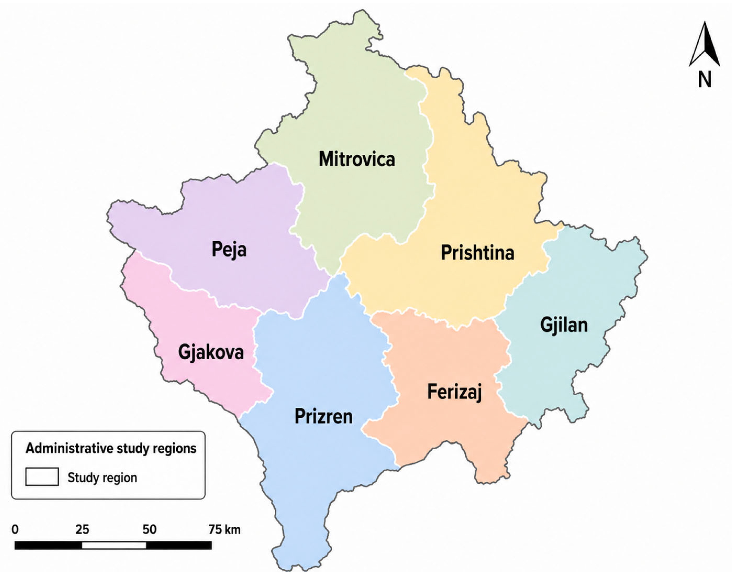
A cross-sectional study was conducted between December 2021 and April 2022 on ruminant farms located across the seven geographical regions of Kosovo: Prishtina, Mi trovica, Peja, Gjakova, Prizren, Ferizaj, and Gjilan. For the purposes of this study, a farm-species unit was defined as one ruminant species sampled within one farm. Therefore, a farm keeping more than one ruminant species could contribute more than one unit when the species were sampled independently. The study included cattle, sheep, and goat farms. From each farm-species unit, three sample types were collected: raw milk, fecal material, and animal feed. The study was designed to assess the occurrence of Listeria spp. in these matrices and to compare their distribution according to geographical region and ruminant species. Sampling was performed directly on the selected farms using a consistent sampling framework across all regions. Animal species and feed type were recorded during each farm visit. Geographical distribution of the seven study regions is shown in Figure 1.

2.2. Farm Selection and Sampling Framework

No formal registry-based sampling frame was used. In each of the seven geographical regions of Kosovo, veterinarians conducting the fieldwork identified cattle, sheep, and goat farms locally and randomly selected farms from those identified for inclusion in the study. All farms approached agreed to participate, and no farms declined participation. No formal statistical sample-size calculation was performed. The sampling framework comprised nine farm-species units in each region: five cattle units, two sheep units, and two goat units. This allocation reflected the greater number of cattle farms compared with sheep and goat farms across the regions of Kosovo, while ensuring representation of all these ruminant species in each region. Overall, 60 distinct farms provided 63 farm-species units, consisting of 35 cattle, 14 sheep, and 14 goat units. Most farms contributed one farm-species unit. However, three mixed-species farms contributed two units each: one farm contributed separate cattle and goat units, while two farms contributed separate cattle and sheep units. At these farms, feed, fecal material, and raw milk were collected independently for each animal species. One raw-milk sample, one pooled fecal sample, and one animal-feed sample were collected from each of the 63 farm-species units, resulting in 63 samples from each matrix and 189 samples in total. The distribution of farm-species units and collected samples by ruminant species is presented in Table 1.

2.3. Farm Questionnaire

During each farm visit, a structured questionnaire was administered through an interview with the farm owner or manager. Information was collected on farm location, years of operation, herd or flock size, animal species and age, the presence of other animals, housing and grazing systems, and the introduction of newly acquired animals. The questionnaire also covered milking practices, milk storage and marketing, barn hygiene and ventilation, rodent presence, feeding practices, silage production and management, water source, and the most frequently reported animal health problems. Information on milking practices referred to the routine milking of animals by the farmers. Farms where routine milking was not practiced could still have lactating animals from which milk was collected specifically for this study. Questionnaire data were summarized descriptively to characterize the surveyed farms. Because only three microbiological samples were positive, no inferential analysis of associations between farm-management variables and Listeria detection was performed.

2.4. Sample Collection and Transportation

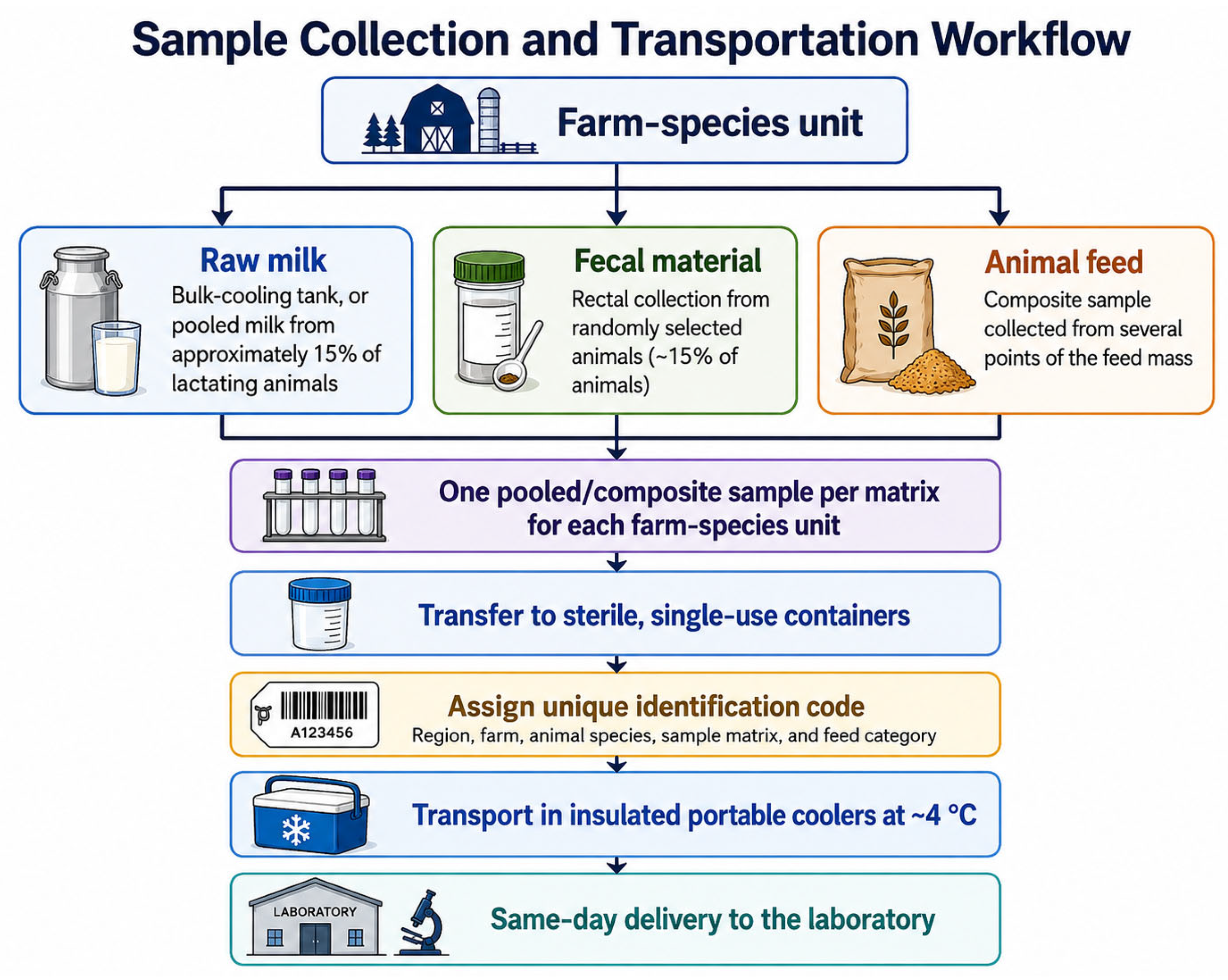
Raw milk, fecal material, and animal-feed samples were collected directly from each farm-species unit using sterile gloves and sterile, single-use containers. Appropriate precautions were taken to prevent cross-contamination among farms, animal species, and sample matrices. Feed samples were collected from the feed offered to the animals at the time of the farm visit. Silage was obtained from several points across the exposed surface of the storage unit and combined to form one composite sample. The same approach was used for hay, straw, and mixed silage–hay samples, with material collected from different locations within the available feed mass to improve sample representativeness. Fecal material was collected directly from the rectum of randomly selected animals representing approximately 15% of the cattle, sheep, or goats present in each farm-species unit. Individual portions were combined to produce one pooled fecal sample for each unit and were transferred immediately to a sterile container. Raw milk was collected from the bulk-cooling tank when available. On farms without a bulk-cooling tank, including farms where routine milking was not practiced, milk was collected manually from approximately 15% of the available lactating animals and pooled to obtain a raw-milk sample representing the farm-species unit. In this way, one raw-milk sample was collected from each of the 63 farm-species units. Therefore, both the milk and fecal samples represented the farm-species unit rather than individual animals. The sampling design was intended for farm-level screening. Therefore, the pooled fecal and raw-milk samples and the composite feed sample were used to characterize each farm-species unit and were not intended to estimate individual-animal prevalence or within-farm distribution of Listeria spp. Each sample was assigned a unique identification code linking it to the geographical region, farm, animal species, sample matrix, and feed category. Immediately after collection, samples were placed in insulated portable coolers and maintained at approximately 4 °C during transportation. All samples were delivered to the laboratory on the day of collection. An overview of the sample collection and transportation workflow is presented in Figure 2.

2.5. Sample Storage and Preparation

Upon arrival at the laboratory, all samples were stored at −20 °C for approximately one to two weeks before microbiological analysis. Approximately 24 h before analysis, the samples were removed from frozen storage and allowed to thaw at room temperature (15–20 °C). Following thawing, the detection and isolation of Listeria spp. was performed according to the procedure described in Section 2.8. Frozen storage and subsequent thawing represented preanalytical steps and were considered when interpreting the microbiological findings.

2.6. Classification and Distribution of Animal-Feed Samples

The 63 animal-feed samples were classified according to the type of stored feed provided to the animals at the time of sampling. Four feed categories were identified: silage, hay, straw, and a combination of silage and hay. Access to pasture was recorded separately as part of the farm feeding and management practices and was not included as a feed-sample category, because pasture vegetation was not collected for laboratory analysis. The distribution of the collected feed samples according to ruminant species and feed category is presented in Table 2.

2.7. Determination of Water Activity of Animal-Feed Samples

At the time of sampling, a separate portion of each composite animal-feed sample was reserved for water activity determination and was not subjected to freezing and thawing. The portion intended for microbiological analysis was handled according to the storage procedure described in Section 2.5. The water activity (aw) of the collected animal-feed samples was measured using a calibrated Novasina LabMaster aw neo instrument (Novasina AG, Lachen, Switzerland), equipped with a resistive electrolytic sensor, in accordance with ISO 18787 [16]. Before measurement, each sample was homogenized, and an appropriate quantity was transferred into a clean sample cup. The sample was placed in the measuring chamber, and the water activity value was recorded after equilibrium had been reached. Measurements were performed once for each sample.

2.8. Detection and Isolation of Listeria spp.

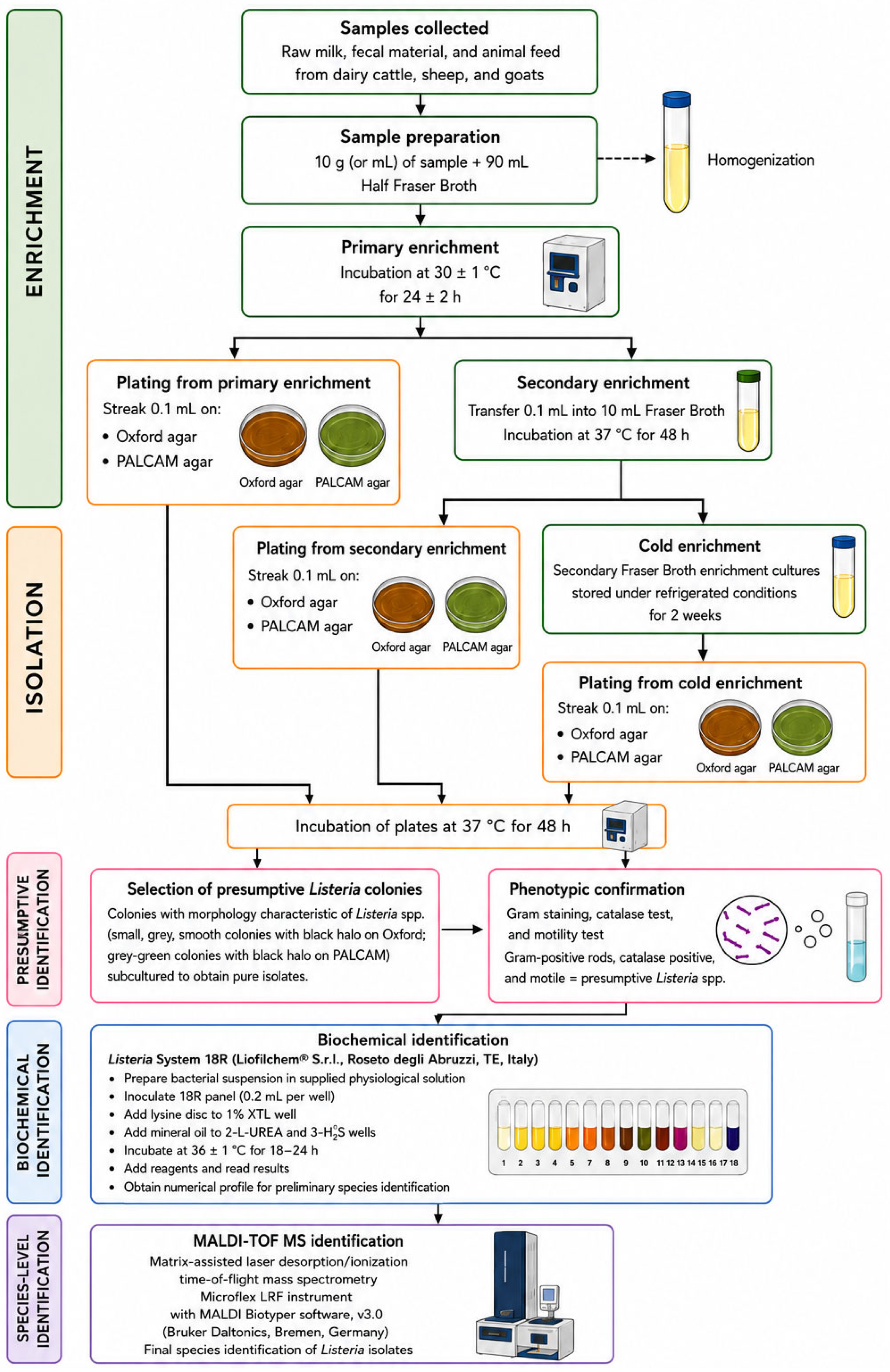
The detection and isolation of Listeria spp. were performed according to ISO 11290-1:2017 [17] with an additional cold-enrichment step performed as described below. Under aseptic conditions, a 10 g portion of each sample was transferred into a sterile stomacher bag containing 90 mL of Half Fraser Broth (Liofilchem® S.r.l., Roseto degli Abruzzi, Te, Italy). The sample broth mixture was homogenized using a peristaltic homogenizer (bioMérieux, Marcy-l’Étoile, France) operated at the normal speed setting. The homogenized samples were incubated at 30 °C for 24 h for primary enrichment. Following primary enrichment, 0.1 mL of the enriched culture was inoculated onto Oxford and Palcam (Liofilchem® S.r.l., Roseto degli Abruzzi, Te, Italy) selective agar plates. The plates were incubated at 37 °C for 48 h and subsequently examined for colonies exhibiting morphological characteristics of Listeria spp. In parallel, 0.1 mL of the primary enrichment culture was transferred into a tube containing 10 mL of Fraser Broth. The tubes were vortexed and incubated at 37 °C for 48 h for secondary enrichment. Following secondary enrichment, 0.1 mL of each Fraser Broth (Liofilchem® S.r.l., Roseto degli Abruzzi, Te, Italy) culture was inoculated onto Oxford and Palcam agar plates. The plates were incubated at 37 °C for an additional 48 h and examined for colonies displaying presumptive Listeria morphology. Following the standard enrichment and plating procedure, the Fraser Broth enrichment cultures were stored under refrigerated conditions for two weeks as an additional precautionary cold-enrichment step to provide a further opportunity for recovery of Listeria. The cultures were subsequently reinoculated onto Oxford and Palcam agar plates, incubated at 37 °C for 48 h, and examined again for presumptive Listeria colonies. This additional cold-enrichment step represented a modification of the standard ISO 11290-1:2017 procedure. Colonies with characteristic morphology from either the initial or post-cold-enrichment plates were selected for further confirmation and species level identification [17].

2.9. Phenotypic and Biochemical Confirmation of Presumptive Listeria Isolates

Colonies displaying morphological characteristics consistent with Listeria spp. on Oxford and Palcam agar were selected from both the initial and post-cold-enrichment plates and subcultured to obtain pure isolates. The isolates were examined by Gram staining and tested for catalase activity and motility. Gram-positive, short rod-shaped isolates showing a positive catalase reaction and characteristic motility were considered presumptive members of the genus Listeria and retained for further identification. Presumptive isolates were subsequently examined using the Listeria System 18R biochemical identification panel (Liofilchem® S.r.l., Roseto degli Abruzzi, Te, Italy) according to the manufacturer’s instructions. A bacterial suspension was prepared in the sterile physiological solution supplied with the kit using two to three well-isolated, morphologically similar colonies. An aliquot of 0.2 mL of the suspension was transferred into each reaction well. A xylose disc was placed in the designated 15-XYL well, while microbiological grade mineral oil was added to the 2-UREA and 3-H2S wells. The inoculated panels were incubated at 36 ± 1 °C for 18–24 h. Following incubation, the required supplementary reagents were added according to the manufacturer’s instructions. Biochemical reactions were evaluated based on color changes in the individual wells, and the resulting numerical profile was used for preliminary species identification. The isolates were subsequently subjected to MALDI-TOF mass spectrometry for final species-level identification.

2.10. Identification of Listeria Isolates by MALDI-TOF MS

The isolates preliminarily identified using the Listeria System 18R were subjected to matrix-assisted laser desorption/ionization time-of-flight mass spectrometry (MALDI-TOF MS) for confirmatory species-level identification. Analysis was performed using a Microflex LRF mass spectrometer equipped with MALDI Biotyper software, version 3.0 (Bruker Daltonics, Bremen, Germany). Four isolates originating from the three positive samples were subjected to MALDI-TOF MS analysis. The isolate from sample 07/Gj/DH/1/F (BB Lab ID 4) was identified as Listeria innocua with a recorded score of 2.38. Two isolates from sample 06/Gj/4/S (BB Lab IDs 24 and 26), recovered from the two selective media, were identified as Listeria innocua with recorded scores of 2.00 and 2.30, respectively. The isolate from sample 06/Gj/3/S (BB Lab ID 28) was identified as Listeria innocua with a recorded score of 2.05. None of these isolates was flagged for repeat MALDI-TOF MS analysis. These results were used for final species-level identification. The reference database version and formal score-threshold criterion applied during the analysis were not available in the retained laboratory records.

2.11. Statistical Analysis

Descriptive statistics were used to summarize the microbiological findings and selected farm characteristics obtained from the questionnaire. The occurrence of Listeria spp. was expressed as the number and percentage of positive samples and was summarized according to sample matrix, ruminant species, geographical region, and feed category. All percentages were reported together with their corresponding numerator and denominator. Because feed, fecal material, and raw milk represented different sample matrices, the combined result across the 189 collected samples was treated as a descriptive sample-level detection proportion rather than as a conventional prevalence estimate. Matrix-specific percentages were calculated using the number of samples analyzed within each respective matrix as the denominator, whereas species- and region-specific percentages were calculated using the corresponding number of collected samples. Co-occurrence was defined as the detection of Listeria spp. in more than one of the three matrices (feed, fecal material, and raw milk) collected from the same farm-species unit. At mixed-species farms, co-occurrence was evaluated separately for each sampled animal species. Herds and flock sizes were summarized separately for cattle, sheep, and goat farms using the mean and standard deviation, together with the median and range. Water activity (aw) values of the feed samples were summarized using the mean, standard deviation, median, minimum, and maximum, both overall and according to feed category. An overview of the laboratory workflow is shown in Figure 3.

3. Results

3.1. Questionnaire-Based Characteristics of the Surveyed Farms

The main characteristics of the surveyed cattle, sheep, and goat farms, as reported by the farmers through the questionnaire, are presented below.

3.1.1. Cattle Farms

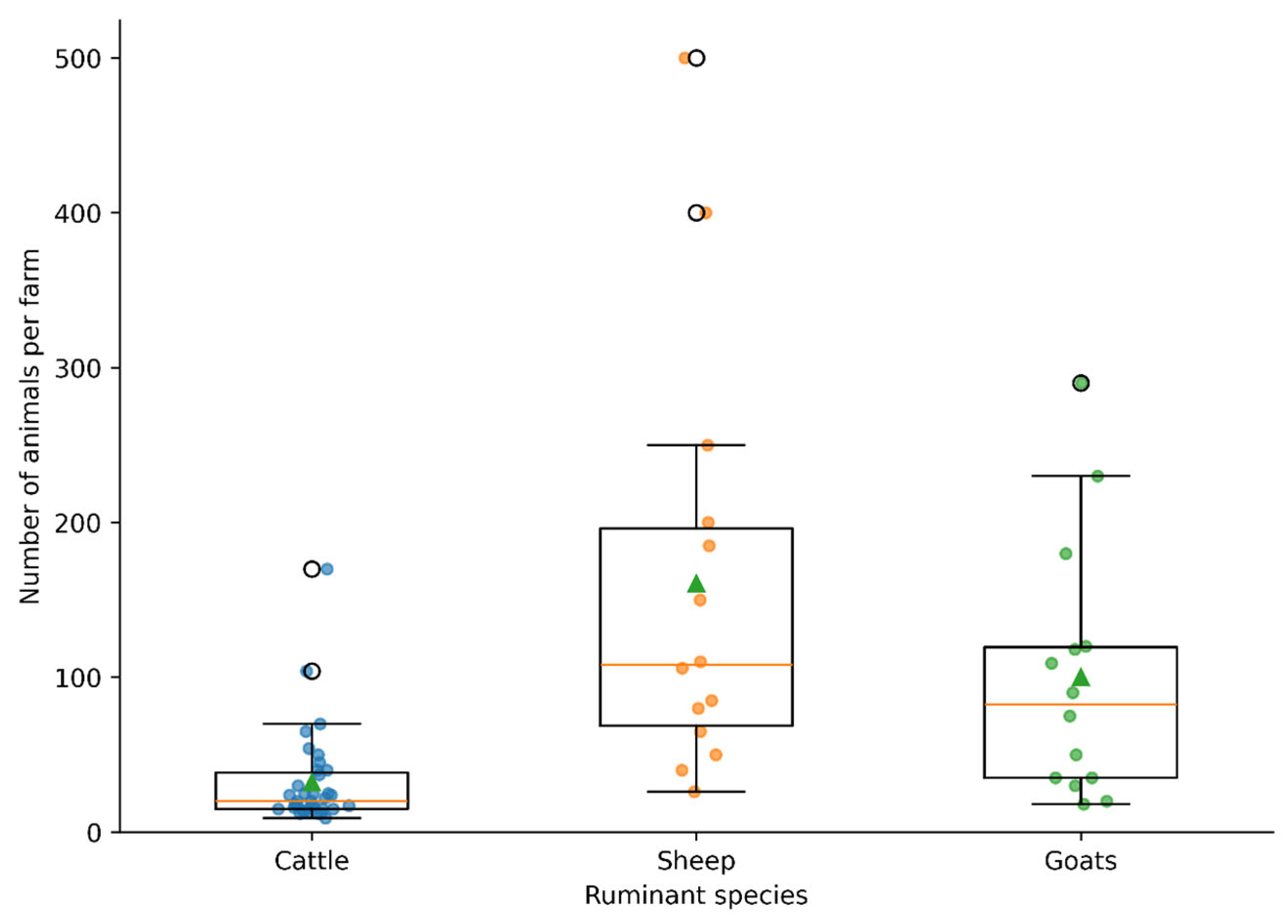
A total of 35 dairy cattle farms were included in the study, with five farms selected from each of the seven regions of Kosovo: Prishtina, Mitrovica, Peja, Prizren, Ferizaj, Gjilan, and Gjakova. Consequently, each region represented 14.3% of the surveyed farms. Altogether, the farms comprised 1122 dairy cattle. Herd size varied considerably, ranging from 9 to 170 animals per farm, with a mean herd size of 32.1 ± 31.4 animals and a median of 20 animals. Simmental was the most frequently represented breed. Sixteen farms (45.7%) kept only Simmental cattle, while three farms (8.6%) kept only Holstein cattle. Mixed herds consisting of Simmental and Holstein cattle were recorded on 12 farms (34.3%). Two farms (5.7%) kept Simmental, Holstein, and Montbéliarde cattle, whereas another two farms (5.7%) kept Holstein and Montbéliarde cattle. Overall, Simmental cattle were present on 30 farms (85.7%), Holstein cattle on 19 farms (54.3%), and Montbéliarde cattle on 4 farms (11.4%). The reported age of the animals ranged from 2 to 9 years. The tied housing system was the predominant management system and was used on 28 farms (80.0%). Four farms (11.4%) used a free-housing system, while three farms (8.6%) applied a semi-free system. Automatic milking was reported on 34 farms (97.1%), whereas one farm (2.9%) reported the use of mechanical milking. Silage was the main type of feed provided on 31 farms (88.6%). Hay was reported as the main feed on four farms (11.4%), including one farm in the Prishtina region and three farms in the Gjilan region. These findings indicate that the surveyed dairy farms were predominantly characterized by Simmental or mixed Simmental–Holstein herds, tied housing, automatic milking, and silage-based feeding.

3.1.2. Sheep Farms

A total of 14 sheep farms were included in the study, with two farms selected from each of the same seven regions of Kosovo. Therefore, each region accounted for 14.3% of the surveyed farms. Overall, the farms comprised 2247 sheep. Herd size ranged from 26 to 500 animals, with a mean of 160.5 ± 139.9 sheep per farm and a median herd size of 108 animals. The largest proportion of the total sheep population was recorded in the Prizren region, where two surveyed farms included 900 sheep, representing 40.1% of all animals included in the study. This was followed by Peja with 435 sheep (19.4%), Gjakova with 310 sheep (13.8%), Gjilan with 235 sheep (10.5%), Mitrovica with 146 sheep (6.5%), Ferizaj with 145 sheep (6.5%), and Prishtina with 76 sheep (3.4%). The reported age of the animals ranged from 2 to 5 years. Routine milking was not practiced on 10 of 14 sheep farms (71.4%). Regular milking was reported on three farms (21.4%), including two farms in the Prizren region and one farm in the Gjakova region. One farm in the Gjilan region practiced milking only occasionally (7.2%). However, lactating animals were present at all sheep farms at the time of sampling, and one pooled raw-milk sample was collected from each farm specifically for this study. Pasture was included in the feeding system of all surveyed sheep farms. Pasture combined with hay was reported on 11 farms (78.6%), whereas pasture combined with silage was used on three farms (21.4%). Overall, the surveyed sheep farms were characterized by considerable variation in herd size, predominantly pasture and hay-based feeding, and limited milking activity.

3.1.3. Goat Farms

A total of 14 goat farms were included in the study, with two farms selected from each of the seven regions of Kosovo. Altogether, the surveyed farms comprised 1400 goats. Herd size ranged from 18 to 290 animals, with a mean herd size of 100.0 ± 83.2 goats and a median of 82.5 goats. The largest proportion of animals was recorded in the Prishtina region, with 410 goats (29.3%), followed by Gjilan with 339 goats (24.2%), Ferizaj with 200 goats (14.3%), Gjakova with 136 goats (9.7%), Prizren with 125 goats (8.9%), Peja with 120 goats (8.6%), and Mitrovica with 70 goats (5.0%). Mixed herds consisting of Alpine and Red goats of Has were the most common and were recorded on five farms (35.7%). Four farms (28.6%) kept only Alpine goats, while another four farms (28.6%) kept only Red goats of Has. Dwarf goats were kept on one farm (7.1%). Overall, Alpine goats and Red goats of Has were each present on nine farms (64.3%). The reported age of animals ranged from 2 to 7 years. All surveyed farms used a free-range housing system, although the duration and frequency of access to pasture differed between farms. On most farms, goats were released onto pasture throughout the year, except during the coldest winter months, while some farms provided pasture access only during the warmer months or occasionally. Mechanical milking was practiced on 12 farms (85.7%), while one farm (7.1%) used an automatic milking system. Routine milking was not practiced on one farm (7.1%) because the goats were mainly kept for breeding. However, lactating animals were present at the time of sampling, and a pooled raw-milk sample was collected manually for this study. Milk was mainly used for household consumption, private sale, or on-farm cheese production. Hay was the sole feed reported on nine farms (64.3%). Silage alone was used on two farms (14.3%), straw on two farms (14.3%), and a combination of hay and silage on one farm (7.1%). The herd and flock characteristics of the surveyed cattle, sheep, and goat farms are summarized in Table 3, and the distribution of herd and flock sizes among the surveyed farms is shown in Figure 4.

3.2. Water Activity of the Animal-Feed Samples

Water activity (aw) was measured in all 63 animal-feed samples. The aw ranged from 0.5079 to 0.9363, with a mean of 0.775 ± 0.148 and a median of 0.858. Silage samples showed the highest water activity, with a mean value of 0.887 ± 0.033, a median of 0.892, and a range of 0.777–0.9363. Hay samples had a mean of 0.620 ± 0.096, with values ranging from 0.521 to 0.845 and a median of 0.589. The two straw samples had aw values of 0.5079 and 0.695, resulting in a mean of 0.601 ± 0.132. The single sample consisting of a combination of silage and hay had an aw value of 0.823. Because the numbers of straw and combined silage–hay samples were limited, the results according to feed category were interpreted descriptively, as presented in Table 4.

3.3. Occurrence of Listeria spp. in the Collected Samples

Presumptive Listeria colonies were recovered from 3/189 matrix-level samples examined (1.6%). This combined proportion is presented descriptively across the three sample matrices and should not be interpreted as a conventional prevalence estimate. In one sample, characteristic colonies were observed in both Palcam and Oxford agar, whereas the remaining two samples yielded presumptive colonies one on the Palcam agar and the other on Oxford agar. Following species-level identification, all recovered isolates were confirmed as Listeria innocua. No Listeria monocytogenes was detected in any of the samples examined. Positive samples are shown in Table 5.

Distribution of Listeria innocua—Positive Samples

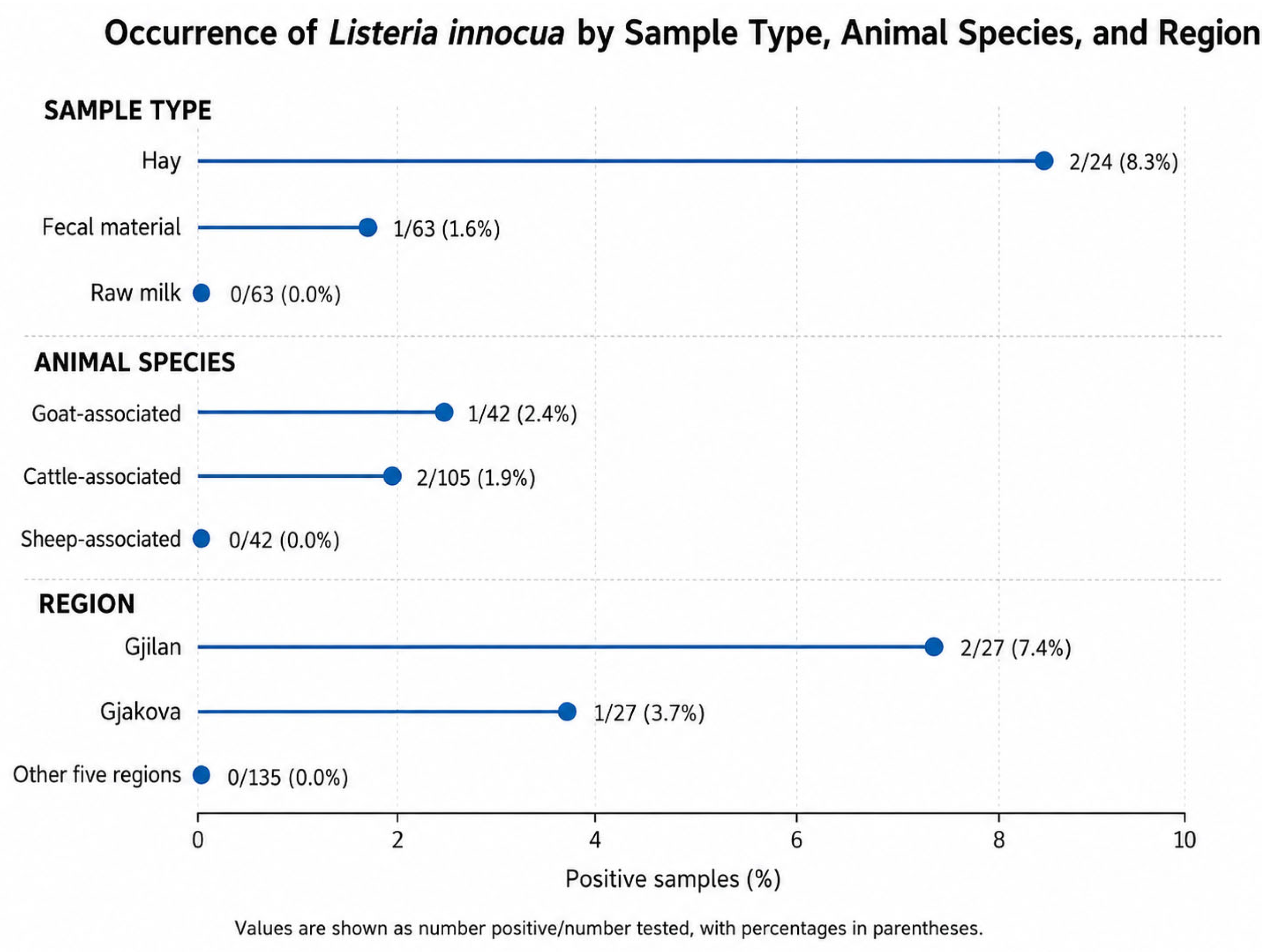
The discordant growth observed between Palcam and Oxford agar may reflect differences in recovery between the two selective media; however, the exact reason for this difference could not be determined from the present data. Of the three Listeria innocua positive samples, two were hay samples collected from cattle farms in the Gjilan region. The two positive hay samples originated from separate farms and represented 2/63 feed samples (3.2%) and 2/24 hay samples (8.3%). The water activity values of these two hay samples were 0.6213 and 0.5603, respectively. The third positive sample was a fecal sample collected from a goat farm in the Gjakova region, representing 1/63 fecal samples (1.6%). No Listeria innocua was detected in raw-milk samples (0/63; 0%) under the conditions of this cross-sectional study. According to animal species, Listeria innocua was detected in 2/105 cattle-associated samples (1.9%) and 1/42 goat-associated samples (2.4%), whereas no positive samples were detected among the 42 sheep-associated samples (0/42; 0%). At the regional level, 2/27 samples collected in Gjilan were positive (7.4%), while 1/27 samples collected in Gjakova was positive (3.7%). No positive samples were detected among the 27 samples analyzed from each of the other five regions (0/27; 0% in each region). Because only three positive samples were identified, these findings are presented descriptively and should not be interpreted as evidence of geographical differences in Listeria occurrence. As the three positive samples originated from separate farms, no co-occurrence across feed, fecal material, and raw milk was observed within the same farm-species unit. Descriptive distribution of the three Listeria innocua positive samples identified in the study is shown in Figure 5.

4. Discussion

The present study investigated the occurrence of Listeria spp. in animal feed, fecal material, and raw milk collected from cattle, sheep, and goat farms across Kosovo. Listeria spp. was detected in 3/189 matrix-level samples examined (1.6%), all of which were identified as Listeria innocua, while Listeria monocytogenes was not detected. This combined value represents a descriptive sample-level detection proportion across the three matrices rather than a conventional prevalence estimate. Given the small number of positive samples, these findings are descriptive and do not support conclusions regarding differences among sample matrices, animal species, feed categories, or geographical regions. Previous studies have reported higher detection frequencies of Listeria spp. in ruminant-production environments. Gana et al. detected Listeria spp. in 14.6% of samples collected from cattle farms in South Africa, with Listeria innocua accounting for 11.3% and Listeria monocytogenes for 3.4% [18]. Zhao et al. similarly reported that Listeria innocua occurred more frequently than Listeria monocytogenes in cattle and sheep associated farm and slaughter environments in China, with overall occurrences of 9.7% and 1.8%, respectively [19]. More recently, Maobelo et al. detected Listeria monocytogenes in 2.5% and other Listeria species in 9.2% of samples collected from cattle farms [20]. Comparisons among studies should, however, be made cautiously because differences in geographical and climatic conditions, farm structure and management, sampling season, sample size, matrices examined, sampling frequency, storage conditions, and laboratory procedures may influence detection frequencies. The exclusive recovery of Listeria innocua in the present study is consistent with investigations in which this species was recovered more frequently than Listeria monocytogenes. Manqele et al., for example, reported that Listeria innocua represented 77.1% of the Listeria isolates recovered from cattle farms and retail beef products [21]. Although Listeria innocua is generally considered less pathogenic than Listeria monocytogenes, some strains have been reported to carry virulence-associated genes and show pathogenic potential. Genomic studies of farm- and food chain-associated Listeria innocua have also identified stress-, antimicrobial resistance- and virulence-related determinants [22,23]. However, virulence, antimicrobial resistance, hemolytic activity, and genomic characteristics were not evaluated in the present study; therefore, no conclusions can be drawn regarding pathogenic potential or genetic relatedness of the recovered isolates. Two of the three positive samples were hay samples collected from separate cattle farms. Hay may become contaminated during harvesting, field drying, transportation, storage, or distribution, or through environmental sources such as soil, manure, equipment, rodents, wild birds, dust, or moisture. Previous cattle farm investigations have recovered Listeria spp. from feed, silage, water, and fecal material [18,20,24]. However, only two hay samples were positive in the present study, while no Listeria spp. was detected in the 36 silage samples. Therefore, the findings do not support the conclusion that hay represented a greater contamination risk than silage or other feed categories. Similarly, the occurrence of two positive samples in Gjilan and one in Gjakova should be interpreted descriptively rather than as evidence of geographical differences. Water activity provided additional context for interpreting the two positive hay samples. Hay had a mean aw of 0.620, while the two positive hay samples had aw values of 0.6213 and 0.5603. Experimental studies have reported minimum water-activity values supporting growth of Listeria monocytogenes and Listeria innocua at approximately 0.90–0.92, depending on the composition of the medium [25]. The measured values of the two positive hay samples were therefore below values generally associated with active growth of Listeria spp. at the time of testing. However, water activity was measured at a single time point and does not reflect moisture conditions throughout harvesting or storage. Although the portion used for water-activity determination was reserved separately and was not subjected to freezing and thawing, earlier periods of greater moisture cannot be excluded. Consequently, the water-activity findings should be regarded as contextual information and cannot establish the timing or mechanism of contamination, survival, or persistence [26]. The recovery of Listeria innocua from one pooled goat-fecal sample is consistent with previous evidence that clinically healthy ruminants may carry and shed members of the genus Listeria. Palacios-Gorba et al. recovered Listeria innocua from 4.7% of fecal samples obtained from domestic dairy ruminants and demonstrated that different Listeria species may be carried asymptomatically in feces, tonsils, or udders [27]. Nevertheless, only one pooled fecal sample was positive in the present study, and the sampling design does not permit conclusions regarding individual-animal carriage, shedding frequency, or within-farm prevalence. Listeria spp. was not detected in any of the 63 raw-milk samples analyzed under the conditions of this cross-sectional study. This finding applies only to the samples examined and should not be interpreted as evidence that raw milk from the surveyed population was free of Listeria or as evidence of its long-term absence. Condoleo et al. examined 372 bulk tank milk samples from 87 sheep farms and did not detect Listeria monocytogenes in any sample [28]. In contrast, Sepahvand et al. detected Listeria spp. in raw milk from sheep and goats and identified Listeria monocytogenes in 5.6% of sheep and 3.8% of goat milk samples [29]. A recent global analysis estimated a pooled Listeria monocytogenes prevalence of approximately 3.4% in raw milk and demonstrated considerable variation among geographical areas, study designs and detection methods [30]. These findings further emphasize that the non-detection result in a single cross-sectional survey should be interpreted within the specific sampling and laboratory conditions of the study. No co-occurrence of Listeria spp. was observed among feed, fecal material, and raw milk from the same farm-species unit. Therefore, the present study provided no evidence of a feed–animal–milk contamination sequence within the surveyed farms. However, the absence of co-occurrence does not exclude intermittent contamination or transmission because each farm was visited only once and only one pooled or composite sample from each matrix was examined. The standardized collection of feed, fecal material, and raw milk within the same farm-species framework represents a strength of the study, as does the use of selective enrichment, phenotypic and biochemical confirmation, and MALDI-TOF MS for final species identification. Several limitations should be considered. Only one pooled fecal sample, one pooled raw-milk sample, and one composite feed sample were collected from each farm-species unit during a single sampling visit. This design was appropriate for farm-level screening but may have missed intermittent shedding or localized contamination and does not allow estimation of individual-animal prevalence. The relatively small numbers of sheep and goat farm-species units limit the representativeness of the findings for these production sectors, and the results should therefore be interpreted as a cross-sectional description of the surveyed farms rather than as population-level estimates for Kosovo. In addition, because sampling was conducted between December 2021 and April 2022, the findings reflect conditions during that period and may not fully represent current farm-management or microbiological conditions. Samples used for microbiological analysis were stored at −20 °C for approximately one to two weeks before analysis and were subsequently thawed at room temperature. Although Listeria spp. can survive frozen storage, freezing, storage duration, and thawing may cause sublethal injury and reduce recovery, particularly when organisms are present at low concentrations or are physiologically stressed. Therefore, the effect of the preanalytical freezing and thawing procedure on detection sensitivity cannot be excluded and should be considered when interpreting both the small number of positive samples and the negative findings. The small number of positive samples also prevented inferential comparisons among matrices, animal species, regions, and feed categories, and questionnaire-derived farm-management variables could therefore only be interpreted descriptively. The study also did not include enumeration, antimicrobial testing, virulence gene detection or molecular typing. Specific positive and negative control strains used during enrichment, selective plating, biochemical identification, and MALDI-TOF MS analysis were not documented, and detailed MALDI-TOF MS metadata regarding the reference database version and formal score-threshold criterion were not available in the retained laboratory records. Nevertheless, all four isolates originating from the three positive samples were identified as Listeria innocua, with recorded MALDI-TOF MS scores ranging from 2.00 to 2.38, and none were flagged for repeat analysis. Future investigations should include larger sample numbers, repeated seasonal sampling, analysis of freshly collected samples, direct comparison of fresh and frozen samples to assess the effect of preanalytical storage on Listeria recovery, and molecular characterization of recovered isolates to better understand the occurrence, persistence, genetic diversity, and potential significance of Listeria spp. within ruminant-production environments in Kosovo.

5. Conclusions

This study provides a cross-sectional assessment of the occurrence of Listeria spp. in animal feed, fecal material, and raw milk from cattle, sheep, and goat farms across all seven geographical regions of Kosovo. Listeria spp. was detected in 3/189 matrix-level samples analyzed (1.6%); this combined proportion is descriptive and should not be interpreted as a conventional prevalence estimate. All recovered isolates were identified as Listeria innocua, while Listeria monocytogenes was not detected in any of the samples examined. Two positive samples were hay samples collected from separate cattle farms, representing 2/63 feed samples (3.2%) and 2/24 hay samples (8.3%), while one positive sample was a pooled fecal sample from a goat farm, representing 1/63 fecal samples (1.6%). Listeria spp. was not detected in the raw milk samples analyzed (0/63; 0%) under the conditions of this cross-sectional survey. However, this finding should not be interpreted as evidence of the long-term absence of Listeria from raw milk on the surveyed farms. No co-occurrence among feed, fecal material, and raw milk was observed within the same farm-species unit. However, the single-visit sampling design and the use of one pooled or composite sample per matrix do not allow conclusions regarding long-term absence, seasonal variation, intermittent contamination, individual-animal carriage or transmission pathways among feed, animals, and milk. The detection of Listeria innocua in hay and fecal material indicates the occasional presence of members of the genus Listeria within the surveyed ruminant-farm environments, but the small number of positive samples does not support comparisons among regions, animal species, sample matrices, or feed categories. Future studies should include larger sample numbers, repeated seasonal sampling, analysis of freshly collected samples, and molecular characterization of recovered isolates to better assess temporal variation, persistence, and possible transmission pathways within ruminant-production environments in Kosovo.

Author Contributions

Conceptualization, B.N., A.H., and B.B.; methodology, B.N., A.H., and B.B.; formal analysis, E.G., A.G.T., A.T., D.H., and B.S.; investigation, B.N., A.H., A.G.T., and E.G.; writing—original draft. B.N. and E.G.; writing, review and editing, E.G.; supervision, A.H. and B.N.; funding acquisition B.N. All authors have read and agreed to the published version of the manuscript.

Funding

This research received no extra funding.

Institutional Review Board Statement

Ethical approval was not required for this study, as confirmed by the Scientific Ethics Committee of the Faculty of Agriculture and Veterinary, University of Prishtina.

Informed Consent Statement

Written informed consent was obtained from the owner of the animals involved in this study.

Data Availability Statement

The original contributions presented in this study are included in the article. Further inquiries can be directed to the corresponding author.

Acknowledgments

ChatGPT (https://chatgpt.com/, accessed on 19 August 2026) was used to assist in preparation of graphical materials and in refining the terminology and labels used within the figures.

Conflicts of Interest

The authors declare no conflicts of interest.

References

  1. Oliver, S.P.; Jayarao, B.M.; Almeida, R.A. Foodborne pathogens in milk and the dairy farm environment. Food safety and public health implications. Foodborne Pathog. Dis. 2005, 2, 115–129. [Google Scholar] [CrossRef] [Scilit]
  2. McAuley, C.M.; McMillan, K.K.; Moore, S.C.; Fegan, N.; Fox, E.M. Prevalence and characterization of foodborne pathogens from. Australian dairy farm environments. J. Dairy Sci. 2014, 97, 7402–7412. [Google Scholar] [CrossRef] [Scilit] [PubMed]
  3. Vilar, M.K.; Yus, E.; Sanjuán, M.L.; Diégues, F.J.; Rodrígues-Otero, J.L. Prevalence of and risk factors for Listeria species on dairy farms. J. Dairy Sci. 2007, 90, 5083–5088. [Google Scholar] [CrossRef] [Scilit] [PubMed]
  4. Queiroz, O.C.M.; Ogunade, I.M.; Weinberg, Z.; Adesogan, A.T. Silage review: Foodborne pathogens in silage and their mitigation by silage additives. J. Dairy Sci. 2018, 101, 4132–4142. [Google Scholar] [CrossRef] [Scilit] [PubMed]
  5. Konosonoka, I.H.; Jemeljanovs, A.; Osmane, B.; Ikauniece, D.; Gulbe, G. Incidence of Listeria spp. in dairy cows feed and raw milk in Latvia. Int. Sch. Res. Not. 2012, 2012, 435187. [Google Scholar] [CrossRef] [Scilit] [PubMed]
  6. Nightingale, K.K.; Schukken, Y.H.; Nightingale, C.R.; Fortes, E.D.; Ho, A.J.; Her, Z.; Gröhn, Y.T.; McDonough, P.L.; Wiedmann, M. Ecology and transmission of Listeria monocytogenes infecting ruminants and in the farm environment. Appl. Environ. Microbiol. 2004, 70, 4458–4467. [Google Scholar] [CrossRef] [Scilit] [PubMed]
  7. Nightingale, K.K.; Fortes, E.D.; Ho, A.J.; Schukken, Y.H.; Gröhn, Y.T.; Wiedmann, M. Evaluation of farm management practices as risk factors for clinical listeriosis and fecal shedding of Listeria monocytogenes in ruminants. J. Am. Vet. Med. Assoc. 2005, 227, 1808–1814. [Google Scholar] [CrossRef] [Scilit] [PubMed]
  8. Ho, A.J.; Ivanek, R.; Gröhn, Y.T.; Nightingale, K.K.; Wiedmann, M. Listeria monocytogenes fecal shedding in dairy cattle shows high levels of day-to-day variation and includes outbreaks and sporadic cases of shedding of specific L. monocytogenes subtypes. Prev. Vet. Med. 2007, 80, 287–305. [Google Scholar] [CrossRef] [Scilit] [PubMed]
  9. Esteban, J.I.; Oporto, B.; Aduriz, G.; Juste, R.A.; Hurtado, A. Faecal shedding and strain diversity of Listeria monocytogenes in healthy ruminants and swine in Northern Spain. BMC Vet. Res. 2009, 5, 2. [Google Scholar] [CrossRef] [Scilit] [PubMed]
  10. Mohammed, H.O.I.; Stipetic, K.; McDonough, P.L.; Gonzales, R.N.; Nydam, D.V.; Atwill, E.R. Identification of potential on-farm sources of Listeria monocytogenes in herds of dairy cattle. Am. J. Vet. Res. 2009, 70, 383–388. [Google Scholar] [CrossRef] [Scilit] [PubMed]
  11. Latorre, A.A.; Van Kessel, J.S.; Karns, J.S.; Zurakowski, M.J.; Pradhan, A.K.; Boor, K.J.; Jayarao, B.M.; Houser, B.A.; Daugherty, C.S.; Schukken, Y.H. Biofilm in milking equipment on a dairy farm as a potential source of bulk-tank milk contamination with Listeria monocytogenes. J. Dairy Sci. 2010, 93, 2792–2802. [Google Scholar] [CrossRef] [Scilit] [PubMed]
  12. Schoder, D.; Melzner, D.; Schmalwieser, A.; Zangana, A.; Winter, P.; Wagner, M. Important vectors for Listeria monocytogenes transmission at farm dairies manufacturing fresh sheep and goat cheese from raw milk. J. Food Prot. 2011, 74, 919–924. [Google Scholar] [CrossRef] [Scilit] [PubMed]
  13. Castro, H.; Jaakkonen, A.; Hakkinen, M.; Korkeala, H.; Lindström, M. Occurrence, persistence and contamination routes of Listeria monocytogenes genotypes on three Finnish dairy cattle farms: A longitudinal study. Appl. Environ. Microbiol. 2018, 84, e02000-17. [Google Scholar] [CrossRef] [Scilit] [PubMed]
  14. Sauders, B.D.; Overdevest, J.; Fortes, E.; Windham, K.; Schukken, Y.; Lembo, A.; Wiedmann, M. Diversity of Listeria species in urban and natural environments. Appl. Environ. Microbiol. 2012, 78, 4420–4433. [Google Scholar] [CrossRef] [Scilit] [PubMed]
  15. Fox, E.; O’Mahony, T.; Clancy, M.; Dempsey, R.; O’Brien, M.; Jordan, K. Listeria monocytogenes in the Irish dairy farm environment. J. Food Prot. 2009, 72, 1450–1456. [Google Scholar] [CrossRef] [Scilit] [PubMed]
  16. ISO 18787; Foodstuffs—Determination of Water Activity. ISO: Geneva, Switzerland, 2017.
  17. ISO 11290-1:2017; Microbiology of the Food Chain—Horizontal Method for the Detection and Enumeration of Listeria monocytogenes and of Listeria spp.—Part 1: Detection Method. ISO (International Organization for Standardization): Geneva, Switzerland, 2017.
  18. Gana, J.; Gcebe, N.; Moerane, R.; Ngoshe, Y.B.; Tshuma, T.; Moabelo, K.; Adesiyun, A.A. A comparative study on the occurrence, genetic characteristics, and factors associated with the distribution of Listeria species on cattle farms and beef abattoirs in Gauteng Province, South Africa. Trop. Anim. Health Prod. 2024, 56, 88. [Google Scholar] [CrossRef] [Scilit] [PubMed]
  19. Zhao, Q.; Hu, P.; Li, Q.; Zhang, S.; Li, H.; Chang, J.; Jiang, Q.; Zheng, Y.; Li, Y.; Liu, Z.; et al. Prevalence and transmission characteristics of Listeria species from ruminants in farm and slaughtering environments in China. Emerg. Microbes Infect. 2021, 10, 356–364. [Google Scholar] [CrossRef] [Scilit] [PubMed]
  20. Moabelo, K.C.; Gcebe, N.; Gana, J.; Ngoshe, Y.B.; Moerane, R.; Adesiyun, A. A Detection and molecular characterization of Listeria monocytogenes and Listeria spp. isolates recovered from cattle farms in Mpumalanga Province, South Africa. Vet. Med. Int. 2025, 2025, 4708466. [Google Scholar] [CrossRef] [Scilit] [PubMed]
  21. Manqele, A.; Gcebe, N.; Pierneef, R.E.; Moerane, R.E.; Adesiyun, A. A Identification of Listeria species and MILVA typing of Listeria innocua and Listeria monocytogenes isolates from cattle farms and retail beef products. Pathogens 2023, 12, 147. [Google Scholar] [CrossRef] [Scilit] [PubMed]
  22. Gana, J.; Gcebe, N.; Pierneef, R.E.; Chen, Y.; Moerane, R.; Adesiyun, A.A. Genomic characterization of Listeria innocua isolates recovered from cattle farms, beef abattoirs, and retail outlets in Gauteng Province, South Africa. Pathogens 2023, 12, 1062. [Google Scholar] [CrossRef] [Scilit] [PubMed]
  23. Rivu, S.; Shourav, A.H.; Ahmed, S. Whole-genome sequencing reveals circulation of potentially virulent Listeria innocua strains with novel genomic features in cattle-farm environments in Dhaka, Bangladesh. Infect. Genet. Evol. 2024, 126, 105692. [Google Scholar] [CrossRef] [Scilit] [PubMed]
  24. Idland, L.; Granquist, E.G.; Aspholm, M.; Lindbäck, T. The prevalence of Campylobacter spp., Listeria monocytogenes, and Shiga toxin-producing Escherichia coli in Norwegian dairy cattle farms: A comparison between free-stall and tie-stall housing systems. J. Appl. Microbiol. 2022, 132, 3959–3972. [Google Scholar] [CrossRef] [Scilit] [PubMed]
  25. Nolan, D.A.; Chamblin, D.C.; Troller, J.A. Minimal water activity levels for growth and survival of Listeria monocytogenes and Listeria innocua. Int. J. Food Microbiol. 1992, 16, 323–335. [Google Scholar] [CrossRef] [Scilit] [PubMed]
  26. Hua, Z.; Thapa, B.B.; Younce, F.; Tang, J.; Zhu, M.J. Impacts of water activity on survival of Listeria innocua and Enterococcus faecium NRRL B-2354 in almonds during steam treatments. Int. J. Food Microbiol. 2024, 413, 110592. [Google Scholar] [CrossRef] [Scilit] [PubMed]
  27. Palacios-Gorba, C.; Moura, A.; Markovich, Y.; Tessaud-Rita, N.; Gómez-Martín, Á.; Bracq-Dieye, H.; Gomis, J.; Vales, G.; Pastor-Martín, M.; Thouvenot, P.; et al. Genomic characterization of Listeria spp. isolated from tonsils, udder, and feces of domestic dairy ruminants in Spain. Microbes Infect. 2023, 25, 105079. [Google Scholar] [CrossRef] [Scilit] [PubMed]
  28. Condoleo, R.; Giangolini, G.; Chiaverini, A.; Patriarca, D.; Scaramozzino, P.; Mezher, Z. Occurrence of Listeria monocytogenes and Escherichia coli in raw sheep’s milk from farm bulk tanks in Central Italy. J. Food Prot. 2020, 83, 1929–1933. [Google Scholar] [CrossRef] [Scilit] [PubMed]
  29. Sepahvand, F.; Rashidian, E.; Jaydari, A.; Rahimi, H. Short Communication: Prevalence of Listeria monocytogenes in raw milk of healthy sheep and goats. Vet. Med. Int. 2022, 2022, 3206172. [Google Scholar] [CrossRef] [Scilit] [PubMed]
  30. Li, X.; Zheng, J.; Zhao, W.; Wu, Y. Prevalence of Listeria monocytogenes in milk and dairy-product supply chains: A global systematic review and meta-analysis. Foodborne Pathog. Dis. 2024, 21, 526–535. [Google Scholar] [CrossRef] [Scilit] [PubMed]
Figure 1. Geographical distribution of the seven study regions in Kosovo included in the cross-sectional survey conducted between December 2021 and April 2022.
Ruminants 06 00071 g001
Figure 2. Overview of sample collection, coding, transport, storage, and preparation of raw-milk, fecal material, and animal-feed samples from each farm-species unit.
Ruminants 06 00071 g002
Figure 3. Overview of the laboratory workflow used for enrichment, isolation, phenotypic and biochemical confirmation, and MALDI-TOF MS identification of Listeria spp. from raw milk, fecal material, and animal-feed samples.
Ruminants 06 00071 g003
Figure 4. Distribution of herd and flock sizes among the surveyed cattle, sheep, and goat farms. Individual points represent the number of animals recorded on each farm. The horizontal line within each box represents the median, and the triangle indicates the mean.
Ruminants 06 00071 g004
Figure 5. Descriptive distribution of the three Listeria innocua positive samples identified in the study. The figure is presented for visualization only and does not imply analytical comparison among regions, matrices, or animal species.
Ruminants 06 00071 g005
Table 1. Distribution of farm-species units and collected samples by ruminant species.
Ruminant SpeciesUnits per Region, nTotal Farm-Species Units, nRaw Milk Samples, nFecal Samples, nFeed Samples, nTotal Samples, n
Cattle 535353535105
Sheep21414141442
Goat21414141442
Total963636363189
Note: The study included 60 distinct farms representing 63 farm-species units. A farm-species unit was defined as one ruminant species sampled within one farm. Three mixed-species farms contributed two independently sampled units each.
Table 2. Distribution of animal-feed samples according to ruminant species and feed category.
Ruminant SpeciesSilage, nHay, nStraw, nSilage and Hay, nTotal, n
Cattle 3140035
Sheep3110014
Goats292114
Total36242163
Table 3. Herd and flock characteristics of the surveyed cattle, sheep, and goat farms.
CharacteristicCattle Farms, n = 35Sheep Farms, n = 14Goat Farms, n = 14
Total number of animals112222471400
Herd/flock size, mean ± SD32.1 ± 31.4160.5 ± 139.9100.0 ± 83.2
Herd/flock size, median (range)20 (9–170)108 (26–500)82.5 (18–290)
Table 4. Water activity of the animal-feed samples according to feed category.
Feed CategorynMean ± SDMedianRange
Silage360.887 ± 0.0330.8920.777–0.9363
Hay240.620 ± 0.0960.5890.521–0.845
Straw20.601 ± 0.1320.6010.5079–0.695
Silage–hay combination10.8230.8230.823
Overall630.775 ± 0.1480.8580.5079–0.9363
Table 5. Selective culture results and final identification of presumptive Listeria-positive samples.
Sample CodePalcam AgarOxford AgarFinal Identification
06/Gj/3/SNegative Positive Listeria innocua
06/Gj/4/SPositive PositiveListeria innocua
07/Gj/Dh/1/FPositive Negative Listeria innocua
Disclaimer/Publisher’s Note: The statements, opinions and data contained in all publications are solely those of the individual author(s) and contributor(s) and not of MDPI and/or the editor(s). MDPI and/or the editor(s) disclaim responsibility for any injury to people or property resulting from any ideas, methods, instructions or products referred to in the content.