Review

Beyond Mutation Detection: Cell-Free DNA for Functional Inference and Adaptive Oncology

Independent Researcher, Chicago, IL 60611, USA
Submission received: 26 March 2026 / Revised: 4 May 2026 / Accepted: 8 May 2026 / Published: 2 June 2026

Abstract

Liquid biopsy has evolved beyond its original role as a minimally invasive approach for mutation detection and is now being developed as a broader analytical framework for cancer detection, stratification, and longitudinal monitoring. Improvements in next-generation sequencing, assay chemistry, and computational analysis have increased analytical sensitivity, including in settings with low tumor fraction and very low variant allele abundance. These advances have expanded the utility of cfDNA analysis in measurable residual disease assessment and in the detection of low-abundance tumor-derived signals across multiple clinical contexts. At the same time, the field has shifted toward interpreting cfDNA as a carrier of higher-order biological information rather than solely a substrate for mutation calling. Fragmentation profiles, nucleosome positioning, and chromatin accessibility patterns derived from plasma DNA have been used to infer transcriptional and regulatory states, raising the possibility that cfDNA may capture functional tumor states not readily accessible through genotype-focused assays alone. These developments have prompted growing interest in chromatin-informed cfDNA analysis as a means of identifying pathway activity, enhancer usage, transcription factor occupancy, and potentially actionable biological dependencies. However, the translational relevance of many such inferences remains incompletely established, and preanalytical variability, limited cross-cohort generalizability, and the gap between analytical performance and clinical utility continue to constrain clinical translation. This review examines the role of cfDNA in adaptive oncology, highlighting recent analytical advances, assessing the current evidence supporting their biological and clinical utility, and considering the extent to which cfDNA-derived regulatory inference may contribute to adaptive oncology and therapeutic decision-making.

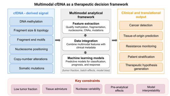
Graphical Abstract

1. Introduction

Liquid biopsy has substantially expanded the analytical toolkit available for cancer monitoring by enabling minimally invasive access to tumor-derived material in blood and other biofluids. Tumors release multiple analytes into circulation, including cell-free DNA (cfDNA), circulating tumor DNA (ctDNA), circulating tumor cells, and extracellular vesicles. Collectively, these analytes can provide a dynamic view of disease that partly mitigates the sampling bias and temporal limitations inherent to tissue biopsy [1,2,3,4]. Among them, cfDNA has emerged as a central component of precision oncology because it can be repeatedly sampled and profiled for genomic, epigenomic, and structural features associated with tumor evolution and treatment response [2,3]. In this review, cfDNA refers to extracellular DNA recovered from plasma using standard isolation workflows, which predominantly capture nucleosome-associated fragments; however, longer cfDNA molecules consistent with multinucleosomal structures have been detected using long-read sequencing approaches, suggesting that short-read workflows may underrepresent the full size spectrum of circulating DNA [3,4,5]. Extracellular vesicle-associated DNA is not specifically resolved in most current sequencing protocols and is therefore not considered separately.
The field was initially driven by the demonstration that circulating DNA could reveal tumor-derived genomic alterations, enabling minimal residual disease (MRD) assessment. With the introduction of ultra-deep sequencing, molecular barcoding, and duplex error-correction approaches, ctDNA assays achieved sensitivity sufficient for clinically relevant detection at very low allele fractions. These technical advances enabled practical applications in advanced disease, recurrence monitoring, and increasingly, early detection [5,6,7,8]. Nevertheless, mutation-centered approaches provide only a partial view of tumor biology and are less informative in tumors shaped by epigenetic dysregulation, transcriptional plasticity, or adaptive resistance programs that arise without recurrent genomic alterations [9,10].
This limitation has motivated a broader interpretation of cfDNA. Plasma DNA retains structural and epigenetic features related to its chromatin origin, including information linked to nucleosome positioning, transcription factor occupancy, chromatin accessibility, and fragmentation topology [11,12,13,14,15]. cfDNA should therefore not be regarded solely as sequence material. Under appropriate analytical conditions, it may also function as a readout of chromatin organization and regulatory state.
Accordingly, fragmentomics has expanded from descriptive analysis of fragment size into a broader framework encompassing fragment-length distributions, fragment-end motifs, nucleosome phasing, regional fragmentation patterns, and methylation-associated cleavage signatures [13,14,16,17]. These features may provide access to functional aspects of tumor biology that are not readily captured by mutation profiling alone [9,10,11,12,14]. Artificial intelligence and machine learning have further accelerated this area by enabling multimodal integration of fragmentation, methylation, copy-number, and sequence-derived features for cancer detection, tissue-of-origin prediction, and biological interpretation [15,18,19,20,21,22,23]. Some recent models aim to move beyond classification by inferring regulatory activity, pathway dysregulation, and aspects of transcriptional state from cfDNA data [12,14,24,25,26].
A related and more speculative extension is the use of cfDNA-derived chromatin features to nominate candidate therapeutic vulnerabilities, particularly when integrated with CRISPR dependency maps, regulatory atlases, and orthogonal tumor profiling data [12,14,24,25]. At present, this does not justify treating cfDNA as a stand-alone target discovery platform. It does, however, support the view that cfDNA may serve as a useful discovery layer for generating and prioritizing mechanistically grounded therapeutic hypotheses.
This review evaluates how cfDNA is being positioned for this broader role in oncology. This review first considers the biological basis of cfDNA as an informational substrate, then examines multimodal and AI-enabled analytical strategies, and finally assesses how genomic and epigenomic cfDNA features may be applied to resistance monitoring, patient stratification, target nomination, and treatment optimization. The methodological and translational limitations that currently constrain the field are also addressed.

2. Literature Search and Selection

This article is a narrative review. Studies were identified through searches on PubMed, Web of Science, and Google Scholar using combinations of keywords including “cell-free DNA,” “cfDNA,” “ctDNA,” “fragmentomics,” “DNA methylation,” “nucleosome positioning,” “liquid biopsy,” “multimodal analysis,” and “machine learning.”
The search focused primarily on publications from 2015 to 2025, with inclusion of earlier seminal studies where relevant. Articles were selected based on relevance to cfDNA biology, analytical methodologies, and translational oncology. Priority was given to peer-reviewed original studies, clinical investigations, and recent high-impact reviews. This review does not follow a systematic methodology and does not include a formal meta-analysis.

3. Biological Foundations and Translational Utility of cfDNA in Adaptive Oncology

3.1. Biological Foundations of cfDNA as an Informational Substrate

3.1.1. Origins and Release Mechanisms

Most circulating cfDNA is thought to arise from apoptotic cells, in which endonuclease-mediated chromatin cleavage generates nucleosome-protected double-stranded DNA fragments associated with histones and other chromatin proteins [11,16,27]. As a result, a substantial fraction of plasma cfDNA exists as cell-free nucleosomes. This is a central observation because it provides the biological basis for interpreting cfDNA as a structured analyte rather than merely a degradation product.
Although cfDNA predominantly consists of mononucleosomal fragments generated during apoptotic DNA fragmentation, longer cfDNA molecules have also been detected using long-read sequencing approaches. These are frequently consistent with multinucleosomal structures, indicating that short-read methods may underrepresent higher-order chromatin-associated fragments in circulation [28].
Additional release mechanisms contribute further heterogeneity. Necrosis typically generates longer and more heterogeneous fragments because chromatin degradation is less orderly [2,29]. NETosis contributes extracellular chromatin derived from neutrophils and links cfDNA biology to inflammation, immune signaling, and thrombosis [30,31]. Active secretion through extracellular vesicles may also contribute DNA cargo, including nucleosomal and supercoiled forms, particularly in settings of inflammation or treatment-induced stress [2,27,32,33].
While apoptosis remains a major contributor, accumulating evidence indicates that these additional mechanisms may contribute substantially in tumor contexts, particularly under conditions of hypoxia, inflammation, and rapid cellular turnover [2,29,30,31]. The plasma cfDNA pool should therefore be understood as a composite output of multiple cell death and secretion processes rather than a uniform analyte. Non-tumor-derived cfDNA—including DNA released during NETosis or via extracellular vesicles—can contribute to background signals and complicate interpretation of tumor-specific features.

3.1.2. Fragmentation Biology, Nucleosome Footprints, and Regulatory Information

cfDNA fragmentation is non-random and is shaped by chromatin architecture, nuclease activity, local DNA accessibility, and methylation state [11,12,13,16,17]. Genome-wide analyses have shown that plasma DNA preserves an in vivo nucleosome footprint: nucleosome-protected regions are relatively shielded from cleavage, whereas open chromatin regions are more accessible and generate shorter fragments with characteristic endpoint distributions [11,12,13]. Nuclease activity contributes an additional layer of specificity. DNASE1L3, for example, has been associated with C-rich fragment ends, while altered nuclease activity in cancer influences end-motif composition and motif diversity [16,17]. Preferential cleavage at methylated CpG sites further links fragmentation patterns to local epigenetic states.
At actively transcribed loci, nucleosome depletion near transcription start sites and phased fragmentation patterns in adjacent regions have been used to infer transcriptional activity directly from plasma DNA [12,14]. Collectively, these observations underpin fragmentomics and nucleosomics as analytical strategies that use fragment length, endpoints, end motifs, and nucleosome phasing to reconstruct regulatory landscapes from cfDNA [11,12,13,14,16,17,27]. Although the biological rationale is strong, the degree to which such inferences are robust across platforms, cohorts, and clinical settings remains an active question.

3.1.3. Distinctive Features of ctDNA and the Relevance of Epigenetic Signals

Tumor-derived cfDNA differs from background plasma DNA across several feature classes, including fragment size, end-motif composition, methylation state, chromatin accessibility signatures, and genomic content [13,14,15,16,17,18,27]. These differences may offer a richer representation of tumor biology than mutation profiling alone, particularly in cancers driven by epigenetic instability, lineage plasticity, or adaptive resistance mechanisms that do not depend on recurrent coding mutations [9,10,14,15,18,19,20].
However, biological richness should not be conflated with clinical utility. Only a subset of circulating molecules is tumor-derived, and many high-dimensional cfDNA features are strongly influenced by tumor fraction, tissue admixture, preanalytical handling, library preparation, sequencing depth, and computational modeling choices [2,7,21,22,34]. Consequently, the field has produced increasingly detailed characterizations of fragment length, end motifs, nucleosome spacing, chromatin accessibility, and vesicle-associated DNA without yet achieving comparable gains in routine treatment decision-making. At present, the strongest clinical use cases remain mutation detection, methylation-based classification, and residual disease assessment. Many higher-order structural and epigenomic features are better regarded as biologically informative research characteristics than as validated clinical endpoints [15,18,19,20,34,35,36].
The critical issue is therefore not whether cfDNA can generate additional descriptors, but whether these descriptors can be converted into reproducible functional inferences—such as pathway activation, lineage transition, resistance state, or therapeutic vulnerability—that improve patient stratification or treatment selection. Until such relationships are prospectively demonstrated and analytically standardized, much of the apparent richness of cfDNA remains descriptive rather than decisional, consistent with current clinical consensus positions [37].

3.2. Multimodal cfDNA Analysis and AI/ML-Enabled Translation

3.2.1. Rationale for Multimodal Integration

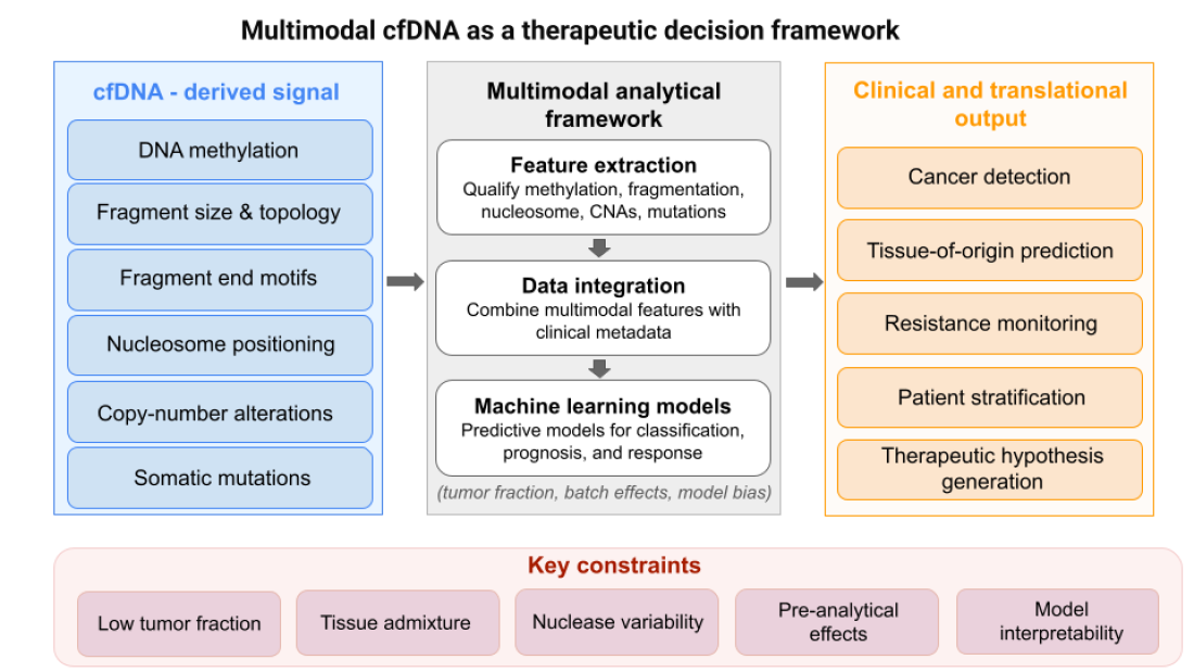
Multimodal cfDNA analysis arises from a fundamental constraint: no single feature class adequately captures the complexity of tumor-derived signals in plasma, particularly in low-tumor-fraction settings. Current frameworks therefore integrate partially independent signals—including DNA methylation, fragment size, fragment-end motifs, copy-number alterations, and somatic variants—within unified computational models [15,18,19,20]. In principle, this approach can improve analytical sensitivity when individual features are too weak or noisy to support stable inference on their own.
However, multimodal integration should not be assumed to confer greater biological insight or clinical utility. Combining analytes introduces additional technical variability, increases model complexity, and complicates attribution of model outputs to specific biological features. In practice, reported gains are often modest and highly dependent on preprocessing, cohort composition, and model training, raising the possibility that performance improvements reflect technical rather than biological integration [21].
The central translational question is therefore not whether additional cfDNA features can be combined, but whether multimodal models yield outputs that are reproducible, interpretable, and clinically actionable. At present, such advantages have not been consistently demonstrated in prospective or interventional settings, and remain limited by challenges in cross-cohort generalizability and lack of harmonized reference datasets [18,19,20,21,34,35].

3.2.2. Representative Multimodal Research Platforms

Several recent studies illustrate the analytical potential of multimodal cfDNA modeling. THEMIS demonstrated that enzyme-mediated methylome sequencing can preserve fragmentation information while simultaneously capturing whole-genome methylation, fragment size, fragment-end motifs, and copy-number alterations from a single cfDNA sample [15]. MESA extended this approach by incorporating chromatin-derived features, including nucleosome occupancy and window protection scores, supporting the view that plasma DNA retains structurally informative signatures of tumor chromatin organization [18]. Additional platforms, such as AlphaLiquid and SPOT-MAS, further suggest that integrated models combining methylation, copy number, and fragmentation may outperform single-feature approaches for cancer detection and tissue-of-origin prediction [19,20].
A conceptual extension of this work is that certain fragmentation-derived features may support functional interpretation rather than classification alone. For example, promoter fragmentation entropy and nucleosome-depleted region signals have been linked to inferred gene-expression states, raising the possibility that cfDNA may encode aspects of regulatory architecture in addition to tumor presence [14]. These observations are biologically informative, but most mechanistic applications remain investigational and require validation across independent datasets and clinical contexts.

3.2.3. Clinical Translation and Commercial Assays

Clinical translation of AI- and ML-based cfDNA analysis has advanced most clearly in cancer detection and tissue-of-origin classification. Its role in treatment selection or target nomination remains substantially less developed. Galleri, developed through the CCGA and PATHFINDER programs, is a prominent example of a methylation-based multi-cancer early detection assay that relies on machine-learning classification [34,35]. Fragmentomic approaches such as DELFI [13] further illustrate the potential of genome-wide fragmentation analysis for cancer detection, while also highlighting current limitations in sensitivity and specificity across clinical contexts.
In colorectal cancer screening, Shield and the PREEMPT CRC validation study further demonstrate that blood-based classification frameworks can be deployed in large prospective settings [38,39].
By contrast, multimodal platforms such as THEMIS and MESA remain primarily research-oriented. Their principal contribution has been to show that a single cfDNA dataset can encode multiple layers of biologically relevant information, including methylation, fragmentation, copy-number changes, and chromatin-related structure [15,18]. However, this analytical breadth has not yet translated into treatment-directing applications supported by comparable prospective validation.
Accordingly, the more meaningful distinction in the field is not simply between unimodal and multimodal assays, but between platforms optimized for clinically validated detection and those designed to extract richer biological information that may eventually support adaptive oncology. The latter remain conceptually important but largely investigational [15,18,34,35].

3.3. Mining Genomic and Epigenomic cfDNA Features for Adaptive Oncology

3.3.1. Resistance Monitoring Through Serial cfDNA Profiling

A core objective of adaptive oncology is to modify treatment according to the evolving molecular state of the tumor rather than relying solely on baseline tissue profiling. cfDNA is particularly well suited to this application because it enables repeated, minimally invasive monitoring of tumor-derived genomic and epigenomic signals over time [2,5,34]. This advantage has driven its clinical adoption.
Importantly, plasma cfDNA mutation analysis is no longer confined to exploratory use. In selected settings, clinical guidelines and disease-specific literature support its use when tumor tissue is unavailable, insufficient, or impractical to re-biopsy, consistent with current recommendations from the European Society for Medical Oncology [37]. This establishes a defined, context-dependent role for cfDNA in real-world molecular genotyping for certain cancers.
Its clearest clinical utility lies in the serial detection of resistance-associated alterations. Such changes may emerge before or near the time of overt progression and can therefore inform earlier therapeutic intervention than conventional imaging or repeat tissue biopsy alone [2,5]. Representative examples include ESR1 mutation monitoring in hormone receptor-positive metastatic breast cancer, prospectively demonstrated in PADA-1 [40]; plasma detection of EGFR T790M to guide osimertinib therapy in EGFR-mutant non-small cell lung cancer [41]; emergent KRAS-mediated resistance during anti-EGFR therapy in metastatic colorectal cancer [42]; and BRCA1/2 reversion mutations associated with resistance to platinum therapy or PARP inhibition [43].
These examples support the use of cfDNA as a practical readout of molecular escape mechanisms and, in selected contexts, as a tool for therapy switching or rational combination strategies. Nonetheless, the strength of evidence remains uneven across tumor types and therapeutic settings, and plasma findings do not uniformly obviate the need for tissue confirmation.

3.3.2. Copy-Number, Structural Variant, and Synthetic Lethality Inference

The contribution of cfDNA to adaptive oncology extends beyond single-nucleotide variants. Plasma sequencing can identify clinically relevant copy-number alterations, amplifications, structural rearrangements, gene fusions, and subclonal drivers that emerge during metastatic progression or treatment [2,44,45].
Examples include ERBB2 or MET amplification and oncogenic kinase rearrangements, which may refine patient stratification and uncover treatment opportunities not captured by limited genotyping panels [44,45]. cfDNA profiling may also contribute to synthetic-lethal inference. Detection of BRCA1/2 alterations remains relevant to PARP inhibitor selection, whereas BRCA1/2 reversion mutations can signal acquired resistance and loss of therapeutic vulnerability [43].
However, the broader translational value of structural and copy-number inference still depends on sufficient tumor fraction, assay sensitivity, analytical standardization, and the limited availability of prospective evidence that these findings improve treatment assignment beyond established mutation-focused workflows [2,43,44,45].

3.3.3. Epigenomic Inference

Epigenomic cfDNA profiling extends adaptive oncology beyond mutation tracking by attempting to capture tumor states not adequately explained by genotype alone [14,15,18,19,20]. Methylome- and fragmentome-derived features may, in principle, reflect pathway activation, tumor-suppressor silencing, lineage plasticity, epithelial-to-mesenchymal transition programs, and dependence on chromatin regulators. This is particularly relevant when resistance is driven by transcriptional reprogramming or lineage switching rather than newly acquired coding mutations [10,46,47].
Although the biological rationale is compelling, the translational significance of such inferences remains uncertain. Combined analyses of methylation, nucleosome organization, and fragmentation can reveal meaningful tumor states, but the extent to which these features can be converted into robust treatment-directing biomarkers has not yet been established [15,18,19,20,34,35].
A more plausible near-term role for epigenomic cfDNA lies in risk stratification and therapeutic hypothesis generation. In this context, cfDNA-derived patterns may help identify lineage switching, therapy-associated dedifferentiation, or chromatin-regulator dependence, which can then be integrated with orthogonal data to refine patient selection and guide follow-up investigation [14,15,18,46,47].

3.3.4. cfDNA as a Real-Time Therapeutic Control System

This treatment-directed perspective reframes liquid biopsy as more than a diagnostic assay and positions cfDNA as a potential real-time therapeutic monitoring system. Within this framework, methylation programs may suggest sensitivity to targeted or epigenetic therapies, chromatin accessibility signatures may inform immunotherapy responsiveness, and serial cfDNA measurements may reveal emerging resistant subclones or epigenetic state transitions before they become radiographically apparent [14,15,18,19,20,34].
The near-term translational objective is therefore not limited to disease detection, but extends to dynamic treatment optimization through repeated cfDNA-guided assessment. Whether this concept can be implemented reliably in routine clinical practice remains uncertain. Even so, the shift in perspective is important because it recasts cfDNA from a passive marker of disease burden into a potentially actionable tool for therapeutic control.
The principal cfDNA signal classes discussed in this review, together with the biology they may capture, their potential translational relevance, and their current evidentiary maturity, are summarized in Table 1.

3.3.5. Target Nomination

A more ambitious extension of the field is the proposition that cfDNA may contribute to target discovery. Framed cautiously, this is plausible. cfDNA can reveal tumor states associated with actionable dependencies, including pathway activation, lineage switching, enhancer engagement, and transcription factor programs not evident from mutation analysis alone [12,14,24,25].
One way to implement this framework is to integrate cfDNA-derived regulatory signals with functional dependency datasets. Enhancer activity and chromatin accessibility inferred from cfDNA can be mapped to candidate genes and pathways and then intersected with CRISPR-based dependency maps, such as the Cancer Dependency Map, to identify genes that are both transcriptionally active and functionally essential for tumor survival. In this framework, cfDNA provides a minimally invasive readout of tumor regulatory state, while orthogonal functional screens support prioritization of candidate targets grounded in both regulatory activity and dependency.
However, cfDNA is better regarded as a prioritization tool rather than a stand-alone discovery platform. Its value lies in narrowing the search space by identifying candidate vulnerabilities that can then be evaluated through integration with CRISPR dependency maps, tumor profiling, and functional validation.

3.4. Future Directions: From Multimodal Biomarkers to Therapeutic Decision Engines

The translational landscape of cfDNA currently spans a spectrum from clinically established mutation-based applications to research-stage efforts aimed at functional inference and therapeutic guidance. Most target-related uses remain provisional and continue to require orthogonal validation, as summarized in Table 1.

3.4.1. Foundation-Style Multimodal Models

The next generation of cfDNA models is increasingly being developed to be multimodal by design, integrating methylation, fragmentomics, copy number, mutations, and clinical variables within unified analytical frameworks [15,18,19,20]. The objective is not simply to improve binary classification, but to align complementary biological signals with clinically relevant outputs spanning diagnosis, prognosis, resistance emergence, and likely treatment sensitivity [15,18].
A logical extension of this trend is the development of foundation-style models trained on large cfDNA datasets. In principle, such systems could generate integrated predictions across several clinically relevant dimensions rather than a single narrow endpoint. However, their value will depend not only on predictive performance, but also on interpretability, biological robustness, and prospective validation.
If such frameworks mature, they may support patient stratification, improve pharmacodynamic monitoring, enrich biomarker-driven trials, and help prioritize rational combination strategies [19,20,34,35]. At present, however, these applications remain aspirational rather than established.

3.4.2. Integration into Biomarker-Driven Clinical Trials

An important next step is the integration of genomic and epigenomic cfDNA features into biomarker-driven clinical trials [19,20,34,35,38,39]. These readouts could improve patient selection, enable earlier identification of likely responders or non-responders, and support the use of molecular residual disease or early plasma ctDNA response as intermediate endpoints.
If validated prospectively, this approach could make trials smaller, faster, and more biologically focused while improving alignment between therapy mechanism and enrolled population [34,35,38,39]. However, its utility will depend on assay reproducibility, interpretability, and evidence that cfDNA-based readouts improve trial decisions rather than merely add correlates.

3.5. Limitations and Pitfalls of cfDNA Analysis

Despite its promise, cfDNA analysis remains subject to substantial biological, technical, and analytical constraints that limit its clinical translation.

3.5.1. Preanalytical Variability and Fragmentation Sensitivity

Confounding and batch effects further complicate interpretation. Higher-order cfDNA features, particularly fragment-end motifs and fragmentation profiles, are highly sensitive to pre-analytical variables such as delays in blood processing, plasma separation, storage conditions, and freeze–thaw cycles. These factors can alter apparent nuclease signatures and introduce systematic biases independent of tumor biology [2,16,17]. Such effects reflect nuclease-driven variability and are especially relevant for fragmentomic analyses, where signal interpretation depends on subtle differences in fragmentation patterns. These observations underscore the importance of standardized collection and processing protocols, including rapid plasma separation, controlled storage conditions, and harmonized library preparation workflows, to ensure reproducibility across studies and clinical settings.

3.5.2. Biological and Clinical Confounders

cfDNA reflects contributions from multiple tissues, and its composition is influenced by physiological and pathological conditions. Clinical variables such as age, liver function, systemic inflammation, and tissue turnover can affect cfDNA abundance and fragmentation patterns, introducing background variation that may overlap with tumor-derived signals [2,29]. In this context, tissue admixture and hematopoietic background represent major constraints, particularly for fragmentomic and epigenomic analyses.

3.5.3. Clonal Hematopoiesis

Clonal hematopoiesis of indeterminate potential (CHIP) represents a well-recognized source of non-tumor-derived mutations in plasma. Somatic variants arising from hematopoietic clones can be misinterpreted as tumor-derived, leading to false-positive findings in mutation-based analyses [2,43]. This further highlights the contribution of hematopoietic background signals to cfDNA measurements. While sequencing of matched leukocyte DNA can mitigate this issue, it is not uniformly implemented across cfDNA workflows.

3.5.4. Analytical and Cohort-Level Variability

Cases and controls may differ not only in cancer status, but also in collection site, preanalytical handling, sequencing chemistry, and clinical characteristics. As a result, classifiers may capture technical or demographic artifacts rather than tumor biology. These issues are particularly pronounced in retrospective and multicenter datasets lacking harmonization and independent validation [21,46,47,48]. In addition, differences in sequencing platforms and computational pipelines further limit comparability across studies.

3.5.5. Model Reproducibility and Clinical Translation

Machine learning models trained on cfDNA data often show reduced performance when applied to independent cohorts, reflecting overfitting, cohort-specific biases, and lack of standardization. Consequently, cross-cohort generalization remains a major limitation. More broadly, prospective evidence demonstrating that AI-guided cfDNA strategies improve clinical outcomes remains limited, and the gap between analytical performance and treatment-directed utility persists [21,34,35].
In addition, risks of data leakage—where information from training datasets is inadvertently incorporated into model evaluation—can lead to overly optimistic performance estimates. This issue is particularly relevant in high-dimensional cfDNA analyses and underscores the need for rigorous validation using independent, prospectively collected cohorts.

3.5.6. Technological, Regulatory, and Cost Considerations

Beyond biological and analytical constraints, practical barriers to clinical implementation remain substantial. High-sensitivity cfDNA assays often require deep sequencing and complex library preparation workflows, which increase cost and limit scalability in routine clinical settings [2,35]. In addition, multimodal analyses generate high-dimensional datasets that require specialized bioinformatics infrastructure and expertise, which may not be uniformly available across clinical laboratories [21].
Regulatory requirements further constrain clinical adoption. Assays intended for clinical use must meet rigorous standards for analytical validity, clinical validity, and clinical utility, as defined by regulatory agencies such as the U.S. Food and Drug Administration and the European Medicines Agency. Demonstrating clinical utility is particularly challenging for multimodal and AI-based approaches, where model interpretability and reproducibility remain active areas of investigation [21,34].
Finally, prospective clinical validation remains limited. Many cfDNA studies are retrospective or case–control in design, and few have demonstrated improved patient outcomes in interventional settings. As a result, integration of cfDNA-based multimodal frameworks into clinical decision-making, particularly in the context of adaptive oncology, will require carefully designed prospective trials and standardized evaluation criteria [34,35].

3.6. Conclusions

At present, the strongest clinical validation of cfDNA continues to be in detection and classification. The more consequential long-term opportunity lies in adaptive oncology, where genomic and epigenomic cfDNA features may eventually support resistance monitoring, patient stratification, and treatment redirection [18,21].
Future progress in this area will depend on prospective interventional studies demonstrating clinical utility, as well as the harmonization of preanalytical and analytical workflows to ensure reproducibility across cohorts and clinical settings.

Funding

This research received no external funding.

Institutional Review Board Statement

Not applicable.

Informed Consent Statement

Not applicable.

Data Availability Statement

No new data were created or analyzed in this study. Data sharing is not applicable to this article.

Acknowledgments

AI-assisted tools were used exclusively for language editing and formatting. The author takes full responsibility for the content and integrity of the manuscript.

Conflicts of Interest

The author declares no conflicts of interest.

References

  1. Pantel, K.; Alix-Panabières, C. Liquid biopsy and minimal residual disease—Latest advances and implications for cure. Nat. Rev. Clin. Oncol. 2019, 16, 409–424. [Google Scholar] [CrossRef]
  2. Wan, J.C.M.; Massie, C.; Garcia-Corbacho, J.; Mouliere, F.; Brenton, J.D.; Caldas, C.; Pacey, S.; Baird, R.; Rosenfeld, N. Liquid biopsies come of age: Towards implementation of circulating tumour DNA. Nat. Rev. Cancer 2017, 17, 223–238. [Google Scholar] [CrossRef]
  3. Bettegowda, C.; Sausen, M.; Leary, R.J.; Kinde, I.; Wang, Y.; Agrawal, N.; Bartlett, B.R.; Wang, H.; Luber, B.; Alani, R.M.; et al. Detection of circulating tumor DNA in early- and late-stage human malignancies. Sci. Transl. Med. 2014, 6, 224ra24. [Google Scholar] [CrossRef]
  4. Diehl, F.; Schmidt, K.; Choti, M.A.; Romans, K.; Goodman, S.; Li, M.; Thornton, K.; Agrawal, N.; Sokoll, L.; Szabo, S.A.; et al. Circulating mutant DNA to assess tumor dynamics. Nat. Med. 2008, 14, 985–990. [Google Scholar] [CrossRef]
  5. Abbosh, C.; Birkbak, N.J.; Wilson, G.A.; Jamal-Hanjani, M.; Constantin, T.; Salari, R.; Le Quesne, J.; Moore, D.A.; Veeriah, S.; Rosenthal, R.; et al. Phylogenetic ctDNA analysis depicts early-stage lung cancer evolution. Nature 2017, 545, 446–451. [Google Scholar] [CrossRef]
  6. Cohen, J.D.; Li, L.; Wang, Y.; Thoburn, C.; Afsari, B.; Danilova, L.; Douville, C.; Javed, A.A.; Wong, F.; Mattox, A.; et al. Detection and localization of surgically resectable cancers with a multi-analyte blood test. Science 2018, 359, 926–930. [Google Scholar] [CrossRef] [PubMed]
  7. Newman, A.M.; Lovejoy, A.F.; Klass, D.M.; Kurtz, D.M.; Chabon, J.J.; Scherer, F.; Stehr, H.; Liu, C.L.; Bratman, S.V.; Say, C.; et al. Integrated digital error suppression for improved detection of circulating tumor DNA. Nat. Biotechnol. 2016, 34, 547–555. [Google Scholar] [CrossRef] [PubMed]
  8. Schmitt, M.W.; Kennedy, S.R.; Salk, J.J.; Fox, E.J.; Hiatt, J.B.; Loeb, L.A. Detection of ultra-rare mutations by next-generation sequencing. Proc. Natl. Acad. Sci. USA 2012, 109, 14508–14513. [Google Scholar] [CrossRef]
  9. Guo, L.; Lee, Y.T.; Zhou, Y.; Huang, Y. Targeting epigenetic regulatory machinery to overcome cancer therapy resistance. Semin. Cancer Biol. 2022, 83, 487–502. [Google Scholar] [CrossRef]
  10. Davies, A.; Zoubeidi, A.; Beltran, H.; Selth, L.A. The transcriptional and epigenetic landscape of cancer cell lineage plasticity. Cancer Discov. 2023, 13, 1771–1788. [Google Scholar] [CrossRef]
  11. Snyder, M.W.; Kircher, M.; Hill, A.J.; Daza, R.M.; Shendure, J. Cell-free DNA comprises an in vivo nucleosome footprint that informs its tissues-of-origin. Cell 2016, 164, 57–68. [Google Scholar] [CrossRef]
  12. Ulz, P.; Thallinger, G.G.; Auer, M.; Graf, R.; Kashofer, K.; Jahn, S.W.; Abete, L.; Pristauz, G.; Petru, E.; Geigl, J.B.; et al. Inferring expressed genes by whole-genome sequencing of plasma DNA. Nat. Genet. 2016, 48, 1273–1278. [Google Scholar] [CrossRef]
  13. Cristiano, S.; Leal, A.; Phallen, J.; Fiksel, J.; Adleff, V.; Bruhm, D.C.; Østrup Jensen, S.; Medina, J.E.; Hruban, C.; White, J.R.; et al. Genome-wide cell-free DNA fragmentation in patients with cancer. Nature 2019, 570, 385–389. [Google Scholar] [CrossRef]
  14. Esfahani, M.S.; Hamilton, E.G.; Mehrmohamadi, M.; Nabet, B.Y.; Alig, S.K.; King, D.A.; Steen, C.B.; Macaulay, C.W.; Schultz, A.; Nesselbush, M.C.; et al. Inferring gene expression from cell-free DNA fragmentation profiles. Nat. Biotechnol. 2022, 40, 585–597. [Google Scholar] [CrossRef] [PubMed]
  15. Bie, F.; Wang, Z.; Li, Y.; Guo, W.; Hong, Y.; Han, T.; Lv, F.; Yang, S.; Li, S.; Li, X.; et al. Multimodal analysis of cell-free DNA whole-methylome sequencing for cancer detection and localization. Nat. Commun. 2023, 14, 6042. [Google Scholar] [CrossRef]
  16. Serpas, L.; Chan, R.W.Y.; Jiang, P.; Ni, M.; Sun, K.; Rashidfarrokhi, A.; Soni, C.; Sisirak, V.; Lee, W.; Cheng, S.H.; et al. Dnase1l3 deletion causes aberrations in length and end-motif frequencies in plasma DNA. Proc. Natl. Acad. Sci. USA 2019, 116, 641–649. [Google Scholar] [CrossRef]
  17. Zhou, Z.; Ma, M.-J.L.; Chan, R.W.Y.; Lam, W.K.J.; Peng, W.; Gai, W.; Hu, X.; Ding, S.C.; Ji, L.; Zhou, Q.; et al. Fragmentation landscape of cell-free DNA revealed by deconvolution of end motifs. Proc. Natl. Acad. Sci. USA 2023, 120, e2220982120. [Google Scholar] [CrossRef]
  18. Li, Y.; Xu, J.; Chen, C.; Lu, Z.; Wan, D.; Li, D.; Li, J.S.; Sorg, A.J.; Roberts, C.C.; Mahajan, S.; et al. Multimodal epigenetic sequencing analysis (MESA) of cell-free DNA for non-invasive colorectal cancer detection. Genome Med. 2024, 16, 9. [Google Scholar] [CrossRef] [PubMed]
  19. Kim, S.Y.; Jeong, S.; Lee, W.; Jeon, Y.; Kim, Y.J.; Park, S.; Lee, D.; Go, D.; Song, S.-H.; Lee, S.; et al. Cancer signature ensemble integrating cfDNA methylation, copy number, and fragmentation facilitates multi-cancer early detection. Exp. Mol. Med. 2023, 55, 2445–2460. [Google Scholar] [CrossRef] [PubMed]
  20. Pham, T.M.Q.; Phan, T.H.; Jasmine, T.X.; Tran, T.T.T.; Huynh, L.A.K.; Vo, T.L.; Nai, T.H.T.; Tran, T.T.; Truong, M.H.; Tran, N.C.; et al. Multimodal analysis of genome-wide methylation, copy number aberrations, and end motif signatures enhances detection of early-stage breast cancer. Front. Oncol. 2023, 13, 1127086. [Google Scholar] [CrossRef]
  21. Luo, X.; Xie, S.; Hong, F.; Li, X.; Wei, Y.; Zhou, Y.; Su, W.; Yang, Y.; Tang, L.; Dao, F.; et al. From multi-omics to deep learning: Advances in cfDNA-based liquid biopsy for multi-cancer screening. Biomark. Res. 2025, 14, 3. [Google Scholar] [CrossRef] [PubMed]
  22. Tsui, W.H.A.; Ding, S.C.; Jiang, P.; Lo, Y.M.D. Artificial intelligence and machine learning in cell-free-DNA-based diagnostics. Genome Res. 2025, 35, 1–19. [Google Scholar] [CrossRef] [PubMed]
  23. Wang, H.Y.; Lin, W.Y.; Zhou, C.; Yang, Z.A.; Kalpana, S.; Lebowitz, M.S. Integrating artificial intelligence for advancing multiple-cancer early detection via serum biomarkers: A narrative review. Cancers 2024, 16, 862. [Google Scholar] [CrossRef]
  24. Lai, K.; Dilger, K.; Cunningham, R.; Lam, K.T.; Boquiren, R.; Truong, K.; Louie, M.C.; Rava, R.; Abdueva, D. Extracting regulatory active chromatin footprint from cell-free DNA. Commun. Biol. 2024, 7, 1086. [Google Scholar] [CrossRef] [PubMed]
  25. Wang, Y.; Hou, Y.; Xiong, Z.; Lu, L.; Zong, Z.; Wang, B.; Chen, H.; Zhang, W.; Zhou, X. Fragment dispersity index analysis of cfDNA fragments reveals chromatin accessibility and enables early cancer detection. Cell Rep. Methods 2025, 5, 101083. [Google Scholar] [CrossRef]
  26. Shen, H.; Yang, M.; Liu, J.; Chen, K.; Li, X. Development of a deep learning model for cancer diagnosis by inspecting cell-free DNA end-motifs. npj Precis. Oncol. 2024, 8, 160. [Google Scholar] [CrossRef]
  27. Qi, T.; Pan, M.; Shi, H.; Wang, L.; Bai, Y.; Ge, Q. Cell-free DNA fragmentomics: The novel promising biomarker in cancer screening. Int. J. Mol. Sci. 2023, 24, 1503. [Google Scholar] [CrossRef]
  28. Yu, S.C.Y.; Deng, J.; Qiao, R.; Cheng, S.H.; Peng, W.; Lau, S.L.; Choy, L.Y.L.; Leung, T.Y.; Wong, J.; Wong, V.W.-S.; et al. Comparison of single molecule, real-time sequencing and nanopore sequencing for analysis of the size, end-motif, and tissue-of-origin of long cell-free DNA in plasma. Clin. Chem. 2023, 69, 168–179. [Google Scholar] [CrossRef]
  29. Thierry, A.R.; El Messaoudi, S.; Gahan, P.B.; Anker, P.; Stroun, M. Origins, structures, and functions of circulating DNA in oncology. Cancer Metastasis Rev. 2016, 35, 347–376. [Google Scholar] [CrossRef]
  30. Wang, H.; Kim, S.J.; Lei, Y.; Wang, S.; Wang, H.; Huang, H.; Zhang, H.; Tsung, A. Neutrophil extracellular traps in homeostasis and disease. Signal Transduct. Target. Ther. 2024, 9, 235. [Google Scholar] [CrossRef]
  31. Zhou, Y.; Xu, Z.; Liu, Z. Impact of neutrophil extracellular traps on thrombosis formation: New findings and future perspective. Front. Cell Infect. Microbiol. 2022, 12, 910908. [Google Scholar] [CrossRef]
  32. García-Silva, S.; Gallardo, M.; Peinado, H. DNA-Loaded Extracellular Vesicles in Liquid Biopsy: Tiny Players With Big Potential? Front. Cell Dev. Biol. 2021, 8, 622579. [Google Scholar] [CrossRef]
  33. Lin, B.; Jiao, Z.; Dong, S.; Yan, W.; Jiang, J.; Du, Y.; Weng, X.; Wang, H.; Hu, Z.; Liu, Y.; et al. Whole-genome methylation profiling of extracellular vesicle DNA in gastric cancer identifies intercellular communication features. Nat. Commun. 2025, 16, 8084. [Google Scholar] [CrossRef]
  34. Pantel, K.; Alix-Panabières, C. Minimal residual disease as a target for liquid biopsy in patients with solid tumours. Nat. Rev. Clin. Oncol. 2025, 22, 65–77. [Google Scholar] [CrossRef]
  35. Vittone, J.; Gill, D.; Goldsmith, A.; Klein, E.A.; Karlitz, J.J. A multi-cancer early detection blood test using machine learning detects early-stage cancers lacking USPSTF-recommended screening. npj Precis. Oncol. 2024, 8, 91. [Google Scholar] [CrossRef]
  36. Battaglin, F.; Lenz, H.J. Clinical Applications of Circulating Tumor DNA Profiling in GI Cancers. JCO Oncol. Pract. 2024, 20, 1481–1490. [Google Scholar] [CrossRef]
  37. Pascual, J.; Attard, G.; Bidard, F.C.; Curigliano, G.; De Mattos-Arruda, L.; Diehn, M.; Italiano, A.; Lindberg, J.; Merker, J.D.; Montagut, C.; et al. ESMO recommendations on the use of circulating tumour DNA assays for patients with cancer: A report from the ESMO Precision Medicine Working Group. Ann. Oncol. 2022, 33, 750–768. [Google Scholar] [CrossRef]
  38. Chung, D.C.; Gray, D.M.; Singh, H.; Issaka, R.B.; Raymond, V.M.; Eagle, C.; Hu, S.; Chudova, D.I.; Talasaz, A.; Greenson, J.K. A Cell-free DNA Blood-Based Test for Colorectal Cancer Screening. N. Engl. J. Med. 2024, 390, 973–983. [Google Scholar] [CrossRef] [PubMed]
  39. Shaukat, A.; Burke, C.A.; Chan, A.T.; Grady, W.M.; Gupta, S.; Katona, B.W.; Ladabaum, U.; Liang, P.S.; Liu, J.J.; Putcha, G.; et al. Clinical Validation of a Circulating Tumor DNA-Based Blood Test to Screen for Colorectal Cancer. JAMA 2025, 334, 56–63. [Google Scholar] [CrossRef] [PubMed]
  40. Bidard, F.-C.; Hardy-Bessard, A.-C.; Dalenc, F.; Bachelot, T.; Pierga, J.-Y.; de la Motte Rouge, T.; Sabatier, R.; Dubot, C.; Frenel, J.-S.; Ferrero, J.M.; et al. Switch to fulvestrant and palbociclib versus no switch in advanced breast cancer with rising ESR1 mutation during aromatase inhibitor and palbociclib therapy (PADA-1): A randomised, open-label, multicentre, phase 3 trial. Lancet Oncol. 2022, 23, 1367–1377. [Google Scholar] [CrossRef]
  41. Oxnard, G.R.; Thress, K.S.; Alden, R.S.; Lawrance, R.; Paweletz, C.P.; Cantarini, M.; Yang, J.C.-H.; Barrett, J.C.; Jänne, P.A. Association between plasma genotyping and outcomes of treatment with osimertinib (AZD9291) in advanced non-small-cell lung cancer. J. Clin. Oncol. 2016, 34, 3375–3382. [Google Scholar] [CrossRef]
  42. Parseghian, C.M.; Sun, R.; Woods, M.; Napolitano, S.; Lee, H.M.; Alshenaifi, J.; Willis, J.; Nunez, S.; Raghav, K.P.; Morris, V.K.; et al. Resistance Mechanisms to Anti–Epidermal Growth Factor Receptor Therapy in RAS/RAF Wild-Type Colorectal Cancer Vary by Regimen and Line of Therapy. J. Clin. Oncol. 2023, 41, 460–471. [Google Scholar] [CrossRef] [PubMed]
  43. Weigelt, B.; Comino-Méndez, I.; de Bruijn, I.; Tian, L.; Meisel, J.L.; García-Murillas, I.; Fribbens, C.; Cutts, R.; Martelotto, L.G.; Ng, C.K.Y.; et al. Diverse BRCA1 and BRCA2 reversion mutations in circulating cell-free DNA of therapy-resistant breast or ovarian cancer. Clin. Cancer Res. 2017, 23, 6708–6720. [Google Scholar] [CrossRef] [PubMed]
  44. Jang, H.; Jeong, M.-K. Copy Number Alteration Profiling from Plasma cfDNA WES in Advanced NSCLC. Int. J. Mol. Sci. 2025, 26, 11111. [Google Scholar] [CrossRef]
  45. Hasegawa, N.; Kohsaka, S.; Kurokawa, K.; Shinno, Y.; Takeda Nakamura, I.; Ueno, T.; Kojima, S.; Kawazu, M.; Suehara, Y.; Ishijima, M.; et al. Highly sensitive fusion detection using plasma cell-free RNA in non-small-cell lung cancers. Cancer Sci. 2021, 112, 4393–4403. [Google Scholar] [CrossRef]
  46. Jamshidi, A.; Liu, M.C.; Klein, E.A.; Venn, O.; Hubbell, E.; Beausang, J.F.; Gross, S.; Melton, C.; Fields, A.P.; Liu, Q.; et al. Evaluation of cell-free DNA approaches for multi-cancer early detection. Cancer Cell 2022, 40, 1537–1549.e12. [Google Scholar] [CrossRef]
  47. Zhang, K.; Fu, R.; Liu, R.; Su, Z. Circulating cell-free DNA-based multi-cancer early detection. Trends Cancer 2024, 10, 161–174. [Google Scholar] [CrossRef]
  48. De Falco, A.; Grisolia, P.; Giuffrida, R.; Iannarone, C.; Graziano, C.; Scrima, M.; Cossu, A.M.; Tufano, R.; Yow, M.V.; Brown, C.M.; et al. Knowledge-informed multimodal cfDNA analysis improves sensitivity and generalization in cancer detection. bioRxiv 2025. [Google Scholar] [CrossRef]

Table 1. cfDNA feature classes, inferred biology, and translational relevance for adaptive oncology and therapeutic hypothesis generation.
cfDNA Feature ClassInferred Biological InformationPotential Translational ApplicationsCurrent Evidence MaturityMajor LimitationsReferences
Somatic mutationsTumor genotype, emerging resistance alterations, clonal evolutionTherapy selection, serial resistance monitoring, measurable residual disease assessmentClinically established in selected settingsLimited by low tumor fraction and reduced sensitivity at low variant allele frequency; confounded by clonal hematopoiesis; does not capture non-mutational resistance mechanisms such as epigenetic reprogramming or lineage plasticity[2,5,7,34,36,37,40,41,42,43]
Copy-number alterations and amplificationsStructural instability, focal amplification events, subclonal genomic evolutionPatient stratification, resistance profiling, context-dependent inference of pathway activationClinically relevant but less standardized than mutation testingLimited by tumor fraction-dependent signal dilution and admixture with background cfDNA, leading to noisy plasma CNA profiles; constrained in detecting subclonal events; prospective evidence for treatment direction remains incompletely established[2,43,44]
Structural variants and gene fusionsRearrangements, oncogenic fusions, and other structural driversDetection of actionable rearrangements, resistance profiling, complementary genotyping when tissue is limitedUseful in selected contextsLimited by assay design and breakpoint coverage; constrained by low tumor fraction and cfDNA fragmentation, reducing recovery of junction-spanning reads; structural resolution in plasma may remain incomplete[2,41,45]
Fragment size, fragment ends, and fragmentation topologyNuclease activity, chromatin accessibility, tumor-associated fragmentation patternsCancer detection, tissue-of-origin modeling, exploratory biological inferenceStrong analytical evidence, but limited routine clinical decision utilityHighly sensitive to preanalytical variability and nuclease activity (e.g., DNASE1L3-dependent fragmentation patterns); limited by tumor fraction-dependent signal dilution, reducing resolution of tumor-specific nucleosome phasing; fragmentation signatures may reflect mixed cellular contributions, complicating biological interpretation[13,16,17,21,22,27]
Nucleosome positioning and promoter fragmentation featuresChromatin organization, nucleosome depletion, inferred transcriptional activityFunctional-state inference, pathway nomination, exploratory therapeutic hypothesis generationResearch stageLimited by dependence on aggregate fragmentation patterns; affected by low tumor fraction and hematopoietic cfDNA background; inference of transcriptional activity relies on computational modeling and lacks standardized validation[11,12,14,24,25]
DNA methylation patternsLineage identity, tissue of origin, tumor class, silencing states, epigenomic reprogrammingMulti-cancer early detection, classification, risk stratification, exploratory therapeutic redirectionClinically advanced for detection and classification; investigational for treatment directionLimited by admixture of tumor and non-tumor cfDNA requiring robust deconvolution; dependent on reference methylation atlases; epigenomic signals are not consistently actionable; treatment-direction evidence remains incompletely established[15,18,19,20,34,35,38,39]
Multimodal integrated cfDNA modelsComposite tumor state derived from combined methylation, fragmentation, CNA, and mutational signalsImproved classification, adaptive-oncology modeling, exploratory target nomination frameworksPromising, but not yet treatment-directing in routine practiceLimited by high-dimensional feature space with risk of overfitting and data leakage; affected by cohort-specific biases and reduced cross-cohort generalization; integration complicates feature attribution and biological interpretability[15,18,19,20,21,46,47,48]
This table summarizes the major categories of cfDNA-derived signals discussed in this review, the biology they may capture, their potential clinical or drug-discovery relevance, and their current evidentiary maturity.
Disclaimer/Publisher’s Note: The statements, opinions and data contained in all publications are solely those of the individual author(s) and contributor(s) and not of MDPI and/or the editor(s). MDPI and/or the editor(s) disclaim responsibility for any injury to people or property resulting from any ideas, methods, instructions or products referred to in the content.