Review

The Two Faces of Saccharomyces cerevisiae RAD9 Function in Homologous Recombination: Suppressor and Promoter of Genome Instability


Department of Nanoscale Science and Engineering, State University of New York at Albany, 257 Fuller Road, Albany, NY 12203, USA
Submission received: 7 December 2025 / Revised: 9 February 2026 / Accepted: 4 March 2026 / Published: 9 April 2026

Abstract

Recombinogenic DNA damage can initiate chromosomal rearrangements that can alter gene expression or accelerate cancer progression in higher eukaryotes. Thus, there is a critical need to identify genes that suppress chromosomal rearrangements and environmental exposures that promote genetic instability. Cell cycle checkpoints modulate the cell cycle so that DNA repair occurs before the replication or segregation of damaged chromosomes. Saccharomyces cerevisiae (budding yeast) RAD9 was the first cell cycle checkpoint gene identified, which initiated intensive research studies into the mechanisms of checkpoint activation and the phenotypes of checkpoint mutants. The budding yeast Rad9 protein serves as both an adaptor and scaffold that facilitates downstream effector activation to orchestrate a DNA damage response at multiple stages of the cell cycle, which facilitates double-strand break (DSB) repair by sister chromatid recombination. However, the role of RAD9 in homologous recombination and in suppressing gross chromosomal rearrangements (GCRs) is not completely understood. In this review we discuss how RAD9 can promote genome instability resulting from aberrant DNA replication intermediates, while suppressing DSB-associated rearrangements. We also discuss possible mechanisms accounting for the synergistic increase in genomic instability in double mutants defective in both RAD9 and recombinational repair. We emphasize that while there is an overlap between checkpoint and recombinational repair pathways, RAD9 and checkpoint pathways can function independently to suppress chromosomal instability. These studies thus elucidate checkpoint mechanisms that control homologous recombination between repeated sequences.

1. Introduction

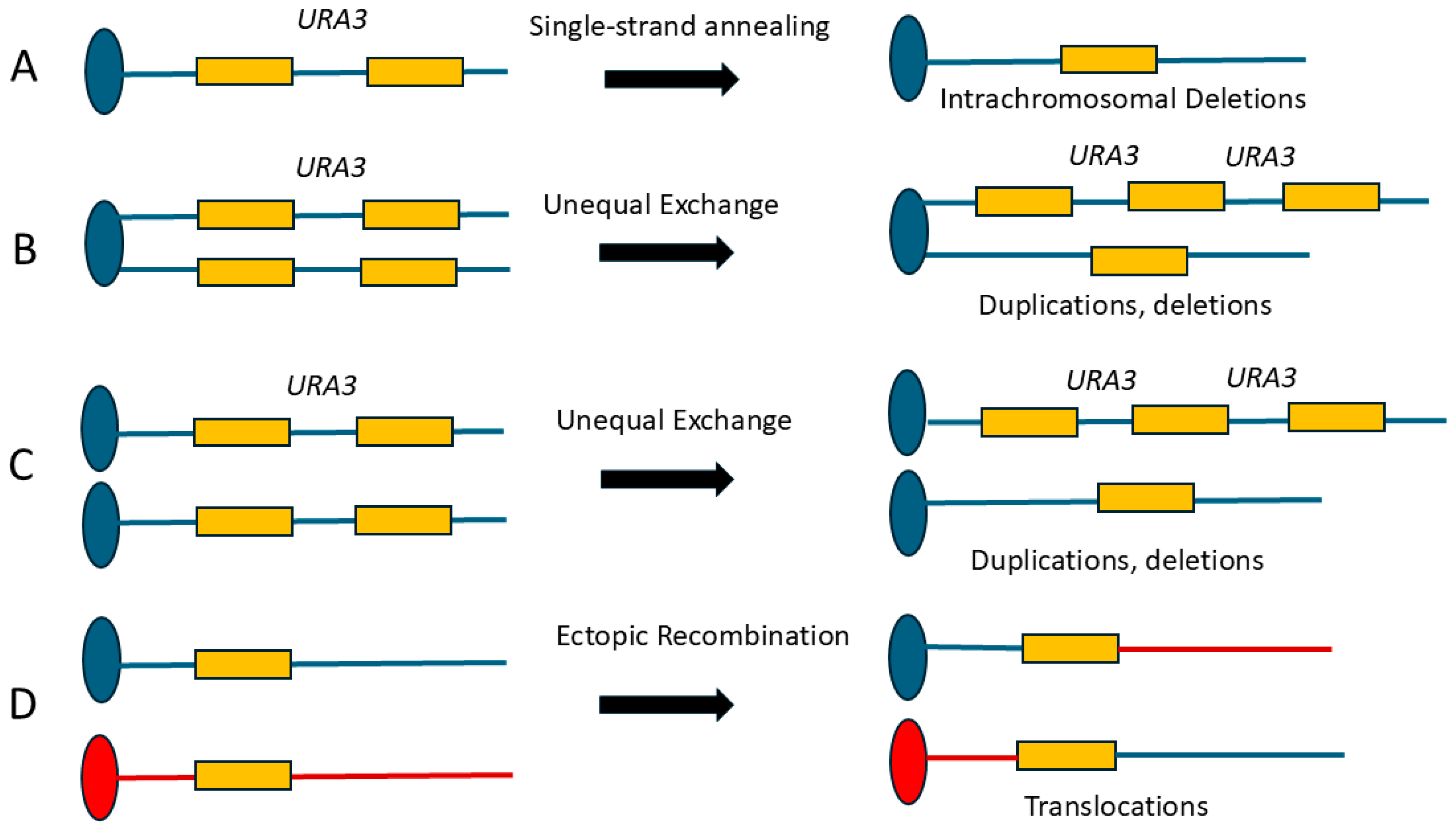
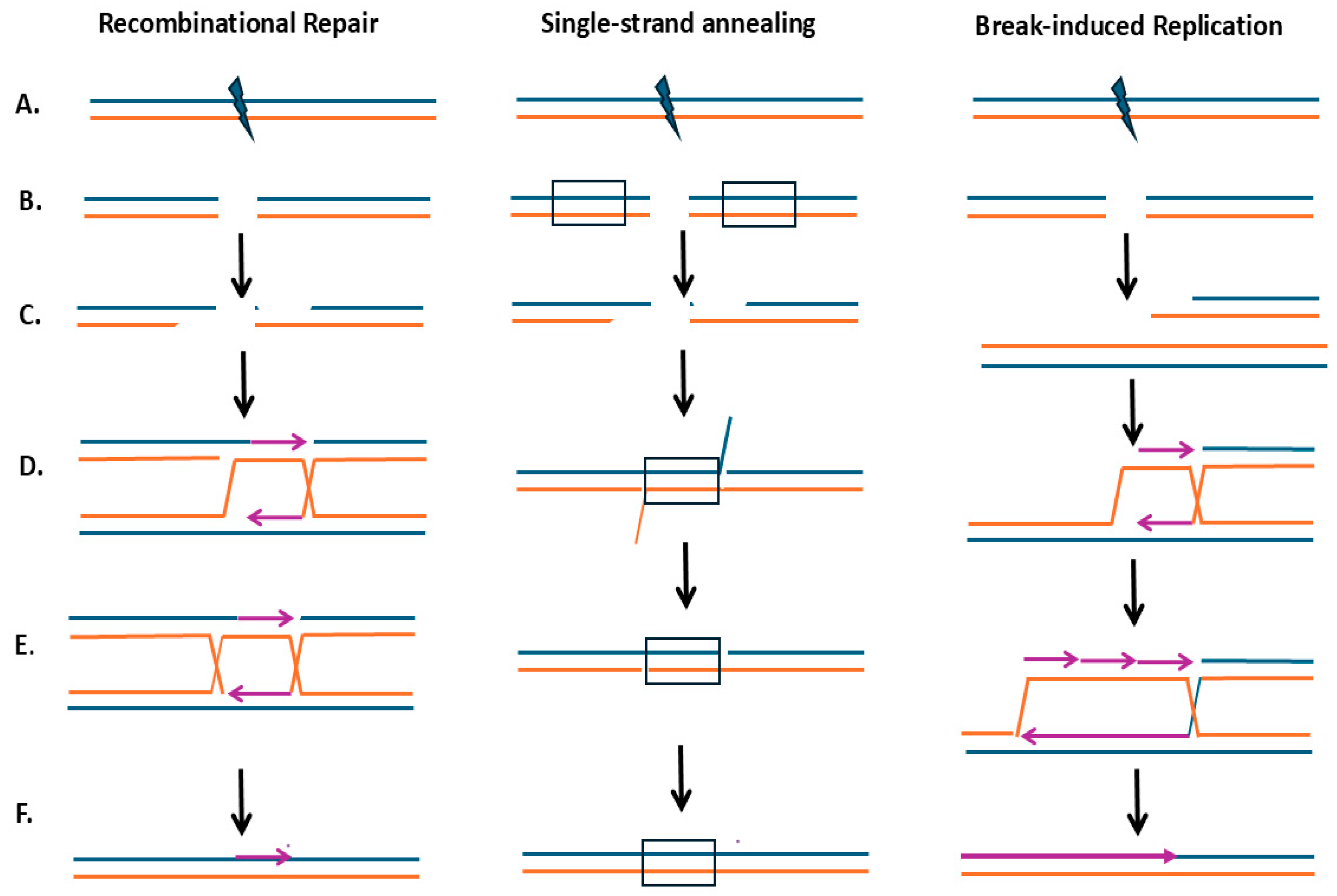
Homologous recombination (HR) is an evolutionarily conserved mechanism by which genetic information is exchanged between similar DNA sequences. Double-strand breaks (DSBs) are efficient initiators of HR, and HR pathways for DSB repair (Figure 1) include gap repair mechanisms, single-strand annealing (SSA), and break-induced replication (BIR) [1]. Additional HR pathways can be stimulated by endogenous and exogenous DNA damage and serve in multiple cellular pathways to maintain genome stability, including error-free template-switching and DNA replication maintenance, reviewed in [2]. However, repetitive DNA, which is ubiquitous in eukaryotic organisms, can also serve as HR substrates, potentially generating chromosomal rearrangements (Figure 2). Examples include deletions and inversions generated by intrachromatid recombination between direct or indirect repeats, respectively. In addition, unequal recombination between sister chromatids or homologs can generate duplications and deletions, while recombination between repeats on non-homologous chromosomes can generate translocations (Figure 2). Thus, HR initiated by DNA lesions, such as DSBs, can reshape the genome via recombination between repeated sequences [3,4].
Because sister chromatids are essentially identical, they are ideal substrates for recombinational repair since equal sister chromatid recombination (SCR) does not generate genetic changes. In Saccharomyces cerevisiae (budding yeast), the higher radiation resistance of G2 diploid cells compared to G1 diploid cells [5,6], and the significant role of HR in DSB repair, supports the assertion that sister chromatids are the preferred substrates for recombinational repair [6]. Since sister chromatids are transient in the cell cycle, mechanisms have evolved to delay or arrest the cell cycle before sister chromatid segregation, providing time for HR-mediated DNA repair. However, DNA replication blocks in the S phase can generate toxic recombination intermediates and BIR can also generate genome rearrangements [7]. In this review article, we discuss HR pathways that function in G2/M which enhance genome stability, and HR pathways that function in the S phase which enhance genetic instability. We discuss RAD9’s functions in facilitating HR pathways including checkpoint activation, DNA end resection, and inhibition of helicases.
Hartwell and Weinert [8] coined cell cycle checkpoints as “control mechanisms that enforce dependency in the cell cycle.” RAD9 [9] is the first identified cell cycle checkpoint gene. Cell cycle checkpoints arrest or delay the cell cycle so that DNA repair is completed before DNA lesions are replicated or segregated to the next cell cycle. DNA damage signals include DSBs [10,11,12], stalled replication forks [13], unresolved Holliday structures [14], ssDNA at telomeres [15,16], fragile DNA sites [17], and unresolved DNA replication intermediates, such as 5′ single-strand flaps [18] and trapped topoisomerase-DNA structures [19,20]. Checkpoint function is particularly important in DSB repair, where the persistence of a single DSB can confer lethality [4,21]. As a consequence of G2/M checkpoint inactivation, irradiated rad9 cells form microcolonies on agar plates. Such microcolonies accumulate inviable cells due to mis-segregation and the loss of chromosomal fragments [22]. Arresting rad9 mutants in G2/M with the microtubule inhibitor nocodazole before radiation exposure can suppress the X-ray and UV sensitivities exhibited by rad9. These seminal observations thus underscore the importance of DNA damage checkpoint function in genetic stability.
Cell cycle checkpoints function at G1/S, intra-S phase and at G2/M. Components of these checkpoints are well-conserved from yeast to mammalian cells. In general, protein sensors recognize DNA damage, which, in turn, activate apical kinases that signal to effector kinases, amplifying the DNA damage signal (for review, see [23,24]). These kinases, in turn, activate factors that arrest the cell cycle, modulate DNA repair, and control the transcriptional response to DNA damage [25,26]. While both apical and effector kinases can directly and indirectly promote their own self-regulation [27], phosphatases can directly remove phosphates from activated targets, and once DNA repair is completed, the absence of the DNA damage switches off the activation signal (for review, see [28]). Thus, DNA damage-associated checkpoint pathways can be generalized as the recognition of DNA damage, transmission of the DNA damage signal, and induction or activation of repair function.
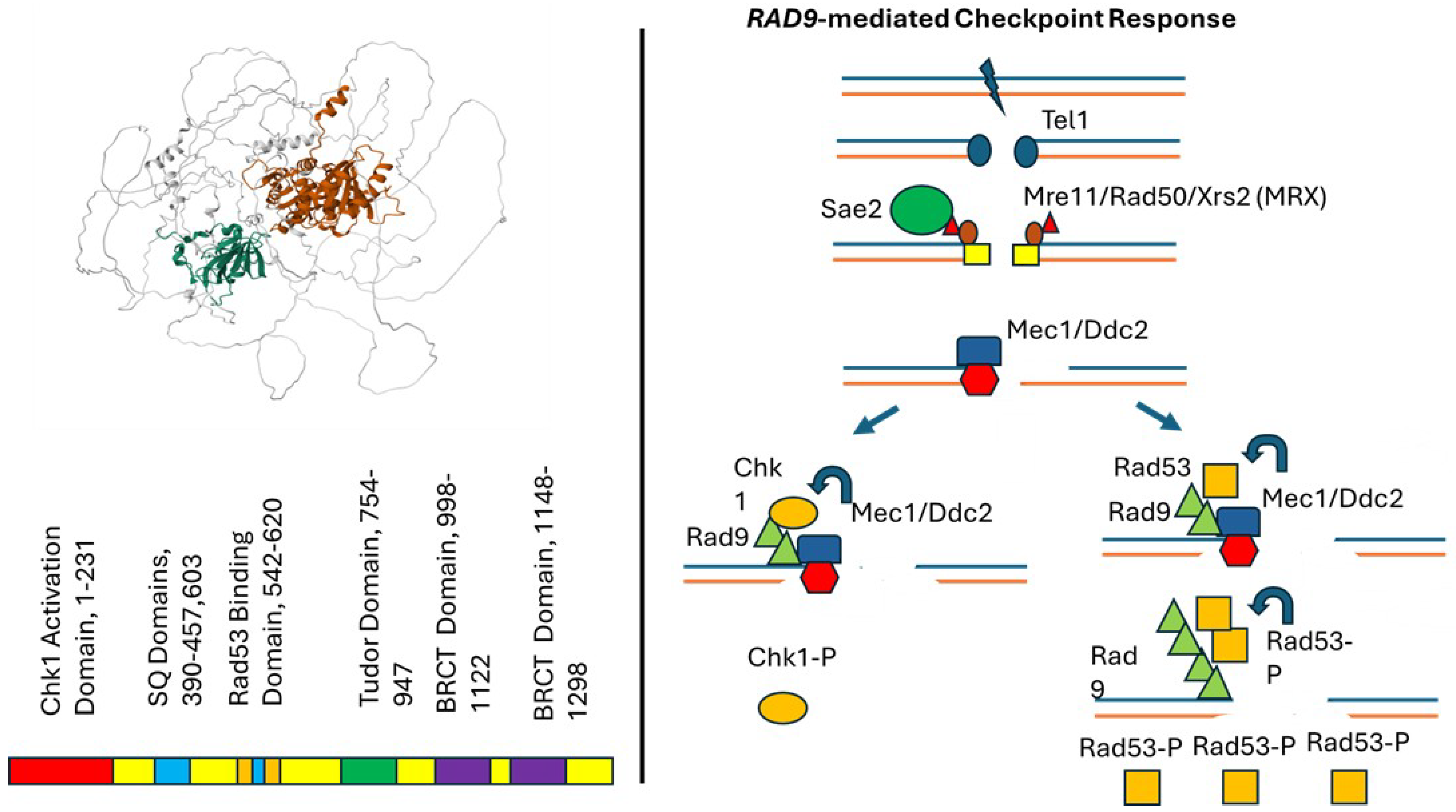
The central players in this pathway in budding yeast are the PI-3K-like kinase encoding genes TEL1 [29] and MEC1 [30], which are the orthologs of the ataxia telangiectasia mutated (ATM) and ATM-related gene (ATR), respectively [31]. DSB signaling involves the rapid recruitment of the Mre11/Sae2 complex and subsequent resection, which leads to ssDNA bound to single-strand binding protein (RPA) (Figure 3). Sae2 activation, in turn, is controlled by the Cdk2 so that resection occurs at the G2 stage of the cell cycle [32]. The ssDNA is then a substrate for binding by the Mec1-Ddc2 complex, which is facilitated by the 9-1-1 complex. Mec1 also phosphorylates DNA repair proteins including Sgs1 [33], Rad51 [34], Rad55 [35], and Exo1 [33] that facilitate sister chromatid recombination. The Mec1 kinase phosphorylates Rad53 [33,36], the CHK2 ortholog, and Chk1 [37,38]. Rad9 serves to bind Rad53 and facilitates Mec1’s phosphorylation of Rad53, which then triggers Rad53 autophosphorylation and its release from Rad9 [39]. Rad53, in turn, phosphorylates effector proteins, whose functions are to inhibit late replication firing [13], upregulate deoxynucleotide levels [40,41] and facilitate formation of Rad51 filaments [32,35]. Cell cycle arrest is achieved by Mec1-dependent activation of Chk1, which in turn phosphorylates Pds1 (securin), which blocks the degradation of cohesin by anaphase-promoting complex (APCCdc20) [42]. Rad53 (Chk2) inhibits Cdc20-Pds1 interaction [42] and phosphorylates Dun1, which in turn inhibits mitotic exit by activating the Bfa1-Bub2 complex [43]. The checkpoint pathway also coordinates with Cdc5 and Cdc14 to activate Holliday junction resolvases, such as Mus81 and Yen1, respectively, so that joint molecules are processed before sister chromatid segregation [44]. Additional roles of the checkpoint response are to inhibit de novo telomere addition [45], promote the mobility of broken chromatids [46], and inhibit asymmetric resection [47], which could promote genome rearrangements [48]. Thus, checkpoints serve to both arrest the cell cycle and facilitate DNA repair.
An intermediate substrate in both checkpoint signaling and recombinational repair is ssDNA bound to single-strand binding protein [49,50]. ssDNA generated by DSB resection is a substrate for Rad51 filament formation, which is facilitated by Rad52, Rad55, and Rad57 (for review, see [51]). Rad51-coated DNA then catalyzes DNA strand invasion and, with the assistance of Rad54, generates Holliday intermediates. The Sgs1 helicase can abort recombination intermediates [52], while additional proteins, such as Yen1, can resolve recombination intermediates to yield cross-over events [53]. DSB processing thus produces intermediates which could either bind to HR repair proteins or checkpoint proteins. While Rad9 controls resection through its interaction with the Mrell complex [54], additional proteins are phosphorylated by Cdk to ensure that recombination is completed before the segregation of sister chromatids. Thus, particular intermediates participate in both DSB repair mechanisms and checkpoint signaling.
While the checkpoint response and RAD50 group genes are required for efficient DSB gap repair and ionizing radiation resistance, other DNA damage-associated or spontaneous HR events have different requirements for individual genes within the RAD50 group. Differential requirements result from the type of initiating DNA lesion; the length, chromosomal position and orientation of the homologous sequences; and the final resolution of the recombination intermediate. In addition, different recombinogenic agents can delay or arrest the cell cycle at distinct stages. While all HR events require RAD52 [51], RAD51 mutations only modestly affect spontaneous SCR [55] and spontaneous unequal sister chromatid recombination (uSCR) events are RAD51-independent [56]; additional pathways have been identified that participate in uSCR [57]. On the other hand, homolog recombination between heteroalleles is RAD51-dependent [51]. Rad50-mediated resection delays HO endonuclease-catalyzed mating-type switching [58] but is not required to initiate X-ray-associated cross-overs [59]. Other studies have also shown multiple pathways for both SSA and BIR events [60,61]. RAD9 functionality at all stages of the cell cycle [62] thus underscores the importance of understanding how checkpoints function to facilitate both spontaneous and DNA damage-associated HR events.

2. Rad9 Protein and Function

The yeast RAD9 gene encodes a protein of 1309 amino acids, is an ortholog of the human 53BP1 and the Schizosaccharomyces pombe crb2+ genes [63,64], and shows a similarity with the human BRCA1 [64]. Overall, the protein is unstable in budding yeast and poorly expressed [65]. An artificial intelligence (AI)-generated alpha-fold structure [66] of the Rad9 protein is shown in Figure 3. The relevant structure includes five major features; these include the Chk1-activating domain (CAD), the Mec1/Tel1-phosphorylated SQ/TQ cluster domain (SCD), the Rad53 binding domain, the hydrophobic tandem Tudor domains, and the BRCT (BRCA1 c-terminal) domain [67]. The Tudor domain and BRCT domain are also conserved in Crb2 and 53BP1 [68,69]. This structure indicates both predicted and disordered domains; high confidence is shown for predicted structures of the Tudor domains, which extend from amino acids 754 to 947, and the BRCT domains, which extend from amino acids 998 to 1122 and from 1148 to 1298 (Figure 3, [67])
The tandem Tudor domain is composed of two β barrels [68]. The first β barrel is formed by five β sheets that are encompassed by amino acids 786–790 (β1), 797–806 (β 2), 811–816 (β3), 819–824 (β4), and 828–830 (β5). The second β barrel is formed by three β sheets that are encompassed by residues 768–771 (β 00), 837–841 (β 10), and 844–853 (β 20). The tandem Tudor domains are highly flexible and evolutionarily conserved to recognize chromatin at DSBs; however, the precise manner by which the Tudor domains 53BP1 and Crb2 bind to modified chromatin is different from that of budding yeast [68].
The BRCT domains are found in proteins that form scaffolds that facilitate protein interactions [69,70]. The BRCT domain in Rad9 is highly hydrophobic and exhibits strong similarities to the BRCT domain found in the tumor suppressor BRCA1 [71,72]. The human BRCT motif shares 15% absolute identity with the first Rad9 BRCT repeat. The importance of these similarities is illustrated by observations that a rad9 mutant, which contains leucine substitutions at either the highly conserved phenylalanine (F1104) in the first motif or tryptophan (W1280) in the second motif, is UV sensitive; the double mutant is as sensitive as the mutant lacking the entire BRCT domain and fails to exhibit full checkpoint activation [70].
Rad9 protein binds to methylated histone H3 on nucleosomes in undamaged cells via its tandem Tudor domain [69], and Rad9’s tandem BRCT domain enhances its concentration at DSBs by interacting with phosphorylated histone H2A (S119). Rad9 is further recruited to DNA damage sites by the Dpb11 (TOPBP1), which interacts with the 9-1-1 complex that is loaded onto ssDNA-dsDNA junctions [72]. Rad9 binding to chromatin limits resection of DSBs by inhibiting the Sgs1 helicase [73]. Rad9 phosphorylation by Mec1 induces Rad9 multimerization via its BRCT domain and enables Rad9 to recruit Rad53 [74]; thus, Rad9 serves as an adaptor so that Mec1 can phosphorylate Rad53 [75]. The oligomerized Rad9 can also serve as a scaffold for the aggregation of multiple Rad53 molecules, which facilitates Rad53 trans autophosphorylation leading to the subsequent release of Rad53 [75]. Rad9 thus controls one branch of the checkpoint pathway, the other of which is controlled by Mrc1, whose role in checkpoint activation is restricted to DNA replication forks. Through its CAD, Rad9 can also function as an adaptor for Mec1-mediated Chk1 activation [76].
RAD9 mediates DNA damage-activated checkpoints at G1 [77,78], S [18,79,80], and G2/M stages of the cell cycle [22,81]. Rad9-associated checkpoint activation is regulated by 98 phosphorylation sites mediated by Mec1, Rad53, Cdc28 -Clb2, and ubiquitylation on position K1139 [67]. Specific phosphorylation sites serve to activate Rad9 function while others function to dampen the checkpoint response. For example, cyclin-dependent kinase (CDK) phosphorylation of Rad9 at multiple sites enables interaction with BRCT domains of another scaffold protein, Dpb11, which in turn facilitates the interaction with the 9-1-1 complex that recognizes ssDNA/dsDNA junctions [81]. Additional CDK-mediated phosphorylation of Rad9 at the T125 and T143 sites regulates Rad9 interaction with Chk1; phosphorylation of T143 stimulates Chk1, whereas phosphorylation of T125 inhibits Chk1 interaction [82]. Thus, Rad9 interactions with both Rad53 and Chk1 are tightly controlled by phosphorylation by multiple regulators.
RAD9 facilitates the completion of DNA replication initiated during the S phase. First, it facilitates chromosomal DNA replication when there are large distances between origins, a function which does not require recombination [83]. Second, it backs up MRC1’s function to extend the length of checkpoint signaling during replication stress [84]. Third, it is required for recombination-mediated resolution of aberrant structures that are formed in DNA replication. For example, rad9 exhibits synthetic lethality with rad27 [85], a mutant defective in the processing of 5′ flaps generated during lagging strand synthesis [86]. The hyper-recombination of rad27 and the synthetic lethality with rad27 and rad52 [87] suggest that HR is required to resolve such aberrant structures that would otherwise confer lethality. Fourth, RAD9 is required for BIR that requires extensive replication of chromosomal arms, since the rate of DNA synthesis initiated by BIR is slower than bidirectional DNA replication [88]. These observations indicate that RAD9-mediated checkpoint function is triggered by multiple types of DNA replication stress.
The structural similarities between budding yeast Rad9 and the human 53BP1 provoke the question whether these proteins share DNA repair functions. The most striking similarity is that both genes are required for NHEJ through their function in inhibiting DNA end resection [54,89,90]. In budding yeast, ligation of a linearized plasmid to form an autonomous circular plasmid is inefficient in rad9 mutants, and this phenotype can be partially rescued by delaying cells in either G1 or G2 [89]. In addition, rad9 mutants are defective in rejoining DSBs generated in vivo by expression of EcoR1 endonuclease [90]. Alternatively, 53BP1 is required for NHEJ mechanisms involved in VDJ recombination and in telomere fusions [91].
RAD9 functions in controlling cell cycle progression and DNA end resection are thus two mechanisms by which HR pathways can be affected. Nonetheless, RAD9’s function in controlling HR between sister chromatids and homologs, and ectopic recombination between repeated sequences, is not fully understood. Depending on the assay to measure mitotic HR, rad9 mutants may exhibit enhanced recombination, decreased recombination, or no effect. Ascertaining phenotypes is complicated by the approximately 10-fold increase in chromosome loss observed in rad9 mutants, compared to the wild type [92,93]. In addition, particular phenotypes may depend on whether the assay measures spontaneous or DNA damage-associated recombination or whether the assay is performed in haploid or diploid cells [94]. Different results obtained from haploid and diploid cells may result in an inhibition of NHEJ in cells expressing both MATα and MATa [95,96]. Our effort to elucidate these apparently contradictory phenotypes will include: (1) summarizing different recombination assays that exhibit rad9 phenotypes, (2) describing RAD9 and recombinational repair pathway interactions, and (3) comparing phenotypes of rad9 mutants with mutants in other checkpoint genes. Finally, we will present possible mechanisms that may elucidate these phenotypes and future experiments.

3. HR Phenotypes of rad9 Mutants

The RAD9 requirement for HR and NHEJ events is shown in Table 1. While differences in strain ploidy and recombination constructs may initially render it difficult to draw conclusions, overall, there are several emerging patterns. RAD9-dependent HR events include those that require a cell cycle delay to complete DSB repair, including SSA, gap repair, and BIR and those that require cell cycle delay to complete DNA replication. RAD9-independent HR events include those that do not require a cell cycle delay. Overall, RAD9 promotes genetic stability when cell cycle delay promotes error-free SCR, while RAD9 promotes genetic instability when DNA replication stress generates recombinogenic DNA lesions that initiate HR-mediated and NHEJ-mediated ectopic recombination. RAD9 promotion of genetic instability is particularly evident in strains that are deficient in selective S phase checkpoint mutants, such as mec1 and mrc1, which exhibit higher frequencies of replication fork collapse and are deficient in signaling at stalled DNA replication forks (for review, see [97]). Secondary considerations are the kinetics of the completion of the DSB-initiated recombination event [98]. In addition, RAD9 may promote the outcome of the recombination event due to the Rad9 protein interactions with DNA helicases and nucleases [99]. We discussed these RAD9 functions in the context of rad9 phenotypes ascribed for SCR, homolog recombination, ectopic recombination and GCRs.

3.1. RAD9-Mediated Checkpoint Functions That Promote Genetic Stability

3.1.1. DSB-Associated SCR Is Mediated by the Rad9-Mediated Pathway That Proceeds Through Rad53

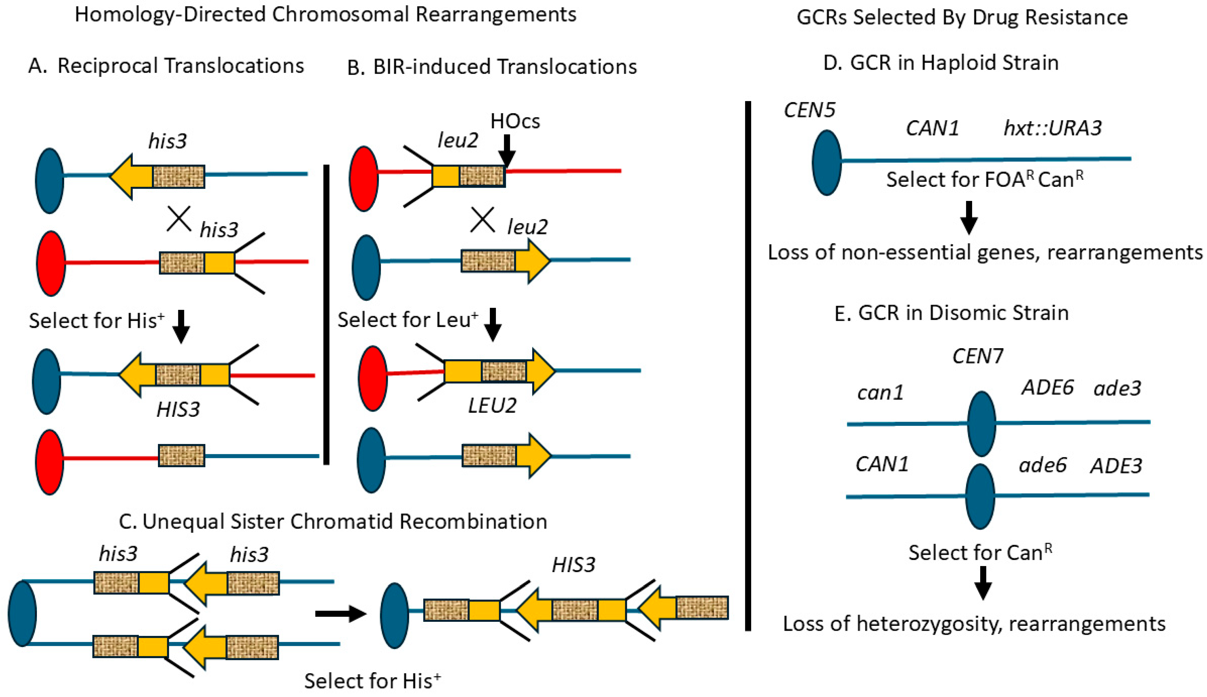
Recombination between sister chromatids proceeds by multiple mechanisms, including template-switching and DSB-initiated HR. While RAD9 is not required for spontaneous sister chromatid recombination (SCR), RAD9 is required for DSB-associated SCR as demonstrated in assays to measure either equal or unequal sister chromatid recombination (uSCR, Figure 4) [103,104]. Both X-ray and HO-induced DSBs do not stimulate as many uSCR events in rad9 mutants as they do in the wild type [103,104]. RAD9 is also required for DSB-initiated equal SCR in a pathway that involves stabilization of cohesin; cohesin is absolutely required for the repair of DSBs in yeast [113] and participates in the DNA damage response [114]. This observation is further supported by observations that one downstream effector of Mec1 activation, Rad53, which contributes to cohesin stabilization and the maintenance of cell cycle arrest, is required for X-ray-associated uSCR [115]. While Chk1 is also a downstream effector of Mec1 activation, chk1 mutants are not X-ray-sensitive [116], nor do they exhibit a clear defect in sister chromatid cohesion [117] or in X-ray-associated uSCR [115]. This does not exclude that Chk1 has a redundant function in conferring X-ray resistance. These results suggest that Rad53 plays a key role in the RAD9-mediated checkpoint pathway that facilitates DSB-associated SCR.
Consistent with the RAD9-dependence of X-ray-associated SCR, rad9 mutants exhibit fewer DNA damage-associated uSCR events after exposure to selective chemical agents, particularly those that indirectly generate DSBs, such as methyl methanesulfonate (MMS) and camptothecin, a topoisomerase inhibitor [107]. However, rad9 mutants exhibit only a minor decrease in UV-associated uSCR [103] and no decrease in the 4-nitroquinoline 1-oxide (4-NQO)-associated uSCR events [107]; 4-NQO is a UM-mimetic agent that forms bulky DNA adducts. One possible explanation is that 4-NQO-induced bulky DNA damage slows replication and promotes SCR without initiating DNA breaks [118]. Thus, these studies demonstrate that the RAD9 dependence of DNA damage-associated uSCR depends on the recombinogenic agent.

3.1.2. RAD9 Suppresses rDNA Rearrangements in orc1-4 Mutants

While RAD9 promotes DSB-associated SCRs, it suppresses rDNA repeat instability that results from insufficient DNA replication firing due to non-functional origin recognition complex (Orc) proteins, as present at the restrictive temperature in orc1 diploid mutants [102]. RAD9 confers lethality in orc1-4 diploid mutants at the restrictive temperatures by triggering a permanent arrest. However, the viable colonies obtained in the rad9 orc1-4 diploid mutant contain reduced numbers of rDNA repeat units. When the Orc1 protein is present in limited amounts at the restrictive temperature, the multitude of Orc1 binding sites at rDNA limits replication initiation at other chromosomal origins. The rDNA is particularly vulnerable to recombinogenic lesions due to the potential collision of replication and transcription complexes and due to topological stress [119]. Such recombinogenic lesions could promote uSCR, unequal homolog recombination, and intrachromosomal recombination at the rDNA locus. Accumulation of DNA breaks at the rDNA locus induces a RAD9-mediated arrest and may precede DSBs induced at other chromosomal sites [102]. A reduced rDNA copy number thus lessens the competition of binding sites for Orc proteins and decreases the DNA damage signals. Although the exact mechanism by which RAD9 suppresses rDNA repeat instability is unknown, one tenable hypothesis is that recombinogenic lesions are channeled to an equal SCR pathway and thus maintain the rDNA copy number.

3.1.3. RAD9 Is Required for SSA When There Are Significant Mismatches Between Repeats

Enzymatic-induced DSBs have been used to study the HR-mediated repair of DSBs. However, efficient digestion of a recognition site on both sister chromatids destroys an undamaged sister chromatid template. In the absence of SCR, HR-mediated repair of DSBs using non-tandem repeats is referred to as SSA (Figure 1). RAD9 is not required for the completion of SSA when an enzymatic-induced DSB initiates recombination between two non-tandem repeats. A study demonstrated this conclusion using one strain that contained tandem his3 fragments, where an HO endonuclease cut site (HOcs) was inserted in one his3 fragment, and another strain contained a disrupted URA3 gene where an HOcs was inserted between ura3 repeats [101]. However, RAD9 is required for intrachromosomal recombination between homeologous repeats that contain 3% divergent DNA [100] in which an HOcs was inserted between the 200 bp ura3 repeat units. The RAD9 dependence of SSA mediated by homeologous repeats is suppressed by nocodazole, suggesting that the critical factor for completing recombinational repair between divergent repeats is maintenance in G2 [100]. The authors suggest that SSA between heterologous repeats requires mismatch repair proteins, which require an extended G2. While RAD9 is not explicitly required for SSA, other checkpoint proteins, such as Mec1, do affect SSA by phosphorylating Slx4 [120] and affecting Rad1/Rad10 cleavage on non-homologous 3′ tails [121]. Thus, while checkpoint proteins regulate SSA, the RAD9 function has not been explicitly defined.

3.1.4. Rad9 Counters Sgs1 and Mph1 During DSB-Associated Homolog Recombination

The overall conclusion of the RAD9 dependence of homolog recombination is that RAD9 is not required for spontaneous recombination but does alter the type of DSB-induced recombination event. Several strain constructs used to measure rates of spontaneous homolog recombination include those that 1) simply measure gene conversion events between two heteroalleles and 2) those that measure cross-overs between two heteroalleles of two or more genes. In the first type of construct, RAD9 is not required for spontaneous homolog recombination between heteroalleles in either diploids or in a haploid disomic strain for chromosome VII [106]. In the second strain construction, CanR and Thr+ recombinants were selected in a diploid that was heterozygous with the wild type at both CAN1 and HOM3. The authors observed a two-fold increase in rates of spontaneous homolog recombination, compared to a wild-type diploid [105]. Thus, RAD9 has a minimal effect on recombination between homologs.
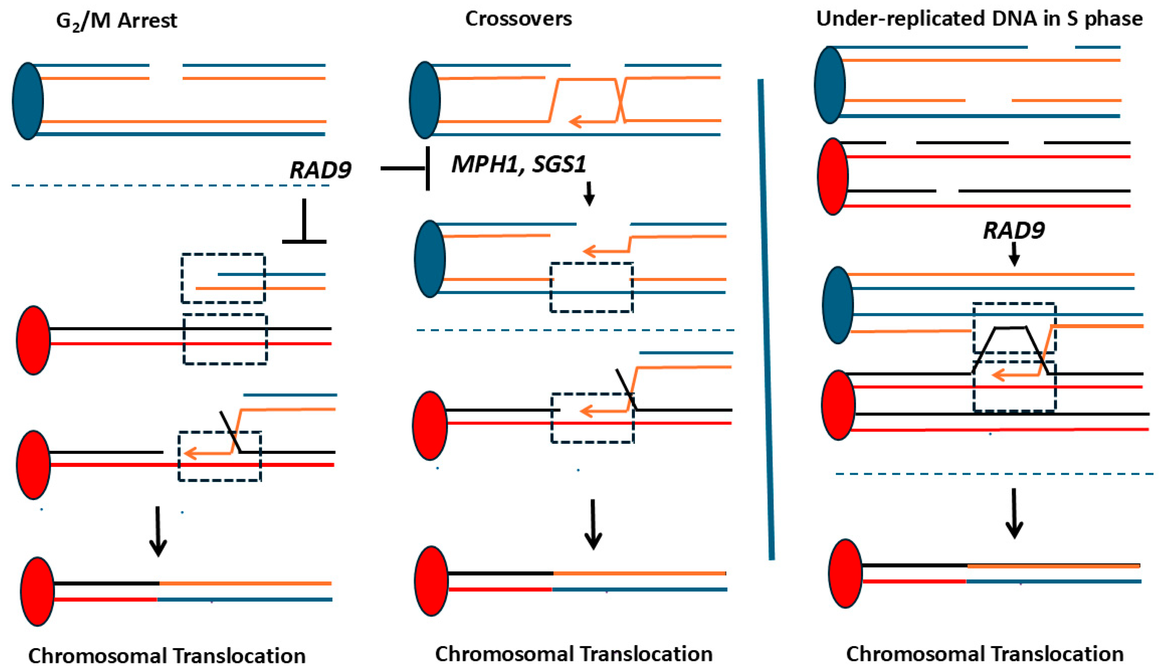
However, RAD9 does alter the types of recombination events that are induced by an I-Sce1 restriction endonuclease when the endonuclease recognition site is placed in one copy of an ade2 gene in a diploid strain containing two ade2 heteroalleles flanked by different markers [99]. In this strain construction, gene conversion events, cross-overs, and break-induced replication (BIR) events could be identified by scoring the presence of the ade2 allele and whether the flanking marker was present. Based on these studies, the authors demonstrated that the rad9 mutant exhibited a higher percentage of short-track gene conversion and a reduced frequency of break-induced replication and cross-over events. By Chip analysis, they also showed that Rad9 limits the Sgs1 and Mph1 helicase binding, suggesting that Rad9 facilitates the recombinogenic repair of DSBs by stable annealing of the recipient and donor strands during recombination [99].

3.1.5. RAD9 Suppresses Ectopic Recombination That Generates Translocations

RAD9 suppresses translocations generated by HR between his3 repeated sequences located at centromere-linked loci on non-homologous chromosomes II and IV (Figure 4). However, the RAD9-mediated suppression is keenly observed after cells are exposed to X-ray mimetic agents, compared to exposure to compounds that primarily cause base pair damage and adducts. Compared to the wild-type diploid, the rate of spontaneous homology-directed translocations in homozygous rad9 diploid mutants increases by seven-fold [103,105]. A modest but significant increase is also observed in rad9 haploids, compared to the wild type [103]. The hyper-Rec phenotype of the rad9 mutant is further enhanced if cells are exposed to radiation; an approximately thousand-fold stimulation was observed after rad9 diploid mutants are exposed to 15.6 krads [103]. The radiation-associated recombination is partially suppressed by pre-arresting cells with the microtubule inhibitor nocodazole before UV or X-ray exposure. These data suggest that cell cycle delay is sufficient to partially suppress DNA damage-associated translocations.
Many homology-mediated translocations in rad9 mutants may result from recombinogenic acentric and centromere-containing chromosomal fragments that are inherited in repeated cell cycles (Figure 5). Consistent with this model, many radiation-associated translocations are non-reciprocal events, also referred to as half cross-overs (HCs). Although BIR can theoretically generate these HCs, BIR has not been observed to transverse centromeric DNA [122]. Instead, it is likely that multiple rearrangements generate radiation-associated His+ recombinants, especially those where the His+ phenotype is unstable. This interpretation is supported by independent observations that DSBs can efficiently remodel the genome [4] and that chromosomal instability in rad9 mutants can be initiated at telomeric sites [106], which would subsequently trigger recombination at other loci.
An increase in homology-directed translocations in rad9 diploids has also been observed in independent studies [109]. In a strain construction to measure the loss of heterozygosity (LOH) on chromosome III, where the URA3 gene is positioned on one chromosome III homolog, rad9 mutants exhibit a higher frequency of 5-fluoroorotic acid resistant (Ura-) isolates containing an accompanying chromosomal rearrangement [109]. While the increase in the frequency of spontaneous translocations was a modest three-fold, the increase observed in diploid orc1-4 rad9 mutants was 42-fold. Many of these cross-over events occurred at Ty1 sequences. While the rad9 mutants did not exhibit an increase in LOH due to gene conversion, LOH events due to cross-over events increased three-fold in the rad9 diploid and 14-fold in the orc1-4 rad9 diploid mutant, compared to the wild type. These data thus support the notion that rad9 mutants exhibit more ectopic recombination.

3.1.6. RAD9 and RAD50 Group Genes Are Separate Pathways for Suppressing Ectopic Events in Diploid Strains

Models for DSB-initiated checkpoint signaling and HR repair of DSBs suggest that there is cross-talk between the two pathways. The Mre11/Rad50/Xrs2 complex, which facilitates DSB recission, is required in both DSB-mediated checkpoint signaling and in DSB repair. Because Rad9 is required for Mec1-mediated activation of RAD53, the model would suggest recombinational repair and RAD9-mediated checkpoint pathways participate in the same pathway for suppressing homology-directed translocations [50,123]. Pertaining to haploid mutants, RAD9 is epistatic to recombinational repair [124] in conferring ionizing radiation resistance. While RAD9 suppresses frequencies of spontaneous homology-directed translocations by seven-fold, diploid double mutants defective in either RAD9 andeither RAD51, RAD55, or RAD57 exhibit a synergistic (57-78-fold) increase in the frequencies of spontaneous ectopic recombination directed by his3 sequences (Table 2, [105]). In addition, diploid mutants defective in RAD9 and either MRE11 or XRS2 exhibit synergistic (57-fold) increases in spontaneous his3 homology-directed translocations [125]. These studies suggest that RAD9 and RAD50 group genes participate in independent pathways for suppressing spontaneous HR between repeated sequences in diploid strains. While the exact identity of spontaneous recombination events is unknown, one possibility is lesions generated by the base excision repair of uracil [126].
One possible interpretation of the synergistic increase in spontaneous translocation in rad9 rad51 double mutants is that the ectopic recombination between the repeated sequences is mediated by annealing homologous single-stranded DNA. Mutations in Rad9 confer more ssDNA while RAD51 inhibits SSA [127,128]. Since Rad9 inhibits resection of DSBs and Rad51 inhibits SSA, an increase in ssDNA and lack of inhibition to reanneal these sequences may increase recombination events. Similar interactions would also apply to the interaction of rad9 and the other rad mutants. An alternative explanation is that more DNA lesions accumulate in rad51 mutants, and these lesions are tolerated in rad9 mutants. At present, we cannot distinguish between the two and it is possible that both mechanisms are important in promoting chromosomal rearrangements that occur by single-strand annealing.
Table 2. Synergistic effects of rad9 and DNA repair mutants on generating homology-mediated translocations.
Genotype of Single and Double MutantRecombination AssayPloidyFold Increase Relative to WT 1Fold Increase Compared to rad9Fold Increase Compared to Single DNA Metabolism MutantReferences
rad9Directed translocationsDiploid71NA[103,125]
rad9 rad51Directed translocationsDiploid5785.3[125]
rad9 rad55Directed translocationsDiploid77116.2[125]
rad9 rad57Directed translocationsDiploid78115.3[125]
rad9 rad54Directed translocationsDiploid55824[125]
rad9 mre11Directed translocationsDiploid5782[125]
rad9 mec1Directed translocationsDiploid610.3[125]
rad9GCRHaploid61NA[129]
rad9 sgs1GCRHaploid213 (3%)379.7[129]
1 Wild type strain is the corresponding Rad+ haploid or diploid form which the single or the double mutant is derived from.

3.1.7. Rad9 and Sgs1 Suppress HR Between Divergent Genes and Ty1 Elements

Schmidt and Kolodner [129] observed that SGS1 suppresses the formation of gross genome rearrangements (GCRs) that occur by recombination between the CAN1 positioned on chromosome V and the LYP1 or ALP1 genes positioned on chromosome XIV. CAN1, LYP1, and ALP1 encode basic amino acid transporters, which share more than 50% sequence identity. The haploid strains contain URA3 and CAN1 genes located on the non-essential chromosomal V arm; double selection against both URA3 and CAN1 using 5-fluororotic acid (5-FOA) and canavanine, respectively, generates drug-resistant isolates containing chromosomal rearrangements. While most FOAR and CanR isolates result from NHEJ events, in sgs1 mutants, the rate of GCRs is increased 22-fold above the wild type, while the sgs1 rad9 mutant exhibited a 213-fold increase above the wild type. While only ~3% of these drug-resistant isolates were translocations due to HR between these sequences, no homology-directed translocations were observed in either the sgs1 or the rad9 mutants; translocations found in the single mutants are due to NHEJ or micro-homology end-joining [129]. These experiments thus indicate that SGS1 and RAD9 constitute independent pathways in suppressing GCRs in haploid strains through multiple mechanisms, including homeologous recombination between sequences.
RAD9 and SGS1 were also observed to suppress DSB-initiated ectopic rearrangements that directly result from BIR. Vasan et al. [108] characterized recombinants that resulted from HCs initiated by galactose-induced DSBs at a HOcs positioned on chromosome III in a chromosome III disomic strain. Deletion of RAD9 and SGS1 conferred a higher frequency of recombinants that resulted from compromised BIR leading to cascades of genome instability [108]. One interpretation is that rad9 mutants exhibit greater mis-segregation of chromosomal fragments while Sgs1 serves as an anti-recombinogenic factor that reverses recombination intermediates generated by BIR. Additionally, Rad9 protects replication intermediates from excessive degradation [130]. Thus, aborted BIR (Figure 5) may lead to aberrant recombination events at ectopic loci. Recombination sites involving HC during compromised HC include Ty1 elements. These data thus indicate that rad9 mutants exhibit both higher frequencies of spontaneous and DSB-associated recombination between Ty1 sequences.

3.2. RAD9-Mediated Checkpoint Functions That Promote HR-Mediated Genetic Instability

3.2.1. HR Promoted by DNA Replication Impediments

Copy number variation (CNV) has also been measured in checkpoint mutants containing multiple juxtaposed CUP1 repeats (Table 3). In these haploid strains, nicotinamide-mediated suppression of the histone deacetylase H3K56ac induces CUP1 transcriptional induction, initiating a replication fork impediment and CNV contraction. CNV contraction is RAD52-dependent and proceeds through HR. Interestingly, MRC1, which facilitates replication fork stability, but not RAD9, suppresses CNV [131]. In addition, rad27 mutants exhibit enhanced CNV, suggesting aberrant replication intermediates are leading to recombinogenic lesions. Considering that the double mus81 yen1 mutant defective in Holliday junction resolution does not exhibit CNV; an attractive mechanism is that CUP1 transcriptional induction induces replication fork cleavage in the S phase, initiating a replication restart mechanism. Thus, RAD9 can suppress instability at the rDNA locus while facilitating instability at CUP1 repeats [131].

3.2.2. HR Promoted by S Phase Checkpoint Defects Resulting from dNTP Insufficiency

Various mutants defective in the stabilization of DNA replication intermediates accumulate recombinogenic lesions in the S phase, due to DNA replication fork collapse, failure to adequately process Okazaki fragments or resulting from lower levels of dNTPs. This is particularly true of mec1 hypomorphs, such as mec1-21 and mec1-srf mutants [134], which exhibit decreased viability when RAD52 is inactivated. The hyper-recombination of mec1-21 is suppressed by mutations in SML1, an inhibitor of ribonucleotide reductase, suggesting that elevated dNTP levels reduce the accumulation of recombinogenic substrates [135]. Unlike rad9 mutants, which do not exhibit higher rates of spontaneous uSCR and heteroallelic recombination, mec1-21 mutants exhibit hyper-recombination in spontaneous uSCR and heteroallelic recombination. RAD9 is required for the hyper-recombination phenotypes that are exhibited by mec1-21 mutants and mec1-21 rad9 double mutants exhibit similar rates of homology-directed translocations as rad9 diploids. An attractive model is that RAD9 is required to delay the cell cycle so that recombinogenic DNA damage produced by replication fork collapse or replication errors can be repaired. However, additional observations indicate that RAD9 is required to protect stalled or collapsed replication forks from excessive degradation [130], and that rad9 mutants exhibit hyper-resection leading to Mec1-mediated Sgs1 phosphorylation [136]. Thus, a combination of factors may function to ensure that recombinogenic lesions generated during DNA replication can be adequately repaired so that replication fork progression may be completed.
Phenotypes of mec1-21 rad9 mutants are partially mimicked by mec1-21 chk1 and mec1-21 pds1 mutants, while higher frequencies of homology-directed recombination observed in rad9 diploids are also exhibited by rad53 diploids [116]. For example, mutations in either PDS1 or CHK1 reduce the hyper-recombination phenotype of mec1-21, and also increase radiation sensitivity in mec1-21, as observed in mec1-21 rad9 mutants. One interpretation of these results is that mutations in either CHK1 or PDS1 confer toleration to under-replicated DNA [137], which would otherwise be a substrate for HR proteins. These observations suggest that RAD9 functions in promoting genome instability observed in S phase checkpoints function through downstream effectors.

3.2.3. HR-Associated Translocations Mediated by BIR

BIR is a mechanism by which DNA replication is initiated by HR factors at the site of a DSB (Figure 1). The 3′ end of the DSB invades a template, creating a Holliday junction, and is subsequently converted into a one-directional DNA replication fork that can proceed to the telomere [138]. The subsequent replication is highly error-prone and results from conservative DNA replication [139]. One cellular assay to measure BIR uses two overlapping fragments of lys2, positioned on chromosomes III and V (Figure 4). An HOcs has been positioned adjacent to the lys2 fragment on chromosome V. Induction of a galactose-inducible HO endonuclease creates a DSB that initiates recombination with the lys2 fragment on chromosome III. The chromosome V:chromosome III half-translocations can be scored by LYS2 and the loss of non-essential markers on chromosome V [140].
The genetic requirements for BIR have been determined by high-throughput screens and targeted knockouts of DNA recombination and replication genes [140]. Importantly, recombination genes required for gap repair are also required for BIR. Replication genes that are required include PIF1, which encodes a helicase and blocks telomere formation, and DNA polymerase delta. The rate of replication fork movement is slower than bidirectional replication initiated at ORIs in the genome. The slower rate of replication triggers a RAD9-mediated checkpoint response, and together with the induction of a spindle assembly checkpoint, replication can proceed to the telomere. Deleting both the spindle assembly checkpoint genes and RAD9 lead to a dramatic decrease in the efficiency of BIR. These studies show that particular genetic instability events induced by BIR require RAD9 [140].

4. RAD9 Role in Suppressing GCRs

RAD9 functions in suppressing gross GCRs that result from fusion of indirect repeated sequences, which do not require HR functions. This was demonstrated in a chromosome VII disomic strain where one copy of chromosome VII contains a telomeric CAN1 gene and internal short, inverted repeats (Figure 4). Spontaneous CanR mutants containing rearrangements generated by fusion of these inverted repeats can then be selected and screened based on the sectored colony phenotype; sectors typically contain unstable dicentric and acentric chromosomes [112,133]. rad9 mutants exhibit 17-fold higher frequencies of CanR isolates containing such rearrangements. The authors suggest that inverted repeat fusions result from aberrant template-switching events during the S phase.
However, RAD9 and G2/M checkpoint genes have a minor role in suppressing spontaneous Chr V rearrangements, compared to S phase checkpoint mutants [111,132,141]. The GCR assay used a haploid strain containing URA3 and CAN1 genes located on the non-essential chromosomal V arm (Figure 4); double selection against both URA3 and CAN1 using 5-fluororotic acid (5-FOA) and canavanine, respectively, generates drug-resistant isolates containing chromosomal rearrangements. The rad9 mutant exhibits slightly elevated rates (three-fold) of GCRs, while the double rad9 exol mutant exhibits a slightly more elevated rate of GCR, compared to the single mutants [110]. Even when the URA3 gene is placed in a position adjacent to the Ty1 element, the overall rate of GCRs in the rad9 mutant is only two-fold greater than that observed in the wild type (Table 3, [110]). Since non-homologous end-joining (NHEJ) is a major mechanism for generating GCRs, one possible explanation for the modest increase in GCRs in rad9 mutants is the rad9 deficiency in NHEJ [89,90]). Another contributing factor is that rad9 mutants exhibit chromosome loss [92,93], which would be lethal in haploid strains.
While RAD9 function in suppressing spontaneous double drug-resistant isolates is minor, its function in suppressing frequencies of DNA damage-associated GCRs is significant [132,142]. The rad9 mutant exhibits a ~168-fold increase in GCR frequency after exposure 0.07% MMS, while the wild type exhibits 68-fold increase in GCR frequency after exposure to the same MMS concentration. While the difference between 168 and 68-fold seems minor, the overall frequency of MMS-associated GCRs in the rad9 mutant are approximately ten-fold higher than the MMS-associated frequency observed in wild type [132,142].MMS is known to significantly impede DNA replication [143], supporting observations that S phase DNA errors are the major initiating cause for GCRs.
Comparing the genetic control and DNA damage inducibility of GCRs and HR-directed translocations reveals important similarities and differences. The major similarity is that S phase checkpoint defects confer the highest increases in frequencies of either GCRs or HR-mediated rearrangements, likely due to recombinogenic structures generated during replication. Additionally, the higher rates of spontaneous GCRs and HR-directed translocations observed in the mec1 mutant can be synergistically increased by knocking out RAD51 [144], suggesting that the checkpoint and recombinational repair pathway are independent in suppressing both NHEJ-mediated and HR-mediated rearrangements. RAD9 suppresses both DNA damage-associated GCRs and HR-directed translocations, particularly when cells are exposed to the X-ray mimetic chemical, MMS. Both frequencies of GCRs and HR-directed translocations are stimulated by diverse DNA damaging agents, ranging from simple alkylating agents to both X-rays and UV.
The differences between the genetic control and DNA damage inducibility of GCRs and HR-directed translocations are also notable. DNA damage-associated and spontaneous GCRs have different requirements for yKu70 [142,145,146], a gene required for NHEJ; meanwhile, the highest frequencies of radiation-associated, HR-directed translocations are observed in diploid and not haploid strains [94], in which NHEJ is repressed. Indeed, in yku70 haploid mutants, DNA damage-associated GCRs are abolished [142] while a five- to six-fold increase in X-ray-associated, homology-directed translocations are observed in haploid yku70 mutants [147]. These observations underscore observations that DSBs are important lesions in remodeling the yeast genome even when NHEJ is abolished.

5. Similarities of Rad9 Orthologs in Promoting Genetic Stability

The budding yeast RAD9 ortholog in Schizosaccharomyces pombe (fission yeast) is crb2+ and the human ortholog is 53BP1, which encodes proteins that share structural and functional similarities. Similar to budding yeast Rad9, 53BP1 and Crb2 contain Tudor(2) and BRCT(2) domains, which function to bind chromatin and form a scaffold for the assembly of additional checkpoint proteins [148]. 53BP1 and Crb2 are also recognized and phosphorylated by cyclin-dependent kinases and ATM. These proteins promote HR-mediated repair of DSBs and cross-overs and enable checkpoint-mediated arrest [149,150]. They differ from budding yeast Rad9, however, in how they regulate HR and promote NHEJ [149,150].
In S. pombe, crb2 mutants are defective in DSB repair and exhibit an enhanced loss of linear mini-chromosomes [149]. Haploid strains are SSA-defective, as measured in a plasmid-based assay. In an independent study, the X-ray sensitivity of the crb2 mutant is suppressed by topoisomerase III mutations, suggesting that the irradiated crb2 cells accumulate toxic recombination intermediates [151]. Thus, S. pombe crb2+ may function at multiple stages in HR. Since S. pombe diploids are unstable [152], some diploid-based budding yeast genetic instability assays cannot be performed.
While 53BP1 is a well-known marker for DSBs in mammalian cells, 53BP1 has opposing roles in promoting HR; while it limits resection and thus promotes NHEJ, it promotes DSB repair by HR at heterochromatin in G2 cells [150]. The role of limiting end resection, however, serves as important in VD(J) class-switch recombination and at unprotected telomeres [153]. Thus, both Crb2 and 53BP1 function to promote HR in particular contexts. These studies indicate evolutionary conservations of some functions of budding yeast RAD9.

6. Conclusions

RAD9 functions at all stages of the cell cycle and is required for efficient DNA damage-associated checkpoint function. Salient functions include controlling cell cycle progression and limiting the resection of DSBs. These functions are coordinated with cyclin-dependent kinases so that resection is limited to the G2/M stage of the cell cycle, thus biasing sister chromatids as preferred substrates for recombinogenic repair. However, the literature has often presented seemingly contradictory observations on the role of RAD9 in suppressing recombination-associated genome instability. In this review, we have highlighted particular RAD9 functions that suppress genetic instability and those that promote genetic instability. The RAD9 function to suppress genetic instability is particularly profound when cells are exposed to radiation and radiomimetic drugs. Rad9 mutants thus exhibit a dramatic increase in both homology-directed translocations and GCRs after exposure to recombinogenic agents, and this phenotype can be partially suppressed by triggering the spindle assembly checkpoint to maintain cells in G2/M. However, during replication stress, RAD9 functions to promote cell cycle delay so that DNA replication can be completed. This function, however, increases the frequency of chromosomal rearrangements that result from either HR or NHEJ, and is required for BIR to proceed to completion. The purpose of this review was to document the multiple recombination assays that have elucidated these phenotypes in budding yeast. The phenotypes differ in haploid or diploid strains and reveal novel insights into the independence of HR-mediated pathways and the checkpoint pathway in suppressing HR-mediated pathways. Nonetheless, there are still outstanding questions concerning how ploidy influences RAD9-mediated signaling, when RAD9-mediated signaling predominates over other signaling pathways at stalled replication forks, and elucidating how checkpoints regulate HR depending on the length of homology and the percentage of mismatched sequences. This later question is particularly relevant considering the abundance of repeated sequences in the eukaryotic genome. Considering that S. cerevisiae RAD9 has both mammalian and S. pombe orthologs, it will be important to compare yeast phenotypes with those observed in mammalian cells.

Funding

This research was supported by grants from the National Institutes of Health, R01CA70105 and R21ES015954, R15ES02685 and a grant from the Center for Advancement of Nanotechnology (CATN).

Institutional Review Board Statement

Not applicable.

Informed Consent Statement

Not applicable.

Data Availability Statement

Data provided in this review is provided with references attached. Unpublished data may be provided upon request.

Conflicts of Interest

The author declares no conflicts of interest.

Abbreviations

BIRBreak-induced replication
CNVCopy number variation
DSBdouble-strand breaks
5-FOA5-fluororotic acid
GCRgross chromosomal rearrangements
HChalf cross-over
HOcsHO endonuclease cut site
HRhomologous recombination
LOHloss of heterozygosity
MMEJmicrohomology-mediated end-joining
MMSmethyl methanesulfonate
NHEJnon-homologous end-joining
4-NQO4-nitroquinoline 1-oxide
OrcOrigin recognition complex
SSBsingle-strand breaks
SCRsister chromatid recombination
uSCRunequal sister chromatid recombination

References

  1. Mehta, A.; Haber, J.E. Sources of DNA double-strand breaks and models of recombinational DNA repair. Cold Spring Harb. Perspect. Biol. 2014, 6, a016428. [Google Scholar] [CrossRef] [PubMed]
  2. Krejci, L.; Altmannova, V.; Spirek, M.; Zhao, X. Homologous recombination and its regulation. Nucleic Acids Res. 2012, 40, 5795–5818. [Google Scholar] [CrossRef] [PubMed]
  3. Jinks-Robertson, S.; Petes, T.D. Mitotic recombination in yeast: What we know and what we don’t know. Curr. Opin. Genet. Dev. 2021, 71, 78–85. [Google Scholar] [CrossRef] [PubMed]
  4. Argueso, J.L.; Westmoreland, J.; Mieczkowski, P.A.; Gawel, M.; Petes, T.D.; Resnick, M.A. Double-strand breaks associated with repetitive DNA can reshape the genome. Proc. Natl. Acad. Sci. USA 2008, 105, 11845–11850. [Google Scholar] [CrossRef]
  5. Brunborg, G.; Resnick, M.A.; Williamson, D.H. Cell-cycle-specific repair of DNA double strand breaks in Saccharomyces cerevisiae. Radiat. Res. 1980, 82, 547–558. [Google Scholar] [CrossRef]
  6. Kadyk, L.C.; Hartwell, L.H. Sister chromatids are preferred over homologs as substrates for recombinational repair in Saccharomyces cerevisiae. Genetics 1992, 132, 387–402. [Google Scholar] [CrossRef]
  7. Lambert, S.; Carr, A.M. Replication stress and genome rearrangements: Lessons from yeast models. Curr. Opin. Genet. Dev. 2013, 23, 132–139. [Google Scholar] [CrossRef]
  8. Hartwell, L.H.; Weinert, T.A. Checkpoints: Controls that ensure the order of cell cycle events. Science 1989, 246, 629–634. [Google Scholar] [CrossRef]
  9. Prakash, L. Lack of chemically induced mutation in repair-deficient mutants of yeast. Genetics 1974, 78, 1101–1118. [Google Scholar] [CrossRef]
  10. Pizzul, P.; Casari, E.; Gnugnoli, M.; Rinaldi, C.; Corallo, F.; Longhese, M.P. The DNA damage checkpoint: A tale from budding yeast. Front Genet. 2022, 13, 995163. [Google Scholar] [CrossRef]
  11. Chen, X.; Niu, H.; Yu, Y.; Wang, J.; Zhu, S.; Zhou, J.; Papusha, A.; Cui, D.; Pan, X.; Kwon, Y.; et al. Enrichment of Cdk1-cyclins at DNA double-strand breaks stimulates Fun30 phosphorylation and DNA end resection. Nucleic Acids Res. 2016, 44, 2742–2753. [Google Scholar] [CrossRef]
  12. Waterman, D.P.; Haber, J.E.; Smolka, M.B. Checkpoint Responses to DNA Double-Strand Breaks. Annu. Rev. Biochem. 2020, 89, 103–133. [Google Scholar] [CrossRef] [PubMed]
  13. Tercero, J.A.; Diffley, J.F. Regulation of DNA replication fork progression through damaged DNA by the Mec1/Rad53 checkpoint. Nature 2001, 412, 553–557. [Google Scholar] [CrossRef] [PubMed]
  14. Kaur, H.; Gn, K.; Lichten, M. Unresolved Recombination Intermediates Cause a RAD9-Dependent Cell Cycle Arrest in Saccharomyces cerevisiae. Genetics 2019, 213, 805–818. [Google Scholar] [CrossRef] [PubMed]
  15. Garvik, B.; Carson, M.; Hartwell, L. Single-stranded DNA arising at telomeres in cdc13 mutants may constitute a specific signal for the RAD9 checkpoint. Mol. Cell. Biol. 1995, 15, 6128–6138. [Google Scholar] [CrossRef]
  16. Lazzaro, V.; Sapountzi, M.; Granata, A.; Pellicioli, M.; Vaze, J.E.; Haber, J.; Plevani, P.; Lydall, D.; Muzi-Falconi, M. Histone methyltransferase Dot1 and Rad9 inhibit single-stranded DNA accumulation at DSBs and uncapped telomeres. EMBO J. 2008, 27, 1502–1512. [Google Scholar] [CrossRef]
  17. Andreadis, C.; Nikolaou, C.; Fragiadakis, G.S.; Tsiliki, G.; Alexandraki, D. Rad9 interacts with Aft1 to facilitate genome surveillance in fragile genomic sites under non-DNA damage-inducing conditions in S. cerevisiae. Nucleic Acids Res. 2014, 42, 12650–12667. [Google Scholar] [CrossRef]
  18. Paulovich, A.G.; Margulies, R.U.; Garvik, B.M.; Hartwell, L.H. RAD9, RAD17, and RAD24 are required for S phase regulation in Saccharomyces cerevisiae in response to DNA damage. Genetics 1997, 145, 45–62. [Google Scholar] [CrossRef]
  19. Levin, N.A.; Bjornsti, M.A.; Fink, G.R. A novel mutation in DNA topoisomerase I of yeast causes DNA damage and RAD9-dependent cell cycle arrest. Genetics 1993, 133, 799–814. [Google Scholar] [CrossRef]
  20. Pommier, Y.; Barcelo, J.M.; Rao, V.A.; Sordet, O.; Jobson, A.G.; Thibaut, L.; Miao, Z.H.; Seiler, J.A.; Zhang, H.; Marchand, C.; et al. Repair of topoisomerase I-mediated DNA damage. Prog. Nucleic Acid. Res. Mol. Biol. 2006, 81, 179–229. [Google Scholar]
  21. Bennett, C.B.; Lewis, A.L.; Baldwin, K.K.; Resnick, M.A. Lethality induced by a single site-specific double-strand break in a dispensable yeast plasmid. Proc. Natl. Acad. Sci. USA 1993, 90, 5613–5617. [Google Scholar] [CrossRef] [PubMed]
  22. Weinert, T.A.; Hartwell, L.H. Cell cycle arrest of cdc mutants and specificity of the RAD9 checkpoint. Genetics 1993, 134, 63–80. [Google Scholar] [CrossRef] [PubMed]
  23. Cussiol, J.R.; Soares, B.L.; Oliveira, F.M.B. From yeast to humans: Understanding the biology of DNA Damage Response (DDR) kinases. Genet. Mol. Biol. 2019, 43, e20190071. [Google Scholar] [CrossRef] [PubMed]
  24. Lanz, M.C.; Dibitetto, D.; Smolka, M.B. DNA damage kinase signaling: Checkpoint and repair at 30 years. EMBO J. 2019, 16, e101801. [Google Scholar] [CrossRef]
  25. Aboussekhra, A.; Vialard, J.E.; Morrison, D.E.; de la Torre-Ruiz, M.A.; Cernakova, L.; Fabre, F.; Lowndes, N.F. A novel role for the budding yeast RAD9 checkpoint gene in DNA damage-dependent transcription. EMBO J. 1996, 15, 3912–3922. [Google Scholar] [CrossRef]
  26. Jaehnig, E.J.; Kuo, D.; Hombauer, H.; Ideker, T.G.; Kolodner, R.D. Checkpoint kinases regulate a global network of transcription factors in response to DNA damage. Cell Rep. 2013, 4, 174–188. [Google Scholar] [CrossRef]
  27. Clerici, M.; Trovesi, C.; Galbiati, A.; Lucchini, G.; Longhese, M.P. Mec1/ATR regulates the generation of single-stranded DNA that attenuates Tel1/ATM signaling at DNA ends. EMBO J. 2014, 33, 198–216. [Google Scholar] [CrossRef]
  28. Casari, E.; Tisi, R.; Longhese, M.P. Checkpoint activation and recovery: Regulation of the 9-1-1 axis by the PP2A phosphatase. DNA Repair. 2025, 151, 103854–103864. [Google Scholar] [CrossRef]
  29. Lustig, A.J.; Petes, T.D. Identification of yeast mutants with altered telomere structure. Proc. Natl. Acad. Sci. USA 1986, 83, 1398–1402. [Google Scholar] [CrossRef]
  30. Kato, R.; Ogawa, H. An essential gene, ESR1, is required for mitotic cell growth, DNA repair, and meiotic recombination in Saccharomyces cerevisiae. Nucleic Acids Res. 1994, 22, 3104–3112. [Google Scholar] [CrossRef]
  31. Shiloh, Y. ATM and related protein kinases: Safeguarding genome integrity. Nat. Rev. 2003, 3, 3155–3168. [Google Scholar] [CrossRef] [PubMed]
  32. Ira, G.; Pellicioli, A.; Balijja, A.; Wang, X.; Fiorani, S.; Carotenuto, W.; Liberi, G.; Bressan, D.; Wan, L.; Hollingsworth, N.M.; et al. DNA end resection, homologous recombination and DNA damage checkpoint activation require CDK1. Nature 2004, 431, 1011–1017. [Google Scholar] [CrossRef] [PubMed]
  33. Sanford, E.J.; Comstock, W.J.; Faça, V.M.; Vega, S.C.; Gnügge, R.; Symington, L.S.; Smolka, M.B. Phosphoproteomics reveals a distinctive Mec1/ATR signaling response upon DNA end hyper-resection. EMBO J. 2021, 40, e104566. [Google Scholar] [CrossRef] [PubMed]
  34. Flott, S.; Kwon, Y.; Pigli, Y.Z.; Rice, P.A.; Sung, P.; Jackson, S.P. Regulation of Rad51 function by phosphorylation. EMBO Rep. 2011, 12, 833–839. [Google Scholar] [CrossRef]
  35. Herzberg, K.; Bashkirov, V.I.; Rolfsmeier, M.; Haghnazari, E.; McDonald, W.H.; Anderson, S.; Bashkirova, E.V.; Yates, J.R.; Heyer, W.D. Phosphorylation of Rad55 on serines 2, 8, and 14 is required for efficient homologous recombination in the recovery of stalled replication forks. Mol. Cell. Biol. 2006, 26, 8396–8409. [Google Scholar] [CrossRef]
  36. Sanchez, Y.; Desany, B.A.; Jones, W.J.; Liu, Q.; Wang, B.; Elledge, S.J. Regulation of RAD53 by the ATM-like kinases MEC1 and TEL1 in yeast cell cycle checkpoint pathways. Science 1996, 271, 357–360. [Google Scholar] [CrossRef]
  37. Sanchez, Y.; Bachant, J.; Wang, H.; Hu, F.; Liu, D.; Tetzlaff, M.; Elledge, S.J. Control of the DNA damage checkpoint by chk1 and rad53 protein kinases through distinct mechanisms. Science 1999, 286, 1166–1171. [Google Scholar] [CrossRef]
  38. Chen, Y.; Caldwell, J.M.; Pereira, E.; Baker, R.W.; Sanchez, Y. ATR/Mec1 phosphorylation-independent activation of Chk1 in vivo. J. Biol. Chem. 2009, 284, 182–190. [Google Scholar] [CrossRef]
  39. Toh, G.W.; Lowndes, N.F. Role of the Saccharomyces cerevisiae Rad9 protein in sensing and responding to DNA damage. Biochem. Soc. Trans. 2003, 31, 242–246. [Google Scholar] [CrossRef]
  40. Zhao, X.; Chabes, A.; Domkin, V.; Thelander, L.; Rothstein, R. The ribonucleotide reductase inhibitor Sml1 is a new target of the Mec1/Rad53 kinase cascade during growth and in response to DNA damage. EMBO J. 2001, 20, 3544–3553. [Google Scholar] [CrossRef]
  41. Zhao, X.; Rothstein, R. The Dun1 checkpoint kinase phosphorylates and regulates the ribonucleotide reductase inhibitor Sml1. Proc. Natl. Acad. Sci. USA 2002, 99, 3746–3751. [Google Scholar] [CrossRef] [PubMed]
  42. Agarwal, R.; Tang, Z.; Yu, H.; Cohen-Fix, O. Two distinct pathways for inhibiting Pds1 ubiquitination in response to DNA damage. J. Biol. Chem. 2003, 278, 45027–45033. [Google Scholar] [CrossRef] [PubMed]
  43. Hu, F.; Wang, Y.; Liu, D.; Li, Y.; Qin, J.; Elledge, S.J. Regulation of the Bub2/Bfa1 GAP complex by Cdc5 and cell cycle checkpoints. Cell 2001, 107, 655–665. [Google Scholar] [CrossRef] [PubMed]
  44. Matos, J.; West, S.C. Holliday junction resolution: Regulation in space and time. DNA Repair 2014, 19, 176–181. [Google Scholar] [CrossRef]
  45. Makovets, S.; Blackburn, E.H. DNA damage signaling prevents deleterious telomere addition at DNA breaks. Nat. Cell Biol. 2009, 11, 1383–1386. [Google Scholar] [CrossRef]
  46. Dion, V.; Kalck, V.; Horigome, C.; Towbin, B.D.; Gasser, S.M. Increased mobility of double-strand breaks requires Mec1, Rad9 and the homologous recombination machinery. Nat. Cell Biol. 2012, 14, 502–509. [Google Scholar] [CrossRef]
  47. Ngo, G.H.; Balakrishnan, L.; Dubarry, M.; Campbell, J.L.; Lydall, D. The 9-1-1 checkpoint clamp stimulates DNA resection by Dna2-Sgs1 and Exo1. Nucleic Acids Res. 2014, 42, 10516–10528. [Google Scholar] [CrossRef]
  48. Marcomini, I.; Shimada, K.; Delgoshaie, N.; Yamamoto, I.; Seeber, A.; Cheblal, A.; Horigome, C.; Naumann, U.; Gasser, S.M. Asymmetric Processing of DNA Ends at a Double-Strand Break Leads to Unconstrained Dynamics and Ectopic Translocation. Cell Rep. 2018, 24, 2614–2628.e4. [Google Scholar] [CrossRef]
  49. Lazzaro, F.; Giannattasio, M.; Puddu, F.; Granata, M.; Pellicioli, A.; Plevani, P.; Muzi-Falconi, M. Checkpoint mechanisms at the intersection between DNA damage and repair. DNA Repair 2009, 8, 1055–1067. [Google Scholar] [CrossRef]
  50. Finn, K.; Lowndes, N.F.; Grenon, M. Eukaryotic DNA damage checkpoint activation in response to double-strand breaks. Cell. Mol. Life Sci. 2012, 69, 1447–1473. [Google Scholar] [CrossRef]
  51. Krogh, B.O.; Symington, L.S. Recombination proteins in yeast. Annu. Rev. Genet. 2004, 38, 233–271. [Google Scholar] [CrossRef] [PubMed]
  52. Gangloff, S.; McDonald, J.P.; Bendixen, C.; Arthur, L.; Rothstein, R. The yeast type I topoisomerase Top3 interacts with Sgs1, a DNA helicase homolog: A potential eukaryotic reverse gyrase. Mol. Cell. Biol. 1994, 14, 8391–8398. [Google Scholar] [CrossRef] [PubMed]
  53. Ip, S.C.; Rass, U.; Blanco, M.G.; Flynn, H.R.; Skehel, J.M.; West, S.C. Identification of Holliday junction resolvases from humans and yeast. Nature 2008, 20, 357–361. [Google Scholar] [CrossRef] [PubMed]
  54. Ferrari, M.; Dibitetto, D.; De Gregorio, G.; Eapen, V.V.; Rawal, C.C.; Lazzaro, F.; Tsabar, M.; Marini, F.; Haber, J.E.; Pellicioli, A. Functional interplay between the 53BP1-ortholog Rad9 and the Mre11 complex regulates resection, end-tethering and repair of a double-strand break. PLoS Genet. 2015, 11, e1004928. [Google Scholar] [CrossRef]
  55. Claussin, C.; Porubský, D.; Spierings, D.C.; Halsema, N.; Rentas, S.; Guryev, V.; Lansdorp, P.M.; Chang, M. Genome-wide mapping of sister chromatid exchange events in single yeast cells using Strand-seq. Elife 2017, 6, e30560. [Google Scholar] [CrossRef]
  56. Fasullo, M.; Giallanza, P.; Dong, Z.; Cera, C.; Bennett, T. Saccharomyces cerevisiae rad51 mutants are defective in DNA damage-associated sister chromatid exchanges but exhibit increased rates of homology-directed translocations. Genetics 2001, 158, 959–972. [Google Scholar] [CrossRef]
  57. Dong, Z.; Fasullo, M. Multiple recombination pathways for sister chromatid exchange in Saccharomyces cerevisiae: Role of RAD1 and the RAD52 epistasis group genes. Nucleic Acids Res. 2003, 31, 2576–2785. [Google Scholar] [CrossRef]
  58. Ivanov, E.L.; Sugawara, N.; White, C.I.; Fabre, F.; Haber, J.E. Mutations in XRS2 and RAD50 delay but do not prevent mating-type switching in Saccharomyces cerevisiae. Mol. Cell. Biol. 1994, 14, 3414–3425. [Google Scholar]
  59. Westmoreland, J.W.; Resnick, M.A. Recombinational repair of radiation-induced double-strand breaks occurs in the absence of extensive resection. Nucleic Acids Res. 2016, 29, 695–704. [Google Scholar] [CrossRef]
  60. Ivanov, E.L.; Sugawara, N.; Fishman-Lobell, J.; Haber, J.E. Genetic requirements for the single-strand annealing pathway of double-strand break repair in Saccharomyces cerevisiae. Genetics 1996, 142, 693–704. [Google Scholar] [CrossRef]
  61. Prado, F.; Aguilera, A. Role of reciprocal exchange, one-ended invasion crossover and single-strand annealing on inverted and direct repeat recombination in yeast: Different requirements for the RAD1, RAD10, and RAD52 genes. Genetics 1995, 139, 109–123. [Google Scholar] [CrossRef] [PubMed]
  62. Mathiasen, D.P.; Lisby, M. Cell cycle regulation of homologous recombination in Saccharomyces cerevisiae. FEMS Microbiol. Rev. 2014, 38, 172–184. [Google Scholar] [CrossRef] [PubMed]
  63. Saka, Y.; Esashi, F.; Matsusaka, T.; Mochida, S.; Yanagida, M. Damage and replication checkpoint control in fission yeast is ensured by interactions of Crb2, a protein with BRCT motif, with Cut5 and Chk1. Genes Dev. 1997, 11, 3387–3400. [Google Scholar] [CrossRef] [PubMed]
  64. Alpha-Bazin, B.; Lorphelin, A.; Nozerand, N.; Charier, G.; Marchetti, C.; Bérenguer, F.; Couprie, J.; Gilquin, B.; Zinn-Justin, S.; Quéméneur, E. Boundaries and physical characterization of a new domain shared between mammalian 53BP1 and yeast Rad9 checkpoint proteins. Protein Sci. 2005, 14, 1827–1839. [Google Scholar] [CrossRef]
  65. Huh, W.K.; Falvo, J.V.; Gerke, L.C.; Carroll, A.S.; Howson, R.W.; Weissman, J.S.; O’Shea, E.K. Global analysis of protein localization in budding yeast. Nature 2003, 425, 686–691. [Google Scholar] [CrossRef]
  66. Jumper, J.; Evans, R.; Pritzel, A.; Green, T.; Figurnov, M.; Ronneberger, O.; Tunyasuvunakool, K.; Bates, R.; Žídek, A.; Potapenko, A.; et al. Highly accurate protein structure prediction with AlphaFold. Nature 2021, 596, 583–589. [Google Scholar] [CrossRef]
  67. Usui, T.; Foster, S.S.; Petrini, J.H. Maintenance of the DNA-damage checkpoint requires DNA-damage-induced mediator protein oligomerization. Mol Cell 2009, 33, 147–159. [Google Scholar] [CrossRef]
  68. Lancelot, N.; Charier, G.; Couprie, J.; Duband-Goulet, I.; Alpha-Bazin, B.; Quémeneur, E.; Ma, E.; Marsolier-Kergoat, M.C.; Ropars, V.; Charbonnier, J.B.; et al. The checkpoint Saccharomyces cerevisiae Rad9 protein contains a tandem tudor domain that recognizes DNA. Nucleic Acids Res. 2007, 35, 5898–5912. [Google Scholar] [CrossRef]
  69. Manke, I.A.; Lowery, D.M.; Nguyen, A.; Yaffe, M.B. BRCT repeats as phosphopeptide-binding modules involved in protein targeting. Science 2003, 302, 636–639. [Google Scholar] [CrossRef]
  70. Soulier, J.; Lowndes, N.F. The BRCT domain of the S. cerevisiae checkpoint protein Rad9 mediates a Rad9-Rad9 interaction after DNA damage. Curr. Biol. 1999, 9, 551–554. [Google Scholar] [CrossRef]
  71. Pfander, B.; Diffley, J.F. Dpb11 coordinates Mec1 kinase activation with cell cycle-regulated Rad9 recruitment. EMBO J. 2011, 30, 4897–4907. [Google Scholar] [CrossRef] [PubMed]
  72. Bonetti, D.; Villa, M.; Gobbini, E.; Cassani, C.; Tedeschi, G.; Longhese, M.P. Escape of Sgs1 from Rad9 inhibition reduces the requirement for Sae2 and functional MRX in DNA end resection. EMBO Rep. 2015, 16, 351–361. [Google Scholar] [CrossRef] [PubMed]
  73. Schwartz, M.F.; Duong, J.K.; Sun, Z.; Morrow, J.S.; Pradhan, D.; Stern, D.F. Rad9 phosphorylation sites couple Rad53 to the Saccharomyces cerevisiae DNA damage checkpoint. Mol. Cell 2002, 9, 1055–1065. [Google Scholar] [CrossRef] [PubMed]
  74. van den Bosch, M.; Lowndes, N.F. Remodeling the Rad9 checkpoint complex: Preparing Rad53 for action. Cell Cycle 2004, 3, 119–122. [Google Scholar] [CrossRef]
  75. Vialard, J.E.; Gilbert, C.S.; Green, C.M.; Lowndes, N.F. The budding yeast Rad9 checkpoint protein is subjected to Mec1/Tel1-dependent hyperphosphorylation and interacts with Rad53 after DNA damage. EMBO J. 1998, 17, 5679–5688. [Google Scholar] [CrossRef]
  76. Blankley, R.T.; Lydall, D. A domain of Rad9 specifically required for activation of Chk1 in budding yeast. J. Cell Sci. 2004, 117, 601–618. [Google Scholar] [CrossRef]
  77. Siede, W.; Friedberg, A.S.; Friedberg, E.C. RAD9-dependent G1 arrest defines a second checkpoint for damaged DNA in the cell cycle of Saccharomyces cerevisiae. Proc. Natl. Acad. Sci. USA 1993, 90, 7985–7989. [Google Scholar] [CrossRef]
  78. Nnakwe, C.C.; Altaf, M.; Côté, J.; Kron, S.J. Dissection of Rad9 BRCT domain function in the mitotic checkpoint response to telomere uncapping. DNA Repair 2009, 8, 1452–1461. [Google Scholar] [CrossRef]
  79. Bacal, J.; Moriel-Carretero, M.; Pardo, B.; Barthe, A.; Sharma, S.; Chabes, A.; Lengronne, A.; Pasero, P. Mrc1 and Rad9 cooperate to regulate initiation and elongation of DNA replication in response to DNA damage. EMBO J. 2018, 37, e99319. [Google Scholar] [CrossRef]
  80. Weinert, T.A.; Kiser, G.L.; Hartwell, L.H. Mitotic checkpoint genes in budding yeast and the dependence of mitosis on DNA replication and repair. Genes Dev. 1994, 15, 652–665. [Google Scholar] [CrossRef]
  81. Wang, G.; Tong, X.; Weng, S.; Zhou, H. Multiple phosphorylation of Rad9 by CDK is required for DNA damage checkpoint activation. Cell Cycle 2012, 11, 3792–3800. [Google Scholar] [CrossRef] [PubMed]
  82. Abreu, C.M.; Kumar, R.; Hamilton, D.; Dawdy, A.W.; Creavin, K.; Eivers, S.; Finn, K.; Balsbaugh, J.L.; O’Connor, R.; Kiely, P.A.; et al. Site-specific phosphorylation of the DNA damage response mediator rad9 by cyclin-dependent kinases regulates activation of checkpoint kinase 1. PLoS Genet. 2013, 9, e1003310. [Google Scholar] [CrossRef] [PubMed]
  83. Theis, J.F.; Irene, C.; Dershowitz, A.; Brost, R.L.; Tobin, M.L.; di Sanzo, F.M.; Wang, J.Y.; Boone, C.; Newlon, C.S. The DNA damage response pathway contributes to the stability of chromosome III derivatives lacking efficient replicators. PLoS Genet. 2010, 6, e1001227. [Google Scholar] [CrossRef] [PubMed]
  84. Pardo, B.; Crabbé, L.; Pasero, P. Signaling pathways of replication stress in yeast. FEMS Yeast Res. 2017, 17, fow101. [Google Scholar] [CrossRef]
  85. Vallen, E.A.; Cross, F.R. Mutations in RAD27 define a potential link between G1 cyclins and DNA replication. Mol. Cell Biol. 1995, 15, 4291–4302. [Google Scholar] [CrossRef]
  86. Medina-Rivera, M.; Phelps, S.; Sridharan, M.; Becker, J.; Lamb, N.A.; Kumar, C.; Sutton, M.D.; Bielinsky, A.; Balakrishnan, L.; Surtees, J.A. Elevated MSH2 MSH3 expression interferes with DNA metabolism in vivo. Nucleic Acids Res. 2023, 51, 12185–12206. [Google Scholar] [CrossRef]
  87. Debrauwère, H.; Loeillet, S.; Lin, W.; Lopes, J.; Nicolas, A. Links between replication and recombination in Saccharomyces cerevisiae: A hypersensitive requirement for homologous recombination in the absence of Rad27 activity. Proc. Natl. Acad. Sci. USA 2001, 98, 8263–8269. [Google Scholar]
  88. Liu, L.; Yan, Z.; Osia, B.A.; Twarowski, J.; Sun, L.; Kramara, J.; Lee, R.S.; Kumar, S.; Elango, R.; Li, H.; et al. Tracking break-induced replication shows that it stalls at roadblocks. Nature 2021, 590, 655–659. [Google Scholar] [CrossRef]
  89. de la Torre-Ruiz, M.; Lowndes, N.F. The Saccharomyces cerevisiae DNA damage checkpoint is required for efficient repair of double strand breaks by non-homologous end joining. FEBS Lett. 2000, 467, 311–315. [Google Scholar] [CrossRef]
  90. Lewis, L.K.; Kirchner, J.M.; Resnick, M.A. Requirement for end-joining and checkpoint functions, but not RAD52-mediated recombination, after EcoRI endonuclease cleavage of Saccharomyces cerevisiae DNA. Mol. Cell Biol. 1998, 18, 1891–1902. [Google Scholar] [CrossRef]
  91. Lei, T.; Du, S.; Peng, Z.; Chen, L. Multifaceted regulation and functions of 53BP1 in NHEJ-mediated DSB repair (Review). Int. J. Mol. Med. 2022, 50, 90. [Google Scholar] [CrossRef] [PubMed]
  92. Klein, H.L. Spontaneous chromosome loss in Saccharomyces cerevisiae is suppressed by DNA damage checkpoint functions. Genetics 2001, 159, 1501–1509. [Google Scholar] [CrossRef] [PubMed]
  93. Weinert, T.A.; Hartwell, L.H. Characterization of RAD9 of Saccharomyces cerevisiae and evidence that its function acts posttranslationally in cell cycle arrest after DNA damage. Mol. Cell. Biol. 1990, 10, 6554–6564. [Google Scholar] [CrossRef] [PubMed]
  94. Fasullo, M.; Dave, P. Mating type regulates the radiation- associated stimulation of reciprocal translocation events in Saccharomyces cerevisiae. Mol. Gen. Genet. 1994, 243, 63–70. [Google Scholar] [CrossRef]
  95. Heude, M.; Fabre, F. a/alpha-control of DNA repair in the yeast Saccharomyces cerevisiae: Genetic and physiological aspects. Genetics 1993, 133, 489–498. [Google Scholar] [CrossRef]
  96. Clikeman, J.A.; Khalsa, G.J.; Barton, S.L.; Nickoloff, J.A. Homologous recombinational repair of double-strand breaks in yeast is enhanced by MAT heterozygosity through yKU-dependent and -independent mechanisms. Genetics 2001, 157, 579–589. [Google Scholar] [CrossRef]
  97. Segurado, M.; Tercero, J.A. The S-phase checkpoint: Targeting the replication fork. Biol. Cell 2009, 101, 617–627. [Google Scholar] [CrossRef]
  98. Lyndaker, A.M.; Goldfarb, T.; Alani, E. Mutants defective in Rad1-Rad10-Slx4 exhibit a unique pattern of viability during mating-type switching in Saccharomyces cerevisiae. Genetics 2008, 179, 1807–1821. [Google Scholar] [CrossRef]
  99. Ferrari, M.; Rawal, C.C.; Lodovichi, S.; Vietri, M.Y.; Pellicioli, A. Rad9/53BP1 promotes DNA repair via crossover recombination by limiting the Sgs1 and Mph1 helicases. Nat. Commun. 2020, 11, 3181. [Google Scholar] [CrossRef]
  100. George, C.M.; Lyndaker, A.M.; Alani, E. The DNA damage checkpoint allows recombination between divergent DNA sequences in budding yeast. DNA Repair 2011, 10, 1086–1094. [Google Scholar] [CrossRef]
  101. DeMase, D.; Zeng, L.; Cera, C.; Fasullo, M. The Saccharomyces cerevisiae PDS1 and RAD9 checkpoint genes control different DNA double-strand break repair pathways. DNA Repair 2005, 4, 59–69. [Google Scholar] [CrossRef] [PubMed]
  102. Ide, S.; Watanabe, K.; Watanabe, H.; Shirahige, K.; Kobayashi, T.; Maki, H. Abnormality in initiation program of DNA replication is monitored by the highly repetitive rRNA gene array on chromosome XII in budding yeast. Mol. Cell. Biol. 2007, 27, 568–578. [Google Scholar] [CrossRef] [PubMed]
  103. Fasullo, M.; Bennett, T.; AhChing, P.; Koudelik, J. The Saccharomyces cerevisiae RAD9 checkpoint reduces the DNA damage-associated stimulation of directed translocations. Mol. Cell. Biol. 1998, 18, 1190–1200. [Google Scholar] [CrossRef] [PubMed]
  104. Conde, F.; Refolio, E.; Cordón-Preciado, V.; Cortés-Ledesma, F.; Aragón, L.; Aguilera, A.; San-Segundo, P.A. The Dot1 histone methyltransferase and the Rad9 checkpoint adaptor contribute to cohesin-dependent double-strand break repair by sister chromatid recombination in Saccharomyces cerevisiae. Genetics 2009, 182, 437–446. [Google Scholar] [CrossRef]
  105. Craven, R.J.; Greenwell, P.W.; Dominska, M.; Petes, T.D. Regulation of genome stability by TEL1 and MEC1, yeast homologs of the mammalian ATM and ATR genes. Genetics 2002, 161, 493–507. [Google Scholar] [CrossRef]
  106. Beyer, T.; Weinert, T. Ontogeny of Unstable Chromosomes Generated by Telomere Error in Budding Yeast. PLoS Genet. 2016, 12, e1006345. [Google Scholar] [CrossRef]
  107. Fasullo, M.; Zeng, L.; Giallanza, P. Enhanced stimulation of chromosomal translocations by radiomimetic DNA damaging agents and camptothecin in Saccharomyces cerevisiae rad9 checkpoint mutants. Mutat. Res. 2004, 547, 123–132. [Google Scholar] [CrossRef]
  108. Vasan, S.; Deem, A.; Ramakrishnan, S.; Argueso, J.L.; Malkova, A. Cascades of genetic instability resulting from compromised break-induced replication. PLoS Genet. 2014, 10, e1004119. [Google Scholar] [CrossRef]
  109. Watanabe, K.; Morishita, J.; Umezu, K.; Shirahige, K.; Maki, H. Involvement of RAD9-dependent damage checkpoint control in arrest of cell cycle, induction of cell death, and chromosome instability caused by defects in origin recognition complex in Saccharomyces cerevisiae. Eukaryot. Cell 2002, 1, 200–212. [Google Scholar] [CrossRef]
  110. Putnam, C.D.; Kolodner, R.D. Pathways and Mechanisms that Prevent Genome Instability in Saccharomyces cerevisiae. Genetics 2017, 206, 1187–1225. [Google Scholar] [CrossRef]
  111. Myung, K.; Kolodner, R.D. Suppression of genome instability by redundant S-phase checkpoint pathways in Saccharomyces cerevisiae. Proc. Natl. Acad. Sci. USA 2002, 99, 4500–4507. [Google Scholar] [CrossRef] [PubMed]
  112. Kaochar, S.; Shanks, L.; Weinert, T. Checkpoint genes and Exo1 regulate nearby inverted repeat fusions that form dicentric chromosomes in Saccharomyces cerevisiae. Proc. Natl. Acad. Sci. USA 2010, 107, 21605–21610. [Google Scholar] [CrossRef] [PubMed]
  113. Ström, L.; Karlsson, C.; Lindroos, H.B.; Wedahl, S.; Katou, Y.; Shirahige, K.; Sjögren, C. Postreplicative formation of cohesion is required for repair and induced by a single DNA break. Science 2007, 317, 242–245. [Google Scholar] [CrossRef] [PubMed]
  114. Litwin, I.; Pilarczyk, E.; Wysocki, R. The Emerging Role of Cohesin in the DNA Damage Response. Genes 2018, 9, 581–605. [Google Scholar] [CrossRef]
  115. Fasullo, M.; Dong, Z.; Sun, M.; Zeng, L. Saccharomyces cerevisiae RAD53 (CHK2) but not CHK1 is required for double-strand break-initiated SCE and DNA damage-associated SCE after exposure to X rays and chemical agents. DNA Repair 2005, 4, 1240–1251. [Google Scholar] [CrossRef]
  116. Fasullo, M.; Sun, M. The Saccharomyces cerevisiae checkpoint genes RAD9, CHK1 and PDS1 are required for elevated homologous recombination in a mec1 (ATR) hypomorphic mutant. Cell Cycle 2008, 7, 2418–2426. [Google Scholar] [CrossRef]
  117. Covo, S.; Chiou, E.; Gordenin, D.A.; Resnick, M.A. Suppression of allelic recombination and aneuploidy by cohesin is independent of Chk1 in Saccharomyces cerevisiae. PLoS ONE 2014, 9, e113435. [Google Scholar] [CrossRef]
  118. Minca, E.C.; Kowalski, D. Replication fork stalling by bulky DNA damage: Localization at active origins and checkpoint modulation. Nucleic Acids Res. 2011, 39, 2610–2623. [Google Scholar] [CrossRef]
  119. Egidi, A.; Di Felice, F.; Camilloni, G. Saccharomyces cerevisiae rDNA as super-hub: The region where replication, transcription and recombination meet. Cell. Mol. Life Sci. 2020, 77, 4787–4798. [Google Scholar] [CrossRef]
  120. Flott, S.; Alabert, C.; Toh, G.W.; Toth, R.; Sugawara, N.; Campbell, D.G.; Haber, J.E.; Pasero, P.; Rouse, J. Phosphorylation of Slx4 by Mec1 and Tel1 regulates the single-strand annealing mode of DNA repair in budding yeast. Mol. Cell. Biol. 2007, 27, 6433–6445. [Google Scholar] [CrossRef]
  121. Toh, G.W.; Sugawara, N.; Dong, J.; Toth, R.; Lee, S.E.; Haber, J.E.; Rouse, J. Mec1/Tel1-dependent phosphorylation of Slx4 stimulates Rad1-Rad10-dependent cleavage of non-homologous DNA tails. DNA Repair 2010, 9, 718–726. [Google Scholar] [CrossRef] [PubMed]
  122. Morrow, D.M.; Connelly, C.; Hieter, P. Break copy" duplication: A model for chromosome fragment formation in Saccharomyces cerevisiae. Genetics 1997, 147, 371–382. [Google Scholar] [CrossRef] [PubMed]
  123. Frigerio, C.; Di Nisio, E.; Galli, M.; Colombo, C.V.; Negri, R.; Clerici, M. The Chromatin Landscape around DNA Double-Strand Breaks in Yeast and Its Influence on DNA Repair Pathway Choice. Int. J. Mol. Sci. 2023, 24, 3248–3272. [Google Scholar] [CrossRef]
  124. Game, J.C.; Williamson, M.S.; Spicakova, T.; Brown, J.M. The RAD6/BRE1 histone modification pathway in Saccharomyces confers radiation resistance through a RAD51-dependent process that is independent of RAD18. Genetics 2006, 173, 1951–1968. [Google Scholar] [CrossRef] [PubMed]
  125. Zeng, L.; Sun, M.; Fasullo, M. Checkpoint and recombination pathways independently suppress rates of spontaneous homology-directed chromosomal translocations in budding yeast. Front. Genet. 2025, 6, 1479307. [Google Scholar] [CrossRef]
  126. Guillet, M.; Boiteux, S. Endogenous DNA abasic sites cause cell death in the absence of Apn1, Apn2 and Rad1/Rad10 in Saccharomyces cerevisiae. EMBO J. 2002, 21, 2833–2841. [Google Scholar] [CrossRef]
  127. Wu, Y.; Kantake, N.; Sugiyama, T.; Kowalczykowski, S.C. Rad51 protein controls Rad52-mediated DNA annealing. J. Biol. Chem. 2008, 283, 14883–14892. [Google Scholar] [CrossRef]
  128. Gallagher, D.N.; Pham, N.; Tsai, A.M.; Janto, N.V.; Choi, J.; Ira, G.; Haber, J.E. A Rad51-independent pathway promotes single-strand template repair in gene editing. PLoS Genet. 2020, 16, e1008689. [Google Scholar] [CrossRef]
  129. Schmidt, K.H.; Wu, J.; Kolodner, R.D. Control of translocations between highly diverged genes by Sgs1, the Saccharomyces cerevisiae homolog of the Bloom’s syndrome protein. Mol. Cell. Biol. 2006, 26, 5406–5420. [Google Scholar] [CrossRef]
  130. Villa, M.; Bonetti, D.; Carraro, M.; Longhese, M.P. Rad9/53BP1 protects stalled replication forks from degradation in Mec1/ATR-defective cells. EMBO Rep. 2018, 19, 351–367. [Google Scholar] [CrossRef]
  131. Whale, A.J.; King, M.; Hull, R.M.; Krueger, F.; Houseley, J. Stimulation of adaptive gene amplification by origin firing under replication fork constraint. Nucleic Acids Res. 2022, 50, 915–936. [Google Scholar] [CrossRef] [PubMed]
  132. Myung, K.; Datta, A.; Kolodner, R.D. Suppression of spontaneous chromosomal rearrangements by S phase checkpoint functions in Saccharomyces cerevisiae. Cell 2001, 104, 397–408. [Google Scholar] [CrossRef] [PubMed]
  133. Paek, A.L.; Kaochar, S.; Jones, H.; Elezaby, A.; Shanks, L.; Weinert, T. Fusion of nearby inverted repeats by a replication-based mechanism leads to formation of dicentric and acentric chromosomes that cause genome instability in budding yeast. Genes Dev. 2009, 23, 2861–2875. [Google Scholar] [CrossRef] [PubMed]
  134. Merrill, B.J.; Holm, C. A requirement for recombinational repair in Saccharomyces cerevisiae is caused by DNA replication defects of mec1 mutants. Genetics. 1999, 153, 595–605. [Google Scholar] [CrossRef]
  135. Fasullo, M.; Tsaponina, O.; Sun, M.; Chabes, A. Elevated dNTP levels suppress hyper-recombination in Saccharomyces cerevisiae S-phase checkpoint mutants. Nucleic Acids Res. 2010, 38, 1195–1203. [Google Scholar] [CrossRef]
  136. Xie, B.; Sanford, E.J.; Hung, S.H.; Wagner, M.; Heyer, W.D.; Smolka, M.B. Multi-step control of homologous recombination via Mec1/ATR suppresses chromosomal rearrangements. EMBO J. 2024, 43, 3027–3043. [Google Scholar] [CrossRef]
  137. Karumbati, A.S.; Wilson, T.E. Abrogation of the Chk1-Pds1 checkpoint leads to tolerance of persistent single-strand breaks in Saccharomyces cerevisiae. Genetics 2005, 169, 1833–1844. [Google Scholar] [CrossRef]
  138. Bosco, G.; Haber, J.E. Chromosome break-induced DNA replication leads to nonreciprocal translocations and telomere capture. Genetics 1998, 150, 1037–1047. [Google Scholar] [CrossRef]
  139. Osia, B.; Twarowski, J.; Jackson, T.; Lobachev, K.; Liu, L.; Malkova, A. Migrating bubble synthesis promotes mutagenesis through lesions in its template. Nucleic Acids Res. 2022, 8, 6870–6889. [Google Scholar] [CrossRef]
  140. Liu, L.; Lee, R.S.; Twarowski, J.M.; Emagbetere, T.; Thomas, J.; Wells, J.M.; Seuferer, G.J.; Lobachev, K.; Malkova, A. Genome-wide screen reveals dependence of break induced replication on several distinct checkpoints. Nat. Commun. 2025, 17, 494. [Google Scholar] [CrossRef]
  141. Kolodner, R.D.; Putnam, C.D.; Myung, K. Maintenance of genome stability in Saccharomyces cerevisiae. Science 2002, 297, 552–557. [Google Scholar] [CrossRef] [PubMed]
  142. Myung, K.; Kolodner, R.D. Induction of genome instability by DNA damage in Saccharomyces cerevisiae. DNA Repair 2003, 2, 243–258. [Google Scholar] [CrossRef] [PubMed]
  143. Bonner, J.N.; Zhao, X. Replication-Associated Recombinational Repair: Lessons from Budding Yeast. Genes 2016, 17, 48–68. [Google Scholar] [CrossRef] [PubMed]
  144. Pennaneach, V.; Kolodner, R.D. Recombination and the Tel1 and Mec1 checkpoints differentially effect genome rearrangements driven by telomere dysfunction in yeast. Nat. Genet. 2004, 36, 612–617. [Google Scholar] [CrossRef]
  145. Banerjee, S.; Smith, S.; Myung, K. Suppression of gross chromosomal rearrangements by yKu70-yKu80 heterodimer through DNA damage checkpoints. Proc. Natl. Acad. Sci. USA 2006, 103, 1816–1821. [Google Scholar] [CrossRef]
  146. Chen, C.; Kolodner, R.D. Gross chromosomal rearrangements in Saccharomyces cerevisiae replication and recombination defective mutants. Nat. Genet. 1999, 23, 81–85. [Google Scholar] [CrossRef]
  147. Fasullo, M.; St Amour, C.; Zeng, L. Enhanced stimulation of chromosomal translocations and sister chromatid exchanges by either HO-induced double-strand breaks or ionizing radiation in Saccharomyces cerevisiae yku70 mutants. Mutat. Res. 2005, 578, 158–169. [Google Scholar] [CrossRef]
  148. Du, L.L.; Nakamura, T.M.; Russell, P. Histone modification-dependent and -independent pathways for recruitment of checkpoint protein Crb2 to double-strand breaks. Genes Dev. 2006, 20, 1583–1596. [Google Scholar]
  149. Blaikley, E.J.; Tinline-Purvis, H.; Kasparek, T.R.; Marguerat, S.; Sarkar, S.; Hulme, L.; Hussey, S.; Wee, B.Y.; Deegan, R.S.; Walker, C.A.; et al. The DNA damage checkpoint pathway promotes extensive resection and nucleotide synthesis to facilitate homologous recombination repair and genome stability in fission yeast. Nucleic Acids Res. 2014, 42, 5644–5656. [Google Scholar] [CrossRef]
  150. Kakarougkas, A.; Ismail, A.; Klement, K.; Goodarzi, A.A.; Conrad, S.; Freire, R.; Shibata, A.; Lobrich, M.; Jeggo, P.A. Opposing roles for 53BP1 during homologous recombination. Nucleic Acids Res. 2013, 41, 9719–9731. [Google Scholar] [CrossRef]
  151. Caspari, T.; Murray, J.M.; Carr, A.M. Cdc2-cyclin B kinase activity links Crb2 and Rqh1-topoisomerase III. Genes Dev. 2002, 16, 1195–1208. [Google Scholar] [CrossRef]
  152. Park, J.M.; Pinski, D.F.; Forsburg, S.L. Diploidy confers genomic instability in Schizosaccharomyces pombe. Genetics 2025, 230, iyaf078. [Google Scholar] [CrossRef]
  153. Kabrani, E.; Saha, T.; Di Virgilio, M. DNA repair and antibody diversification: The 53BP1 paradigm. Trends Immunol. 2023, 10, 782–791. [Google Scholar] [CrossRef]
Figure 1. Double-strand break (DSB) repair pathways including sister chromatid recombinational repair (left panel), single-strand annealing (middle panel), and break-induced replication (right panel). (A) The initiation of the DNA lesion, (B) the position of the break, (C) the resection of the break revealing 3′ overhangs, (D) homology search and annealing complementary DNA, (E) DNA single-strand gaps that are filled by DNA polymerases where newly synthesized strands are shown in purple, (F) resolution, ligation of nicks, and reconstitution of the chromatid. Single strands of the 5′-3′ polarity are shown in blue and 3′-5′ polarity is shown in orange. The newly synthesized DNA is shown in purple, where the arrow indicates the 3′ end.
Dna 06 00019 g001
Figure 2. Arrangements of repeated sequences aligned in direct orientations on (A) chromosome, (B) sister chromatids, (C) homologs, and (D) non-homologous chromosomes. Recombination between these repeats generates deletion, duplication, and translocation. The chromosome is shown as a single line representing duplex DNA. For simplicity, the left arms of the chromosomes are not illustrated. The oval represents the centromere, and the rectangular box represents a repeated sequence. The blue and red colors are indicative of two different non-homologous chromosomes. Yellow color indicates the repeated sequence. The URA3 is shown as an example of a gene located between repeated sequences.
Dna 06 00019 g002
Figure 3. Domain structures and alpha-fold predicted structure of the Rad9 protein (1309 amino acids) and Rad9-mediated checkpoint pathway. On the left are the domains within the amino acid sequence, starting from the N-terminal to the C-terminal end. Each color code represents a different domain; red, blue, orange, green and purple represent the Chk1-activating domain, the SQ domain, the Rad53 binding domain, the Tudor domain, the BRCT domain, respectively. The yellow shaded region represents amino acids between these domains. The alpha-fold structure is shown on the left, where the highest confidence structures are colored green and deep brown; these two domains cover both the Tudor and the BRCT domains, respectively. On the right is the Rad9-mediated checkpoint pathway. The checkpoint pathway is initiated by a double-strand break (DSB), followed by Tel1 binding, MRX recruitment of Sae2 and nucleases, and Mec1 Ddc2 recruitment. Rad9 protein concentrates at the restricted DSB by binding to modified chromatin and then serves as a scaffold to recruit Chk1 (left) and Rad53 (right). Mec1 phosphorylates Chk1 and Rad53; Rad53p catalyzes its own phosphorylation and the polyphosphorylated Rad53 is released from the Rad9 scaffold. The proteins are designated with specific colors and shapes. Rad50 is represented as the yellow rectangle, Mrell is the brown oval, and Xrs2 is the red triangle. Mec1 is the blue rectangle and Ddc2 is the red hexagon.
Dna 06 00019 g003
Figure 4. Yeast constructs for selecting chromosome rearrangements. The left side presents strain constructs where prototrophic selection can yield recombinants resulting from (A) reciprocal translocations, (B) break-induced replication (BIR)-induced translocations, and (C) uSCR. The right side presents constructs where drug selection can yield recombinants resulting from (D) gross gene rearrangements (GCRs) and (E) loss of heterozygosity (LOH) and GCRs. In all panels, the lines represent duplex DNA, the ovals centromere, and the left side of chromosomes are not presented unless they contain relevant markers. Gene fragments are presented by an arrow, where the 5′Δ fragment lacks a feather and the 3′Δ fragment lacks an arrow. The textured brown rectangles represent homologous sequences. The blue and red colors represent different chromosomes and a translocation is depicted as a merger of both the red and the blue colors.
Dna 06 00019 g004
Figure 5. RAD9 functions in suppressing and promoting homology-directed translocations. The left panel shows mechanisms by which RAD9 suppresses homology-directed translocations. RAD9 can promote G2/M arrest, allowing for addition time in the cell cycle to facilitate DSB and DNA repair; otherwise, acentric chromosome fragments are segregated and HR between repeated sequences promotes translocations. In the middle panel, RAD9 promotes cross-overs by inhibiting helicase function of MPH1 and SGS1, thereby promoting SCR. In the last panel, RAD9 promotes ectopic recombination in S phase by promoting HR between single-stranded DNA present in repeated DNA sequences. The blue dashed lines represent cell division. Black arrows indicate step-wise progression of the pathway; yellow arrows represent the direction of DNA synthesis. Blue and orange are the Watson and Crick strands on one chromosome; red and black are the Watson and Crick strands on the other non-homologous chromosomes. Sequence homology is designated by a black-dashed boxes. The ovals represent the centromeres, and the left arms of the chromosomes are not illustrated.
Dna 06 00019 g005
Table 1. Recombination phenotypes of rad9 mutants.
Genetic Assay DNA Damaging Agent and Assay rad9 Phenotype 1Ploidy SpecificityReference
Intrachromatid recombination
SSA between homeologous repeatsHO-induced DSBsDecreasedHaploid[100]
SSAHO-induced DSBsNo changeHaploid[100,101]
Deletions at the rDNA locusSpontaneousIncreased in orc1-4 mutantsHaploid[102]
Sister chromatid recombination
uSCRSpontaneousNo changeHaploid[103]
uSCRX-raysDecreasedHaploid[103]
uSCRMMSDecreasedHaploid[103]
Equal SCR1-Sce1 induced breakDecreasedHaploid[104]
Homolog recombination between heteroalleles
Homolog recombination occurring between two heteroallelesSpontaneousTwo-fold increaseDiploid[105]
Allelic recombination SpontaneousNo changeDiploid[105,106]
Recombination between repeats on non-homologous chromosomes
Directed CEN4::II translocations SpontaneousIncreasedHaploid and diploid [103]
Directed CEN4::II translocationsRadiation (X-ray, UV)IncreasedDiploid[103]
Directed CEN4::II translocationsTopoisomerase inhibitorsIncreasedDiploid[107]
Directed CEN4::II translocationsMMSIncreasedDiploid[103,107]
Directed CEN4::II translocations4-NQONo changeDiploid[107]
Directed CEN4::II translocationsHO-induced breaksNo changeDiploid[103]
Directed BIR-generated translocationsHO-induced breaksDecreaseHaploid[108]
Chromosome III translocationsSpontaneousIncreased in orc1-4 mutantsDiploid[109]
Ectopic Gene ConversionSpontaneousNo changeHaploid and diploid strainFasullo (unpublished)
Gross chromosomal rearrangements (GCRs)
GCRSpontaneousIncreasedHaploid[110]
GCRMMSIncreasedHaploid[111]
GCRSpontaneousIncreasedDisome for VII[112]
Non-homologous end-joining between cohesive ends
Chromosome BreaksEcoR1-induced breaksDecreaseHaploid[90]
Plasmid BreakspRS315 digested with BamH1DecreaseHaploid[89]
1 Rate or frequency in comparison to wild type, see reference for more detail.
Table 3. Fold stimulation of homology-directed translocations and GCRs generated in rad9, checkpoint, and rad mutants.
GenotypeRAD52-Dependent RearrangementsGross Chromosomal Rearrangements (GCRs)
his3 repeat-directed translocations 1CUP1 copy number variation (CNV) 2yel069c::URA3
CAN1 3
yel072w::URA3
CAN 4
CenVII ade6 ADE3/
hxk2:CAN1 CenVII ADE6 ade3 5.
WT11111
rad970.861.923
mrc1 4.12194.2
rad53 sml1101271642
rad53.30.91271927
rad27 2.11100140
rad52<0.10.081260.673
mec1-2123
mec1-21 rad51100
mec1 sml1 11947.697
mec1 sml1 rad51 1271
1 Rate of spontaneous recombination in diploid wild type is 3 × 10⁻⁸, fold increase is (rate in mutant)/(rate in wild type), see references [103,125]; 2 CNV is measured by Southern blot by comparing the intensity of variation in wild type with that of the mutant, see references [131]; 3 rate of spontaneous GCRs in haploid wild type is 3.5 × 10⁻¹⁰, fold increase is (rate in mutant)/(rate in wild type), see references [110,132]; 4 rate of GCRs in haploid wild type is 2 × 10⁻⁸, fold increase is (rate in mutant)/(rate in wild type), see references [110]; 5 rate of GCRs in Chr VII disome is 2.3 × 10⁻⁵, fold increase is (rate in mutant)/(rate in wild type), see references [112,133].
Disclaimer/Publisher’s Note: The statements, opinions and data contained in all publications are solely those of the individual author(s) and contributor(s) and not of MDPI and/or the editor(s). MDPI and/or the editor(s) disclaim responsibility for any injury to people or property resulting from any ideas, methods, instructions or products referred to in the content.