Article

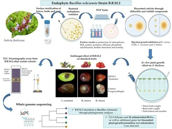
Unveiling the Genomic Basis of Antagonism and Plant Growth Promotion in the Novel Endophyte Bacillus velezensis Strain B.B.Sf.2

by
Dimitra Douka
1,
Tasos-Nektarios Spantidos
1,
Panagiotis Katinakis
1 and
Anastasia Venieraki
2,*
1
Laboratory of General and Agricultural Microbiology, Department of Crop Science, Agricultural University of Athens, Iera Odos 75, 11855 Athens, Greece
2
Laboratory of Plant Pathology, Department of Crop Science, Agricultural University of Athens, Iera Odos 75, 11855 Athens, Greece
*
Author to whom correspondence should be addressed.
Submission received: 11 March 2025 / Revised: 15 April 2025 / Accepted: 16 April 2025 / Published: 4 May 2025

Abstract

:
Background/Objectives: The agriculture sector faces significant challenges due to global climate change, environmental stressors, and rapid population growth, compounded by unsustainable farming practices. This study investigates the potential of the endophytic bacterial strain B.B.Sf.2, isolated from the bark of Salvia fruticosa and identified as Bacillus velezensis through phylogenomic analyses. Methods: To address these issues, eco-friendly techniques, such as the application of plant-associated microbes, are gaining attention. Genome mining revealed numerous secondary metabolite biosynthetic gene clusters associated with plant growth promotion, biocontrol, colonization, and defense elicitation. Results: The strain exhibited strong antagonistic activity against phytopathogens, mediated by diffusible and volatile compound production, along with plant-growth-promoting traits and environmental adaptability. Genome mining revealed numerous secondary metabolite biosynthetic gene clusters associated with plant growth promotion, biocontrol, colonization, and defense elicitation. B.B.Sf.2 effectively inhibited Colletotrichum species causing olive anthracnose and suppressed Botrytis cinerea, the gray mold pathogen, in post-harvest studies on infected fruits. Bioautography of ethyl acetate extracts demonstrated bioactivity against B. cinerea, attributed to iturin-like metabolites. The extracts maintained bioactive properties regardless of fungal interaction. Furthermore, the strain significantly promoted the growth of Arabidopsis thaliana via diffusible and volatile compounds. Conclusions: Our results highlight the multifunctional potential of B.B.Sf.2 as a biocontrol and growth-promoting agent, warranting further evaluation in field applications to enhance sustainable agriculture.

Graphical Abstract

1. Introduction

Sustainable farming is considered one of the greatest challenges that will exist in the 21st century in agriculture. Global climate change, environmental stress, and rapid population growth will be the main threats to worldwide food stability [1,2,3]. In addition, the conventional agricultural practices applied to date (use of agrochemicals) have caused significant environmental and health problems and reduced the soil’s beneficial microbial diversity [2,4,5]. As can be seen, traditional agriculture is gradually being deemed unsustainable to meet the needs of the ever-growing human population. In recent years, there has been an increased demand for more environmentally friendly techniques in agriculture to ensure environmental stability. Much research is now directed towards beneficial microorganisms that interact with plants (plant-growth-promoting microorganisms, PGPMs), promoting plant development and protecting plants from abiotic and biotic stresses [6].
PGPMs can act as biostimulants and biological control agents (BCAs), through direct and indirect modes of action. They can make significant nutrients bioavailable for plants and produce phytohormones and plant growth regulators. Furthermore, they can show antagonistic activity against phytopathogens by producing antimicrobial secondary metabolites, inducing plant defense, and resource competition for habitat and essential nutrients [7,8,9,10]. Among the PGPMs, the subcategory of plant-growth-promoting endophytic bacteria (PGPEB) deserves particular attention. PGPEBs occupy inner plant tissues without inducing disease symptoms, offering multiple benefits to the plant more effectively due to their site of thriving and acting [11]. Medicinal plants are considered a great source of endophytic bacteria with properties for plant growth and biological control potential [12,13]. Moreover, cultivable endophytic bacteria broaden their action since they can colonize the outer plant tissues as well (such as roots and fruits) [14,15].
Bacillus species are highly versatile, thriving in diverse ecological niches and frequently occurring within the PGPEB community [14,16,17]. From a biotechnological perspective, Bacillus species exhibit a diverse secondary metabolism, producing a broad range of structurally distinct antimicrobial compounds—such as bacteriocins, lipopeptides, polyketides, and siderophores—while also enhancing plant growth [17,18,19,20]. In addition, bacteria of this genus show great colonization ability and endospore formation; highly resistant structures to various environmental stresses. All the above-mentioned features are potent enough for Βacillus species to be commercialized successfully as biofertilizers and biological control agents [18,19].
The objectives of present study were (a) endophytic bacteria isolation from plant tissues of the asymptomatic medicinal plant Salvia fruticosa; (b) detection of the most antagonistic bacterial strain against a number of fungal plant pathogens in vitro; (c) investigating plant-growth-promoting capabilities using in vitro biochemical assays and plant growth evaluation experiments; (d) whole-genome sequencing and genome mining analysis to identify genetic traits linked to plant growth promotion, biocontrol, and colonization capabilities; and (e) evaluation of biocontrol potential and antagonistic mechanisms against post-harvest fungal pathogens Colletotrichum acutatum and Botrytis cinerea by employing laboratory-based assays and detached fruit bioassays.

2. Materials and Methods

2.1. Plant Material and Isolation of Endophytic Bacterial Strains

The plant Salvia fruticosa with no visible pathogenicity symptoms was collected from one of the open fields of the Agricultural University of Athens in Attica, Greece. Plant tissues, including leaves, roots, and bark, underwent surface sterilization based on the procedure reported by Kusari et al. (2012) [21]. The excised tissues were disinfected by using 70% ethanol for 60 sec and washing solution (5% sodium hypochlorite solution (v/v) and 0.1% Tween20) for 180 sec. A final sterilization step involved immersion in 70% ethanol for 30 s, followed by three washes with sterile distilled water. To ensure the success of the disinfection, aliquots from the last rinse were cultured on nutrient agar (NA, Conda) at 30 °C for 10 days and monitored for any microbial growth. Plant tissues were ground using a sterile mortar and pestle, and the resulting homogenate was spread onto Petri plates containing nutrient agar (NA, Conda) supplemented with cycloheximide at a concentration of 100 μg/mL. Plates were incubated at 30 °C for 72 h. As an additional sterility control, intact surface-disinfected tissues were also placed on agar without prior maceration. Emerging bacterial colonies were collected daily over the 3-day incubation period and subcultured onto fresh nutrient agar for isolation. For each plate, colonies were selected based on their appearance time and distinct morphological traits, including pigmentation, diameter, and form. The bacterial cultures were kept at 4 °C (Whirlpool, EU) on NA medium and further stored as 40-fold glycerol stock at −80 °C (Thermo Scientific Forma FDE series, H-drive) and cultured at 30 °C (Gallenkamp Orbital Incubator) in nutrient broth (NB, Conda).

2.2. Examination of Plant-Growth-Promoting and Colonization-Related Traits

To assess the beneficial bacterial properties, several key functional traits were examined. These included siderophore production, phosphate solubilization, acetoin biosynthesis, enzymatic activities, motility, biofilm formation, and IAA production.

2.2.1. Siderophore Production Assay

The production of siderophores was assessed using the Chrome Azurol S (CAS) agar assay. A 10 μL aliquot of an overnight bacterial culture was spotted onto CAS agar plates and incubated at 30 °C for three days. The production of siderophores was designated by the development of an orange halo surrounding the colony [22].

2.2.2. Phosphate Solubilization Assay

The bacterial ability to solubilize tricalcium phosphate was tested using Pikovskaya medium. A 10 μL bacterial culture was spotted on the medium and incubated at 30 °C for seven days. Indicating phosphate solubilization was a transparent halo surrounding the bacterial colony [23].

2.2.3. Acetoin Production Assay

Acetoin production was assessed using the Voges–Proskauer (VP) test [24]. Acetoin biosynthesis showed a positive result when the liquid medium turned pink-red.

2.2.4. Cellulase Production Assay

For cellulose production testing bacterial inoculation was conducted on CYEA medium supplemented with 1% carboxymethyl cellulose (CMC) and incubated at 30 °C for three days. The plates were subsequently treated with Congo Red solution (1 mg/mL) after the incubation period, followed by 1 M NaCl to remove unbound dye, revealing a clear halo around cellulolytic colonies [25].

2.2.5. Urease Production Assay

Urease activity plays a role in nitrogen metabolism and environmental adaptation. To assess urease production, 10 μL of overnight bacterial culture was spotted onto Urea Base Christensen ISO 6579 [26], ISO 19250 [27] medium (Conda, Madrid, Spain) and incubated. Indicative of ureolytic activity was a pink halo around the colony [28].

2.2.6. Protease Production Assay

Proteolytic activity is essential for bacterial nutrient acquisition and plant colonization. To assess protease activity, bacterial cultures were spot-inoculated (10 μL of over-night-grown culture) onto CYEA medium (composed of 0.5% casein, 0.25% yeast extract, 0.1% glucose, and 1.5% agar) supplemented with 7% skim milk powder. The plates were incubated at 30 °C for a period of 3 days. Protease secretion was defined by a clear halo surrounding bacterial colonies following three days of incubation at 30 °C [29].

2.2.7. Motility Assays (Swarming and Swimming)

Bacterial motility is a crucial factor for successful plant colonization. Swarming and swimming motility were examined by spot inoculating 5 μL and 3 μL, respectively, of bacterial cultures onto nutrient agar medium amended with 0.5% agar (for swarming motility) and 0.3% agar (for swimming motility). Plates were incubated at 30 °C for 24 h, and motility was assessed based on bacterial colony expansion.

2.2.8. Biofilm Formation Assay

Biofilm formation enhances bacterial survival and plant colonization. This ability was tested using a crystal violet staining assay in 96-well PVC plates. A 1:100 dilution of an overnight bacterial culture was inoculated into wells (five replicates) and incubated at 30 °C for one day. The wells were washed, treated with crystal violet dye, and then destained with an ethanol–acetone solution. The intensity of the resulting purple color indicated biofilm formation [30].

2.2.9. Indole-Related Compounds Production Assay and Quantitative Analysis of IAA Production

The Salkowski method [31] was used to examine the production of indole-related compounds. The Salkowski reagent consisted of 2% 0.5 M FeCl3 and 35% HCLO4, was mixed with the supernatant from a 48 h bacterial culture grown in LB supplemented with 1% L-Tryptophan (Sigma-Aldrich®, Merk KGaA, Burlington, MA, USA) in a ratio of 1:2, and the mixture remained at room temperature in the dark for 30 min.
The development of a red coloration from an initial orange hue signified the presence of indole-associated compounds. For the quantitative determination of IAA production, a UV spectrophotometer was used to measure absorbance at 530 nm following the same Salkowski assay conditions [32,33].
All assays were performed in triplicate across three independent experiments to ensure reproducibility and reliability.

2.3. Evaluation of Antagonistic Activity

All isolated strains were screened for their antagonistic activity against Rhizoctonia solani Kühn BPIC2531, and the most promising strains were further tested against fungal plant pathogens Botrytis cinerea, Fusarium oxysporum f.sp. radicis-lycopersici (FORL), Colletotrichum acutatum, and Verticillium dahliae using dual culture assay on NA medium (Conda) in order to evaluate the best antagonistic strains. A mycelial plug (6 mm) was excised from a 10-day fungal culture grown on PDA medium (Conda) and positioned 3 cm across a 5 μL bacterial spot from an overnight NB culture. Single-culture conditions of each microorganism were used as experimental controls. All treated plates were incubated at 25 °C for 15 days, with the experiment conducted in triplicate; fungal cultures grown on PDA without bacterial application served as controls. The mycelial radius of each phytopathogenic fungus was calculated in order to determine the bacterial inhibition effect on fungal growth by the type Ι% = (ρ1 − ρ2)/ρ1 × 100 [34]. The experiment was performed 3 times. As a control, we used 3 fungal cultures on PDA medium without bacterial treatment.
To examine the role of bacterial volatiles on fungal plant pathogens inhibition, the modified protocol of Hunziker et al. (2015) [35] combined with that of Yuan et al. (2012) was used [36]. All isolated strains were tested against four phytopathogenic fungi B. cinerea, F. oxysporum f.sp. radicis-lycopersici (FORL), C. acutatum, and V. dahliae using dual culture assay on I plate containing PDA on the one half and ½MS with 0.8% agar and 1.5% sucrose on the other. A mycelial plug (6 mm) was excised from a 10-day fungal culture on the one half with PDA medium (Conda). Then, 3 spots of 20 μL of bacterial inoculant (108 CFU/mL) were inoculated on the other half. As a control, 3 fungal cultures on PDA medium without bacterial treatment were used. The mycelial area of each phytopathogenic fungus was calculated to determine the inhibition effect of bacterial volatiles on fungal phytopathogenic growth by the type Ι% = (E1 − E2)/E1 × 100.

2.4. In Vitro Plant Growth Effect on A. thaliana Col-0 Seedlings

The surface sterilization of A. thaliana Col-0 seeds followed the protocol described in a previous study [37]. The seeds were washed in 70% ethanol for 0.5 min, followed by a washing solution containing 5% (v/v) aqueous solution of 5% sodium hypochlorite solution and 0.1% Tween 20 for 1.5 min. For 30 s, the seeds were re-immersed in 70% ethanol before being rinsed three times in sterile dH2O.
Seeds were germinated on Murashige and Skoog medium at half-strength concentration medium (½MS) medium (MS0222, Duchefa Biochemie, Haarlem, The Netherlands), including vitamins, encompassing 0.6% agar and 1.5% sucrose. All plates were maintained at 4 °C for 2 days post-sowing, then incubated at 22–25 °C under a 16:8 light-dark cycle in a growth chamber, oriented at 70° or horizontally (I plates) until used for transplantation. For our experiment, following the established preparation protocol, 3-day-old seedlings were placed on ½ MS medium (0.8% agar, 1% sucrose; 6 seedlings/dish). Then, a 10 μL bacterial suspension (108 CFU/mL) was spotted 3 cm from each root apex, and the plates were incubated at 70° for 9 days. As a control, we used plants without bacterial treatment. Weight and photographic measurements of the plants were taken to determine morphological characteristics.
To examine the role of bacterial volatiles on plant growth, we followed the protocol of Asari et al., (2016) [38] with some modifications. Seeds were surface-sterilized and sown on one half of an I plate containing ½MS including vitamins amended with 0.8% agar and 1.5% sucrose (6 seeds/Petri dish) and were placed at 4 °C for 48 h. Then, 3 spots of 20 μL of bacterial inoculant (108 CFU/mL) were inoculated on the other half. The plates were sealed with double parafilm and placed horizontally in a growth chamber (16 h light: 8 h dark photoperiod, 22–25 °C) with a 14-day incubation period preceding fresh weight and leaf area determination. As a control, we used plants without bacterial treatment. The plants’ morphological traits were deciphered by weighing and photographing them.

2.5. In Vivo Biological Control Against Colletotrichum acutatum and Botrytis cinerea on Detached Fruits

Olives (Olea europaea var. Kalamata), grape berries (Vitis vinifera var. Fraoula), and strawberries (Fragaria ananassa var. Camarosa) were selected based on the absence of visible injuries or infection and the homogeneity of their morphological characteristics (color, size, and maturity).
For the preparation of the fruits, they were rinsed with tap water and dH2O and then immersed in a 3% (v/v) washing solution (5% sodium hypochlorite solution) for 4 min. After being thoroughly cleaned with sterilized dH2O, they were dried in a sterile laminar flow chamber. Then, the corresponding empty Petri dish was filled with treated fruits, which received a 3 mm × 3 mm artificial wound.
For the grape berries (Vitis vinifera var. Fraoula) and olives (Olea europaea var. Kalamata) experiments based on Lian et al. (2017) [39] protocol. A 20 μL aliquot of the bacterial culture, containing vegetative cells at a concentration of 108 CFU/mL, was inoculated into the wound of each fruit. Following inoculation, fruits were placed in Petri dishes within high-humidity chambers (>90% humidity, sterilized transparent containers lined with wet filter paper). After 1 h, 20 μL of the fungal phytopathogen spore suspension (106 spores/mL) was re-inoculated into the same wound, and the fruits were incubated for 4 more days. For the control fruits in this assessment, we inoculate the wounded fruits by using only bacterial inoculants in the absence of fungal phytopathogen (B. cinerea or C. acutatum) and vice versa. For the strawberries (Fragaria ananassa var. Camarosa), based on the Essghaier et al. (2008) [40] protocol, the bacterial inoculum was applied to the fruits by spraying with the suspension 108 CFU/mL and left them to dry. After 1 h, the fruits were sprayed with the conidial suspension of B. cinerea (106 spores/mL), and the fruits were further incubated for 3 days. As controls in this assay, we inoculated the harvested fruits by using only bacterial inoculants in the absence of B. cinerea and vice versa. All the experiments consisted of 36 fruits (12 fruits per treatment, with 3 biological replications). We used the following rating scale to describe the mycelial and rot coverage of each fruit and consist of 4 classes: 0 = healthy fruit, 1 = 1–20%, 2 = 20–50%, 3 = 50–75%, 4 = 75% and above according to the visible fruit surface from the observer. Disease incidence (%) was calculated as the percentage of infected fruit according to the type; Di = (A/Β) × 100, where A is the number of infected fruits and Β the total number of fruits. The disease severity index was calculated according to the formula Ds = [Σ(Ci)/n*Z] × 100, where Σ(Ci) total number of fruits in each corresponding class, n is the total number of fruits and Z is the number of the highest class.

2.6. Extraction of Agar-Diffusible Compounds and Bioactivity Through TLC-Bioautography Testing

For agar-diffusible compounds from B.B.Sf.2, the protocols of Bertrand et al. (2013) and Costa et al. (2019) [41,42] were used with some modifications. For the experiment, we used Petri dishes with single culture of B.B.Sf.2 and dual culture of B.B.Sf.2 -Botrytis cinerea treatments, incubated for 4 days at 25 °C.
Next, with a sterile scalpel, the zones of each treatment—the inhibition zones of the double cultures and the same weighting and location zones of the single cultures—were removed, cut, and weighed. They were vortexed and sonicated in a water bath (Elmasonic S30H, Elmasonic Schmidbauer GmbH, Singen, Germany) for 30 min at room temperature in bottles containing ethyl acetate and 0.1% formic acid (Millipore Sigma, Burlington, MA, USA). The resulting solution was filtered with Whisman’s filter and dried in the vacuum evaporation chamber (Genevac HT-4, SP Scientific, Ipswich, Suffolk, United Kingdom). The dried material was reconstituted with 100 μL methanol of HPLC grade and then filtered by Whatman Uniflo® (Millipore Sigma, Burlington, MA, USA).
Thin-layer chromatography (TLC) was performed on extracts obtained from both monoculture and co-culture samples, following the protocol described by Calvo et al., 2019 [43]. A chloroform–methanol–water mixture (65:25:4, v/v/v) v and v) was employed as the mobile phase while Silica gel 60 F254 plates (20 × 20 cm; layer thickness, 0.20 mm; Merck) served as the stationary phase. For bioautography assay, the developed chromatograms were covered with B. cinerea spore suspension (107 spores/mL) in PDA semisolid agar medium (0.8% w/v). Tetrazolium salt dye solution (2.5 mg/mL) (Sigma-Aldrich®, Merk Merck KGaA, Burlington, MA, USA) was sprayed onto the TLC plate and incubated at 25 °C for 48 h. This dye solution stains living cells purple.
Then, the TLC plate was documented photographically, and the retention factor (Rf) values corresponding to the visible clear zones were recorded as indicative of bioactive compounds showing antifungal activity. The Rf values were determined based on the formula: Rf (retention factor) = traveling distance of the solute/traveling distance of the solvent front.

2.7. Phylogenetic Taxonomy on 16S rRNA Sequence

A Nucleospin® Microbial DNA (Macherey-Nagel GmbH and Co. KG, Düren, Germany) kit was used to extract bacterial DNA from an overnight culture. The 16S rRNA gene fragment was amplified using the following primers: 5′-AGAGTTTGATCCTGGCTCAG-3′ and 5′-ACGGCTACCTTGTTACGACTT-3′ [44]. To identify the genus and the phylogenetically related strains deposited in GenBank database, BLASTn search (BLAST+ 2.16.0) was performed.

2.8. Whole-Genome Sequencing (WGS)

In order to isolate genomic DNA of the strain an overnight liquid culture was grown and the protocol of the PureLink® Genomic DNA Mini Kit (Thermo Fisher Scientific, Carlsbad, CA, USA) was used. The sequencing was performed by SNPsaurus (Eugene, OR, USA) using an Illumina HiSeq2000 platform. A library was generated using a Nextera XT DNA Library Prep Kit. The trimming of the sequencing was prepared with BBDuk and the assembling with SPAdes-3.12.0, considering the default parameters [45].
The final de novo genome of the B.B.Sf.2 strain (NCBI accession: JAEACM000000000.1) was assembled in 7 scaffolds with a total genome size of 4,047,560 bp.
The phylogenetic identity of B.B.Sf.2 was calculated by digital DNA:DNA hybridization (dDNAH) using the GGGD 2.1 web service according to the recommended formula and orthologous mean nucleotide identity (OrthoANI) [46]. For the analysis of the ANI and the dDDH, the threshold values for species separation were proposed by the standard analysis (95–96% and 70%, respectively) [46,47].
Phylogenomic analysis of the strain was conducted using the Type (Strain) Genome Server (TYGS; https://tygs.dsmz.de, accessed on 28 November 2024), and the phylogenetic tree was constructed employing the FastME algorithm based on Genome BLAST Distance Phylogeny (GBDP) distance calculations [48].

2.9. In Silico Genomic Analysis

Carbohydrate-active enzymes (CAZymes) present in the core genome were identified using the dbCAN2 meta server, in conjunction with Hotpep, HMMER, and DIAMOND algorithms for accurate annotation for CAZymes [49]. For the prediction and annotation of secondary metabolites’ biosynthetic gene clusters, we used the antiSMASH 6.0 server [50].
The ClusterBlast and Known ClusterBlast analysis through the antiSMASH 6.0 server were used for comparative gene cluster procedure based on the Biosynthetic Gene Cluster (MIBiG) database [51].

2.10. Statistical Analysis

Statistical analyses and plots were performed using GraphPad Prism software v.10.4.1 (GraphPad Software, San Diego, CA, USA). The normality of the data was assessed using the Shapiro–Wilk test, and the homogeneity of variances was evaluated with Levene’s test. Differences between the bacterial treatment and the control were analyzed using a two-tailed independent samples Student’s t-test, with statistical significance set at p < 0.05. The average values are indicated by plots, standard deviation by error bars, and statistically significant differences by asterisks.

3. Results

3.1. Isolation of Endophytic Bacteria from Salvia fruticosa

A total of 100 single bacterial colonies were isolated from leaves, barks, and roots of the medicinal plant Salvia fruticosa (Table S1). Macroscopical and stereoscopic testing were performed to evaluate the characteristics of these colonies.
Massive biocontrol testing through a multiple-culture system in vitro was carried out to determine whether any bacterial isolates had antagonistic activity against the fungal plant pathogen Rhizoctonia solani. B.B.Sf.2 showed the most promising activity among the other isolates, in terms of the size of the inhibition zone, so it was selected for further characterization.
BLASTn analysis of the 16S rRNA sequence (PQ867398.1) and comparison to type strains of the GenBank database of NCBI revealed that this strain is more closely related to Bacillus species, with identity > 97% to Bacillus velezensis strain HBPT71 (acc. number PQ805259.1), Bacillus velezensis strain HBPT105 (PQ804066.1), B. velezensis strain HBPT139 (PQ804358.1), B. subtilis strain GX S-12 (KU904290.1) and Bacillus sp. strain PGR 008 (MH158241.1). Comparative analysis of the 16S rRNA gene sequence (PQ867398) using BLASTn against type strain sequences in the NCBI GenBank database indicated that the strain exhibits > 97% sequence identity to B. velezensis strain HBPT71 (acc. number PQ805259.1), B. velezensis strain HBPT105 (PQ804066.1), B. velezensis strain HBPT139 (PQ804358.1), B. subtilis strain GX S-12 (KU904290.1) and Bacillus sp. strain PGR 008 (MH158241.1), suggesting a close phylogenetic affiliation with this group.

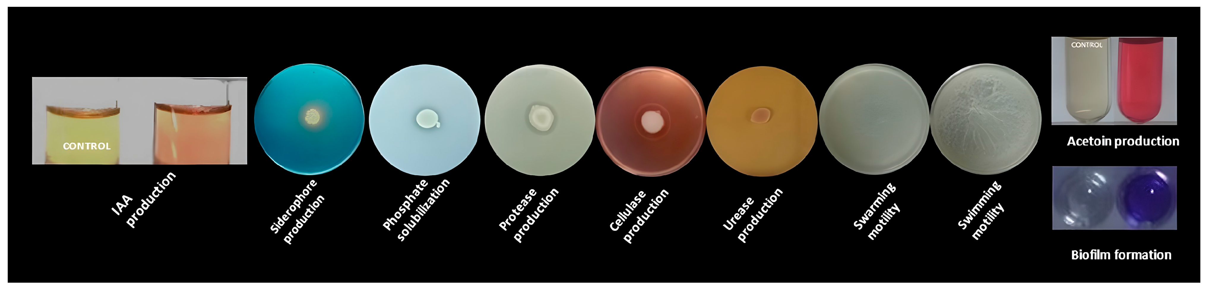
3.2. In Vitro Traits Associated with B.B.Sf.2 Plant Growth Promotion and Biocontrol Potential

The B.B.Sf.2 strain was evaluated for environmental, in vitro characteristics indicative of plant-growth-promoting and biocontrol capabilities. B.B.Sf.2, as depicted in Figure 1, could mobilize chelated iron and precipitated phosphate and was capable of producing acetoin in vitro. It could also secret cellulase and protease, but it could not produce urease. B.B.Sf.2 also could produce indole-related compounds and exhibited excellent swarming, swimming motility, and biofilm formation in polystyrene wells. The results confirm the production of IAA by the bacterial strain. The concentration of IAA production is 57.42 ± 1.25 μg/mL. The value is presented as mean ± SD of three replications (Table S2).
Furthermore, the strain B.B.Sf.2 was tested for its antagonistic capability against various fungal plant pathogens, including B. cinerea, FORL, C. acutatum, and V. dahliae in vitro (Figure 2).
The results reveal that B.B.Sf.2 showed remarkable biocontrol against a wide range of fungal plant pathogens but proved to be more effective via diffusible compound secretion than VOC emissions (Table 1).

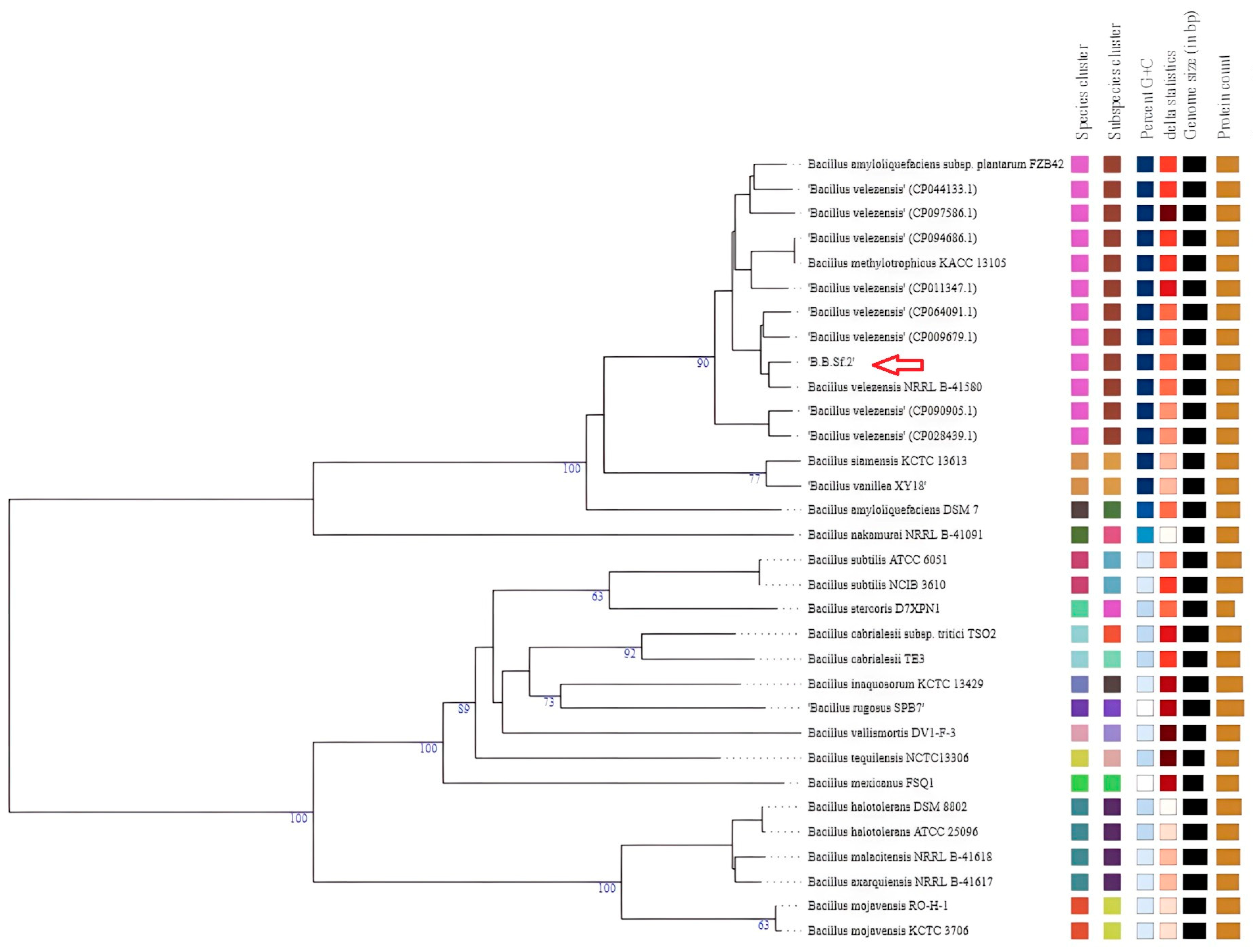
3.3. Phylogenomic Taxonomy of the Bacterial Endophytic Strain B.B.Sf.2

To further verify the classification of the B.B.Sf.2 strain, a phylogenomic approach was employed. Initially, whole-genome sequence (WGS) analysis of the B.B.Sf.2 strain, conducted via the Type (Strain) Genome Server (TYGS) in comparison with other Bacillus genomes, revealed a close taxonomic relationship to B. velezensis (Figure 3). OrthoANI values between the B.B.Sf.2 strain and ten B. velezensis reference strains ranged from 97.62% to 99.40%, while digital DNA–DNA hybridization (dDDH) values ranged from 79.7% to 95%. These values exceed the generally accepted species delineation thresholds [46,47]. Accordingly, the ANI and dDDH results were congruent with findings from genome-based phylogenetic analysis, supporting the classification of strain B. velezensis (Table 2).

3.4. Genetic Basis of Plant Growth, Colonization, and Biological Control Activity of the B.B.Sf.2 Strain

The analysis of the whole genome (WGS) of the B.B.Sf.2 strain (B. velezensis) showed the existence of genes involved in biological control, plant growth stimulation, and colonization. Specifically, in the genome of the B.B.Sf.2 strain, genes related to plant-growth-promoting properties, such as indoleacetic acid biosynthesis (trpA-trpF), phosphorus solubilization (phoA, phoB, phoD, phyC), and the production of 2, 3 butanediol and acetoin (bdhA, budA, alsS, ilvN, ilvB) were found, as well as spermidine involved in abiotic stress mitigation (speE, speB). Furthermore, genes related to biofilm formation (wecB, csrA, rpoN, remA, YlbF), chemotaxis (CheA, CheC, CheD, CheV, CheW, CheY), swarming motility (swrA, swrB), the production of exopolysaccharides (epsG), as well as genes related to flagellum synthesis and function (fliS, fliD, fliW, flgL, flgK, flgN, flgM, flgB, fliE, fliG, fliJ, flbD, fliM, fliQ, fliR) were found. Additionally, 112 genes encoding CAZymes were identified in B.B.Sf.2 (B. velezensis); 40 of these belonged to families of glycoside hydrolases, 38 to glycosyltransferases, 3 to lyases, 12 to polysaccharide esterases, 5 to growth factors and 14 belonged to families of carbohydrate-binding proteins. Approximately 27.67% of the aforementioned CAZymes contain an amino-terminal of the signal peptide that facilitates the export across the cytoplasmic membrane. Among the CAZymes, enzymes involved in the cell wall hydrolysis of the fungi were found, such as chitosanase (GH8, GH46), chitinase (GH18), and glucanase (GH51), as well as enzymes responsible for endophytic colonization such as β-glucosidase (GH1, GH3) and various pectinases (PL1, PL9) (Table S3).

3.5. Detection of Secondary Metabolites Gene Clusters

The genome of bacterial strain Β.Β.Sf.2 was further analyzed using antiSMASH in order to detect secondary metabolites’ biosynthetic gene clusters. According to antiSMASH analysis, Β.Β.Sf.2 possesses 16 individual BGCs (Table 3), across 14 biosynthetic regions. These include biosynthetic gene clusters (BGCs) producing either known or novel/non-registered compounds in the MIBiG database.
Regarding the non-registered compounds, 2 BGCs of terpenes were identified in regions 6.2 and 7.2. Based on the ClusterBlust analysis, the first showed 95% homology with other B. velezensis strains (strains ΝΥ12-2, CACC316, etc.), as well as the second showed a high homology of 100% with other B. velezensis strains (strains SRCM102755, DH8043, etc.). Also, in region 1.2, a non-ribosomal peptide synthetase (NRPS) BGC was identified that was not linked to any characterized compound; however, ClusterBlast analysis revealed 100% sequence similarity to BGCs previously reported in other B. velezensis strains as JSRB166, SRCM102755, etc. (Figure 4). Similarly, in region 7.3, a BGC of type III PKS was identified with 100% gene similarity compared with other strains (strain AP183, SQR9, etc.), as well as in region 5.1, a BGC of phosphonate was found with 100% homology with other B. velezensis (strain ATR2, CN026, etc.). Furthermore, in region 6.1, a gene cluster was located that had a similarity of 7% with a known BGC encoding butirosin polyketide synthase A. The antiSMASH Cluster Blust analysis showed the presence of the putative butirosin A gene cluster of the B.B.Sf.2 strain, which had high gene similarity (100%) with a new PKS gene cluster found in several B. velezensis strains (Bac57, JSRB 166, and other strains). Regarding the remaining known clusters, they presented 100% homology with the deposited BGCs and similar gene organization to other B. velezensis strains representing biosynthetic genes of bacilysin, mersacidin, macrolactin H, bacillaene, difficidin, and fengycin, as well as surfactin (82% homology) with the reference strains of the MIBIG database (Table 3). More specifically, in region 1.1, 2 BGCs were identified where the first showed 100% homology with amylocyclicin, while the second showed 100% homology with bacillibactin. Similarly, in region 7.1 were located 2 biosynthetic clusters, the first concerned the BGC of fengycin/plipastatin, while the second presented as an iturin like cluster showed high homology with the known BGC of bacillomycin D (100% homology), mycosubtilin (100% homology) and iturin A (88% homology). However, the predicted amino acid sequence indicated that this BGC encodes Bacillomycin D synthases due to the presence of amino acids Pro4, Glu5, and Thr7, which are unique to Bacillomycin D, while the BGC of fengycin was related to the compound plipastatin because of the D configuration of Tyr9.

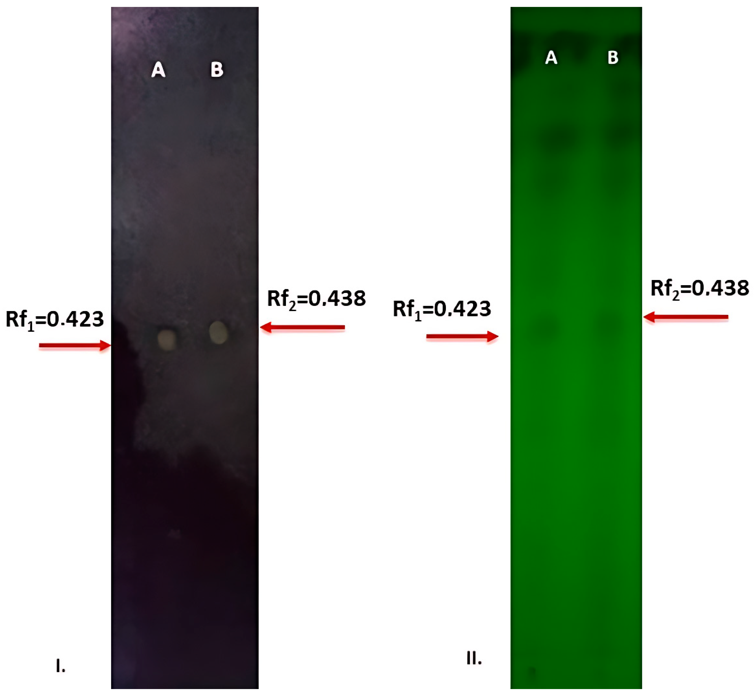
3.6. TLC-Bioautography Assay

As described above, the B.B.Sf.2 strain exhibited strong antagonistic activity in vitro through diffusible metabolites, particularly against B. cinerea. However, it seems difficult to determine whether these secondary metabolites were secreted by the B.B.Sf.2 as a single culture or in its interaction with the fungus (dual culture). Thus, we performed ethyl acetate extracts from single and dual cultures. After TLC-bioautography analysis, it was noticed that both secretomes exhibited the same patterning active spots of antagonistic activity against B. cinerea with Rf1 = 0.423 and Rf2 = 0.438 for extracts from single and dual cultures, respectively (Figure 5).

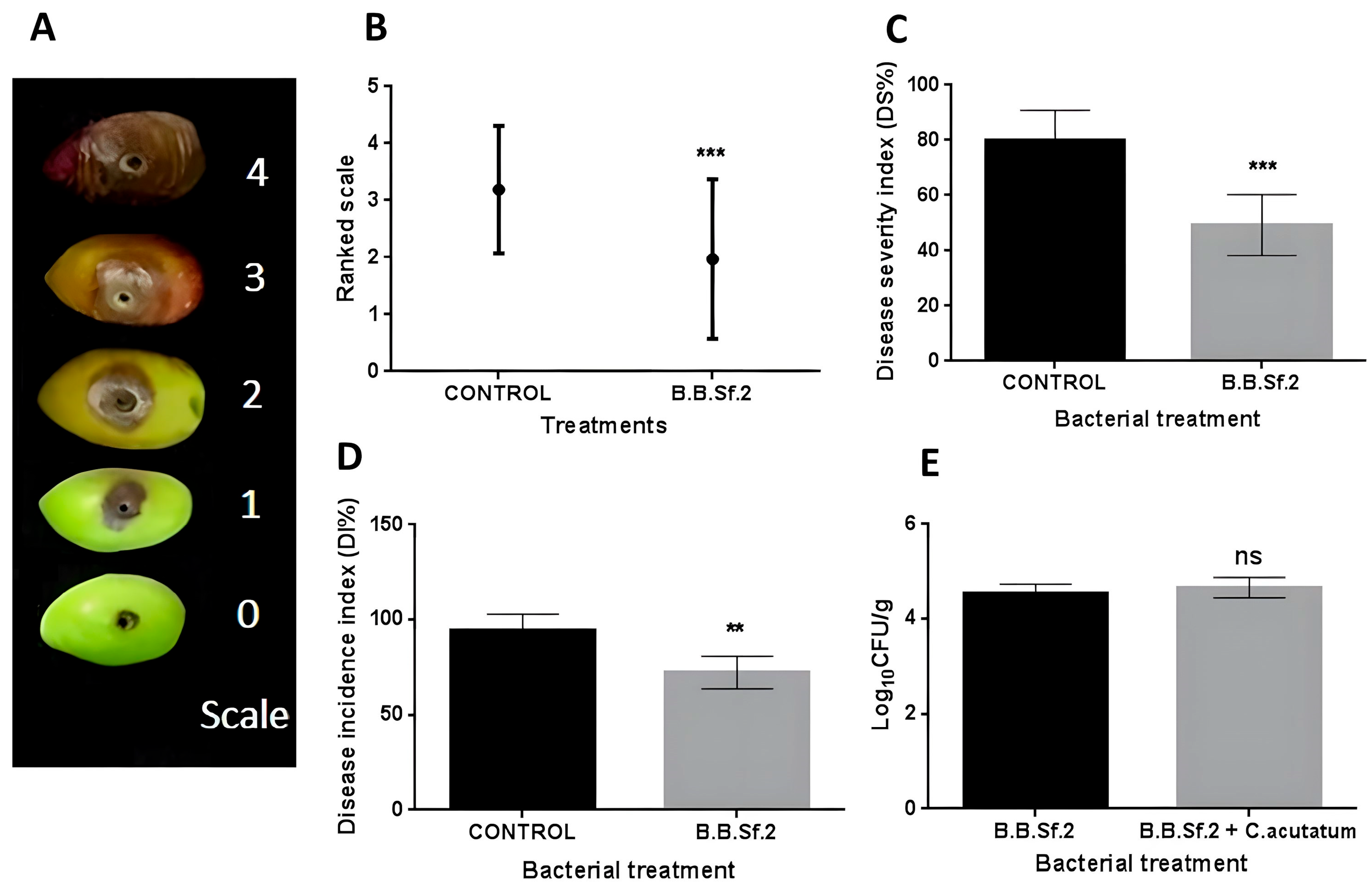
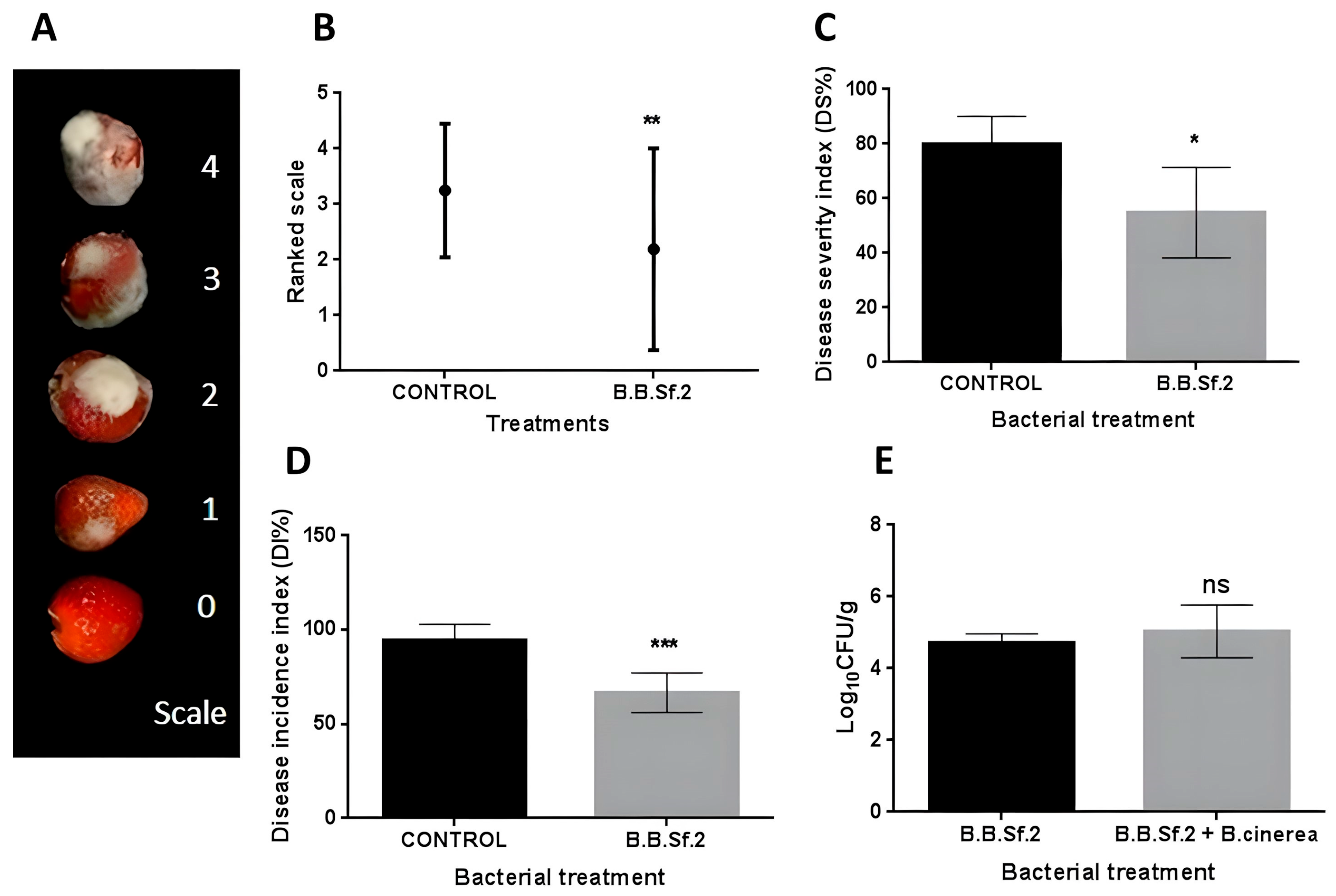
3.7. Biological Control Activity of the B.B.Sf.2 Strain on Detached Fruits

As mentioned above, the B.B.Sf.2 strain possesses antifungal activity against four significant phytopathogenic fungi in vitro. In the next step, the in vivo biological activity of the strain was examined on artificially infected detached fruits. The purpose of this experiment is to reveal the potential protective activity when the bacterial strains were applied as a preventative treatment.
Artificially wounded olive fruits were inoculated with 20 μL of bacterial culture suspension (108 CFU/mL), and when dried, they were treated with 20 μL of spore suspension (106 spores/mL) of the phytopathogen C. acutatum. The results show that the disease incidence index (Di) was significantly reduced after treatment with B.B.Sf.2 strains (Figure 6). The Di% percentage of the strain was 72.21%, significantly lower than the 94.44% of the control. The disease intensity index (Ds) was significantly reduced after the treatment of the B.B.Sf.2 strain (Ds = 49.30%) compared with the control (Ds = 79.85%). Regarding the bacterial inoculum application upon fruits, in the absence of a phytopathogenic fungus, the strain did not cause any symptoms of pathogenicity.
In addition, the antagonistic efficiency of the B.B.Sf.2 strain against the gray mold disease caused by B. cinerea was tested on detached grape berries and strawberries. Artificially wounded grape berries were inoculated with 20 μL of bacterial culture suspension (108 CFU/mL), and when dried, they were treated with 20 μL of spore suspension (106 spores/mL) of the phytopathogen B. cinerea, as described above. According to our results, the antagonistic activity regarding disease incidence index (Di) was quite low, where B.B.Sf.2 showed a significant reduction with a rate of 91.66% compared with the control (Di = 100%). However, the results differ in the disease severity index (Ds), where the B.B.Sf.2 strain indicated a significant reduction in the Ds (approximately 43.74%) compared with the control (Ds = 80.55%). Lastly, no pathogenicity symptoms were observed during the bacterial inoculum application on the fruits (Figure 7).
The application of bacterial inoculum upon the strawberries was different from the previous assays, which were performed by spraying to cover the whole fruit surface, due to the fruit-specific conditions. The antagonistic activity of the strains according to Di index values was relatively low, where the B.B.Sf.2 strain (Di = 66.66%) proved to be effective. The Di value of B.B.Sf.2 was significantly lower than that of the control (Di = 94.44%). In addition, the Ds index value of the B.B.Sf.2 strain significantly reduced according to the Ds index value of the control (Ds = 79.85%). Finally, it should be emphasized that the fruits to which only the tested strains were applied did not show any pathogenicity symptoms (Figure 8).

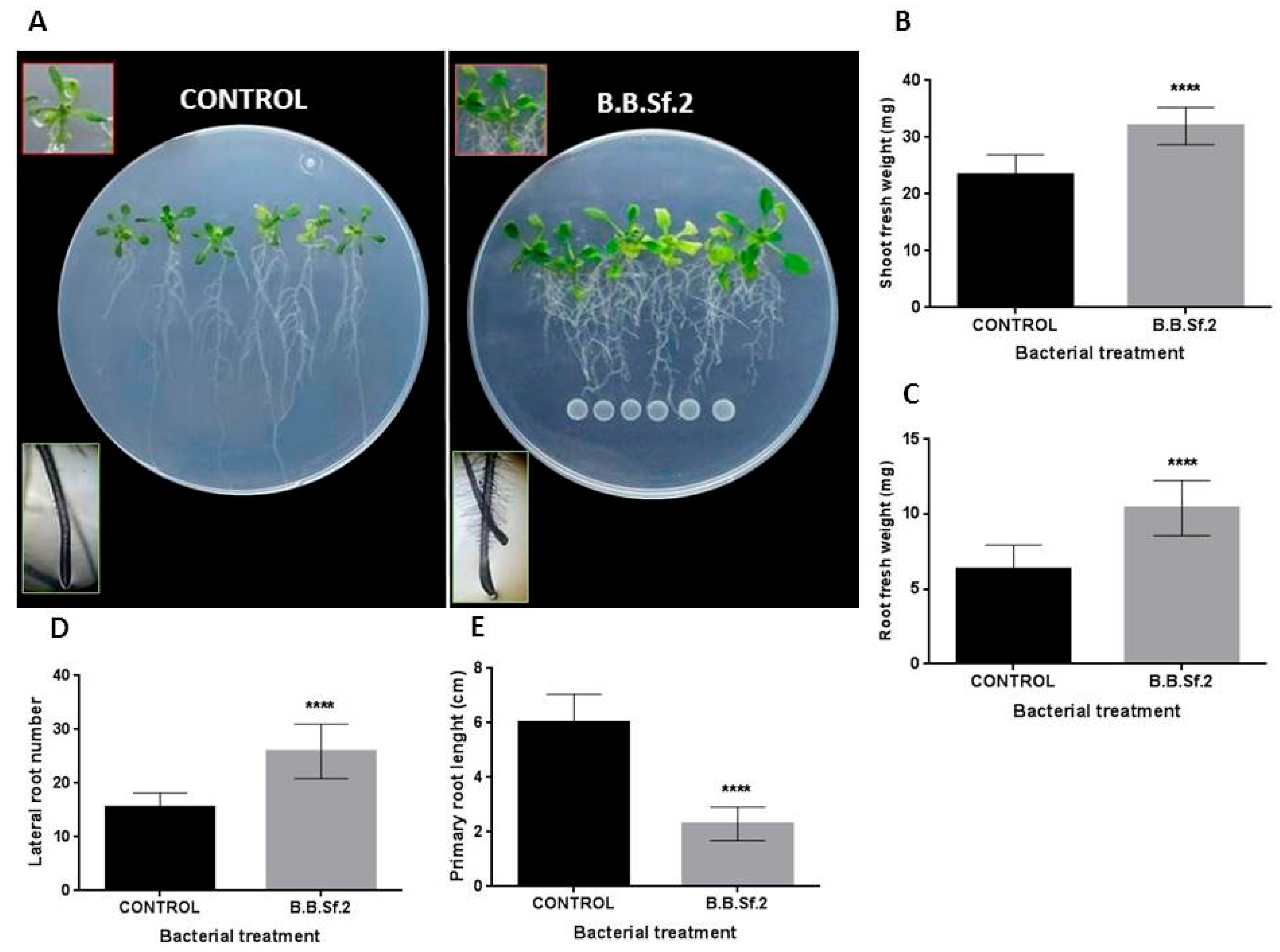
3.8. B.B.Sf.2 Possesses Plant-Growth-Promoting Potential on the Model Plant A. thaliana In Vitro

Apart from the promising biocontrol activity of the B.B.Sf.2 strain, the plant-growth-promoting effect was investigated in vitro on A. thaliana seedlings growing vertically on MS agar medium. To examine the effect mediated through diffusible and/or volatile compounds by bacterial inoculant, we used plates with transplanting seedlings with and without center partition. After 9 days of co-culture, it was found that the bacterial suspension inoculated below the root tip was capable of stimulating plant biomass production and increasing the shoot fresh weight. Also, the B.B.Sf.2 strain triggered morphological alterations in the root system architecture. The most obvious morphological modifying characteristics in the root system appear to be the limitation of the primary root and the stimulation of lateral root formation; as was noted, the total root surface area increased compared with the untreated plants (Figure 9). Furthermore, the volatile compounds experiment showed that the B.B.Sf.2 strain affected the growing seedlings positively after 14 days of co-culture in I-plates by increasing shoot fresh weight and leaf area.

4. Discussion

Medicinal and aromatic plants host a diverse range of endophytic bacteria that contribute to biocontrol and promote plant growth [13,52,53]. Bacillus species are commonly isolated from medicinal plants due to their adaptability and beneficial plant interactions. These bacteria serve as valuable agents for biostimulation and disease management [54,55,56]. Salvia fruticosa, a perennial medicinal plant, is renowned for its antimicrobial, antitumor, and antioxidant properties [57,58,59]. This study is the first to analyze endophytic bacteria from S. fruticosa for biocontrol and growth-promoting traits, highlighting strain B.B.Sf.2 for its potential benefits.
The endophytic bacterial strains that we studied were isolated from S. fruticosa leaves, roots, and bark tissues, with the B.B.Sf.2 strain showing the strongest antagonistic effect against Rhizoctonia solani in vitro and selected for further study. It exhibited broad antifungal activity via diffusible and volatile compounds and demonstrated key plant-growth-promoting traits, including acetoin production, iron chelation, phosphate solubilization, and lytic enzyme secretion. Additionally, B.B.Sf.2 displayed swarming, swimming, and biofilm formation, enhancing its ability to colonize plant tissues [11,60,61,62,63].
Taxonomic analysis of 16S rRNA gene sequences revealed a high similarity to Bacillus amyloliquefaciens, B. subtilis, and B. velezensis, which are often indistinguishable by traditional methods [64]. Advances in whole-genome sequencing and phylogenomic analysis now allow precise species differentiation [65]. In this study, phylogenomic analysis placed B.B.Sf.2 within a distinct B. velezensis cluster, confirmed by ANI and dDDH values, leading to its classification as B. velezensis.

4.1. Genome Mining and Functional Insights

Genome mining provided crucial insights into plant-growth-promoting (PGP) and biological control activity of the B.B.Sf.2 strain, confirming several in vitro findings. Genes encoding acetoin and 2,3-butanediol (alsD, ilvB, alsR, bdhA, acuC) were identified, supporting plant growth stimulation and induced systemic resistance (ISR). Additionally, genes involved in indole-3-acetic acid (IAA) synthesis (trpA–trpF), phosphorus solubilization, and siderophore production (e.g., bacillibactin) were detected, enhancing nutrient bioavailability and soil fertility [66,67].
Carbohydrate-active enzyme (CAZyme) analysis revealed genes for enzymatic degradation of complex compounds, aiding bacterial survival and plant disease suppression by disrupting pathogen cell walls and triggering plant immunity. Chitinase and chitosanase, identified in the B.B.Sf.2 genome, serve as key elicitors of plant defense and exhibit strong antifungal activity. Additional enzymes, including endoglucanase, β-glucosidase, α-amylase, β-xylosidase, and various pectate lyases, contribute to microbial endophytism [60,68,69,70,71,72].
Genes supporting motility and colonization were also identified. Flagellar biosynthesis (fli, flh), swarming regulation (SwrA, SwrB), and chemotaxis (che) genes facilitate movement and habitat occupation. Biofilm formation was associated with rpoN, remA, and YlbF, while csrA regulated biofilm disaggregation and planktonic cell dispersion, optimizing bacterial adaptability [73,74,75,76,77,78,79].

4.2. Biosynthetic Gene Clusters and Secondary Metabolite Potential

AntiSMASH analysis of the B.B.Sf.2 genome revealed its potential to synthesize a diverse range of antimicrobial secondary metabolites. Sixteen biosynthetic gene clusters (BGCs) were identified across 14 genomic regions. Among these, 11 were known, while five were uncharacterized, potentially encoding novel bioactive compounds. The evolution of such genes is often driven by mutations or horizontal gene transfer (HGT), facilitating the acquisition of external DNA through mobile genetic elements [80,81,82].
BlastN analysis identified an NRPS gene cluster in genomic region 1.2, containing genes such as amino acid adenylation domain proteins (02160, 02155), ABC transporter ATP-binding protein (02150), saccharopine dehydrogenase NADP-binding domain protein (02145), DUF3810 domain protein, and thioesterase (02135). This cluster showed ~95% sequence similarity to one found in the phylogenetically distant Paenibacillus sp. strain FSL-W7 1279, suggesting acquisition via HGT. According to antiSMASH predictions, this NRPS cluster may synthesize a peptide with the sequence (D-cys)-Ser-cys-ala-X-asn-(D-asn) [83].
Additionally, a putative PKS gene cluster in region 6.1 exhibited only 7% similarity to butirosin A, an aminoglycoside antibiotic from Bacillus circulans [84]. The low sequence similarity suggests that this cluster may encode a novel antibacterial compound, warranting further investigation.

4.3. Secondary Metabolite and Antimicrobial Potential of B.B.Sf.2

The known biosynthetic gene clusters (BGCs) in the B.B.Sf.2 genome include genes for polyketide synthases (PKS), non-ribosomal peptide synthetases (NRPS), and ribosomally synthesized and post-translationally modified peptides (RiPPs). Among the identified PKS, we found clusters responsible for producing difficidin, macrolactin H, and bacillaene. Difficidin, encoded by the dif cluster, is an antibacterial agent active against Erwinia amylovora and Xanthomonas oryzae [85,86,87]. Bacillaene, which inhibits protein synthesis, is encoded by the bae operon, and macrolactin H, known for its antagonistic properties, is encoded by the mln operon [85,87,88,89,90,91,92].
Two RiPPs were also identified: mersacidin, a type II lantibiotic active against Staphylococcus aureus, and amylocyclicin, a circular bacteriocin inhibiting Gram-positive bacteria [85,88,93]. The mersacidin gene cluster (mrs) in B.B.Sf.2 shares high similarity with that of Bacillus sp. HIL Y-85, while the FZB42 strain possesses only partial mersacidin genes [88,93,94,95]. Amylocyclicin, encoded by the acn operon, is effective against B. subtilis but not Gram-negative bacteria [85,96].
In addition, several sfp-dependent NRPs were identified, including bacilysin, bacillibactin, surfactin, bacillomycin D, and plipastatin. Bacilysin is a dipeptide with broad activity against bacterial and fungal cell walls [97,98], while bacillibactin, a siderophore, is involved in iron scavenging and direct antibiosis [99,100]. Surfactin, a multifunctional lipopeptide, is essential for motility, biofilm development, and root colonization [101,102,103]. Plipastatin, an antifungal lipopeptide, is chemically similar to fengycin but exhibits stronger activity against filamentous fungi [104,105]. The B.B.Sf.2 genome also contains an iturinic-type BGC, with similarity to mycosubtilin and bacillomycin D, which exhibits antifungal activity against several plant pathogens [106,107,108,109,110].
Genome mining of B.B.Sf.2 revealed its potential to produce various antimicrobial compounds. However, the presence of BGCs alone does not confirm natural product synthesis. To verify this, we analyzed ethyl extracts from single- or dual-cultured B.B.Sf.2 and B. cinerea on solid medium. TLC and bioautography showed active spots matching iturinic-like lipopeptides [111]. These experiments are conducted under more natural environmental conditions than in vitro experiments, providing an alternative approach to controlling post-harvest diseases [112,113,114,115,116]. In vivo, B.B.Sf.2 demonstrated antifungal activity against postharvest pathogens like B. cinerea and C. acutatum, reducing disease incidence and severity in detached fruits. Zhou et al. (2020) [117] showed that the cell suspension of B. amyloliquefaciens NCPSJ7 strain reduced the Di index and diameter lesion of grapes infected by B. cinerea. Similarly, B. safensis B3 significantly reduced the Ds index of gray mold in strawberries (by approximately 73%) [118]. Moreover, according to Wang et al. (2020) [119], B. amyloliquefaciens HG01 strain’s cell suspension applied to artificially infected loquat fruits by C. acutatum was able to reduce significantly the Di index and the fungus diameter lesion. The effectiveness of the bacterial cell suspension is likely due to its multiple modes of action, unlike bacterial supernatant. These include competition for colonization sites and nutrients, continuous production of antifungal secondary metabolites, and the induction of host defense responses [100,102,120]. These results support B.B.Sf.2 as a promising biological control agent for postharvest diseases, consistent with previous studies on Bacillus strains. The struggle for nutrients and habitat between pathogens and BCAs is the primary mechanism for controlling postharvest decay [121]. The bacterial cell suspension applied to fruits successfully colonized, supported by in vitro and in silico analyses. The B.B.Sf.2 strain produced siderophores like bacillobactin, helping to compete for iron with pathogens [116]. Mycoparasitism, involving enzymes like protease, cellulase, chitinase, and glucanase, also contributes to fungal suppression [118,119]. Additionally, Bacillus spp. VOCs are recognized for their ability to inhibit fungal pathogens such as B. cinerea and C. gloeosporioides through fungistatic activity [113,122,123]. B.B.Sf.2 reduced the mycelium growth of B. cinerea (18.42%) and C. acutatum (12.74%) via volatile emissions. B.B.Sf.2 also produces antimicrobial metabolites, such as surfactin, plipastatin, and bacillomycin D; the latter was detected in bioautography assays. Bacillomycin D’s bioactivity against postharvest fungi, such as Rhizopus stolonifer and B. cinerea, reduces disease indexes and induces resistance in tomatoes [108,110,124,125]. However, TLC-bioautography has limitations, so further chemical analysis is required.
Besides biocontrol, Bacillus spp. promotes plant growth. In vitro tests with Arabidopsis demonstrated that B.B.Sf.2 increased seedling and lateral root growth and expanded total root area while restricting primary root elongation. This effect is linked to phytohormones and volatile compounds like acetoin and 2,3-butanediol, which regulate root architecture and enhance plant biomass [126,127,128,129,130,131,132,133].

5. Conclusions

In this study, we presented the novel bacterial endophyte B.B.Sf.2 from the bark of Salvia fruticosa, which was classified as B. velezensis by whole-genome phylogenomic analyses. After the bacterial isolation and screening, it was found that of those isolated from the S. fruticosa, the B.B.Sf.2 strain was the most antagonistic among others, against a number of fungal plant pathogens in vitro. These analyses revealed the presence of genes associated with biological control activity, plant growth stimulation, and stress tolerance. Genes related to biofilm formation, swarming motility, chemotaxis, exopolysaccharide production, and flagellum synthesis were also identified, highlighting the strain’s colonization potential. Additionally, 112 genes encoding CAZymes were detected, including enzymes responsible for the degradation of fungal cell walls and endophytic colonization, suggesting a role in plant–microbe interactions. The antiSMASH analysis identified 16 biosynthetic gene clusters across 14 genomic regions, including known and novel compounds. Several BGCs showed 100% homology with other B. velezensis strains, such as those that encode phosphonates, NRPs synthetases, and type III PKS, while others exhibited partial similarity to known clusters like butirosin. Additionally, BGCs for bacilysin, mersacidin, macrolactin H, bacillaene, difficidin, fengycin, surfactin, and iturin-like compounds were detected, with specific amino acid signatures confirming the identity of Bacillomycin D and plipastatin. B. velezensis B.B.Sf.2 exhibits strong antimicrobial activity against B. cinerea and C. acutatum on infected fruits and promotes A. thaliana growth through multiple mechanisms. Further studies are required to evaluate its field application potential. This research supports its development as a promising biostimulant and biopesticide.

Supplementary Materials

The following supporting information can be downloaded at https://www.mdpi.com/article/10.3390/dna5020023/s1, Table S1: Endophytic bacterial strains of Salvia fruticosa plant tissues (bark, leaves, roots) and 16S rRNA gene accession numbers of selected strains with the most promising beneficial traits. Table S2: In vitro plant-growth-promoting traits. Table S3: Description of CAZymes gene families in the genome of B.B.Sf.2 (B. velezensis).

Author Contributions

Conceptualization, A.V. and P.K.; methodology, D.D. and A.V.; investigation and formal analysis, D.D. and P.K; validation: A.V. and P.K.; writing—original draft, D.D. and P.K.; elaborating the research questions, analyzing the data, formal analysis, software, and writing and reviewing the article; D.D., T.-N.S., A.V., and P.K.; supervision and funding acquisition, P.K. and A.V. All authors have read and agreed to the published version of the manuscript.

Funding

This research received no external funding.

Institutional Review Board Statement

Not applicable.

Informed Consent Statement

Not applicable.

Data Availability Statement

All 16S rRNA gene sequences’ accession numbers are available in the NCBI database. The Bacillus velezensis strain B.B.Sf.2 whole-genome project is available in the NCBI database under the accession numbers JAEACM000000000.1 (GenBank), SAMN16949412 (BioSample) and PRJNA681330 (BioProject).

Conflicts of Interest

The authors declare no conflicts of interest.

References

  1. Battisi, D.S.; Naylor, R.L. Historical Warnings of Future Food Insecurity with Unprecented Seasonal Heat, Science. Sci. J. Sci. 2009, 323, 240–244. [Google Scholar]
  2. Berg, G. Plant–microbe interactions promoting plant growth and health: Perspectives for controlled use of microorganisms in agriculture. Appl. Microbiol. Biotechnol. 2009, 84, 11–18. [Google Scholar] [CrossRef] [PubMed]
  3. Kumar, A.; Verma, J.P. Does plant-Microbe interaction confer stress tolerance in plants: A review? Microbiol. Res. 2018, 207, 41–52. [Google Scholar] [CrossRef] [PubMed]
  4. Leach, A.W.; Mumford, J.D. Pesticide Environmental Accounting: A method for assessing the external costs of individual pesticide applications. Environ. Pollut. 2008, 151, 139–147. [Google Scholar] [CrossRef] [PubMed]
  5. Meena, R.S.; Kumar, S.; Datta, R.; Lal, R.; Vijayakumar, V.; Brtnicky, M.; Sharma, M.P.; Yadav, G.S.; Jhariya, M.K.; Jangir, C.K. Impact of agrochemicals on soil microbiota and management: A review. Land 2020, 9, 34. [Google Scholar] [CrossRef]
  6. Mishra, J.; Singh, R.; Arora, N.K. Plant Growth-Promoting Microbes: Diverse Roles in Agriculture and Environmental Sustainability. In Probiotics and Plant Health; Kumar, V., Kumar, M., Sharma, S., Prasad, R., Eds.; Springer: Singapore, 2017; pp. 71–111. [Google Scholar]
  7. Abhilash, P.C.; Dubey, R.K.; Tripathi, V.; Gupta, V.K.; Singh, H.B. Plant growth-promoting microorganisms for environmental sustainability. Trends Biotechnol. 2016, 34, 847–850. [Google Scholar] [CrossRef]
  8. Yadav, A.N.; Kumar, R.; Kumar, S.; Kumar, V.; Sugitha, T.; Singh, B.; Chauahan, V.S.; Dhaliwal, H.S.; Saxena, A.K. Beneficial microbiomes: Biodiversity and potential biotechnological applications for sustainable agriculture and human health. J. Appl. Biol. Biotechnol. 2017, 5, 45–57. [Google Scholar]
  9. Lugtenberg, B.J.; Chin-A-Woeng, T.F.; Bloemberg, G.V. Microbe-plant interactions: Principles and mechanisms. Antonie Van Leeuwenhoek 2002, 81, 373–383. [Google Scholar] [CrossRef]
  10. El-Saadony, M.T.; Saad, A.M.; Soliman, S.M.; Salem, H.M.; Ahmed, A.I.; Mahmood, M.; El-Tahan, A.M.; Ebrahim, A.A.M.; El-Mageed, T.A.A.; Negm, S.H.; et al. Plant growth-promoting microorganisms as biocontrol agents of plant diseases: Mechanisms, challenges and future perspectives. Front. Plant Sci. 2022, 13, 923880. [Google Scholar] [CrossRef]
  11. Santoyo, G.; Moreno-Hagelsieb, G.; del Carmen Orozco-Mosqueda, M.; Glick, B.R. Plant growth-promoting bacterial endophytes. Microbiol. Res. 2016, 183, 92–99. [Google Scholar] [CrossRef]
  12. Liu, X.; Dou, G.; Ma, Y. Potential of endophytes from medicinal plants for biocontrol and plant growth promotion. J. Gen. Plant Pathol. 2016, 82, 165–173. [Google Scholar] [CrossRef]
  13. Wu, W.; Chen, W.; Liu, S.; Wu, J.; Zhu, Y.; Qin, L.; Zhu, B. Beneficial relationships between endophytic bacteria and medicinal plants. Front. Plant Sci. 2021, 12, 646146. [Google Scholar] [CrossRef] [PubMed]
  14. Adeleke, B.S.; Babalola, O.O.; Glick, B.R. Plant growth-promoting root-colonizing bacterial endophytes. Rhizosphere 2021, 20, 100433. [Google Scholar] [CrossRef]
  15. Kumari, M.; Qureshi, K.A.; Jaremko, M.; White, J.F.; Singh, S.K.; Sharma, V.K.; Singh, K.K.; Santoyo, G.; Puopolo, G.; Kumar, A. Deciphering the role of endophytic microbiome in postharvest diseases management of fruits: Opportunity areas in commercial up-scale production. Front. Plant Sci. 2022, 13, 1026575. [Google Scholar] [CrossRef] [PubMed]
  16. Márquez, R.; Blanco, E.L.; Aranguren, Y. Bacillus strain selection with plant growth-promoting mechanisms as potential elicitors of systemic resistance to gray mold in pepper plants. Saudi J. Biol. Sci. 2020, 27, 1913–1922. [Google Scholar] [CrossRef] [PubMed]
  17. Fira, D.; Dimkić, I.; Berić, T.; Lozo, J.; Stanković, S. Biological control of plant pathogens by Bacillus species. J. Biotechnol. 2018, 285, 44–55. [Google Scholar] [CrossRef]
  18. Lopes, R.; Tsui, S.; Gonçalves, P.J.; de Queiroz, M.V. A look into a multifunctional toolbox: Endophytic Bacillus species provide broad and underexploited benefits for plants. World J. Microbiol. Biotechnol. 2018, 34, 1–10. [Google Scholar] [CrossRef]
  19. Miljaković, D.; Marinković, J.; Balešević-Tubić, S. The significance of Bacillus spp. in disease suppression and growth promotion of field and vegetable crops. Microorganisms 2020, 8, 1037. [Google Scholar] [CrossRef]
  20. Iqbal, S.; Begum, F.; Rabaan, A.A.; Aljeldah, M.; Al Shammari, B.R.; Alawfi, A.; Alshengeti, A.; Sulaiman, T.; Khan, A. Classification and Multifaceted Potential of Secondary Metabolites Produced by Bacillus subtilis Group: A Comprehensive Review. Molecules 2023, 28, 927. [Google Scholar] [CrossRef]
  21. Kusari, P.; Kusari, S.; Spiteller, M.; Kayser, O. Endophytic fungi harbored in Cannabis sativa L.: Diversity and potential as biocontrol agents against host plant-specific phytopathogens. Fungal Divers. 2013, 60, 137–151. [Google Scholar] [CrossRef]
  22. Schwyn, B.; Neilands, J.B. Universal chemical assay for the detection and determination of siderophores. Anal. Biochem. 1987, 160, 47–56. [Google Scholar] [CrossRef] [PubMed]
  23. Pikovskaya, R.I. Mobilization of Phosphorus in Soil Connection with the Vital Activity of Some Microbial Species. Microbiology 1948, 17, 362–370. [Google Scholar]
  24. Baysal, Ö.; Lai, D.; Xu, H.H.; Siragusa, M.; Çalışkan, M.; Carimi, F.; Tör, M. A proteomic approach provides new insights into the control of soil-borne plant pathogens by Bacillus species. PLoS ONE 2013, 8, e53182. [Google Scholar] [CrossRef]
  25. Xu, S.J.; Kim, B.S. Biocontrol of Fusarium crown and root rot and promotion of growth of tomato by Paenibacillus strains isolated from soil. Mycobiology 2014, 42, 158–166. [Google Scholar] [CrossRef] [PubMed]
  26. ISO 6579:2017; Microbiology of the Food Chain—Horizontal Method for the Detection, Enumeration and Serotyping of Salmonella. International Organization for Standardization (ISO): Geneva, Switzerland, 2017.
  27. ISO 19250:2010; Water Quality—Detection of Salmonella. International Organization for Standardization (ISO): Geneva, Switzerland, 2010.
  28. Mobley, H.L.; Hausinger, R.P. Microbial ureases: Significance, regulation, and molecular characterization. Microbiol. Rev. 1989, 53, 85–108. [Google Scholar] [CrossRef]
  29. Kumar, R.S.; Ayyadurai, N.; Pandiaraja, P.; Reddy, A.; Venkateswarlu, Y.; Prakash, O.; Sakthivel, N. Characterization of antifungal metabolite produced by a new strain Pseudomonas aeruginosa PUPa3 that exhibits broad-spectrum antifungal activity and biofertilizing traits. J. Appl. Microbiol. 2005, 98, 145–154. [Google Scholar] [CrossRef] [PubMed]
  30. O’Toole, G.A.; Kolter, R. Initiation of biofilm formation in Pseudomonas fluorescens WCS365 proceeds via multiple, convergent signalling pathways: A genetic analysis. Mol. Microbiol. 1998, 28, 449–461. [Google Scholar] [CrossRef]
  31. Baldan, E.; Nigris, S.; Romualdi, C.; D’Alessandro, S.; Clocchiatti, A.; Zottini, M.; Stevanato, P.; Squartini, A.; Baldan, B. Beneficial bacteria isolated from grapevine inner tissues shape Arabidopsis thaliana roots. PLoS ONE 2015, 10, e0140252. [Google Scholar] [CrossRef]
  32. Ehmann, A. The Van Urk-Salkowski reagent—A sensitive and specific chromogenic reagent for silica gel thin-layer chromatographic detection and identification of indole derivatives. J. Chromatogr. 1977, 132, 267–276. [Google Scholar] [CrossRef]
  33. Li, W.; Sun, L.; Wu, H.; Gu, W.; Lu, Y.; Liu, C.; Zhang, J.; Li, W.; Zhou, C.; Geng, H.; et al. Bacillus velezensis YXDHD1-7 Prevents Early Blight Disease by Promoting Growth and Enhancing Defense Enzyme Activities in Tomato Plants. Microorganisms 2024, 12, 921. [Google Scholar] [CrossRef]
  34. Lamsal, K.; Kim, S.W.; Kim, Y.S.; Lee, Y.S. Application of rhizobacteria for plant growth promotion effect and biocontrol of anthracnose caused by Colletotrichum acutatum on pepper. Mycobiology 2012, 40, 244–251. [Google Scholar] [CrossRef] [PubMed]
  35. Hunziker, L.; Bönisch, D.; Groenhagen, U.; Bailly, A.; Schulz, S.; Weisskopf, L. Pseudomonas strains naturally associated with potato plants produce volatiles with high potential for inhibition of Phytophthora infestans. Appl. Environ. Microbiol. 2015, 81, 821–830. [Google Scholar] [CrossRef]
  36. Yuan, J.; Raza, W.; Shen, Q.; Huang, Q. Antifungal activity of Bacillus amyloliquefaciens NJN-6 volatile compounds against Fusarium oxysporum f. sp. cubense. Appl. Environ. Microbiol. 2012, 78, 5942–5944. [Google Scholar] [CrossRef] [PubMed]
  37. Palacio-Rodríguez, R.; Coria-Arellano, J.L.; López-Bucio, J.; Sánchez-Salas, J.; Muro-Pérez, G.; Castañeda-Gaytán, G.; Sáenz-Mata, J. Halophilic rhizobacteria from Distichlis spicata promote growth and improve salt tolerance in heterologous plant hosts. Symbiosis 2017, 73, 179–189. [Google Scholar] [CrossRef]
  38. Asari, S.; Matzén, S.; Petersen, M.A.; Bejai, S.; Meijer, J. Multiple effects of Bacillus amyloliquefaciens volatile compounds: Plant growth promotion and growth inhibition of phytopathogens. FEMS Microbiol. Ecol. 2016, 92, fiw070. [Google Scholar] [CrossRef] [PubMed]
  39. Lian, Q.; Zhang, J.; Gan, L.; Ma, Q.; Zong, Z.; Wang, Y. The Biocontrol Efficacy of Streptomyces pratensis LMM15 on Botrytis cinerea in Tomato. Biomed. Res. Int. 2017, 2017, 9486794. [Google Scholar] [CrossRef]
  40. Essghaier, B.; Fardeau, M.L.; Cayol, J.L.; Hajlaoui, M.R.; Boudabous, A.; Jijakli, H.; Sadfi-Zouaoui, N. Biological control of grey mould in strawberry fruits by halophilic bacteria. J. Appl. Microbiol. 2009, 106, 833–846. [Google Scholar] [CrossRef]
  41. Bertrand, S.; Schumpp, O.; Bohni, N.; Bujard, A.; Azzollini, A.; Monod, M.; Gindro, K.; Wolfender, J.L. Detection of metabolite induction in fungal co-cultures on solid media by high-throughput differential ultra-high pressure liquid chromatography–time-of-flight mass spectrometry fingerprinting. J. Chromatogr. A 2013, 1292, 219–228. [Google Scholar] [CrossRef]
  42. Costa, J.H.; Wassano, C.I.; Angolini, C.F.F.; Scherlach, K.; Hertweck, C.; Pacheco Fill, T. Antifungal potential of secondary metabolites involved in the interaction between citrus pathogens. Sci. Rep. 2019, 9, 18647. [Google Scholar] [CrossRef]
  43. Calvo, H.; Mendiara, I.; Arias, E.; Blanco, D.; Venturini, M. The role of iturin A from B. amyloliquefaciens BUZ-14 in the inhibition of the most common postharvest fruit rots. Food. Microbiol. 2019, 82, 62–69. [Google Scholar] [CrossRef]
  44. Weisburg, W.G.; Barns, S.M.; Pelletier, D.A.; Lane, D.J. 16S ribosomal DNA amplification for phylogenetic study. J. Bacteriol. 1991, 173, 697–703. [Google Scholar] [CrossRef] [PubMed]
  45. Bankevich, A.; Nurk, S.; Antipov, D.; Gurevich, A.A.; Dvorkin, M.; Kulikov, A.S.; Lesin, V.M.; Nikolenko, S.I.; Pham, S.; Prjibelski, A.D.; et al. SPAdes: A new genome assembly algorithm and its applications to single-cell sequencing. J. Comput. Bio. 2012, 19, 455–477. [Google Scholar] [CrossRef] [PubMed]
  46. Yoon, S.H.; Ha, S.M.; Kwon, S.; Lim, J.; Kim, Y.; Seo, H.; Chun, J. Introducing EzBioCloud: A taxonomically united database of 16S rRNA gene sequences and whole-genome assemblies. Int. J. Syst. Evol. Microbiol. 2017, 67, 1613. [Google Scholar] [CrossRef]
  47. Meier-Kolthoff, J.P.; Auch, A.F.; Klenk, H.P.; Göker, M. Genome sequence-based species delimitation with confidence intervals and improved distance functions. BMC Bioinform. 2013, 14, 60. [Google Scholar] [CrossRef]
  48. Lefort, V.; Desper, R.; Gascuel, O. FastME 2.0: A comprehensive, accurate, and fast distance-based phylogeny inference program. Mol. Biol. Evol. 2015, 32, 2798–2800. [Google Scholar] [CrossRef] [PubMed]
  49. Zhang, H.; Yohe, T.; Huang, L.; Entwistle, S.; Wu, P.; Yang, Z.; Busk, P.K.; Xu, Y.; Yin, Y. dbCAN2: A meta server for automated carbohydrate-active enzyme annotation. Nucleic Acids Res. 2018, 46, W95–W101. [Google Scholar] [CrossRef]
  50. Blin, K.; Shaw, S.; Steinke, K.; Villebro, R.; Ziemert, N.; Lee, S.Y.; Medema, M.H.; Weber, T. antiSMASH 5.0: Updates to the secondary metabolite genome mining pipeline. Nucleic. Acids. Res. 2019, 47, W81–W87. [Google Scholar] [CrossRef]
  51. Medema, M.H.; Kottmann, R.; Yilmaz, P.; Cummings, M.; Biggins, J.B.; Blin, K.; De Bruijn, I.; Chooi, Y.H.; Claesen, J.; Coates, R.C.; et al. Minimum information about a biosynthetic gene cluster. Nat. Chem. Biol. 2015, 11, 625–631. [Google Scholar] [CrossRef]
  52. Castronovo, L.M.; Vassallo, A.; Mengoni, A.; Miceli, E.; Bogani, P.; Firenzuoli, F.; Fani, R.; Maggini, V. Medicinal plants and their bacterial microbiota: A review on antimicrobial compounds production for plant and human health. Pathogens 2021, 10, 106. [Google Scholar] [CrossRef]
  53. Aswani, R.; Jishma, P.; Radhakrishnan, E.K. Endophytic bacteria from the medicinal plants and their potential applications. In Microbial Endophytes: Prospects for Sustainable Agriculture; Kumar, A., Singh, V.K., Eds.; Woodhead Publishing: Sawston, Cambridge, UK, 2020; pp. 15–36. [Google Scholar]
  54. Etesami, H.; Jeong, B.R.; Glick, B.R. Potential use of Bacillus spp. as an effective biostimulant against abiotic stresses in crops—A review. Curr. Res. Biotechnol. 2023, 5, 100128. [Google Scholar] [CrossRef]
  55. Shafi, J.; Tian, H.; Ji, M. Bacillus species as versatile weapons for plant pathogens: A review. Biotechnol. Biotechnol. Equip. 2017, 31, 446–459. [Google Scholar] [CrossRef]
  56. Dunlap, C.A. Taxonomy of registered Bacillus spp. strains used as plant pathogen antagonists. Biol. Control 2019, 134, 82–86. [Google Scholar] [CrossRef]
  57. Papageorgiou, V.; Gardeli, C.; Mallouchos, A.; Papaioannou, M.; Komaitis, M. Variation of the chemical profile and antioxidant behavior of Rosmarinus officinalis L. and Salvia fruticosa Miller grown in Greece. J. Agric. Food Chem. 2008, 56, 7254–7264. [Google Scholar] [CrossRef] [PubMed]
  58. Duletić-Laušević, S.; Aradski, A.A.; Šavikin, K.; Knežević, A.; Milutinović, M.; Stević, T.; Vukojević, J.; Marković, S.; Marin, P.D. Composition and biological activities of Libyan Salvia fruticosa Mill. and S. lanigera Poir. extracts. S. Afr. J. Bot. 2018, 117, 101–109. [Google Scholar] [CrossRef]
  59. Zimowska, B.; Bielecka, M.; Abramczyk, B.; Nicoletti, R. Bioactive products from endophytic fungi of sages (Salvia spp). Agriculture 2020, 10, 543. [Google Scholar] [CrossRef]
  60. Egamberdieva, D.; Eshboev, F.; Shukurov, O.; Alaylar, B.; Arora, N.K. Bacterial bioprotectants: Biocontrol traits and induced resistance to phytopathogens. Microbiol. Res. 2023, 14, 689–703. [Google Scholar] [CrossRef]
  61. Fadiji, A.E.; Babalola, O.O. Elucidating mechanisms of endophytes used in plant protection and other bioactivities with multifunctional prospects. Front. Bioeng. Biotechnol. 2020, 8, 467. [Google Scholar] [CrossRef]
  62. Danhorn, T.; Fuqua, C. Biofilm formation by plant-associated bacteria. Annu. Rev. Microbiol. 2007, 61, 401–422. [Google Scholar] [CrossRef]
  63. Venieraki, A.; Tsalgatidou, P.C.; Georgakopoulos, D.; Dimou, M.; Katinakis, P. Swarming motility in plant-associated bacteria. Hell. Plant Prot. J. 2016, 9, 16–27. [Google Scholar] [CrossRef]
  64. Rooney, A.P.; Price, N.P.; Ehrhardt, C.; Swezey, J.L.; Bannan, J.D. Phylogeny and molecular taxonomy of the Bacillus subtilis species complex and description of Bacillus subtilis subsp. inaquosorum subsp. nov. Int. J. SystEvolMicrobiol. 2009, 59, 2429–2436. [Google Scholar]
  65. Gilchrist, C.A.; Turner, S.D.; Riley, M.F.; Petri, W.A., Jr.; Hewlett, E.L. Whole-genome sequencing in outbreak analysis. Clin Microbiol Rev. 2015, 28, 541–563. [Google Scholar] [CrossRef] [PubMed]
  66. Zaid, D.S.; Cai, S.; Hu, C.; Li, Z.; Li, Y. Comparative genome analysis reveals phylogenetic identity of Bacillus velezensis HNA3 and genomic insights into its plant growth promotion and biocontrol effects. Microbiol. Spectr. 2022, 10, e02169-21. [Google Scholar] [CrossRef] [PubMed]
  67. Tang, J.; Li, Y.; Zhang, L.; Mu, J.; Jiang, Y.; Fu, H.; Zhang, Y.; Cui, H.; Yu, X.; Ye, Z. Biosynthetic Pathways and Functions of Indole-3-Acetic Acid in Microorganisms. Microorganisms 2023, 11, 2077. [Google Scholar] [CrossRef] [PubMed]
  68. Hibbing, M.E.; Fuqua, C.; Parsek, M.R.; Peterson, S.B. Bacterial competition: Surviving and thriving in the microbial jungle. Nat. Rev. Microbiol. 2010, 8, 15–25. [Google Scholar] [CrossRef]
  69. Mishra, A.K.; Sharma, K.; Misra, R.S. Elicitor recognition, signal transduction and induced resistance in plants. J. Plant Interact. 2012, 7, 95–120. [Google Scholar] [CrossRef]
  70. Mikitova, V.; Jopcik, M.; Rajninec, M.; Libantova, J. Complex transcription regulation of acidic chitinase suggests fine-tuning of digestive processes in Droserabinata. Planta 2025, 261, 2. [Google Scholar] [CrossRef]
  71. Fukamizo, T.; Shinya, S. Chitin/Chitosan-Active Enzymes Involved in Plant-Microbe Interactions. Adv. Exp. Med. Biol. 2019, 1142, 253–272. [Google Scholar]
  72. Carro, L.; Menéndez, E. Knock, knock-let the bacteria in: Enzymatic potential of plant associated bacteria. In Molecular Aspects of Plant Beneficial Microbes in Agriculture; Academic Press: Cambridge, MA, USA, 2020; pp. 169–178. [Google Scholar]
  73. Phillips, A.M.; Sanchez, S.; Sysoeva, T.A.; Burton, B.M.; Kearns, D.B. Molecular and Cell Biological Analysis of SwrB in Bacillus subtilis. J. Bacteriol. 2021, 203, e0022721. [Google Scholar] [CrossRef]
  74. Kearns, D.B.; Chu, F.; Rudner, R.; Losick, R. Genes governing swarming in Bacillus subtilis and evidence for a phase variation mechanism controlling surface motility. Mol. Microbiol. 2004, 52, 357–369. [Google Scholar] [CrossRef]
  75. Ghelardi, E.; Salvetti, S.; Ceragioli, M.; Gueye, S.A.; Celandroni, F.; Senesi, S. Contribution of surfactin and SwrA to flagellin expression, swimming, and surface motility in Bacillus subtilis. Appl. Environ. Microbiol. 2012, 78, 6540–6544. [Google Scholar] [CrossRef]
  76. Dąbrowska, G.B.; Tylman-Mojżeszek, W.; Mierek-Adamska, A.; Richert, A.; Hrynkiewicz, K. Potential of Serratia plymuthica IV-11-34 strain for biodegradation of polylactide and poly (ethylene terephthalate). Int. J. Biol. Macromol. 2021, 193, 145–153. [Google Scholar] [CrossRef] [PubMed]
  77. Winkelman, J.T.; Bree, A.C.; Bate, A.R.; Eichenberger, P.; Gourse, R.L.; Kearns, D.B. RemA is a DNA-binding protein that activates biofilm matrix gene expression in Bacillus subtilis. Mol. Microbiol. 2013, 88, 984–997. [Google Scholar] [CrossRef] [PubMed]
  78. Carabetta, V.J.; Tanner, A.W.; Greco, T.M.; Defrancesco, M.; Cristea, I.M.; Dubnau, D. A complex of YlbF, YmcA and YaaT regulates sporulation, competence and biofilm formation by accelerating the phosphorylation of Spo0A. Mol. Microbiol. 2013, 88, 283–300. [Google Scholar] [CrossRef]
  79. Guttenplan, S.B.; Kearns, D.B. Regulation of flagellar motility during biofilm formation. FEMS Microbiol. Rev. 2013, 37, 849–871. [Google Scholar] [CrossRef] [PubMed]
  80. Ochman, H.; Lawrence, J.G.; Groisman, E.A. Lateral gene transfer and the nature of bacterial innovation. Nature 2000, 405, 299–304. [Google Scholar] [CrossRef]
  81. Li, Z.; Song, C.; Yi, Y.; Kuipers, O.P. Characterization of plant growth-promoting rhizobacteria from perennial ryegrass and genome mining of novel antimicrobial gene clusters. BMC Genom. 2020, 21, 157. [Google Scholar] [CrossRef]
  82. Wang, D.; Xu, Z.; Zhang, G.; Xia, L.; Dong, X.; Li, Q.; Liles, M.R.; Shao, J.; Shen, Q.; Zhang, R. A genomic island in a plant beneficial rhizobacterium encodes novel antimicrobial fatty acids and a self-protection shield to enhance its competition. Environ. Microbiol. 2019, 21, 3455–3471. [Google Scholar] [CrossRef]
  83. Su, Z.; Liu, G.; Liu, X.; Li, S.; Lu, X.; Wang, P.; Zhao, W.; Zhang, X.; Dong, L.; Qu, Y. Functional analyses of the Bacillus velezensis HMB26553 genome provide evidence that its genes are potentially related to the promotion of plant growth and prevention of cotton Rhizoctonia damping-off. Cells 2023, 12, 1301. [Google Scholar] [CrossRef]
  84. Llewellyn, N.M.; Li, Y.; Spencer, J.B. Biosynthesis of butirosin: Transfer and deprotection of the unique amino acid side chain. Chem. Biol. 2007, 14, 379–386. [Google Scholar] [CrossRef]
  85. Zhao, X.; Kuipers, O.P. Identification and classification of known and putative antimicrobial compounds produced by a wide variety of Bacillales species. BMC Genom. 2016, 17, 882. [Google Scholar] [CrossRef]
  86. Wu, L.; Wu, H.; Chen, L.; Yu, X.; Borriss, R.; Gao, X. Difficidin and bacilysin from Bacillus amyloliquefaciens FZB42 have antibacterial activity against Xanthomonas oryzae rice pathogens. Sci. Rep. 2015, 5, 12975. [Google Scholar] [CrossRef] [PubMed]
  87. Aleti, G.; Sessitsch, A.; Brader, G. Genome mining: Prediction of lipopeptides and polyketides from Bacillus and related Firmicutes. Comput. Struct. Biotechnol. J. 2015, 13, 192–203. [Google Scholar] [CrossRef] [PubMed]
  88. Schmitz, S.; Hoffmann, A.; Szekat, C.; Rudd, B.; Bierbaum, G. The lantibiotic mersacidin is an autoinducing peptide. Appl. Environ. Microbiol. 2006, 72, 7270–7277. [Google Scholar] [CrossRef] [PubMed]
  89. Caulier, S.; Nannan, C.; Gillis, A.; Licciardi, F.; Bragard, C.; Mahillon, J. Overview of the Antimicrobial Compounds Produced by Members of the Bacillus subtilis Group. Front Microbiol. 2019, 10, 302. [Google Scholar] [CrossRef]
  90. Schneider, K.; Chen, X.H.; Vater, J.; Franke, P.; Nicholson, G.; Borriss, R.; Süssmuth, R.D. Macrolactin is the polyketide biosynthesis product of the pks2 cluster of Bacillus amyloliquefaciens FZB42. J. Nat. Prod. 2007, 70, 1417–1423. [Google Scholar] [CrossRef]
  91. Chen, J.; Liu, T.; Wei, M.; Zhu, Z.; Liu, W.; Zhang, Z. Macrolactin a is the key antibacterial substance of Bacillus amyloliquefaciens D2WM against the pathogen Dickeya chrysanthemi. Eur. J. Plant Pathol. 2019, 155, 393–404. [Google Scholar] [CrossRef]
  92. Chen, L.; Wang, X.; Liu, Y. Contribution of macrolactin in Bacillus velezensis CLA178 to the antagonistic activities against Agrobacterium tumefaciens C58. Arch Microbiol. 2021, 203, 1743–1752. [Google Scholar] [CrossRef]
  93. Altena, K.; Guder, A.; Cramer, C.; Bierbaum, G. Biosynthesis of the lantibiotic mersacidin: Organization of a type B lantibiotic gene cluster. Appl. Environ. Microbiol. 2000, 66, 2565–2571. [Google Scholar] [CrossRef]
  94. Herzner, A.M.; Dischinger, J.; Szekat, C.; Josten, M.; Schmitz, S.; Yakéléba, A.; Reinartz, R.; Jansen, A.; Sahl, H.G.; Piel, J.; et al. Expression of the lantibiotic mersacidin in Bacillus amyloliquefaciens FZB42. PLoS ONE 2011, 6, e22389. [Google Scholar] [CrossRef]
  95. Stadermann, K.B.; Blom, J.; Borgmeier, C.; Sciberras, N.; Herbold, S.; Kipker, M.; Meurer, G.; Molck, S.; Petri, D.; Pelzer, S.; et al. First complete genome sequence of Bacillus glycinifermentans B-27. J. Biotechnol. 2017, 257, 187–191. [Google Scholar] [CrossRef]
  96. Chowdhury, S.P.; Hartmann, A.; Gao, X.; Borriss, R. Biocontrol mechanism by root-associated Bacillus amyloliquefaciens FZB42–a review. Front. Microbiol. 2015, 6, 780. [Google Scholar] [CrossRef] [PubMed]
  97. Nannan, C.; Vu, H.Q.; Gillis, A.; Caulier, S.; Nguyen, T.T.T.; Mahillon, J. Bacilysin within the Bacillus subtilis group: Gene prevalence versus antagonistic activity against Gram-negative foodborne pathogens. J. Biotechnol. 2021, 327, 28–35. [Google Scholar] [CrossRef]
  98. Özcengiz, G.; Öğülür, İ. Biochemistry, genetics and regulation of bacilysin biosynthesis and its significance more than an antibiotic. N. Biotechnol. 2015, 32, 612–619. [Google Scholar] [CrossRef]
  99. Chen, X.H.; Koumoutsi, A.; Scholz, R.; Borriss, R. More than anticipated–production of antibiotics and other secondary metabolites by Bacillus amyloliquefaciens FZB42. J. Mol. Microbiol. Biotechnol. 2009, 16, 14–24. [Google Scholar] [CrossRef]
  100. Dimopoulou, A.; Theologidis, I.; Benaki, D.; Koukounia, M.; Zervakou, A.; Tzima, A.; Diallinas, G.; Hatzinikolaou, D.G.; Skandalis, N. Direct antibiotic activity of bacillibactin broadens the biocontrol range of Bacillus amyloliquefaciens MBI600. Msphere 2021, 6, 10–1128. [Google Scholar] [CrossRef]
  101. Hoff, G.; Arguelles Arias, A.; Boubsi, F.; Pršić, J.; Meyer, T.; Ibrahim, H.M.; Steels, S.; Luzuriaga, P.; Legras, A.; Franzil, L.; et al. Surfactin stimulated by pectin molecular patterns and root exudates acts as a key driver of the Bacillus-plant mutualistic interaction. MBio 2021, 12, e0177421. [Google Scholar] [CrossRef] [PubMed]
  102. Ongena, M.; Jacques, P. Bacillus lipopeptides: Versatile weapons for plant disease biocontrol. Trends Microbiol. 2008, 16, 115–125. [Google Scholar] [CrossRef]
  103. Seydlová, G.; Svobodová, J. Review of surfactin chemical properties and the potential biomedical applications. Cent. Eur. J. Med. 2008, 3, 123–133. [Google Scholar] [CrossRef]
  104. Kiesewalter, H.T.; Lozano-Andrade, C.N.; Wibowo, M.; Strube, M.L.; Maróti, G.; Snyder, D.; Jørgensen, T.S.; Larsen, T.O.; Cooper, V.S.; Weber, T.; et al. Genomic and chemical diversity of Bacillus subtilis secondary metabolites against plant pathogenic fungi. mSystems. 2021, 6, e00770-20. [Google Scholar] [CrossRef]
  105. Fazle Rabbee, M.; Baek, K.H. Antimicrobial activities of lipopeptides and polyketides of Bacillus velezensis for agricultural applications. Molecules 2020, 25, 4973. [Google Scholar] [CrossRef]
  106. Koumoutsi, A.; Chen, X.H.; Henne, A.; Liesegang, H.; Hitzeroth, G.; Franke, P.; Vater, J.; Borriss, R. Structural and functional characterization of gene clusters directing nonribosomal synthesis of bioactive cyclic lipopeptides in Bacillus amyloliquefaciens strain FZB42. J. Bacteriol. 2004, 186, 1084–1096. [Google Scholar] [CrossRef] [PubMed]
  107. Gong, Q.; Zhang, C.; Lu, F.; Zhao, H.; Bie, X.; Lu, Z. Identification of bacillomycin D from Bacillus subtilis fmbJ and its inhibition effects against Aspergillus flavus. Food Control 2014, 36, 8–14. [Google Scholar] [CrossRef]
  108. Gu, Q.; Yang, Y.; Yuan, Q.; Shi, G.; Wu, L.; Lou, Z.; Huo, R.; Wu, H.; Borriss, R.; Gao, X. Bacillomycin D produced by Bacillus amyloliquefaciens is involved in the antagonistic interaction with the plant-pathogenic fungus Fusarium graminearum. Appl. Environ. Microbiol. 2017, 83, e01075-17. [Google Scholar] [CrossRef]
  109. Jin, P.; Wang, H.; Tan, Z.; Xuan, Z.; Dahar, G.Y.; Li, Q.X.; Miao, W.; Liu, W. Antifungal mechanism of bacillomycin D from Bacillus velezensis HN-2 against Colletotrichum gloeosporioides Penz. Pestic. Biochem. Physiol. 2020, 163, 102–107. [Google Scholar] [CrossRef] [PubMed]
  110. Lin, F.; Huang, Z.; Chen, Y.; Zhou, L.; Chen, M.; Sun, J.; Lu, Z.; Lu, Y. Effect of combined Bacillomycin D and chitosan on growth of Rhizopus stolonifer and Botrytis cinerea and cherry tomato preservation. J. Sci. Food Agric. 2021, 101, 229–239. [Google Scholar] [CrossRef] [PubMed]
  111. Zeriouh, H.; Romero, D.; García-Gutiérrez, L.; Cazorla, F.M.; de Vicente, A.; Perez-Garcia, A. The iturin-like lipopeptides are essential components in the biological control arsenal of Bacillus subtilis against bacterial diseases of cucurbits. Mol. Plant Microbe Interact. 2011, 24, 1540–1552. [Google Scholar] [CrossRef]
  112. Köhl, J.; Kolnaar, R.; Ravensberg, W.J. Mode of action of microbial biological control agents against plant diseases: Relevance beyond efficacy. Front. Plant Sci. 2019, 10, 845. [Google Scholar] [CrossRef]
  113. Lastochkina, O.; Garshina, D.; Allagulova, C.; Fedorova, K.; Koryakov, I.; Vladimirova, A. Application of Endophytic Bacillus subtilis and Salicylic Acid to Improve Wheat Growth and Tolerance under Combined Drought and Fusarium Root Rot Stresses. Agronomy 2020, 10, 1343. [Google Scholar] [CrossRef]
  114. Chen, K.; Tian, Z.; He, H.; Long, C.; Jiang, F. Bacillus species as potential biocontrol agents against citrus diseases. Biol. Control 2020, 151, 104419. [Google Scholar] [CrossRef]
  115. Kumar, A.P.; Swamy, J.S.; Gouthami, P.; Santhoshini, C.N.R. A Review on Bio-Based Control of Post-Harvest Diseases. Int. J. Plant Soil Sci. 2023, 35, 212–222. [Google Scholar] [CrossRef]
  116. Dukare, A.S.; Paul, S.; Nambi, V.E.; Gupta, R.K.; Singh, R.; Sharma, K.; Vishwakarma, R.K. Exploitation of microbial antagonists for the control of postharvest diseases of fruits: A review. Crit. Rev. Food. Sci. Nutr. 2019, 59, 1498–1513. [Google Scholar] [CrossRef] [PubMed]
  117. Zhou, Q.; Fu, M.; Xu, M.; Chen, X.; Qiu, J.; Wang, F.; Yan, R.; Wang, J.; Zhao, S.; Xin, X. Application of antagonist Bacillus amyloliquefaciens NCPSJ7 against Botrytis cinerea in postharvest Red Globe grapes. Food Sci. Nutr. 2020, 8, 1499–1508. [Google Scholar] [CrossRef] [PubMed]
  118. Hassan, E.A.; Mostafa, Y.S.; Alamri, S.; Hashem, M.; Nafady, N.A. Biosafe management of Botrytis grey mold of strawberry fruit by novel bioagents. Plants 2021, 10, 2737. [Google Scholar] [CrossRef]
  119. Wang, X.L.; Yuan, Z.X.; Shi, Y.Q.; Cai, F.H.; Zhao, J.R.; Wang, J.Z.; Wang, Y.P. Bacillus amyloliquefaciens HG01 induces resistance in loquats against anthracnose rot caused by Colletotrichum acutatum. Postharvest Biol. Technol. 2020, 160, 111034. [Google Scholar] [CrossRef]
  120. Nifakos, K.; Tsalgatidou, P.C.; Thomloudi, E.E.; Skagia, A.; Kotopoulis, D.; Baira, E.; Delis, C.; Papadimitriou, K.; Markellou, E.; Venieraki, A.; et al. Genomic analysis and secondary metabolites production of the endophytic Bacillus velezensis Bvel1: A biocontrol agent against Botrytis cinerea causing bunch rot in post-harvest table grapes. Plants 2021, 10, 1716. [Google Scholar] [CrossRef]
  121. Wenneker, M.; Thomma, B.P. Correction to: Latent postharvest pathogens of pome fruit and their management: From single measures to a systems intervention approach. Eur. J. Plant Pathol. 2020, 156, 663–681. [Google Scholar] [CrossRef]
  122. Arrebola, E.; Sivakumar, D.; Korsten, L. Effect of volatile compounds produced by Bacillus strains on postharvest decay in citrus. Biol. Control. 2010, 53, 122–128. [Google Scholar] [CrossRef]
  123. Zheng, M.; Shi, J.; Shi, J.; Wang, Q.; Li, Y. Antimicrobial effects of volatiles produced by two antagonistic Bacillus strains on the anthracnose pathogen in postharvest mangos. Biol. Control. 2013, 65, 200–206. [Google Scholar] [CrossRef]
  124. Lin, F.; Xue, Y.; Huang, Z.; Jiang, M.; Lu, F.; Bie, X.; Miao, S.; Lu, Z. Bacillomycin D inhibits growth of Rhizopus stolonifer and induces defense-related mechanism in cherry tomato. Appl. Microbiol. Biotechnol. 2019, 103, 7663–7674. [Google Scholar] [CrossRef]
  125. Xue, Y.; Sun, J.; Lu, F.; Bie, X.; Li, Y.; Lu, Y.; Lu, Z.; Lin, F. Transcriptomic analysis reveals that bacillomycin D-C16 induces multiple pathways of disease resistance in cherry tomato. BMC Genom. 2023, 24, 218. [Google Scholar]
  126. Brock, A.K.; Berger, B.; Mewis, I.; Ruppel, S. Impact of the PGPB Enterobacter radicincitans DSM 16656 on growth, glucosinolate profile, and immune responses of Arabidopsis thaliana. Microb. Ecol. 2013, 65, 661–670. [Google Scholar] [CrossRef]
  127. López-Bucio, J.; Campos-Cuevas, J.C.; Hernández-Calderón, E.; Velásquez-Becerra, C.; Farías-Rodríguez, R.; Macías-Rodríguez, L.; Valencia-Cantero, E. Bacillus megaterium rhizobacteria promote growth and alter root-system architecture through an auxin-and ethylene-independent signaling mechanism in Arabidopsis thaliana. Mol. Plant Microbe Interact. 2007, 20, 207–217. [Google Scholar] [CrossRef] [PubMed]
  128. Dahmani, M.A.; Desrut, A.; Moumen, B.; Verdon, J.; Mermouri, L.; Kacem, M.; Coutos-Thevenot, P.; Kaid-Harche, M.; Berges, T.; Vriet, C. Unearthing the plant growth-promoting traits of Bacillus megaterium RmBm31, an endophytic bacterium isolated from root nodules of Retama monosperma. Front. Plant Sci. 2020, 11, 124. [Google Scholar] [CrossRef] [PubMed]
  129. Chu, T.N.; Bui, L.V.; Hoang, M.T.T. Pseudomonas PS01 isolated from maize rhizosphere alters root system architecture and promotes plant growth. Microorganisms 2020, 8, 471. [Google Scholar] [CrossRef] [PubMed]
  130. Vacheron, J.; Desbrosses, G.; Bouffaud, M.L.; Touraine, B.; Moenne-Loccoz, Y.; Muller, D. Plant growth-promoting rhizobacteria and root system functioning. Front. Plant Sci. 2013, 4, 356. [Google Scholar] [CrossRef]
  131. Thomloudi, E.E.; Tsalgatidou, P.C.; Baira, E.; Papadimitriou, K.; Venieraki, A.; Katinakis, P. Genomic and metabolomic insights into secondary metabolites of the novel Bacillus halotolerans Hil4, an endophyte with promising antagonistic activity against gray mold and plant growth promoting potential. Microorganisms 2021, 9, 2508. [Google Scholar] [CrossRef]
  132. Ryu, C.M.; Farag, M.A.; Hu, C.H.; Reddy, M.S.; Wei, H.X.; Paré, P.W.; Kloepper, J.W. Bacterial volatiles promote growth in Arabidopsis. Proc. Natl. Acad. Sci. USA 2003, 100, 4927–4932. [Google Scholar] [CrossRef]
  133. He, A.L.; Zhao, L.Y.; Ren, W.; Li, H.R.; Paré, P.W.; Zhao, Q.; Zhang, J.L. A volatile producing Bacillus subtilis strain from the rhizosphere of Haloxylon ammodendron promotes plant root development. Plant Soil. 2023, 486, 661–680. [Google Scholar] [CrossRef]
Figure 1. In vitro traits associated with plant growth promotion and biocontrol potential of the endophytic bacterial strain B.B.Sf.2: indole-related compounds; siderophore production; phosphate solubilization; protease production; cellulase production; urease production; swarming motility; swimming motility; acetoin production; and biofilm formation. Negative controls are shown on the left side of figures for indole-related compounds, acetoin production and biofilm formation. All assays yielded positive results, apart from the result of the urease production assay, which was negative.
Dna 05 00023 g001
Figure 2. Biocontrol activity of the endophytic bacterial strain B.B.Sf.2 against Verticillium dahliae, Colletotrichum acutatum, Botrytis cinerea, and Fusarium oxysporum f. sp. radicis-lycopersici (FORL) in vitro through diffusible secondary metabolites and VOCs. Representative images of the dual cultures where ‘C’ indicates the control pathogens and ‘T’ the bacterial treatments.
Dna 05 00023 g002
Figure 3. The phylogenomic tree, constructed using the Type (Strain) Genome Server (TYGS; https://tygs.dsmz.de/, accessed on 28 November 2024), illustrates the phylogenetic placement of strain B.B.Sf.2 among closely related Bacillus species. Tree construction was performed using FastME [46] based on Genome BLAST Distance Phylogeny (GBDP) distances. Branch lengths correspond to GBDP distance formula d5, and branch support values (pseudo-bootstrap) greater than 60% are shown, based on 100 replicates. The tree was midpoint-rooted, and genome accession numbers are indicated in parentheses. The strains that belong to the same species cluster or subspecies cluster are indicated by the same color. The B.B.Sf.2 strain is marked with a red arrow. The length of the black and brown boxes indicates the genome size and protein count.
Dna 05 00023 g003
Figure 4. Image where the genomic regions and corresponding biosynthetic gene clusters (BGCs) identified in the B. velezensis B.B.Sf.2 genome through antiSMASH analysis are presented. Core biosynthetic genes are highlighted in red. Additional biosynthetic genes are shown in pink. Transport-related genes are marked in blue. Regulatory genes are indicated in green. Other genes are represented in gray. The analysis also references the most closely related known and unknown BGCs from the MIBiG database, along with the best ClusterBlast strains’ hits. Gene similarities, as determined by the antiSMASH server, are provided in parentheses. Additionally, core biosynthetic genes responsible for cyclic lipopeptide production and their associated amino acid sequences are detailed.
Dna 05 00023 g004
Figure 5. Thin-layer chromatography (TLC)–bioautography development of B.B.Sf.2 diffusible metabolites extracted by ethyl acetate: (I) Developing TLC chromatography of ethyl acetate extracts (20 μL) overlaid with agar containing spore suspension of B. cinerea after incubation at 25 °C for 3 days. (II) Developing TLC chromatography of ethyl acetate extracts (20 μL) under UV light. (A) Active spots of extracts from single bacterial culture and (B) active spots of extracts from dual bacterial–fungal culture.
Dna 05 00023 g005
Figure 6. Activity of B.B.Sf.2 inoculant in suppressing olive anthracnose disease. Olives were artificially wounded and inoculated with 20 μL of bacterial culture (108 CFU/mL). Then, they were infected by C. acutatum by using fungal inoculant 20 μL (106 spores/mL). The treated olives were further incubated for 4 days. Control treatment consisted of the pathogen and the bacterial suspension inoculated alone, as well. (A) The images illustrate the pathogenicity scale of olives; (B) Ranked scale calculated by 36 olives by using ANOVA; (C) Ds index (%) and (D) DI index (%) calculated in 3 independent experiments each consisting of 12 berries; (E) population dynamics of B.B.Sf.2 in the wound tissue of olives (Log10CFU/g) were given by 3 independent experiments using 3 olives per experiment (n = 9). Data represent mean ± standard deviation (SD) from three biological replicates. Statistical analysis was performed using an unpaired two-tailed Student’s t-test after confirming normality (Shapiro–Wilk test) and homogeneity of variances (Levene’s test). Significance levels are denoted as follows: ns (non-significant), ** p <0.01, *** p < 0.001. Error bars represent standard deviations.
Dna 05 00023 g006
Figure 7. Activity of B.B.Sf.2 inoculant in suppressing gray mold disease. Grape berries were artificially wounded and inoculated with 20 μL of bacterial culture (108 CFU/mL). Then, they were infected by B. cinerea by using fungal inoculant 20 μL (106 spores/mL). The treated grape berries were further incubated for 4 days. Control treatment consisted of the pathogen and the bacterial suspension inoculated alone, as well. (A) The images illustrate the pathogenicity scale of grape berries; (B) Ranked scale calculated by 36 berries by using ANOVA; (C) Ds index (%) and (D) DI index (%) calculated in 3 independent experiments each consisting of 12 berries; (E) population dynamics of B.B.Sf.2 in the wound tissue of grape berries (Log10CFU/g) were given by 3 independent experiments using 3 berries per experiment (n = 9). Data represent mean ± standard deviation (SD) from three biological replicates. Statistical analysis was performed using an unpaired two-tailed Student’s t-test after confirming normality (Shapiro–Wilk test) and homogeneity of variances (Levene’s test). Significance levels are denoted as follows: ns (non-significant), * p < 0.05, **** p < 0.0001. Error bars represent standard deviations.
Dna 05 00023 g007
Figure 8. Activity of B.B.Sf.2 inoculant in suppressing gray mold disease in strawberries. Bacterial suspension (108 CFU/mL) was applied by spraying upon strawberries and when they dried after 1 h, fungal suspension of B. cinerea spores (106 spores/mL) was applied by spraying, and then fruits were incubated for 3 days. Control treatment consisted of the pathogen and the bacterial suspension inoculated alone, as well: (A) The images illustrate the pathogenicity scale of strawberries. (B) Ranked scale calculated by 36 berries by using ANOVA. (C) Ds index (%) and (D) DI index (%) calculated in 3 independent experiments each consisting of 12 strawberries; (E) population dynamics of B.B.Sf.2 in the strawberries (Log10CFU/g) were given by 3 independent experiments using 3 berries per experiment (n = 9). Data represent mean ± standard deviation (SD) from three biological replicates. Statistical analysis was performed using an unpaired two-tailed Student’s t-test after confirming normality (Shapiro–Wilk test) and homogeneity of variances (Levene’s test). Significance levels are denoted as follows: ns (non-significant), * p < 0.05, ** p < 0.01, *** p < 0.001.
Dna 05 00023 g008
Figure 9. The growth effect of the endophytic bacterial strain B.B.Sf.2 on A. thaliana Col-0 seedlings in vitro. An inoculant of 10 μL bacterial suspension (108 CFU/mL) was applied below the seedlings and they were further incubated for 9 days (16 h light: 8 h dark photoperiod, 22–25 °C): (A) illustration of macroscopic images of the seedlings and root tip images using a stereoscope (left-below panel) for the two treatments; (B) shoot fresh weight (mg); (C) root fresh weight (mg); (D) total number of lateral roots; (E) primary root length. Data represent the mean ± standard deviation (SD) from 12 biological replicates (n = 12), obtained from a single representative experiment. Statistical significance was determined using an unpaired two-tailed Student’s t-test. ****, p < 0.0001.
Dna 05 00023 g009
Table 1. Fungal growth inhibition index by B.B.Sf.2 strain on Verticillium dahliae, Colletotrichum acutatum, Botrytis cinerea, and Fusarium oxysporum f. sp. radicis-lycopersici (FORL) through diffusible secondary metabolites and VOCs.
Fungal Growth Inhibition (%)
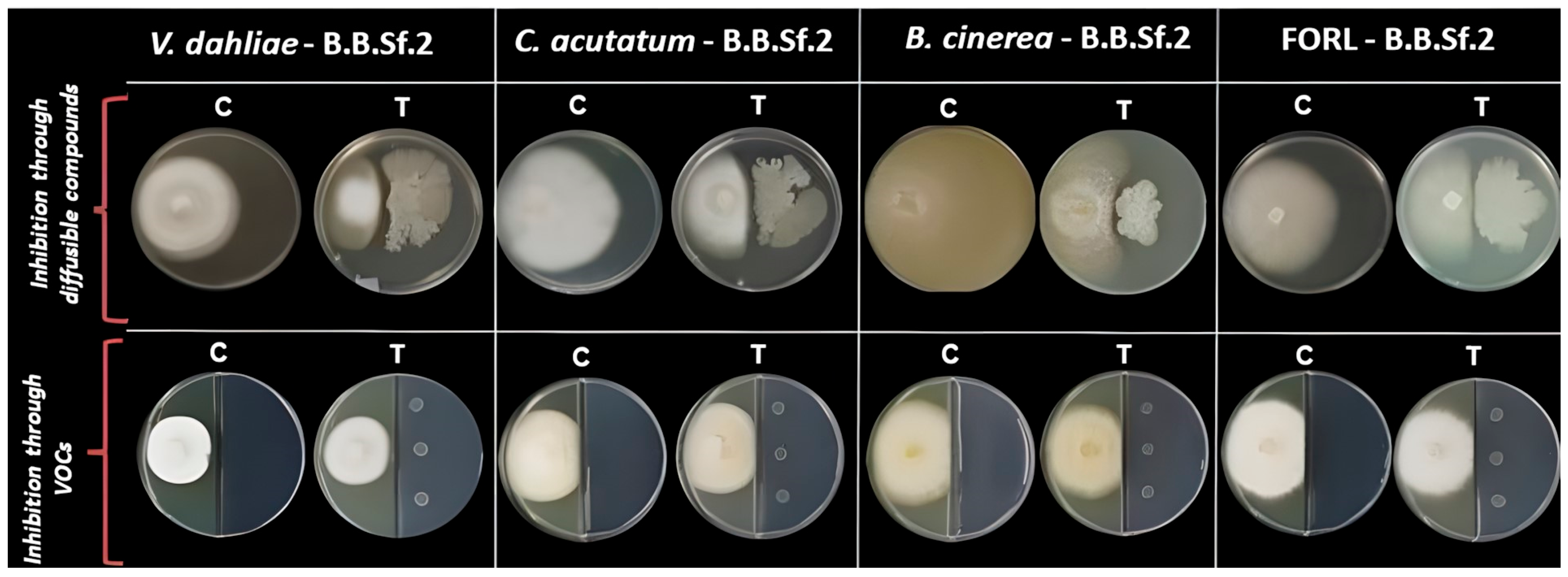
PhytopathogenDiffusible CompoundsVOCs
Verticillium dahliae64.72 ± 3.721.50 ± 0.39
Colletotrichum acutatum63.15 ± 4.5412.74 ± 7.20
Botrytis cinerea66.77 ± 7.6418.42 ± 1.51
FORL70.02 ± 5.5013.19 ± 3.01
Table 2. Taxonomic identification of the endophytic bacterial strain B.B.Sf.2 was established based on its genomic relatedness to closely related B. velezensis strains, as determined by average nucleotide identity by OrthoANI and dDDH analysis.
Bacillus velezensis StrainsorthoANI %dDDH %
Β.Β.Sf.2100100
B.velezensis strain C1 (CP064091.1)99.0993
B.velezensis strain JS25R (CP009679.1)99.1992.9
B.velezensis FZB42′ (NC 009725.2)98.3585.5
B.velezensis strain YJ11-1-4 (CP011347.1)98.3485.1
B.velezensis strain MBI600 (CP094686.1)98.3585
B.velezensis strain UA2208 (CP097586.1)98.384.7
B.velezensis strain FJAT-46737 (CP044133.1)98.284.5
B. velezensisstrain 8-2 (CP028439.1)97.7379.8
B.velezensis strain Yao (CP090905.1)97.6279.7
B.velezensis strain NRRL B-41580 T (NZ_LLZC00000000.1) 199.4095
1 Type strains are indicated with a T.
Table 3. Presentation of predicted secondary metabolite BGCs detected in the genome of B.B.Sf.2 (B. velezensis) strain byantiSMASHanalysis, where NRPS = non-ribosomal peptide synthetase, PKS = polyketide synthetase, and RiPP = ribosomally synthesized and post-translationally modified peptides.
RegionsMost Similar Known ClusterMIBiG ID
(% of Genes Show Similarity)
SizePredicted Biosynthetic Gene ClustersMetabolite
1.1acnBGC0000616 (100%)51,791 ntRiPPAmylocyclicin
dhbBGC0001185 (100%)NRPSBacillibactin
1.2--68,421 ntNRPS-
1.3bacBGC0001184 (100%)41,419 ntOtherBacilycin
1.4mrsBGC0000527 (100%)23,189 ntLanthipeptideMersacidin
4.1srfBGC0000433 (82%)65,408 ntNRPSSurfactin
5.1--12,148 ntPhosphonate-
6.1btrBGC0000693 (7%)41,245 ntPKSButirosin A
6.2--20,741 ntTerpene-
6.3pksxBGC0000181 (100%)86,374 ntTransAT-PKSMacrolactin H
6.4baeBGC0001089 (100%)102,629 ntNRPS,transAT-PKSBacillaene
7.1fenBGC0001095 (93%)137,509 ntNRPSFengycin
bmyBGC0001090 (100%)NRPS, PolyketideBacillomycinD
7.2--21,884 ntTerpene-
7.3--41,101 ntT3PKS-
7.4difBGC0000176 (100%)93,790 ntTransAT-PKSDifficidin
Disclaimer/Publisher’s Note: The statements, opinions and data contained in all publications are solely those of the individual author(s) and contributor(s) and not of MDPI and/or the editor(s). MDPI and/or the editor(s) disclaim responsibility for any injury to people or property resulting from any ideas, methods, instructions or products referred to in the content.

