


Abstract
Severe acute respiratory syndrome coronavirus-2 (SARS-CoV-2) caused a global public health emergency in humans from 2020 to 2023 and was associated with over 7 million human deaths. Besides humans, SARS-CoV-2 has been detected in a wide range of animals, including companion, farm, zoo, and wild animals. At least 61 animal species from 29 animal families of 12 animal orders have tested SARS-CoV-2 positive. Documented evidence reported that not only human-to-animal transmission but also animal-to-human transmission events occurred. During the course of the pandemic progression in humans, SARS-CoV-2 strains in animals evolved in parallel with those in humans. Continued monitoring of SARS-CoV-2 in animals is needed to safeguard both human and animal health. In this study, we report investigation of two outbreaks of SARS-CoV-2 Omicron variant infection in tigers and gorillas in two zoological institutions. In the first zoo, six tigers tested positive by SARS-CoV-2 real-time RT-PCR and shed viral nucleic acid in feces for up to two weeks. Three of the tigers showed intermittent shedding patterns, while the other tigers shed only for 7–10 days. No other species, including cheetah, otter, lion, anteater, gibbon, and tamarin, tested positive. During the outbreak at the second zoo, a total of six gorillas were tested positive for SARS-CoV-2, while other primates housed in the same building (colobus and orangutan) tested negative. Follow-up testing revealed that two gorillas tested positive for SARS-CoV-2 over a one-month period (30 and 33 days, respectively), while the other four gorillas had positive SARS-CoV-2 PCR results for 14 to 25 days. Four gorillas had intermittent shedding patterns. Notably, compared to tigers, gorillas had a prolonged duration of fecal viral shedding. Sequencing was performed on the positive samples, and analysis indicated that strains detected in tigers and gorillas belonged to SARS-CoV-2 Omicron BQ.1.10 and XBB.1.16, respectively. Overall, this study offers valuable insights into the duration of viral RNA shedding for SARS-CoV-2 Omicron in zoo animals, facilitating accurate diagnostic evaluation and management of infected tigers and gorillas.
1. Introduction
Coronavirus disease 2019 (COVID-19) is a contagious disease caused by severe acute respiratory syndrome coronavirus 2 (SARS-CoV-2). COVID-19 was first observed at the end of 2019 in China and caused global pandemic a couple months later [1]. SARS-CoV-2 is an enveloped single-stranded positive-sense RNA virus in the genus of Betacoronavirus within the family Coronaviridae. In general, zoonotic coronaviruses usually need intermediate hosts to gain adaptations for infectivity of humans. It has been known that SARS-CoV and Middle East respiratory syndrome coronavirus (MERS-CoV) evolved through palm civets and camels before infecting humans [2]. At the beginning of the COVID-19 pandemic, one bat coronavirus strain, RaTG13, was found to have the highest nucleotide identity (96.2%) with SARS-CoV-2 [3]. A subsequent surveillance study reported that another bat coronavirus strain, BANAL-20-52, shares an even higher nucleotide identity (96.8%) with SARS-CoV-2 than RgTG13 [4]. It remains unclear how SARS-CoV-2 evolved from a bat coronavirus, probably through point mutations and/or recombination events in bats or other intermediate hosts.
As one of three human pandemic coronaviruses, SARS-CoV-2 caused 762,133,867 infections with 6,811,298 associated deaths during 2019–2023 worldwide [5]. In addition to the devastating impact on humans, SARS-CoV-2 also causes infection in a varietiy of animal species. To date, four major categories of animals have tested positive for SARS-CoV-2 including domesticated pets (dogs, cats, domestic ferrets, and hamsters), captive wildlife (tigers, lions, snow leopards, cougars/pumas, hyenas, gorillas, red foxes, lynxes, fishing cats, binturongs, leopard cats, Indian leopards, etc.), farm animals (minks, cattle, buffalo, etc.), and free-ranging wildlife (white-tailed deer, mule deer, giant anteaters, etc.) [6]. Similar to infection in humans, SARS-CoV-2 mainly causes respiratory illness in animals. In addition to respiratory signs [7], SARS-CoV-2 also causes enteric signs in animals, such as vomiting and diarrhea [8]. Depending on the animal species, clinical presentations in animals infected by SARS-CoV-2 vary from asymptomatic to mild or severe respiratory symptoms, pneumonia, enteric illness, and death [7,8,9,10,11].
Most SARS-CoV-2 animal cases represent reverse zoonoses, and the strains in animals evolved alongside the pandemic’s progression in humans [8,10,12,13,14]. In addition to human-to-animal transmission events, animal-to-animal [12,14] and even animal-to-human transmission [12,15,16,17] events have been documented for SARS-CoV-2, highlighting the importance of a one health approach.
Regardless of host, among these SARS-CoV-2 variants, the Delta variant has been more virulent, while the Omicron variant is more transmissible [18,19,20,21,22,23]. Soon after the emergence of the Omicron variant in humans, it was also detected in animals [24]. In the present study, we report the characterization (genome and replication) of Omicron variants in two zoo animal species: tigers and gorillas.
2. Material and Methods
2.1. Samples
Fecal samples from six tigers, (1.5) H, K, M, Suk, Sum, and N, with an age range of 1 to 16 years, and from other animals (anteater, cheetah, gibbon, lion, otter, and tamarin) at Zoo #1, as well as from six gorillas, B, Ko, Ka, A, J, and N (1.5), with an age range of 5 to 35 years at Zoo #2, which were submitted for diagnostic testing, were used in this study. Fecal samples were collected in tube containers and shipped overnight on ice packs. Two tigers (Sum + N) were housed together, so individual feces could not be identified.
2.2. RNA Extraction
Fecal samples were swabbed into 0.5 mL of 1× phosphate-buffered saline (PBS) solution, briefly vortexed and centrifuged for 2 min at 6800 RCF. A total of 200 µL of supernatant from the fecal suspension solution was used for extraction of total nucleic acids using the MagMax Pathogen RNA/DNA kit following the kit instructions (Catalog # 4462359, Thermo Fisher Scientific, Austin, TX, USA) as described previously [25]. Nucleic acid extraction was processed using an automated extraction instrument, the Thermo Scientific KingFisher Flex (Thermo Fisher Scientific, Waltham, MA, USA), with the MagMAX Pathogen High Volume 96DW protocol. Viral RNA was eluted in 90 µL of elution buffer and subsequently used for the rRT-PCR assay.
2.3. Real-Time RT-PCR Testing
The presence of SARS-CoV-2 nucleic acid was assessed using a real-time RT-PCR assay, as previously described [25]. Real-time RT-PCR was performed using N2 CDC primers (2019-nCoV RUO Kit, 500 rxn, IDT# 10006713, IDT, Coralville, IA, USA) on an Applied Biosystems™ (ABI) 7500 Real-Time PCR System (Thermo Fisher Scientific, Foster City, CA, USA) [25] or a Bio-Rad CFX Opus 96 Real-Time PCR instrument (Bio-Rad Laboratories, Hercules, CA, USA). The sequences of primers and probe were as follows: 2019-nCoV_N2-forward primer: TTACAAACATTGGCCGCAAA, 2019-nCoV_N2-reverse primer: GCGCGACATTCCGAAGAA, and 2019-nCoV_N2-probe: FAM-ACAATTTGCCCCCAGCGCTTCAG-BHQ1 [25,26]. On the Bio-Rad CFX Opus 96 Real-Time PCR System, the PCR reaction was performed in a 24 µL volume, containing 6.0 µL of the TaqPath™ 1-Step Multiplex Master Mix, 2.0 µL of the N2 primer and probe mixture, 1.0 µL of the Xeno (Thermo Fisher Scientific, Austin, TX, USA), 1.0 µL of the LIZ assay (Thermo Fisher Scientific, Austin, TX, USA), 8.0 µL of the nuclease-free water, and 6.0 µL of the nucleic acid template. Xeno, together with the LIZ™ assay, was employed to serve as an internal positive control. Additionally, one negative rRT-PCR control (nuclease-free water) and one positive rRT-PCR control were assayed with every run of PCR reaction. Following the completion of the run, fluorescence thresholds were analyzed to ensure the Ct values were appropriately recorded. The PCR conditions were as follows: Uracil-N-glycosylase (UNG) incubation at 25 °C for 2 min; reverse transcription at 48 °C for 10 min; polymerase activation at 95 °C for 10 min; and 40 cycles of amplification at 95 °C for 15 sec followed by 60 °C for 45 s.
2.4. Whole-Genome Sequencing and Analysis
PCR amplicons were amplified using a CDC-based method [27] and the Artic method [28], and sequencing library was prepared using the Nextera XT DNA Library Preparation Kit (Illumina, San Diego, CA, USA), followed by sequencing using MiSeq Reagent Kit V2 (Illumina, San Diego, CA, USA) at 300 cycles on MiSeq platform (Illumina, San Diego, CA, USA). The raw FastQ data were assembled into Fasta format using both SPAdes [29] and CLC genomics workbench (https://www.qiagen.com/zh-cn/products/discovery-and-translational-research/next-generation-sequencing/informatics-and-data/analysis-and-visualization/clc-genomics-workbench, accessed on 9 December 2025), and a local blast was run against the NCBI NT database. Sequence alignment and phylogenetic tree analysis were performed using MEGA software, version 7.0.26 [30]. The online Pangolin COVID-19 Lineage Assigner tool was used to classify strains included in the phylogenetic tree [31].
Assembled genomes of tiger TX-UIUC-31115 and gorilla IL-UIUC-39037 strains were deposited into GISAID [32]. Other online sequences used for comparison and analysis in this study are publicly accessible in GISAID under https://doi.org/10.55876/gis8.250828um.
3. Results
3.1. Clinical Infections in Tigers and Gorillas
At Zoo 1, six tigers (1.5) ranged in age from 1 to 16 years. Four tigers (H, K, M, Suk) were housed individually, and the two youngest tigers (Sum + N) were housed together. All tigers had previously been vaccinated with the Zoetis mink coronavirus vaccine subunit with a two-dose regimen in 2021 [33]. The first tiger initially presented with a single episode of vomiting and intermittent cough in January 2023. Symptoms progressed to include hyporexia, increased coughing, and wheezing with clear nasal discharge. Fecal samples were collected from all tigers for COVID-19 testing. Five of the six tigers were asymptomatic when they initially tested positive. For the tigers that were asymptomatic when the initial positive sample was collected, clinical signs developed within 1–3 days. All tigers developed cough, wheezing, hyporexia and varying degrees of lethargy, with symptoms being more severe in the oldest tiger and milder in the youngest tigers. One tiger, the most severely affected one, also developed tachypnea. Clinical signs persisted for 2 to 11 days. Depending on the severity of clinical signs, treatment ranged from close monitoring to the administration of meloxicam and cefovecin sodium.
At Zoo #2, six gorillas (1.5), with an age range of 5 to 35 years, were housed together and had previously been vaccinated with the Zoetis mink coronavirus vaccine subunit with a two-dose regime in 2021 [33]. In the first week of August 2023, all six developed varying clinical signs consistent with COVID-19 infection, such as lethargy, hyporexia, clear nasal discharge, and infrequent dry unproductive cough. Clinical signs resolved in less than 2 weeks either without treatment or with a combination of maropitant, ondansetron, ibuprofen, and oral vitamin C, based on individual clinical signs.
3.2. Detection and Follow-Up Testing for SARS-CoV-2 in Tigers
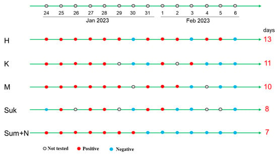
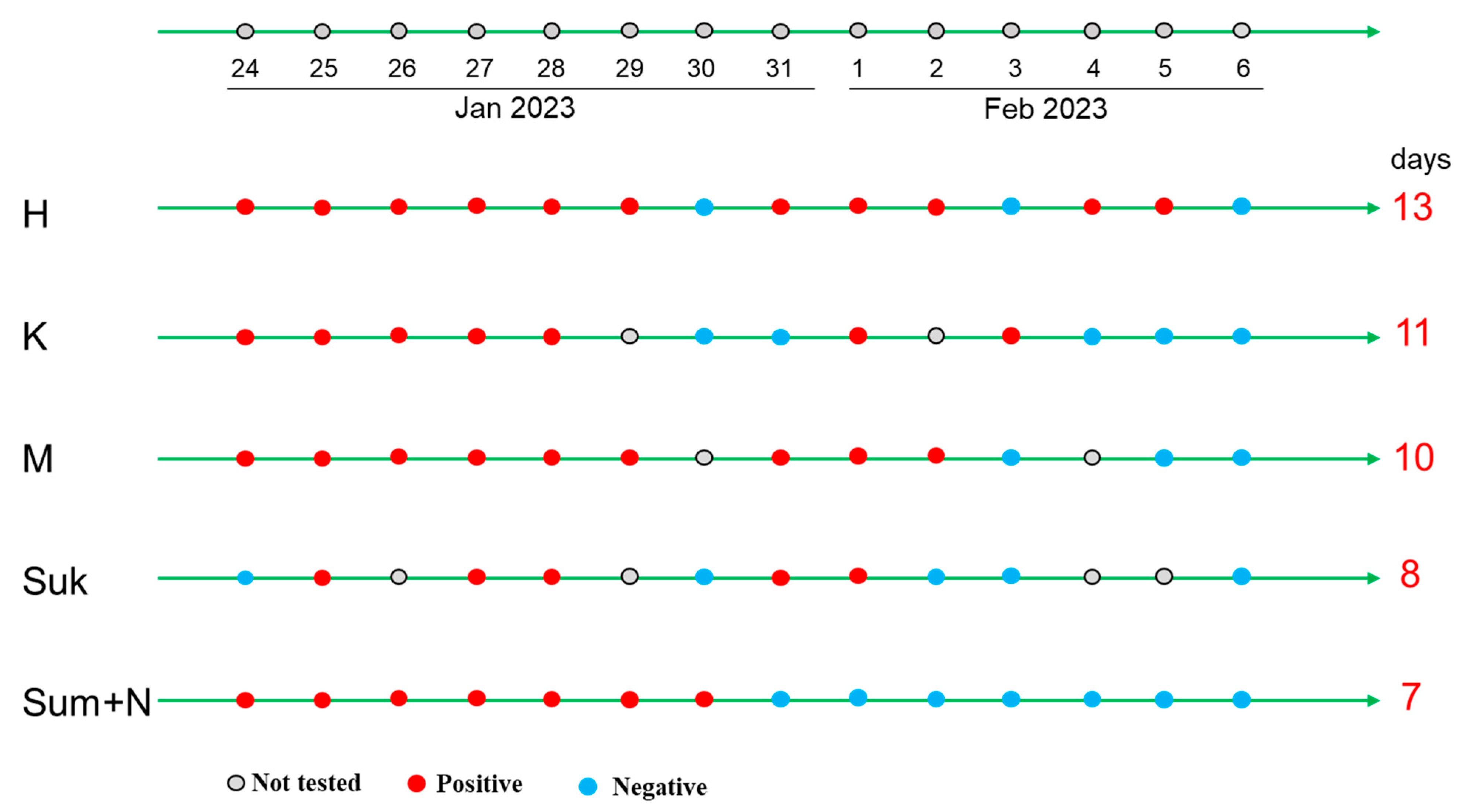
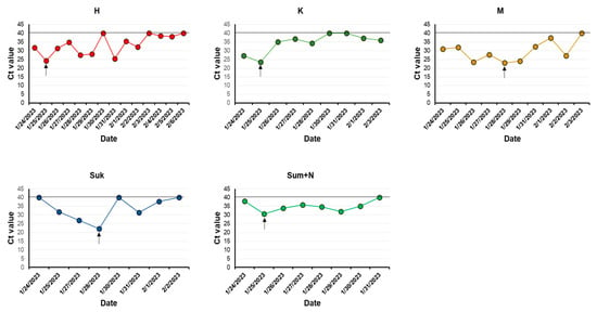
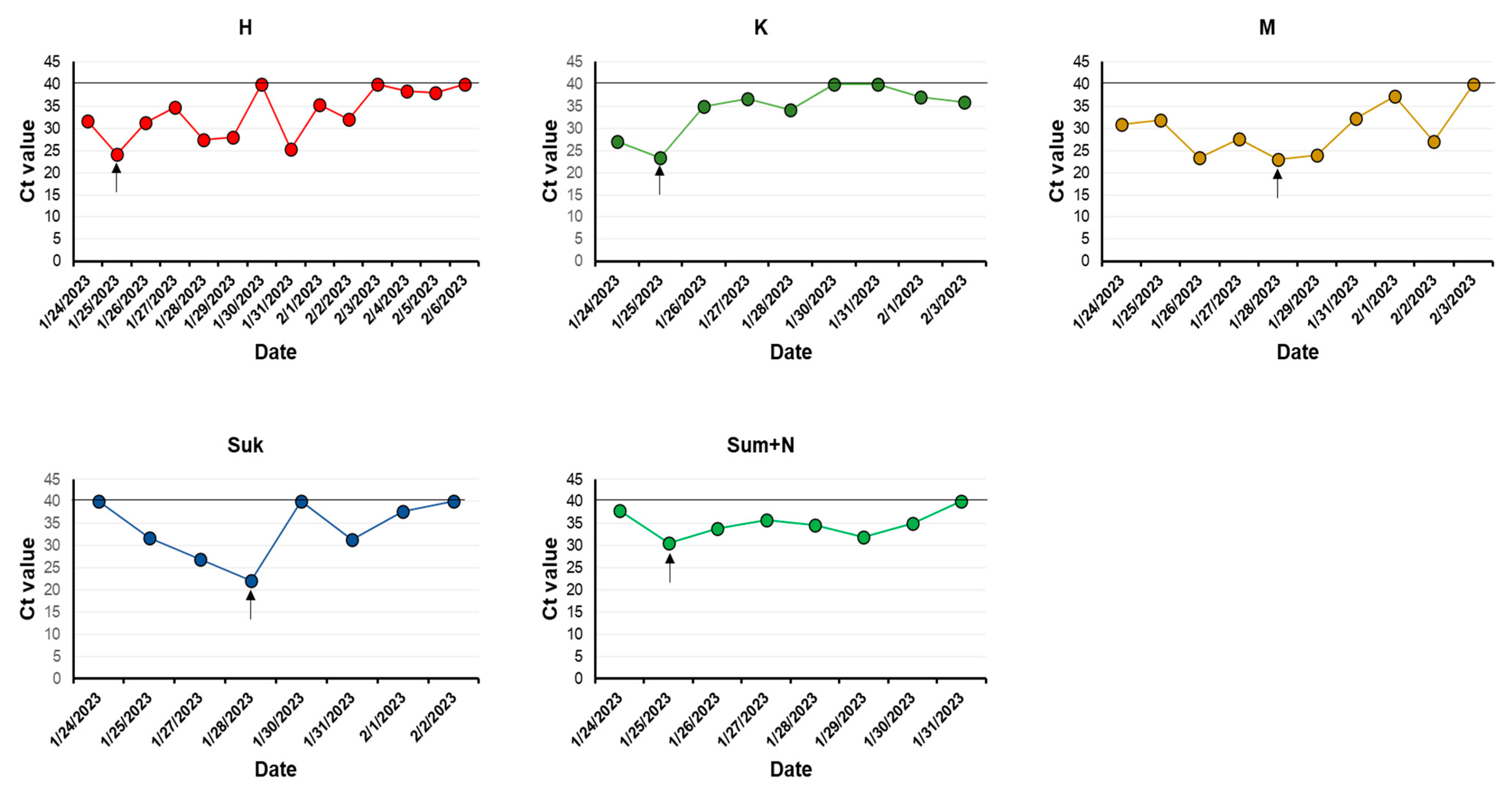
For the SARS-CoV-2 real-time RT-PCR testing in the first zoo, five tigers (H, K, M, Sum + N,) initially tested positive, with Ct values between 26.97 and 37.93 (Figure 1 and Figure 2), while the sixth tiger (Suk) tested positive the following day. These tigers exhibited the highest viral shedding on the second (H, K, Sum + N) or fifth (M and Suk) day post-PCR positive (Figure 2).
Figure 1.
Test timeline and results for tigers H, K, M, Suk, and Sum + N at Zoo #1 using real-time RT-PCR. The duration of positive SARS-CoV-2 results was calculated from the first to the last day of positivity. Gray solid circles indicate no samples were collected for testing, red solid circles indicate a positive PCR result, and teal solid circles indicate a negative PCR result.
Figure 2.
Follow-up real-time RT-PCR result (Ct value) for tigers H, K, M, Suk, and Sum + N at Zoo #1. The day with the highest viral shedding is highlighted with an arrow. Timepoints with a negative result are noted y a line at a Ct value of 40.
Shedding periods of the six tigers for SARS-CoV-2 ranged from one week to two weeks (Figure 1). Tiger H had the longest shedding duration (13 days), followed by Tiger K (11 days) and Tiger M (10 days). The remaining three tigers (Suk and Sum + N) shed viruses for 7 to 8 days. During these periods, three tigers (H, K, and Suk) showed an intermittent shedding pattern, with one or two consecutive negative result before resuming shedding (Figure 1 and Figure 2). SARS-CoV-2 was not detected in samples from other zoo animals, including anteater, cheetah, gibbon, lion, otter, and tamarin.
3.3. Detection and Follow-Up Testing SARS-CoV-2 in Gorillas
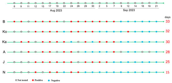
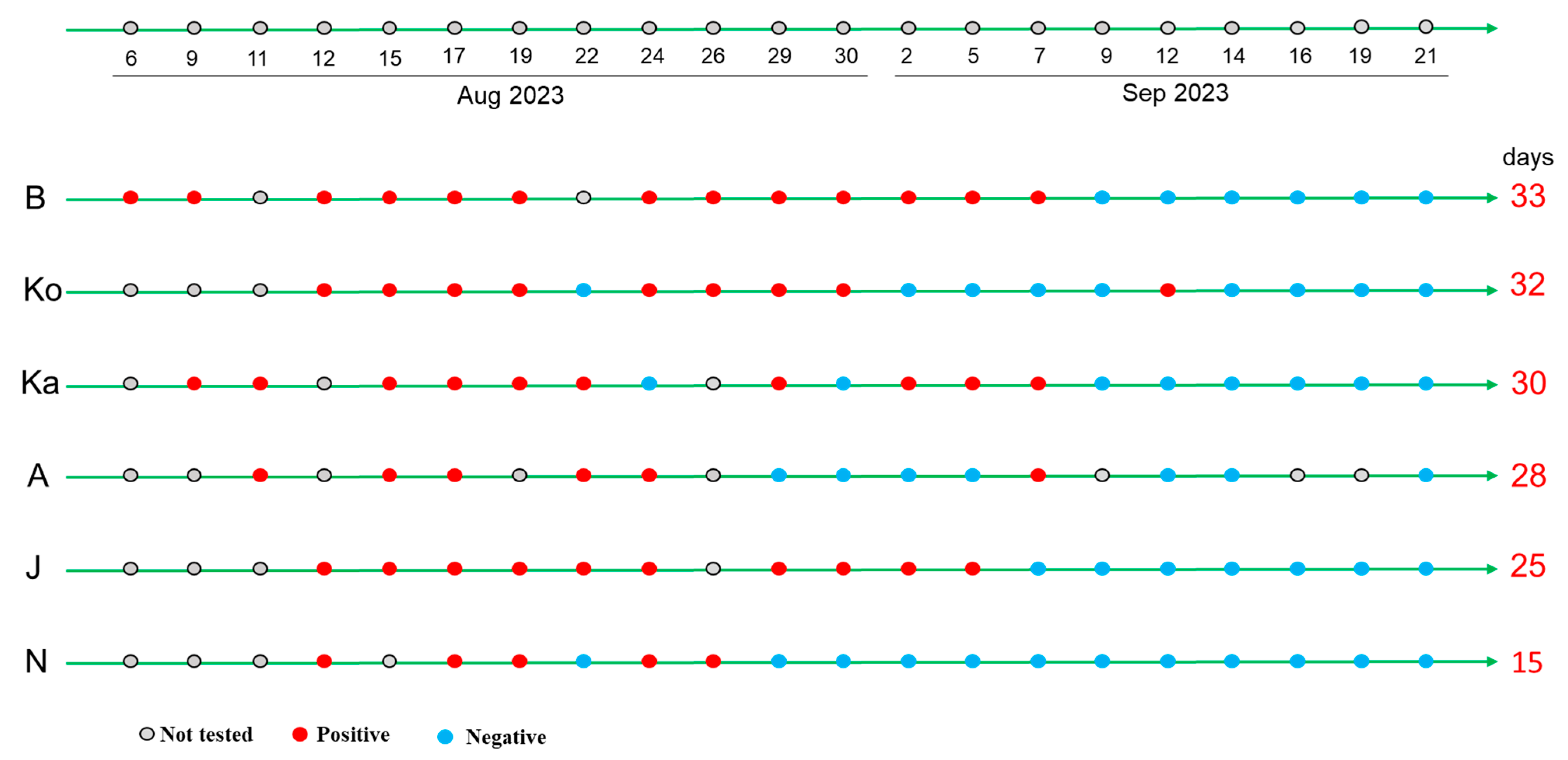
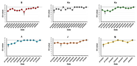
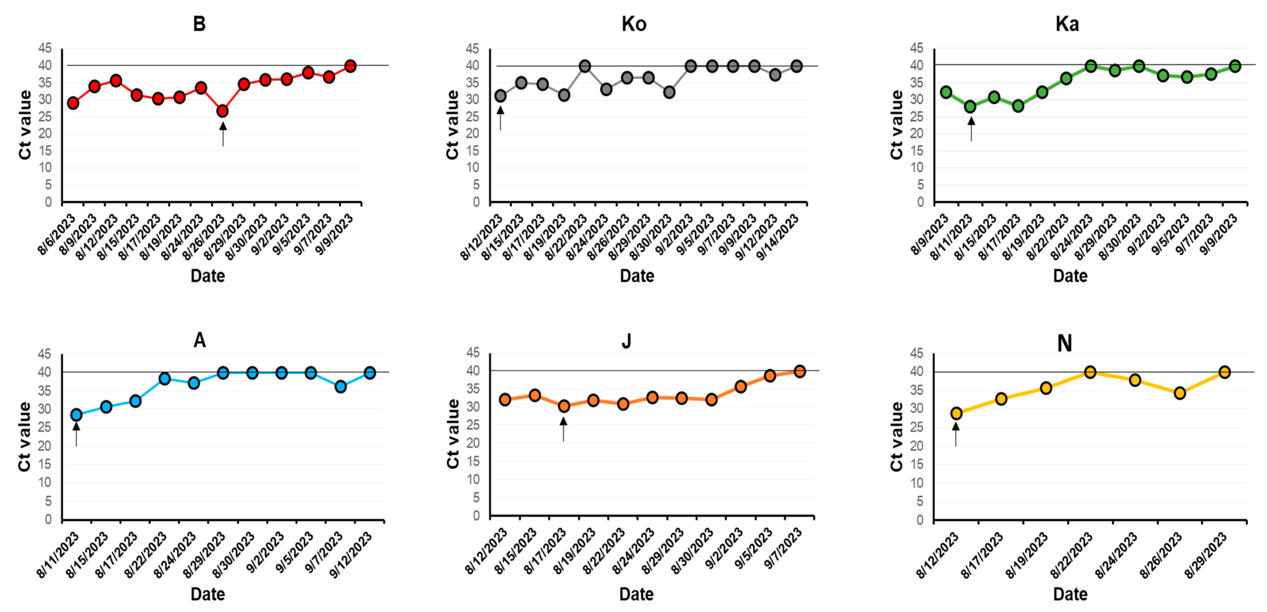
A total of six gorillas at Zoo #2 tested positive for SARS-CoV-2. The first positive case was detected in gorilla B using real-time PCR (Ct value 29; Figure 3 and Figure 4). Three days later, another gorilla (Ka) tested positive, and within six days, all six gorillas were confirmed positive. The index gorilla B exhibited the highest viral load 20 days after the initial positive PCR (Figure 4), while gorilla J had the highest viral load 5 days after the initial positive PCR. Four gorillas (Ko, A, N, and Ka) shed the highest amounts of virus in feces during their first or second positive PCR test, which occurred five to six days after the initial detection in the index gorilla B (Figure 3 and Figure 4).
Figure 3.
Test timeline and results for gorillas B, Ko, Ka, A, J, and N at Zoo #2 by real-time RT-PCR. The duration for positive SARS-CoV-2 results was calculated from the first to the last day of positivity. Gray solid circles indicate no samples collected for testing, red solid circles indicated a positive PCR result, and teal solid circles indicate a negative PCR result.
Figure 4.
Follow-up real-time RT-PCR result (Ct values) for gorillas B, Ko, Ka, A, J, and N at Zoo #2. The day with the highest viral shedding is indicated with an arrow. Time points with a negative result are indicated by a line at a Ct value of 40.
Follow-up testing showed that the index gorilla B shed virus more than one month (33 days) and other two gorillas, Ko and Ka, shed virus for 32 and 30 days, respectively. The remaining three gorillas A, J, and N, shed virus 28, 25, and 15 days, respectively. Overall, these data showed that gorillas had longer viral shedding compared to tigers.
3.4. Sequence and Phylogenetic Analysis
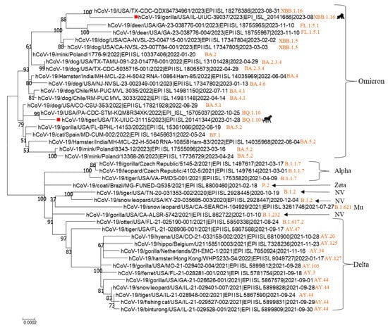
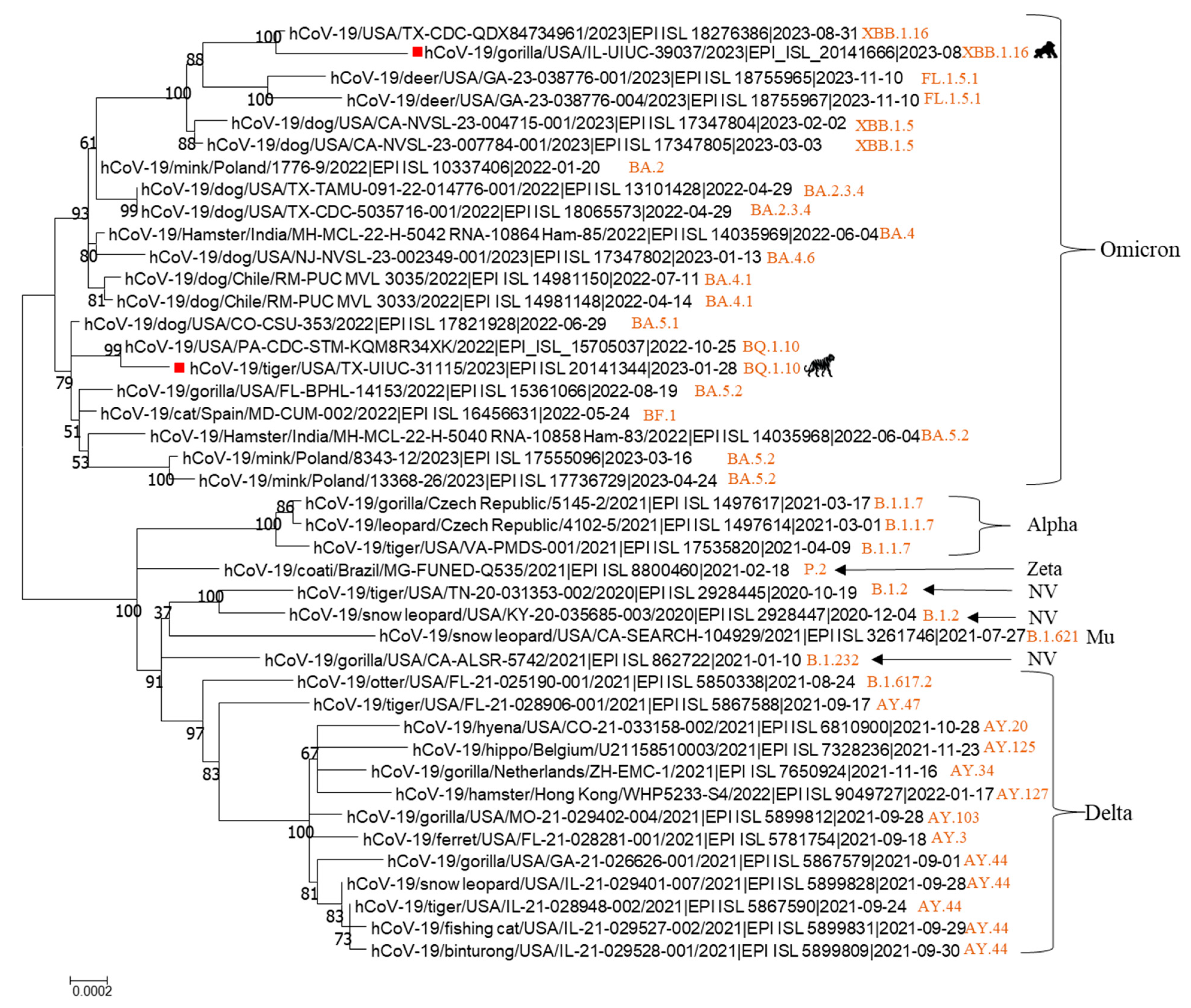
Whole-genome sequencing of SARS-CoV-2 was performed on samples from tigers and gorillas that tested positive, and sequences were deposited into GISAID with accession numbers EPI_ISL_20141344 and EPI_ISL_20141666. Online NCBI blast search indicated that the tiger SARS-CoV-2 TX-UIUC-31115 strain showed the highest nucleotide identity of 99.97% with several human strains, while the gorilla strain SARS-CoV-2 IL-UIUC-39037 had the highest nucleotide identity of 99.77% with several human strains. Further analysis using the online Pangolin COVID-19 Lineage Assigner tool (https://ngdc.cncb.ac.cn/ncov/online/tool/lineage-assigner?lang=en, accessed on 9 December 2025) revealed that the tiger TX-UIUC-31115 strain belonged to Omicron BQ.1.10 lineage, while the gorilla IL-UIUC-39037 strain was classified as Omicron XBB.1.16 lineage. Phylogenetic tree analysis also showed that both tiger and gorilla strains clustered together with human SARS-CoV-2 isolates in the branch of Omicron (Figure 5). These results indicate that different lineages of the Omicron variant infected wildlife under human care.
Figure 5.
Phylogenetic tree analysis of complete genomes of SARS-CoV-2 variant strains in various animal species. The tiger and gorilla SARS-CoV-2 strains in the present study are indicated by a solid red square and corresponding animal icons. SARS-CoV-2 lineage of each strain was noted with orange color in the tree.
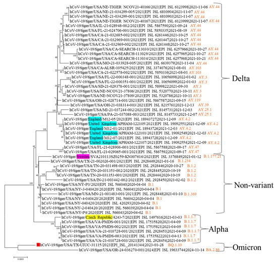
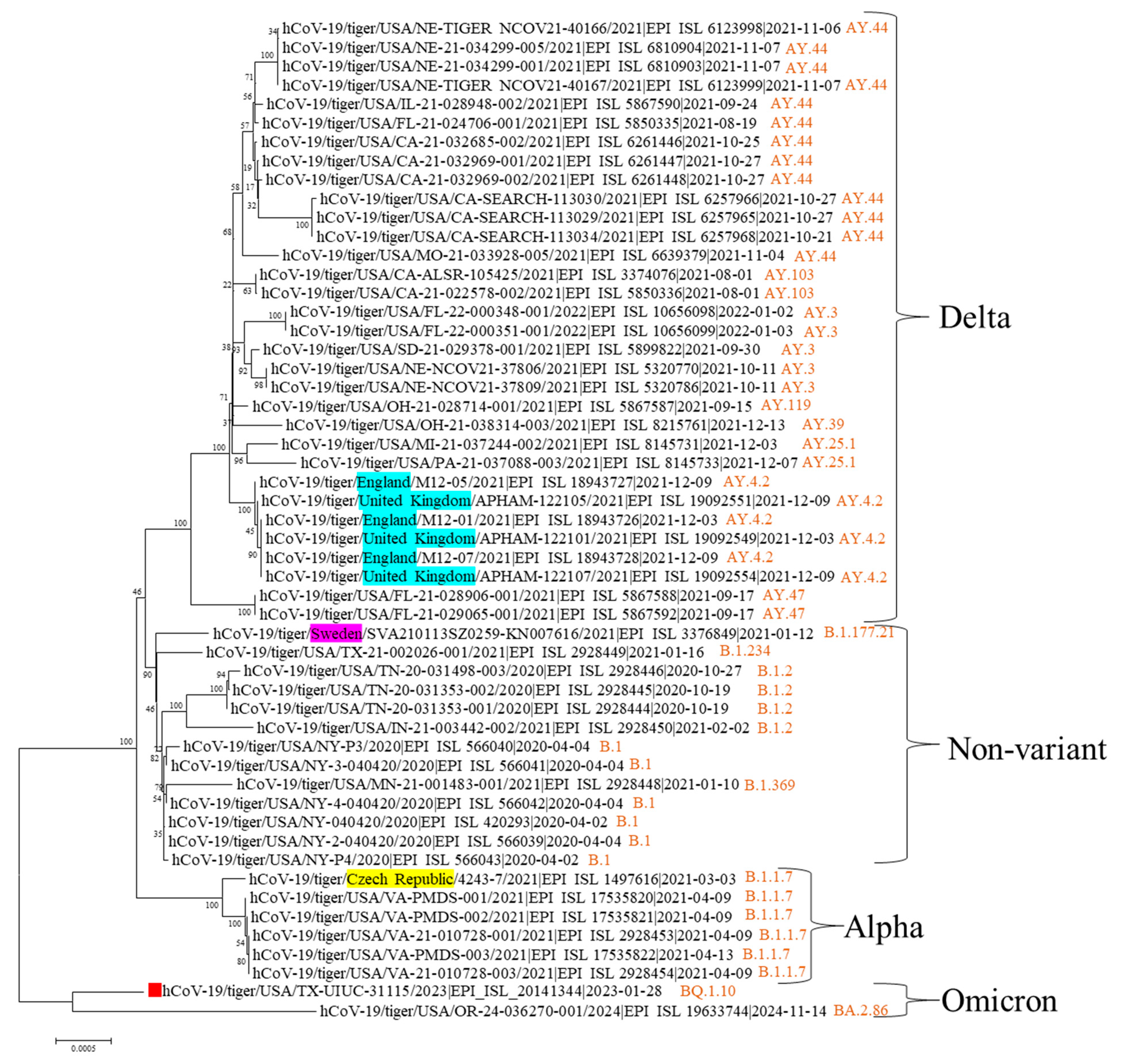
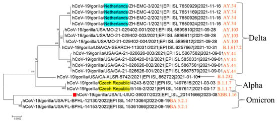
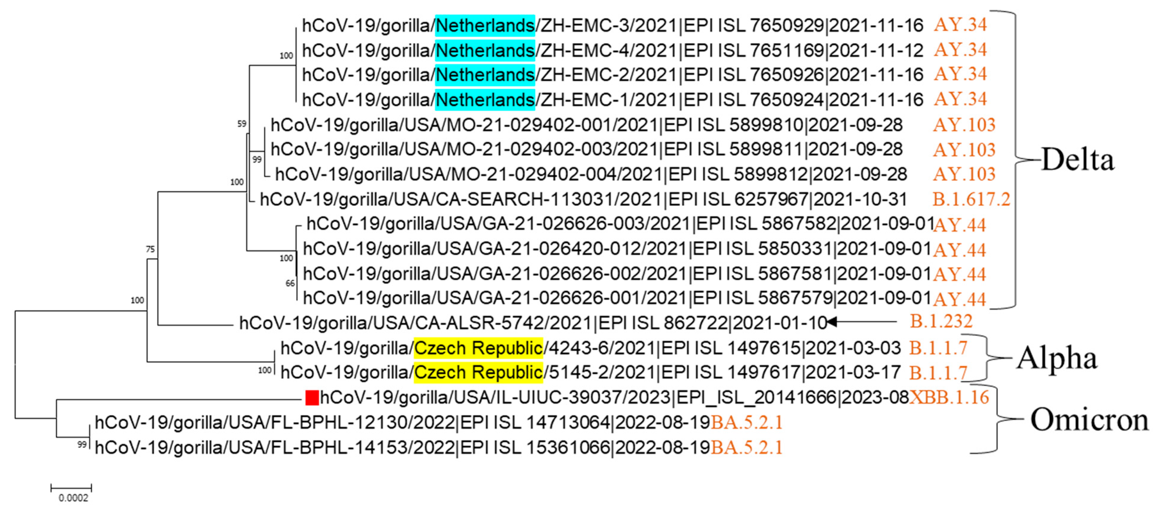
By analyzing the deposited sequences in GISAID, we found that tigers had been infected with SARS-CoV-2 of non-variants (B.1, B.1.2, B.1.234, B.1.369, and B.1.177.21), Alpha (B.1.1.7), Delta (AY.103, AY.119, AY.25.1, AY.3, AY.39, AY.4.2, and AY.44, AY.47), and Omicron (BA.2.86 and BQ.1.10) (Figure 6). While more tigers were infected by Delta variants, only a few deposited sequences showed Omicron infection. Similarly, gorillas were infected by SARS-CoV-2 of non-variant (B.1.232), and three different variants including Alpha (B.1.1.7), Delta (AY.103, AY.34, AY.44, and B.1.617.2), and Omicron (BA.5.2.1 and XBB.1.16) (Figure 7). These data indicate that tigers and gorillas can be infected by different variants of SARS-CoV-2.
Figure 6.
Phylogenetic tree analysis of complete genomes of SARS-CoV-2 non-variant and variant strains in tigers. Strains from other non-USA countries are highlighted in different colors. The tiger SARS-CoV-2 strain from the present study is indicated with a red solid square. SARS-CoV-2 lineage of each strain was noted with orange color in the tree.
Figure 7.
Phylogenetic tree analysis of complete genome of SARS-CoV-2 non-variant and variant strains. Strains from other non-USA countries are highlighted in different colors. The gorilla SARS-CoV-2 strain from the present study is indicated with a red solid square. SARS-CoV-2 lineage of each strain was noted with orange color in the tree.
4. Discussion
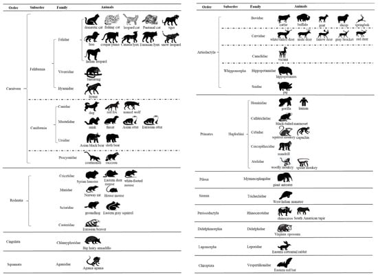
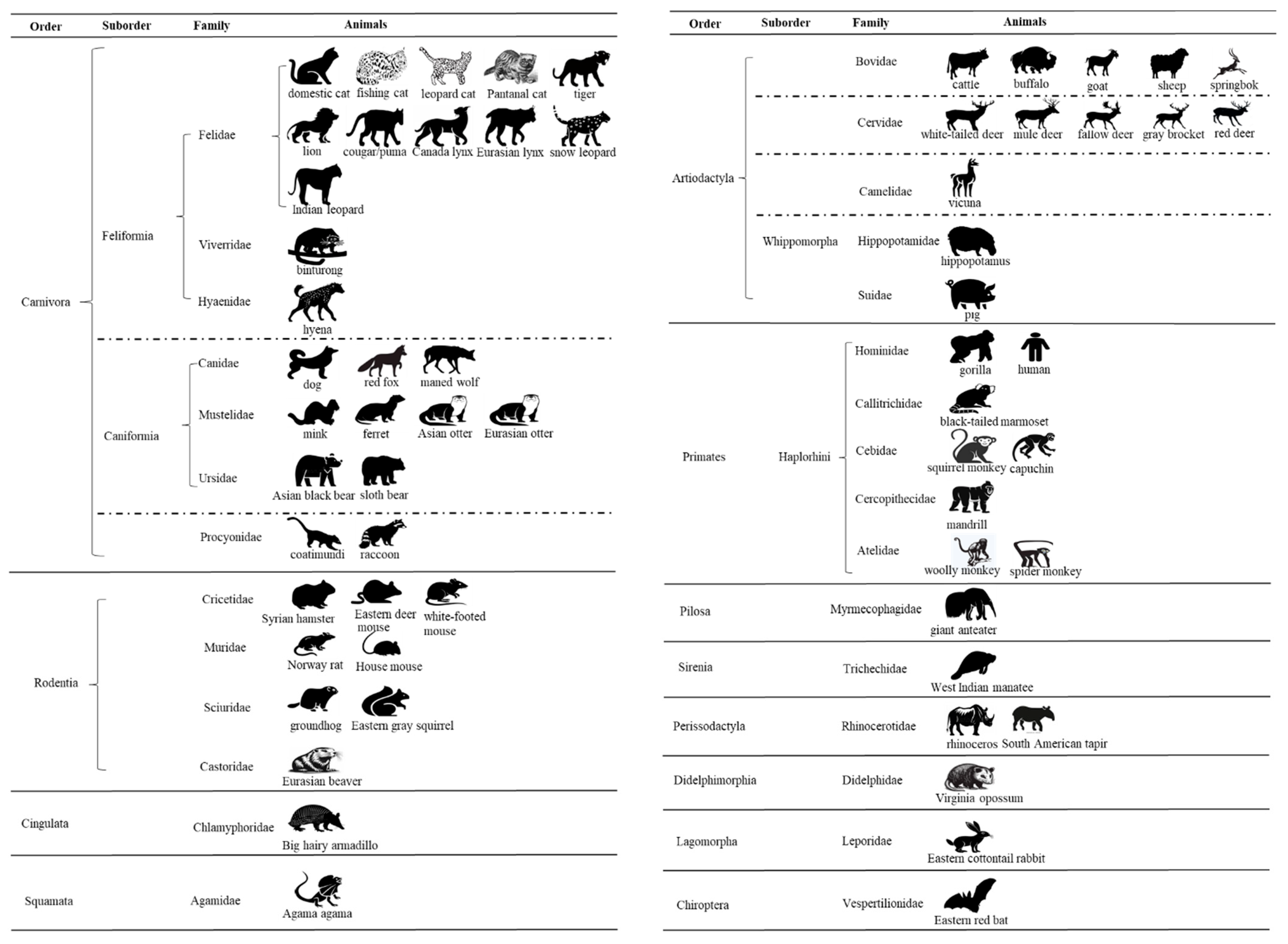
Since February 2020, when the first report indicated that dogs in the Canidae family within order Carnivora tested positive for SARS-CoV-2 [10], animals from 29 families across 12 orders (Carnivora, Rodentia, Cingulata, Squamata, Artiodactyla, Primates, Pilosa, Sirenia, Perissodactyla, Didelphimorphia, Lagomorpha, and Chiroptera) have tested positive for SARS-CoV-2 [34]. Among these animal orders and families, there are at least 61 animal species that have tested positive for SARS-CoV-2 through molecular testing (e.g., RT-PCR) and/or serological testing [34,35] (Figure 8). The order Carnivora has the highest number of SARS-CoV-2-positive animal species (24), followed by Artiodactyla (13), Rodentia (8), Primates (7), Perissodactyla (2), and seven animal orders with one species each. Experimental studies previously reported that pigs were not susceptible to SARS-CoV-2 infection [36,37], or had low susceptibility even when higher inoculation doses were used [38]. ACE2 receptor modeling and analysis also classified pigs into the low-susceptibility group [39]. However, a previous study from Nigeria reported that pigs tested positive for SARS-CoV-2 by RT-PCR and serological assays under non-experimental conditions [40], suggesting that multiple factors contribute to the susceptibility of animal to SARS-CoV-2.
Figure 8.
Based on the FAO website [34] and our recent publication [35], 61 animal species across 12 orders and 29 families have tested positive for SARS-CoV-2 by molecular PCR, serological test, or both. Three species—chicken, duck, and turkey—that tested positive by molecular PCR were not included in the figure.
Tigers have been documented to be infected with both non-variant and variant strains of SARS-CoV-2. In April 2020, two Malayan and three Amur tigers at Bronx Zoo were the first to be reported SARS-CoV-2-positive, with four of them presenting clinical respiratory signs; sequencing confirmed infection with the B.1 lineage [13]. Subsequently, five tigers with respiratory signs at two U.S. zoos (Knoxville zoo in Tennessee and Fort Wayne Zoo in Indiana) tested positive for SARS-CoV-2, and sequence analysis identified the B.1.2 lineage [7,41]. Additional reports include two tigers at Boras Zoo in Sweden infected with the B.1.177.21 lineage and one tiger at the Wildcat Sanctuary in Minnesota infected with the B.1.369 lineage [7].
In addition to these non-variant strains, tigers have also been infected with SARS-CoV-2 variants. In 2021, five Malayan and one Sumatran tiger at two zoos (Virginia Zoo in the US and Prague Zoo in the Czech Republic) developed respiratory signs, including labored breathing and coughing, and were confirmed to be infected with B.1.1.7 (Alpha) variant [7,42]. Three Amur tigers at a wildlife park in England and two tigers at Brookfield Zoo in Chicago, Illinois, were reported to be infected with the B.1.617.2 (Delta) variant [43,44]. Moreover, at least eight additional U.S. states reported tigers infected with Delta variants (Figure 6), suggesting a high infection rate in this species.
By contrast, only two outbreaks involving tigers have been attributed to the Omicron variant. In the present study, we reported that six tigers were infected with the Omicron BQ.1.10 lineage in 2023 (Figure 6). In 2024, tigers from a zoo in Oregon also tested positive for the Omicron sublineage BA.2.86. Compared with the widespread Delta variant, the much lower number of reported Omicron infection in tigers in the U.S. (Figure 6) may reflect the presence of protective immunity induced by vaccination and/or prior SARS-CoV-2 infection.
Gorillas with SARS-CoV-2 infections have been reported in different countries, including the U.S., Czech Republic, and the Netherlands. Strains infecting gorillas included all variants—B.1.232, Alpha, Delta, and Omicron. The SARS-CoV-2 outbreak in gorillas was first documented at the San Diego Zoo Safari Park in January 2021 [45], and sequencing analysis confirmed that the animals were infected with the B.1.232 variant (Figure 7). Following this, the Alpha variant was detected in five gorillas at the Prague Zoological Garden in the Czech Republic during February–March 2021 [46], and the Delta variant was detected in gorillas at zoos in three states (Georgia, California, and Missouri) in the U.S. (Figure 7) and the Netherlands during September–November 2021 [47]. The Omicron variant has been detected at zoos in two states (Florida and Illinois) in the U.S (Figure 7).
Our study reports the detection and sequencing of Omicron variants in two zoo animal species under human care at zoological institutions: tiger and gorilla. In contrast to the relatively short viral shedding periods observed in tigers (up to 2 weeks in feces), gorillas shed SARS-CoV-2 for a considerably longer time, extending beyond one month. Variability in shedding duration has been documented across individual animals and different species, both with non-variant and variants of SARS-CoV-2. For example, four tigers at Bronx Zoo infected with a non-variant B.1 lineage strain in 2020 shed virus for 12, 14, 22, and 26 days, which appeared to correlate with the duration of clinical signs (2, 5, 16, 1 day, respectively) [7]. At the same zoo, three lions infected with the non-variant B.33 lineage shed virus for 16, 34, and 39 days. Similarly, one of three tigers at Tennessee zoo shed a non-variant in feces for up to 29 days [48]. All three snow leopards infected with a B.1.2 lineage strain at a Kentucky zoo shed virus for at least one month (30, 32, and 33 days) [49]. By contrast, two tigers at Brookfield Zoo in Chicago infected with the Delta variant (AY.44 lineage) shed virus in feces for only 5 and 6 days, whereas two lions from the same zoo shed virus for 14 and 20 days [44]. Lions at Utah’s Hogle Zoo infected with the Delta variant shed virus for up to 33 days in feces and as long as 14 weeks in respiratory specimen after illness onset [50]. Collectively, these data indicate that the durations of viral shedding in zoo animals are highly variable, underscoring the importance of follow-up monitoring of SARS-CoV-2-positive animals to safeguard their health.
It remains unknown how these animals acquired SARS-CoV-2 infection. It is possible that they were exposed through contaminated feed, water, or close contact with an infected human. Our study showed two different lineages of Omicron variants infecting zoo tigers and gorillas. Continued surveillance and investigation of SARS-CoV-2 in animals is warranted.
Author Contributions
Conceptualization, L.W., X.D., M.A. and K.T.; methodology, L.W. and S.K.; software, L.W.; validation, L.W.; formal analysis, L.W.; investigation, L.W., A.B.-M. and M.A.; resources, L.W., A.B.-M., M.A. and K.T.; data curation, L.W.; writing—original draft preparation, L.W.; writing—review and editing, L.W., S.K., A.B.-M., X.D., M.A. and K.T.; visualization, L.W.; supervision, L.W.; project administration, L.W.; funding acquisition, L.W. All authors have read and agreed to the published version of the manuscript.
Funding
This research was funded by the American Rescue Plan Act through USDA APHIS (APHIS/NIFA Collaborative Award# 2023-70432-39482).
Institutional Review Board Statement
Ethical review and approval were not required for this study, as all animal fecal samples used for the analyses were collected non-invasively by the two zoos.
Informed Consent Statement
Not applicable.
Data Availability Statement
All data related to the current study is provided in the manuscript, and all genome sequences in this study are publicly accessible in GISAID; see Supplemental Table epi_set_250828um (https://doi.org/10.55876/gis8.250828um).
Acknowledgments
We gratefully acknowledge the authors from the originating laboratories who obtained the specimens and the submitting laboratories that generated the genetic sequence and metadata, and shared them via the GISAID initiative, on which this research is based. All sequence data contributors for this study are listed at https://doi.org/10.55876/gis8.250828um. We thank National Veterinary Service Laboratories (NVSL) for their confirmatory support in both cases.
Conflicts of Interest
The authors declare no conflicts of interest.
References
- Guan, W.J.; Ni, Z.Y.; Hu, Y.; Liang, W.H.; Ou, C.Q.; He, J.X.; Liu, L.; Shan, H.; Lei, C.L.; Hui, D.S.C.; et al. Clinical Characteristics of Coronavirus Disease 2019 in China. N. Engl. J. Med. 2020, 382, 1708–1720. [Google Scholar] [CrossRef] [Scilit]
- Cui, J.; Li, F.; Shi, Z.L. Origin and evolution of pathogenic coronaviruses. Nat. Rev. Microbiol. 2019, 17, 181–192. [Google Scholar] [CrossRef] [Scilit]
- Zhou, P.; Yang, X.L.; Wang, X.G.; Hu, B.; Zhang, L.; Zhang, W.; Si, H.R.; Zhu, Y.; Li, B.; Huang, C.L.; et al. A pneumonia outbreak associated with a new coronavirus of probable bat origin. Nature 2020, 579, 270–273. [Google Scholar] [CrossRef] [Scilit] [PubMed]
- Temmam, S.; Vongphayloth, K.; Baquero, E.; Munier, S.; Bonomi, M.; Regnault, B.; Douangboubpha, B.; Karami, Y.; Chretien, D.; Sanamxay, D.; et al. Bat coronaviruses related to SARS-CoV-2 and infectious for human cells. Nature 2022, 604, 330–336. [Google Scholar] [CrossRef] [Scilit] [PubMed]
- Du, J.; Lang, H.M.; Ma, Y.; Chen, A.W.; Qin, Y.Y.; Zhang, X.P.; Huang, C.Q. Global trends in COVID-19 incidence and case fatality rates (2019-2023): A retrospective analysis. Front. Public. Health 2024, 12, 1355097. [Google Scholar] [CrossRef] [Scilit]
- EFSA Panel on Animal Health and Welfare (AHAW); Nielsen, S.S.; Alvarez, J.; Bicout, D.J.; Calistri, P.; Canali, E.; Drewe, J.A.; Garin-Bastuji, B.; Gonzales Rojas, J.L.; Gortázar, C.; et al. SARS-CoV-2 in animals: Susceptibility of animal species, risk for animal and public health, monitoring, prevention and control. EFSA J. 2023, 21, e07822. [Google Scholar] [CrossRef] [Scilit]
- Bartlett, S.L.; Koeppel, K.N.; Cushing, A.C.; Bellon, H.F.; Almagro, V.; Gyimesi, Z.S.; Thies, T.; Hard, T.; Denitton, D.; Fox, K.Z.; et al. Global Retrospective Review of Severe Acute Respiratory Syndrome SARS Cov-2 Infections in Nondomestic Felids: March 2020-February 2021. J. Zoo Wildl. Med. 2023, 54, 607–616. [Google Scholar] [CrossRef] [Scilit]
- Garigliany, M.; Van Laere, A.S.; Clercx, C.; Giet, D.; Escriou, N.; Huon, C.; van der Werf, S.; Eloit, M.; Desmecht, D. SARS-CoV-2 Natural Transmission from Human to Cat, Belgium, March 2020. Emerg. Infect. Dis. 2020, 26, 3069–3071. [Google Scholar] [CrossRef] [Scilit]
- Jahid, M.J.; Bowman, A.S.; Nolting, J.M. SARS-CoV-2 Outbreaks on Mink Farms-A Review of Current Knowledge on Virus Infection, Spread, Spillover, and Containment. Viruses 2024, 16, 81. [Google Scholar] [CrossRef] [Scilit]
- Sit, T.H.C.; Brackman, C.J.; Ip, S.M.; Tam, K.W.S.; Law, P.Y.T.; To, E.M.W.; Yu, V.Y.T.; Sims, L.D.; Tsang, D.N.C.; Chu, D.K.W.; et al. Infection of dogs with SARS-CoV-2. Nature 2020, 586, 776–778. [Google Scholar] [CrossRef] [Scilit]
- Kuroda, Y.; Ozaki, M.; Sakai, Y.; Uchida-Fujii, E.; Hanada, I.; Yamamoto, T.; Tatemoto, K.; Hirata, Y.; Sato, Y.; Katano, H.; et al. An outbreak of SARS-CoV-2 omicron variant and deaths of three lions in a zoo. One Health 2024, 19, 100870. [Google Scholar] [CrossRef] [Scilit] [PubMed]
- Oude Munnink, B.B.; Sikkema, R.S.; Nieuwenhuijse, D.F.; Molenaar, R.J.; Munger, E.; Molenkamp, R.; van der Spek, A.; Tolsma, P.; Rietveld, A.; Brouwer, M.; et al. Transmission of SARS-CoV-2 on mink farms between humans and mink and back to humans. Science 2021, 371, 172–177. [Google Scholar] [CrossRef] [Scilit]
- McAloose, D.; Laverack, M.; Wang, L.; Killian, M.L.; Caserta, L.C.; Yuan, F.; Mitchell, P.K.; Queen, K.; Mauldin, M.R.; Cronk, B.D.; et al. From People to Panthera: Natural SARS-CoV-2 Infection in Tigers and Lions at the Bronx Zoo. mBio 2020, 11, e02220-20. [Google Scholar] [CrossRef] [Scilit]
- Hale, V.L.; Dennis, P.M.; McBride, D.S.; Nolting, J.M.; Madden, C.; Huey, D.; Ehrlich, M.; Grieser, J.; Winston, J.; Lombardi, D.; et al. SARS-CoV-2 infection in free-ranging white-tailed deer. Nature 2022, 602, 481–486. [Google Scholar] [CrossRef] [Scilit] [PubMed]
- Siegrist, A.A.; Richardson, K.L.; Ghai, R.R.; Pope, B.; Yeadon, J.; Culp, B.; Behravesh, C.B.; Liu, L.; Brown, J.A.; Boyer, L.V. Probable Transmission of SARS-CoV-2 from African Lion to Zoo Employees, Indiana, USA, 2021. Emerg. Infect. Dis. 2023, 29, 1102–1108. [Google Scholar] [CrossRef] [Scilit] [PubMed]
- Sila, T.; Sunghan, J.; Laochareonsuk, W.; Surasombatpattana, S.; Kongkamol, C.; Ingviya, T.; Siripaitoon, P.; Kositpantawong, N.; Kanchanasuwan, S.; Hortiwakul, T.; et al. Suspected Cat-to-Human Transmission of SARS-CoV-2, Thailand, July-September 2021. Emerg. Infect. Dis. 2022, 28, 1485–1488. [Google Scholar] [CrossRef] [Scilit]
- Yen, H.L.; Sit, T.H.C.; Brackman, C.J.; Chuk, S.S.Y.; Gu, H.; Tam, K.W.S.; Law, P.Y.T.; Leung, G.M.; Peiris, M.; Poon, L.L.M.; et al. Transmission of SARS-CoV-2 delta variant (AY.127) from pet hamsters to humans, leading to onward human-to-human transmission: A case study. Lancet 2022, 399, 1070–1078. [Google Scholar] [CrossRef] [Scilit]
- Andre, M.; Lau, L.S.; Pokharel, M.D.; Ramelow, J.; Owens, F.; Souchak, J.; Akkaoui, J.; Ales, E.; Brown, H.; Shil, R.; et al. From Alpha to Omicron: How Different Variants of Concern of the SARS-Coronavirus-2 Impacted the World. Biology 2023, 12, 1267. [Google Scholar] [CrossRef] [Scilit]
- Hu, F.H.; Jia, Y.J.; Zhao, D.Y.; Fu, X.L.; Zhang, W.Q.; Tang, W.; Hu, S.Q.; Wu, H.; Ge, M.W.; Du, W.; et al. Clinical outcomes of the severe acute respiratory syndrome coronavirus 2 Omicron and Delta variant: Systematic review and meta-analysis of 33 studies covering 6 037 144 coronavirus disease 2019-positive patients. Clin. Microbiol. Infect. 2023, 29, 835–844. [Google Scholar] [CrossRef] [Scilit]
- Balint, G.; Voros-Horvath, B.; Szechenyi, A. Omicron: Increased transmissibility and decreased pathogenicity. Signal Transduct. Target. Ther. 2022, 7, 151. [Google Scholar] [CrossRef] [Scilit]
- Ulloa, A.C.; Buchan, S.A.; Daneman, N.; Brown, K.A. Estimates of SARS-CoV-2 Omicron Variant Severity in Ontario, Canada. JAMA 2022, 327, 1286–1288. [Google Scholar] [CrossRef] [Scilit]
- Wrenn, J.O.; Pakala, S.B.; Vestal, G.; Shilts, M.H.; Brown, H.M.; Bowen, S.M.; Strickland, B.A.; Williams, T.; Mallal, S.A.; Jones, I.D.; et al. COVID-19 severity from Omicron and Delta SARS-CoV-2 variants. Influenza Other Respir. Viruses 2022, 16, 832–836. [Google Scholar] [CrossRef] [Scilit]
- Mahase, E. Covid-19: Hospital admission 50-70% less likely with omicron than delta, but transmission a major concern. BMJ 2021, 375, n3151. [Google Scholar] [CrossRef] [Scilit]
- Vandegrift, K.J.; Yon, M.; Surendran Nair, M.; Gontu, A.; Ramasamy, S.; Amirthalingam, S.; Neerukonda, S.; Nissly, R.H.; Chothe, S.K.; Jakka, P.; et al. SARS-CoV-2 Omicron (B.1.1.529) Infection of Wild White-Tailed Deer in New York City. Viruses 2022, 14, 2770. [Google Scholar] [CrossRef] [Scilit]
- Wang, L.; Olmstead, C.; Terio, K.; Fredrickson, R. SARS-CoV-2 Real-Time RT-PCR Assay in Animals. In Animal Coronaviruses; Wang, L., Ed.; Springer: New York, NY, USA, 2022; pp. 151–157. [Google Scholar]
- Lu, X.; Wang, L.; Sakthivel, S.K.; Whitaker, B.; Murray, J.; Kamili, S.; Lynch, B.; Malapati, L.; Burke, S.A.; Harcourt, J.; et al. US CDC Real-Time Reverse Transcription PCR Panel for Detection of Severe Acute Respiratory Syndrome Coronavirus 2. Emerg. Infect. Dis. 2020, 26, 1654–1665. [Google Scholar] [CrossRef] [Scilit] [PubMed]
- Paden, C.R.; Tao, Y.; Queen, K.; Zhang, J.; Li, Y.; Uehara, A.; Tong, S. Rapid, Sensitive, Full-Genome Sequencing of Severe Acute Respiratory Syndrome Coronavirus 2. Emerg. Infect. Dis. 2020, 26, 2401–2405. [Google Scholar] [CrossRef] [Scilit] [PubMed]
- Ulhuq, F.R.; Barge, M.; Falconer, K.; Wild, J.; Fernandes, G.; Gallagher, A.; McGinley, S.; Sugadol, A.; Tariq, M.; Maloney, D.; et al. Analysis of the ARTIC V4 and V4.1 SARS-CoV-2 primers and their impact on the detection of Omicron BA.1 and BA.2 lineage-defining mutations. Microb. Genom. 2023, 9, mgen000991. [Google Scholar] [CrossRef] [Scilit]
- Prjibelski, A.; Antipov, D.; Meleshko, D.; Lapidus, A.; Korobeynikov, A. Using SPAdes De Novo Assembler. Curr. Protoc. Bioinform. 2020, 70, e102. [Google Scholar] [CrossRef] [Scilit] [PubMed]
- Kumar, S.; Stecher, G.; Tamura, K. MEGA7: Molecular Evolutionary Genetics Analysis Version 7.0 for Bigger Datasets. Mol. Biol. Evol. 2016, 33, 1870–1874. [Google Scholar] [CrossRef] [Scilit]
- O’Toole, A.; Scher, E.; Underwood, A.; Jackson, B.; Hill, V.; McCrone, J.T.; Colquhoun, R.; Ruis, C.; Abu-Dahab, K.; Taylor, B.; et al. Assignment of epidemiological lineages in an emerging pandemic using the pangolin tool. Virus Evol. 2021, 7, veab064. [Google Scholar] [CrossRef] [Scilit]
- Khare, S.; Gurry, C.; Freitas, L.; Schultz, M.B.; Bach, G.; Diallo, A.; Akite, N.; Ho, J.; Lee, R.T.; Yeo, W.; et al. GISAID’s Role in Pandemic Response. China CDC Wkly. 2021, 3, 1049–1051. [Google Scholar] [CrossRef] [Scilit]
- Pagliarani, S.; Tuling, J.; Pham, P.H.; Leacy, A.; Delnatte, P.; Lillie, B.N.; Masters, N.; Sookhoo, J.; Babiuk, S.; Wootton, S.K.; et al. SARS-COV-2 Vaccination Response in Non-Domestic Species Housed at the Toronto Zoo. Vaccines 2025, 13, 1037. [Google Scholar] [CrossRef] [Scilit] [PubMed]
- Food and Agriculture Organization of the United Nations. SARS-CoV-2 in Animals Situation Update. Available online: https://www.fao.org/animal-health/situation-updates/sars-cov-2-in-animals (accessed on 23 March 2025).
- Arvidson, M.; Subedi, Y.R.; Kayastha, S.; Mitchell, A.; Alvarado, K.; Deng, X.F.; Terio, K.; Allender, M.; Wang, L.Y. SARS-CoV-2 Serological Surveillance of Both Vaccinated and Unvaccinated Zoo Animals with the Identification of a Sloth Bear and a Tapir with Previous Infection. Viruses 2025, 17, 1459. [Google Scholar] [CrossRef] [Scilit] [PubMed]
- Schlottau, K.; Rissmann, M.; Graaf, A.; Schon, J.; Sehl, J.; Wylezich, C.; Hoper, D.; Mettenleiter, T.C.; Balkema-Buschmann, A.; Harder, T.; et al. SARS-CoV-2 in fruit bats, ferrets, pigs, and chickens: An experimental transmission study. Lancet Microbe 2020, 1, e218–e225. [Google Scholar] [CrossRef] [Scilit]
- Shi, J.; Wen, Z.; Zhong, G.; Yang, H.; Wang, C.; Huang, B.; Liu, R.; He, X.; Shuai, L.; Sun, Z.; et al. Susceptibility of ferrets, cats, dogs, and other domesticated animals to SARS-coronavirus 2. Science 2020, 368, 1016–1020. [Google Scholar] [CrossRef] [Scilit]
- Pickering, B.S.; Smith, G.; Pinette, M.M.; Embury-Hyatt, C.; Moffat, E.; Marszal, P.; Lewis, C.E. Susceptibility of Domestic Swine to Experimental Infection with Severe Acute Respiratory Syndrome Coronavirus 2. Emerg. Infect. Dis. 2021, 27, 104–112. [Google Scholar] [CrossRef] [Scilit]
- Damas, J.; Hughes, G.M.; Keough, K.C.; Painter, C.A.; Persky, N.S.; Corbo, M.; Hiller, M.; Koepfli, K.P.; Pfenning, A.R.; Zhao, H.; et al. Broad host range of SARS-CoV-2 predicted by comparative and structural analysis of ACE2 in vertebrates. Proc. Natl. Acad. Sci. USA 2020, 117, 22311–22322. [Google Scholar] [CrossRef] [Scilit]
- Happi, A.N.; Ayinla, A.O.; Ogunsanya, O.A.; Sijuwola, A.E.; Saibu, F.M.; Akano, K.; George, U.E.; Sopeju, A.E.; Rabinowitz, P.M.; Ojo, K.K.; et al. Detection of SARS-CoV-2 in Terrestrial Animals in Southern Nigeria: Potential Cases of Reverse Zoonosis. Viruses 2023, 15, 1187. [Google Scholar] [CrossRef] [Scilit]
- Grome, H.N.; Meyer, B.; Read, E.; Buchanan, M.; Cushing, A.; Sawatzki, K.; Levinson, K.J.; Thomas, L.S.; Perry, Z.; Uehara, A.; et al. SARS-CoV-2 Outbreak among Malayan Tigers and Humans, Tennessee, USA, 2020. Emerg. Infect. Dis. 2022, 28, 833–836. [Google Scholar] [CrossRef] [Scilit] [PubMed]
- Mitchell, P.K.; Martins, M.; Reilly, T.; Caserta, L.C.; Anderson, R.R.; Cronk, B.D.; Murphy, J.; Goodrich, E.L.; Diel, D.G. SARS-CoV-2 B.1.1.7 Variant Infection in Malayan Tigers, Virginia, USA. Emerg. Infect. Dis. 2021, 27, 3171–3173. [Google Scholar] [CrossRef] [Scilit]
- Seekings, A.H.; Shipley, R.; Byrne, A.M.P.; Shukla, S.; Golding, M.; Amaya-Cuesta, J.; Goharriz, H.; Vitores, A.G.; Lean, F.Z.X.; James, J.; et al. Detection of SARS-CoV-2 Delta Variant (B.1.617.2) in Domestic Dogs and Zoo Tigers in England and Jersey during 2021. Viruses 2024, 16, 617. [Google Scholar] [CrossRef] [Scilit]
- Allender, M.C.; Adkesson, M.J.; Langan, J.N.; Delk, K.W.; Meehan, T.; Aitken-Palmer, C.; McEntire, M.M.; Killian, M.L.; Torchetti, M.; Morales, S.A.; et al. Multi-species outbreak of SARS-CoV-2 Delta variant in a zoological institution, with the detection in two new families of carnivores. Transbound. Emerg. Dis. 2022, 69, e3060–e3075. [Google Scholar] [CrossRef] [Scilit] [PubMed]
- Gorilla Troop at the San Diego Zoo Safari Park Test Positive for COVID-19. Available online: https://sandiegozoowildlifealliance.org/story-hub/2021/01/11/gorilla-troop-at-the-san-diego-zoo-safari-park-test-positive-for-covid-19 (accessed on 11 January 2026).
- Nagy, A.; Stará, M.; Vodicka, R.; Cernikova, L.; Jirincova, H.; Krivda, V.; Sedlák, K. Reverse-zoonotic transmission of SARS-CoV-2 lineage alpha (B.1.1.7) to great apes and exotic felids in a zoo in the Czech Republic. Arch. Virol. 2022, 167, 1681–1685. [Google Scholar] [CrossRef] [Scilit]
- Dusseldorp, F.; Bruins-van-Sonsbeek, L.G.R.; Buskermolen, M.; Niphuis, H.; Dirven, M.; Whelan, J.; Munnink, B.B.O.; Koopmans, M.; Fanoy, E.B.; Sikkema, R.S.; et al. SARS-CoV-2 in lions, gorillas and zookeepers in the Rotterdam Zoo, the Netherlands, a One Health investigation, November 2021. Eurosurveillance 2023, 28, 2200741. [Google Scholar] [CrossRef] [Scilit] [PubMed]
- Cushing, A.C.; Sawatzki, K.; Grome, H.N.; Puryear, W.B.; Kelly, N.; Runstadler, J. Duration of antigen shedding and development of antibody titers in malayan tigers (panthera tigris jacksoni) natu-rally infected with SARS-CoV-2. J. Zoo Wildl. Med. 2021, 52, 1224–1228. [Google Scholar] [CrossRef] [Scilit] [PubMed]
- Wang, L.; Gyimesi, Z.S.; Killian, M.L.; Torchetti, M.; Olmstead, C.; Fredrickson, R.; Terio, K.A. Detection of SARS-CoV-2 clade B.1.2 in three snow leopards. Transbound. Emerg. Dis. 2022, 69, e3346–e3351. [Google Scholar] [CrossRef] [Scilit]
- Oltjen, H.; Crook, E.; Lanier, W.A.; Rettler, H.; Oakeson, K.F.; Young, E.L.; Torchetti, M.; Van Wettere, A.J. SARS-CoV-2 delta variant in African lions (Panthera leo) and humans at Utah’s Hogle Zoo, USA, 2021–2022. Zoonoses Public Health 2024, 71, 807–816. [Google Scholar] [CrossRef] [Scilit]
Disclaimer/Publisher’s Note: The statements, opinions and data contained in all publications are solely those of the individual author(s) and contributor(s) and not of MDPI and/or the editor(s). MDPI and/or the editor(s) disclaim responsibility for any injury to people or property resulting from any ideas, methods, instructions or products referred to in the content. |
© 2026 by the authors. Licensee MDPI, Basel, Switzerland. This article is an open access article distributed under the terms and conditions of the Creative Commons Attribution (CC BY) license.