Review

Self-Powered Bioelectrical Materials for Tissue Repair: A Charge-Centered Perspective

1
School of Medical Instrumentation, Shenyang Pharmaceutical University, Shenyang 110016, China
2
Vita Tech Innovation Center, Tsinghua Changgung Hospital, School of Clinical Medicine, Tsinghua University, Beijing 100084, China
3
School of Nanoscience and Engineering, University of Chinese Academy of Sciences, Beijing 101408, China
4
School of Biomedical Engineering, Tsinghua Medicine, Tsinghua University, Beijing 100084, China
5
Beijing Key Laboratory of Clinical Innovation and Translation for Active Implantable and Interventional Medical Devices, Tsinghua Changgung Hospital, Beijing 100084, China
*
Authors to whom correspondence should be addressed.
These authors contributed equally to this work.
Micro 2026, 6(3), 59; https://doi.org/10.3390/micro6030059
Submission received: 25 May 2026 / Revised: 18 June 2026 / Accepted: 1 July 2026 / Published: 3 August 2026
(This article belongs to the Section Microscale Biology and Medicines)

Abstract

Electrical cues are essential regulators of tissue repair processes such as wound healing, nerve regeneration, and bone remodeling. Implantable electrical stimulation systems have therefore attracted increasing interest; however, conventional devices typically rely on external power supplies or batteries, leading to limitations including bulky system integration, finite lifetime, mechanical mismatch, and elevated risks of infection and revision surgery. Herein, we propose a conceptual classification of implantable electrical stimulation materials based on their relationship with electric charges, categorizing them into charge-storing materials, charge-conducting materials, and charge-generating materials. Among these, charge-generating materials represent an emerging class capable of autonomously converting endogenous mechanical, chemical, thermal, or optical energy into electrical signals, enabling self-powered and self-sustained electrical stimulation without external energy input. This review systematically summarizes the underlying mechanisms, material design strategies, and recent advances of representative charge-generating systems, including piezoelectric, triboelectric, and electrochemical materials. Their applications in tissue repair are critically discussed, highlighting unique advantages in device miniaturization, long-term operation, and intelligent responsiveness. Finally, current challenges and future perspectives are outlined to guide the development of next-generation self-powered bioelectronic therapies.

1. Introduction

Tissue injury and organ damage caused by trauma, disease, or aging remain major clinical challenges. Owing to the limited regenerative capacity of most adult tissues, functional recovery is often incomplete, highlighting the urgent need for advanced therapeutic strategies that can actively guide repair and regeneration [1,2].
Beyond biochemical regulation, increasing evidence has identified bioelectric signals as a fundamental physical regulatory layer in regenerative processes [3,4]. Endogenous electric fields (EFs), arising from transepithelial potential gradients established by asymmetric ion transport, are rapidly generated after tissue injury and serve as instructive cues at wound sites [5,6]. Such injury-induced EFs have been widely observed across skin, nerve, and bone tissues, suggesting that electrical signaling represents a conserved mechanism underlying tissue homeostasis and repair [7,8,9,10].
Physiological-strength EFs play critical roles in directing key cellular behaviors, including electrotactic migration, proliferation, and lineage-specific differentiation, as well as regulating the secretion of cytokines and growth factors involved in tissue reconstruction [11,12,13,14]. For instance, keratinocytes migrate cathodally to accelerate re-epithelialization [15,16,17], while fibroblast activation and macrophage polarization are modulated toward pro-regenerative phenotypes [18]. In electrically excitable and mechanoresponsive tissues such as nerve and bone, bioelectric cues further influence axonal extension and osteogenic differentiation, underscoring the broad biological relevance of electrical regulation [19,20].
These insights have driven the development of electrical stimulation (ES)–based therapies, including implantable devices and electroactive biomaterials, to enhance wound healing, nerve regeneration, and osseointegration [21,22,23]. However, conventional ES systems typically rely on external power supplies or wired electronics, which introduce significant translational limitations such as bulky device integration, infection risk, foreign body reactions, interfacial electrochemical side effects, and insufficient long-term stability [24,25]. These challenges motivate the shift from device-centered electrotherapy toward material-based approaches capable of delivering localized, sustained, and physiologically compatible electrical cues.
In this context, bioelectrical repair materials have emerged as a promising strategy by integrating electrical functionality directly into regenerative biomaterial platforms. Based on their charge-related behavior, electroactive biomaterials can be conceptually classified into three categories: (i) charge-storing (electret) materials, which retain quasi-permanent electrostatic charges; (ii) charge-conducting materials, which facilitate electron and/or ion transport; and (iii) charge-generating (self-powered) materials, which autonomously convert mechanical or biochemical energy from physiological environments into electrical outputs [26,27,28,29]. These paradigms represent progressive levels of bioelectrical functionality, evolving from passive charge retention to active energy conversion. Understanding their mechanisms, advantages, and limitations is essential for the rational design of next-generation self-powered bioelectronic therapies for regenerative medicine.
In this review, we aim to provide a comprehensive and system-level overview of bioelectrical repair materials for tissue regeneration from the perspective of charge–material relationships. We first introduce a conceptual classification of implantable electroactive materials into charge-storing, charge-conducting, and charge-generating categories, highlighting their progressive functional evolution from passive electrical interfaces to self-powered therapeutic systems. We then focus on charge-generating materials, summarizing their underlying energy-conversion mechanisms, material design strategies, and representative examples. Their applications in tissue repair and regeneration are critically discussed, together with current challenges related to output performance, biocompatibility, and translational feasibility. Finally, we outline future directions toward intelligent, self-powered, and personalized bioelectronic therapies, aiming to guide the rational design of next-generation materials for regenerative medicine, while objectively acknowledging the rigorous translational bottlenecks and the current lack of clinical validation that must be addressed prior to their practical application.

2. Classification of Bioelectrical Repair Materials Based on Charge–Material Relationships

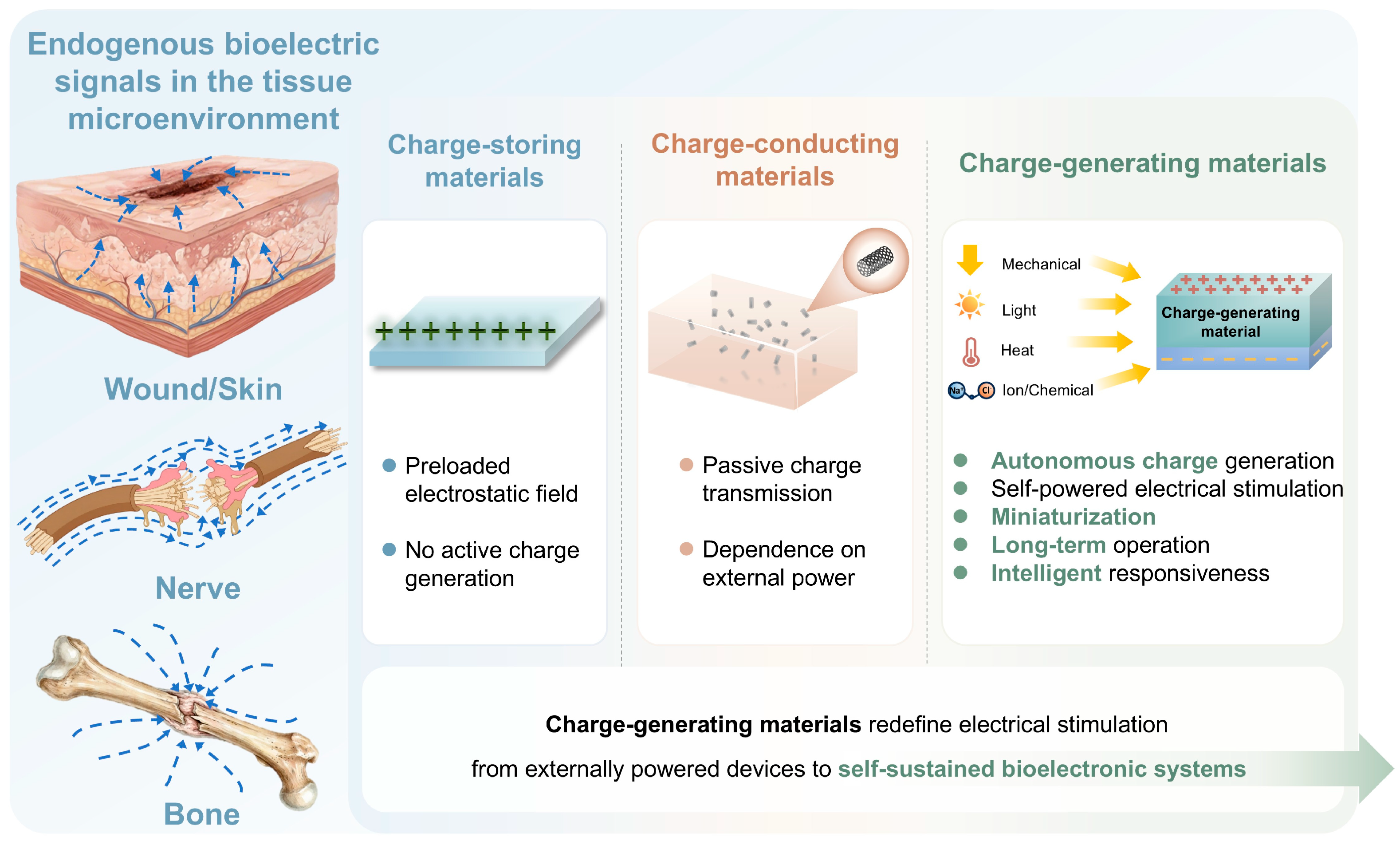
Electrical stimulation strategies for tissue repair can be broadly categorized according to how electrical charges are stored, transported, or generated within biomaterial systems. While traditional classifications often focus on material composition or fabrication methods, such approaches may obscure the fundamental relationship between electrical functionality and biological regulation. To provide a more therapeutically relevant framework, bioelectrical repair materials can be conceptually classified based on their interaction with electric charges, as schematically illustrated in Figure 1. This classification links material categories to their primary electrical functions and provides a function-oriented perspective for understanding electroactive biomaterials. By distinguishing whether a material primarily stores, transports, or generates electrical charges, this framework may facilitate the evaluation of their suitability for different therapeutic objectives and implantable bioelectronic applications.
From this perspective, electroactive biomaterials can be divided into three categories: charge-storing materials, charge-conducting materials, and charge-generating materials. These categories represent progressively increasing levels of bioelectrical functionality, ranging from passive electrostatic stimulation to active and autonomous electrical signal generation. It is worth noting that this charge-centered classification relies strictly on the material’s in vivo operational mode and thermodynamic behavior, rather than its fundamental chemical composition. Consequently, certain materials may exhibit dual identities depending on their structural design and application scenarios. A quintessential example is poly(vinylidene fluoride) (PVDF) and its copolymers. When PVDF is subjected to high-voltage corona polarization in vitro to trap quasi-permanent space charges within its pores or interfaces, and is subsequently used to provide a static, gradually decaying electric field without continuous mechanical input, it functions strictly as a charge-storing material (electret). Conversely, when PVDF is deployed in a dynamically active physiological environment (e.g., attached to a beating heart or articulating joints), where continuous mechanical strain dynamically alters its β-phase non-centrosymmetric crystal structure to induce transient dipole reorientation and generate real-time alternating electrical signals, it transitions into an active charge-generating material (piezoelectric generator). Thus, this functional taxonomy accurately captures the dynamic bioelectrical role the material plays during the tissue repair process.
Importantly, this classification does not aim to replace conventional material-based taxonomies, but rather to highlight the distinct therapeutic roles, advantages, and limitations associated with different electrical stimulation paradigms. Such a charge-centered framework provides a foundation for understanding why charge-generating materials, as self-powered systems, hold particular promise for long-term and implantable regenerative therapies.

2.1. Charge-Storing Materials: Passive Electrostatic Stimulation

Charge-storing materials, commonly referred to as electret materials, represent the earliest class of bioelectrical repair materials explored for tissue regeneration. Logically, they operate in a passive electrostatic mode. Their fundamental distinction from conducting or generating materials lies in the “pre-loaded” and “static dissipation” nature of their charge origins. These materials retain quasi-permanent electrostatic charges or frozen dipoles originating entirely from artificial charge injection or external field polarization during in vitro processing [27,34]. During in vivo operation, the materials themselves do not undergo any form of energy conversion, nor do they exhibit spontaneous dynamic charge separation behaviors. When integrated into biomaterial scaffolds or wound dressings, charge-storing materials can partially mimic endogenous injury-induced electric fields and provide passive electrostatic stimulation to surrounding cells [35,36]. Their simplicity, ease of fabrication, and device-free nature have enabled broad investigation in wound healing, bone repair, and neural applications [36,37]. However, because the electrical output of charge-storing materials is inherently static and gradually decays over time, their stimulation modes remain limited, and precise temporal or spatial regulation of bioelectric cues is difficult to achieve [27,36]. These intrinsic constraints define both the therapeutic potential and the performance ceiling of charge-storing systems.
The concept of electrets dates back to the early 19th century. The earliest concept of electrets was proposed by Faraday [38], who studied the phenomenon of materials retaining electric charges under the influence of external electric fields. In the 1970s, researchers further developed this theory, particularly regarding the charge storage and release mechanisms in dielectric materials. They established the theory that electrets achieve permanent polarization through the alignment of polar molecules and the displacement of space charges [39]. The working principle of electret materials is based on their unique internal structure. Typically composed of porous materials, their cavities are filled with air or other gases. Charges can be injected and permanently retained on the surfaces of these cavities. The storage and release of charges occur through the phenomenon of electrical breakdown. When an external electric field exceeds a certain critical threshold, the gas within the cavities breaks down, causing charges to be injected onto the cavity surfaces and forming macroscopic dipoles [40].
Among representative electret materials, common examples include polypropylene (PP) [41], polytetrafluoroethylene (PTFE) [30], poly(methyl methacrylate) (PMMA) [42], and poly(vinylidene fluoride) (PVDF) [43]. They have demonstrated tremendous efficacy in applications such as wound dressings [36] and bone repair [44,45]. However, despite these promising observations, electret materials possess inherent limitations that restrict their effectiveness in dynamic biological environments, such as difficulty in processing or high energy consumption during processing [46,47], sensitivity of the material’s charge stability to environmental factors [34], or the stored charge diminishes over time [48]. Moreover, electret materials function as passive charge reservoirs and lack the capacity for energy conversion, preventing sustained or adaptive electrical regulation in response to tissue motion or metabolic activity. These shortcomings will significantly hinder the application of these materials and highlight the need for alternative material systems with enhanced electrical functionality.

2.2. Charge-Conducting Materials: Electrical Interfaces for Externally Powered Systems

Charge-conducting materials form the foundational electrical interfaces in most conventional electrical stimulation systems for regenerative medicine [49]. Logically, they operate in a passive non-autonomous transport mode. The fundamental essence of these materials is to serve as low-impedance physical electron or ion conduits, facilitating efficient charge transport [50]. They inherently lack both the capacity to pre-store static charges in vitro and the capability to convert physiological energy in situ. In this paradigm, the materials themselves do not generate electrical stimuli; rather, the directional movement of their internal delocalized charges (electrons, holes, or free ions) and the subsequent transmission of electrical signals are strictly dependent on external electric fields applied by active external systems (e.g., implantable batteries, wired power supplies) or driven by intrinsic potential fields within the physiological environment [20,51]. Such systems have demonstrated strong capability in delivering programmable and precisely controlled electrical stimulation, particularly in neural and musculoskeletal applications [52,53]. Once the external energy input is removed, however, the material immediately loses its capacity to actively modulate the bioelectrical microenvironment. This absolute dependence on external power introduces substantial translational challenges, including bulky device architectures, mechanical mismatch with soft tissues, interfacial electrochemical reactions, and risks of infection or device failure [54,55]. As a result, charge-conducting materials are increasingly viewed as essential components of device-centered electrotherapy rather than self-sufficient therapeutic systems [56].
Conductive materials in biomedical material systems refer to a class of materials capable of efficiently transmitting electrical charges (electrons or ions). Compared with electret systems that rely on static charge retention, conductive materials enable active charge transport, offering a more direct strategy for electrical regulation in regenerative biomaterials. These materials provide a low-impedance physical pathway, coupling electrical signals from external sources or physiological potential gradients to cells and tissues, thereby modulating the behavior of electrically sensitive cells [49].
Generally speaking, conductive materials can be categorized into the following three types based on the type of charge carrier: (1) Electronically conductive materials: These rely primarily on electrons as charge carriers. Representative examples include metallic nanoparticles (NPs) [57], carbon-based nanomaterials (such as graphene [58] and carbon nanotubes (CNTs) [59]), and conductive polymers (such as polypyrrole (PPy) [60] and polyaniline (PANI) [61]). The conductivity of these materials originates from the delocalized migration of electrons within their energy bands or the movement of electrons within π-conjugated systems, exhibiting high electronic conductivity [49]. (2) Ionic conductive materials: In conductive hydrogels (CHs) [62] or certain biological polymer networks, charge is primarily transferred through the migration of ions (e.g., Na+, K+, Ca2+). This mechanism closely resembles electrical conduction in native tissues and is generally associated with good biocompatibility [63]. (3) Electron-ion hybrid conductive materials: Certain conductive polymers [64,65] or composite hydrogel [66,67] systems can simultaneously support both electron and ion migration, enabling signal conversion and synergistic conduction at bio/electronic interfaces. This provides a balanced solution for signal transmission efficiency and biocompatibility.
It follows that the fundamental distinction among these three categories of conductive materials lies in their electrical conduction mechanisms. The core function of conductive materials is to provide low-impedance pathways for sustaining stable electrical stimulation and shaping electrically active microenvironments. This enables their application in cardiac pacing [68,69] or the promotion of electrically sensitive behaviors such as wound healing [70,71], angiogenesis [72,73], or nerve regeneration [74,75]. However, their functionality remains intrinsically limited by several factors. First, these conductive materials are fundamentally passive, relying on external power sources to deliver electrical stimulation, which restricts their long-term and autonomous use in vivo. Second, repeated ionic doping and dedoping processes during operation can induce structural instability, leading to degradation of electrical and mechanical performance over time. Moreover, the spatial and temporal profiles of electrical stimulation are typically externally defined, providing limited adaptability to dynamic biological microenvironments. Collectively, these limitations constrain the applicability of conductive materials in sustained bioelectrical repair and motivate the development of alternative material strategies.

2.3. Charge-Generating Materials: Self-Powered Electrical Stimulation Platforms

Charge-generating materials constitute an emerging and transformative class of bioelectrical repair materials that operate in an active in situ energy conversion mode [76,77]. This category fundamentally departs from the static charge release of electrets and the passive transport of conducting systems. Its core distinction lies in possessing a real-time transduction mechanism and self-driven capability: rather than relying on pre-stored electrostatic charges or external active circuits, these materials function as active transducers. They autonomously harvest unordered dynamic energy from the body—including mechanical deformation (e.g., tissue motion, physiological micro-vibrations), biochemical reactions (e.g., metabolite redox processes, ion concentration gradients), or exogenous optical/thermal stimuli—and convert it into electrical outputs [78,79]. By driving the relative displacement of positive and negative charge centers within non-centrosymmetric lattices, or by inducing the spontaneous transfer of oppositely charged species at heterogeneous interfaces, they continuously generate spontaneous direct current or pulsed electrical signals in real time. Their output characteristics are intrinsically tied to the dynamic response mechanisms of internal crystals or molecules, enabling profound and adaptive coupling with physiological microenvironmental stimuli. This conceptual shift from externally powered devices toward material-based therapeutic systems offers intrinsic advantages in miniaturization, long-term operation, and reduced system complexity.
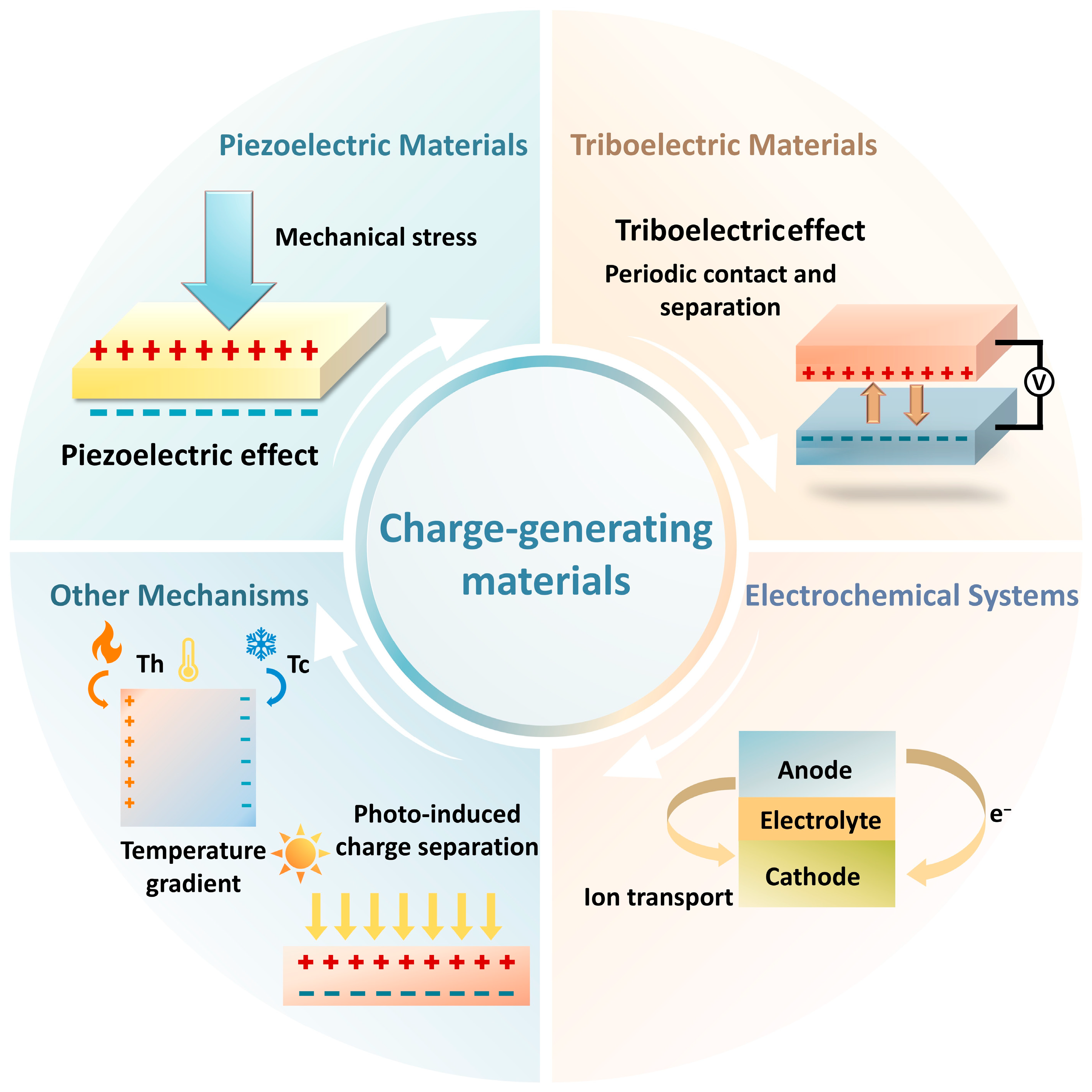
This property represents a fundamental shift in the design philosophy of bioelectrical materials—from passive electrical environments to autonomous electrical regulation. As summarized in Figure 2, charge-generating materials can produce electrical outputs through several representative energy conversion mechanisms. Mechanical deformation can induce charge polarization in piezoelectric materials, while periodic contact and separation between different surfaces result in triboelectric charge transfer. In addition, electrochemical reactions can drive electron flow through redox processes, and other physical stimuli such as temperature gradients or light irradiation may also induce charge separation. These mechanisms collectively enable the harvesting of diverse physiological energy sources and their conversion into localized electrical signals. Compared with traditional conductive materials, self-powered materials theoretically offer multiple promising features, including energy autonomy, sustained electrical output, and adaptive coupling with dynamic biological microenvironments. These characteristics enable long-term, minimally invasive, and potentially closed-loop bioelectrical stimulation, making them particularly attractive for next-generation bioelectrical repair strategies [78].
The piezoelectric effect refers to the phenomenon where certain materials with non-centrosymmetric crystal structures generate electric charges and voltage when subjected to mechanical stress. The specific mechanism varies depending on the material’s structural nature: in traditional inorganic materials, it originates from the displacement of positive and negative charge centers within non-centrosymmetric crystal structures [80,81]; however, in organic polymers and molecular crystals, it is typically driven by macromolecular shear polarization, dipole orientation [82,83], or the mechanical deformation of macroscopic dipoles [84,85]. Both mechanisms result in a potential difference across the material’s surface, enabling the conversion of mechanical energy into electrical energy. Common piezoelectric materials include inorganic piezoelectric ceramics [86,87] conventional non-degradable piezoelectric polymers [88] emerging biodegradable piezoelectric materials [89,90], and flexible organic molecular crystals [91,92]. Based on this effect, these materials are frequently used to fabricate energy generators or sensors for harvesting energy from external environments. The essence of the triboelectric effect lies in the charge transfer and interfacial accumulation process triggered by differences in surface physicochemical properties—such as work functions and ionic affinities—when two materials contact and separate. Based on this principle, periodic contact-separation or relative sliding between materials achieved through structural design can efficiently convert mechanical friction energy into electrical energy, forming the operational foundation of triboelectric nanogenerators (TENGs) [93]. In self-powered systems, electrochemical effects typically refer to processes that generate electrical energy through chemical or biochemical reactions, such as biofuel cells [94] and enzyme-catalyzed batteries [95]. Under this mechanism, continuous electrical output is achieved by driving electron flow through redox reactions, making it particularly suitable for long-term self-powered applications.
Biological tissues are inherently dynamic systems characterized by continuous motion, fluid flow, and electrochemical gradients. Self-powered materials can harvest these endogenous energy sources and transform them into localized electrical stimuli, enabling spatiotemporally adaptive bioelectrical modulation without external power supply. Such autonomous energy conversion not only improves electrical sustainability but also allows electrical outputs to be naturally coupled with physiological activities, which more closely resemble endogenous bioelectric signaling during tissue repair.
Consequently, electrogenic materials introduce a promising conceptual paradigm in bioelectrical regeneration, aiming to shift electrical stimulation towards a more self-sustained, self-responsive mode integrated with tissue dynamics. However, it is crucial to recognize that these systems are still predominantly in the preclinical proof-of-concept stage. Their claim of superiority must be tempered by significant translational limitations. Currently, there is a distinct absence of robust clinical validation to confirm their long-term therapeutic efficacy and safety in human patients. Furthermore, critical challenges such as the standardization of dynamic electrical outputs, unpredicted device degradation, and long-term biocompatibility in complex in vivo environments remain unresolved. Therefore, given the immense theoretical potential alongside these formidable translational hurdles, this review will comprehensively focus on their mechanisms, material design, and recent developments.

3. Charge-Generating Materials: Mechanisms and Design Strategies

The defining feature of charge-generating materials lies in their ability to autonomously transform diverse forms of endogenous energy into biologically relevant electrical signals [79,96]. This capability fundamentally distinguishes them from charge-storing and charge-conducting systems and underpins their potential as self-powered electrical stimulation platforms for tissue repair. Rather than serving as passive electrical reservoirs or transmission interfaces, charge-generating materials function as active transducers that dynamically couple physiological processes—such as mechanical deformation, biochemical reactions, thermal gradients, or metabolic activity—with localized electrical stimulation [33,97].
From a materials design perspective, the performance of charge-generating systems is governed by both their intrinsic energy-conversion mechanisms and their interactions with complex biological environments. Key considerations include the efficiency and stability of electrical output, the compatibility of generated signals with physiological electric fields, and the ability to integrate seamlessly with soft and dynamic tissues [78,97]. Accordingly, diverse strategies have been developed to harness different energy sources in vivo, giving rise to multiple classes of charge-generating materials with distinct operational principles and therapeutic profiles. In the following sections, we systematically discuss representative charge-generating mechanisms and corresponding material design strategies, highlighting their advantages, limitations, and implications for regenerative bioelectronic applications.

3.1. Piezoelectric Materials: Converting Physiological Mechanical Stimuli into Electrical Signals

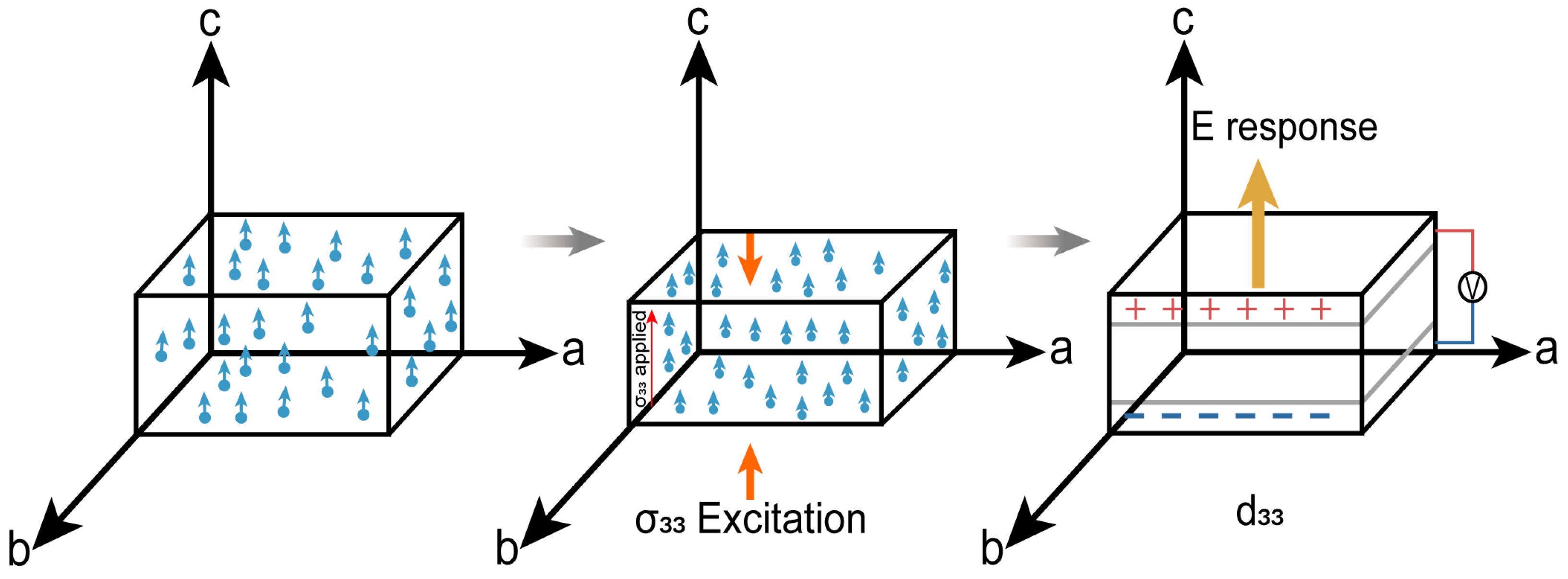
Piezoelectric materials are among the most extensively investigated charge-generating systems for self-powered bioelectrical stimulation, owing to their intrinsic ability to convert mechanical deformation into electrical signals [20]. This property arises from non-centrosymmetric crystal or molecular structures, in which applied mechanical stress induces relative displacement of positive and negative charge centers in crystals, or the stress-induced reorientation of molecular dipoles in polymers, resulting in surface charge accumulation and electric potential generation [32,98]. The fundamental electromechanical conversion process of piezoelectric materials is illustrated in Figure 3. In biological environments, such electromechanical coupling enables piezoelectric materials to directly harvest endogenous mechanical energy—such as tissue deformation, body movement, or fluid-induced stress—and translate it into localized electrical cues relevant for tissue repair [51,99].
From a regenerative medicine perspective, the appeal of piezoelectric materials lies in their natural compatibility with mechanically active tissues. Bone, nerve, and muscle tissues continuously experience dynamic mechanical stimuli, providing abundant and sustainable energy sources for in situ electrical generation [79,100]. Electrical signals generated by piezoelectric materials have been shown to regulate osteogenic differentiation, enhance axonal growth, and modulate cell migration and proliferation, thereby partially recapitulating endogenous bioelectric signaling during tissue repair [101,102]. Importantly, this mechanically triggered electrical output enables a direct coupling between tissue activity and stimulation intensity, offering a biomimetic and self-adaptive mode of electrical regulation.
A wide range of piezoelectric materials has been explored for bioelectrical repair applications, including inorganic ceramics, polymer-based piezoelectrics, and emerging biodegradable piezoelectric systems [103,104]. Piezoelectric ceramics, such as lead zirconate titanate and barium titanate, exhibit high piezoelectric coefficients and robust electrical output, but their intrinsic brittleness and limited biodegradability constrain their long-term implantability [105,106]. In contrast, polymeric piezoelectric materials, particularly poly(vinylidene fluoride) and its copolymers, offer superior flexibility, processability, and mechanical compatibility with soft tissues, albeit at the expense of reduced electrical output [56,107]. Recent efforts have further extended piezoelectric concepts to biodegradable materials, enabling transient electrical stimulation platforms that degrade after fulfilling therapeutic functions [78,108].
Despite these advantages, several challenges limit the translational potential of piezoelectric materials for sustained bioelectrical repair. The electrical output generated under physiological mechanical stimuli is often relatively low and highly dependent on loading conditions, making precise quantification and standardization difficult [78,104]. Moreover, the predictability of piezoelectric performance during the dynamic degradation process in complex biological environments, as well as the correlation between generated electrical signals and therapeutic efficacy, remains insufficiently understood [100,109]. Addressing these challenges requires rational material design strategies, including structural optimization, composite engineering, and integration with biological interfaces, to enhance electromechanical efficiency and ensure reliable electrical stimulation in vivo.

3.2. Triboelectric Materials: Contact-Induced Electrical Generation for Dynamic Stimulation

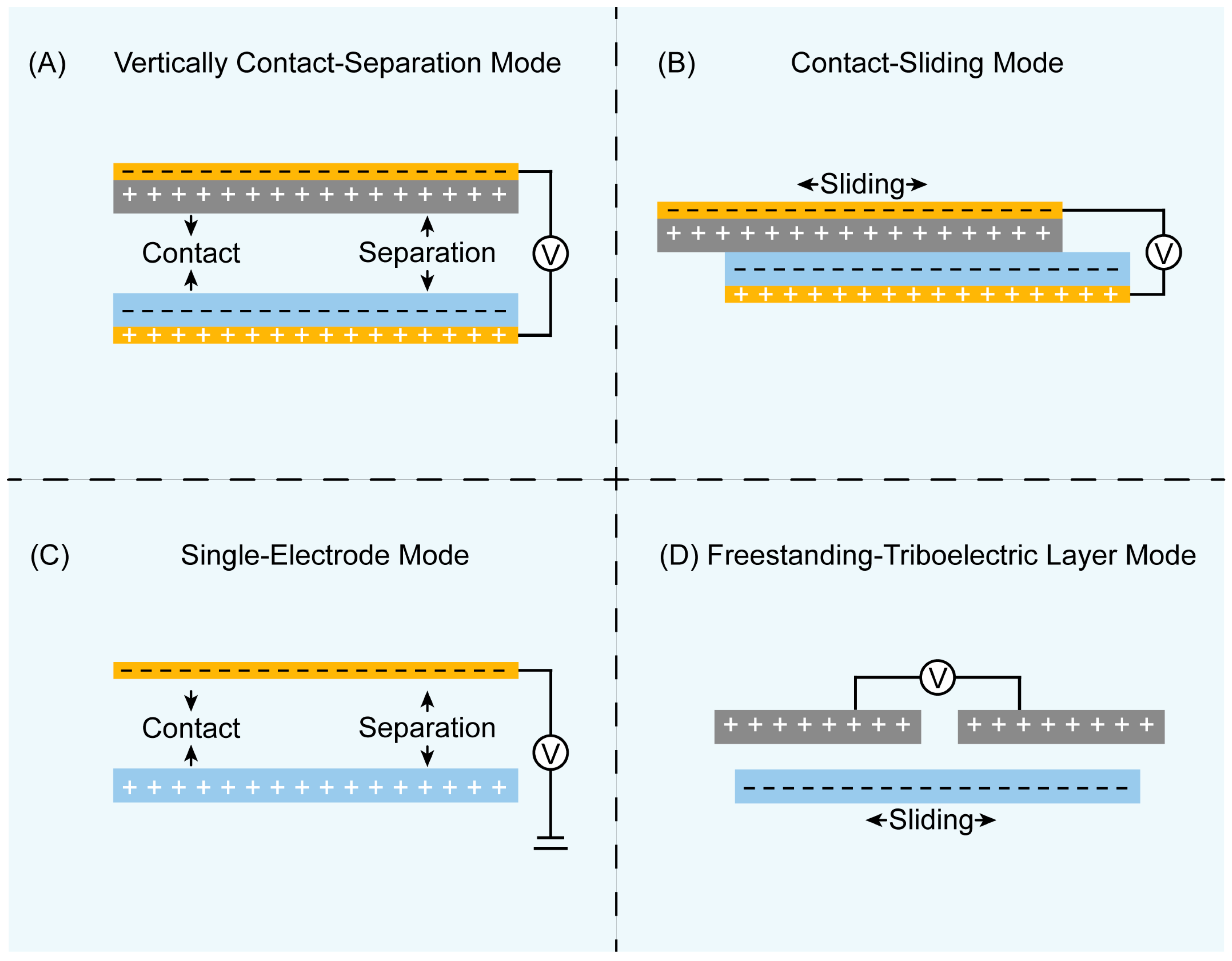
Triboelectric materials represent a rapidly emerging class of charge-generating systems that produce electrical signals through contact-induced charge transfer and electrostatic induction [110]. When two materials with different surface physicochemical properties—such as work function, electron affinity, or ionic adsorption behavior—come into contact and subsequently separate, interfacial charge redistribution occurs, resulting in the accumulation of opposite charges on each surface [111,112]. Repeated contact–separation or sliding motions can therefore convert mechanical friction energy into pulsed electrical outputs, forming the fundamental operating principle of triboelectric nanogenerators (TENGs) [96,113,114]. The working mechanism of triboelectric charge generation is illustrated in Figure 4.
In biological contexts, triboelectric materials are particularly attractive for harvesting low-frequency and irregular mechanical motions, including joint movement, tissue deformation, and organ pulsation [32,52]. Unlike piezoelectric systems that rely on bulk deformation, triboelectric systems can generate electrical signals from subtle interfacial motions, making them well suited for dynamic soft tissues and surface-contact environments [110,112]. The inherently pulsed and high-voltage characteristics of triboelectric outputs have been exploited to stimulate cell migration, enhance wound healing, and modulate tissue microenvironments, enabling self-powered electrical stimulation without external energy input [77,115].
A variety of triboelectric material combinations and structural designs have been explored for biomedical applications, including polymer–polymer, polymer–metal, and polymer–hydrogel interfaces [32,113]. Soft and flexible polymers, such as polydimethylsiloxane and fluorinated polymers, are frequently employed owing to their mechanical compliance and tunable surface properties [52,112]. Structural engineering strategies, including micro/nanostructuring and multilayer architectures, are commonly adopted to enhance charge density and output performance [112]. These design approaches allow triboelectric systems to be integrated into wearable or implantable platforms for localized bioelectrical stimulation.
Despite their promising capabilities, triboelectric materials face several challenges for long-term bioelectrical repair. Electrical output is highly dependent on contact stability and surface integrity, which may be compromised under repetitive motion or in moist physiological environments [56,78]. In addition, the pulsed and often high-voltage nature of triboelectric signals complicates precise control and quantitative correlation with biological responses [76,77]. Addressing these challenges requires advances in surface chemistry, structural durability, and signal modulation to enable reliable, biocompatible, and therapeutically relevant triboelectric stimulation in vivo [116].

3.3. Electrochemical and Bio-Galvanic Systems: Continuous Electrical Output from Physiological Environments

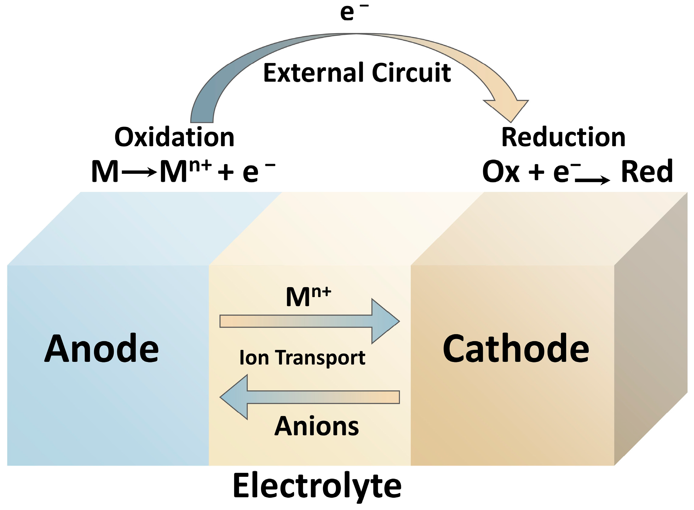
Electrochemical and bio-galvanic systems constitute a distinct class of charge-generating materials that produce electrical signals through chemical or biochemical redox reactions [77,117]. In these systems, electrical energy is generated by driving electron flow between electrodes with different electrochemical potentials, typically mediated by physiological electrolytes or biofluids [77,118]. The fundamental electrochemical mechanism underlying bio-galvanic electrical generation is illustrated in Figure 5. Unlike mechanically driven piezoelectric or triboelectric materials, electrochemical systems can generate continuous and relatively stable electrical outputs, making them particularly suitable for long-term self-powered electrical stimulation in vivo, although their operational lifespan is ultimately dictated by electrode consumption or biocatalyst stability.
From a biological standpoint, electrochemical charge generation is inherently compatible with physiological environments rich in ionic species, metabolites, and redox-active molecules. Bio-galvanic systems can directly harvest chemical energy from body fluids or metabolic processes, enabling sustained microcurrent generation without reliance on external power sources [119,120]. Such continuous electrical stimulation has been shown to influence cell proliferation, inflammation modulation, angiogenesis, and tissue remodeling, aligning well with the requirements of chronic tissue repair and regeneration [121,122].
Representative electrochemical systems for bioelectrical repair include metal-based galvanic couples, biofuel cells, and enzyme-catalyzed electrochemical platforms [119,123]. Metal-based bio-galvanic systems generate electrical output through spontaneous redox reactions between dissimilar electrodes immersed in physiological electrolytes, while biofuel cells utilize endogenous metabolites—such as glucose or lactate—as energy sources through enzymatic or catalytic pathways [117,124]. These approaches offer distinct advantages in terms of energy autonomy and sustained electrical output, and have been explored in applications ranging from wound healing to bone regeneration [56,121].
Despite their promise, electrochemical and bio-galvanic systems face important challenges that limit their translational potential. The long-term stability and safety of redox reactions in vivo remain critical concerns, particularly with respect to electrode degradation, by-product accumulation, and local tissue responses [20,125]. In addition, controlling electrical output magnitude and duration in complex biological environments is non-trivial, and the coupling between generated microcurrents and specific therapeutic outcomes is not yet fully understood [77,126]. Future progress will require careful material selection, interface engineering, and system-level optimization to ensure biocompatible, predictable, and therapeutically effective electrochemical stimulation.

3.4. Other Emerging Energy-Conversion Mechanisms (Thermoelectric, Photoelectric)

Beyond piezoelectric, triboelectric, and electrochemical systems, several emerging energy-conversion mechanisms have been explored to enable self-powered bioelectrical stimulation for tissue repair [79,117]. These approaches expand the conceptual landscape of charge-generating materials by harvesting alternative endogenous or externally accessible energy sources, including thermal gradients, light and so on [127,128]. Although most of these strategies remain at an early stage of development, they provide valuable insights into the versatility and future potential of self-powered bioelectronic materials.
Thermoelectric materials generate electrical signals by converting temperature gradients into electrical potential through the Seebeck effect [48]. In biological systems, subtle temperature differences between tissues or between implanted materials and surrounding environments can serve as continuous energy sources [78,129]. Thermoelectric systems have been investigated for long-term, low-power electrical stimulation, particularly in scenarios where mechanical motion is limited [117]. However, the relatively small temperature gradients available in vivo and the low conversion efficiency of biocompatible thermoelectric materials currently constrain their practical output [79,117].
Photoelectric materials represent another emerging strategy, in which light energy is converted into electrical signals to stimulate cellular activity [130]. Photoresponsive systems enable spatiotemporally precise electrical modulation and have been explored for neural stimulation and wound healing applications, especially when combined with minimally invasive or transdermal light delivery [96,131]. Nevertheless, light penetration depth in biological tissues and the need for external optical sources limit the applicability of photoelectric stimulation for deeply implanted systems [96,131].
Collectively, these emerging mechanisms underscore the expanding toolbox of self-powered bioelectrical materials and highlight opportunities for hybrid or multi-modal energy-conversion strategies [76,79]. Future advances in material chemistry, device architecture, and biological integration may enable these alternative mechanisms to complement established charge-generating platforms, further enriching the design space of self-powered bioelectronic therapies for tissue repair.
Despite their distinct operating principles, all charge-generating systems face common challenges when deployed in complex physiological environments. In vivo conditions are characterized by high ionic strength, continuous fluid exchange, and abundant proteins and cells, all of which may influence charge generation and electrical output. For triboelectric systems, ionic shielding can partially neutralize surface charges and weaken electrostatic induction, while protein adsorption may alter interfacial properties and reduce charge-transfer efficiency [132,133]. Piezoelectric materials may also experience attenuation of local electric fields due to ionic screening of mechanically induced surface charges [134,135]. In electrochemical systems, protein fouling, biofilm formation, and electrode passivation can increase interfacial resistance and compromise long-term charge-transfer performance [136,137,138]. Therefore, improving the stability and efficiency of charge generation under physiological conditions remains a critical challenge for the clinical translation of self-powered bioelectrical materials, motivating ongoing efforts in surface engineering, interface optimization, and protective material design. In this context, bioorthogonal click chemistry may provide precise and biocompatible tools for scaffold functionalization and cell–biomaterial interface construction, thereby facilitating the integration of electroactive platforms with regenerative microenvironments [139].

4. Biomedical Applications of Charge-Generating Materials

The therapeutic relevance of charge-generating materials ultimately lies in their ability to transform autonomous electrical signal generation into meaningful biological outcomes in tissue repair and regeneration. Building upon the mechanistic principles discussed in the previous section, self-powered electrical stimulation platforms have been explored across a range of regenerative applications, where electrical cues play critical roles in coordinating cellular behavior, tissue remodeling, and microenvironmental regulation [77,140]. By directly coupling endogenous energy sources with localized electrical outputs, these systems offer a distinct advantage in delivering sustained and physiologically adaptive stimulation without external power supply [79,97].
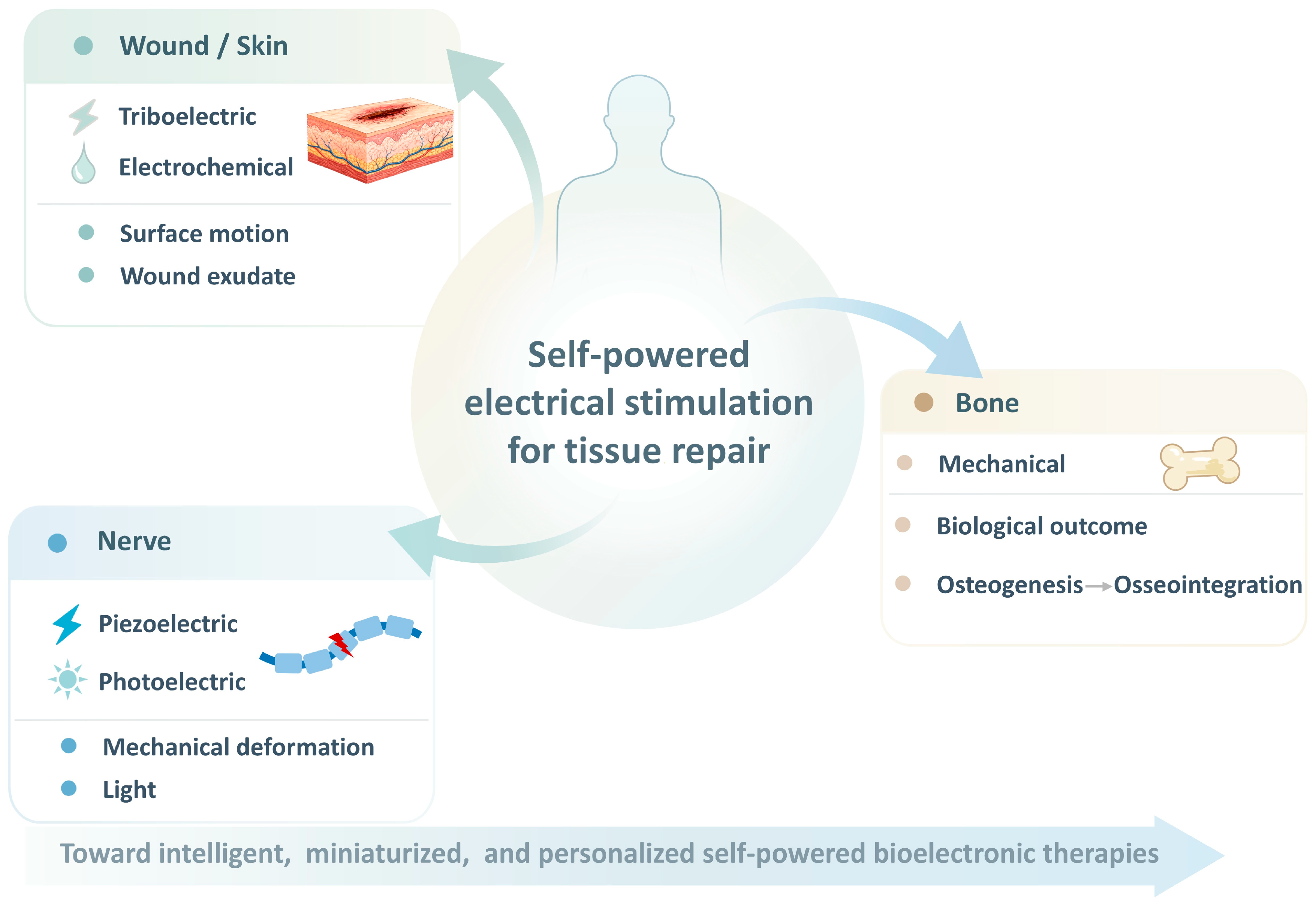
Importantly, different tissues exhibit distinct electrical sensitivities, mechanical environments, and regenerative demands, necessitating tailored stimulation strategies [77,141]. Self-powered materials enable electrical signals to be intrinsically synchronized with tissue-specific dynamics, such as motion, loading, or metabolic activity, thereby providing a more biomimetic mode of intervention [77,142]. In this section, we summarize representative biomedical applications of self-powered electrical stimulation, focusing on wound healing, nerve regeneration, bone repair, and related therapeutic contexts. An overview of representative regenerative applications enabled by self-powered charge-generating materials is illustrated in Figure 6. Emphasis is placed on how material design and energy-conversion mechanisms influence biological performance, as well as on current limitations that must be addressed to advance clinical translation.

4.1. Wound Healing and Soft Tissue Repair

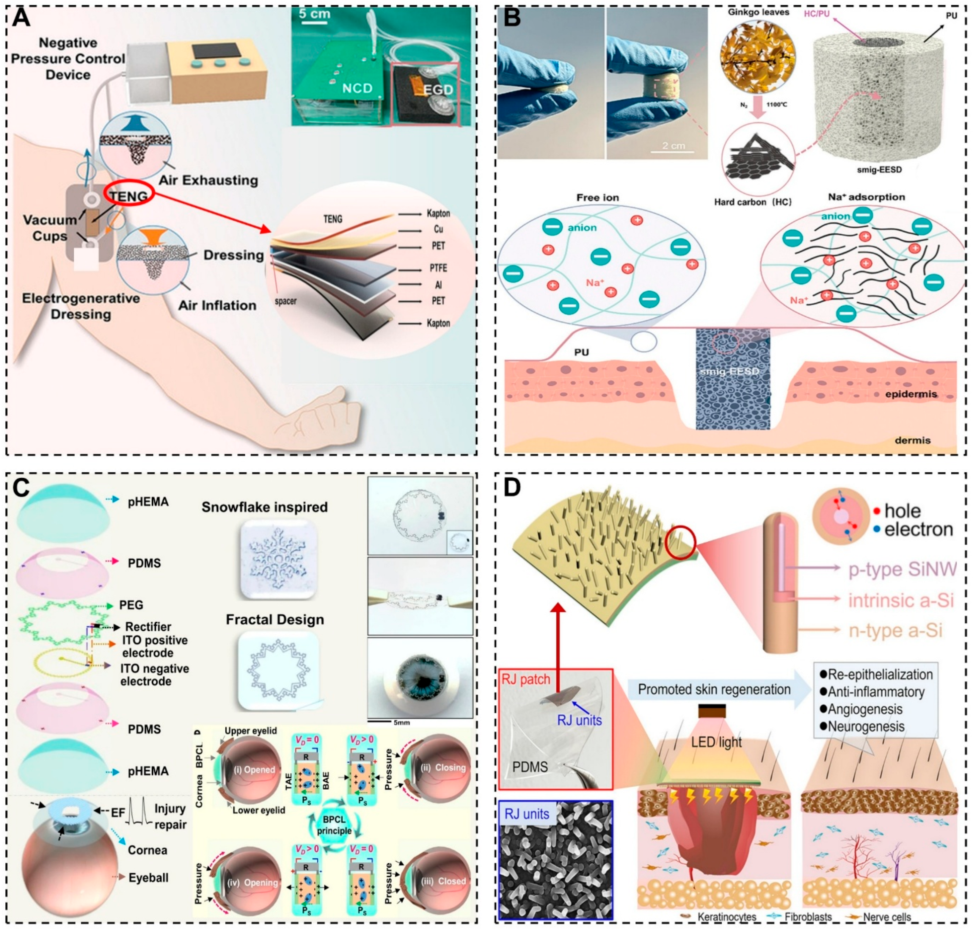
Wound healing is a highly dynamic cascade strictly dependent on bioelectric signals [126]. Epithelial injury disrupts the transepithelial potential and rapidly establishes an endogenous electric field (EEF), which guides the directional migration of cells to close the wound [143]. However, in chronic wounds, the dysregulation of electrical signals often leads to healing stagnation [144]. To actively reshape this electrophysiological microenvironment, Li, Zhang, and co-workers developed an electrogenerative dressing (EGD) based on a triboelectric nanogenerator (TENG) [145]. The ingenuity of this design lies in its ability to actively convert the macroscopic mechanical deformation from clinical negative-pressure suction into a continuous therapeutic electric field, thereby forcefully driving macrophage polarization toward a reparative phenotype while maintaining highly efficient exudate drainage [12] (Figure 7A).
Nevertheless, mechanical force-driven mechanisms are not suitable for all clinical scenarios. Targeting static wounds where external motion is difficult to apply, Li et al. further achieved a paradigm shift from “mechanical force dependency” to “microenvironment adaptability” by developing a self-manipulating sodium ion gradient-based endogenic electrical stimulation dressing (smig-EESD). This system non-invasively amplifies the endogenous electric field by passively adsorbing natural ions from wound exudates, enabling a completely battery-free and self-sustained power supply [24] (Figure 7B). For delicate and highly sensitive soft tissues, piezoelectric materials demonstrate distinct advantages. Yao, Lin, and co-workers developed a snowflake-inspired piezoelectric contact lens (BPCL). This device stands out for its extreme sensitivity in capturing the minute physiological motion of “blinking” and converting it into localized electric fields, realizing non-invasive electrical repair of corneal injuries [146] (Figure 7C).
Furthermore, while traditional self-powered dressings primarily focus on angiogenesis or macrophage polarization, the flexible amorphous silicon radial junction (RJ) patch developed by Liu, Chen, and co-workers pioneers a novel repair pathway targeting the “neuro-cutaneous axis”. Its uniqueness lies in utilizing wireless photoelectric signals under near-infrared light to precisely promote synaptic growth and neural network reconstruction of damaged sensory nerves in the deep wound bed, effectively addressing the clinical dilemma of mismatched regeneration speeds between neural and skin tissues, ultimately achieving high-quality full-thickness skin regeneration accompanied by neural functional recovery [147] (Figure 7D). In the future, intelligent patches integrating multiplexed adaptive regulation of physiological microenvironments will inevitably dictate the evolutionary direction of soft tissue repair materials [148].

4.2. Nerve Regeneration and Neural Interfaces

Nervous system injuries, encompassing both peripheral nerve injuries (PNI) and traumatic brain injuries (TBI), present formidable clinical challenges, primarily due to the extremely limited intrinsic regenerative capacity of neurons and the formation of inhibitory glial scars post-injury [22,149]. Given that electrical signaling is the fundamental language of neural activity, restoring physiological electric cues via self-powered bioelectronics has emerged as a transformative therapeutic strategy [150].
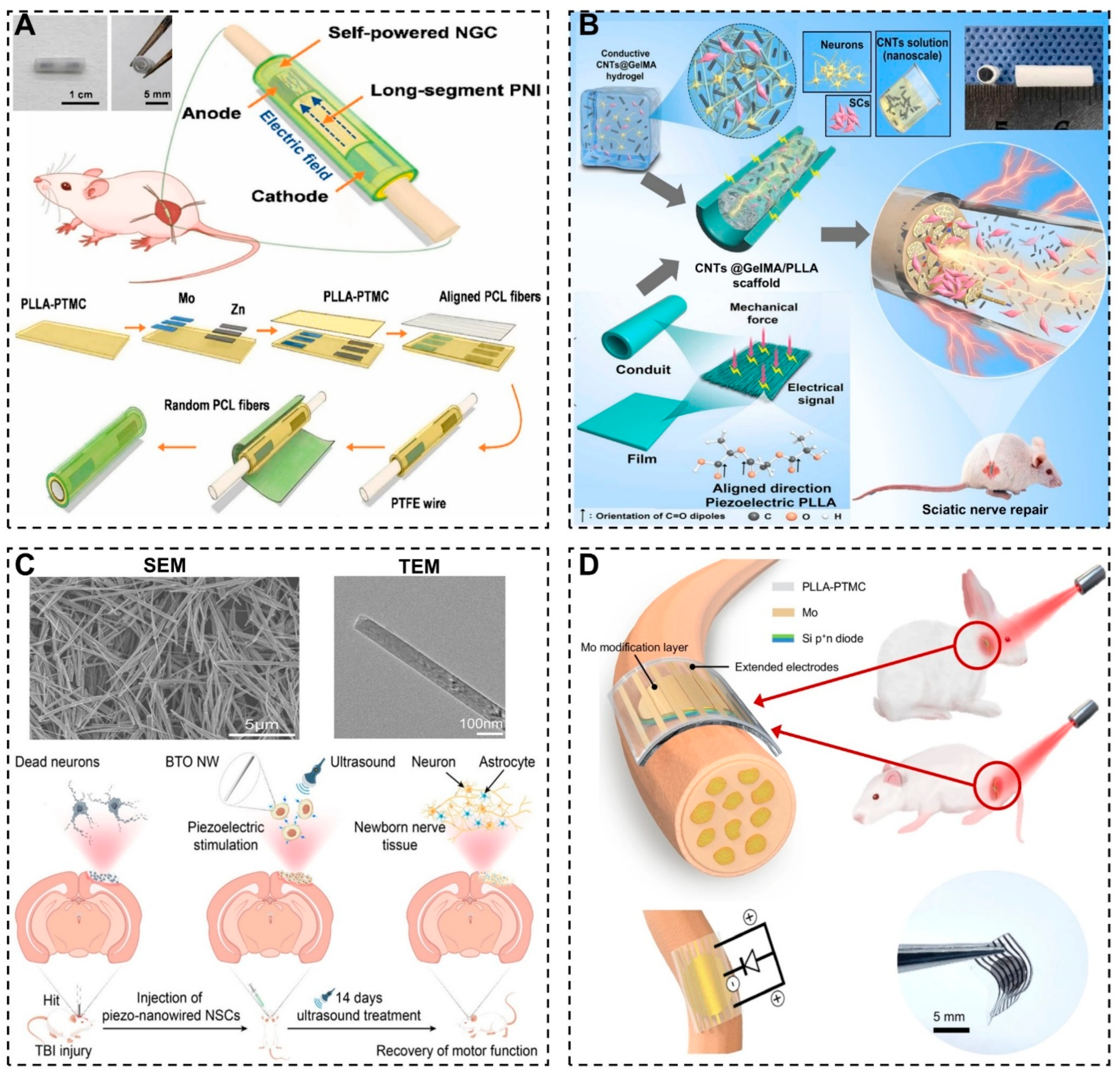
In terms of providing sustained direct-current (DC) electrical stimulation, Yin and co-workers developed a biodegradable nerve guidance conduit based on zinc-molybdenum micro-batteries. Without requiring any external energy penetration, this system relies solely on the spontaneous redox reactions of the metallic conduit walls within body fluids to provide long-lasting microcurrents for axonal extension [151] (Figure 8A). To fully harness the abundant biomechanical energy in vivo, Li, Zhao, and co-workers engineered a wirelessly self-powered and electroconductive composite scaffold (CNTs@GelMA/PLLA). The ingenuity of this design lies in seamlessly integrating a soft, biomimetic conductive microenvironment with endogenous piezoelectric stimulation, which actively translates the macroscopic mechanical forces of natural body movements into localized therapeutic electrical signals, thereby significantly promoting Schwann cell myelination and accelerating peripheral nerve regeneration [152] (Figure 8B).
In the repair of central nervous system injuries (e.g., traumatic brain injury), the requirement for therapeutic targeting precision is exceedingly high. To this end, Liu, Qiu, and co-workers proposed a “piezo-nanowired stem cell” strategy. By anchoring high-aspect-ratio piezoelectric nanowires onto the stem cell membrane, they achieved ultra-high spatiotemporal resolution electrical modulation at the single-cell membrane level under ultrasound penetration, substantially improving the reconstruction rate of neural circuits [153] (Figure 8C). Additionally, in the realm of advanced light-driven stimulation, Yin, Wang, and co-workers designed a fully biodegradable and flexible neural interface. This design pioneered a new pathway for transdermal wireless optoelectronic neuromodulation by efficiently converting near-infrared light into localized electrical stimuli to promote peripheral nerve regeneration [127] (Figure 8D). Ultimately, delivering high-fidelity spatiotemporal electrical signals while strictly avoiding neuronal excitotoxicity will determine the clinical translation potential of these neuro-electroceuticals.

4.3. Bone Regeneration and Osteointegration

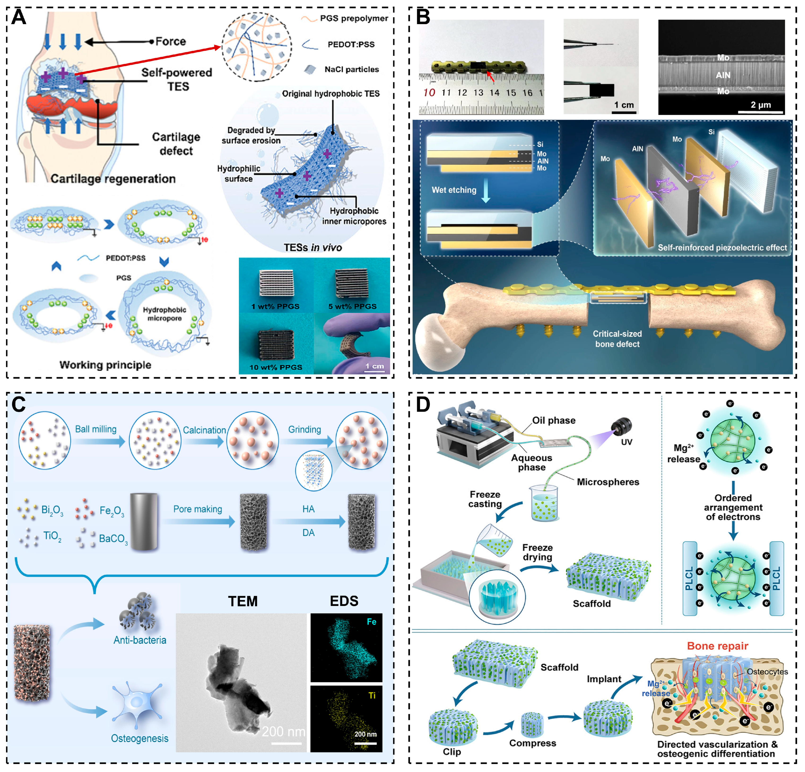
Bone tissue naturally possesses a unique hierarchical structure and inherent piezoelectric properties, and its remodeling process is highly dependent on the electromechanical coupling of mechanical loading and electrical signaling (Wolff’s Law) [154]. Traditional implantable electrical stimulators often face the pain points of bulky size and absolute reliance on external batteries, whereas self-powered biomaterials have opened a new paradigm for bone repair via in situ energy harvesting. Addressing the anti-moisture challenge of triboelectric materials in vivo, You and co-workers proposed a 3D-printed “encapsulation-free” triboelectric scaffold. It leverages internal hydrophobic micropores to stably generate electricity in humid bodily fluids, while the surface dynamically turns hydrophilic upon degradation to promote cell adhesion, perfectly resolving the in vivo aquatic failure dilemma of traditional TENGs [54] (Figure 9A).
To break free from the constraints of bulky 3D scaffolds, Tian, Li, and co-workers introduced a minimalist engineering concept of “scaffold-free” repair. Relying merely on an ultra-thin, self-reinforced aluminum nitride piezoelectric microchip attached to the bone surface, it can acutely capture physiological micro-vibrations to induce vascularized repair of large segmental bone defects within four weeks [37] (Figure 9B). Furthermore, targeting the severe clinical challenge of implant-associated infections in bone defects, Zhou, Wang, and co-workers pioneered a multimodal defect-rich nanoreactor. Driven by deep-penetrating ultrasound, this strategy stands out by not only initiating sono-piezoelectric tandem catalysis to eradicate resistant biofilms via reactive oxygen species, but, more profoundly, modulating local iron metabolism to rescue the osteogenic differentiation of stem cells, thereby achieving simultaneous infection clearance and robust bone regeneration [155] (Figure 9C). Meanwhile, pursuing the chemical-to-electrical energy conversion pathway, He and co-workers pioneered a “battery scaffold”. While degrading in vivo and releasing magnesium ions, this porous scaffold converts disordered chemical energy into directional, closed-loop osteogenic microcurrents, achieving long-lasting and stable electrical stimulation even without the involvement of mechanical loading [140] (Figure 9D). Future designs of orthopedic electroactive materials must be dedicated to ensuring that the dynamic degradation and electrical attenuation curves of the materials flawlessly intersect with the mechanical climbing trajectory of nascent bone.

4.4. Comparative Analysis of Charge-Generating Mechanisms for Tissue-Specific Electrical Stimulation

Self-powered electrical stimulation platforms have demonstrated considerable potential across diverse regenerative applications. However, their therapeutic performance is highly dependent on the biological, mechanical, and electrical characteristics of specific tissues. In addition to tissue-specific biological requirements, the electrical output profiles of different charge-generating mechanisms should also be considered. Triboelectric systems generally produce pulsed electrical outputs with relatively high voltage but motion- and interface-dependent current, making them suitable for dynamic tissues or applications where intermittent mechanical motion is available [156,157]. Piezoelectric systems generate load-dependent alternating electrical signals that are directly coupled with mechanical deformation, which is particularly relevant for mechanically active tissues such as bone, cartilage, and peripheral nerves [37,156]. In contrast, electrochemical and bio-galvanic systems usually provide more continuous microcurrent outputs driven by redox reactions in physiological electrolytes, making them attractive for chronic repair scenarios that require sustained stimulation [158,159]. Representative studies have reported outputs ranging from high-voltage pulsed signals in triboelectric systems to deformation-dependent piezoelectric potentials and sustained microcurrents in electrochemical platforms. However, direct comparison of power density across different studies should be interpreted cautiously, because reported values are strongly affected by device geometry, testing frequency, loading conditions, electrolyte composition, and normalization methods [37,156,158,159]. Therefore, representative quantitative metrics should be considered together with output mode and stability when selecting charge-generating mechanisms for tissue-specific regenerative applications. Soft tissues such as skin and wounds possess accessible surfaces and dynamic microenvironments, where endogenous electric cues play a critical role in regulating cell migration, inflammation resolution, and angiogenesis [160,161]. In these contexts, triboelectric and electrochemical systems are particularly advantageous. Due to their conformal integration with soft tissues, these systems can translate mild mechanical or biochemical activation into physiological-level electric fields and safe microcurrents (typically in the microampere range), making them exceptionally well suited for wound healing applications without triggering undesirable Joule heating or tissue damage [20,162].
Neural tissues, in contrast, require more precise spatial and temporal control of electrical signals to guide axonal extension and neurite outgrowth while avoiding unintended neuronal excitation [147]. Piezoelectric systems, which convert mechanical deformation into electrical output, can seamlessly harness abundant physiological motions (e.g., respiratory movements of the chest wall and muscle contractions) to provide activity-dependent stimulation [104]. However, for central nervous system (CNS) repair, where natural macroscopic motion is severely restricted by bony encasements (a typical low-strain tissue environment) [163], deep-penetrating external triggers such as ultrasound are imperative to remotely actuate these piezoelectric implants [149,153]. Photoelectric platforms offer superior spatiotemporal precision for neural modulation through light-triggered charge generation, although their application is constrained by limited light penetration in biological tissues [127]. Electrochemical systems can also deliver continuous stimulation but require careful control of electrical output to ensure neural safety and maintain long-term electrode–tissue interface stability [151,164].
Bone regeneration represents a distinct scenario in which mechanical loading and electrical signaling are intrinsically coupled. Piezoelectric materials are particularly well suited for this load-bearing environment, as they can convert mechanical stress into electrical signals that mimic the native electromechanical behavior of bone tissue [165]. Electrochemical systems can further complement this strategy by generating sustained microcurrents that support long-term osteogenesis and osseointegration [140]. Nevertheless, variations in patient mobility and mechanical loading conditions may influence the magnitude and consistency of the generated electrical stimulation [104].
Overall, these comparisons suggest that no single self-powered mechanism is universally optimal for all regenerative scenarios. Instead, effective bioelectrical repair strategies require careful matching between tissue-specific regenerative demands and the characteristics of different charge-generating systems, including their energy sources, electrical output profiles, and stimulation modes. Such a comparative perspective is essential for guiding material selection, device design, and translational development in self-powered bioelectronic therapies, as summarized in Table 1.

5. Challenges and Future Perspectives

Despite rapid advances in self-powered bioelectrical materials for tissue repair, several fundamental challenges must be addressed before these systems can be translated from promising laboratory demonstrations to clinically viable therapies. Unlike externally powered electrical stimulation devices, self-powered platforms rely on endogenous energy sources and material-intrinsic transduction mechanisms, which introduce unique complexities in output stability, signal predictability, and therapeutic standardization [104,166]. Bridging the gap between material innovation and clinical application therefore requires a holistic understanding that integrates materials science, bioelectrical regulation, and translational medicine.
One of the primary challenges lies in the quantitative characterization and standardization of self-generated electrical signals in vivo. Electrical outputs are highly dependent on tissue-specific mechanical activity, biochemical environments, and patient-to-patient variability, complicating direct comparisons across studies and therapeutic contexts [105,167]. In addition, long-term biocompatibility and functional stability remain critical concerns, particularly with respect to material degradation, interfacial reactions, and potential immune responses during prolonged implantation [79,168].
Among these challenges, several bottlenecks should be prioritized for future development. First, the standardization of self-generated electrical outputs represents a fundamental prerequisite for comparing different charge-generating systems. Parameters such as voltage, current density, charge density, power density, stimulation duration, frequency, and waveform should be reported under physiologically relevant and standardized testing conditions. Second, quantitative correlations between generated electrical signals and biological outcomes remain insufficiently established. Defining how specific electrical parameters regulate cell migration, proliferation, differentiation, immune modulation, angiogenesis, or tissue remodeling is essential for identifying effective therapeutic windows and avoiding under- or over-stimulation. Third, long-term biocompatibility and functional stability must be systematically evaluated, particularly for degradable, electrochemically active, or chronically implanted systems. In this regard, future studies should not only demonstrate short-term regenerative efficacy but also assess material degradation, interfacial reactions, immune responses, output attenuation, and tissue safety over clinically relevant timescales.
Beyond these material-level challenges, the clinical translation of self-powered bioelectrical materials also faces distinct regulatory and manufacturing barriers compared with conventional battery-powered implants. Conventional implantable stimulators are typically based on mature battery modules and established electronic architectures, whereas self-powered systems introduce material-integrated energy conversion as an active therapeutic function. Therefore, regulatory evaluation should not only address conventional requirements for implantable devices, such as biocompatibility, electrical safety, electromagnetic compatibility, sterilization, and long-term reliability, but also validate the stability, controllability, and safety of self-generated electrical outputs under physiologically relevant conditions [169,170]. For degradable or electrochemically active systems, additional attention should be paid to degradation products, reaction by-products, electrode consumption, and time-dependent changes in output performance [169]. Moreover, when self-powered platforms are integrated with wireless communication, sensing, or closed-loop control modules, electromagnetic coexistence, cybersecurity, and data integrity may represent additional regulatory considerations [170,171,172]. From a manufacturing perspective, scale-up remains challenging because electrical outputs are highly sensitive to micro/nanostructure, interfacial chemistry, polarization state, electrode geometry, encapsulation quality, and sterilization processes [173,174]. Batch-to-batch reproducibility, scalable fabrication of hierarchical structures, in-line quality control of electromechanical or electrochemical performance, and reliable integration with flexible or biodegradable substrates are therefore key bottlenecks that must be addressed before clinical translation [173,174].
From a forward-looking perspective, the future of self-powered bioelectrical therapies is likely to be shaped by advances in intelligent material design, system integration, and personalized medicine [175]. Strategies that enable adaptive or feedback-regulated electrical stimulation, hybrid energy-conversion mechanisms, and seamless integration with regenerative biomaterials may further enhance therapeutic efficacy and reliability [78,176]. Ultimately, addressing these challenges will be essential for establishing self-powered bioelectrical materials as a robust and clinically impactful platform for next-generation regenerative therapies.

Author Contributions

Conceptualization, X.Z., X.C. and Z.L.; methodology, X.Z., Z.W., J.L. and C.C.; software, X.Z. and Z.W.; validation, X.Z., Z.W. and W.M.; formal analysis, X.Z., Z.W. and J.L.; investigation, X.Z., Z.W., J.L. and C.C.; resources, X.C. and Z.L.; data curation, X.Z. and Z.W.; writing—original draft preparation, X.Z. and Z.W.; writing—review and editing, J.L., C.C., W.M., X.C. and Z.L.; visualization, X.Z., Z.W., J.L. and C.C.; supervision, X.C. and Z.L.; project administration, X.C. and Z.L.; funding acquisition, X.C. and Z.L. All authors have read and agreed to the published version of the manuscript.

Funding

Beijing Nova Program 20240484655; National Natural Science Foundation of China U25A20417; Beijing Natural Science Foundation L245015, Z240022.

Data Availability Statement

No new data were created or analyzed in this study.

Conflicts of Interest

The authors declare no conflict of interest.

References

  1. Ishikawa, K.; Ueyama, Y.; Mano, T.; Koyama, T.; Suzuki, K.; Matsumura, T. Self-setting barrier membrane for guided tissue regeneration method: Initial evaluation of alginate membrane made with sodium alginate and calcium chloride aqueous solutions. J. Biomed. Mater. Res. 1999, 47, 111–115. [Google Scholar] [CrossRef]
  2. Sen, C.K. Human Wound and Its Burden: Updated 2020 Compendium of Estimates. Adv. Wound Care 2021, 10, 281–292. [Google Scholar] [CrossRef] [PubMed]
  3. Zhao, M. Electrical fields in wound healing—An overriding signal that directs cell migration. Semin. Cell Dev. Biol. 2009, 20, 674–682. [Google Scholar] [CrossRef] [PubMed]
  4. Liang, Y.; Tian, H.; Liu, J.; Lv, Y.; Wang, Y.; Zhang, J.; Huang, Y. Application of stable continuous external electric field promotes wound healing in pig wound model. Bioelectrochemistry 2020, 135, 107578. [Google Scholar] [CrossRef] [PubMed]
  5. Levin, M.; Pezzulo, G.; Finkelstein, J.M. Endogenous Bioelectric Signaling Networks: Exploiting Voltage Gradients for Control of Growth and Form. Annu. Rev. Biomed. Eng. 2017, 19, 353–387. [Google Scholar] [CrossRef] [PubMed]
  6. Martin-Granados, C.; McCaig, C.D. Harnessing the Electric Spark of Life to Cure Skin Wounds. Adv. Wound Care 2014, 3, 127–138. [Google Scholar] [CrossRef] [PubMed]
  7. Cheng, H.; Bai, J.; Zhou, X.; Chen, N.; Jiang, Q.; Ren, Z.; Li, X.; Su, T.; Liang, L.; Jiang, W.; et al. Electrical stimulation with polypyrrole-coated polycaprolactone/silk fibroin scaffold promotes sacral nerve regeneration by modulating macrophage polarisation. Biomater. Transl. 2024, 5, 157–174. [Google Scholar] [CrossRef] [PubMed]
  8. Borgens, R.B.; Vanable, J.W.; Jaffe, L.F. Bioelectricity and regeneration: Large currents leave the stumps of regenerating newt limbs. Proc. Natl. Acad. Sci. USA 1977, 74, 4528–4532. [Google Scholar] [CrossRef] [PubMed]
  9. Fukada, E.; Yasuda, I. On the Piezoelectric Effect of Bone. J. Phys. Soc. Jpn. 1957, 12, 1158–1162. [Google Scholar] [CrossRef]
  10. Aaron, R.K.; Ciombor, D.M.; Simon, B.J. Treatment of nonunions with electric and electromagnetic fields. Clin. Orthop. Relat. Res. 2004, 419, 21–29. [Google Scholar] [CrossRef] [PubMed]
  11. Zhao, M.; Song, B.; Pu, J.; Wada, T.; Reid, B.; Tai, G.; Wang, F.; Guo, A.; Walczysko, P.; Gu, Y.; et al. Electrical signals control wound healing through phosphatidylinositol-3-OH kinase-gamma and PTEN. Nature 2006, 442, 457–460. [Google Scholar] [CrossRef] [PubMed]
  12. Luo, R.; Liang, Y.; Yang, J.; Feng, H.; Chen, Y.; Jiang, X.; Zhang, Z.; Liu, J.; Bai, Y.; Xue, J.; et al. Reshaping the Endogenous Electric Field to Boost Wound Repair via Electrogenerative Dressing. Adv. Mater. 2023, 35, 2208395. [Google Scholar] [CrossRef] [PubMed]
  13. Levin, M. Endogenous bioelectrical networks store non-genetic patterning information during development and regeneration. J. Physiol. 2014, 592, 2295–2305. [Google Scholar] [CrossRef] [PubMed]
  14. Koul, S.; Devecka, L.A.; Pierce, M.C.; Vazquez, M. A Microscale–Optical Interface to Examine Electric Field-Induced Cell Motility Within Whole-Eye Facsimiles. Micro 2025, 5, 10. [Google Scholar] [CrossRef] [PubMed]
  15. Nishimura, K.Y.; Isseroff, R.R.; Nuccitelli, R. Human keratinocytes migrate to the negative pole in direct current electric fields comparable to those measured in mammalian wounds. J. Cell Sci. 1996, 109, 199–207. [Google Scholar] [CrossRef] [PubMed]
  16. Abedin-Do, A.; Zhang, Z.; Douville, Y.; Méthot, M.; Bernatchez, J.; Rouabhia, M. Electrical stimulation promotes the wound-healing properties of diabetic human skin fibroblasts. J. Tissue Eng. Regen. Med. 2022, 16, 643–652. [Google Scholar] [CrossRef] [PubMed]
  17. Urabe, H.; Akimoto, R.; Kamiya, S.; Hosoki, K.; Ichikawa, H.; Nishiyama, T. Effects of pulsed electrical stimulation on growth factor gene expression and proliferation in human dermal fibroblasts. Mol. Cell Biochem. 2021, 476, 361–368. [Google Scholar] [CrossRef] [PubMed]
  18. Rouabhia, M.; Park, H.; Meng, S.; Derbali, H.; Zhang, Z. Electrical stimulation promotes wound healing by enhancing dermal fibroblast activity and promoting myofibroblast transdifferentiation. PLoS ONE 2013, 8, e71660. [Google Scholar] [CrossRef] [PubMed]
  19. Calvey, C.; Zhou, W.; Stakleff, K.S.; Sendelbach-Sloan, P.; Harkins, A.B.; Lanzinger, W.; Willits, R.K. Short-Term Electrical Stimulation to Promote Nerve Repair and Functional Recovery in a Rat Model. J. Hand Surg. 2015, 40, 314–322. [Google Scholar] [CrossRef] [PubMed]
  20. Guillot-Ferriols, M.; Lanceros-Méndez, S.; Gómez Ribelles, J.L.; Gallego Ferrer, G. Electrical stimulation: Effective cue to direct osteogenic differentiation of mesenchymal stem cells? Biomater. Adv. 2022, 138, 212918. [Google Scholar] [CrossRef] [PubMed]
  21. Ashrafi, M.; Alonso-Rasgado, T.; Baguneid, M.; Bayat, A. The efficacy of electrical stimulation in lower extremity cutaneous wound healing: A systematic review. Exp. Dermatol. 2017, 26, 171–178. [Google Scholar] [CrossRef] [PubMed]
  22. Zuo, K.J.; Gordon, T.; Chan, K.M.; Borschel, G.H. Electrical stimulation to enhance peripheral nerve regeneration: Update in molecular investigations and clinical translation. Exp. Neurol. 2020, 332, 113397. [Google Scholar] [CrossRef] [PubMed]
  23. Qin, W.; Li, L.; Niu, W.; Wang, W.-R.; Wu, D.-W.; Song, C.-G.; Gao, C.-H.; Mu, Z.; Tay, F.R.; Jiao, K.; et al. Effects of Electric Field-Modulated Conductive Hydrogel on Osseoperception and Osseointegration of Dental Implants. Adv. Funct. Mater. 2024, 34, 2400256. [Google Scholar] [CrossRef]
  24. Luo, R.; Fan, Y.; Qi, Y.; Bai, Y.; Xiao, M.; Lv, Y.; Liang, J.; Tang, M.; Zhang, J.; Li, Z.; et al. Self-Manipulating Sodium Ion Gradient-Based Endogenic Electrical Stimulation Dressing for Wound Repair. Adv. Mater. 2025, 37, 2419149. [Google Scholar] [CrossRef] [PubMed]
  25. Li, J.; Wang, Y.; Wang, H.; Liu, Q.; Song, B. Regulation of Skin Wound Healing by Bioelectric Stimulation. Dermatol. Bull. 2023, 40, 430–437. [Google Scholar]
  26. Sabzpoushan, S.; Woias, P. Electret-based energy harvesters: A review. Nano Energy 2024, 131, 110167. [Google Scholar] [CrossRef]
  27. Sessler, G.M. Introduction. In Electrets; Sessler, G.M., Ed.; Springer: Berlin/Heidelberg, Germany, 1987; pp. 1–12. [Google Scholar]
  28. Mahanty, B.; Ghosh, S.K.; Garain, S.; Mandal, D. An effective flexible wireless energy harvester/sensor based on porous electret piezoelectric polymer. Mater. Chem. Phys. 2017, 186, 327–332. [Google Scholar] [CrossRef]
  29. Capron, B.A.; Hess, D.W. Microscopic Models of Piezoelectric Polymers. IEEE Trans. Ultrason. Ferroelectr. Freq. Control 1986, 33, 33–40. [Google Scholar] [CrossRef] [PubMed]
  30. Bi, M.; Wang, S.; Wang, X.; Ye, X. Freestanding-electret rotary generator at an average conversion efficiency of 56%: Theoretical and experimental studies. Nano Energy 2017, 41, 434–442. [Google Scholar] [CrossRef]
  31. Wu, P.; Xu, C.; Zou, X.; Yang, K.; Xu, Y.; Li, X.; Li, X.; Wang, Z.; Luo, Z. Capacitive-Coupling-Responsive Hydrogel Scaffolds Offering Wireless In Situ Electrical Stimulation Promotes Nerve Regeneration. Adv. Mater. 2024, 36, 2310483. [Google Scholar] [CrossRef] [PubMed]
  32. Sun, M.; Li, Z.; Yang, C.; Lv, Y.; Yuan, L.; Shang, C.; Liang, S.; Guo, B.; Liu, Y.; Li, Z.; et al. Nanogenerator-based devices for biomedical applications. Nano Energy 2021, 89, 106461. [Google Scholar] [CrossRef]
  33. Zheng, M.; Wang, X.; Yue, O.; Bai, Z.; Cui, B.; Liu, X. Electrochemical biomaterials for self-powered implantable “tissue batteries”: A tutorial review. Nano Res. 2023, 16, 5447–5463. [Google Scholar] [CrossRef]
  34. Huang, J.A.; Wu, X.; Wang, X.; Yan, X.; Lin, L. A novel high-sensitivity electrostatic biased electric field sensor. J. Micromechanics Microengineering 2015, 25, 095008. [Google Scholar] [CrossRef]
  35. Hamza, M.; Kanwal, Q.; Hussain, M.I.; Khan, K.; Asghar, A.; Liu, Z.; Liu, C.; Chen, Z. Recent progress in 3D printed piezoelectric materials for biomedical applications. Mater. Sci. Eng. R Rep. 2025, 164, 100962. [Google Scholar] [CrossRef]
  36. Yao, G.; Mo, X.; Yin, C.; Lou, W.; Wang, Q.; Huang, S.; Mao, L.; Chen, S.; Zhao, K.; Pan, T.; et al. A programmable and skin temperature–activated electromechanical synergistic dressing for effective wound healing. Sci. Adv. 2022, 8, eabl8379. [Google Scholar] [CrossRef] [PubMed]
  37. Li, W.; Mo, Y.; Xie, S.; Fu, L.; Ye, Y.; Zhou, Y.; Lai, J.; Wang, W.; Lu, Y.; Li, G.; et al. Self-reinforced piezoelectric chip for scaffold-free repair of critical-sized bone defects. Nat. Commun. 2025, 16, 5800. [Google Scholar] [CrossRef] [PubMed]
  38. Faraday, M. Experimental Researches in Electricity. Philos. Trans. R. Soc. Lond. 1832, 122, 125–162. [Google Scholar] [CrossRef]
  39. Zhu, X.; Qian, Z.; Chen, X.; Liu, L.; Sheng, C.; Gu, W. Electrohydrodynamics-Printed Silver Nanoparticle Flexible Pressure Sensors with Improved Gauge Factor. IEEE Sens. J. 2021, 21, 5836–5844. [Google Scholar] [CrossRef]
  40. Fang, P.; Qiu, X.; Wirges, W.; Gerhard, R.; Zirkel, L. Polyethylene-naphthalate (PEN) ferroelectrets: Cellular structure, piezoelectricity and thermal stability. IEEE Trans. Dielectr. Electr. Insul. 2010, 17, 1079–1087. [Google Scholar] [CrossRef]
  41. Nie, J.; Ji, M.; Chu, Y.; Meng, X.; Wang, Y.; Zhong, J.; Lin, L. Human pulses reveal health conditions by a piezoelectret sensor via the approximate entropy analysis. Nano Energy 2019, 58, 528–535. [Google Scholar] [CrossRef]
  42. Ma, X.; Xie, Z.; Liu, Z.; Liu, X.; Cao, T.; Zheng, Z. Polymer Brush Electrets. Adv. Funct. Mater. 2013, 23, 3239–3246. [Google Scholar] [CrossRef]
  43. Zhang, N.; Dong, X.; He, S.; Liang, Z.; Li, W.; Qian, Q.; Jiang, C. Ultra-high electrostriction and ferroelectricity in poly (vinylidene fluoride) by ‘printing of charge’ throughout the film. Nat. Commun. 2025, 16, 744. [Google Scholar] [CrossRef]
  44. Cui, J.; Yu, B.; Li, D.; Fu, Z.; Yang, X.; Jiang, L.; Wang, X.; Lin, K. Remodeling Electrophysiological Microenvironment for Promoting Bone Defect Repair via Electret Hybrid Electrospun Fibrous Mat. Adv. Fiber Mater. 2024, 6, 1855–1873. [Google Scholar] [CrossRef]
  45. Sun, Y.; Zhang, Y.; Guo, Y.; Lao, A.; Li, D.; Lin, K.; Shen, S.G.F. A self-powered electret nanogenerator coordinates nerve- and bone-repair microenvironments for bone regeneration. Nano Today 2024, 58, 102430. [Google Scholar] [CrossRef]
  46. Labeeba, A.C.C.; Ramesan, M.T. A brief review on the electrical properties of polymethyl methacrylate/polyaniline blends. J. Adv. Mater. Interfaces 2023, 1, 55–71. Available online: https://www.researchgate.net/publication/373391449_A_brief_review_on_the_electrical_properties_of_polymethyl_methacrylate_polyaniline_blends (accessed on 30 June 2026).
  47. Ahbab, N.; Naz, S.; Xu, T.-B.; Zhang, S. A Comprehensive Review of Piezoelectric PVDF Polymer Fabrications and Characteristics. Micromachines 2025, 16, 386. [Google Scholar] [CrossRef] [PubMed]
  48. Costa, C.M.; Cardoso, V.F.; Martins, P.; Correia, D.M.; Gonçalves, R.; Costa, P.; Correia, V.; Ribeiro, C.; Fernandes, M.M.; Martins, P.M.; et al. Smart and Multifunctional Materials Based on Electroactive Poly(vinylidene fluoride): Recent Advances and Opportunities in Sensors, Actuators, Energy, Environmental, and Biomedical Applications. Chem. Rev. 2023, 123, 11392–11487. [Google Scholar] [CrossRef] [PubMed]
  49. Lee, S.; Ozlu, B.; Eom, T.; Martin, D.C.; Shim, B.S. Electrically conducting polymers for bio-interfacing electronics: From neural and cardiac interfaces to bone and artificial tissue biomaterials. Biosens. Bioelectron. 2020, 170, 112620. [Google Scholar] [CrossRef] [PubMed]
  50. Ferrigno, B.; Bordett, R.; Duraisamy, N.; Moskow, J.; Arul, M.R.; Rudraiah, S.; Nukavarapu, S.P.; Vella, A.T.; Kumbar, S.G. Bioactive polymeric materials and electrical stimulation strategies for musculoskeletal tissue repair and regeneration. Bioact. Mater. 2020, 5, 468–485. [Google Scholar] [CrossRef] [PubMed]
  51. Tandon, B.; Blaker, J.J.; Cartmell, S.H. Piezoelectric materials as stimulatory biomedical materials and scaffolds for bone repair. Acta Biomater. 2018, 73, 1–20. [Google Scholar] [CrossRef] [PubMed]
  52. Radhakrishnan, S.; Joseph, N.; Vighnesh, N.P.; Sabarinath, P.J.; John, J.; John, H.; Padmanabhan, N.T. Recent updates on triboelectric nanogenerator based advanced biomedical technologies: A short review. Results Eng. 2022, 16, 100782. [Google Scholar] [CrossRef]
  53. Ning, C.; Zhou, Z.; Tan, G.; Zhu, Y.; Mao, C. Electroactive polymers for tissue regeneration: Developments and perspectives. Prog. Polym. Sci. 2018, 81, 144–162. [Google Scholar] [CrossRef] [PubMed]
  54. Luo, B.; Wang, S.; Song, X.; Chen, S.; Qi, Q.; Chen, W.; Deng, X.; Ni, Y.; Chu, C.; Zhou, G.; et al. An Encapsulation-Free and Hierarchical Porous Triboelectric Scaffold with Dynamic Hydrophilicity for Efficient Cartilage Regeneration. Adv. Mater. 2024, 36, 2401009. [Google Scholar] [CrossRef] [PubMed]
  55. Kao, F.-C.; Hung, S.-F.; Yang, C.-C.; Parashar, P.; Huang, C.-J.; Hsieh, M.-K.; Liao, J.C.; Lai, P.-L.; Fu, T.-S.; Tsai, T.-T.; et al. Ultrasound-driven triboelectric and piezoelectric nanogenerators in biomedical application. J. Phys. Energy 2024, 6, 022002. [Google Scholar] [CrossRef]
  56. Liu, S.; Manshaii, F.; Chen, J.; Wang, X.; Wang, S.; Yin, J.; Yang, M.; Chen, X.; Yin, X.; Zhou, Y. Unleashing the Potential of Electroactive Hybrid Biomaterials and Self-Powered Systems for Bone Therapeutics. Nano-Micro Lett. 2024, 17, 44. [Google Scholar] [CrossRef] [PubMed]
  57. Koo, W.-T.; Kim, S.-J.; Jang, J.-S.; Kim, D.-H.; Kim, I.-D. Catalytic Metal Nanoparticles Embedded in Conductive Metal–Organic Frameworks for Chemiresistors: Highly Active and Conductive Porous Materials. Adv. Sci. 2019, 6, 1900250. [Google Scholar] [CrossRef] [PubMed]
  58. Liu, X.; Pang, K.; Qin, H.; Liu, Y.; Liu, Y.; Gao, C.; Xu, Z. Hyperbolic Graphene Framework with Optimum Efficiency for Conductive Composites. ACS Nano 2022, 16, 14703–14712. [Google Scholar] [CrossRef] [PubMed]
  59. Oluwalowo, A.; Nguyen, N.; Zhang, S.; Park, J.G.; Liang, R. Electrical and thermal conductivity improvement of carbon nanotube and silver composites. Carbon 2019, 146, 224–231. [Google Scholar] [CrossRef]
  60. Zhao, Y.; Liang, Y.; Ding, S.; Zhang, K.; Mao, H.-q.; Yang, Y. Application of conductive PPy/SF composite scaffold and electrical stimulation for neural tissue engineering. Biomaterials 2020, 255, 120164. [Google Scholar] [CrossRef] [PubMed]
  61. Wang, Y.; Song, J.; Wong, W.-Y. 3D nanostructured conductive PANI/MXene hydrogels for durable aqueous Zn-ion batteries. J. Mater. Chem. A 2024, 12, 943–949. [Google Scholar] [CrossRef]
  62. Green, R. Elastic and conductive hydrogel electrodes. Nat. Biomed. Eng. 2019, 3, 9–10. [Google Scholar] [CrossRef] [PubMed]
  63. Wang, Y.; Wu, T.; Mo, X.; Wang, Y. Recent advances and biomedical applications of conductive hydrogels for wound repair. J. Mater. Chem. B 2025, 13, 11148–11165. [Google Scholar] [CrossRef] [PubMed]
  64. Dimov, I.B.; Sautter, A.; Lövenich, W.; Neumann, C.; Malliaras, G.G. Adhesive cutaneous conducting polymer electrodes. Appl. Phys. Rev. 2022, 9, 021401. [Google Scholar] [CrossRef]
  65. Yuk, H.; Lu, B.; Lin, S.; Qu, K.; Xu, J.; Luo, J.; Zhao, X. 3D printing of conducting polymers. Nat. Commun. 2020, 11, 1604. [Google Scholar] [CrossRef] [PubMed]
  66. Zhang, H.; Guo, J.; Wang, Y.; Sun, L.; Zhao, Y. Stretchable and Conductive Composite Structural Color Hydrogel Films as Bionic Electronic Skins. Adv. Sci. 2021, 8, 2102156. [Google Scholar] [CrossRef] [PubMed]
  67. Wang, Y.; Yu, Y.; Zhao, F.; Feng, Y.; Feng, W. Multi-functional and multi-responsive layered double hydroxide-reinforced polyacrylic acid composite hydrogels as ionic skin sensors. Adv. Compos. Hybrid Mater. 2023, 6, 65. [Google Scholar] [CrossRef]
  68. Chakraborty, P.; Oved, H.; Bychenko, D.; Yao, Y.; Tang, Y.; Zilberzwige-Tal, S.; Wei, G.; Dvir, T.; Gazit, E. Nanoengineered Peptide-Based Antimicrobial Conductive Supramolecular Biomaterial for Cardiac Tissue Engineering. Adv. Mater. 2021, 33, 2008715. [Google Scholar] [CrossRef] [PubMed]
  69. An, Z.; Wu, J.; Li, S.H.; Chen, S.; Lu, F.L.; Xu, Z.Y.; Sung, H.W.; Li, R.K. Injectable conductive hydrogel can reduce pacing threshold and enhance efficacy of cardiac pacemaker. Theranostics 2021, 11, 3948–3960. [Google Scholar] [CrossRef] [PubMed]
  70. Zheng, H.; Wang, S.; Cheng, F.; He, X.; Liu, Z.; Wang, W.; Zhou, L.; Zhang, Q. Bioactive anti-inflammatory, antibacterial, conductive multifunctional scaffold based on MXene@CeO2 nanocomposites for infection-impaired skin multimodal therapy. Chem. Eng. J. 2021, 424, 130148. [Google Scholar] [CrossRef]
  71. Li, S.; Wang, L.; Zheng, W.; Yang, G.; Jiang, X. Rapid Fabrication of Self-Healing, Conductive, and Injectable Gel as Dressings for Healing Wounds in Stretchable Parts of the Body. Adv. Funct. Mater. 2020, 30, 2002370. [Google Scholar] [CrossRef]
  72. Tu, Z.; Chen, M.; Wang, M.; Shao, Z.; Jiang, X.; Wang, K.; Yao, Z.; Yang, S.; Zhang, X.; Gao, W.; et al. Engineering Bioactive M2 Macrophage-Polarized Anti-Inflammatory, Antioxidant, and Antibacterial Scaffolds for Rapid Angiogenesis and Diabetic Wound Repair. Adv. Funct. Mater. 2021, 31, 2100924. [Google Scholar] [CrossRef]
  73. Wang, F.; Qiu, J.; Guan, S.; Chen, S.; Nie, X.; Fu, Z.; Yao, F.-Z.; Gong, W.; Wang, K.; Liu, X. An ultrasound-responsive hydrogel with piezoelectric-enhanced electrokinetic effect accelerates neurovascular regeneration for diabetic wound healing. Mater. Today 2025, 84, 48–64. [Google Scholar] [CrossRef]
  74. Escobar, A.; Serafin, A.; Carvalho, M.R.; Culebras, M.; Cantarero, A.; Beaucamp, A.; Reis, R.L.; Oliveira, J.M.; Collins, M.N. Electroconductive poly(3,4-ethylenedioxythiophene) (PEDOT) nanoparticle-loaded silk fibroin biocomposite conduits for peripheral nerve regeneration. Adv. Compos. Hybrid Mater. 2023, 6, 118. [Google Scholar] [CrossRef]
  75. Xuan, H.; Wu, S.; Jin, Y.; Wei, S.; Xiong, F.; Xue, Y.; Li, B.; Yang, Y.; Yuan, H. A Bioinspired Self-Healing Conductive Hydrogel Promoting Peripheral Nerve Regeneration. Adv. Sci. 2023, 10, 2302519. [Google Scholar] [CrossRef] [PubMed]
  76. Swain, S.; Misra, R.; Rautray, T. Nanoscale Generators for Tissue Healing: A Perspective. Int. J. Nanomed. 2024, 19, 11859–11882. [Google Scholar] [CrossRef] [PubMed]
  77. Chai, M.; Li, Y.; Li, Y.; Zuo, Y.; Li, J. Effect of electrical stimulation generated by self-powered systems for tissue repair. Acta Biomater. 2025, 201, 1–33. [Google Scholar] [CrossRef] [PubMed]
  78. Razack, R.K.; Poovadichalil, N.M.; Sadasivuni, K.K. Toward autonomous medicine: A comprehensive review of biomedical energy harvesting and wearable sensing systems. Nano Energy 2025, 145, 111422. [Google Scholar] [CrossRef]
  79. Li, X.; Zhang, Y.; Deng, Z.; Zhao, X.; Zhang, S.; Shan, Y.; Guo, B.; Han, Y. Polymer-based stimuli-responsive biomaterials featuring self-generated electric fields for tissue repair. Prog. Mater. Sci. 2026, 159, 101660. [Google Scholar] [CrossRef]
  80. He, Q.; Briscoe, J. Piezoelectric Energy Harvester Technologies: Synthesis, Mechanisms, and Multifunctional Applications. ACS Appl. Mater. Interfaces 2024, 16, 29491–29520. [Google Scholar] [CrossRef] [PubMed]
  81. Dragan, D. Ferroelectric, dielectric and piezoelectric properties of ferroelectric thin films and ceramics. Rep. Prog. Phys. 1998, 61, 1267. [Google Scholar] [CrossRef]
  82. Bowen, C.R.; Kim, H.A.; Weaver, P.M.; Dunn, S. Piezoelectric and ferroelectric materials and structures for energy harvesting applications. Energy Environ. Sci. 2014, 7, 25–44. [Google Scholar] [CrossRef]
  83. Ni, X.; Cui, Y.; Salehi, M.; Nai, M.L.S.; Zhou, K.; Vyas, C.; Huang, B.; Bartolo, P. Piezoelectric Biomaterials for Bone Regeneration: Roadmap from Dipole to Osteogenesis. Adv. Sci. 2025, 12, e14969. [Google Scholar] [CrossRef] [PubMed]
  84. Vishnoi, S.; Guerin, S. Predicting and Rationalizing Piezoelectricity in Racemic Bioorganic Molecular Crystals. Angew. Chem. Int. Ed. 2026, 65, e24346. [Google Scholar] [CrossRef] [PubMed]
  85. Sødahl, E.D.; Walker, J.; Berland, K. Piezoelectric Response of Plastic Ionic Molecular Crystals: Role of Molecular Rotation. Cryst. Growth Des. 2023, 23, 729–740. [Google Scholar] [CrossRef]
  86. Xu, L.; Lin, J.; Yang, Y.; Zhao, Z.; Shi, X.; Ge, G.; Qian, J.; Shi, C.; Li, G.; Wang, S.; et al. Ultrahigh thermal stability and piezoelectricity of lead-free KNN-based texture piezoceramics. Nat. Commun. 2024, 15, 9018. [Google Scholar] [CrossRef] [PubMed]
  87. Wu, J.; Ma, X.; Zhou, D.; Qi, H.; Zhang, R.; Huo, C.; Chen, L.; Li, H.; Deng, S.; Fan, L.; et al. High-Entropy High-Temperature High-Piezoelectricity Ceramics. Adv. Mater. 2025, 37, 2419134. [Google Scholar] [CrossRef] [PubMed]
  88. Xu, T.; Jin, L.; Ao, Y.; Zhang, J.; Sun, Y.; Wang, S.; Qu, Y.; Huang, L.; Yang, T.; Deng, W.; et al. All-polymer piezo-ionic-electric electronics. Nat. Commun. 2024, 15, 10876. [Google Scholar] [CrossRef] [PubMed]
  89. Wu, P.; Chen, P.; Xu, C.; Wang, Q.; Zhang, F.; Yang, K.; Jiang, W.; Feng, J.; Luo, Z. Ultrasound-driven in vivo electrical stimulation based on biodegradable piezoelectric nanogenerators for enhancing and monitoring the nerve tissue repair. Nano Energy 2022, 102, 107707. [Google Scholar] [CrossRef]
  90. Li, Y.; Chen, J.; Liu, S.; Wang, Z.; Zhang, S.; Mao, C.; Wang, J. Biodegradable piezoelectric polymer for cartilage remodeling. Matter 2024, 7, 1631–1643. [Google Scholar] [CrossRef]
  91. Arora, K.; Saini, D.; Naskar, S.; Sahoo, S.C.; Mandal, D.; Neelakandan, P.P. Harnessing the Piezo-Phototronic Effect in Flexible Chiral Organic Single Crystals for Light-Responsive Tactile Sensing. J. Am. Chem. Soc. 2025, 147, 25498–25507. [Google Scholar] [CrossRef] [PubMed]
  92. Baptista, R.M.F.; Silva, B.; Oliveira, J.; Almeida, B.; Castro, C.; Rodrigues, P.V.; Machado, A.; Gomes, E.d.M.; Belsley, M. Piezoelectric and Pyroelectric Properties of Organic MDABCO-NH4Cl3 Perovskite for Flexible Energy Harvesting. Micro 2024, 4, 196–205. [Google Scholar] [CrossRef]
  93. Pan, S.; Zhang, Z. Fundamental theories and basic principles of triboelectric effect: A review. Friction 2019, 7, 2–17. [Google Scholar] [CrossRef]
  94. Ji, K.; Liang, Z.; Wang, P.; Li, Z.; Ma, Q.; Su, X. Mxene-based capacitive enzyme-free biofuel cell Self-Powered sensor for lead ion detection in human plasma. Chem. Eng. J. 2024, 495, 153598. [Google Scholar] [CrossRef]
  95. Luo, X.; Li, S.; Wu, Y.; Tan, F.; Li, C.; Gu, W.; Liu, J.; Zhu, C. Hybrid enzymatic and nanozymatic biofuel cells for wearable and implantable biosensors. Trends Anal. Chem. 2025, 185, 118169. [Google Scholar] [CrossRef]
  96. Cui, X.; Wu, L.; Zhang, C.; Li, Z. Implantable Self-Powered Systems for Electrical Stimulation Medical Devices. Adv. Sci. 2025, 12, 2412044. [Google Scholar] [CrossRef] [PubMed]
  97. Wang, L.; Chen, K.; Fan, Y.; Yin, L. Novel implantable devices delivering electrical cues for tissue regeneration and functional restoration. Med. Nov. Technol. Devices 2022, 16, 100146. [Google Scholar] [CrossRef]
  98. Marino, A.; Genchi, G.G.; Sinibaldi, E.; Ciofani, G. Piezoelectric Effects of Materials on Bio-Interfaces. ACS Appl. Mater. Interfaces 2017, 9, 17663–17680. [Google Scholar] [CrossRef] [PubMed]
  99. Fernandez-Yague, M.A.; Trotier, A.; Demir, S.; Abbah, S.A.; Larrañaga, A.; Thirumaran, A.; Stapleton, A.; Tofail, S.A.M.; Palma, M.; Kilcoyne, M.; et al. A Self-Powered Piezo-Bioelectric Device Regulates Tendon Repair-Associated Signaling Pathways through Modulation of Mechanosensitive Ion Channels. Adv. Mater. 2021, 33, 2008788. [Google Scholar] [CrossRef] [PubMed]
  100. Deng, X.; Zhuang, Y.; Cui, J.; Wang, L.; Zhan, H.; Wang, X.; Lin, K.; Yuan, C. Open Challenges and Opportunities in Piezoelectricity for Tissue Regeneration. Adv. Sci. 2025, 12, e10349. [Google Scholar] [CrossRef] [PubMed]
  101. Wu, L.; Gao, H.; Han, Q.; Guan, W.; Sun, S.; Zheng, T.; Liu, Y.; Wang, X.; Huang, R.; Li, G. Piezoelectric Materials for Neuroregeneration: A Review. Biomater. Sci. 2023, 11, 7296–7310. [Google Scholar] [CrossRef] [PubMed]
  102. Liu, L.; Li, Z.; Zhu, T.; Sun, Y.; Xu, J. Advances in applications of low-dimensional piezoelectric materials in musculoskeletal system. Mater. Today Bio 2025, 33, 102065. [Google Scholar] [CrossRef] [PubMed]
  103. Habib, M.; Lantgios, I.; Hornbostel, K. A review of ceramic, polymer and composite piezoelectric materials. J. Phys. D Appl. Phys. 2022, 55, 423002. [Google Scholar] [CrossRef]
  104. Wang, X.; Stefanello, S.T.; Shahin, V.; Qian, Y. From Mechanoelectric Conversion to Tissue Regeneration: Translational Progress in Piezoelectric Materials. Adv. Mater. 2025, 37, 2417564. [Google Scholar] [CrossRef] [PubMed]
  105. Yuan, H.; Han, P.; Tao, K.; Liu, S.; Gazit, E.; Yang, R. Piezoelectric Peptide and Metabolite Materials. Research 2019, 2019, 9025939. [Google Scholar] [CrossRef] [PubMed]
  106. Fakhry, M.; Soppera, O.; Berling, D. Innovative Elaboration of Polyvinylidene Fluoride Thin Films via Dip-Coating: Beta Phase Optimization, Humidity Control, Nanoparticles Addition, and Topographic Analysis. Micro 2025, 5, 12. [Google Scholar] [CrossRef]
  107. Chen, N.; Su, Y.; Zhao, R.; Deng, X. Bioelectric cues from piezoelectric materials in stem-cell adhesion and migration. Front. Cell Dev. Biol. 2025, 13, 1707436. [Google Scholar] [CrossRef] [PubMed]
  108. Bai, Y.; Meng, H.; Li, Z.; Wang, Z.L. Degradable piezoelectric biomaterials for medical applications. Med Mat 2024, 1, 40–49. [Google Scholar] [CrossRef]
  109. Maridevaru, M.C.; Lu, H.; Roy, S.; Yan, Y.; Wang, F.; Soe, S.K.; Ullah, Z.; Sang, H.; Shang, J.; Guo, B. Development of Polymer-Based Piezoelectric Materials for the Bone Tissue Regeneration. Macromol. Biosci. 2025, 25, e2500031. [Google Scholar] [CrossRef] [PubMed]
  110. Cheng, T.; Shao, J.; Wang, Z.L. Triboelectric nanogenerators. Nat. Rev. Methods Prim. 2023, 3, 39. [Google Scholar] [CrossRef]
  111. Zhang, Z.; Wu, T.; Sun, E.; Chen, Y.; Wang, N. Ferroelectric Material in Triboelectric Nanogenerator. Materials 2024, 17, 2834. [Google Scholar] [CrossRef] [PubMed]
  112. Han, Y.; Kong, Y.; Zhao, B.; Kwan, C.H.; Sun, S. Developments in nanogenerator-based human body motion energy harvesting systems. Alex. Eng. J. 2025, 114, 366–380. [Google Scholar] [CrossRef]
  113. Liu, H.; Li, Y.; Sun, Q.; Yang, J.; Zhao, Y.; Cui, X.; Tian, Y. Triboelectric wearable devices for accelerated wound healing. Chem. Eng. J. 2024, 497, 154628. [Google Scholar] [CrossRef]
  114. Chilabi, H.J.; Abdullah, L.C.; Al-Ashtari, W.; As’arry, A.; Salleh, H.; Supeni, E.E. Rotational Triboelectric Energy Harvester Utilizing Date-Seed Waste as Tribopositive Layer. Micro 2026, 6, 3. [Google Scholar] [CrossRef]
  115. Qin, X.; Shi, H.; Wen, Z.; Chu, B.; Li, H.; Wang, H.; He, Y.; Sun, X. Triboelectric-Responsive Drug Delivery Hydrogel for Accelerating Infected Wound Healing. Adv. Healthc. Mater. 2024, 13, 2303474. [Google Scholar] [CrossRef] [PubMed]
  116. Zhao, E.; Wang, T.; Wang, Y.; Zeng, F.; Chen, L.; Zhu, Z.; Tang, W. Active learning assisted piezoelectric materials synthesis on the basis of composite decision-making. Med Mat 2024, 1, 95–103. [Google Scholar] [CrossRef]
  117. Quan, Z.; Yu, H.; Li, H.; Sun, S.; Xu, Y. Self-Powered Materials for the Treatment of Skin Wounds: Material Categorization, Binding Strategies, Power Supply Mechanisms, and Therapeutic Effects. Small 2025, 21, e2501608. [Google Scholar] [CrossRef] [PubMed]
  118. Guan, S.; Hou, Z.; Zhang, X.; Cao, Y.; Qian, S.; Liu, X.; Wang, F.; Zhu, H.; Li, D.; Chu, P.K.; et al. Galvanic cell metasurface modulating electron transfer on polymer implants for sterilization and osteointegration. Mater. Sci. Eng. R Rep. 2025, 163, 100929. [Google Scholar] [CrossRef]
  119. Huang, X.; Hou, H.; Yu, B.; Bai, J.; Guan, Y.; Wang, L.; Chen, K.; Wang, X.; Sun, P.; Deng, Y.; et al. Fully Biodegradable and Long-Term Operational Primary Zinc Batteries as Power Sources for Electronic Medicine. ACS Nano 2023, 17, 5727–5739. [Google Scholar] [CrossRef] [PubMed]
  120. Wu, H.; Wang, Y.; Li, H.; Hu, Y.; Liu, Y.; Jiang, X.; Sun, H.; Liu, F.; Xiao, A.; Chang, T.; et al. Accelerated intestinal wound healing via dual electrostimulation from a soft and biodegradable electronic bandage. Nat. Electron. 2024, 7, 299–312. [Google Scholar] [CrossRef]
  121. Ma, X.; Zhou, Y.; Xin, M.; Yuan, H.; Chao, D.; Liu, F.; Jia, X.; Sun, P.; Wang, C.; Lu, G.; et al. A Mg Battery-Integrated Bioelectronic Patch Provides Efficient Electrochemical Stimulations for Wound Healing. Adv. Mater. 2024, 36, 2410205. [Google Scholar] [CrossRef] [PubMed]
  122. Zhang, S.; Jiang, T.; Han, F.; Cao, L.; Li, M.; Ge, Z.; Sun, H.; Wu, H.; Wu, W.; Zhou, N.; et al. A wearable self-powered microneedle system based on conductive drugs for infected wound healing: A new electrical stimulation delivery strategy. Chem. Eng. J. 2024, 480, 148347. [Google Scholar] [CrossRef]
  123. Lin, Z.; Wu, Y.; Wang, Y.; Su, P.; Li, X.; Zou, Y.; Chen, K.; Li, Y.; Zhou, J.; Ye, T.; et al. Flexible Patterned Fuel Cell Patches Stimulate Nerve and Myocardium Restoration. Adv. Mater. 2025, 37, 2416410. [Google Scholar] [CrossRef] [PubMed]
  124. Lin, X.; Jia, Q.; Lin, X.; Shi, J.; Gong, W.; Shen, K.; Liu, B.; Sun, L.; Fan, Z. Galvanic Cell Bipolar Microneedle Patches for Reversing Photoaging Wrinkles. Adv. Mater. 2025, 37, 2500552. [Google Scholar] [CrossRef] [PubMed]
  125. Sun, X.; Yang, Y.n.; Liu, Q.; Zheng, D.; Shao, C.; Wang, Y.; Lv, J.; Yang, T.; Lu, Y.; Ren, Q.; et al. Accelerating wound healing with flexible zinc ion microbatteries coupled with endogenous electrical fields. Nano Energy 2024, 123, 109425. [Google Scholar] [CrossRef]
  126. Meng, X.; Xiao, X.; Jeon, S.; Cho, D.S.; Zhang, K.; Kwon, Y.H.; Mo, H.; Park, Y.; Park, B.-J.; Kim, D.; et al. Self-contracting, battery-free triboelectric wound healing strip with strong wet adhesion. Nat. Commun. 2025, 16, 7220. [Google Scholar] [CrossRef] [PubMed]
  127. Sun, P.; Li, C.; Yang, C.; Sun, M.; Hou, H.; Guan, Y.; Chen, J.; Liu, S.; Chen, K.; Ma, Y.; et al. A biodegradable and flexible neural interface for transdermal optoelectronic modulation and regeneration of peripheral nerves. Nat. Commun. 2024, 15, 4721. [Google Scholar] [CrossRef] [PubMed]
  128. Qin, Y.; Jia, S.; Shi, X.-L.; Gao, S.; Zhao, J.; Ma, H.; Wei, Y.; Huang, Q.; Yang, L.; Chen, Z.-G.; et al. Self-Powered Thermoelectric Hydrogels Accelerate Wound Healing. ACS Nano 2025, 19, 15924–15940. [Google Scholar] [CrossRef] [PubMed]
  129. Xin, J.; Gao, L.; Zhang, W.; Song, X.; Yang, Y.; Li, W.; Zhou, X.; Zhang, H.; Wang, Z.; Wang, Z.; et al. A thermogalvanic cell dressing for smart wound monitoring and accelerated healing. Nat. Biomed. Eng. 2026, 10, 80–93. [Google Scholar] [CrossRef] [PubMed]
  130. Yuan, B.; Aziz, M.R.F.; Li, S.; Wu, J.; Li, D.; Li, R.-K. An electro-spun tri-component polymer biomaterial with optoelectronic properties for neuronal differentiation. Acta Biomater. 2022, 139, 82–90. [Google Scholar] [CrossRef] [PubMed]
  131. Wang, H.; Tian, J.; Jiang, Y.; Liu, S.; Zheng, J.; Li, N.; Wang, G.; Dong, F.; Chen, J.; Xie, Y.; et al. A 3D biomimetic optoelectronic scaffold repairs cranial defects. Sci. Adv. 2023, 9, eabq7750. [Google Scholar] [CrossRef] [PubMed]
  132. Jeon, S.-B.; Seol, M.-L.; Kim, D.; Park, S.-J.; Choi, Y.-K. Self-Powered Ion Concentration Sensor with Triboelectricity from Liquid–Solid Contact Electrification. Adv. Electron. Mater. 2016, 2, 1600006. [Google Scholar] [CrossRef]
  133. Zhang, J.; Lin, S.; Zheng, M.; Wang, Z.L. Triboelectric Nanogenerator as a Probe for Measuring the Charge Transfer between Liquid and Solid Surfaces. ACS Nano 2021, 15, 14830–14837. [Google Scholar] [CrossRef] [PubMed]
  134. Shi, J.; Starr, M.B.; Xiang, H.; Hara, Y.; Anderson, M.A.; Seo, J.-H.; Ma, Z.; Wang, X. Interface Engineering by Piezoelectric Potential in ZnO-Based Photoelectrochemical Anode. Nano Lett. 2011, 11, 5587–5593. [Google Scholar] [CrossRef] [PubMed]
  135. Qian, W.; Yang, W.; Zhang, Y.; Bowen, C.R.; Yang, Y. Piezoelectric Materials for Controlling Electro-Chemical Processes. Nano-Micro Lett. 2020, 12, 149. [Google Scholar] [CrossRef] [PubMed]
  136. Newbold, C.; Richardson, R.; Millard, R.; Huang, C.; Milojevic, D.; Shepherd, R.; Cowan, R. Changes in biphasic electrode impedance with protein adsorption and cell growth. J. Neural Eng. 2010, 7, 056011. [Google Scholar] [CrossRef] [PubMed]
  137. Harris, A.R.; Carter, P.; Cowan, R.; Wallace, G.G. Impact of Protein Fouling on the Charge Injection Capacity, Impedance, and Effective Electrode Area of Platinum Electrodes for Bionic Devices. ChemElectroChem 2021, 8, 1078–1090. [Google Scholar] [CrossRef]
  138. Xiao, X.; Xia, H.-q.; Wu, R.; Bai, L.; Yan, L.; Magner, E.; Cosnier, S.; Lojou, E.; Zhu, Z.; Liu, A. Tackling the Challenges of Enzymatic (Bio)Fuel Cells. Chem. Rev. 2019, 119, 9509–9558. [Google Scholar] [CrossRef] [PubMed]
  139. Hu, Y.; Chen, Y.; Tang, H.; Zhang, G.; Ma, M.; Pan, Z.; Bai, Q.; Lin, W.; Cao, R.; Wang, L.; et al. Precision synthesis strategies and emerging applications of bioorthogonal click chemistry in tissue engineering and regenerative medicine. Med Mat 2025, 2, 33–54. [Google Scholar] [CrossRef]
  140. Wu, Q.; Xia, P.; Kong, W.; Liu, G.; Yuan, X.; Chen, Y.; He, Y. A battery scaffold for tissue regeneration. Cell Biomater. 2026, 2, 100308. [Google Scholar] [CrossRef]
  141. Tang, J.; Zhang, Y.; Zhao, Q.; Yang, W.; Liao, H.; Li, Y.; Zuo, Y. A dynamic enhanced electrical stimulation system coordinates non-neural cells via single-input cues for neuro-vascularized bone regeneration. Chem. Eng. J. 2025, 526, 171189. [Google Scholar] [CrossRef]
  142. Wang, T.; Ouyang, H.; Luo, Y.; Xue, J.; Wang, E.; Zhang, L.; Zhou, Z.; Liu, Z.; Li, X.; Tan, S.; et al. Rehabilitation exercise–driven symbiotic electrical stimulation system accelerating bone regeneration. Sci. Adv. 2024, 10, eadi6799. [Google Scholar] [CrossRef] [PubMed]
  143. Wu, H.; Xiao, G.; He, X.; Ju, J.; Zhang, J.; Sathishkumar, G.; Yu, L.; Zhang, K.; Rao, X.; Lu, Z.; et al. A Wound Exudate-Activated Yarn Battery for Antimicrobial Electrical Fabric Dressing. Adv. Funct. Mater. 2024, 34, 2405114. [Google Scholar] [CrossRef]
  144. Hwang, H.J.; Choi, Y.; Kim, S.; Lee, S.H.; Choi, S.J.; Kwon, H.-Y.; Kwon, D.; Park, S.; Lee, H.; Ok, M.-R.; et al. Synergistic integration of electrical stimulation, reactive oxygen species regulation, and pro-angiogenic for accelerated wound healing. Nano Energy 2024, 131, 110200. [Google Scholar] [CrossRef]
  145. Peng, C.; Lin, Y.; Jiang, Z.; Liu, Y.; Zhou, L.; Liu, Z.; Tang, L.; Yang, B. Recent Advances in Triboelectric Materials for Active Health Applications. Electron. Mater. 2025, 6, 16. [Google Scholar] [CrossRef]
  146. Yao, G.; Mo, X.; Liu, S.; Wang, Q.; Xie, M.; Lou, W.; Chen, S.; Pan, T.; Chen, K.; Yao, D.; et al. Snowflake-inspired and blink-driven flexible piezoelectric contact lenses for effective corneal injury repair. Nat. Commun. 2023, 14, 3604. [Google Scholar] [CrossRef] [PubMed]
  147. Wang, S.; Liu, Z.; Chen, C.; Zhang, S.; Hu, R.; Cao, Y.; Xu, J.; Chen, J.; Yu, L. Flexible Amorphous Silicon Radial Junction Patches Promote Skin Regeneration by Offering Wireless Photoelectric Neuromodulation. ACS Nano 2025, 19, 18996–19008. [Google Scholar] [CrossRef] [PubMed]
  148. Aliyev, A.; Israyilova, A.; Hasanova, U.; Gakhramanova, Z.; Ahmadova, A. Nanotechnology in Wound Healing: A New Frontier in Regenerative Medicine. Micro 2025, 5, 60. [Google Scholar] [CrossRef]
  149. Wang, W.; Li, K.; Ma, W.; Li, Y.; Liu, F.; Kong, Y.; Wang, L.; Yi, F.; Sang, Y.; Li, G.; et al. Ultrasound-activated piezoelectric nanostickers for neural stem cell therapy of traumatic brain injury. Nat. Mater. 2025, 24, 1137–1150. [Google Scholar] [CrossRef] [PubMed]
  150. Casal, D.; Casimiro, M.H.; Ferreira, L.M.; Leal, J.P.; Rodrigues, G.; Lopes, R.; Moura, D.L.; Gonçalves, L.; Lago, J.B.; Pais, D.; et al. Review of Piezoelectrical Materials Potentially Useful for Peripheral Nerve Repair. Biomedicines 2023, 11, 3195. [Google Scholar] [CrossRef] [PubMed]
  151. Yu, B.; Bai, J.; Guan, Y.; Huang, X.; Liang, L.; Ren, Z.; Song, X.; Zhang, T.; Yang, C.; Dai, F.; et al. Fully biodegradable and self-powered nerve guidance conduit based on zinc-molybdenum batteries for peripheral nerve repair. Biosens. Bioelectron. 2024, 263, 116578. [Google Scholar] [CrossRef] [PubMed]
  152. Yang, Y.; Yin, X.; Wang, H.; Qiu, W.; Li, L.; Li, F.; Shan, Y.; Zhao, Z.; Li, Z.; Guo, J.; et al. Engineering a wirelessly self-powered and electroconductive scaffold to promote peripheral nerve regeneration. Nano Energy 2023, 107, 108145. [Google Scholar] [CrossRef]
  153. Li, K.; Wang, W.; Ma, W.; Li, Y.; Zhang, J.; Yin, A.; Wang, L.; Li, B.; Wang, Q.; Li, G.; et al. Piezo-Nanowired Stem Cells: Ultrasound-Powered Neuronal Commitment for Rapid Neural Circuit Reconstruction after Traumatic Brain Injury. ACS Nano 2025, 19, 36943–36957. [Google Scholar] [CrossRef] [PubMed]
  154. Lay, R.; Deijs, G.S.; Malmström, J. The intrinsic piezoelectric properties of materials—A review with a focus on biological materials. RSC Adv. 2021, 11, 30657–30673. [Google Scholar] [CrossRef] [PubMed]
  155. Zheng, F.; Wan, X.; Zhang, Y.; Yue, Y.; Li, Q.; Zhang, Z.; Li, S.; Xu, H.; Su, Q.; Chen, X.; et al. A multimodal defect-rich nanoreactor triggers sono-piezoelectric tandem catalysis and iron metabolism disruption for implant infections. Sci. Adv. 2025, 11, eads8694. [Google Scholar] [CrossRef] [PubMed]
  156. Yao, G.; Kang, L.; Li, C.; Chen, S.; Wang, Q.; Yang, J.; Long, Y.; Li, J.; Zhao, K.; Xu, W.; et al. A self-powered implantable and bioresorbable electrostimulation device for biofeedback bone fracture healing. Proc. Natl. Acad. Sci. USA 2021, 118, e2100772118. [Google Scholar] [CrossRef] [PubMed]
  157. Zhang, W.; Qin, X.; Li, G.; Zhou, X.; Li, H.; Wu, D.; Song, Y.; Zhao, K.; Wang, K.; Feng, X.; et al. Self-powered triboelectric-responsive microneedles with controllable release of optogenetically engineered extracellular vesicles for intervertebral disc degeneration repair. Nat. Commun. 2024, 15, 5736. [Google Scholar] [CrossRef] [PubMed]
  158. Wu, Z.; Xu, W.; Zhang, H.; Wang, Z.; Yin, Z.; Zeng, X.; He, M.; Deng, G.; Cheng, J.; Hu, W. Body-Fluid-Driven Magnesium–Molybdenum Battery for Wound Healing. ACS Appl. Energy Mater. 2024, 7, 4768–4778. [Google Scholar] [CrossRef]
  159. Togonon, J.J.H.; Esparcia, E.A., Jr.; del Rosario, J.A.D.; Ocon, J.D. Development of Magnesium Anode-Based Transient Primary Batteries. ChemistryOpen 2021, 10, 471–476. [Google Scholar] [CrossRef] [PubMed]
  160. Liang, Y.; Qiao, L.; Qiao, B.; Guo, B. Conductive hydrogels for tissue repair. Chem. Sci. 2023, 14, 3091–3116. [Google Scholar] [CrossRef] [PubMed]
  161. Yue, Y.; Xu, R.; Li, N. Integrating piezoelectric dressings with botanicals as emerging smart dressings for diabetic wound healing. Nanoscale 2025, 17, 20044–20056. [Google Scholar] [CrossRef] [PubMed]
  162. Barman, S.R.; Chan, S.-W.; Kao, F.-C.; Ho, H.-Y.; Khan, I.; Pal, A.; Huang, C.-C.; Lin, Z.-H. A self-powered multifunctional dressing for active infection prevention and accelerated wound healing. Sci. Adv. 2023, 9, eadc8758. [Google Scholar] [CrossRef] [PubMed]
  163. Shen, J.; Wu, S.; Wang, Y.; Yan, Z.; Liu, T.; Sun, X.; Qian, Y. Mechano-bioactive hydrogel bioelectronics for mechanical-electrical-bioenergetic conversion and glia-modulating neural regeneration. Nat. Commun. 2025, 16, 11582. [Google Scholar] [CrossRef] [PubMed]
  164. Alves-Sampaio, A.; Collazos-Castro, J.E. Biphasic Electrical Stimulation of Schwann Cells on Conducting Polymer-Coated Carbon Microfibers. Int. J. Mol. Sci. 2025, 26, 8102. [Google Scholar] [CrossRef] [PubMed]
  165. Chen, X.; Zhang, S.; Peng, S.; Qian, Y.; Zhou, J. Piezoelectric materials for bone implants: Opportunities and challenges. Nano Energy 2025, 138, 110841. [Google Scholar] [CrossRef]
  166. Zhu, C.; Wang, E.; Li, Z.; Ouyang, H. Advances in Symbiotic Bioabsorbable Devices. Adv. Sci. 2025, 12, 2410289. [Google Scholar] [CrossRef] [PubMed]
  167. Sun, L.; Hao, R.; Van Van, K.; Tian, F.; Xue, J. Electrically Conductive Hydrogels for Wound Healing. Adv. Wound Care 2026, 15, 399–413. [Google Scholar] [CrossRef] [PubMed]
  168. Wang, X.; Sun, Y.; Han, C.; Meng, X.; Wen, K.; Wu, J.; Min, P.; Li, K.; Zhang, Y. Research trends of piezoelectric materials in neurodegenerative disease applications. Bioact. Mater. 2025, 52, 366–392. [Google Scholar] [CrossRef] [PubMed]
  169. ISO 10993-1; Biological Evaluation of Medical Devices—Part 1: Evaluation and Testing Within a Risk Management Process. FDA: Silver Spring, MD, USA, 2023; pp. 1–66.
  170. U.S. Food and Drug Administration. Electromagnetic Compatibility (EMC) of Medical Devices; FDA-2015-D-3787; FDA: Silver Spring, MD, USA, 2022; pp. 1–20.
  171. U.S. Food and Drug Administration. Radio Frequency Wireless Technology in Medical Devices—Guidance for Industry and FDA Staff; FDA-2006-D-0300; FDA: Silver Spring, MD, USA, 2013; pp. 1–24.
  172. U.S. Food and Drug Administration. Cybersecurity in Medical Devices: Quality Management System Considerations and Content of Premarket Submissions; FDA-2021-D-1158; FDA: Silver Spring, MD, USA, 2026; pp. 1–60.
  173. Li, Y.; Li, N.; De Oliveira, N.; Wang, S. Implantable bioelectronics toward long-term stability and sustainability. Matter 2021, 4, 1125–1141. [Google Scholar] [CrossRef]
  174. Shuvo, M.M.H.; Titirsha, T.; Amin, N.; Islam, S.K. Energy Harvesting in Implantable and Wearable Medical Devices for Enduring Precision Healthcare. Energies 2022, 15, 7495. [Google Scholar] [CrossRef]
  175. Hua, L.; Ke, Y.; Li, T.; Pei, M.; Zhuang, S.; Pan, R.; Yang, F.; Wang, S. Piezoelectric interfaces pioneering autonomous and personalized diagnostics. Med Mat 2025, 2, 145–181. [Google Scholar] [CrossRef]
  176. Deng, K.; Luo, R.; Chen, Y.; Liu, X.; Xi, Y.; Usman, M.; Jiang, X.; Li, Z.; Zhang, J. Electrical Stimulation Therapy—Dedicated to the Perfect Plastic Repair. Adv. Sci. 2025, 12, 2409884. [Google Scholar] [CrossRef] [PubMed]
Figure 1. Conceptual classification of implantable electrical stimulation materials based on charge–material relationships. Schematic illustration of implantable electrical stimulation materials categorized according to their relationship with electric charges, including charge-storing materials, charge-conducting materials, and charge-generating materials. Charge-storing materials provide preloaded electrostatic fields but suffer from charge decay and limited stimulation modes [30]. Charge-conducting materials act as passive electrical interfaces that rely on external power sources, leading to challenges such as bulky system integration, mechanical mismatch, and infection risks [31]. In contrast, charge-generating materials are capable of autonomously converting endogenous mechanical, chemical, thermal, or optical energy into electrical signals, enabling self-powered and self-sustained electrical stimulation [32]. This classification provides a system-level perspective that highlights the unique therapeutic potential of charge-generating materials for implantable bioelectronic applications [33].
Micro 06 00059 g001
Figure 2. Overview of representative mechanisms for charge generation in self-powered biomaterials. Piezoelectric materials convert mechanical stress into electrical polarization through lattice deformation. Triboelectric materials generate electrical signals via periodic contact–separation–induced charge transfer. Electrochemical systems produce electrical energy through redox reactions and ion transport between electrodes [64]. Additional mechanisms, including thermoelectric and photo-induced charge separation, further expand the strategies for harvesting physiological energy sources and generating localized bioelectrical signals [34].
Micro 06 00059 g002
Figure 3. Piezoelectric mechanism for electromechanical energy conversion. Mechanical deformation of a piezoelectric material induces displacement of charge centers in its non-centrosymmetric structure, generating surface polarization and an electrical potential. This process enables the direct conversion of mechanical stimuli into electrical signals for self-powered bioelectrical stimulation.
Micro 06 00059 g003
Figure 4. Working mechanism of the contact–separation mode triboelectric nanogenerator. (A) Vertically Contact-Separation Mode; (B) Contact-Sliding Mode; (C) Single-Electrode Mode; (D) Freestanding-Triboelectric Layer Mode.
Micro 06 00059 g004
Figure 5. Schematic illustration of electrochemical charge generation in bio-galvanic systems. Electrical energy is generated through redox reactions occurring at electrodes with different electrochemical potentials. Oxidation reactions at the anode release electrons, which flow through an external circuit toward the cathode where reduction reactions occur. Meanwhile, ionic transport within the electrolyte maintains charge balance and completes the electrochemical circuit. Such systems can harvest chemical energy from physiological fluids or metabolites to produce continuous microcurrents for self-powered bioelectrical stimulation.
Micro 06 00059 g005
Figure 6. Overview of regenerative applications enabled by self-powered charge-generating materials. Self-powered electrical stimulation platforms can harvest endogenous mechanical, chemical, or physiological energy and convert it into localized electrical signals to modulate tissue repair processes. These electrical cues regulate multiple cellular behaviors—including migration, proliferation, differentiation, and angiogenesis—thereby promoting regeneration in diverse tissues. Representative applications include wound healing, nerve regeneration, bone repair, and other electroactive tissue systems. The integration of energy harvesting and bioelectrical modulation provides a biomimetic strategy for autonomous and sustained regenerative stimulation.
Micro 06 00059 g006
Figure 7. Self-powered charge-generating materials for wound healing and soft tissue repair. (A) TENG-based electrogenerative dressing converts negative-pressure-induced mechanical deformation into therapeutic electric fields to regulate macrophage polarization and enhance wound healing [12]. Copyright 2023, John Wiley and Sons. (B) Ion-gradient-based smig-EESD amplifies endogenous electric fields via passive Na+ adsorption from wound exudate, enabling source-free electrical stimulation [24]. Copyright 2025, John Wiley and Sons. (C) Snowflake-inspired piezoelectric contact lens generates electrical cues from blinking motion to promote corneal epithelial regeneration [146]. Copyright 2023, Springer Nature. (D) Flexible amorphous silicon radial junction patch produces photoelectric stimulation under near-infrared light to enhance neuro-cutaneous regeneration [147]. Copyright 2025, American Chemical Society.
Micro 06 00059 g007
Figure 8. Self-powered charge-generating materials for nerve regeneration and neural interfaces. (A) Biodegradable Zn–Mo microbattery-based nerve guidance conduit generates sustained DC electric fields via spontaneous redox reactions to promote axonal extension in long-gap peripheral nerve injury [151]. Copyright 2024, Elsevier. (B) CNTs @GelMA/PLLA conductive scaffold converts biomechanical motion into piezoelectric stimulation, enhancing Schwann cell myelination and peripheral nerve regeneration [152]. Copyright 2022, Elsevier. (C) Piezoelectric nanowired stem cells enable ultrasound-triggered electrical stimulation at the single-cell level, facilitating neural circuit reconstruction in traumatic brain injury [153]. Copyright 2025, Springer Nature. (D) Flexible biodegradable optoelectronic interface produces localized electrical cues under near-infrared irradiation to promote peripheral nerve regeneration [127]. Copyright 2024, Springer Nature.
Micro 06 00059 g008
Figure 9. Self-powered charge-generating materials for bone regeneration and osteointegration. (A) Encapsulation-free triboelectric scaffold with hydrophobic micropores enables stable electricity generation in wet environments, promoting cartilage and bone tissue regeneration [54]. Copyright 2024, John Wiley and Sons. (B) Ultrathin self-reinforced AlN piezoelectric microchip converts physiological micro-vibrations into electrical stimulation to accelerate vascularized bone repair in critical-sized defects [37]. Copyright 2025, Springer Nature. (C) Ultrasound-activated piezoelectric nanoreactor induces sono-piezoelectric catalytic therapy to eliminate bacterial infection while enhancing osteogenic differentiation [155]. Copyright 2025, The American Association for the Advancement of Science. (D) Biodegradable battery scaffold releases Mg2+ during degradation and generates sustained endogenous microcurrents to support long-term bone regeneration [140]. Copyright 2025, Elsevier.
Micro 06 00059 g009
Table 1. Tissue-specific design considerations for charge-generating materials in regenerative bioelectronic therapy. Application–Mechanism Mapping of Self-Powered Electrical Stimulation in Tissue Repair.
ApplicationApplicationSuitable Charge-Generating MechanismsTypical Electrical Output FeaturesKey AdvantagesMajor Challenges
Wound healingElectrotaxis, inflammation modulation, angiogenesisTriboelectric, Electrochemical, PiezoelectricLow-intensity endogenous-like electric fields or microcurrents, continuous or pulsed stimulationConformal integration with soft tissues, self-powered activation under subtle motionOutput stability in moist environments, lack of standardized stimulation parameters
Nerve regenerationAxonal guidance, neurite outgrowthPiezoelectric, Photoelectric, ElectrochemicalDirectional and spatiotemporally controlled electrical cuesActivity-responsive stimulation and precise spatial modulationPrecise signal control and long-term electrode–tissue interface stability
Bone regenerationOsteogenic differentiation, mechanotransductionPiezoelectric, ElectrochemicalLoad-dependent or sustained electrical stimulationMimics native bone electromechanical couplingLimited electrial output under insufficient mechanical loading
Disclaimer/Publisher’s Note: The statements, opinions and data contained in all publications are solely those of the individual author(s) and contributor(s) and not of MDPI and/or the editor(s). MDPI and/or the editor(s) disclaim responsibility for any injury to people or property resulting from any ideas, methods, instructions or products referred to in the content.