Article

Tailoring of Albumin Nanoparticles Modified with Mannose for Effective Targeting in Immunosuppressive Tumor Microenvironment

by
Alyona B. Kuznetsova
,
Valentina I. Gorbacheva
,
Ekaterina P. Kolesova
and
Vera S. Egorova
*
Scientific Center for Translation Medicine, Sirius University of Science and Technology, Olympiyskiy ave., b.1, 354340 Sirius, Krasnodar Region, Russia
*
Author to whom correspondence should be addressed.
Micro 2025, 5(2), 30; https://doi.org/10.3390/micro5020030
Submission received: 15 April 2025 / Revised: 14 May 2025 / Accepted: 11 June 2025 / Published: 13 June 2025

Abstract

:
In the tumor microenvironment, M2 tumor-associated macrophages play a crucial role in promoting tumor growth, vascularization, and metastasis through their anti-inflammatory and tissue-repairing functions. To reprogram M2 cells into a more benign M1 phenotype and enhance the patient’s intrinsic immune response against cancer, siRNA and small molecules are used, which can be encapsulated into nanoparticles to enhance their stability, circulation time, and bioavailability. Albumin nanoparticles are ideal candidates for the delivery of such cargo because of their low toxicity, biocompatibility, biodegradability, prolonged circulation in the bloodstream, and feasible particle modification. In this study, we optimized a one-step desolvation method using the standard cross-linker glutaraldehyde and D-mannose as a second cross-linker for the synthesis of mannosylated albumin nanoparticles. The obtained nanoparticles demonstrated favorable physical characteristics, high encapsulation efficiency, and the most effective targeting into activated M2 macrophages overexpressing the mannose receptor in comparison to M1 macrophages and cancer cells in vitro.

1. Introduction

Over recent decades, researchers have made significant progress in the treatment of cancer; however, problems such as low overall response rate, therapy resistance, and the high metastatic potential of some diseases still persist, and therefore the development of new therapeutic approaches is still needed. One of the most actively studied state-of-the-art therapeutic strategies is combatting tumor-associated macrophages (TAMs), immune cells abundantly represented in the tumor microenvironment. Conventionally, TAMs are classified into two populations of cells that perform diametrically opposite functions: pro-inflammatory M1 macrophages, which produce pro-inflammatory cytokines (IL6, IL12, TNFα) and stimulate the antitumor immune response, and anti-inflammatory M2 macrophages, which suppress inflammation and immune response, promoting the immune escape of cancer cells [1,2]. M2 macrophages predominate in active cancerogenesis and metastatic diseases, promoting tumor progression and increasing the invasiveness of cancer cells, angiogenesis, and tumor metastasis due to the release of serine proteinases, cathepsins [3], and metalloproteases [4], as well as angiogenic factors such as vascular endothelial growth factor (VEGF), angiopoietin [5], placental growth factor (PIGF) [6], and platelet-derived growth factor (PDGF)-BB [7]. Proteases released into the tumor microenvironment (TME) cleave the protein components of the extracellular matrix (laminin, collagen V, collagen I, E-cadherin), thereby promoting cancer cell migration through the bloodstream and, as a result, tumor metastasis [8,9,10], while the release of angiogenic factors into the extracellular space enhances angiogenesis and vascular permeability [11,12]. In this context, it is also known that hypoxia and acidosis, conditions typical for the tumor microenvironment (TME), further promote M2 polarization and tumor progression [13,14,15].
The suppression of M2 macrophages can be applied in combination with tumor chemotherapy and significantly enhances its effectiveness. The strategies to combat TAMs include blocking TAM recruitment and the depletion of TAMs, primarily by inhibiting the chemoattractive cytokine CSF-1 [16,17,18]; the reprogramming of M2 towards M1 macrophages [19,20]; and blocking the production of pathogenic molecules, thereby decreasing the levels of these molecules in the TME [21,22]. These strategies often rely on the use of small molecules and small interfering RNAs (siRNAs), but their targeted delivery to the TME is complicated by the rapid degradation and clearance of these molecules after systemic administration and the strong negative charge of naked siRNA prohibiting its internalization through cell membranes. Consequently, an urgent task is the development and optimization of a feasible and simple method for synthesizing nanoparticles that would ensure the targeted delivery of small molecules to the immunosuppressive TME.
Albumin nanoparticles (ANPs) are widely used for drug delivery. Their biocompatibility, biodegradability, low toxicity, prolonged circulation in the bloodstream, accumulation in tumors due to enhanced permeability and retention effects, high loading efficiency, prolonged drug release, and the possibility of particle modification make albumin nanoparticles promising nanocarriers [23,24,25]. A number of studies have shown that ANPs covered with mannose residues can be used for the efficient delivery of siRNAs [24,26] and small molecules [27,28] into M2 macrophages overexpressing mannose receptor CD206 (MRC1), to reprogram macrophages into the more benign M1 phenotype. In addition, some studies have used mannosylated ANPs to target small molecules into M1 macrophages to alleviate inflammation [29].
In this study, we optimized a one-step method for the synthesis of mannosylated albumin nanoparticles and demonstrated the effective targeted delivery of nanoparticles carrying fluorescently labeled siRNA to activated M2 macrophages in vitro.

2. Materials and Methods

2.1. Reagents and Cell Lines

Most of the reagents were purchased from Sigma-Aldrich (Saint Louis, MO, USA), glutaraldehyde was purchased from PanReac AppliChem (Barcelona, Spain), Lysotracker red was purchased from Invitrogen (Waltham, MA, USA), and Cyanine5.5 NHS ester was obtained from Lumiprobe (Moscow, Russia). The RPMI-1640 culture medium and versene solution were purchased from PanEco (Moscow, Russia). Fetal bovine serum (FBS) and penicillin–streptomycin solution were purchased from Gibco (Waltham, MA, USA).
Fluorescein (FAM)-modified siRNA molecules were purchased from the DNA-Synthesis company (Moscow, Russia).
The cell lines THP-1, MDA-MB-231, and A549 were purchased from the American Type Culture Collection (ATCC, Manassas, VA, USA).

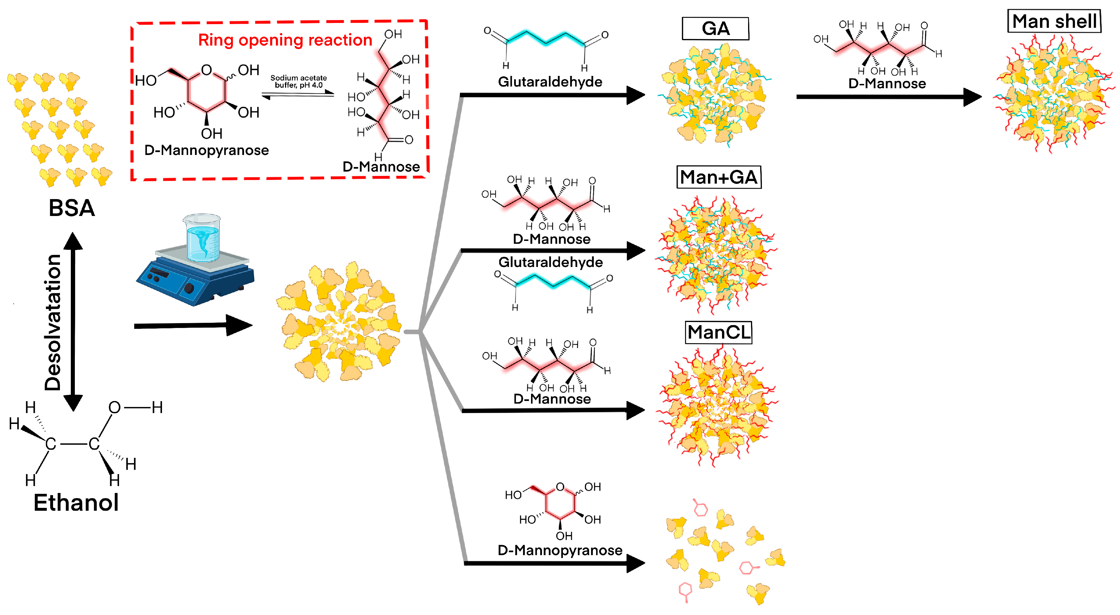
2.2. Fabrication of Mannosylated Albumin Nanoparticles Using Desolvation Method

D-Mannose stock solution (5.46 g/L) was prepared in acidic sodium acetate buffer, pH 4.0. Just before the synthesis, the mannose stock solution was incubated at 60 °C for 1 h to open the mannose ring.
A bovine serum albumin (BSA) solution with a concentration of 20 mg/mL was stirred continuously on a magnetic stirrer throughout the synthesis. Then, 96% ethanol (EtOH/BSA solution ratio 4:1) was added as a desolvating agent at a rate of 2 mL/min. At the next step, a cross-linking agent was added: 0.1% glutaraldehyde (GA) solution in 96% ethanol (final concentration 0.009%, in accordance with the previously published protocol [30]), and/or mannose stock solution (5.46 g/L, in cyclic form or in the open ring form according to protocol in [26]). The obtained solution was stirred for 24 h for nanoparticle formation. Then, the obtained albumin nanoparticles (ANP) were centrifuged, washed three times with sterile bidistilled water, and resuspended in water. The concentration of ANPs was estimated after evaporation and weighing.
In total, four types of nanoparticles were prepared. Mannose was used as a single cross-linker agent for the preparation of 2 types of ANPs: 5 µL and 10 µL of mannose stock solution (0.03 mg and 0.06 mg, respectively) were added to the mixture of BSA and ethanol to produce the ANPs ManCL0.03 and ManCL0.06, respectively. A third type of ANP was prepared using glutaraldehyde and mannose together as cross-linker agents. The resulting ANPs were indicated as Man0.03+GA. The fourth type of mannosylated ANPs were prepared according to standard protocol with GA [31] and covered with mannose residues after synthesis (ANP Man shell).
ANP labeling with fluorescent dye Cy5.5 was performed with the addition of Cy5.5 dye to an ANP suspension in a ratio of 2 µg of dye: 10 mg of ANPs and stirring for 24 h.
For the encapsulation of phosphoramidite (FAM)-labeled scramble small interfering RNA (siRNA), the following FAM-labeled siRNA oligonucleotides were annealed, added to BSA solution (5 µg of siRNA per 1 mL of BSA solution), and incubated under stirring for 1 h: 5′-FAM-AGGCUAGUGCGCACUAAUUGGTT, 5′-FAM-CCAAUUAGUGCGCACUAGCCUTT. The remaining steps of the synthesis were identical to the standard synthesis protocol.

2.3. Nanoparticle Characterization

Nanoparticle size and zeta potential (in bidistilled water) were measured using dynamic light scattering (DLS; Zetasizer Ultra, Malvern, UK). Mannosylated ANPs were visualized by scanning transmission electron microscopy (STEM). In brief, the ANP suspension was incubated for 2 min on a grid (230 mesh, carbon/formvar, EMCN, Beijing, China) and then washed twice with bidistilled water. Uranyl acetate solution (1%) was used as the staining agent. Electron micrographs were obtained using a scanning electron microscope (Crossbeam 550, Carl Zeiss, Jena, Germany) with a scanning transmission electron microscope detector.
The encapsulation efficiency of mannosylated ANPs with FAM-siRNA was calculated by the following formula: fluorescence intensity of ANP suspension before centrifugation/fluorescence intensity of supernatant after centrifugation × 100%.

2.4. Cell Lines

The THP-1 human leukemia cell line was used as a model of human macrophages. Cells were passaged in RPMI-1640 medium, containing 10% FBS, 0.3 g/L L-glutamine, and 0.05% beta-mercaptoethanol. The cells were activated by the addition of 100 ng/mL phorbol 12-myristate 13-acetate (PMA) to the medium and 24 h incubation, and then the cells were washed with PBS and cultivated for 48 h in fresh medium with 100 ng/mL bacterial LPS or 20 ng/mL IL-4 and 10 µM all-trans-retinoic acid (ATRA) added to obtain polarization into the M1 or M2 phenotype. M1/M2 polarization was confirmed by increased expression levels of the M1 markers C-C chemokine receptor type 7 (CCR7) and C-C chemokine ligand 3 (CCL3), and the M2 markers CD206 and VSIG4 (V-set and immunoglobulin domain containing protein 4), analyzed by qPCR (see Supplementary Figure S1).
Cancer cell (triple-negative breast cancer cell line MDA-MB-231 and lung adenocarcinoma cell line A549) passaging was performed using RPMI-1640 medium according standard protocols.

2.5. Evaluation of the Internalization Efficiency of Cy 5.5-Labeled Mannosylated ANPs into M1 and M2 Macrophages

THP-1 cells were plated in 12-well plates (300,000 cells per well) and polarized into the M1 or M2 phenotype according to standard protocol for 48 h. The polarized macrophages were treated with Cy 5.5-labeled ANP ManCL0.03 and Man0.03+GA (0.5 ng/cell) for different time periods (0.5 h, 1.5 h, and 3 h), and then the cells were washed three times with PBS and incubated for additional 2 h in fresh medium to internalize residual ANPs from the cell surface. Then, the cells were washed three times with PBS, detached with versene solution, and resuspended in PBS, and the cell suspension was used for flow cytometry on a Novocyte 3000 instrument (Agilent, Santa Clara, CA, USA).
Gates were set in the forward scatter (FSC) versus side scatter (SSC) plot. Untreated cells were used as a negative control. For all experiments, identical settings were used.
For confocal microscopy, THP-1 cells were plated in 3 cm glass-bottomed dishes, polarized into the M1 or M2 phenotype according to standard protocol for 48 h and treated with all four types of mannosylated ANPs (ManCL0.03, ManCL0.06, Man0.03+GA, Man shell, 0.5 ng/cell) according to the same protocol. Confocal fluorescence images were acquired using an inverted point-scanning confocal microscope (LSM 980 Airyscan on an Axio Observer 7, Carl Zeiss Microscopy GmbH, Jena, Germany) with 20× objective lens (Carl Zeiss Microscopy GmbH, Jena, Germany).
Microphotograph analysis was performed using FIJI software (a distribution of ImageJ v1.53c, National Institutes of Health, Bethesda, MD, USA). Statistical analysis was performed with GraphPad Prism 10.0 software (GraphPad Software Inc., San Diego, CA, USA).

2.6. Evaluation of the Internalization Efficiency of Mannosylated ANP Man0.03+GA and Control ANPs Encapsulating FAM-Labeled Scramble siRNA in M1 and M2 Macrophages and in Cancer Cells

THP-1 cells were plated in 12-well plates (300,000 cells per well) and polarized into the M1 or M2 phenotype according to standard protocol for 48 h. Cancer cells MDA-MB-231 and A549 were plated in 12-well plates (300,000 cells per well) 24 h before the start of the experiment. The cells were treated with ANP Man0.03+GA or control ANPs, encapsulating FAM-labeled scramble siRNA (0.5 ng/cell) and incubated with ANP for different time periods (5, 10, 15, 30, 45 min). Then, the cells were washed three times with PBS, incubated for additional 2 h in fresh medium to internalize residual ANPs from the cell surface, washed three times with PBS, detached with versene solution, and resuspended in PBS, and the cell suspension was used for flow cytometry on a Novocyte 3000 instrument (Agilent, Santa Clara, CA, USA).
Statistical analysis was performed with GraphPad Prism 10.0 software (GraphPad Software Inc., San Diego, CA, USA).

2.7. Evaluation of Localization of FAM-Labeled siRNA Within the M2 Cells

THP-1 cells were plated in 3 cm glass-bottomed dishes, polarized into the M2 phenotype according to standard protocol for 48 h, and treated with Man0.03+GA ANPs, encapsulating FAM-labeled siRNA (0.5 ng/cell). The M2 cells were incubated with ANPs for 24, 72, and 144 h. At different timepoints, the cells were washed from nanoparticles and treated with Lysotracker red (Invitrogen, Waltham, MA, USA) according to the manufacturer’s protocol to visualize lysosomes. Confocal fluorescence images were acquired using an inverted point-scanning confocal microscope (LSM 980 Airyscan on an Axio Observer 7, Carl Zeiss Microscopy GmbH, Jena, Germany) with a 63× oil immersion objective lens (Carl Zeiss Microscopy GmbH, Jena, Germany).

2.8. Determination of ANP Internalization Pathway

THP-1 cells were plated in 12-well plates (300,000 cells per well) and polarized into the M2 phenotype according to standard protocol for 48 h. The M2 cells were pretreated with endocytosis inhibitors for 1 h. The working concentrations of the inhibitors used are presented in Table 1. Then, the cells were washed with PBS and Cy5.5-labeled ANP Man0.03+GA, and control ANPs were added at a concentration of 0.5 ng/cell. The cells were incubated with nanoparticles for three hours and then washed with PBS, covered with fresh medium, and left for another 2 h to internalize residual ANPs from the cell surface. Finally, the cells were washed, detached with versene solution, and used for flow cytometry on a Novocyte 3000 instrument (Agilent, Santa Clara, CA, USA).

3. Results and Discussion

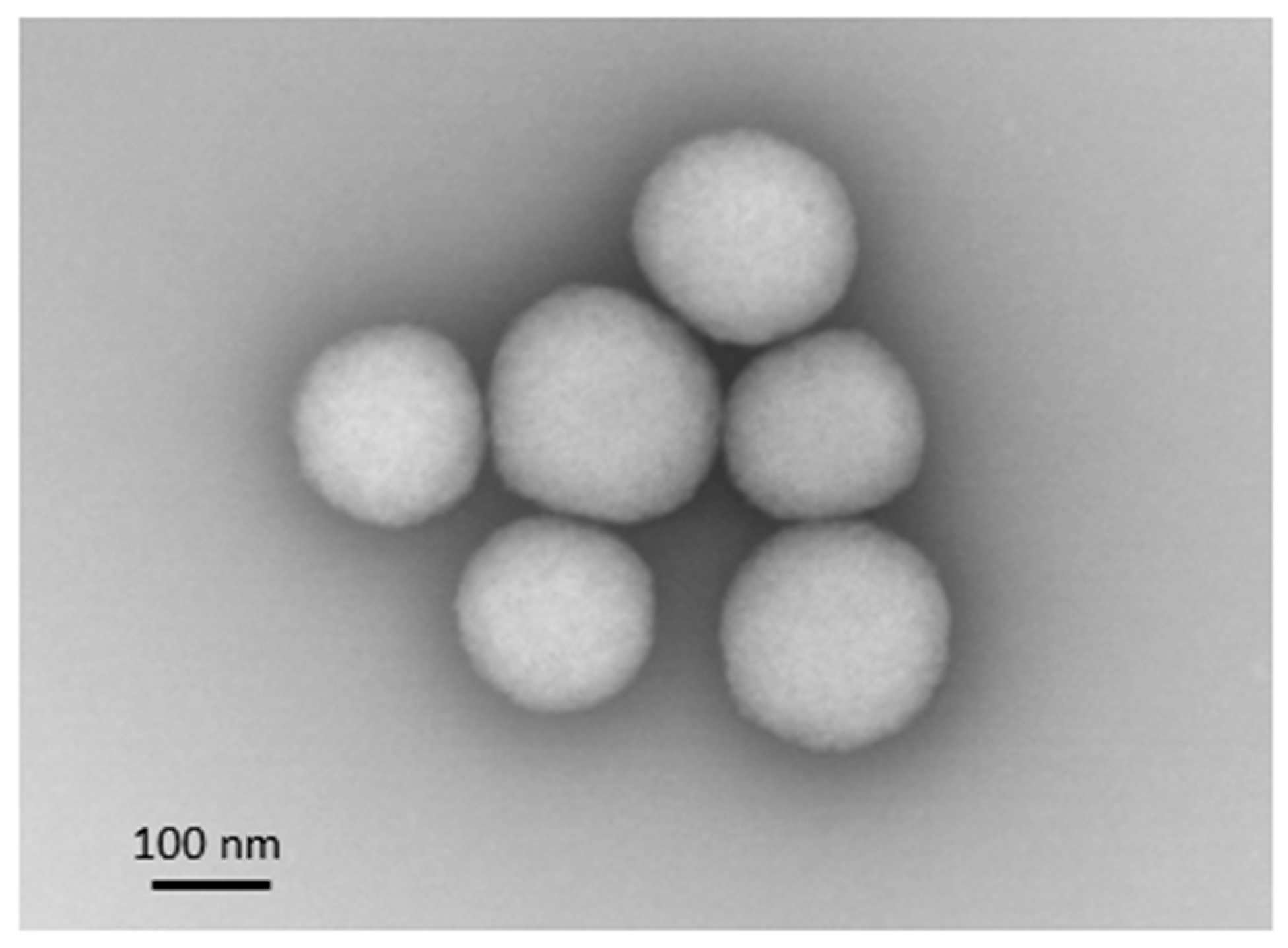
3.1. Mannosylated ANP Synthesis and Characterization

Four types of mannosylated ANPs were produced with the addition of mannose in the open ring form. The addition of intact mannose solution prevented ANP formation. Therefore, pyranose mannose is not capable of stabilizing the albumin nanoparticles. Schematics of mannosylated ANP synthesis are presented in Figure 1.
According to the DLS results (Table 2), the nanoparticle size decreased from 200 nm to 158 nm with increasing mannose concentration when it was used as a cross-linker, and the zeta potential tended to decrease; however, the PI value increased. The use of mannose as a cross-linker did not allow long-term stability to be achieved; the nanoparticles tended to form aggregates. This is why the additional cross-linker, glutaraldehyde, was introduced. The use of two cross-linkers simultaneously allowed us to obtain stable nanoparticles with a hydrodynamic radius of 130 nm, PI less than 0.1, and a zeta potential of −14.3, while the use of GA only gives nanoparticles with diameter of 109 nm and a zeta potential of −30 mV. In addition, standard ANPs with GA were incubated with mannose in acidic buffer after synthesis, resulting in the fabrication of ANPs with a mannose outer shell. These ANPs were characterized by an increased hydrodynamic radius of up to 154 nm and a positive zeta potential (12.67), which can be explained by the protonation of albumin amino acid residues.
The spherical shape and uniform size of the nanoparticles were confirmed by scanning transmission electron microscopy. A representative electron micrograph of Man0.03+GA ANP is presented in Figure 2.
The encapsulation efficiency of Man0.03+GA ANP with FAM-siRNA was 33 ± 5%, which corresponded to 1 µg siRNA/mg ANP.

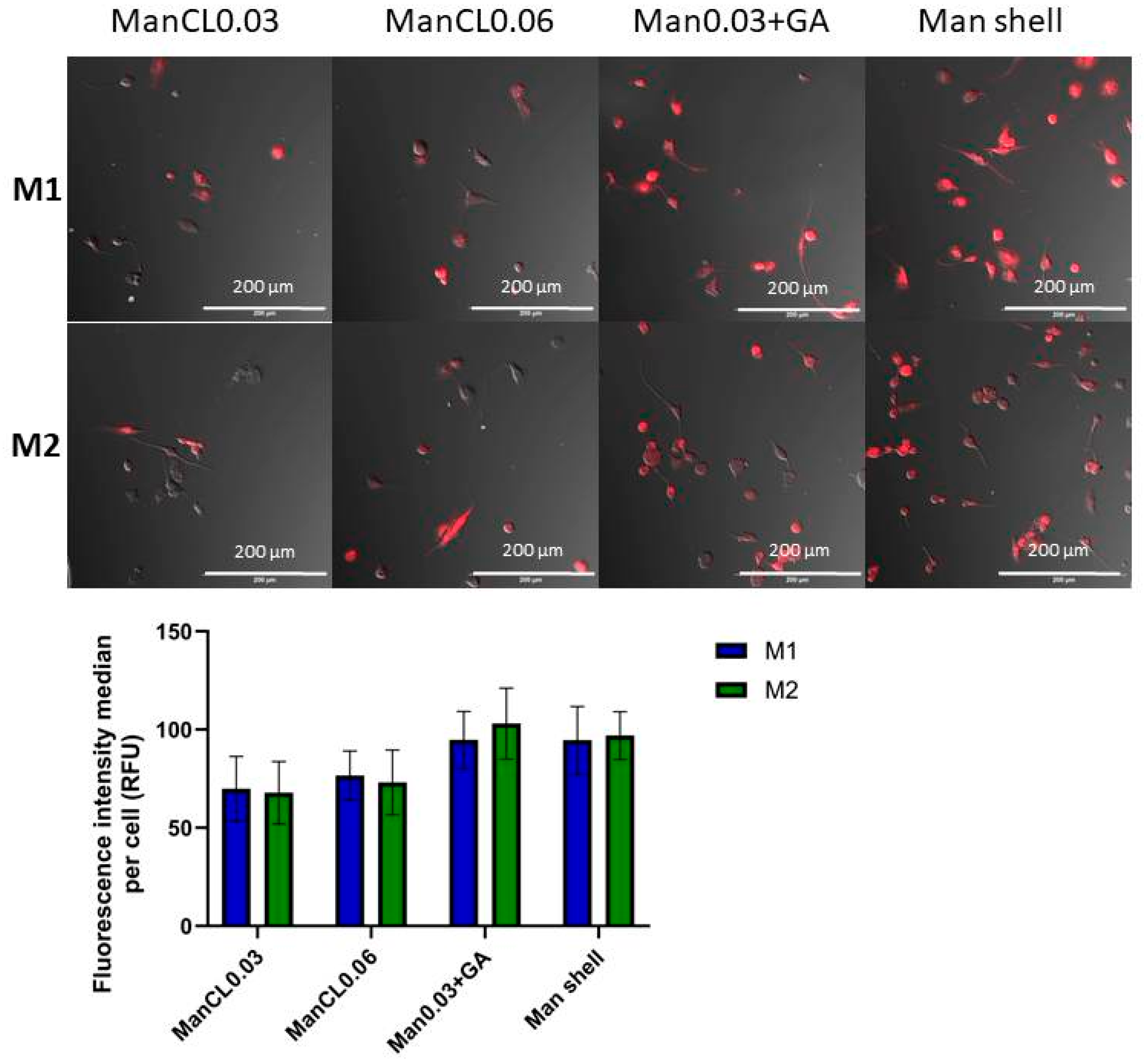
3.2. Efficiency of the Internalization of Mannosylated ANPs into M1 and M2 Macrophages

All types of mannosylated ANPs obtained using the described protocol were labeled with Cy5.5 dye and used to evaluate the efficiency of internalization into M1 and M2 macrophages. THP-1 cells polarized with PMA and bacterial LPS or IL-4, respectively, were used as a model of tumor-associated M1 and M2 macrophages and treated with Cy5.5-Man-ANPs for three hours. The cells were visualized using confocal microscopy and the median fluorescence intensity of ANPs per cell was calculated from three experiments. The calculation results showed that the internalization efficiency of Man0.03+GA ANPs (94.7 ± 14.5 and 103.0 ± 18.1 in M1 and M2, respectively) and Man shell (94.5 ± 17.2 and 96.9 ± 12.1 in M1 and M2, respectively) was significantly higher than the internalization efficiency of ANPs prepared using mannose as a cross-linker (ManCL0.03: 69.8 ± 16.5 and 67.8 ± 15.9 in M1 and M2, respectively; ManCL0.06: 76.6 ± 12.4 and 73.1 ± 16.5 in M1 and M2, respectively). The highest level of fluorescence was observed in M2 cells incubated with Man0.03+GA ANP (Figure 3). Therefore, we showed that nanoparticles produced using mannose as a cross-linker together with glutaraldehyde (Man0.03+GA), and ANPs covered with mannose shell after synthesis (Man shell) provided efficient targeting into CD206-expressing M2 cells.
In the next step, we selected two types of ANPs with equal concentrations of mannose: one type of ANP with mannose cross-linker (ManCL0.03) and one type of ANP produced with the addition of GA and mannose (Man0.03+GA). We repeated the evaluation of the ANP internalization efficiency using flow cytometry at the following timepoints: 0.5 h, 1.5 h, and 3 h of incubation. The results confirmed that M2 cells carrying the CD206 mannose receptor internalized the mannosylated ANPs ManCL0.03 and Man+GA more efficiently in comparison to the M1 macrophages (Figure 4). Moreover, the highest fluorescence intensity was observed in M2 cells after Man0.03+GA treatment for three hours. Therefore, we chose the Man0.03+GA ANPs for further experiments, because these nanoparticles can be produced using a simple one-step synthesis protocol and showed maximum efficiency in internalization into M2 macrophages.

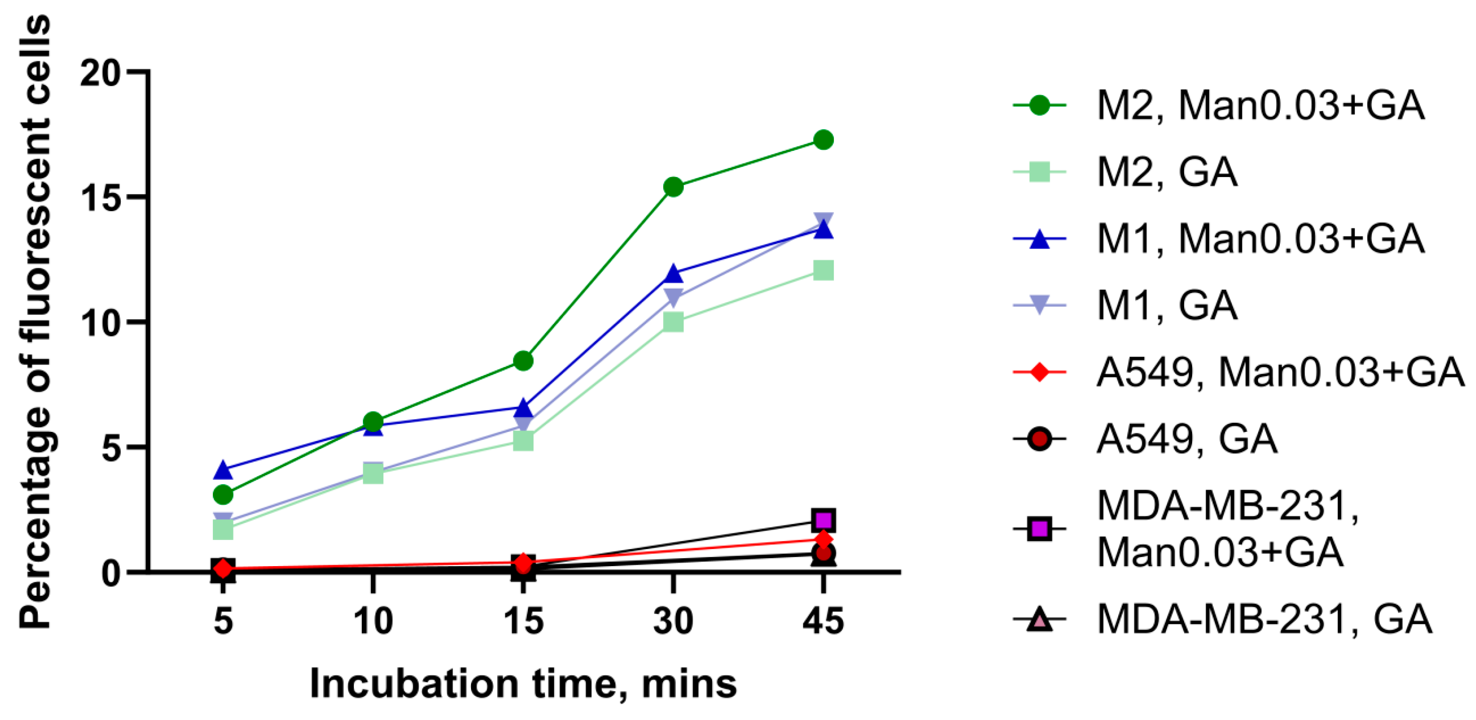
3.3. Comparison of the Internalization Efficiency of Mannosylated ANPs Man0.03+GA and Control ANPs in M1 and M2 Macrophages and in Cancer Cells

To compare the targeting efficacy of our selected mannosylated ANPs Man0.03+GA and control ANPs without mannose residues produced according to a standard protocol, we treated M1 and M2 macrophages as well as A549 lung adenocarcinoma cancer cells and MDA-MB-231 triple-negative breast cancer cells with Man0.03+GA ANPs and control ANPs encapsulating FAM-labeled scramble siRNA for different time periods: 5, 10, 15, 30, and 45 min. Non-functional FAM-labeled siRNA was used as a model to study the behavior of the therapeutic cargo of the nanoparticles inside the cell. MDA-MB-231 cells were chosen as a model of the most aggressive triple-negative breast cancer, and the choice of the lung adenocarcinoma cell line is justified by the fact that, according to literature data on nanoparticle biodistribution, a significant proportion of nanoparticles accumulate in the lungs [30].
The results of the experiment are presented in Figure 5. We can see that the internalization of both mannosylated and control ANPs to macrophages (percentage of fluorescent cells 10–15%) is significantly more effective than internalization to cancer cells (1–2%). This result correlates with the natural phagocyte function of macrophages. Moreover, we can see that the percentage of fluorescent M2 cells, treated with mannosylated ANPs, is about 30% higher than the percentage of fluorescent M2 cells treated with control ANPs. Therefore, we can confirm the efficient targeting of mannosylated ANPs to M2 macrophages. The percentages of fluorescent M1 cells treated with control ANPs and mannosylated ANPs were comparable with the percentage of fluorescent M2 cells treated with control ANPs.

3.4. Analysis of Localization of FAM-Labeled siRNA Inside the M2 Cells

The next stage of the work was to study the localization of the fluorescent dye-labeled cargo of mannosylated ANPs inside the cell. For this purpose, macrophages polarized into M2 phenotype were treated with ANP Man0.03+GA, encapsulating FAM-labeled siRNA, for 24 h. Then, the cells were washed, stained with the lysosome dye Lysotracker red, and imaged on a confocal microscope or left to grow in medium until timepoints 72 h and 144 h (6 days) were reached, and then a similar procedure was performed. The resulting micrographs are shown in Figure 6. After 24 h of the incubation of cells with nanoparticles, we see the complete co-localization of FAM dye and the red lysotracker signal. Therefore, all nanoparticles are localized in the lysosomes. However, at timepoints 72 h and 144 h, we observe a more diffuse distribution of the FAM signal and its redistribution towards the cell periphery, which may indicate nanoparticle disruption, dye detachment, and the release of dye and siRNA from the lysosomes into the cell cytoplasm. The decrease in the FAM fluorescence intensity may indicate the cleavage of the dye from albumin molecules and its bleaching in the acidic lysosomal environment.

3.5. Determination of Endocytosis Pathway of Mannosylated ANPs Man0.03+GA and Control ANPs in M2 Macrophages

The last objective of this work was to study the mechanism of the internalization of mannosylated ANPs and control ANPs into M2 macrophages. It is known that albumin nanoparticles can be internalized by active transport and via clathrin- and caveolin-mediated pathways [32]. Nanoparticles up to 100 nm in size are predominantly taken up by the caveolin-mediated pathway [23,32]. The internalization of CD206 ligands and their delivery to the endosomal system is accomplished by the clathrin-mediated pathway [33]. In our study, we used the following endocytosis inhibitors to discriminate the endocytosis pathway: filipin complex, 5-(N-ethyl-N-isopropyl)-amiloride (EIPA), bafilomycin A, sodium azide, and hydroxychloroquine. Filipin complex, EIPA, and sodium azide are classical inhibitors of caveolin-mediated endocytosis, micropinocytosis, and active transport, respectively [33,34,35,36]. To reduce the efficiency of clathrin-mediated endocytosis, we used the antibiotic bafilomycin A and hydroxychloroquine, which block the acidification of endosomes, thereby preventing their fusion with lysosomes [36].
The results are summarized in Figure 7. The most pronounced reduction in internalization efficiency was observed with bafilomycin A (by 36% and 74% for Man0.03+GA and control ANPs, respectively) and sodium azide (by 24% and 58% for Man0.03+GA and control ANP, respectively), suggesting that ANPs enter macrophages predominantly via clathrin-dependent endocytosis and ATP-dependent transport. A pronounced decrease in mannosylated and control ANP internalization was also observed with EIPA (by 13% and 40% for Man0.03+GA and control ANPs, respectively), indicating the participation of macropinocytosis in ANP uptake, and with hydroxychloroquine (by 16% and 25% for Man0.03+GA and control ANPs, respectively), also indicating the involvement of clathrin-mediated endocytosis. Filipin complex, which blocks caveolin-mediated endocytosis, suppressed the internalization of control ANPs by 20%, but did not affect the internalization of the mannosylated nanoparticles.
The addition of mannose residues, which incorporate into ANPs both as a cross-linker and as a surface modification, somewhat mitigates the effect of endocytosis inhibitors on ANP Man0.03+GA internalization. Apparently, this may be due to changes in the size and charge of the nanoparticles. However, the overall internalization pattern of mannosylated and control ANPs is identical. Therefore, in this study, we demonstrate that the mannosylated albumin nanoparticles Man0.03+GA, as well as standard ANPs, internalize into M2 macrophages using the clathrin-mediated pathway and active transport.

4. Limitations of the Study and Future Perspectives

This study is characterized by some limitations, particularly its in vitro design, absence of in vivo validation, and application of a human monocytic cell line as a model of tumor-associated macrophages. Additional studies should be performed to demonstrate the therapeutic effect of the suggested nanocarrier in vitro and in vivo. However, despite these conceptual limitations, the obtained results can be used as a basis to develop new nanocarriers for the delivery of nucleic acids and small molecules into tumor-associated macrophages and perform adjuvant cancer immunotherapy targeted on immunosuppressive tumor microenvironment.

5. Conclusions

In summary, we have produced a novel nanocarrier on a base of albumin nanoparticles using the optimized one-step method of ANP mannosylation. The resulting albumin nanoparticles, prepared using the desolvation method with the standard cross-linker glutaraldehyde and D-mannose as a second cross-linker (Man0.03+GA), demonstrated long-term stability and a favorable profile of physical characteristics, such as uniform size, spherical shape, good polydispersity index, and z-potential. These nanoparticles are also characterized by high cargo loading capability and provide efficient targeting to M1 and M2 macrophages in vitro. Moreover, we showed the diffuse distribution of labeled siRNA delivered with ANP Man0.03+GA in macrophage cells. Therefore, we propose that the resulting nanoparticles can be a prominent vehicle for appropriate therapeutic cargo and can be used as an adjuvant therapy to reprogram pro-tumor M2 macrophages.

Supplementary Materials

The following supporting information can be downloaded at: https://www.mdpi.com/article/10.3390/micro5020030/s1, Figure S1: M1 and M2 cell polarization.

Author Contributions

A.B.K.: cell culture and treatment, confocal microscopy, STEM analysis, flow cytometry, data calculation; V.I.G. and E.P.K.: nanoparticle synthesis, cross-linker concentration optimization; V.S.E.: funding acquisition, research coordination, supervision, paper writing. All authors have read and agreed to the published version of the manuscript.

Funding

This research was funded by the Russian Science Foundation, grant #23-75-01155.

Institutional Review Board Statement

Not applicable.

Informed Consent Statement

Not applicable.

Data Availability Statement

The data that support the findings of this study are available from the corresponding author upon request.

Conflicts of Interest

The authors declare no conflicts of interest.

Abbreviations

The following abbreviations are used in this manuscript:
TAMTumor-associated macrophage
TMETumor microenvironment
CCR7C-C chemokine receptor type 7
CCL3C-C chemokine ligand 3
CD206Mannose receptor (Cluster of Differentiation 206)
VSIG4V-set and immunoglobulin domain containing protein 4
ANPAlbumin nanoparticle
DLSDynamic light scattering
PIPolydispersity index
STEMScanning transmission electron microscopy
PMAPhorbol 12-myristate 13-acetate
LPSLipopolysaccharide
ATRAAll-trans-retinoic acid
GAGlutaraldehyde
EIPA5-(N-ethyl-N-isopropyl)-amiloride
PBSPhosphate-buffered saline
siRNASmall interfering RNA
FAMFluorescein
SDStandard deviation

References

  1. Kamat, K.; Krishnan, V.; Dorigo, O. Macrophage-derived CCL23 upregulates expression of T-cell exhaustion markers in ovarian cancer. Br. J. Cancer 2022, 127, 1026–1033. [Google Scholar] [CrossRef] [PubMed]
  2. Kuznetsova, A.B.; Kolesova, E.P.; Parodi, A.; Zamyatnin, A.A., Jr.; Egorova, V.S. Reprogramming Tumor-Associated Macrophage Using Nanocarriers: New Perspectives to Halt Cancer Progression. Pharmaceutics 2024, 16, 636. [Google Scholar] [CrossRef] [PubMed]
  3. Yan, D.; Wang, H.W.; Bowman, R.L.; Joyce, J.A. STAT3 and STAT6 Signaling Pathways Synergize to Promote Cathepsin Secretion from Macrophages via IRE1α Activation. Cell Rep. 2016, 16, 2914–2927. [Google Scholar] [CrossRef] [PubMed]
  4. Deryugina, E.I.; Zajac, E.; Juncker-Jensen, A.; Kupriyanova, T.A.; Welter, L.; Quigley, J.P. Tissue-infiltrating neutrophils constitute the major in vivo source of angiogenesis-inducing MMP-9 in the tumor microenvironment. Neoplasia 2014, 16, 771–788. [Google Scholar] [CrossRef]
  5. Zhao, J.; Chen, L.; Shu, B.; Tang, J.; Zhang, L.; Xie, J.; Qi, S.; Xu, Y. Granulocyte/macrophage colony-stimulating factor influences angiogenesis by regulating the coordinated expression of VEGF and the Ang/Tie system. PLoS ONE 2014, 9, e92691. [Google Scholar] [CrossRef]
  6. Song, Y.; Tang, C.; Yin, C. Combination antitumor immunotherapy with VEGF and PIGF siRNA via systemic delivery of multi-functionalized nanoparticles to tumor-associated macrophages and breast cancer cells. Biomaterials 2018, 185, 117–132. [Google Scholar] [CrossRef]
  7. Guo, X.; Zhang, H.; He, C.; Qin, K.; Lai, Q.; Fang, Y.; Chen, Q.; Li, W.; Wang, Y.; Wang, X.; et al. RUNX1 promotes angiogenesis in colorectal cancer by regulating the crosstalk between tumor cells and tumor associated macrophages. Biomark. Res. 2024, 12, 29. [Google Scholar] [CrossRef]
  8. Grabowska, M.M.; Day, M.L. Soluble E-cadherin: More than a symptom of disease. Front. Biosci. (Landmark Ed). 2012, 17, 1948–1964. [Google Scholar] [CrossRef]
  9. Mitrović, A.; Pečar Fonović, U.; Kos, J. Cysteine cathepsins B and X promote epithelial-mesenchymal transition of tumor cells. Eur. J. Cell Biol. 2017, 96, 622–631. [Google Scholar] [CrossRef]
  10. Häuselmann, I.; Roblek, M.; Protsyuk, D.; Huck, V.; Knopfova, L.; Grässle, S.; Bauer, A.T.; Schneider, S.W.; Borsig, L. Monocyte Induction of E-Selectin-Mediated Endothelial Activation Releases VE-Cadherin Junctions to Promote Tumor Cell Extravasation in the Metastasis Cascade. Cancer Res. 2016, 76, 5302–5312. [Google Scholar] [CrossRef]
  11. Lu, Y.; Han, G.; Zhang, Y.; Zhang, L.; Li, Z.; Wang, Q.; Chen, Z.; Wang, X.; Wu, J. M2 macrophage-secreted exosomes promote metastasis and increase vascular permeability in hepatocellular carcinoma. Cell Commun. Signal. 2023, 21, 299. [Google Scholar] [CrossRef] [PubMed]
  12. Gadde, M.; Mehrabi-Dehdezi, M.; Debeb, B.G.; Woodward, W.A.; Rylander, M.N. Influence of Macrophages on Vascular Invasion of Inflammatory Breast Cancer Emboli Measured Using an In Vitro Microfluidic Multi-Cellular Platform. Cancers 2023, 15, 4883. [Google Scholar] [CrossRef] [PubMed]
  13. Xu, J.; Zhang, J.; Zhang, Z.; Gao, Z.; Qi, Y.; Qiu, W.; Pan, Z.; Guo, Q.; Li, B.; Zhao, S.; et al. Hypoxic glioma-derived exosomes promote M2-like macrophage polarization by enhancing autophagy induction. Cell Death Dis. 2021, 12, 373. [Google Scholar] [CrossRef] [PubMed]
  14. Zhou, S.; Lan, Y.; Li, Y.; Li, Z.; Pu, J.; Wei, L. Hypoxic Tumor-Derived Exosomes Induce M2 Macrophage Polarization via PKM2/AMPK to Promote Lung Cancer Progression. Cell Transplant. 2022, 31, 9636897221106998. [Google Scholar] [CrossRef]
  15. Zhou, H.; Yao, J.; Zhong, Z.; Wei, H.; He, Y.; Li, W.; Hu, K. Lactate-Induced CCL8 in Tumor-Associated Macrophages Accelerates the Progression of Colorectal Cancer through the CCL8/CCR5/mTORC1 Axis. Cancers 2023, 15, 5795. [Google Scholar] [CrossRef]
  16. Gomez-Roca, C.; Cassier, P.; Zamarin, D.; Machiels, J.P.; Perez Gracia, J.L.; Stephen Hodi, F.; Taus, A.; Martinez Garcia, M.; Boni, V.; Eder, J.P.; et al. Anti-CSF-1R emactuzumab in combination with anti-PD-L1 atezolizumab in advanced solid tumor patients naïve or experienced for immune checkpoint blockade. J. Immunother. Cancer 2022, 10, e004076. [Google Scholar] [CrossRef]
  17. Cassier, P.A.; Italiano, A.; Gomez-Roca, C.; Le Tourneau, C.; Toulmonde, M.; D’Angelo, S.P.; Weber, K.; Loirat, D.; Jacob, W.; Jegg, A.M.; et al. Long-term clinical activity, safety and patient-reported quality of life for emactuzumab-treated patients with diffuse-type tenosynovial giant-cell tumour. Eur. J. Cancer 2020, 141, 162–170. [Google Scholar] [CrossRef]
  18. Nywening, T.M.; Wang-Gillam, A.; Sanford, D.E.; Belt, B.A.; Panni, R.Z.; Cusworth, B.M.; Toriola, A.T.; Nieman, R.K.; Worley, L.A.; Yano, M.; et al. Targeting tumour-associated macrophages with CCR2 inhibition in combination with FOLFIRINOX in patients with borderline resectable and locally advanced pancreatic cancer: A single-centre, open-label, dose-finding, non-randomised, phase 1b trial. Lancet Oncol. 2016, 17, 651–662. [Google Scholar] [CrossRef]
  19. Reid, T.; Oronsky, B.; Caroen, S.; Quinn, M.; Williams, J.; Cabrales, P.; Abrouk, N. Phase 1 pilot study of RRx-001 + nivolumab in patients with advanced metastatic cancer (PRIMETIME). Front. Immunol. 2023, 14, 1104753. [Google Scholar] [CrossRef]
  20. Byrne, K.T.; Betts, C.B.; Mick, R.; Sivagnanam, S.; Bajor, D.L.; Laheru, D.A.; Chiorean, E.G.; O’Hara, M.H.; Liudahl, S.M.; Newcomb, C.; et al. Neoadjuvant Selicrelumab, an Agonist CD40 Antibody, Induces Changes in the Tumor Microenvironment in Patients with Resectable Pancreatic Cancer. Clin. Cancer Res. 2021, 27, 4574–4586. [Google Scholar] [CrossRef]
  21. Zhao, S.; Ren, S.; Jiang, T.; Zhu, B.; Li, X.; Zhao, C.; Jia, Y.; Shi, J.; Zhang, L.; Liu, X.; et al. Low-Dose Apatinib Optimizes Tumor Microenvironment and Potentiates Antitumor Effect of PD-1/PD-L1 Blockade in Lung Cancer. Cancer Immunol. Res. 2019, 7, 630–643. [Google Scholar] [CrossRef] [PubMed]
  22. Rajan, A.; Abdul Sater, H.; Rahma, O.; Agajanian, R.; Lassoued, W.; Marté, J.L.; Tsai, Y.T.; Donahue, R.N.; Lamping, E.; Bailey, S.; et al. Efficacy, safety, and biomarker analyses of bintrafusp alfa, a bifunctional fusion protein targeting TGF-β and PD-L1, in patients with advanced non-small cell lung cancer. J. Immunother. Cancer 2024, 12, e008480. [Google Scholar] [CrossRef] [PubMed]
  23. Liu, N.; Qi, Y.H.; Cheng, C.T.; Yang, W.B.; Malhotra, A.; Zhou, Q. Potential of siRNA-albumin complex against cancer. Chem. Biol. Interact. 2018, 295, 93–96. [Google Scholar] [CrossRef] [PubMed]
  24. Wang, Y.; Chen, S.; Yang, X.; Zhang, S.; Cui, C. Preparation Optimization of Bovine Serum Albumin Nanoparticles and Its Application for siRNA Delivery. Drug Des. Dev. Ther. 2021, 15, 1531–1547. [Google Scholar] [CrossRef]
  25. Mehta, A.; Dalle Vedove, E.; Isert, L.; Merkel, O.M. Targeting KRAS Mutant Lung Cancer Cells with siRNA-Loaded Bovine Serum Albumin Nanoparticles. Pharm. Res. 2019, 36, 133. [Google Scholar] [CrossRef]
  26. Singh, A.; Chakraborty, S.; Wong, S.W.; Hefner, N.A.; Stuart, A.; Qadir, A.S.; Mukhopadhyay, A.; Bachmaier, K.; Shin, J.W.; Rehman, J.; et al. Nanoparticle targeting of de novo profibrotic macrophages mitigates lung fibrosis. Proc. Natl. Acad. Sci. USA 2022, 119, e2121098119. [Google Scholar] [CrossRef]
  27. Gu, G.J.; Chung, H.; Park, J.Y.; Yoo, R.; Im, H.J.; Choi, H.; Lee, Y.S.; Seok, S.H. Mannosylated-serum albumin nanoparticle imaging to monitor tumor-associated macrophages under anti-PD1 treatment. J. Nanobiotechnol. 2023, 21, 31. [Google Scholar] [CrossRef]
  28. Wang, C.; Xu, X.; Zhang, P.; Xiong, S.; Yuan, J.; Gao, X.; Guan, W.; Wang, F.; Li, X.; Dou, H.; et al. Lipid-coated albumin-paclitaxel nanoparticles loaded with sorcin-siRNA reverse cancer chemoresistance via restoring intracellular calcium ion homeostasis. J. Nanobiotechnol. 2022, 20, 319. [Google Scholar] [CrossRef]
  29. Fu, Z.; Wang, X.; Lu, X.; Yang, Y.; Zhao, L.; Zhou, L.; Wang, K.; Fu, H. Mannose-decorated ginsenoside Rb1 albumin nanoparticles for targeted anti-inflammatory therapy. Front. Bioeng. Biotechnol. 2022, 10, 962380. [Google Scholar] [CrossRef]
  30. Flannagan, R.S.; Jaumouillé, V.; Grinstein, S. The cell biology of phagocytosis. Annu. Rev. Pathol. 2012, 7, 61–98. [Google Scholar] [CrossRef]
  31. Kolesova, E.P.; Egorova, V.S.; Syrocheva, A.O.; Frolova, A.S.; Kostyushev, D.; Kostyusheva, A.; Brezgin, S.; Trushina, D.B.; Fatkhutdinova, L.; Zyuzin, M.; et al. Proteolytic Resistance Determines Albumin Nanoparticle Drug Delivery Properties and Increases Cathepsin B, D, and G Expression. Int. J. Mol. Sci. 2023, 24, 10245. [Google Scholar] [CrossRef] [PubMed]
  32. Wang, Z.; Tiruppathi, C.; Minshall, R.D.; Malik, A.B. Size and dynamics of caveolae studied using nanoparticles in living endothelial cells. ACS Nano 2009, 3, 4110–4116. [Google Scholar] [CrossRef] [PubMed]
  33. Allavena, P.; Chieppa, M.; Monti, P.; Piemonti, L. From pattern recognition receptor to regulator of homeostasis: The double-faced macrophage mannose receptor. Crit. Rev. Immunol. 2004, 24, 179–192. [Google Scholar] [CrossRef] [PubMed]
  34. Mo, Y.; Lim, L.Y. Mechanistic study of the uptake of wheat germ agglutinin-conjugated PLGA nanoparticles by A549 cells. J. Pharm. Sci. 2004, 93, 20–28. [Google Scholar] [CrossRef]
  35. Lin, H.P.; Singla, B.; Ghoshal, P.; Faulkner, J.L.; Cherian-Shaw, M.; O’Connor, P.M.; She, J.X.; Belin de Chantemele, E.J.; Csányi, G. Identification of novel macropinocytosis inhibitors using a rational screen of Food and Drug Administration-approved drugs. Br. J. Pharmacol. 2018, 175, 3640–3655. [Google Scholar] [CrossRef]
  36. Patel, P.C.; Giljohann, D.A.; Daniel, W.L.; Zheng, D.; Prigodich, A.E.; Mirkin, C.A. Scavenger receptors mediate cellular uptake of polyvalent oligonucleotide-functionalized gold nanoparticles. Bioconjugate Chem. 2010, 21, 2250–2256. [Google Scholar] [CrossRef]
Figure 1. Protocol of mannosylated ANP synthesis.
Micro 05 00030 g001
Figure 2. Scanning electron micrograph of Man0.03+GA ANPs.
Micro 05 00030 g002
Figure 3. Photomicrographs of M1 and M2 cells after incubation with mannosylated ANPs for 3 h and the results of photomicrographs analysis. Maximal fluorescence intensity in M2 cells treated with Man0.03+GA was set as 100%.
Micro 05 00030 g003
Figure 4. Internalization of Cy5.5-labeled ManCL0.03 and Man0.03+GA ANP into M1 and M2 macrophages. The data presented are mean ± SD. Maximum mean fluorescence intensity (3 h treatment of M2 cells with ANP Man0.03+GA) was set as 100%.
Micro 05 00030 g004
Figure 5. The internalization efficiency of mannosylated ANPs Man0.03+GA and control ANPs in M1 and M2 macrophages and MDA-MB-231 and A549 cells.
Micro 05 00030 g005
Figure 6. Localization of FAM-labeled siRNA inside the M2 cells.
Micro 05 00030 g006
Figure 7. Determination of endocytosis pathway of control ANPs (A) and mannosylated Man0.03+GA ANPs (B) in M2 macrophages. The data presented are mean ± SD. Fluorescence intensity of control cells without treatment was set as 100%. Significance was analyzed using unpaired t-test; differences were considered statistically significant at p < 0.05. * symbol indicates significance level; ns—non-significant difference.
Micro 05 00030 g007
Table 1. Working concentrations of endocytosis inhibitors.
InhibitorWorking Concentration
Filipin complex2 µM
EIPA24 µM
Bafilomycin A50 µM
Sodium azide1 µM
Hydroxychloroquine200 µM
Table 2. Characteristics of mannosylated ANPs.
Type of Man-ANPManCL0.03ManCL0.06Man+GAMan ShellANP
Hydrodynamic diameter, nm202.1158130.5154109.3
PI0.12030.21150.0940.07990.124
Zeta potential, mV−8.9−15.8−14.312.67−47
Disclaimer/Publisher’s Note: The statements, opinions and data contained in all publications are solely those of the individual author(s) and contributor(s) and not of MDPI and/or the editor(s). MDPI and/or the editor(s) disclaim responsibility for any injury to people or property resulting from any ideas, methods, instructions or products referred to in the content.

