Review

Functional Plasticity of Microbial Siderophores in Iron- and Boron-Rich Niches

by
Valery M. Dembitsky
1,2,*,
Alexander O. Terent’ev
2 and
Sergey V. Baranin
2
1
Bio-Pharm Laboratories, 23615 El Toro Rd X, Lake Forest, CA 92630, USA
2
N.D. Zelinsky Institute of Organic Chemistry, Russian Academy of Sciences, 47 Leninsky Prospect, Moscow 119991, Russia
*
Author to whom correspondence should be addressed.
Appl. Microbiol. 2026, 6(4), 50; https://doi.org/10.3390/applmicrobiol6040050
Submission received: 22 February 2026 / Revised: 21 March 2026 / Accepted: 24 March 2026 / Published: 31 March 2026

Abstract

Siderophores are high-affinity iron-chelating metabolites that underpin microbial survival in iron-limited environments and play central roles in metal homeostasis, ecological competition, and pathogenesis. Traditionally viewed as dedicated Fe(III) scavengers, siderophores are now recognized as structurally and functionally versatile coordination agents whose donor-set architectures—particularly catecholate and α-hydroxycarboxylate motifs—permit conditional interactions beyond iron. In iron- and boron-rich niches, especially marine and mildly alkaline systems where borate availability increases, certain siderophores are chemically capable of forming reversible borate complexes through cis-diol coordination. Although Fe(III) exhibits substantially higher thermodynamic affinity and remains the primary biological target, boron binding represents a predictable secondary property arising from shared oxygen-donor chemistry. This dynamic interplay allows siderophores to cycle between iron-bound, boron-bound, and apo states depending on local redox conditions, pH, and metal availability. Here, we synthesize current knowledge on the structural classes of microbial siderophores, their transport and regulatory mechanisms, and emerging evidence for boron coordination within catecholate and carboxylate systems. By integrating coordination chemistry with microbial ecology, we propose an expanded model in which siderophores function not only as iron acquisition molecules but also as modulators of boron speciation and environmental sensing. This functional plasticity positions siderophores at the intersection of iron and boron biogeochemical cycles and highlights new directions for understanding microbial adaptation in complex metal-rich environments.

1. Introduction

Boron is a ubiquitous yet chemically distinct element in marine and alkaline environments, where it exists primarily as boric acid and borate in a pH-dependent equilibrium [1,2,3,4]. Unlike redox-active metals such as iron, boron functions as a Lewis acid capable of forming reversible tetrahedral complexes with cis-diol-containing ligands. This property enables dynamic interactions with biologically relevant molecules and suggests a potential role for boron in modulating molecular structure and speciation in iron- and boron-rich niches [1,2,3,4,5,6].
Iron is essential for nearly all living organisms but is poorly bioavailable under physiological conditions. At neutral pH and in aerobic environments, ferric iron (Fe3+) has an extremely low solubility (~10−17 M), far below the levels required for microbial growth (10−5–10−7 M). In host systems, iron availability is further restricted by proteins such as transferrin, which reduce free iron concentrations to ~10−24 M [7,8,9]. Despite its abundance in the Earth’s crust, iron is largely inaccessible due to the formation of insoluble oxyhydroxides. To overcome this limitation, microorganisms produce siderophores—low-molecular-weight, high-affinity Fe(III)-chelating compounds that solubilize and transport iron from mineral and biological sources [10,11,12,13,14].
Siderophores bind Fe(III) through oxygen- and nitrogen-containing donor groups, forming ferri-siderophore complexes that are recognized by specific receptors and actively transported into the cell. Their production is tightly regulated by iron availability and plays a central role in microbial adaptation, competition, and pathogenicity [13,14,15,16,17]. Microorganisms may also utilize siderophores produced by other species, and ligand-exchange mechanisms can facilitate iron transfer between complexes at the cell surface [18,19].
In Gram-negative bacteria such as Escherichia coli, ferri-siderophore complexes are transported via outer membrane receptors (e.g., FepA, FecA, FhuA), followed by periplasmic shuttling and ATP-dependent uptake. Iron is subsequently released through hydrolysis or reduction [20,21,22]. Alternative pathways, including reductive iron assimilation, also contribute to iron acquisition in fungi and bacteria. These systems are tightly regulated and interconnected, underscoring the critical importance of siderophores in microbial iron homeostasis [23,24,25].
Within this framework, the coexistence of boron and siderophore-producing microorganisms raises the possibility of previously unrecognized interactions between boron and siderophore ligands. Given the prevalence of cis-diol and catechol functionalities in many siderophores, boron coordination may influence their structure, function, and ecological roles, representing an underexplored aspect of microbial metal–metalloid dynamics.

2. Microbial Types of Siderophores

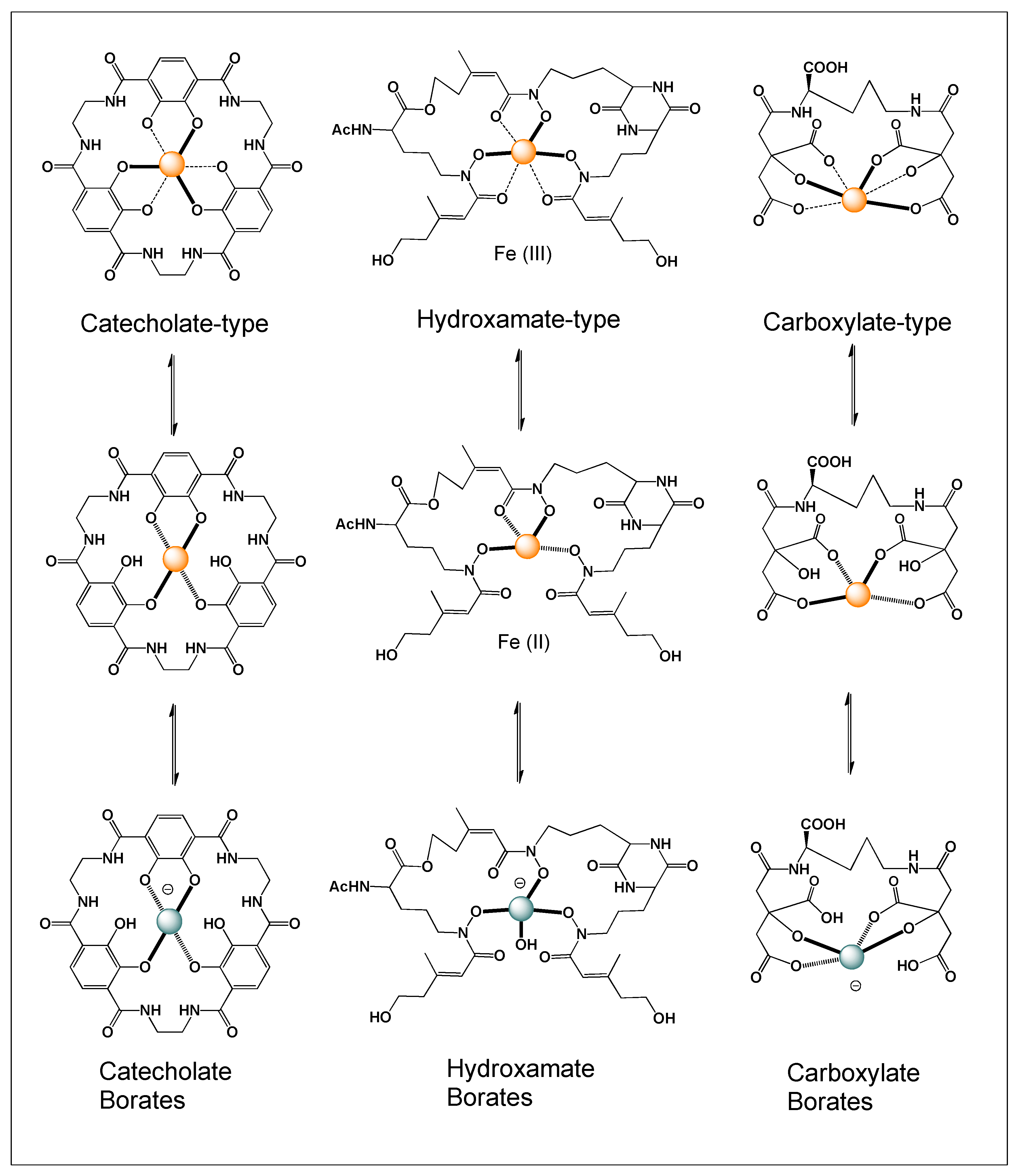
Microbial siderophores constitute a highly diverse class of low-molecular-weight compounds specialized in iron acquisition, and they are commonly categorized based on the nature of their metal-binding functional groups. The three principal classes include catecholates (phenolates), hydroxamates, and carboxylates (Figure 1), although many siderophores exhibit hybrid architectures that incorporate multiple donor types [26,27,28]. Catecholate siderophores, typically derived from 2,3-dihydroxybenzoic acid, coordinate Fe(III) through adjacent phenolic oxygen atoms and are among the strongest known natural iron chelators, often displaying exceptionally high stability constants. Hydroxamate siderophores utilize –C(=O)N–OH functional groups for ferric iron binding and are widely distributed among bacteria and fungi, particularly in terrestrial ecosystems. In contrast, carboxylate-type siderophores, frequently based on α-hydroxycarboxylate motifs such as citrate, coordinate Fe(III) via oxygen-rich donor sets and are prevalent in marine and plant-associated microorganisms.
Beyond primary categories, mixed-type siderophores combine functional groups such as catecholate–hydroxamate or catecholate–carboxylate systems, enhancing coordination versatility. Structurally, they range from linear and cyclic forms to macrocyclic and peptide-based scaffolds, including nonribosomal peptides and amphiphilic variants with lipid tails that promote membrane association [29,30,31,32]. This diversity allows microorganisms to adapt iron acquisition strategies to varying environmental conditions, influencing metal-binding strength, transport efficiency, and receptor specificity. The presence of multiple donor groups further enables effective function under fluctuating pH and competing metal ions [28,29,30,31,32].
Overall, this structural diversity reflects evolutionary optimization for iron acquisition and supports the capacity of certain siderophores to interact conditionally with other elements, including boron [33,34].

3. Bacterial Siderophores

Bacterial siderophores are extracellular iron-chelating molecules produced under iron-limited conditions to enable high-affinity Fe(III) acquisition [15,16,25,29]. A well-characterized example is Escherichia coli, which synthesizes enterobactin, a triscatecholate siderophore with exceptionally high affinity for ferric iron [29,35,36,37]. Enterobactin consists of three 2,3-dihydroxybenzoyl groups linked to a cyclic triserine lactone backbone. Gram-positive actinobacteria also produce diverse siderophores, including hydroxamate-type desferrioxamines in Streptomyces species and carboxymycobactins and exochelins in Mycobacterium tuberculosis [37,38,39].
Among Gram-negative pathogens, Yersinia pestis produces yersiniabactin, enabling iron acquisition from host proteins such as transferrin and lactoferrin [40,41]. Similarly, Pseudomonas aeruginosa synthesizes pyoverdine and pyochelin, which contribute to virulence under iron-limited conditions. Mixed-type siderophores such as aerobactin, containing citrate and hydroxamate groups, are produced by pathogenic E. coli, as well as Shigella flexneri and Klebsiella pneumoniae [42,43,44,45]. Together, these systems highlight the structural diversity and clinical relevance of bacterial siderophores in iron acquisition and pathogenicity

4. Cyanobacterial Siderophores

Cyanobacterial siderophores are predominantly dihydroxamate-type compounds, such as schizokinen and anachelin H, enabling high-affinity Fe(III) acquisition in iron-limited aquatic environments. In unicellular cyanobacteria (e.g., Prochlorococcus, Synechococcus, Synechocystis), iron uptake is mediated by the FutABC (FutA/IdiA) ATP-binding cassette transporter system, where FutA/IdiA functions as a periplasmic iron-binding protein, FutB as the permease, and FutC as the ATPase [46,47]. In freshwater species, the futA2 ortholog encodes a key iron-concentrating protein.
Beyond iron, cyanobacterial siderophores can bind other metal ions, including Cu(II), Ni(II), Co(II), Zn(II), Ga(III), and Al(III). For example, siderophores from Anabaena oryzae can sequester cadmium, contributing to heavy metal mitigation, while the catecholate chromophore of anachelin has been used to generate antimicrobial surfaces, highlighting biomedical applications [48,49,50,51].

5. Fungal Siderophores

Fungal siderophores are primarily hydroxamate-type iron chelators involved in iron acquisition, storage, and virulence. Unlike many bacteria, fungi produce both extracellular siderophores (e.g., ferrichrome, coprogen, fusarinine C, triacetylfusarinine C) for iron uptake and intracellular siderophores for storage and trafficking. Pathogenic fungi such as Aspergillus fumigatus rely on these systems to acquire iron from host proteins and maintain iron homeostasis during infection [52,53,54].
In addition to siderophore-mediated uptake, some fungi employ reductive iron assimilation pathways, reducing Fe(III) to Fe(II) prior to transport. These coordinated systems support growth in iron-limited environments and contribute to oxidative stress resistance, biofilm formation, and pathogenicity, highlighting their ecological and clinical importance [55,56,57].

6. Iron-Boron Affinity and the Formation of Siderophores

Iron and boron exhibit fundamentally different chemical behaviors toward siderophore ligands. Ferric iron (Fe3+), a hard Lewis acid, forms highly stable octahedral complexes with oxygen-donor groups such as catecholates and hydroxamates, with formation constants often exceeding 1030. This high stability underlies the evolutionary optimization of siderophores for iron acquisition. In contrast, boron exists mainly as boric acid or borate and forms weaker, reversible tetrahedral complexes with cis-diol-containing ligands [1,6].
Although some siderophores—particularly those with catecholate or citrate moieties—can coordinate boron, these interactions are significantly less stable and highly dependent on environmental conditions. Boron binding is therefore reversible and readily displaced by Fe(III), indicating that siderophore structure is primarily adapted for iron coordination, with boron interaction arising as a secondary chemical property rather than a specific biological function [1,2,6,58,59,60,61].
However, this hierarchy may shift under non-ideal environmental conditions. In iron-limited systems, such as marine environments with extremely low Fe(III) concentrations or partial reduction to Fe(II), effective metal–ligand binding is reduced. Additional factors, including photochemical processes, pH variation, and competing ligands, can transiently destabilize Fe(III)–siderophore complexes. Under these conditions, siderophore functional groups may become available for alternative interactions.
Within this framework, boron complexation is best understood as a conditional and transient process rather than a direct competitor to Fe(III) binding. The reversible nature of boron–diol interactions supports dynamic exchange under fluctuating environmental conditions, suggesting that siderophores may participate in boron coordination during transient windows of reactivity in natural systems.

7. Cyclic Interconversion of Iron and Boron Siderophores

The interconversion between iron–siderophore and boron–siderophore complexes can be viewed as a conditional, reversible cycle governed by differences in metal affinity and environmental parameters. Siderophores are primarily produced to chelate Fe(III), forming highly stable ferric complexes that are subsequently transported into the cell to support iron acquisition. After uptake, Fe(III) is typically reduced to Fe(II) or released through enzymatic modification of the ligand, regenerating the metal-free (apo) siderophore [13,14,15,16]. In environments enriched in boron—particularly under mildly alkaline conditions where borate species are more prevalent—the apo form of the siderophore, especially those bearing vicinal diol functionalities such as catechol or α-hydroxycarboxylate groups, can transiently bind boron to form borate complexes [3,4,5,62,63,64,65].
Because Fe(III) has a significantly greater thermodynamic affinity for these oxygen-donor ligands than boron, the reintroduction of ferric iron promotes displacement of boron and re-establishment of the Fe(III)–siderophore complex. This dynamic exchange suggests that siderophores can cycle between Fe-bound, boron-bound, and apo states depending on local physicochemical conditions, including metal availability, redox state, and pH, rather than functioning as dedicated boron chelators [62,63,64,65,66,67]. Importantly, such transitions are likely influenced by transient environmental fluctuations, including photochemical reduction processes and microbial metabolic activity. The reversible nature of boron coordination further supports the idea that these interactions are short-lived and condition-dependent. In addition, competition with other metal ions and ligands in natural systems may further modulate the equilibrium between different coordination states. Altogether, this framework highlights the functional plasticity of siderophores and their potential to engage in alternative coordination chemistry under specific ecological conditions [63,64,65,66,67].
In contrast to Fe(III), which forms exceptionally strong complexes with siderophores, Fe(II) exhibits significantly weaker interactions with oxygen-donor ligands such as catecholates and polyols. Reported stability constants for Fe(II) complexes typically fall in the range of log K ≈ 5–15, depending on ligand structure and solution conditions, which is several orders of magnitude lower than those of Fe(III)–siderophore complexes. This marked decrease in binding affinity reflects both the lower charge density and different coordination preferences of Fe(II).
This distinction is particularly relevant in light of photochemical and redox processes. Under environmental conditions, Fe(III)–siderophore complexes can undergo photoreduction or ligand-to-metal charge transfer, generating Fe(II) species with substantially reduced binding strength. As a result, the metal–ligand complex becomes more labile, facilitating dissociation or exchange processes. We now explicitly discuss how this redox-driven weakening of coordination can create a transient thermodynamic window in which alternative interactions, including boron complexation with available polyol functionalities, may become competitive.
Thus, while Fe(III) coordination dominates under equilibrium conditions, the inclusion of Fe(II) chemistry highlights the dynamic nature of these systems and provides additional support for the plausibility of siderophore functional plasticity in fluctuating environmental contexts.

8. Boron Chelation

Boron chelation involves the coordination of boron—most commonly present as boric acid (B(OH)3) or the borate anion (B(OH)4)—by organic ligands that contain appropriate donor groups, particularly vicinal (cis-) diols [68,69,70,71,72]. As a Lewis acid with an empty p-orbital, boron readily accepts electron pairs from oxygen donors, leading to the formation of tetrahedral borate ester complexes. In aqueous environments, this interaction is strongly governed by pH, since the proportion of borate species increases under mildly alkaline conditions (pKa ≈ 9.2). Chelation stabilizes boron in a defined coordination geometry, influences its chemical reactivity, and can affect its solubility, mobility, and bioavailability.
In biological systems, boron coordination commonly occurs in molecules bearing adjacent hydroxyl groups, such as carbohydrates, ribose-containing metabolites, and catechol-type siderophores, where reversible borate ester formation can modulate molecular structure and local chemical behavior [73,74,75,76]. The reversibility of these interactions allows boron to participate in dynamic exchange processes rather than forming permanently bound complexes. In addition, the strength and specificity of boron binding depend on ligand geometry, diol orientation, and local environmental conditions. Such interactions may influence molecular recognition or stabilization of certain conformations without necessarily indicating a dedicated transport mechanism. Furthermore, fluctuations in pH and competing ligands can shift the equilibrium between free and bound boron species. Overall, boron chelation is best understood as a context-dependent and chemically driven process that contributes to local speciation and reactivity rather than serving as a primary biological function.
Although intrinsic stability constants for Fe(III)–siderophore complexes are exceptionally high, their effective (conditional) stability can be substantially reduced by several interrelated factors. In particular, pH plays a critical role by influencing both ligand protonation and metal speciation. At lower pH values, protonation of key donor groups (e.g., catecholates or hydroxamates) decreases their availability for metal coordination, thereby weakening binding affinity. Concurrently, Fe(III) undergoes extensive hydrolysis in aqueous environments, forming hydroxo complexes that compete with ligand coordination and reduce the fraction of freely available Fe(III).
In addition, the presence of competing ligands, including natural organic matter and other metal-binding metabolites, further shifts the equilibrium and diminishes the effective binding strength of siderophores. Changes in ionic strength and salinity, particularly in marine systems, also influence metal–ligand interactions by altering activity coefficients and complex stability. Where available, we have incorporated conditional stability constants and literature estimates to quantify these effects and to illustrate how the apparent affinity of siderophores for Fe(III) may be attenuated under realistic environmental conditions.
Together, these considerations emphasize that the extraordinarily high intrinsic stability of Fe(III)–siderophore complexes does not directly translate to equivalent binding strength in situ. Rather, the effective stability is context-dependent and may be significantly lower in natural systems, thereby opening the possibility for alternative interactions, including transient coordination with other elements such as boron under specific conditions.

9. Hydroxamate-Type Siderophores

Hydroxamate-type siderophores are generally not considered effective boron chelators because their donor architecture is optimized for Fe(III) rather than for borate coordination. Hydroxamate groups [–C(=O)N–OH] bind ferric iron through oxygen atoms arranged to support octahedral Fe(III) complexation, often forming very stable tris-hydroxamate ferric complexes such as ferrichromes or desferrioxamines [16,23,77]. In contrast, boron preferentially forms tetrahedral borate esters with cis-vicinal diols, a structural feature that hydroxamate groups do not inherently provide. The N-substitution in hydroxamates prevents the formation of the adjacent dianionic oxygen donor pair required for strong borate ester stabilization. Consequently, classical hydroxamate siderophores are expected to exhibit negligible or very weak affinity for boron under physiological conditions. Any potential boron interaction would likely be indirect, weak, and reversible, and far less stable than Fe(III) coordination. Therefore, unlike catecholate or α-hydroxycarboxylate siderophores, hydroxamate-type siderophores are not structurally predisposed to form biologically relevant boron–siderophore complexes [20,21,22,23,24,25,77].

10. α-Hydroxycarboxylate-Type Siderophores

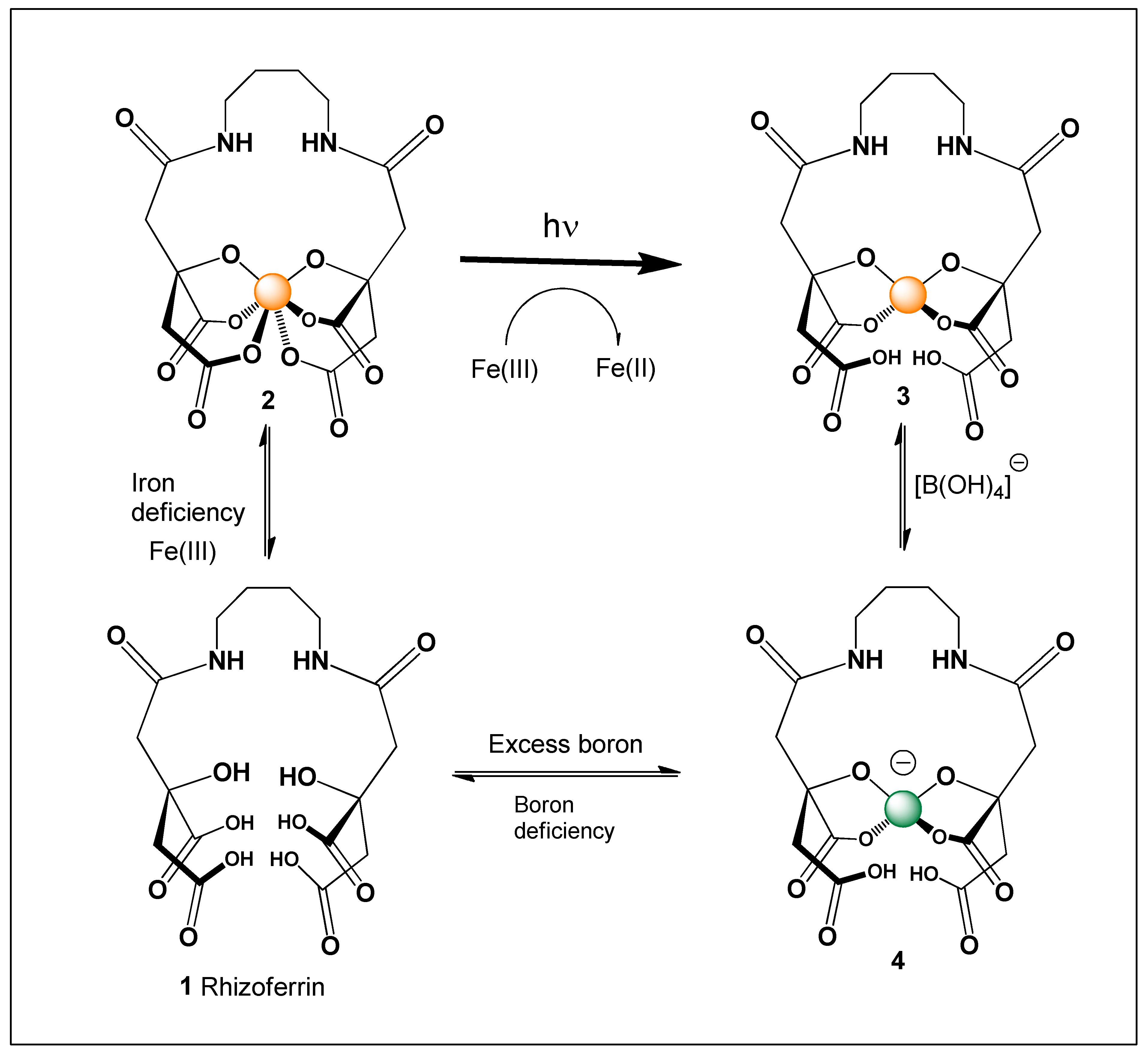
Rhizoferrin (1) is a citrate-derived (α-hydroxycarboxylate-type) siderophore produced by several bacteria and fungi, including rhizosphere-associated and soil-dwelling species. Rhizoferrin siderophore (see Figure 2) is produced by the fungus Rhizopus microsporus [78,79,80,81,82]. Structurally, rhizoferrin consists of two citrate-like units linked through a diamine backbone, generating multiple α-hydroxycarboxylate donor sets capable of chelating Fe(III) (2) in a hexadentate fashion, which, under the influence of light, is reduced to Fe(II) (3). Unlike catecholate siderophores, rhizoferrin coordinates iron through oxygen atoms derived from carboxylate and hydroxyl groups, forming stable Fe(III) complex (2) under circumneutral conditions [78,79,80].
From a structure–activity perspective, rhizoferrin (1) is also a chemically plausible candidate for boron coordination. Its α-hydroxycarboxylate motifs provide vicinal oxygen donors capable of stabilizing tetrahedral borate ester, analogous to well-characterized borodicitrate complex (4). Because boron speciation is pH-dependent (pKa ≈ 9.2 for the boric acid/borate equilibrium), rhizoferrin–boron complex formation would be favored under mildly alkaline conditions where the borate anion (B(OH)4) becomes more prevalent. In such environments, reversible borate ester formation at one or more α-hydroxycarboxylate sites is chemically feasible.
However, Fe(III) exhibits substantially higher thermodynamic affinity for rhizoferrin than boron, indicating that boron coordination would most likely occur transiently in iron-depleted extracellular settings and be displaced upon iron availability [78,79,80,81,82]. No dedicated boron transport function has been demonstrated for rhizoferrin, and direct structural characterization of rhizoferrin–boron complexes remains limited. Nevertheless, its donor-set architecture strongly supports chemically plausible borate formation (see Figure 2). In boron-rich soils or rhizosphere microenvironments, such reversible interactions may contribute to localized boron speciation modulation, buffering effects, or conformational variability of the ligand, rather than serving as a primary boron acquisition pathway.
The thermodynamic stability of the Fe(III)–rhizoferrin complex provides an important reference point when evaluating the plausibility of competing equilibria involving boron. Rhizoferrin is a citrate-based siderophore produced by several fungi and bacteria and coordinates Fe(III) primarily through oxygen donor atoms derived from carboxylate and hydroxyl groups. Experimental studies have shown that the overall formation constant for the Fe(III)–rhizoferrin complex is very high, with reported stability constants in the range of log β ≈ 29–30, reflecting the strong affinity of siderophores for ferric iron under physiological conditions. Such high stability is typical of siderophore–Fe(III) systems and explains their effectiveness in iron acquisition under extremely iron-limited environmental conditions. In comparison, boron–diol or boron–polyol complexes generally exhibit much lower stability constants and are characterized by reversible equilibrium behavior strongly dependent on pH and ligand geometry. Consequently, under conditions where Fe(III) is available, the Fe(III)–rhizoferrin complex is expected to dominate thermodynamically. Any potential interaction between rhizoferrin and boron species would therefore most likely occur only in the absence of ferric iron or under specific environmental conditions where boron concentrations are unusually high and suitable diol motifs remain unoccupied. This large difference in stability constants highlights that the proposed siderophore–boron interaction should be regarded as a secondary or conditional equilibrium, rather than a direct competitor to the highly optimized Fe(III) binding function of siderophores.
Borate esters formed with diols and polyols are known to exhibit relatively modest stability, with reported log K values typically ranging from ~1 to 6, depending on ligand structure, pH, and ionic strength. These values are several orders of magnitude lower than those associated with Fe(III)–siderophore complexes, which generally display log β values in the range of ~30–50 under optimal conditions. This quantitative comparison highlights the strong thermodynamic preference for Fe(III) coordination in canonical siderophore systems.
At the same time, the formation of boron–polyol complexes is highly sensitive to environmental conditions, particularly pH, as borate binding is favored under alkaline conditions where tetrahedral borate species predominate. In addition, the reversible nature of borate ester formation allows for dynamic ligand exchange, which may facilitate transient coordination under fluctuating environmental conditions. It is also important to note that conditional stability constants for Fe(III) complexes can be significantly reduced in natural settings due to proton competition, metal hydrolysis, and competing ligands. Under such conditions, the effective difference in stability between Fe(III)–siderophore and boron–polyol complexes may be less extreme than suggested by intrinsic constants alone.
Taken together, these considerations indicate that while Fe(III) binding remains thermodynamically dominant under standard conditions, boron complexation by polyol-containing ligands cannot be excluded in specific environmental niches. Rather, it is likely to occur under constrained or transient conditions where iron availability is extremely low or where Fe(III) coordination is weakened, thereby providing a plausible physicochemical basis for the proposed functional plasticity of siderophores.

11. Catecholate Siderophores

Catecholate siderophores constitute one of the most chemically potent classes of microbial iron-chelating agents. They are commonly biosynthesized from 2,3-dihydroxybenzoic acid (2,3-DHBA) or related salicylate-derived precursors and are characterized by vicinal phenolic hydroxyl groups forming o-dihydroxybenzene motifs. These functional groups serve as strong oxygen donors, enabling exceptionally tight coordination of Fe(III). The specific arrangement of adjacent hydroxyl groups provides an optimal geometric and electronic environment not only for high-affinity iron binding but also for chemically feasible interactions with boron species [26,27,28,29,30,31,32,62,63,64].
This dual binding capability arises from the shared preference of both Fe(III) and boron for oxygen-rich donor sites, although with markedly different binding strengths. In catecholate systems, deprotonation of the hydroxyl groups enhances electron donation and further stabilizes metal coordination. The rigidity and preorganization of the aromatic scaffold also contribute to efficient chelation by minimizing entropic penalties upon binding. While Fe(III) coordination remains the dominant function, the same structural features allow for transient boron association under suitable conditions. Additionally, environmental factors such as pH and redox state can influence the availability and reactivity of these donor groups. Together, these characteristics highlight the chemical versatility of catecholate siderophores and their potential to participate in multiple coordination processes beyond iron acquisition [28,31].

11.1. Structural Basis of Catecholate Iron Binding

During Fe(III) coordination, catechol groups undergo deprotonation to generate catecholate anions, each donating two oxygen atoms for metal binding. Triscatecholate siderophores such as enterobactin or bacillibactin provide a total of six oxygen donors, enabling hexadentate chelation of Fe(III) in an octahedral geometry with exceptionally high thermodynamic stability, with formation constants often exceeding 1030. Even siderophores containing fewer catechol units, such as aminochelin or vanchrobactin, maintain strong Fe(III) affinity due to the efficiency of their donor groups.
This remarkable binding strength can be attributed to several key factors. First, the strong hard–hard Lewis acid–base interaction between Fe3+ and oxygen donors drives stable complex formation. Second, the chelate effect arising from multidentate coordination significantly enhances overall stability compared to monodentate ligands. Third, π-electron delocalization within the aromatic catecholate system stabilizes the deprotonated ligand and strengthens metal binding. Fourth, favorable entropy changes associated with the formation of a single complex from multiple components further contribute to stability.
Additionally, the preorganized structure of many catecholate siderophores reduces entropic penalties during complex formation. The rigidity of the ligand framework ensures optimal orientation of donor atoms for efficient metal coordination. Cooperative interactions among multiple binding sites can further enhance overall affinity. Environmental factors such as pH can modulate the degree of catechol deprotonation and thus influence binding strength. Altogether, these features explain why catecholate siderophores rank among the most powerful natural Fe(III) chelators.

11.2. Chemical Basis of Boron Chelation

Boron chemistry is fundamentally distinct from that of iron, particularly in terms of speciation, reactivity, and coordination behavior. In aqueous environments, boron is present primarily as trigonal boric acid (B(OH)3) or, under more alkaline conditions, as the tetrahedral borate anion (B(OH)4), with a pKa of approximately 9.2 governing this equilibrium. Unlike iron, boron is not redox-active; instead, it functions as a Lewis acid that accepts electron density from oxygen donors to form tetrahedral borate esters, most commonly with cis-vicinal diols [1,2,6,62,63,64].
Catecholate siderophores are particularly well suited for boron interaction due to their structural features. First, the adjacent phenolic hydroxyl groups form a cis-diol system capable of engaging in borate ester formation. Second, upon deprotonation, these hydroxyl groups provide electron-rich oxygen donors that can coordinate borate in a tetrahedral geometry. Third, the aromatic catechol framework contributes to stabilization of the borate complex through electronic delocalization and resonance effects.
In addition, the rigidity of the aromatic scaffold helps maintain an optimal spatial arrangement for coordination. The pH-dependent deprotonation of catechol groups further enhances their binding capacity under alkaline conditions where borate is more prevalent. Despite these favorable features, boron binding remains weaker and more reversible compared to Fe(III) coordination. Environmental factors such as competing ligands and ionic strength can further influence borate complex formation. Altogether, these characteristics illustrate how catecholate siderophores possess inherent chemical compatibility with boron, even though their primary biological function remains iron acquisition.
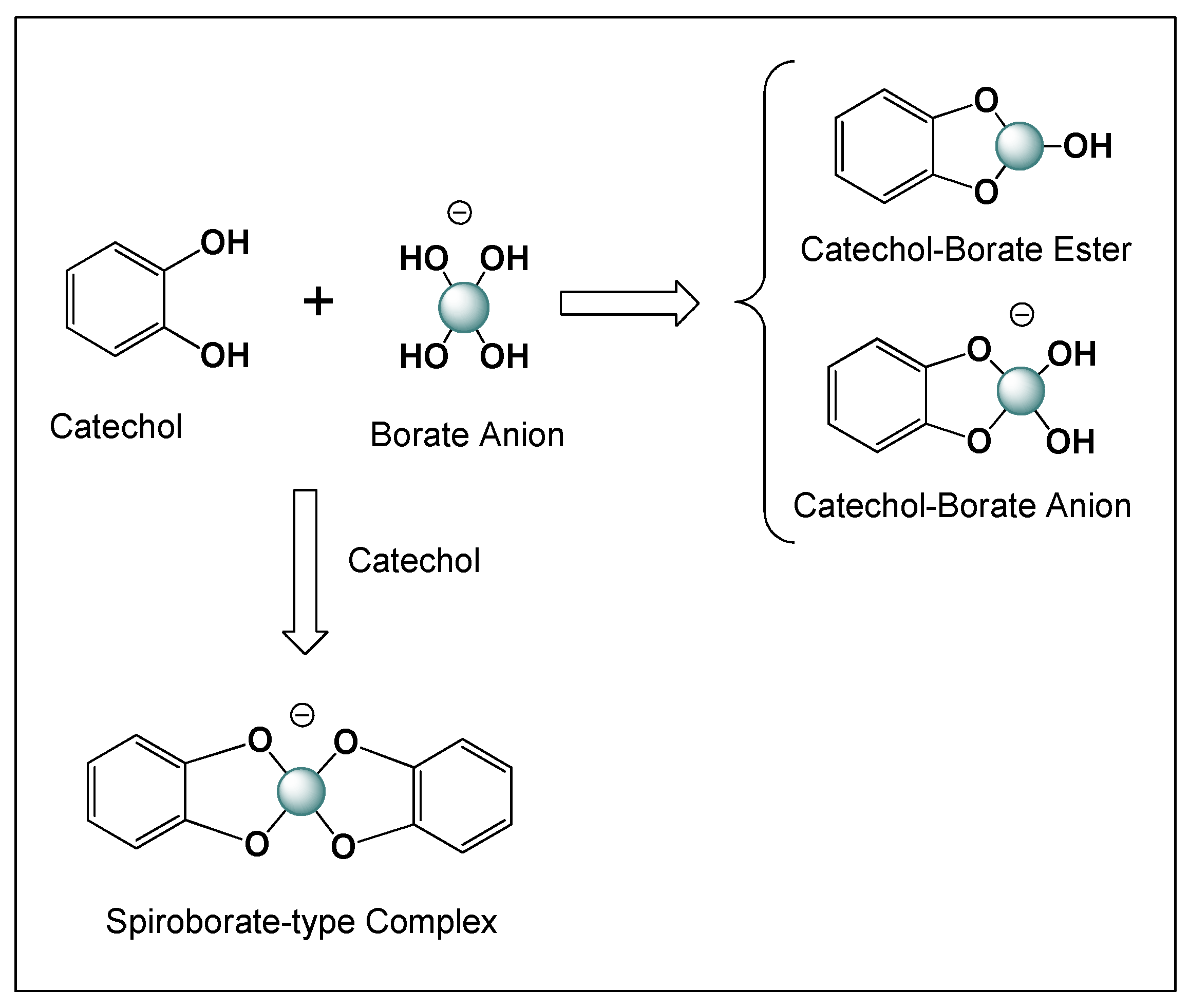
The resulting boron complex typically involves: (i) One boron center coordinated by two oxygen atoms from a single catechol (1:1 complex), or (ii) A spiroborate-type 2:1 complex, in which two catechol units coordinate one boron atom (Figure 3).

11.3. pH Dependence and Environmental Context

Boron coordination by catecholate siderophores is highly dependent on pH, which governs boron speciation and reactivity. At near-neutral pH, boron is present mostly as boric acid, resulting in relatively weak interactions with catecholate ligands. In contrast, under mildly alkaline conditions, the proportion of the borate anion increases, favoring the formation of tetrahedral borate esters with cis-diol-containing groups. Consequently, boron–catecholate interactions are more likely to occur in environments such as marine systems (pH ~8.1), alkaline soils, evaporative basins, and biofilm microenvironments where localized pH can be elevated.
Despite these favorable conditions, the thermodynamic preference for Fe(III) binding remains dominant, as its stability constants with catecholate ligands are orders of magnitude higher than those of boron complexes. As a result, Fe(III) will readily displace boron from catecholate coordination sites when available, particularly under biologically relevant iron-limited conditions. The pH-dependent deprotonation of catechol groups also plays a key role in modulating their binding capacity for both metals. In addition, fluctuations in environmental pH can shift the equilibrium between boric acid and borate, thereby influencing the extent of boron interaction. The reversible nature of borate ester formation further supports the transient character of these complexes. Overall, boron binding by catecholate siderophores is best understood as a conditional process that occurs under specific environmental constraints rather than as a primary function of these molecules.

11.4. Competitive and Conditional Binding

The interaction between Fe(III) and boron in catecholate systems can be described as a dynamic and condition-dependent process. In the iron-free (apo) state, catecholate ligands retain accessible vicinal diol functionalities that are capable of forming reversible borate esters. Upon binding of Fe(III), these coordination sites are occupied by the metal, which displaces any bound boron due to its substantially higher thermodynamic affinity. Conversely, following photochemical reduction or reductive release of iron, the catechol groups are regenerated in a form that can again participate in transient boron coordination.
This cycle highlights the reversible and competitive nature of boron binding in comparison to iron coordination. Boron association is therefore likely to occur primarily in the extracellular environment, where siderophores are exposed to fluctuating chemical conditions. It remains a secondary interaction that depends on the temporary absence or weakening of Fe(III) binding rather than a dedicated biological function. Additionally, the kinetics of metal exchange and environmental variability may further influence the extent of boron association. The transient availability of binding sites plays a critical role in enabling such interactions. Overall, boron coordination should be viewed as a conditional and reversible process that complements, but does not replace, the primary role of siderophores in iron acquisition.

11.5. Structural Diversity and Boron Potential

Catecholate siderophores display variability in their capacity to bind boron, largely influenced by the number and arrangement of catechol donor groups. Multicatecholate systems, in particular, offer enhanced geometric flexibility and multiple coordination sites, which can facilitate more stable and cooperative interactions with boron species (see Table 1). The presence of multiple catechol units increases the likelihood of forming favorable binding geometries, potentially allowing intramolecular stabilization of borate complexes.
This increased donor multiplicity can promote chelation through multidentate interactions, even though boron typically forms tetrahedral complexes with fewer donor atoms. In addition, the spatial organization of catechol groups may enable transient bridging or cooperative binding effects within a single molecule. However, the stability of such boron complexes remains significantly lower than that of corresponding Fe(III) complexes, and is strongly influenced by pH and boron speciation. Multicatecholate architectures may also enhance local boron concentration through weak, reversible interactions. Furthermore, conformational flexibility within these siderophores can modulate accessibility of donor groups and influence binding dynamics. Overall, donor multiplicity plays an important role in determining the extent and nature of boron association, while remaining secondary to the primary function of iron coordination.

11.6. Functional Implications

Although boron coordination does not compete with Fe(III) binding in biological priority, it may exert subtle effects on siderophore behavior. Reversible boron binding can modulate extracellular boron speciation, influence ligand conformation, and potentially affect receptor recognition and uptake processes. It may also contribute to environmental signaling in boron-rich niches. In marine systems, where iron limitation coincides with relatively high boron availability, such interactions may influence local chemical equilibria, particularly within biofilms and particle-associated communities, providing adaptive advantages under fluctuating conditions.

11.7. Integrated Perspective

Catecholate siderophores exemplify multifunctional coordination chemistry, combining high-affinity Fe(III) binding with the capacity for reversible boron interaction. While their primary role is iron acquisition, boron coordination arises as a secondary consequence of their cis-diol-containing structures. These interactions are context-dependent, influenced by pH, metal availability, and redox conditions, and reflect chemical opportunism rather than specific evolutionary selection [1,2,6,58,59,60,61].
This dual reactivity highlights the broader geochemical relevance of siderophores, linking microbial iron homeostasis with environmental boron chemistry. Such versatility may enhance microbial adaptability in heterogeneous environments and underscores the importance of considering siderophores beyond iron cycling alone.
In particular, the presence of cis-diol functionalities enables siderophores to engage in transient borate ester formation without compromising their core role in iron transport. Such interactions are highly context-dependent, influenced by factors such as pH, metal availability, and redox dynamics. The overlap between iron-binding and boron-binding capabilities reflects a form of chemical opportunism rather than specific evolutionary selection for boron utilization. Moreover, this versatility may enhance the adaptability of microorganisms in chemically heterogeneous environments. It also underscores the importance of considering siderophores within a wider geochemical framework beyond iron cycling alone. Ultimately, these properties link microbial metal homeostasis to environmental boron chemistry, particularly in marine and alkaline ecosystems where both elements play significant roles.

12. Comparison of Fe(III) and Boron Stability

Classical siderophores (catecholate, hydroxamate, and mixed-type) form highly stable Fe(III) complexes, with stability constants reaching 1030–1052, reflecting the strong preference of Fe(III) for hexadentate oxygen donors in octahedral geometries. This high affinity enables microorganisms to acquire iron under extremely low environmental concentrations. In contrast, boron interacts with carbohydrates and polyols through reversible borate ester formation with cis-diols, governed by pH, boron speciation, and ligand structure. These complexes are significantly less stable and fall within the range of dynamic covalent interactions [1,2,3,11,12,13,14,15,16].
Accordingly, siderophore complexes function in high-affinity iron acquisition, whereas boron–diol interactions act as reversible structural motifs that modulate molecular conformation, crosslinking, and interfacial organization in biological systems [1,2,11,12,13,14,15,16].

13. Petrobactin Siderophores

Petrobactins (57, see Figure 4) are a product of many microorganisms such as the marine bacterium Marinobacter hydrocarbonoclasticus, Marinobacter aquaeolei, Alteromonas, Bacillus anthracis, and the non-sulfur bacterium Rhodopseudomonas palustris. Structurally, petrobactin is a mixed catecholate-type siderophore distinguished by its 3,4-dihydroxybenzoyl moieties, which confer high-affinity Fe(III) binding while enabling evasion of host immune proteins such as siderocalin. Its unusual 3,4-catechol substitution pattern differentiates it from classical 2,3-dihydroxybenzoyl siderophores and contributes to its role in pathogenicity, particularly in Bacillus anthracis. In marine and soil environments, petrobactin supports iron acquisition under limiting conditions and exemplifies the structural diversity and adaptive versatility of microbial siderophores [83,84,85,86,87].
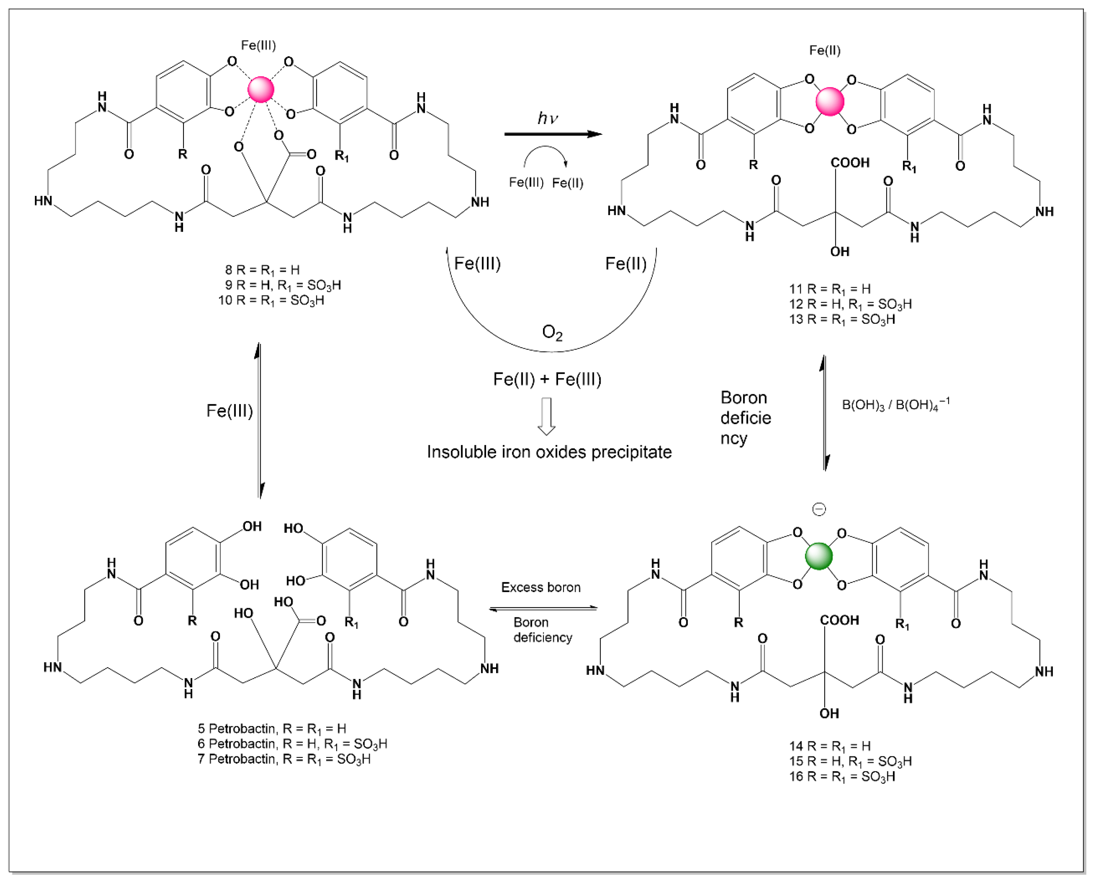
Petrobactin is produced by the oil-degrading marine bacterium Marinobacter hydrocarbonoclasticus and is notable for its photochemical reactivity in the Fe(III)-complexed form [83]. In natural sunlight, the Fe(III)–petrobactin complexes (810) undergoes ligand-to-metal charge transfer (LMCT) excitation mediated by the Fe(III)–α-hydroxy acid motif, resulting in decarboxylation of the ligand and reduction of Fe(III) to Fe(II) (1113) [88,89,90,91,92,93,94,95,96,97,98,99,100,101,102,103,104,105,106,107,108,109,110,111]. This represents one of the earliest demonstrations of light-driven photoreduction in an Fe(III)-siderophore system, raising intriguing questions about the ecological role of photochemistry in siderophore-mediated iron cycling.
Importantly, boron binding alters the conformation of petrobactin relative to both its free and ferric-bound forms. This conformational distinction supports a broader SAR hypothesis emerging from this review: boron–siderophore complexes (1116) may function not primarily in boron nutrition, but as chemically distinct signaling entities, particularly in marine environments where borate is abundant. Such complexes could influence quorum sensing, metal homeostasis, or niche adaptation, providing a selective advantage to boron-tolerant bacteria inhabiting boron-rich marine microniches [1,2,88,89,90].

14. Aminochelin Siderophores

Azotobacter vinelandii produces a diverse suite of iron-chelating siderophores under iron-limited conditions to facilitate nutrient uptake, including protochelin (tricatecholate), azotochelin (dicatecholate), aminochelin (monocatecholate), 2,3-dihydroxybenzoic acid, and the fluorescent azotobactin. These molecules are critical for high-affinity Fe3+ acquisition, and some can bind Mo and V, which are essential for nitrogen fixation [91,92,93,94].
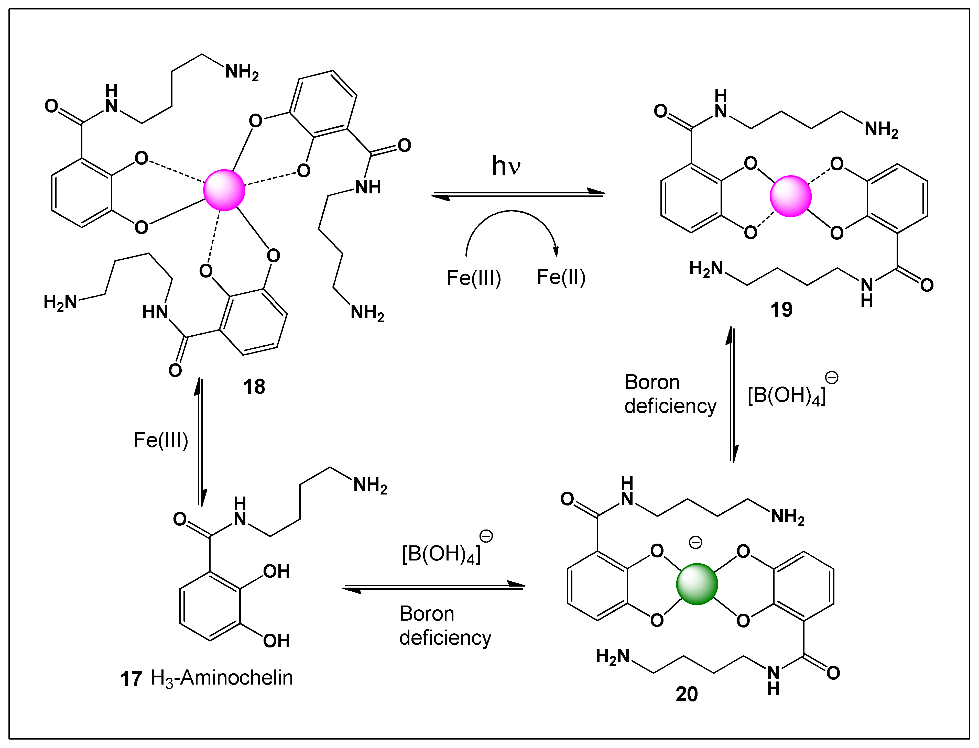
Aminochelin (17, see Figure 5) is a monocatecholamide siderophore produced by Azotobacter vinelandii that forms a complex with Fe (III) (18). It functions at high iron concentrations, forming a 3:1 complex (18, aminochelin: Fe) with an overall stability constant of log β3 41.3 and a value of pFe (III) at 7.45 pH [91,92,93].
Ferric iron (Fe3+) is thermodynamically stable in oxic surface environments, but it is poorly soluble and biologically inaccessible at circumneutral pH because it readily forms insoluble hydroxides and oxides. Many microorganisms overcome this limitation by secreting siderophores that chelate Fe(III) and maintain it in solution. In certain systems, exposure to sunlight initiates ligand-to-metal charge transfer (LMCT) within the Fe(III)–siderophore complex. Upon absorption of photons in the UV–visible range, an electron is transferred from the ligand to the Fe(III) center, transiently reducing it to Fe(II). This photochemical reduction weakens the coordination environment and can lead to partial ligand oxidation and release of Fe(II) [91,92,93].
In the case of Aminochelin, iron coordination typically occurs in a 2:1 ligand-to-metal ratio, forming a dimeric complex in which two aminochelin molecules chelate a single Fe(III) center through their catechol groups. This bis(catecholate) coordination environment is photochemically active, as catechol ligands are known to support LMCT transitions. Upon irradiation, Fe(III) (18) within the aminochelin complex can undergo reduction to Fe(II) (19), accompanied by partial oxidation of the catechol moieties to semiquinone or quinone forms. The photoreduced Fe(II) may then dissociate from the complex and become available for biological uptake. Thus, sunlight-driven reduction of ferric iron within aminochelin dimers provides a mechanistic pathway by which poorly soluble Fe(III) can be converted into bioavailable Fe(II) in illuminated environmental niches such as surface waters and soils [91,92,93,94].
Aminochelin (H3-aminochelin in its fully protonated form) is a monocatecholate siderophore produced by Azotobacter vinelandii that contains a 2,3-dihydroxybenzoyl (catechol) moiety capable of providing vicinal phenolic oxygen donors for boron coordination. From a structure–activity perspective, this adjacent diol arrangement satisfies the fundamental geometric requirement for tetrahedral borate ester formation upon deprotonation, rendering boron binding chemically plausible. However, because aminochelin contains only a single catechol unit, its boron complexes would likely be limited to discrete 1:1 or 2:1 (ligand:boron, 20) species rather than extended boron-bridged assemblies typical of multicatecholate scaffolds. As with other catechol-based systems, boron complexation is expected to be reversible and strongly pH-dependent, becoming more favorable under mildly alkaline conditions where the borate anion predominates. Thermodynamically, Fe(III) exhibits substantially higher affinity for aminochelin than boron, indicating that any aminochelin–boron complexes would most plausibly form transiently in iron-depleted extracellular environments and be displaced upon iron loading. Although direct structural characterization of aminochelin–boron complex (20) remains limited, its catechol donor-set architecture supports chemically feasible borate formation consistent with established catecholate–boron coordination principles.

15. Protochelin Siderophores

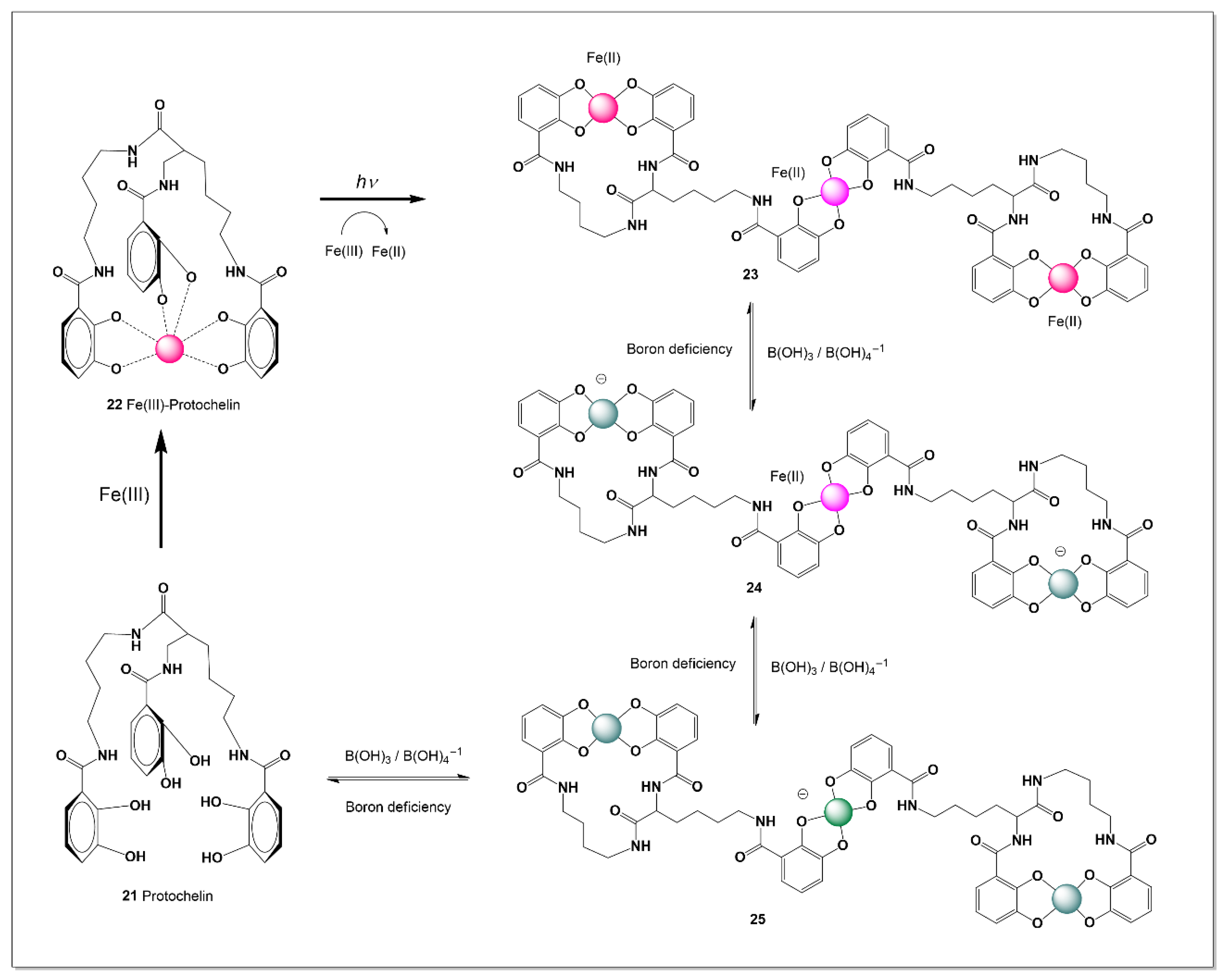
Protochelin (21, see Figure 6) is a triscatecholate siderophore whose three 2,3-dihydroxybenzoyl (catechol) units provide vicinal phenolic oxygen donors capable of tetrahedral borate ester formation (25). From a structure–activity standpoint, this multicatecholate architecture satisfies the geometric and electronic criteria required for boron coordination, rendering boron binding chemically plausible. The presence of multiple catechol sites affords greater binding flexibility than monocatecholate ligands and may permit discrete borate complexes under appropriate conditions. As with other catecholate systems, boron complexation is expected to be reversible and strongly pH-dependent, becoming more favorable under mildly alkaline conditions where the borate anion predominates. Thermodynamically, Fe(III) exhibits substantially higher affinity for protochelin than boron, indicating that any protochelin–boron complexes would most likely form transiently in iron-depleted extracellular environments and be displaced upon iron availability. Although direct structural characterization of protochelin–boron complexes remains limited, its donor-set architecture strongly supports chemically feasible borate formation consistent with established catecholate–boron coordination principles [95,96,97,98,99].
Protochelin contains three catecholate units that form a versatile oxygen-donor framework capable of coordinating metals beyond Fe(III), including Cr, Ni, Cu, Zn, As, Hg, and Pb. This broad binding profile reflects the hard Lewis base character of catecholate oxygens and their adaptability to different metal ions and coordination environments. Notably, strong interactions with chromium species have been reported under certain conditions. Such multi-metal affinity suggests that protochelin-like ligands may play a role in environmental processes, including the mobilization or sequestration of toxic metals, with potential applications in bioremediation [100].
However, several mechanistic considerations complicate the assumption that protochelin can simultaneously coordinate iron, boron, and additional metals within a single stable complex. Boron coordination occurs through formation of tetrahedral borate esters with cis-diol groups and is generally weaker and reversible compared to Fe(III) binding. Consequently, while protochelin may form alternative complexes with different metals under metal-specific conditions, the formation of stable multi-metal–boron–iron assemblies simultaneously is thermodynamically and sterically unlikely in most biological environments.
Therefore, protochelin should be viewed as a chemically flexible ligand capable of binding various metals individually depending on environmental availability and redox state, rather than as a universal multi-metal scaffold operating concurrently with iron and boron. Further spectroscopic, structural, and in situ studies are required to distinguish true inner-sphere coordination from competitive displacement, redox transformation, or transient association phenomena.

16. Catecholate Borate Siderophores

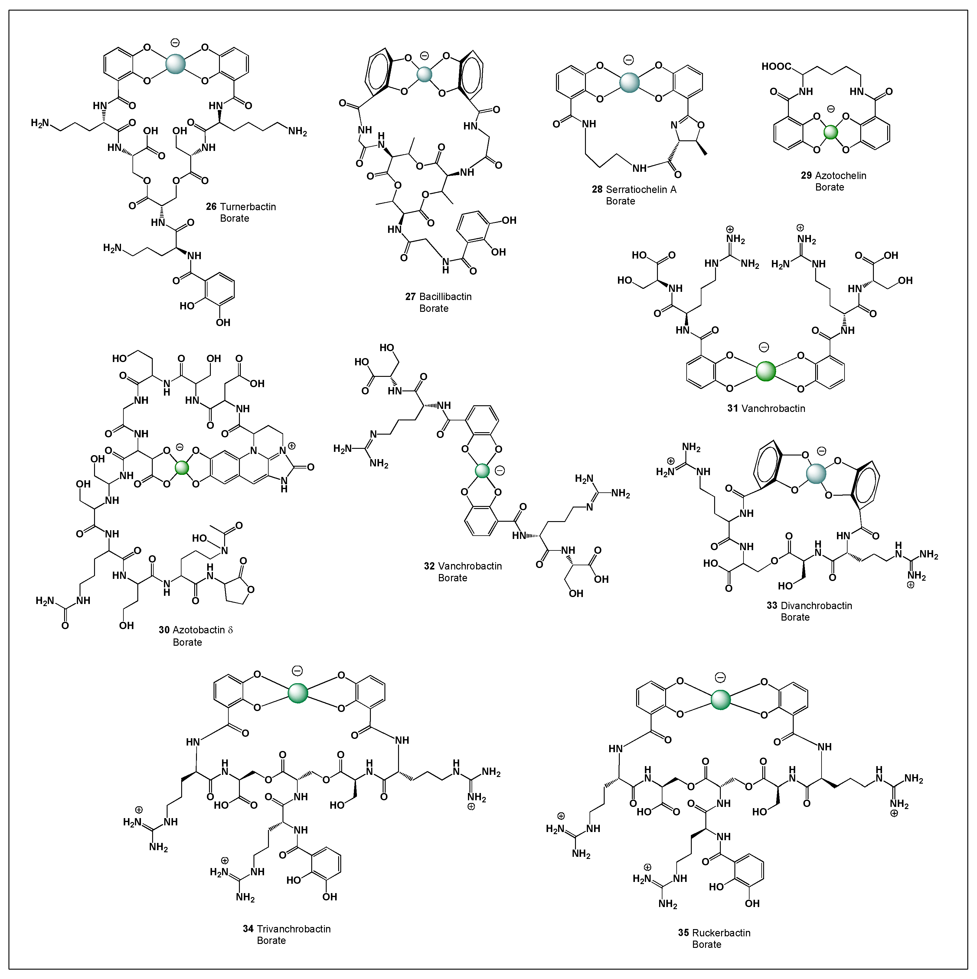
Teredinibacter turnerae, a symbiont of the shipworm Bankia gouldi, produces the catecholate siderophore turnerbactin, composed of 2,3-dihydroxybenzoyl (Figure 7) units linked via a peptide backbone [101,102]. These catechol groups enable high-affinity Fe(III) coordination in an octahedral geometry, supporting iron uptake in iron-limited symbiotic environments. Structurally, turnerbactin also permits reversible boron coordination through catechol-derived borate ester formation under alkaline conditions. However, due to the much higher affinity of Fe(III), such boron interactions are likely transient and environmentally dependent, potentially influencing local boron speciation without serving a transport function.
Bacillus species produce bacillibactin, a triscatecholate siderophore with a cyclic trilactone backbone, synthesized via nonribosomal peptide pathways [103,104,105,106]. Its hexadentate structure enables strong Fe(III) binding and efficient iron uptake. The presence of three catechol units also supports potential boron coordination, including spiroborate-type interactions under suitable conditions. Although not well characterized structurally, such interactions may contribute to boron speciation in soil environments without implying a dedicated biological role.
Serratiochelin A, produced by Serratia marcescens, contains two catechol units that facilitate high-affinity Fe(III) binding and iron acquisition in competitive environments [107,108,109]. In addition to its iron-scavenging role, it has been explored as a sideromycin scaffold for targeted antimicrobial delivery.
From a structure–activity standpoint, the same catechol motifs that confer high Fe(III) affinity also render serratiochelin A chemically capable of coordinating the borate anion (B(OH)4). Upon deprotonation of adjacent phenolic hydroxyl groups, tetrahedral borate ester formation is feasible, particularly under mildly alkaline conditions where borate availability increases. Because Fe(III) exhibits substantially higher thermodynamic affinity for catecholate ligands than boron, any serratiochelin A–boron complexes would likely be reversible, extracellular, and competitively displaced upon iron binding. Although direct structural characterization of serratiochelin A–boron complexes remains limited, its donor-set architecture strongly supports chemically plausible borate formation, suggesting potential roles in transient boron speciation modulation without implying a dedicated boron transport function.
Azotochelin (29) is a catecholate-type siderophore produced by Azotobacter vinelandii that contains 2,3-dihydroxybenzoyl (catechol) units capable of providing vicinal phenolic oxygen donors for boron coordination [110,111,112]. Azotochelin is a tetradentate bis(catecholate) siderophore that forms a coordinatively unsaturated 1:1 Fe(III):ligand complex at neutral pH. Unlike typical hexadentate siderophores, this 1:1 complex is structurally unique. Its stability and reduction potential are influenced by pH and buffer composition, exhibiting structural rearrangements below pH 6.25 [110,111,112].
From a structure–activity perspective, this adjacent diol arrangement satisfies the geometric requirement for tetrahedral borate ester formation upon deprotonation, rendering boron binding chemically plausible. Because azotochelin contains multiple catechol moieties, it could, in principle, form discrete borate complexes, including spiroborate-type species under appropriate conditions. As with other catecholate systems, boron complexation is expected to be reversible and strongly pH-dependent, becoming more favorable under mildly alkaline conditions where the borate anion (B(OH)4) predominates. Thermodynamically, Fe(III) exhibits substantially higher affinity for azotochelin than boron, indicating that any azotochelin–boron complexes would most likely form transiently in iron-depleted extracellular environments and be displaced upon iron availability. Although direct structural characterization of azotochelin–boron complexes remains limited, its donor-set architecture supports chemically feasible borate formation consistent with established catecholate–boron coordination principles.
Azotobactin δ (30) is a fluorescent siderophore (pyoverdine-type) produced by Azotobacter vinelandii. Its structure typically contains a dihydroxyquinoline or related catecholate-like chromophore moiety, along with additional coordinating groups (e.g., hydroxamate and/or carboxylate functionalities) in the peptide side chain. From a structure–activity perspective, boron binding would most plausibly occur at the catechol-like or vicinal oxygen donor sites within the chromophore region, where adjacent oxygen atoms can form tetrahedral borate esters upon deprotonation. As with other catecholate systems, boron complexation is expected to be reversible and strongly pH-dependent, becoming more favorable under mildly alkaline conditions where borate (B(OH)4) predominates. However, Fe(III) exhibits substantially higher thermodynamic affinity for azotobactin δ, indicating that any boron coordination would likely be transient, extracellular, and competitively displaced upon iron loading. Although direct structural characterization of azotobactin δ–boron complexes remains limited, its donor-set architecture supports chemically plausible borate formation, particularly in iron-depleted and boron-rich environments.
Vanchrobactin is a catechol-based siderophore synthesized by several Vibrio species, including pathogens associated with fish and marine habitats. Its structure features 2,3-dihydroxybenzoyl (catechol) moieties connected by a polyamine scaffold, which enables efficient and high-affinity binding of Fe(III). In its ferric state, vanchrobactin forms a multidentate complex in which Fe(III) is coordinated by catecholate oxygen atoms in an octahedral arrangement. This mode of binding provides considerable thermodynamic stability, enhancing iron solubilization and facilitating uptake under iron-depleted conditions in marine systems. The Fe(III)–vanchrobactin complex is specifically recognized by outer membrane receptors and transported into the periplasm, where iron is subsequently released via reduction or ligand exchange processes [112,113,114,115,116].
The biosynthesis of vanchrobactin is typically mediated by nonribosomal peptide synthetase (NRPS) pathways, which assemble its catecholate-containing framework. Once internalized, iron is incorporated into cellular metabolic processes essential for bacterial growth and virulence. The production of vanchrobactin is tightly regulated by iron availability, often through ferric uptake regulator (Fur) proteins. Additionally, competition for iron via siderophore production plays a significant role in microbial interactions within marine ecosystems.
From a structure–activity perspective, vanchrobactin is also a chemically plausible boron-binding ligand. The presence of vicinal phenolic hydroxyl groups within its catechol moieties provides the geometric requirement for tetrahedral borate ester formation upon deprotonation. Under mildly alkaline conditions, where the borate anion (B(OH)4) is more abundant, reversible boron coordination at available catechol sites is feasible. Vanchrobactin borate complexes may adopt two distinct spatial arrangements, commonly described as form A (31) and form B (32), depending on the orientation of the catechol–borate coordination and the overall ligand conformation. These conformers likely arise from rotational flexibility within the backbone and the reversible nature of tetrahedral borate ester formation. Under physiological conditions, the two forms are expected to exist in dynamic equilibrium, interconverting in response to pH, borate concentration, and competing metal ions such as Fe(III).
Divanchrobactin (33) is a dimeric catecholate siderophore produced by marine and fish-pathogenic Vibrio species, particularly strains associated with Vibrio anguillarum. Structurally, divanchrobactin consists of two vanchrobactin-like subunits, each containing 2,3-dihydroxybenzoyl (catechol) moieties linked through a polyamine framework. This bis-catecholate architecture provides multiple vicinal phenolic oxygen donors capable of high-affinity Fe(III) coordination, typically forming stable octahedral ferric complexes essential for iron acquisition in iron-limited marine environments [117,118,119].
Compared with monomeric vanchrobactin, divanchrobactin exhibits increased denticity and potentially enhanced iron-binding stability, which may confer a competitive advantage under severe iron restriction. The presence of multiple catechol groups also renders divanchrobactin chemically capable of reversible borate ester formation under mildly alkaline conditions, although Fe(III) binding remains thermodynamically dominant. Functionally, divanchrobactin contributes to virulence and environmental persistence in marine vibrios, highlighting the adaptive diversification of catecholate siderophores in aquatic ecosystems.
Trivanchrobactin (34) is a triscatecholate siderophore synthesized by certain Vibrio species and can be regarded as an oligomeric form of vanchrobactin. Its structure comprises three 2,3-dihydroxybenzoyl (catechol) groups arranged to create a hexadentate binding site for Fe(III). In the ferric complex, Fe(III) is coordinated by catecholate oxygen atoms in a highly stable octahedral geometry, resulting in strong thermodynamic affinity and effective iron solubilization in iron-deficient marine environments. This multicatecholate framework provides greater binding stability compared to simpler catecholate systems and enables efficient uptake via TonB-dependent outer membrane receptors that recognize ferric catecholate complexes [117,120].
The biosynthesis of trivanchrobactin is believed to involve nonribosomal peptide synthetase (NRPS) pathways similar to those responsible for vanchrobactin assembly. Its oligomeric nature may enhance iron-scavenging efficiency by increasing ligand denticity and coordination strength. After transport into the cell, iron is released through reduction processes that weaken the Fe(III)–ligand interaction. Furthermore, the production of such high-affinity siderophores provides a competitive advantage in marine microbial communities where iron availability is extremely limited.
From a structure–activity perspective, trivanchrobactin also fulfills the geometric and electronic requirements for boron coordination. Each catechol moiety provides vicinal phenolic oxygen donors capable of forming tetrahedral borate esters upon deprotonation. The presence of multiple catechol units increases the potential for discrete borate complex formation, including intramolecular or spiroborate-type interactions, particularly under mildly alkaline conditions where the borate anion (B(OH)4) is more prevalent. In boron-rich marine environments, such transient interactions may contribute to localized boron speciation modulation without implying a dedicated boron transport function.
Trivanchrobactin (34) and ruckerbactin (35) possess the same molecular formula and share a common catecholate–polyamine architecture, differing only in the stereochemical configuration of the arginine residue within their backbone. In particular, vanchrobactin incorporates an L-arginine unit, whereas ruckerbactin contains the D-arginine enantiomer. This difference in stereochemistry does not affect the catechol-based donor groups responsible for Fe(III) binding or potential boron coordination, but it can influence the overall three-dimensional conformation of the molecule. As a result, variations in stereochemistry may impact receptor recognition, transport efficiency, and species-specific uptake mechanisms.
Although their coordination chemistry remains fundamentally similar, subtle conformational differences may alter how these siderophores interact with membrane-bound receptors or transport proteins. Such stereochemical variation is often a consequence of biosynthetic pathway divergence and may confer selective advantages in different microbial niches. In addition, differences in molecular recognition could influence competitive interactions among microorganisms producing structurally related siderophores. The preservation of identical donor sets highlights that metal-binding properties are conserved despite configurational changes in the backbone. Furthermore, these variations may affect the kinetics of complex formation or dissociation without significantly altering overall thermodynamic stability. Overall, the distinction between trivanchrobactin and ruckerbactin is configurational rather than compositional, reflecting evolutionary adaptation without modification of their core coordination capabilities.
Ruckerbactin (35) is a catecholate-type siderophore produced by Yersinia ruckeri and related aquatic pathogens. Structurally, ruckerbactin contains 2,3-dihydroxybenzoyl (catechol) moieties linked through a polyamine backbone, enabling high-affinity coordination of Fe(III). In its ferric form, Fe(III) is chelated by the catecholate oxygen donors in an octahedral geometry, generating a thermodynamically stable complex that facilitates iron solubilization and uptake under iron-limited conditions typical of host tissues and aquatic environments. The Fe(III)–ruckerbactin complex is recognized by specific outer membrane receptors and internalized through TonB-dependent transport systems, after which iron is released intracellularly via reduction or ligand exchange mechanisms [121,122].
From a structure–activity standpoint, ruckerbactin also possesses the fundamental chemical features required for boron coordination. The vicinal phenolic hydroxyl groups within its catechol units can, upon deprotonation, form tetrahedral borate esters with borate (B(OH)4). In boron-containing aquatic environments, such interactions may contribute to localized boron speciation modulation without implying a dedicated boron transport function.

17. Comparative Conformation of Iron and Boron Siderophores

The potential interaction between siderophores and boron must account for key differences in Fe(III) and B(III) coordination chemistry. Siderophores are optimized for Fe(III), which favors octahedral coordination with six oxygen donors, forming highly stable hexadentate complexes. In contrast, boron exists as B(OH)3 or B(OH)4 and prefers trigonal or tetrahedral geometries, involving a smaller central atom and different electronic structure [20,90]. As a result, siderophore ligand frameworks are not ideally suited for boron coordination, and interactions would likely involve only partial binding, potentially introducing geometric strain and reducing stability [20,90,123,124].
These differences also affect binding affinity. Fe(III) forms extremely stable complexes (1030–1052), whereas boron–diol interactions are weaker, reversible, and strongly dependent on pH and ligand structure. Consequently, Fe(III) binding dominates under most conditions, making direct competition with boron unlikely [123,124,125].
Nevertheless, boron interactions may occur under specific environmental conditions, particularly in iron-poor, boron-rich systems such as marine or hypersaline environments. In such cases, binding would likely involve localized diol coordination rather than full hexadentate chelation, forming transient borate esters. While siderophores are specialized for iron acquisition, their oxygen-rich structures allow for possible secondary interactions with boron, which warrant further investigation [20,90,123,124,125,126].

18. Possibility of Siderophore–Boron Complex Formation

The hypothesis that siderophores may interact with boron in boron-rich environments is grounded in coordination chemistry rather than direct experimental evidence. Catecholate siderophores, such as enterobactin, contain vicinal phenolic hydroxyl groups that strongly bind Fe(III) but are also well-known ligands for boron, which forms reversible complexes with cis-diols and polyphenols. Studies of catechol and related compounds demonstrate the formation of borate complexes, including spiroborate structures, particularly under alkaline conditions where borate ions are more abundant [14,15,16,127,128,129].
However, Fe(III)–siderophore complexes are significantly more stable (often >1030) than borate–diol interactions, which are weaker and reversible. Thus, boron coordination is unlikely to compete with iron binding and is better considered a secondary process occurring under specific conditions, such as in iron-poor, boron-rich environments. In these settings, boron may interact with uncomplexed or partially protonated siderophores, potentially influencing conformation, aggregation, or interfacial behavior.
Such conditions are relevant in marine, hypersaline, and geothermal systems, where elevated boron levels may promote transient borate complexation with catechol-containing ligands. Although direct evidence for siderophore–boron complexes remains limited, analogous interactions with polyphenols and carbohydrates support their chemical plausibility. Further studies using spectroscopic and computational approaches are needed to clarify the stability and environmental role of these interactions [1,2,130,131,132].

Possibility of Siderophore–Boron Complex Formation and Its Relation to Boron-Containing Antibiotics from Marine Bacteria

The potential formation of siderophore–boron complexes can be considered in the context of boron-containing natural products from marine microorganisms. Compounds such as borophycin, aplasmomycin, boromycin, and tartrolone derivatives demonstrate that microbial systems can incorporate boron into complex bioactive structures. These molecules typically involve oxygen-rich ligands that coordinate boron through multiple hydroxyl groups, forming stable borate complexes [1,2,133,134,135,136].
This capacity provides a chemical precedent for possible boron interactions with siderophores, which also contain oxygen-donor ligands such as catecholates and hydroxamates. In marine environments with relatively high boron concentrations, reversible coordination with such ligands may occur under suitable conditions. Although Fe(III) binding remains dominant, structural similarities to known boron-binding motifs support the possibility of transient boron interactions in specific niches [11,12,13,125,126,131].
Overall, siderophore–boron interactions may represent part of a broader spectrum of boron-associated chemistry in marine microbial systems. While direct evidence remains limited, further spectroscopic and thermodynamic studies are needed to assess their stability and ecological relevance [1,2,137,138,139].

19. Conclusions

Microbial siderophores are highly optimized Fe(III)-chelating agents, but they also function as versatile coordination platforms capable of interactions beyond iron. Catecholate and α-hydroxycarboxylate siderophores contain vicinal oxygen donors that can form reversible borate esters under suitable conditions. While Fe(III) binding remains dominant, boron coordination emerges as a secondary property based on shared hard Lewis acid–base chemistry.
In iron- and boron-rich environments—such as marine systems and alkaline habitats—this dual reactivity may become relevant. Boron, present as boric acid and borate, can transiently interact with siderophores under mildly alkaline conditions. These interactions are reversible and likely extracellular, potentially influencing boron speciation, ligand conformation, and local chemical equilibria without competing with iron uptake.
This perspective reframes siderophores as multifunctional mediators linking metal homeostasis and geochemical cycling. Their structural diversity and adaptive transport systems support roles in microbial competition, biofilm formation, and pathogenicity. The potential for boron interaction reflects chemical opportunism rather than evolutionary selection, arising from ligand architecture optimized for Fe(III).
Future studies should quantify boron binding to siderophores, assess structural determinants of borate formation, and evaluate environmental relevance using spectroscopic and computational approaches [1,2,137,138,139].
In summary, siderophores exemplify functional plasticity in coordination chemistry—evolved for iron acquisition yet capable of interacting with boron—highlighting their role at the interface of iron and boron biogeochemical cycles.

Author Contributions

Conceptualization, V.M.D.; methodology, V.M.D.; software, A.O.T. and S.V.B.; investigation, V.M.D.; resources, V.M.D. and S.V.B.; writing—original draft preparation, A.O.T., S.V.B. and V.M.D.; writing—review and editing, A.O.T. and V.M.D. All authors have read and agreed to the published version of the manuscript.

Funding

This research received no external funding.

Institutional Review Board Statement

Not applicable.

Informed Consent Statement

Not applicable.

Data Availability Statement

No new data were created or analyzed in this study. Data sharing is not applicable to this article.

Conflicts of Interest

Author Valery M. Dembitsky was employed by the company Bio-Pharm Laboratories. The remaining authors declare that the research was conducted in the absence of any commercial or financial relationships that could be construed as a potential conflict of interest.

References

  1. Dembitsky, V.M.; Al Quntar, A.A.A.; Srebnik, M. Natural and synthetic small boron-containing molecules as potential inhibitors of bacterial and fungal quorum sensing. Chem. Rev. 2011, 111, 209–237. [Google Scholar] [CrossRef] [Scilit] [PubMed]
  2. Dembitsky, V.M.; Smoum, R.; Al-Quntar, A.A.; Ali, H.A.; Pergament, I.; Srebnik, M. Natural occurrence of boron-containing compounds in plants, algae and microorganisms. Plant Sci. 2002, 163, 931–942. [Google Scholar] [CrossRef] [Scilit]
  3. Dembitsky, V.M.; Terent’ev, A.O.; Baranin, S.V. Life with Boron: Microbial Boron-Binding Siderophores, Adaptation, and Function. Microbiol. Res. 2026, 17, 57. [Google Scholar] [CrossRef] [Scilit]
  4. Gaillardet, J.; Lemarchand, D. Boron in the weathering environment. In Boron Isotopes: The Fifth Element; Springer: Berlin/Heidelberg, Germany, 2017; pp. 163–188. [Google Scholar]
  5. Park, H.; Schlesinger, W.H. Global biogeochemical cycle of boron. Glob. Biogeochem. Cycles 2002, 16, 20-1–20-11. [Google Scholar] [CrossRef] [Scilit]
  6. Dembitsky, V.M.; Terent’ev, A.O.; Stolbov, L.A.; Pogodin, P.V.; Filimonov, D.A.; Poroikov, V.V. Salicylic acid and its boron complexes as quorum sensing molecules. Mol. Pharm. 2025, 22, 6499–6509. [Google Scholar] [CrossRef] [Scilit]
  7. Sánchez, M.; Sabio, L.; Gálvez, N.; Capdevila, M.; Dominguez-Vera, J.M. Iron chemistry at the service of life. IUBMB Life 2017, 69, 382–388. [Google Scholar] [CrossRef] [Scilit]
  8. Theil, E.C.; Goss, D.J. Living with iron (and oxygen): Questions and answers about iron homeostasis. Chem. Rev. 2009, 109, 4568–4579. [Google Scholar] [CrossRef] [Scilit]
  9. Babanin, V.F.; Zalutskii, A.A.; Mikhaleva, N.V.; Pukhov, D.E.; Omeljanuk, G.G. State of iron in a living substance. RUDN J. Ecol. Life Saf. 2009, 2, 5–13. [Google Scholar]
  10. Weber, K.A.; Achenbach, L.A.; Coates, J.D. Microorganisms pumping iron: Anaerobic microbial iron oxidation and reduction. Nat. Rev. Microbiol. 2006, 4, 752–764. [Google Scholar] [CrossRef] [Scilit]
  11. Crowley, D.E.; Wang, Y.C.; Reid, C.P.P.; Szaniszlo, P.J. Mechanisms of iron acquisition from siderophores by microorganisms and plants. Plant Soil 1991, 130, 179–198. [Google Scholar] [CrossRef] [Scilit]
  12. Moody, M.D. Microorganisms and iron limitation. Bioscience 1986, 36, 618–623. [Google Scholar] [CrossRef] [Scilit]
  13. Wilson, B.R.; Bogdan, A.R.; Miyazawa, M.; Hashimoto, K.; Tsuji, Y. Siderophores in iron metabolism: From mechanism to therapy potential. Trends Mol. Med. 2016, 22, 1077–1090. [Google Scholar] [CrossRef] [Scilit] [PubMed]
  14. Wandersman, C.; Delepelaire, P. Bacterial iron sources: From siderophores to hemophores. Annu. Rev. Microbiol. 2004, 58, 611–647. [Google Scholar] [CrossRef] [Scilit] [PubMed]
  15. Hider, R.C.; Kong, X. Chemistry and biology of siderophores. Nat. Prod. Rep. 2010, 27, 637–657. [Google Scholar] [CrossRef] [Scilit]
  16. Schalk, I.J. Bacterial siderophores: Diversity, uptake pathways and applications. Nat. Rev. Microbiol. 2025, 23, 24–40. [Google Scholar] [CrossRef] [Scilit]
  17. Gu, S.; Wang, N.; Zheng, Y.; Wang, T.; Shen, Q.; Zhang, F.; Zuo, Y. Integrating microbial siderophores into concepts of plant iron nutrition. Nat. Plants 2025, 12, 26–36. [Google Scholar] [CrossRef] [Scilit]
  18. Kumar, R.; Singh, A.; Srivastava, A. Xenosiderophores: Bridging the gap in microbial iron acquisition strategies. World J. Microbiol. Biotechnol. 2025, 41, 69. [Google Scholar] [CrossRef] [Scilit]
  19. Vijay, K.; Shibasini, M.; Sivasakthivelan, P.; Kavitha, T. Microbial siderophores as molecular shuttles for metal cations: Sources, sinks and application perspectives. Arch. Microbiol. 2023, 205, 322. [Google Scholar] [CrossRef] [Scilit]
  20. Raymond, K.N.; Müller, G.; Matzanke, B.F. Complexation of iron by siderophores: A review of their solution and structural chemistry and biological function. Struct. Chem. 2005, 123, 49–102. [Google Scholar]
  21. Albelda-Berenguer, M.; Monachon, M.; Joseph, E. Siderophores: From natural roles to potential applications. Adv. Appl. Microbiol. 2019, 106, 193–225. [Google Scholar]
  22. Crosa, J.H. Genetics and molecular biology of siderophore-mediated iron transport in bacteria. Microbiol. Rev. 1989, 53, 517–530. [Google Scholar] [CrossRef] [PubMed]
  23. Schalk, I.J.; Lamont, I.L.; Cobessi, D. Structure–function relationships in the bifunctional ferrisiderophore FpvA receptor from Pseudomonas aeruginosa. Biometals 2009, 22, 671–678. [Google Scholar] [CrossRef] [Scilit] [PubMed]
  24. Ghssein, G.; Ezzeddine, Z. A review of Pseudomonas aeruginosa metallophores: Pyoverdine, pyochelin and pseudopaline. Biology 2022, 11, 1711. [Google Scholar] [CrossRef] [Scilit] [PubMed]
  25. Cai, J.; Fang, Y.; Liu, X.; Adjei, M.O.; Fan, B. Revealing microbial siderophores: From genes to applications. Microorganisms 2026, 14, 393. [Google Scholar] [CrossRef] [Scilit]
  26. Philippot, L.; Chenu, C.; Kappler, A.; Rillig, M.C.; Fierer, N. The interplay between microbial communities and soil properties. Nat. Rev. Microbiol. 2024, 22, 226–239. [Google Scholar] [CrossRef] [Scilit]
  27. Alshatari, S.S.; Ziarno, M. From microbes to medicine: Targeting metalloprotein pathways for innovative antibacterial strategies. Int. J. Mol. Sci. 2026, 27, 737. [Google Scholar] [CrossRef] [Scilit]
  28. Sundaray, J.K.; Roy, D.; Mohapatra, M.; Mohanty, D.; Das, I.I.; Parida, C.K. Metagenomic profiling of fish-associated microbiota: Ecological perspectives from freshwater to marine environment—A review. Arch. Microbiol. 2026, 208, 105. [Google Scholar] [CrossRef] [Scilit]
  29. Neilands, J.B. Siderophores: Structure and function of microbial iron transport compounds. J. Biol. Chem. 1995, 270, 26723–26726. [Google Scholar] [CrossRef] [Scilit]
  30. Timofeeva, A.M.; Galyamova, M.R.; Sedykh, S.E. Bacterial siderophores: Classification, biosynthesis, perspectives of use in agriculture. Plants 2022, 11, 3065. [Google Scholar] [CrossRef] [Scilit]
  31. Winkelmann, G. Microbial siderophore-mediated transport. Biochem. Soc. Trans. 2002, 30, 691–696. [Google Scholar] [CrossRef] [Scilit]
  32. Höfte, M. Classes of microbial siderophores. In Iron Chelation in Plants and Soil Microorganisms; Academic Press: London, UK, 1993; pp. 3–26. [Google Scholar]
  33. De Serrano, L.O.; Camper, A.K.; Richards, A.M. An overview of siderophores for iron acquisition in microorganisms living in the extreme. Biometals 2016, 29, 551–571. [Google Scholar] [CrossRef] [Scilit] [PubMed]
  34. Barber, M.F.; Elde, N.C. Buried treasure: Evolutionary perspectives on microbial iron piracy. Trends Genet. 2015, 31, 627–636. [Google Scholar] [CrossRef] [Scilit] [PubMed]
  35. Kramer, J.; Özkaya, Ö.; Kümmerli, R. Bacterial siderophores in community and host interactions. Nat. Rev. Microbiol. 2020, 18, 152–163. [Google Scholar] [CrossRef] [Scilit] [PubMed]
  36. Ali, S.S.; Vidhale, N.N. Bacterial siderophore and their application: A review. Int. J. Curr. Microbiol. Appl. Sci. 2013, 2, 303–312. [Google Scholar]
  37. Schalk, I.J.; Hannauer, M.; Braud, A. New roles for bacterial siderophores in metal transport and tolerance. Environ. Microbiol. 2011, 13, 2844–2854. [Google Scholar] [CrossRef] [Scilit]
  38. Amiri, M.; Golchin, M.; Jamshidian Mojaver, M.; Farzin, H.; Hajizade, A. Enterobactin: A key player in bacterial iron acquisition and virulence and its implications for vaccine development and antimicrobial strategies. Virulence 2025, 16, 2563018. [Google Scholar] [CrossRef] [Scilit]
  39. Guo, C.; Nolan, E.M. Exploring the antibacterial activity and cellular fates of enterobactin–drug conjugates that target Gram-negative bacterial pathogens. Acc. Chem. Res. 2024, 57, 1046–1056. [Google Scholar] [CrossRef] [Scilit]
  40. Levay, P.F.; Viljoen, M. Lactoferrin: A general review. Haematologica 1995, 80, 252–267. [Google Scholar]
  41. Sanchez, L.; Calvo, M.; Brock, J.H. Biological role of lactoferrin. Arch. Dis. Child. 1992, 67, 657–661. [Google Scholar] [CrossRef] [Scilit]
  42. Złoch, M.; Thiem, D.; Gadzała-Kopciuch, R.; Hrynkiewicz, K. Synthesis of siderophores by plant-associated metallotolerant bacteria under exposure to Cd2+. Chemosphere 2016, 156, 312–325. [Google Scholar] [CrossRef] [Scilit]
  43. Vishwakarma, A.; Hait, S. Selective lithium extraction from spent lithium-ion batteries using siderophores produced by Pseudomonas aeruginosa: Efficacy, kinetics, and artificial neural network modeling. Process Biochem. 2024, 146, 115–127. [Google Scholar] [CrossRef] [Scilit]
  44. Holden, V.I.; Bachman, M.A. Diverging roles of bacterial siderophores during infection. Metallomics 2015, 7, 986–995. [Google Scholar] [CrossRef] [Scilit]
  45. Årstøl, E.; Hohmann-Marriott, M.F. Cyanobacterial siderophores—Physiology, structure, biosynthesis, and applications. Mar. Drugs 2019, 17, 281. [Google Scholar] [CrossRef] [Scilit]
  46. Chakraborty, S.; Verma, E.; Singh, S.S. Cyanobacterial siderophores: Ecological and biotechnological significance. In Cyanobacteria; Academic Press: London, UK, 2019; pp. 383–397. [Google Scholar]
  47. Řezanka, T.; Palyzová, A.; Sigler, K. Isolation and identification of siderophores produced by cyanobacteria. Folia Microbiol. 2018, 63, 569–579. [Google Scholar] [CrossRef] [Scilit]
  48. Brick, M.B.; Hussein, M.H.; Mowafy, A.M.; Hamouda, R.A.; Ayyad, A.M.; Refaay, D.A. Significance of siderophore-producing cyanobacteria on enhancing iron uptake potentiality of maize plants grown under iron-deficiency. Microb. Cell Fact. 2025, 24, 3. [Google Scholar]
  49. Sabat, S.; Patra, S.; Swain, S.; Bej, S.; Bishoyi, A.K.; Sahoo, C.R.; Padhy, R.N. Phycocompounds from cyanobacteria: Exploring synergistic effects with conventional anticancer and antimicrobial properties. ACS Omega 2025, 10, 23957–23980. [Google Scholar] [CrossRef] [Scilit]
  50. Jurado-Flores, A.; Heredia-Martínez, L.G.; Torres-Cortes, G.; Díaz-Santos, E. Harnessing microalgae and cyanobacteria for sustainable agriculture: Mechanistic insights and applications as biostimulants, biofertilizers and biocontrol agents. Agriculture 2025, 15, 1842. [Google Scholar] [CrossRef] [Scilit]
  51. Sun, Z.; Liu, X.; Ugya, A.Y.; Liu, H.; Sun, L.; Luo, G. Microalgae and cyanobacteria as a tool for agricultural sustainability: A review of biofertilizer and biostimulant potential. Front. Plant Sci. 2025, 16, 1733394. [Google Scholar]
  52. Haas, H. Fungal siderophore metabolism with a focus on Aspergillus fumigatus. Nat. Prod. Rep. 2014, 31, 1266–1276. [Google Scholar] [CrossRef] [Scilit]
  53. Haas, H. Molecular genetics of fungal siderophore biosynthesis and uptake: The role of siderophores in iron uptake and storage. Appl. Microbiol. Biotechnol. 2003, 62, 316–330. [Google Scholar] [CrossRef] [Scilit]
  54. Pecoraro, L.; Wang, X.; Shah, D.; Song, X.; Kumar, V.; Shakoor, A.; Rani, R. Biosynthesis pathways, transport mechanisms and biotechnological applications of fungal siderophores. J. Fungi 2021, 8, 21. [Google Scholar] [CrossRef] [Scilit]
  55. Johnson, L. Iron and siderophores in fungal–host interactions. Mycol. Res. 2008, 112, 170–183. [Google Scholar] [CrossRef] [Scilit]
  56. Happacher, I.; Aguiar, M.; Yap, A.; Decristoforo, C.; Haas, H. Fungal siderophore metabolism with a focus on Aspergillus fumigatus: Impact on biotic interactions and potential translational applications. Essays Biochem. 2023, 67, 829–842. [Google Scholar]
  57. Petrik, M.; Zhai, C.; Haas, H.; Decristoforo, C. Siderophores for molecular imaging applications. Clin. Transl. Imaging 2017, 5, 15–27. [Google Scholar] [CrossRef] [Scilit]
  58. Gao, S.; Zhang, Y.; Zhou, R.; Shen, T.; Zhang, D.; Guo, Z.; Zou, X. Boronic acid-assisted detection of bacterial pathogens: Applications and perspectives. Coord. Chem. Rev. 2024, 518, 216082. [Google Scholar] [CrossRef] [Scilit]
  59. Khan, S.; Hussain, Z.; Bibi, S.; Kashif, M.; Zubair, M.; Azizi, S.; Maaza, M. Bias-free catalytic photoelectrochemical degradation of Congo red using a boron–iron oxide coated electrode: A green and sustainable approach. Catal. Surv. Asia 2025, 30, 64–76. [Google Scholar] [CrossRef] [Scilit]
  60. Ji, L.; Wu, J.; Zuo, Y.; Gao, W.; Feng, J.; Zhang, Z. Potential of boronic acid derivatization and activity in agrochemical discovery. Molecules 2025, 30, 3018. [Google Scholar] [CrossRef] [Scilit]
  61. Farfán-García, E.D.; Kilic, A.; García-Machorro, J.; Cuevas-Galindo, M.E.; Rubio-Velazquez, B.A.; García-Coronel, I.H.; Soriano-Ursúa, M.A. Antimicrobial (viral, bacterial, fungal, and parasitic) mechanisms of action of boron-containing compounds. In Viral, Parasitic, Bacterial, and Fungal Infections; Academic Press: London, UK, 2023; pp. 733–754. [Google Scholar]
  62. Dembitsky, V.M.; Terent’ev, A.O.; Baranin, S.V.; Gursky, M.E. Aromatic compounds and their fascinating boron complexes as potential quorum sensing molecules. Vietnam J. Chem. 2025, 63, 883–911. [Google Scholar] [CrossRef] [Scilit]
  63. Dembitsky, V.M.; Terent’ev, A.O.; Gursky, M.E.; Baranin, S.V. Fascinating and intriguing biomolecules: The chemistry of boron complexes with carbohydrates, glycolipids, and steroids. Vietnam J. Chem. 2025; in press. [CrossRef] [Scilit]
  64. Dembitsky, V.M.; Terent’ev, A.O.; Baranin, S.V. Boronosteroids as potential antitumor drugs: A review. Tumor Discov. 2025, 5, 14–33. [Google Scholar] [CrossRef] [Scilit]
  65. Kawamura, A.; Guo, J.; Itagaki, Y.; Bell, C.; Wang, Y.I.; Haupert, G.T., Jr.; Nakanishi, K. On the structure of endogenous ouabain. Proc. Natl. Acad. Sci. USA 1999, 96, 6654–6659. [Google Scholar] [CrossRef] [Scilit]
  66. Kawamura, A.; Guo, J.; Maggiali, F.; Berova, N.; Nakanishi, K. Structure of endogenous ouabain. Pure Appl. Chem. 1999, 71, 1643–1648. [Google Scholar] [CrossRef] [Scilit]
  67. Kawamura, A.; Abrell, L.M.; Maggiali, F.; Berova, N.; Nakanishi, K.; Labutti, J.; Hamlyn, J.M. Biological implication of conformational flexibility in ouabain: Observations with two ouabain phosphate isomers. Biochemistry 2001, 40, 5835–5844. [Google Scholar] [CrossRef] [Scilit] [PubMed]
  68. Hunter, J.M.; Nemzer, B.V.; Rangavajla, N.; Biţă, A.; Rogoveanu, O.C.; Neamţu, J.; Mogoşanu, G.D. The fructoborates: Part of a family of naturally occurring sugar–borate complexes—Biochemistry, physiology, and impact on human health: A review. Biol. Trace Elem. Res. 2019, 188, 11–25. [Google Scholar] [CrossRef] [Scilit] [PubMed]
  69. Chapelle, S.; Verchere, J.F. A 11B and 13C NMR determination of the structures of borate complexes of pentoses and related sugars. Tetrahedron 1988, 44, 4469–4482. [Google Scholar] [CrossRef] [Scilit]
  70. Zill, L.P.; Khym, J.X.; Cheniae, G.M. Further studies on the separation of the borate complexes of sugars and related compounds by ion-exchange chromatography. J. Am. Chem. Soc. 1953, 75, 1339–1342. [Google Scholar] [CrossRef] [Scilit]
  71. Verchere, J.F.; Hlaibi, M. Stability constants of borate complexes of oligosaccharides. Polyhedron 1987, 6, 1415–1420. [Google Scholar] [CrossRef] [Scilit]
  72. Davis, H.B.; Mott, C.J. Interaction of boric acid and borates with carbohydrates and related substances. J. Chem. Soc. Faraday Trans. 1 Phys. Chem. Condens. Ph. 1980, 76, 1991–2002. [Google Scholar] [CrossRef] [Scilit]
  73. Hirakawa, Y.; Kakegawa, T.; Furukawa, Y. Borate-guided ribose phosphorylation for prebiotic nucleotide synthesis. Sci. Rep. 2022, 12, 11828. [Google Scholar] [CrossRef] [Scilit]
  74. Imperio, D.; Panza, L. Sweet boron: Boron-containing sugar derivatives as potential agents for boron neutron capture therapy. Symmetry 2022, 14, 182. [Google Scholar] [CrossRef] [Scilit]
  75. Takahashi, Y.; Kim, H.J.; Benner, S.A.; Kakegawa, T.; Furukawa, Y. Ribose accumulation in borate-rich prebiotic environments. Astrobiology 2023, 26, 15311074261417882. [Google Scholar] [CrossRef] [Scilit]
  76. Dembitsky, V.M.; Rosenberg, G.S.; Zanfera, V.M. The Evolutionary Pathway to the Biomembrane: The Role of Low Molecular Weight Polyols in the Formation of the Protomembrane; Editorial & Publishing Department IEVRB RAS: Moscow, Russia; Togliatti, Russia, 2023; p. 128. [Google Scholar]
  77. Wang, M.; Li, H. Structure, function, and biosynthesis of siderophores produced by Streptomyces species. J. Agric. Food Chem. 2025, 73, 4425–4439. [Google Scholar] [CrossRef] [Scilit] [PubMed]
  78. Drechsel, H.; Metzger, J.; Freund, S.; Jung, G.; Boelaert, J.R.; Winkelmann, G. Rhizoferrin—A novel siderophore from the fungus Rhizopus microsporus var. rhizopodiformis. Biol. Met. 1991, 4, 238–243. [Google Scholar] [CrossRef] [Scilit]
  79. Münzinger, M.; Taraz, K.; Budzikiewicz, H.; Drechsel, H.; Heymann, P.; Winkelmann, G.; Meyer, J.M. S,S-rhizoferrin (enantio-rhizoferrin)—A siderophore of Ralstonia (Pseudomonas) pickettii DSM 6297—The optical antipode of R,R-rhizoferrin isolated from fungi. Biometals 1999, 12, 189–193. [Google Scholar] [CrossRef] [Scilit]
  80. Drechsel, H.; Jung, G.; Winkelmann, G. Stereochemical characterization of rhizoferrin and identification of its dehydration products. Biometals 1992, 5, 141–148. [Google Scholar] [CrossRef] [Scilit]
  81. Carrano, C.J.; Drechsel, H.; Kaiser, D.; Jung, G.; Matzanke, B.; Winkelmann, G.; Albrecht-Gary, A.M. Coordination chemistry of the carboxylate-type siderophore rhizoferrin: The iron(III) complex and its metal analogs. Inorg. Chem. 1996, 35, 6429–6436. [Google Scholar] [CrossRef] [Scilit]
  82. Škríba, A.; Patil, R.H.; Hubáček, P.; Dobiáš, R.; Palyzová, A.; Marešová, H.; Havlíček, V. Rhizoferrin glycosylation in Rhizopus microsporus. J. Fungi 2020, 6, 89. [Google Scholar] [CrossRef] [Scilit]
  83. Barbeau, K.; Zhang, G.; Live, D.H.; Butler, A. Petrobactin, a photoreactive siderophore produced by the oil-degrading marine bacterium Marinobacter hydrocarbonoclasticus. J. Am. Chem. Soc. 2002, 124, 378–379. [Google Scholar] [CrossRef] [Scilit]
  84. Manck, L.E.; Park, J.; Tully, B.J.; Poire, A.M.; Bundy, R.M.; Dupont, C.L.; Barbeau, K.A. Petrobactin, a siderophore produced by Alteromonas, mediates community iron acquisition in the global ocean. ISME J. 2022, 16, 358–369. [Google Scholar] [CrossRef] [Scilit]
  85. Homann, V.V.; Edwards, K.J.; Webb, E.A.; Butler, A. Siderophores of Marinobacter aquaeolei: Petrobactin and its sulfonated derivatives. Biometals 2009, 22, 565–571. [Google Scholar] [CrossRef] [Scilit]
  86. Koppisch, A.T.; Browder, C.C.; Moe, A.L.; Shelley, J.T.; Kinkel, B.A.; Hersman, L.E.; Ruggiero, C.E. Petrobactin is the primary siderophore synthesized by Bacillus anthracis str. Sterne under conditions of iron starvation. Biometals 2005, 18, 577–585. [Google Scholar] [CrossRef] [Scilit]
  87. Baars, O.; Morel, F.M.; Zhang, X. The purple non-sulfur bacterium Rhodopseudomonas palustris produces novel petrobactin-related siderophores under aerobic and anaerobic conditions. Environ. Microbiol. 2018, 20, 1667–1676. [Google Scholar] [CrossRef] [Scilit] [PubMed]
  88. Miller, E.P.; Wu, Y.; Carrano, C.J. Boron uptake, localization, and speciation in marine brown algae. Metallomics 2016, 8, 161–169. [Google Scholar] [CrossRef] [Scilit] [PubMed]
  89. Weerasinghe, A.J.; Amin, S.A.; Barker, R.A.; Othman, T.; Romano, A.N.; Parker Siburt, C.J.; Crumbliss, A.L. Borate as a synergistic anion for Marinobacter algicola ferric binding protein, FbpA: A role for boron in iron transport in marine life. J. Am. Chem. Soc. 2013, 135, 14504–14507. [Google Scholar] [CrossRef] [Scilit] [PubMed]
  90. Harris, W.R.; Amin, S.A.; Küpper, F.C.; Green, D.H.; Carrano, C.J. Borate binding to siderophores: Structure and stability. J. Am. Chem. Soc. 2007, 129, 12263–12271. [Google Scholar] [CrossRef] [Scilit]
  91. Page, W.J.; Tigerstrom, M.V. Aminochelin, a catecholamine siderophore produced by Azotobacter vinelandii. Microbiology 1988, 134, 453–460. [Google Scholar] [CrossRef] [Scilit]
  92. Khodr, H.; Hider, R.; Duhme-Klair, A.K. The iron-binding properties of aminochelin, the mono(catecholamide) siderophore of Azotobacter vinelandii. J. Biol. Inorg. Chem. 2002, 7, 891–896. [Google Scholar] [CrossRef] [Scilit]
  93. Cornish, A.S.; Page, W.J. The catecholate siderophores of Azotobacter vinelandii: Their affinity for iron and role in oxygen stress management. Microbiology 1998, 144, 1747–1754. [Google Scholar] [CrossRef] [Scilit]
  94. Cornish, A.S.; Page, W.J. Role of molybdate and other transition metals in the accumulation of protochelin by Azotobacter vinelandii. Appl. Environ. Microbiol. 2000, 66, 1580–1586. [Google Scholar] [CrossRef] [Scilit]
  95. Harrington, J.M.; Bargar, J.R.; Jarzecki, A.A.; Roberts, J.G.; Sombers, L.A.; Duckworth, O.W. Trace metal complexation by the triscatecholate siderophore protochelin: Structure and stability. Biometals 2012, 25, 393–412. [Google Scholar] [CrossRef] [Scilit]
  96. Dubme, A.K.; Hider, R.C.; Khodr, H.H. Synthesis and iron-binding properties of protochelin, the tris(catecholamide) siderophore of Azotobacter vinelandii. Chem. Ber. 1997, 130, 969–973. [Google Scholar] [CrossRef] [Scilit]
  97. Cornish, A.S.; Page, W.J. Production of the triacetecholate siderophore protochelin by Azotobacter vinelandii. Biometals 1995, 8, 332–338. [Google Scholar] [CrossRef] [Scilit]
  98. Dembitsky, V.M.; Terent′ev, A.O.; Baranin, S.V.; Scorei, I.R. Boron’s Double Edge—Antibiotics, Toxins, and the Fine Line Between Them. Molecules 2026, 31, 1021. [Google Scholar] [CrossRef] [Scilit]
  99. Taraz, K.; Ehlert, G.; Geisen, K.; Budzikiewicz, H.; Korth, H.; Pulverer, G. Protochelin, a catecholate siderophore from a bacterium (DMS No. 5746). Z. Naturforsch. B 1990, 45, 1327–1332. [Google Scholar] [CrossRef] [Scilit]
  100. Khan, H.; Khalid, H. A computational study on removal of heavy metal pollutants Cr3+, Cr6+, Ni2+, Cu2+, Zn2+, As3+, Hg2+, and Pb2+ from soil using tricatecholate protochelin siderophore: A DFT study. Chem. Proc. 2025, 18, 74. [Google Scholar]
  101. Han, A.W.; Sandy, M.; Fishman, B.; Trindade-Silva, A.E.; Soares, C.A.; Distel, D.L.; Haygood, M.G. Turnerbactin, a novel triscatecholate siderophore from the shipworm endosymbiont Teredinibacter turnerae T7901. PLoS ONE 2013, 8, e76151. [Google Scholar]
  102. Naka, H.; Haygood, M.G. The dual role of TonB genes in turnerbactin uptake and carbohydrate utilization in the shipworm symbiont Teredinibacter turnerae. Appl. Environ. Microbiol. 2023, 89, e00744-23. [Google Scholar] [CrossRef] [Scilit]
  103. Dimopoulou, A.; Theologidis, I.; Benaki, D.; Koukounia, M.; Zervakou, A.; Tzima, A.; Skandalis, N. Direct antibiotic activity of bacillibactin broadens the biocontrol range of Bacillus amyloliquefaciens MBI600. mSphere 2021, 6, e01128-20. [Google Scholar] [CrossRef] [Scilit]
  104. Chakraborty, K.; Kizhakkekalam, V.K.; Joy, M.; Chakraborty, R.D. Bacillibactin class of siderophore antibiotics from a marine symbiotic Bacillus as promising antibacterial agents. Appl. Microbiol. Biotechnol. 2022, 106, 329–340. [Google Scholar] [CrossRef] [Scilit]
  105. Nalli, Y.; Singh, S.; Gajjar, A.; Mahizhaveni, B.; Dusthackeer, V.N.A.; Shinde, P.B. Bacillibactin class siderophores produced by the endophyte Bacillus subtilis NPROOT3 as antimycobacterial agents. Lett. Appl. Microbiol. 2023, 76, ovac026. [Google Scholar] [CrossRef] [Scilit]
  106. Ma, J.; Ning, X.; Li, J.; Dai, S.; Sun, F.; Li, H.; Ding, Y. Metabolite-mediated alleviation of iron deficiency and growth promotion of Malus hupehensis by Bacillus licheniformis LCDD6 in calcareous soil. Microorganisms 2026, 14, 349. [Google Scholar] [CrossRef] [Scilit]
  107. Schneider, Y.; Jenssen, M.; Isaksson, J.; Hansen, K.Ø.; Andersen, J.H.; Hansen, E.H. Bioactivity of serratiochelin A, a siderophore isolated from a co-culture of Serratia sp. and Shewanella sp. Microorganisms 2020, 8, 1042. [Google Scholar] [CrossRef] [Scilit] [PubMed]
  108. Weakland, D.R.; Smith, S.N.; Bell, B.; Tripathi, A.; Mobley, H.L.T. The Serratia marcescens siderophore serratiochelin is necessary for full virulence during bloodstream infection. Infect. Immun. 2020, 88, e01128-19. [Google Scholar] [CrossRef] [Scilit] [PubMed]
  109. Eze, P.M.; Simons, V.; Seidemann, T.; Wang, L.; Kiffe-Delf, A.L.; Frank, M.; Kalscheuer, R. Serratiochelins A and B from Serratia marcescens show xenosiderophoric characteristics towards Acinetobacter baumannii and Mycobacterium tuberculosis. Trop. J. Pharm. Res. 2021, 20, 2551–2558. [Google Scholar]
  110. Duhme, A.K.; Hider, R.C.; Naldrett, M.J.; Pau, R.N. The stability of the molybdenum–azotochelin complex and its effect on siderophore production in Azotobacter vinelandii. J. Biol. Inorg. Chem. 1998, 3, 520–526. [Google Scholar] [CrossRef] [Scilit]
  111. Karadkhelkar, N.M.; Gupta, P.; Barasa, L.; Chilamakuri, R.; Hlordzi, C.K.; Acharekar, N.; Yoganathan, S. Chemical derivatization leads to the discovery of novel analogs of azotochelin, a natural siderophore, as promising anticancer agents. ChemMedChem 2024, 19, e202300715. [Google Scholar] [CrossRef] [Scilit]
  112. Baranska, N.G.; Parkin, A.; Duhme-Klair, A.K. Electrochemical and solution structural characterization of Fe(III) azotochelin complexes: Examining the coordination behavior of a tetradentate siderophore. Inorg. Chem. 2022, 61, 19172–19182. [Google Scholar] [CrossRef] [Scilit]
  113. Balado, M.; Osorio, C.R.; Lemos, M.L. A gene cluster involved in the biosynthesis of vanchrobactin, a chromosome-encoded siderophore produced by Vibrio anguillarum. Microbiology 2006, 152, 3517–3528. [Google Scholar] [CrossRef] [Scilit]
  114. Souto, A.; Montaos, M.A.; Balado, M.; Osorio, C.R.; Rodriguez, J.; Lemos, M.L.; Jimenez, C. Synthesis and antibacterial activity of conjugates between norfloxacin and analogues of the siderophore vanchrobactin. Bioorg. Med. Chem. 2013, 21, 295–302. [Google Scholar] [CrossRef] [Scilit]
  115. Ntege, W.; Javaid, A.; Banicod, R.J.S.; Tabassum, N.; Kim, T.; Khan, F. Molecular adaptations of ferric uptake regulator (Fur) proteins drive iron homeostasis and pathogenesis in Vibrio spp. Curr. Microbiol. 2026, 83, 178. [Google Scholar] [CrossRef] [Scilit]
  116. Soengas, R.G.; Anta, C.; Espada, A.; Paz, V.; Ares, I.R.; Balado, M.; Jiménez, C. Structural characterization of vanchrobactin, a new catechol siderophore produced by the fish pathogen Vibrio anguillarum serotype O2. Tetrahedron Lett. 2006, 47, 7113–7116. [Google Scholar] [CrossRef] [Scilit]
  117. Sandy, M.; Han, A.; Blunt, J.; Munro, M.; Haygood, M.; Butler, A. Vanchrobactin and anguibactin siderophores produced by Vibrio sp. DS40M4. J. Nat. Prod. 2010, 73, 1038–1043. [Google Scholar] [CrossRef] [Scilit] [PubMed]
  118. Thomsen, E.; Reitz, Z.L.; Stow, P.R.; Dulaney, K.; Butler, A. Ruckerbactin produced by Yersinia ruckeri YRB is a diastereomer of the siderophore trivanchrobactin produced by Vibrio campbellii DS40M4. J. Nat. Prod. 2021, 85, 264–269. [Google Scholar] [CrossRef] [Scilit] [PubMed]
  119. Blunt, J.W.; Copp, B.R.; Keyzers, R.A.; Munro, M.H.G.; Prinsep, M.R. Marine natural products. Nat. Prod. Rep. 2012, 29, 144–222. [Google Scholar] [CrossRef] [Scilit] [PubMed]
  120. Tkachuk, N.; Zelena, L.; Mazur, P. A modern view at some dihydroxybenzoate-capped siderophores: Ecological, technical and medical aspects. Environ. Sci. 2021, 4, 134–140. [Google Scholar] [CrossRef] [Scilit]
  121. Fernández, L.; Marquez, I.; Guijarro, J.A. Identification of specific in vivo-induced (ivi) genes in Yersinia ruckeri and analysis of ruckerbactin, a catecholate siderophore iron acquisition system. Appl. Environ. Microbiol. 2004, 70, 5199–5207. [Google Scholar] [CrossRef] [Scilit]
  122. Thomsen, E.; Thompson, S.; Stow, P.R.; Cukor, M.; Grogan, G.; Duhme-Klair, A.K.; Butler, A. Yersinia ruckeri YRB periplasmic binding protein YiuA selectively recognizes a Fe(III)-mono-catecholate siderophore. Chem. Commun. 2025, 61, 17653–17656. [Google Scholar] [CrossRef] [Scilit]
  123. Raymond, K.N.; Allred, B.E.; Sia, A.K. Coordination chemistry of microbial iron transport. Acc. Chem. Res. 2015, 48, 2496–2505. [Google Scholar] [CrossRef] [Scilit]
  124. Zhao, M.; Liu, C.; Liu, F.C.; Jing, D.W.; Dong, Y.L.; Wang, L.; Ren, L.Y. Adhesion effect and mechanism of siderophore-producing bacteria onto goethite and boron-doped goethite. Colloid Interface Sci. Commun. 2022, 51, 100680. [Google Scholar] [CrossRef] [Scilit]
  125. Sandy, M.; Butler, A. Microbial iron acquisition: Marine and terrestrial siderophores. Chem. Rev. 2009, 109, 4580–4595. [Google Scholar] [CrossRef] [Scilit]
  126. Butler, A.; Theisen, R.M. Iron (III)–siderophore coordination chemistry: Reactivity of marine siderophores. Coord. Chem. Rev. 2010, 254, 288–296. [Google Scholar] [CrossRef] [Scilit]
  127. Rafiee, M.; Nematollahi, D. Electrochemical study of catechol–boric acid complexes. Electrochim. Acta 2008, 53, 2751–2756. [Google Scholar] [CrossRef] [Scilit]
  128. Liu, B.; Liu, J.; Huang, D.; Wei, J.; Di, D. Boric acid modified macroporous adsorption resin and its adsorption properties for catechol compounds. Colloids Surf. A Physicochem. Eng. Asp. 2020, 595, 124674. [Google Scholar] [CrossRef] [Scilit]
  129. Goodwin, R.J.; White, N.G. A Cationic Catechol Derivative Binds Anions in Competitive Aqueous Media. Chem. Asian J. 2024, 19, e202301121. [Google Scholar] [CrossRef] [Scilit] [PubMed]
  130. Geffen, N.; Semiat, R.; Eisen, M.S.; Balazs, Y.; Katz, I.; Dosoretz, C.G. Boron removal from water by complexation to polyol compounds. J. Membr. Sci. 2006, 286, 45–51. [Google Scholar] [CrossRef] [Scilit]
  131. Kim, M.K.; Eom, K.H.; Lim, J.H.; Lee, J.K.; Lee, J.D.; Won, Y.S. Simple boron removal from seawater by using polyols as complexing agents: A computational mechanistic study. Korean J. Chem. Eng. 2015, 32, 2330–2334. [Google Scholar] [CrossRef] [Scilit]
  132. Pizer, R. Boron acid complexation reactions with polyols and α-hydroxy carboxylic acids: Equilibria, reaction mechanisms, saccharide recognition. Inorg. Chim. Acta 2017, 467, 194–197. [Google Scholar] [CrossRef] [Scilit]
  133. Chen, T.S.; Chang, C.J.; Floss, H.G. Biosynthesis of the boron-containing macrolide antibiotic aplasmomycin by Streptomyces griseus. J. Am. Chem. Soc. 1981, 103, 4565–4568. [Google Scholar] [CrossRef] [Scilit]
  134. Pérez, M.; Crespo, C.; Schleissner, C.; Rodríguez, P.; Zúñiga, P.; Reyes, F. Tartrolon D, a cytotoxic macrodiolide from the marine-derived actinomycete Streptomyces sp. MDG-04-17-069. J. Nat. Prod. 2009, 72, 2192–2194. [Google Scholar] [CrossRef] [Scilit]
  135. Dibek, E.; Babayeva, A.; Kürkçü, M.S.; Çöl, N.A.; Çöl, B. Bor içeren bazı biyoaktif bileşikler. J. Boron 2020, 5, 29–39. [Google Scholar] [CrossRef] [Scilit]
  136. Çelikezen, F.Ç.; Şahin, İ.H. Investigation of antimicrobial and antifungal effects of some boron compounds. Bitlis Eren Üniv. Fen Bilim. Derg. 2023, 12, 591–595. [Google Scholar] [CrossRef] [Scilit]
  137. Irschik, H.; Schummer, D.; Gerth, K.; Höfle, G.; Reichenbach, H. The tartrolons, new boron-containing antibiotics from a myxobacterium, Sorangium cellulosum. J. Antibiot. 1995, 48, 26–30. [Google Scholar] [CrossRef] [Scilit]
  138. Huang, Z.; Bai, L.; Liu, J.; Luo, Y. Boron-containing compounds as antimicrobial agents to tackle drug-resistant bacteria. Pharm. Front. 2024, 6, e336–e354. [Google Scholar] [CrossRef] [Scilit]
  139. Konaklieva, M.I.; Plotkin, B.J. Activity of Organoboron Compounds against Biofilm-Forming Pathogens. Antibiotics 2024, 13, 929. [Google Scholar] [CrossRef] [Scilit]
Figure 1. Proposed general scheme for the transformation of ferric iron (Fe3+) siderophores into siderophore borates. Molecular structures of representative examples of different siderophore types. These molecules illustrate the structural diversity of siderophores and the variety of functional groups involved in iron coordination, including catecholates, hydroxamates, and carboxylates. Such ligand systems allow siderophores to form highly stable complexes with ferric iron (Fe3+), facilitating efficient iron acquisition under iron-limited environmental conditions. The diversity of these structures reflects evolutionary adaptations of microorganisms to different ecological niches and strategies for competing for essential trace metals. Divalent and trivalent iron (circle) are marked in brown, and boron is marked in green.
Applmicrobiol 06 00050 g001
Figure 2. Rhizoferrin is derived from conjugating a pair of citric acid molecules. The pair are connected via a diamide linkage between the putrescine (1,4-diaminobutane) and one of the two unhindered carboxylic acid groups of citric acid. The result is a C2-symmetric hexadentate ligand. A related siderophore is staphyloferrin A, where the two citric acid groups are linked by D-ornithine. Divalent and trivalent iron (circle) are marked in brown, and boron is marked in green.
Applmicrobiol 06 00050 g002
Figure 3. Reaction of catechol with boron species leading to the formation of boron–catechol complexes. In an alkaline medium, catechol reacts with a boron anion to form a catechol–borate anion, whereas in an acidic medium it typically forms a catechol–borate ester. Under alkaline conditions, two catechol molecules can coordinate to a single boron center, generating a spiroborate-type complex. These coordination modes arise from the strong affinity of the catechol cis-diol group for boron. The resulting complexes are stabilized by the formation of cyclic B–O bonds. Such interactions are characteristic of boron coordination with vicinal diols and polyphenolic ligands. Boron is marked in green.
Applmicrobiol 06 00050 g003
Figure 4. Proposed photochemical cycle for formation of stable boron(III)–petrobactin complexes from Fe(III)–petrobactin in marine microorganisms. Upon LMCT (Ligand-to-Metal Charge Transfer) excitation of the ferric complex, photoreduction of Fe(III) to Fe(II) is coupled to ligand-centered radical chemistry and can promote partial ligand modification and/or iron release. Under iron-limited, boron-rich seawater conditions, borate competitively binds the vicinal O,O donor sets of the two catecholate units, yielding a tetrahedral borate diester (B–petrobactin) that is conformationally distinct from both apo-petrobactin and Fe–petrobactin. Divalent and trivalent iron (circle) are marked in pink, and boron is marked in green.
Applmicrobiol 06 00050 g004
Figure 5. Proposed cycle of iron–boron complex formation of H3-aminochelin containing a 2,3-dihydroxybenzoyl (catechol) moiety produced by Azotobacter vinelandii. The scheme illustrates the reversible transition between the ferric–aminochelin complex, the metal-free (apo) ligand, and the transient borate complex formed under borate-favorable conditions. This dynamic equilibrium highlights the competitive displacement of boron by Fe(III), emphasizing that iron binding remains thermodynamically dominant while boron coordination occurs conditionally and reversibly in iron-depleted environments. Divalent and trivalent iron (circle) are marked in pink, and boron is marked in green.
Applmicrobiol 06 00050 g005
Figure 6. Proposed cycle of iron complex formation with tetrahedral borate ester. Interestingly, the reduction of Fe(III) to Fe(II) (23) and equal amounts of boron can form a complex containing both Fe(II) and boron atoms (24). This intermediate (24) suggests a transient coexistence of reduced iron and borate coordination within the same ligand framework, reflecting the redox-responsive flexibility of catechol-based systems. Such mixed Fe(II)–boron species may represent short-lived states in environments where photoreduction or enzymatic reduction of iron coincides with elevated borate availability. Divalent and trivalent iron (circle) are marked in pink, and boron is marked in green.
Applmicrobiol 06 00050 g006
Figure 7. Structures of tetrahedral borate esters and catecholate siderophores produced by various microorganisms and containing 2,3-dihydroxybenzoyl units. The figure highlights the geometric compatibility between the cis-vicinal diol arrangement of the 2,3-dihydroxybenzoyl (catechol) motif and the tetrahedral coordination environment preferred by borate. This structural correspondence underpins the chemically plausible formation of reversible borate esters in iron-free states of these siderophores. The comparison also emphasizes how donor-set architecture governs selective metal binding, with Fe(III) favoring octahedral coordination while boron forms tetrahedral complexes under appropriate pH conditions. Boron is marked in green.
Applmicrobiol 06 00050 g007
Table 1. Multicatecholate system existing in microorganisms.
No.TypeBoron Chelation Potential
1Monocatecholate (e.g., aminochelin)Limited; discrete 1:1 or 2:1 complexes
2Bis-catecholate (e.g., vanchrobactin)Moderate; possible spiroborate structures
3Triscatecholate (e.g., enterobactin, bacillibactin, trivanchrobactin)Higher flexibility; multiple borate interactions possible
Disclaimer/Publisher’s Note: The statements, opinions and data contained in all publications are solely those of the individual author(s) and contributor(s) and not of MDPI and/or the editor(s). MDPI and/or the editor(s) disclaim responsibility for any injury to people or property resulting from any ideas, methods, instructions or products referred to in the content.