Review

Oxidative Stress Responses in Microalgae: Modern Insights into an Old Topic

by
Aikaterini Koletti
1,
Dimitrios Skliros
1,
Irene Dervisi
2,
Andreas Roussis
3 and
Emmanouil Flemetakis
1,*
1
Department of Biotechnology, School of Applied Biology and Biotechnology, Agricultural University of Athens, 11855 Athens, Greece
2
Laboratory of Bacteriology, Scientific Directorate of Phytopathology, Benaki Phytopathological Institute (BPI), 14561 Kifissia, Greece
3
Department of Botany, Faculty of Biology, National & Kapodistrian University of Athens, 15784 Athens, Greece
*
Author to whom correspondence should be addressed.
Appl. Microbiol. 2025, 5(2), 37; https://doi.org/10.3390/applmicrobiol5020037
Submission received: 10 March 2025 / Revised: 2 April 2025 / Accepted: 3 April 2025 / Published: 8 April 2025

Abstract

:
Microalgae are the primary producers in aquatic ecosystems, while simultaneously playing a vital role in various industrial sectors. Despite their significant ecological and bioeconomic importance, the impacts of oxidative stress on their populations remain poorly understood. In this mini-review, we summarize recent advancements in understanding oxidative stress modulation in microalgae, with a focus on responses to climate change-related stressors. Additionally, we compare the valuable insights obtained from multi-omics studies on specific biochemical pathways and genes, correlating the responses and mechanisms by which microalgae respond to oxidative stress among different species.

1. Introduction

Microalgae are the primary producers and carbon fixators in aquatic ecosystems, contributing approximately 50% to global photosynthesis by fixing ~50 gigatons of CO2 annually, thus holding great ecological importance [1,2]. They are classified into three main groups: Rhodophytes (red algae), Chlorophytes (green algae), and Cyanobacteria (blue–green algae), which include diatoms [3]. They can thrive in diverse, including extreme, environments and are capable of producing various valuable metabolites [4]. They also hold an increasingly important role in the bioeconomy. Microalgal biomass is consumed by humans or used as a food supplement for animal nutrition [5]. Several microalgal-derived metabolites, such as vitamins, peptides, and carotenoids, have antibacterial and antiviral properties and are used to create high-value products in the nutraceutical, pharmaceutical, and cosmeceutical sectors [6,7]. Additionally, microalgae serve as an alternative and renewable energy source and can be used in sustainable applications, such as integrating wastewater remediation with biofuel production to support water recycling [3,8]. Despite these positive applications, microalgal cultivation and the methods used to extract and purify their metabolites remain costly [9].
Nevertheless, as microscopic organisms, microalgae are highly susceptible to environmental variations. Specifically, suboptimal conditions in factors of their microenvironment, such as temperature, nutrient availability, and salinity, can induce abiotic stress. A common parameter among abiotic stresses is a direct or indirect increase in the intracellular levels of reactive oxygen species (ROS), leading to oxidative stress [10]. The accumulation of ROS affects many cellular functions, by oxidizing biomolecules, destroying their biochemical activity, and destabilizing cellular equilibrium [11]. It also negatively impacts chloroplastic and mitochondrial functions, leading to the activation of programmed cell death mechanisms [12].
Climate change is a global and escalating phenomenon that profoundly impacts aquatic ecosystems, eliciting changes in various factors, including the temperature, light intensity, and the accumulation of human-derived contaminants. These alterations can impose abiotic stresses on microalgae by inevitably inducing oxidative stress, with a detrimental effect on their physiology [13]. Among the primary microalgal adaptation mechanisms to oxidative stress is the production of specific metabolites, several of which are of high value and can be exploited in various industrial fields [14]. The artificial induction of oxidative stress can be used as a tool for the metabolic manipulation of microalgal cultures towards the overproduction of desired metabolites [15]. Therefore, understanding the mechanisms through which oxidative stress is mediated and how microalgal responses are regulated is of particular interest, as it has important ecological and industrial applications.
Recent reviews provide comprehensive and up-to-date insights into the application of oxidative stress, particularly focusing on enhancing the production of desired microalgal products. The generation of ROS in microalgae and the effects of oxidative stress on their physiological and biochemical status have been described, highlighting the potential for exogenous antioxidants to regulate microalgal growth and enhance the accumulation of specific metabolites under non-stress conditions [16]. Additionally, the microalgal responses to environmental stress have been studied as a method to overproduce antioxidants [17]. Other reviews have examined the production and elimination of ROS in microalgae, their involvement in signaling pathways for lipid biosynthesis, and the potential for ROS-induced lipid biosynthesis [18]. Another approach involves studying the responses of microalgae to specific stressors. The induction of oxidative stress by heavy metals and the subsequent antioxidant response of microalgae has been recently examined [19]. Furthermore, a recent mini-review focused on the antioxidant mechanisms of freshwater microalgae, highlighting their species-specific characteristics [20], while Pikula et al. studied oxidative stress and its biomarkers in microalgal ecotoxicology [21]. A review on the management of oxidative stress by microalgae was provided, but this field requires updating due to newly emerged data [22].
Therefore, considering the significant ecological and bioeconomic implications of microalgae, this mini-review aims to provide an updated compilation of the available data on oxidative stress mediation. We examine a broad range of stressors, with an emphasis on those driven by climate change, and offer a comprehensive summary of the putative adaptive molecular and biochemical mechanisms of oxidative stress regulation. Understanding the fundamental mechanisms on which microalgae rely to respond to their continuously altering microenvironment with simulation lab experiments could improve our understanding of their physiology and subsequently lead to more feasible approaches in mass production for human use. We also explore various response mechanisms and pathways, incorporating insights from emerging multi-omics analyses—an aspect not yet extensively reviewed for microalgal oxidative stress responses. In doing so, we attempt to highlight key factors involved in stress sensing and responses while contributing to a broader understanding of the oxidative stress dynamics and their regulation in microalgae.

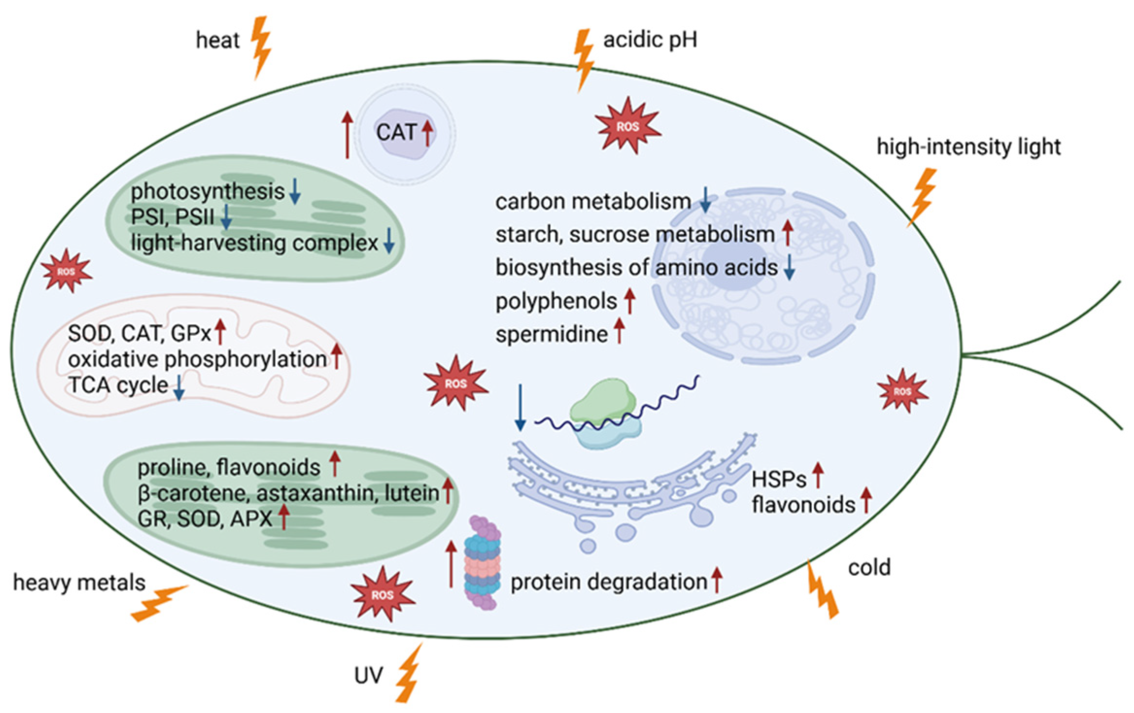
2. Oxidative Stress Mediation in Microalgae

Microalgae, like other organisms, maintain a redox equilibrium between the production and inactivation of ROS [23]. The disruption of this balance and the consequent overaccumulation of ROS lead to oxidative stress and the potential dysbiosis of the population [24].

2.1. Increase in ROS Due to Endogenous Processes

ROS can naturally accumulate in microalgal cells through in vivo processes. As aerobic microorganisms, microalgae rely on oxygen for energy production, which frequently results in ROS generation [25]. A common metabolic process involves the transfer of electrons to molecular oxygen in mitochondria, which unavoidably generates ROS [26]. Additionally, during photosynthesis, microalgae produce molecular oxygen, with several auto-oxidizing enzymes present in the photosystem I (PSI) receptor of the photosynthetic electron transport chain in the chloroplasts [27]. Common cellular ROS produced in microalgal cells include superoxide radicals (O2), singlet oxygen (1O2), hydroxyl radicals (OH•), and hydrogen peroxide (H2O2) [28]. In response to ROS accumulation, the microalgal antioxidant system is activated to maintain the redox balance. However, cell aging and the presence of stressors can overwhelm the scavenging system’s capacity, leading to oxidative stress [16].

2.2. ROS Increase Due to Factors Not Related to Climate Change

ROS can accumulate due to naturally occurring factors that microalgae encounter in their environment. One common challenge in aquatic ecosystems is the interaction with pathogens, including fungi, bacteria, and even viruses. These interactions can induce oxidative stress in microalgal cells. For example, the infection of the green alga Haematococcus pluvialis by the fungus Paraphysoderma sedebokerense has been shown to enhance ROS production [29]. Similarly, the marine diatom Chaetoceros debilis experiences elevated ROS levels and oxidative stress upon infection by the parasitic protist Amoebophrya sp., ultimately leading to cell death [30]. In addition, predation by zooplankton can also lead to stress responses in microalgae. For instance, exposure to metabolites from the grazer Daphnia has been found to increase the ROS levels in Microcystis aeruginosa, a cyanobacterium, leading to oxidative stress [31]. Another significant factor contributing to ROS accumulation is mechanical stress, caused by turbulence or agitation in aquatic environments. Mechanical agitation and hydrodynamic forces in aerated cultures of the red alga Porphyridium cruentum led to increased cell mortality, due to oxidative damage induced by the mechanical stress [32].

2.3. ROS Increase Due to Environmental Factors Linked to Climate Change

ROS accumulation can also be triggered by changes in factors of the microalgal microenvironment. Many of these factors are associated with the ongoing climate change, which exacerbates the oxidative stress that microalgae experience.
The temperature is a critical environmental factor that significantly influences ROS accumulation in microalgal cells, leading to detrimental effects on multiple cellular components and processes [20,33]. High temperatures can induce oxidative stress and increase the ROS levels. For example, various heat stress conditions tested in Auxenochlorella protothecoides UTEX 2341 (Chlorophyta) resulted in a drastic rise in ROS content [34]. Similarly, heat treatment (42 °C for 2 h) induced oxidative stress in another Chlorophyta, the microalga Dunaliella bardawil [35]. Temperature increases in the Arctic and Antarctic regions are particularly concerning, as these areas exhibit rich biodiversity regarding eukaryotic microalgae [36]. Many of these species are obligate cold extremophiles (psychrophiles) and cannot survive higher temperatures [37]. Although green microalgae have been shown to induce the production of heat shock proteins (HSPs) under elevated temperatures, helping to protect the cells from stress by refolding damaged proteins and preventing protein aggregation [38], prolonged elevated temperatures can damage the photosynthetic and respiratory apparatus and induce ROS accumulation, leading to protein misfolding and denaturation [39].
The light quality and quantity have been repeatedly reported to be positively correlated with ROS production, particularly O2 and H2O2 [20]. For instance, the diatom Thalassiosira weissflogii exhibited the increased accumulation of superoxide and hydrogen peroxide in response to an elevated light intensity [40]. Climate change is also altering the percentage of ultraviolet radiation (UVR) reaching the Earth’s surface [41]. Increased UVR exposure can enhance ROS production, leading to cellular damage [42], such as the malfunctioning of photosystem II (PSII) [43].
Metals are essential components of all aquatic systems, but anthropogenic contamination can drastically increase their concentrations, often resulting in pH levels below the optimal value. An acidic pH can induce ROS-producing reactions intracellularly, such as the Fenton and Haber–Weiss reactions [44]. The Haber–Weiss reaction produces HO∙ and is primarily catalyzed by iron or other metals via the Fenton reaction [45]. The Fenton reaction involves the enhanced oxidative potential of H2O2 in the presence of iron or other metals in a low oxidation state, serving as a catalyst under acidic conditions [46]. Some microalgae have the capacity to acclimate to chronic acidic stress, whereas acute stress supersedes their antioxidant mechanisms [44]. However, different microalgal species exhibit varying toxic thresholds for acidic stress and have distinct antioxidant responses [47].
Anthropogenic activities such as mining, the excessive use of fertilizers in agriculture, and the inadequate management of industrial waste contribute to the contamination of aquatic ecosystems with heavy metals [48]. Elevated concentrations of heavy metals can lead to the overproduction of H2O2, resulting in oxidative stress, as reported, for example, in several Chlorophyta. Scenedesmus acuminatus and Chlorella sorokiniana responded to Cu(II) exposure by accumulating H2O2 [49]. Similarly, Scenedesmus obliquus responded to Pb(II) ions with elevated H2O2 levels [50], and Chlorella vulgaris showed increased H2O2 production when exposed to higher Cd(II) concentrations [51].
Nutrient deprivation in microalgae, especially for the macronutrients N, S, and P, can trigger a range of cellular responses, including ROS accumulation [52,53]. This response is often accompanied by lipid accumulation, a characteristic that is especially desirable for the production of commercial bioproducts [54]. Consequently, nutrient stress has been extensively studied in several microalgal species, including the Chlorophyta Dunaliella sp., Haemotococcus sp., and Scenedesmus sp. [52].

3. Oxidative Stress Response

In order to adapt to varying environmental conditions, microalgae have evolved several antioxidant mechanisms (Table 1). These mechanisms help to mitigate the toxic effects of ROS while simultaneously preserving their role as secondary signaling molecules [55,56]. In general, the antioxidant repertoire of microalgae consists of the enzymatic detoxification system and endogenous non-enzymatic antioxidants.

3.1. Oxidative Regulation via Enzymatic Response

The microalgal enzymatic detoxification system comprises several key enzymes, including catalase (CAT), peroxidase (POD), ascorbate peroxidase (APX), superoxide dismutase (SOD), glutathione-S transferase (GST), and glutathione reductase (GR) [15]. Specifically, the porphyrin heme active sites of catalase degrade H2O2 into water and oxygen [22]. The three known groups of catalases are primarily synthesized in the peroxisome and, to a lesser extent, in the chloroplast, cytosol, and mitochondria [77]. Peroxidases are a class of enzymes that catalyze the oxidation of a substrate with hydrogen peroxide, and two types of peroxidase enzymes are present in microalgae: ascorbate peroxidase and glutathione peroxidase [20]. For the metalloenzyme SOD, which catalyzes the neutralization of superoxide radicals by adding H+ to form H2O2 and O2, three major isozymes are reported in microalgae, differing in their protein folding and metal co-factor requirements [78]. Overall, the microalgal antioxidant enzymes eradicate ROS by converting them into H2O2 and subsequently into H2O, helping to preserve cellular structures and functions [79].
Several studies have demonstrated the connection between antioxidant activity and antioxidant enzymes in different microalgal phyla. In Chlorophyta, the strong antioxidant activity of Chlorococcopsis minuta under nutrient stress is also linked to the activity of SOD and CAT [58]. Oxidative stress induced by currently emerging pharmaceutical contaminants, such as erythromycin and clarithromycin, elevates the activity of antioxidant enzymes (SOD, CAT, GP) in microalgae like Chlorella vulgaris and Raphidocelis subcapitata [59,60,61]. Additionally, in Scenedesmus obliquus cultures under H2O2 stress, the expression levels of genes coding for glutathione peroxidase and ascorbate peroxidase were upregulated [76]. The exposure of C. vulgaris cultures to the pesticide glufosinate induced oxidative stress, leading to increased activity of APX, SOD, and CAT, indicating their role in the microalgal defense mechanism against the stressor [62]. In S. obliquus, exposure to a Pb(II) medium led to increased H2O2 and MDA content, along with a significant increase in the enzymatic activity of SOD, CAT, POD, and GR [50]. Furthermore, short- (48 h) and long-term (168 h) heat stress increased the ROS content in A. protothecoides UTEX 2341, resulting in enhanced SOD activity [34]. For N. gaditana (Ochrophyta) cultivated under blue, green, and red filtered light, the expression levels of genes involved in oxidative stress responses remained unaltered under red and blue filtered light, while several of them, including GPx1, GPx3, SOD2_1, and SOD2_3, exhibited significantly lower expression levels under filtered green light [65].

3.2. Oxidative Regulation Through Antioxidant Compounds

In addition to enzymatic defenses, microalgae also produce a variety of non-enzymatic antioxidants that protect them from cellular damage caused by ROS accumulation. These include carotenoids, flavonoids, α-tocopherols, ascorbic acids, reduced glutathione (GSH), proline, hydroquinones, polyamines, and phycocyanins [20]. While many of these compounds are common across microalgal species, some species-specific antioxidant metabolites are produced in response to particular stress conditions [22,80]. Despite the potential negative effects of oxidative damage, abiotic stresses can be utilized for metabolic manipulation and the accumulation of antioxidant compounds in microalgal biomass [14].
Carotenoids are well known antioxidant molecules commonly found in microalgal biomass [81]. Primary carotenoids are naturally produced in microalgal cells, while secondary carotenoids are synthesized and accumulated under stress conditions, such as nutrient deprivation and salinity stress [82]. The stored carotenoids can be utilized by microalgal cells as energy and carbon sources to reactivate their metabolism under less stressful conditions [83]. Simultaneously, carotenoids offer significant protection against oxidative stress during unfavorable conditions. They assist in photoprotection, membrane regulation, and the stabilization of light-harvesting complexes, contributing ~20–30% of the absorbed light energy and broadening the photosystem’s absorption spectrum. Carotenoids can dissipate excess light as heat and provide chemical protection through free radical capture. Additionally, they can stabilize membranes and facilitate electron transfer between molecules like cytochromes and chlorophylls [84]. Specifically, they are highly effective in interacting with singlet oxygen (1O2), transferring its energy to produce ground-state oxygen and a triplet-excited carotenoid. The carotenoid then returns to its ground state by dissipating this energy through interactions with the surrounding solvent. Carotenoids can engage with radical species, particularly 1O2, through three mechanisms: radical addition, the formation of radical cations (via electron transfer), and hydrogen atom abstraction. The effectiveness of their activity depends on several factors, including their structural properties, the concentrations of radicals, and the characteristics of the reaction environment [84,85].
Secondary carotenoids have been reported in several microalgal species, including the green algae Chlorella sorokiniana and Senedesmus sp. and the red alga Porphyridium aerugineum, to accumulate under stress and enhance antioxidant activity [86]. The contribution of carotenoids to microalgal antioxidant activity was indicated through FRAP, TEAC, and AIOLA assays [87]. Tetraselmis tetrathele responded to salinity stress by inducing the biosynthesis of carotenoids [69]. Additionally, the exposure of C. sorokiniana to the fourth-generation fluoroquinolone antibiotic moxifloxacin led to increased levels of chlorophyll a and b and carotenoids [63]. Known secondary carotenoids include astaxanthin, β-carotene, and lutein, which are produced by microalgae when exposed to unfavorable environmental conditions [81,88]. Astaxanthin exhibits potent antioxidant properties due to its readily oxidizable hydroxyl and ketone groups [89]. Its amphiphilic structure facilitates transmembrane passage without compromising membrane integrity, enabling antioxidant activity in both intracellular and extracellular environments [90]. β-Carotene functions as an accessory light-harvesting pigment, protecting the photosynthetic apparatus from photodamage. It shields algae from excessive irradiance by preventing reactive oxygen species formation, quenching triplet-state chlorophyll, and reacting with singlet oxygen [91]. Astaxanthin is primarily produced and accumulated during the red stage of Haematococcus pluvialis cells, constituting 80–99% of the total cellular carotenoids [92], while β-carotene accumulates in Dunaliella salina within lipid droplets inside the chloroplast [81]. Lutein, which is located in the light-harvesting complex, quenches harmful oxidative species and excited chlorophyll molecules [93]. Under conditions of high light intensity, lutein absorbs the excess energy and safely releases it as heat [66]. The exposure of Chlamydomonas reinhardtii and C. vulgaris cultures to high-intensity light stress resulted in the increased synthesis of lutein [67,68].
Another group of microalgal non-enzymatic antioxidants is phenolic compounds. It has been proven that several microalgal species accumulate phenolic compounds under harsh environmental conditions, enhancing their antioxidant capacity [86,87]. For example, Pb-induced oxidative stress in S. obliquus led to an increase in polyphenol content [50]. Polyphenols function as antioxidants by reducing free radicals, with the hydroxyl groups that they contain, converting them into stable molecules [94]. Flavonoids are also important microalgal antioxidants, while certain amino acids, such as proline, also possess antioxidant properties [50,55]. Short-term cold stress resulted in ROS accumulation and increased proline levels in A. protothecoides UTEX 2341 [34]. Proline is well known for its antioxidant properties, and several protective mechanisms have been proposed. These include stabilizing proteins and antioxidant enzymes, directly scavenging ROS, maintaining intracellular redox homeostasis by inducing GSH accumulation, and modulating cellular signaling [95]. Glutathione (GSH), is a low-molecular-weight thiol tripeptide, present in nearly all cellular compartments, playing a crucial role in various cellular processes, including stress-responsive gene expression [96]. Its high reductive potential, driven by a nucleophilic cysteine residue, enables it to scavenge ROS such as H2O2 and singlet oxygen, protecting biomolecules through glutathiolation or reduction. GSH also contributes to heavy metal detoxification, maintaining cellular redox homeostasis [97]. Furthermore, the rise in intracellular ROS levels due to abiotic stress can induce lipid biosynthesis [98]. For instance, a small increase in the H2O2 levels in H. pluvialis, due to exposure to salinity stress, led to lipid accumulation of 18–24% [70]. In addition, the polyamine spermidine showed a protective effect on Chlorella sp. cultures subjected to SO2- and CO2-induced oxidative stress. The growth, PSII structure, chlorophyll synthesis, lipid content, and, consequently, the biomass yield were all recovered in the presence of exogenous spermidine [71]. Spermidine exhibits antioxidant activity through two mechanisms. First, its chemical structure, consisting of a carbon chain with two amino terminal groups, allows it to neutralize the acidifying effects of SO2 and act as a pH buffer. Second, like other polyamines, spermidine promotes and enhances the synthesis and activity of antioxidant enzymes, such as SOD, thereby strengthening the cell’s overall antioxidant capacity [71].

3.3. Genomic Capacity and Cellular Functions Dictate Metabolic Regulation

The insights into the oxidative stress responses in microalgae are confirmed and greatly enhanced by experimental procedures using multi-omics technologies and correlating significant metabolic, molecular, and catalomic profile shifts. An example is the study of Liang et al., which focused on Dunaliella bardawil, a microalga that is widely used in the industrial production of β-carotene. Using RNA-seq analysis, they examined the effects of exposing D. bardawil to heat stress (42 °C for 2 h), finding the upregulation of multiple genes encoding antioxidant enzymes against ROS [35]. This increase in gene expression is crucial, as genes ultimately regulate the pathways that influence enzyme production and therefore activity. Another study explored the response of C. reinhardtii to oxidative stress induced by the photosensitizer Rose Bengal, with a particular focus on the role of GPX5. The researchers used the parental strain CC4348 and a glutathione peroxidase knockout mutant (gpx5) to compare their transcriptomic responses. Upon exposure to the stressor for 30 min, the CC4348 strain showed 708 DEGs, with 322 upregulated and 386 downregulated. Notably, the gpx5 mutant strain exhibited a more intense response, with 1769 DEGs, including 744 upregulated and 1025 downregulated genes. The intense transcriptomic response in the gpx5 mutant emphasizes the crucial role of GPX5 in mitigating oxidative stress, as its mutation led to increased stress levels, the activation of the TCA cycle, and enhanced mitochondrial electron transport in C. reinhardtii [64].
Simultaneously, omics technologies have provided more detailed information regarding the molecular and biochemical adaptation mechanisms of microalgae under oxidative stress, as well as the subcellular organelles participating in these mechanisms. To this end, a general overview of the adaptation mechanisms can be obtained via the enrichment analysis of all data generated by omics techniques. For instance, the exposure of S. obliquus to 8 mg L−1 of H2O2 allowed for the evaluation of changes in proteomic features, leading to the successful identification of 251 differentially expressed proteins (DEPs) [76]. According to a GO annotation and enrichment analysis, the predominant biological processes identified were oxidation–reduction and organonitrogen compound biosynthesis. Among cellular components, the most prevalent were distributed in the chloroplast and plastid parts. In the molecular function group, proteins were involved mostly in oxidoreductase activity, followed by electron carrier activity and chlorophyll binding. According to the KEGG pathway enrichment analysis, five DEPs were involved in pentose and glucoronate interconversion, and another five DEPs were involved in ascorbate and aldarate metabolism, as well as amino sugar and nucleotide sugar metabolism. Proteins involved in galactose metabolism and the biosynthesis of secondary metabolites were also identified as differently expressed [76].
By studying in depth the data derived from omics techniques for specific genes, proteins, or metabolites, it is possible to elucidate the effects of oxidative stress on specific metabolic pathways, although the mechanisms underlying some induced metabolic pathways remain unclear. Several studies have highlighted alterations in carbon metabolism and energy flow as a consequence of oxidative stress. In D. bardawil, adaptation to short-term heat stress involved a shift from aerobic to glycolytic metabolism for energy production [35]. D. salina responded to high cadmium stress by reducing, among other processes, the activity of the TCA cycle, while simultaneously increasing oxidative–phosphorylation gene expression [75]. The exposure of C. reinhardtii to H2O2 led to the identification of several transcripts with decreased abundance, encoding proteins involved in central carbon metabolism [73]. The short-term exposure of Tetraselmis chuii to H2O2 negatively affected the cellular functions of carbon and the energy flow, as the genes encoding for the key factors of glycolysis/gluconeogenesis pyruvate kinase and enolase were both found to be 1.5-fold downregulated, and the urea content was reduced threefold. The downregulation of carbon metabolism was also depicted in the reduction of carbon fixation, as indicated by the reduced expression levels of several transcripts, including a 1.8-fold decrease observed for the gene for ribose 5-phosphate isomerase A (RPIA) [72].
The carbon stream is also affected by the photosynthetic capacity of microalga; this is another crucial cellular process that has been shown to be severely affected by oxidative stress. A common method of assessing the photosynthetic capacity is measuring the maximal photosystem II quantum yield (Fv/Fm). This is determined using pulse-amplitude-modulated (PAM) fluorometry after dark adaptation, where Fv/Fm = (Fm − Fo)/Fm, with Fo and Fm representing the minimal and maximal fluorescence, respectively. In healthy microalgae, the Fv/Fm typically ranges from 0.65 to 0.75, while values below 0.6 indicate stress, and severe damage can reduce it to 0.4 or lower [99]. For instance, when the green microalgae T. chuii and S. obliquus were exposed to H2O2-induced oxidative stress, there was a significant reduction in the Fv/Fm ratio and in the levels of the photosynthetic pigments chlorophyl a and b. This indicates decreased efficiency in light energy conversion at the PSII reaction center [72,76]. In the proteomic analysis of S. obliquus after exposure to H2O2, photosynthesis and photosynthetic antenna proteins were heavily affected. Specifically, cytochrome a-b binding protein, the photosystem I iron–sulfur center (psaC), photosystem II reaction center protein H (psbH), cytochrome b559 subunit alpha (psbE), the cytochrome b6-f complex subunit, and ATP synthase subunit beta (atpB) were significantly downregulated, leading to the diminished photosynthetic capacity of the microalga [76]. Similar observations, regarding both the expression levels of key photosynthetic transcripts and the photosynthetic output, were reported for C. reinhardtii [73,74] and T. chuii [72]. Notably, the short-term exposure of T. chuii to H2O2 led to a strong decrease in photosynthesis, as four genes coding for subunits of PSI and five genes coding for subunits of PSII were downregulated, including the 2.4-fold downregulation of psbQ. Additionally, the expression of several transcripts coding for the light-harvesting complexes was downregulated. The reported downregulation of transcripts associated with photosystems under oxidative stress could be considered as a strategic response to minimize ROS production, conserve energy, prevent photodamage, and promote cellular recovery, leading to enhanced cellular survival [26]. In contrast, the prolonged exposure of T. chuii to H2O2 led to an unsuccessful attempt at carbon metabolism enhancement, as depicted, for example, by the upregulation of petC and petJ by more than twofold [72]. It is possible that some microalgal species respond to prolonged oxidative stress by attempting to shift their energy metabolism towards photosynthesis and glycolysis as a survival mechanism.
Several other pathways are also referred to in related studies as affected by exposure to oxidative stress. The exposure of D. bardawil to heat stress led to the enrichment of the ascorbate–glutathione cycle, the upregulation of genes responsible for chloroplast membranes, and changes in lipid characteristics like the carbon chain length and unsaturation degree [35]. Among the adaptations of T. chuii to H2O2-induced oxidative stress was the upregulation of transcripts related to peroxisome function, endocytosis, starch and sucrose metabolism, the biosynthesis of secondary metabolites, and galactose metabolism. On the other hand, downregulation was observed in the expression levels of genes involved in fatty acid metabolism, such as the genes coding for malonyl CoA-acyl carrier protein transacylase (FABD), β-ketoacylacyl carrier protein synthase III (FABH), and β-ketoacyl-acyl carrier protein (ACP) synthase II (FABF). This downregulation could be an attempt to prevent lipid peroxidation, conserve energy, and reduce ROS production. The biosynthesis of amino acids was also severely affected, as 22 downregulated DEGs were identified. Negative impacts were also observed on several genes coding for ribosome structural proteins and on purine and porphyrin metabolism [72].
Another common observation when studying the responses of microalgae to oxidative stress is the upregulation of genes encoding proteins involved in protein degradation, perhaps for carbon scavenging due to decreased photosynthetic activity. T. chuii responded to exposure to H2O2 by inducing the expression of transcripts responsible for protein targeting in the ER and transcripts coding for the ubiquitin ligase complex and the subsequent ER-associated degradation towards the proteasome [72]. Similar observations have been reported for C. reinhardtii [73]. In contrast to increased protein degradation, some genes encoding specific proteins are overexpressed as a response to oxidative stress, like the molecular chaperones that protect other proteins from damage caused by stress, known as heat shock proteins [100]. For several microalgal species, heat sock proteins are identified as participating in oxidative stress regulation. Heat shock protein 97 (hsp97) was upregulated in A. protothecoides in response to both heat and cold stresses. Moreover, short- and long-term cold stress led to increased expression levels of the heat shock transcriptional factor HSFA1d [34]. Blaby et al. (2015) studied the response of C. reinhardtii to H2O2-induced oxidative stress and identified the increased abundance of 38 transcripts encoding either known or putative molecular chaperones and chaperonins, like HSP20, HSP90, HSP70, and DnaJ proteins [73]. Similar observations are noted for D. bardawil and T. chuii [35,72].
In addition to the mechanisms of oxidative stress regulation in microalgae already discussed, ongoing research continues to uncover new key factors. Recently, selenium-binding protein (SBP) has been identified as a novel stress regulator involved in modulating C. reinhardtii’s early responses to oxidative stress [74]. The researchers compared a wild-type strain of C. reinhardtii with a selenium-binding protein knockout mutant (sbd1) to analyze their transcriptomic responses after exposure to H2O2-induced oxidative stress. The wild-type strain showed 622 DEGs, with 346 upregulated and 276 downregulated. In contrast, the sbd1 mutant strain displayed a minimal response, with only five DEGs identified. Data derived from a metabolomic analysis further highlighted that the sbd1 mutant exhibited the dramatic quenching of the molecular and biochemical responses upon H2O2-induced oxidative stress, as compared to the wild type. Yeast two-hybrid assays indicated that the selenium-binding protein is part of an extensive and conserved protein–protein interaction network involving the fructose-bisphosphate aldolase 3 (FBA3), cysteine endopeptidase 2 (CEP2), and glutaredoxin 6 (GRX6) proteins.

3.4. Applications of Antioxidant Properties

Knowledge of the oxidative stress responses and antioxidant properties in microalgae has applications across various fields. From an ecological perspective, understanding the effects of factors such as rising temperatures can help to predict shifts in microalgal populations under climate change and may provide opportunities to prevent ecosystem collapse [101]. Additionally, species with high antioxidant potential can be used for ecosystem restoration and bioremediation, including water quality improvement [102].
Beyond ecology, knowledge of microalgal antioxidant properties has significant biotechnological applications. For instance, microalgal-derived antioxidants can serve as natural preservatives to extend the shelf lives of food products and enhance food’s quality [103]. These antioxidants also have potential in the pharmaceutical and nutraceutical sectors [104,105], as well as in cosmetics, where recent advancements in exploiting microalgal antioxidant mechanisms could further improve product formulations [106]. Furthermore, antioxidant-rich microalgae can be incorporated into human and animal feed, either directly or as supplements [107]. Lastly, controlled exposure to stressors can be used to trigger lipid accumulation, improving the economic feasibility of microalgal biofuels [108].

4. Conclusions and Future Perspectives

Microalgae, which play significant ecological and bioeconomic roles, are highly susceptible to oxidative stress caused by various abiotic factors in their microenvironment. This presents a major challenge, as the effects of ongoing climate change on global microalgal populations remain uncertain. Conversely, oxidative stress can be harnessed as a tool for the metabolic manipulation of microalgal cultures to induce desired products. Therefore, the study of the mediation and regulation of oxidative stress in microalgae is of great interest. To the best of our knowledge, this review is the first to examine multi-omics data on microalgal responses to oxidative stress, while it also highlights the effects of climate-change-related stressors on microalgae. Additionally, it provides an updated and comprehensive overview of the topic, consolidating recent advancements in understanding oxidative stress in microalgae.
Oxidative stress in microalgae is induced by the accumulation of ROS, which can result from normal cellular functions or suboptimal environmental conditions. These conditions are often associated with climate change and include fluctuations in the temperature, light quality and quantity, and acidic pH levels. Microalgae possess an antioxidant repertoire that includes enzymatic antioxidants like catalase and non-enzymatic antioxidants such as secondary carotenoids (Figure 1).
Recently, the application of omics techniques has generated a wealth of data regarding the regulation of oxidative stress by microalgae. For example, it is well established that oxidative stress negatively affects carbon metabolism and photosynthesis. In recent years, we have learned that many interconnected and overlapping antioxidant mechanisms exist in microalgae, and several metabolic pathways are affected by oxidative stress. However, the antioxidant mechanisms triggered in each case are stress-specific, as the microalga’s response depends on the stressor’s nature, intensity, and duration. For example, T. chuii exhibited different antioxidant adaptations when exposed to short-term and long-term H2O2-induced oxidative stress [72]. Additionally, several microalgal species exhibit unique, species-specific adaptation mechanisms, such as the overaccumulation of specific metabolites. Recent studies have also identified new key stress regulators, such as SBP.
Despite the increasing scientific interest in this topic, many aspects of the mediation and regulation of oxidative stress in microalgae remain unknown. An in-depth study of the responses of specific species to particular stressors is necessary to elucidate their antioxidant adaptations. It is important to underline that microalgae represent a highly diverse group of microorganisms with vastly different characteristics [1]. Depending on their genetic background and the specific microenvironment that they inhabit, each species has developed unique adaptations and metabolic pathways, leading to varied tolerance levels and responses to oxidative stress [55]. Therefore, it is crucial to study and understand each species individually, rather than generalizing about all microalgae, as doing so may overlook their great biotechnological potential. Species of significant ecological interest and those widely cultivated in industry should be prioritized for such research.

Author Contributions

E.F. and A.R. conceived this review. A.K. and D.S. collected and analyzed the literature and drafted the manuscript. A.K., D.S., I.D., A.R. and E.F. revised the manuscript. All authors have read and agreed to the published version of the manuscript.

Funding

This research received no external funding.

Institutional Review Board Statement

Not applicable.

Informed Consent Statement

Not applicable.

Data Availability Statement

The original contributions presented in this study are included in the article. Further inquiries can be directed to the corresponding author(s).

Conflicts of Interest

The authors declare no conflicts of interest.

Abbreviations

The following abbreviations are used in this manuscript:
Glossary
Acute stressa short-term state of stress
Aerobic microorganismsmicroorganisms that utilize oxygen in their metabolic processes
Anthropogenic contaminationpollution or environmental damage caused by human activity
Chronic stressa prolonged state of stress
Metabolic manipulationthe process of altering or controlling the metabolism of organisms or cells to achieve a specific goal, such as enhancing energy production or synthesizing a particular compound
Multi-omic technologiesapproaches that integrate data from various ‘omics’ fields
Nutrient deprivationthe condition where an organism lacks essential nutrients, leading to stress responses or altered metabolic activity
Obligate cold extremophiles (Psychrophiles)organisms that thrive in extremely cold environments, and have evolved specialized mechanisms to survive in these conditions
Pharmaceutical contaminantschemical substances, often derived from pharmaceutical drugs, that enter the environment and can impact ecosystems
Redox equilibriumthe balance between oxidation and reduction reactions in biological or chemical systems
Abbreviations
APXascorbate peroxidase
ATPBATP synthase subunit beta
CATcatalase
Cdcadmium
CEP2cysteine endopeptidase 2
CO2carbon dioxide
FABDmalonyl CoA-acyl carrier protein transacylase
FABFβ-ketoacyl-acyl carrier protein (ACP) synthase II
FABHβ-ketoacylacyl carrier protein synthase III
FBA3Fructose-bisphosphate aldolase 3
GPxglutathione peroxidase
GRglutathione reductase
GRX6glutaredoxin 6
GSHreduced glutathione
GSTglutathione-S transferase
H2O2hydrogen peroxide
HSPsheat shock proteins
HSP97heat shock protein 97
Nnitrogen
OH•hydroxyl radicals
O2superoxide radicals
Pphosphorus
PAMpulse-amplitude modulated
Pblead
PEX1peroxisomal biogenesis factor 1
PODperoxidase
PSIphotosystem I
PSIIphotosystem II
psaCphotosystem I iron-sulfur center
psbEcytochrome b559 subunit alpha
psbHphotosystem II reaction center protein H
RPIAribose 5-phosphate isomerase A
ROSReactive Oxygen Species
SO2sulfur dioxide
SODsuperoxide dismutase
TCA cycletricarboxylic acid cycle
UTEXthe University of Texas at Austin Culture Collection of Algae
UVRultraviolet radiation
1O2singlet oxygen

References

  1. Singh, J.; Saxena, R.C. An Introduction to Microalgae. In Handbook of Marine Microalgae; Elsevier: Amsterdam, The Netherlands, 2015; pp. 11–24. [Google Scholar] [CrossRef]
  2. Yang, X.; Liu, L.; Yin, Z.; Wang, X.; Wang, S.; Ye, Z. Quantifying photosynthetic performance of phytoplankton based on photosynthesis–irradiance response models. Environ. Sci. Eur. 2020, 32, 24. [Google Scholar] [CrossRef]
  3. Khan, A.K.; Kausar, H.; Jaferi, S.S.; Drouet, S.; Hano, C.; Abbasi, B.H.; Anjum, S. An Insight into the Algal Evolution and Genomics. Biomolecules 2020, 10, 1524. [Google Scholar] [CrossRef] [PubMed]
  4. Zhang, Z.; An, M.; Miao, J.; Gu, Z.; Liu, C.; Zhong, B. The Antarctic sea ice alga Chlamydomonas sp. ICE-L provides insights into adaptive patterns of chloroplast evolution. BMC Plant Biol. 2018, 18, 53. [Google Scholar] [CrossRef]
  5. Ścieszka, S.; Klewicka, E. Algae in food: A general review. Crit. Rev. Food Sci. Nutr. 2019, 59, 3538–3547. [Google Scholar] [CrossRef] [PubMed]
  6. Bhalamurugan, G.L.; Valerie, O.; Mark, L. Valuable bioproducts obtained from microalgal biomass and their commercial applications: A review. Environ. Eng. Res. 2018, 23, 229–241. [Google Scholar] [CrossRef]
  7. Levasseur, W.; Perré, P.; Pozzobon, V. A review of high value-added molecules production by microalgae in light of the classification. Biotechnol. Adv. 2020, 41, 107545. [Google Scholar] [CrossRef]
  8. Song, Q.; Kong, F.; Liu, B.-F.; Song, X.; Ren, N.-Q.; Ren, H.-Y. Ozone oxidation of actual waste leachate coupled with culture of microalgae for efficient lipid production under different temperatures. Water Res. 2025, 277, 123305. [Google Scholar] [CrossRef]
  9. Behera, B.; Selvam, S.M.; Paramasivan, B. Research trends and market opportunities of microalgal biorefinery technologies from circular bioeconomy perspectives. Bioresour. Technol. 2022, 351, 127038. [Google Scholar] [CrossRef]
  10. Mittler, R. Abiotic stress, the field environment and stress combination. Trends Plant Sci. 2006, 11, 15–19. [Google Scholar] [CrossRef]
  11. Almeida, A.C.; Gomes, T.; Langford, K.; Thomas, K.V.; Tollefsen, K.E. Oxidative stress in the algae Chlamydomonas reinhardtii exposed to biocides. Aquat. Toxicol. 2017, 189, 50–59. [Google Scholar] [CrossRef]
  12. Darehshouri, A.; Affenzeller, M.; Lütz-Meindl, U. Cell death upon H2O2 induction in the unicellular green alga Micrasterias. Plant Biol. 2008, 10, 732–745. [Google Scholar] [CrossRef]
  13. Kholssi, R.; Lougraimzi, H.; Moreno-Garrido, I. Effects of global environmental change on microalgal photosynthesis, growth and their distribution. Mar. Environ. Res. 2023, 184, 105877. [Google Scholar] [CrossRef] [PubMed]
  14. Paliwal, C.; Mitra, M.; Bhayani, K.; Bharadwaj, S.V.V.; Ghosh, T.; Dubey, S.; Mishra, S. Abiotic stresses as tools for metabolites in microalgae. Bioresour. Technol. 2017, 244, 1216–1226. [Google Scholar] [CrossRef]
  15. Lu, Q.; Li, H.; Zou, Y.; Liu, H.; Yang, L. Astaxanthin as a microalgal metabolite for aquaculture: A review on the synthetic mechanisms, production techniques, and practical application. Algal Res. 2021, 54, 102178. [Google Scholar] [CrossRef]
  16. Zhu, J.; Cai, Y.; Wakisaka, M.; Yang, Z.; Yin, Y.; Fang, W.; Xu, Y.; Omura, T.; Yu, R.; Zheng, A.L.T. Mitigation of oxidative stress damage caused by abiotic stress to improve biomass yield of microalgae: A review. Sci. Total Environ. 2023, 896, 165200. [Google Scholar] [CrossRef]
  17. Gauthier, M.R.; Senhorinho, G.N.A.; Scott, J.A. Microalgae under environmental stress as a source of antioxidants. Algal Res. 2020, 52, 102104. [Google Scholar] [CrossRef]
  18. Wang, L.; Yang, T.; Pan, Y.; Shi, L.; Jin, Y.; Huang, X. The Metabolism of Reactive Oxygen Species and Their Effects on Lipid Biosynthesis of Microalgae. Int. J. Mol. Sci. 2023, 24, 11041. [Google Scholar] [CrossRef]
  19. Danouche, M.; El Ghatchouli, N.; Arroussi, H. Overview of the management of heavy metals toxicity by microalgae. J. Appl. Phycol. 2022, 34, 475–488. [Google Scholar] [CrossRef]
  20. Ugya, A.Y.; Imam, T.S.; Li, A.; Ma, J.; Hua, X. Antioxidant response mechanism of freshwater microalgae species to reactive oxygen species production: A mini review. Chem. Ecol. 2020, 36, 174–193. [Google Scholar] [CrossRef]
  21. Pikula, K.S.; Zakharenko, A.M.; Aruoja, V.; Golokhvast, K.S.; Tsatsakis, A.M. Oxidative stress and its biomarkers in microalgal ecotoxicology. Curr. Opin. Toxicol. 2019, 13, 8–15. [Google Scholar] [CrossRef]
  22. Cirulis, J.T.; Scott, J.A.; Ross, G.M. Management of oxidative stress by microalgae. Can. J. Physiol. Pharmacol. 2013, 91, 15–21. [Google Scholar] [CrossRef]
  23. Nikkanen, L.; Solymosi, D.; Jokel, M.; Allahverdiyeva, Y. Regulatory electron transport pathways of photosynthesis in cyanobacteria and microalgae: Recent advances and biotechnological prospects. Physiol. Plant. 2021, 173, 514–525. [Google Scholar] [CrossRef] [PubMed]
  24. Das, K.; Roychoudhury, A. Reactive oxygen species (ROS) and response of antioxidants as ROS-scavengers during environmental stress in plants. Front. Environ. Sci. 2014, 2, 53. [Google Scholar] [CrossRef]
  25. Chokshi, K.; Pancha, I.; Ghosh, A.; Mishra, S. Oxidative Stress-Induced Bioprospecting of Microalgae. In Systems Biology of Marine Ecosystems; Kumar, M., Ralph, P., Eds.; Springer International Publishing: Cham, Switzerland, 2017; pp. 251–276. [Google Scholar] [CrossRef]
  26. Khorobrykh, S.; Havurinne, V.; Mattila, H.; Tyystjärvi, E. Oxygen and ROS in Photosynthesis. Plants 2020, 9, 91. [Google Scholar] [CrossRef] [PubMed]
  27. Wang, W.; Li, D.; Cao, X.; Xue, S.; Li, C. Liberating photoinhibition through nongenetic drainage of electrons from photosynthesis. Nat. Sci. 2021, 1, e20210038. [Google Scholar] [CrossRef]
  28. Milkovic, L.; Gasparovic, A.C.; Cindric, M.; Mouthuy, P.-A.; Zarkovic, N. Short Overview of ROS as Cell Function Regulators and Their Implications in Therapy Concepts. Cells 2019, 8, 793. [Google Scholar] [CrossRef]
  29. Yan, H.; Ma, H.; Li, Y.; Zhao, L.; Lin, J.; Jia, Q.; Hu, Q.; Han, D. Oxidative stress facilitates infection of the unicellular alga Haematococcus pluvialis by the fungus Paraphysoderma sedebokerense. Biotechnol. Biofuels 2022, 15, 56. [Google Scholar] [CrossRef]
  30. Egerton, T.A. Investigations of Phytoplankton Diversity in Chesapeake Bay. Ph.D. Thesis, Old Dominion University, Norfolk, VA, USA, 2013. [Google Scholar]
  31. Rzymski, P.; Klimaszyk, P.; Jurczak, T.; Poniedziałek, B. Oxidative Stress, Programmed Cell Death and Microcystin Release in Microcystis aeruginosa in Response to Daphnia Grazers. Front. Microbiol. 2020, 11, 1201. [Google Scholar] [CrossRef]
  32. Camacho, F.G.; Gómez, A.C.; Sobczuk, T.M.; Grima, E.M. Effects of mechanical and hydrodynamic stress in agitated, sparged cultures of Porphyridium cruentum. Process Biochem. 2000, 35, 1045–1050. [Google Scholar] [CrossRef]
  33. Barati, B.; Gan, S.-Y.; Lim, P.-E.; Beardall, J.; Phang, S.-M. Green algal molecular responses to temperature stress. Acta Physiol. Plant 2019, 41, 26. [Google Scholar] [CrossRef]
  34. Xing, C.; Li, J.; Yuan, H.; Yang, J. Physiological and transcription level responses of microalgae Auxenochlorella protothecoides to cold and heat induced oxidative stress. Environ. Res. 2022, 211, 113023. [Google Scholar] [CrossRef] [PubMed]
  35. Liang, M.-H.; Jiang, J.-G.; Wang, L.; Zhu, J. Transcriptomic insights into the heat stress response of Dunaliella bardawil. Enzym. Microb. Technol. 2020, 132, 109436. [Google Scholar] [CrossRef] [PubMed]
  36. Cvetkovska, M.; Hüner, N.P.A.; Smith, D.R. Chilling out: The evolution and diversification of psychrophilic algae with a focus on Chlamydomonadales. Polar Biol. 2017, 40, 1169–1184. [Google Scholar] [CrossRef]
  37. Morita, R.Y. Psychrophilic bacteria. Bacteriol. Rev. 1975, 39, 144–167. [Google Scholar] [CrossRef] [PubMed]
  38. Sathasivam, R.; Ki, J.-S. Heat shock protein genes in the green alga Tetraselmis suecica and their role against redox and non-redox active metals. Eur. J. Protistol. 2019, 69, 37–51. [Google Scholar] [CrossRef]
  39. Schroda, M.; Hemme, D.; Mühlhaus, T. The Chlamydomonas heat stress response. Plant J. 2015, 82, 466–480. [Google Scholar] [CrossRef]
  40. Schneider, R.J.; Roe, K.L.; Hansel, C.M.; Voelker, B.M. Species-Level Variability in Extracellular Production Rates of Reactive Oxygen Species by Diatoms. Front. Chem. 2016, 4, 5. [Google Scholar] [CrossRef]
  41. Williamson, C.E.; Zepp, R.G.; Lucas, R.M.; Madronich, S.; Austin, A.T.; Ballare, C.L.; Norval, M.; Sulzberger, B.; Bais, A.F.; McKenzie, R.L.; et al. Solar ultraviolet radiation in a changing climate. Nature Clim. Change 2014, 4, 434–441. [Google Scholar] [CrossRef]
  42. Zhang, Y.; Jiang, H.; Qiu, B. Effects of UVB Radiation on competition between the bloom-forming cyanobacterium Microcystis aeruginosa and the Chlorophyceae Chlamydomonas microsphaera. J. Phycol. 2013, 49, 318–328. [Google Scholar] [CrossRef]
  43. Bouchard, J.N.; Roy, S.; Campbell, D.A. UVB Effects on the Photosystem II-D1 Protein of Phytoplankton and Natural Phytoplankton Communities. Photochem. Photobiol. 2006, 82, 936–951. [Google Scholar] [CrossRef]
  44. Pinto, E.; Sigaud-kutner, T.C.S.; Leitão, M.A.S.; Okamoto, O.K.; Morse, D.; Colepicolo, P. Heavy Metal–Induced Oxidative Stress in Algae. J. Phycol. 2003, 39, 1008–1018. [Google Scholar] [CrossRef]
  45. Kehrer, J.P. The Haber–Weiss reaction and mechanisms of toxicity. Toxicology 2000, 149, 43–50. [Google Scholar] [CrossRef]
  46. Ameta, R.; Chohadia, A.K.; Jain, A.; Punjabi, P.B. Fenton and Photo-Fenton Processes. In Advanced Oxidation Processes for Waste Water Treatment; Elsevier: Amsterdam, The Netherlands, 2018; pp. 49–87. [Google Scholar] [CrossRef]
  47. Bwapwa, J.K.; Jaiyeola, A.T.; Chetty, R. Bioremediation of acid mine drainage using algae strains: A review. S. Afr. J. Chem. Eng. 2017, 24, 62–70. [Google Scholar] [CrossRef]
  48. Ali, H.; Khan, E.; Ilahi, I. Environmental Chemistry and Ecotoxicology of Hazardous Heavy Metals: Environmental Persistence, Toxicity; Bioaccumulation. J. Chem. 2019, 2019, 6730305. [Google Scholar] [CrossRef]
  49. Hamed, S.M.; Selim, S.; Klöck, G.; AbdElgawad, H. Sensitivity of two green microalgae to copper stress: Growth, oxidative and antioxidants analyses. Ecotoxicol. Environ. Saf. 2017, 144, 19–25. [Google Scholar] [CrossRef]
  50. Danouche, M.; El Ghachtouli, N.; El Baouchi, A.; El Arroussi, H. Heavy metals phycoremediation using tolerant green microalgae: Enzymatic and non-enzymatic antioxidant systems for the management of oxidative stress. J. Environ. Chem. Eng. 2020, 8, 104460. [Google Scholar] [CrossRef]
  51. Cheng, J.; Qiu, H.; Chang, Z.; Jiang, Z.; Yin, W. The effect of cadmium on the growth and antioxidant response for freshwater algae Chlorella vulgaris. SpringerPlus 2016, 5, 1290. [Google Scholar] [CrossRef]
  52. Goiris, K.; Van Colen, W.; Wilches, I.; León-Tamariz, F.; De Cooman, L.; Muylaert, K. Impact of nutrient stress on antioxidant production in three species of microalgae. Algal Res. 2015, 7, 51–57. [Google Scholar] [CrossRef]
  53. Procházková, G.; Brányiková, I.; Zachleder, V.; Brányik, T. Effect of nutrient supply status on biomass composition of eukaryotic green microalgae. J. Appl. Phycol. 2014, 26, 1359–1377. [Google Scholar] [CrossRef]
  54. Ruiz-Domínguez, M.C.; Vaquero, I.; Obregón, V.; De La Morena, B.; Vílchez, C.; Vega, J.M. Lipid accumulation and antioxidant activity in the eukaryotic acidophilic microalga Coccomyxa sp. (strain onubensis) under nutrient starvation. J. Appl. Phycol. 2015, 27, 1099–1108. [Google Scholar] [CrossRef]
  55. Barone, M.E.; Parkes, R.; Herbert, H.; McDonnell, A.; Conlon, T.; Aranyos, A.; Fierli, D.; Fleming, G.T.A.; Touzet, N. Comparative Response of Marine Microalgae to H2O2-Induced Oxidative Stress. Appl. Biochem. Biotechnol. 2021, 193, 4052–4067. [Google Scholar] [CrossRef] [PubMed]
  56. Mignolet-Spruyt, L.; Xu, E.; Idänheimo, N.; Hoeberichts, F.A.; Mühlenbock, P.; Brosché, M.; Van Breusegem, F.; Kangasjärvi, J. Spreading the news: Subcellular and organellar reactive oxygen species production and signalling. J. Exp. Bot. 2016, 67, 3831–3844. [Google Scholar] [CrossRef]
  57. Ran, Y.; Sun, D.; Liu, X.; Zhang, L.; Niu, Z.; Chai, T.; Hu, Z.; Qiao, K. Chlorella pyrenoidosa as a potential bioremediator: Its tolerance and molecular responses to cadmium and lead. Sci. Total Environ. 2023, 912, 168712. [Google Scholar] [CrossRef]
  58. Hamid, S.; Sibi, G. Antioxidant System Response in Green Microalga Chlorococcopsis minuta Against Nutrient Stress in Growth Media. Asian J. Biol. Sci. 2018, 11, 210–216. [Google Scholar] [CrossRef]
  59. Aderemi, A.O.; Novais, S.C.; Lemos, M.F.L.; Alves, L.M.; Hunter, C.; Pahl, O. Oxidative stress responses and cellular energy allocation changes in microalgae following exposure to widely used human antibiotics. Aquat. Toxicol. 2018, 203, 130–139. [Google Scholar] [CrossRef] [PubMed]
  60. Guo, J.; Peng, J.; Lei, Y.; Kanerva, M.; Li, Q.; Song, J.; Guo, J.; Sun, H. Comparison of oxidative stress induced by clarithromycin in two freshwater microalgae Raphidocelis subcapitata and Chlorella vulgaris. Aquat. Toxicol. 2020, 219, 105376. [Google Scholar] [CrossRef]
  61. Wang, G.; Zhang, Q.; Li, J.; Chen, X.; Lang, Q.; Kuang, S. Combined effects of erythromycin and enrofloxacin on antioxidant enzymes and photosynthesis-related gene transcription in Chlorella vulgaris. Aquat. Toxicol. 2019, 212, 138–145. [Google Scholar] [CrossRef]
  62. Fayaz, T.; Rana, S.S.; Goyal, E.; Ratha, S.K.; Renuka, N. Harnessing the potential of microalgae-based systems for mitigating pesticide pollution and its impact on their metabolism. J. Environ. Manag. 2024, 357, 120723. [Google Scholar] [CrossRef]
  63. Li, Z.; Gao, X.; Bao, J.; Li, S.; Wang, X.; Li, Z.; Zhu, L. Evaluation of growth and antioxidant responses of freshwater microalgae Chlorella sorokiniana and Scenedesmus dimorphus under exposure of moxifloxacin. Sci. Total Environ. 2023, 858, 159788. [Google Scholar] [CrossRef]
  64. Ma, X.; Zhang, B.; Miao, R.; Deng, X.; Duan, Y.; Cheng, Y.; Zhang, W.; Shi, M.; Huang, K.; Xia, X.-Q. Transcriptomic and Physiological Responses to Oxidative Stress in a Chlamydomonas reinhardtii Glutathione Peroxidase Mutant. Genes 2020, 11, 463. [Google Scholar] [CrossRef]
  65. Patelou, M.; Infante, C.; Dardelle, F.; Randewig, D.; Kouri, E.D.; Udvardi, M.K.; Tsiplakou, E.; Mantecón, L.; Flemetakis, E. Transcriptomic and metabolomic adaptation of Nannochloropsis gaditana grown under different light regimes. Algal Res. 2020, 45, 101735. [Google Scholar] [CrossRef]
  66. Camarena-Bernard, C.; Pozzobon, V. Evolving perspectives on lutein production from microalgae—A focus on productivity and heterotrophic culture. Biotechnol. Adv. 2024, 73, 108375. [Google Scholar] [CrossRef] [PubMed]
  67. Couso, I.; Vila, M.; Vigara, J.; Cordero, B.F.; Vargas, M.Á.; Rodríguez, H.; León, R. Synthesis of carotenoids and regulation of the carotenoid biosynthesis pathway in response to high light stress in the unicellular microalga Chlamydomonas reinhardtii. Eur. J. Phycol. 2012, 47, 223–232. [Google Scholar] [CrossRef]
  68. McClure, D.D.; Nightingale, J.K.; Luiz, A.; Black, S.; Zhu, J.; Kavanagh, J.M. Pilot-scale production of lutein using Chlorella vulgaris. Algal Res. 2019, 44, 101707. [Google Scholar] [CrossRef]
  69. Nezafatian, E.; Farhadian, O.; Yegdaneh, A.; Safavi, M.; Daneshvar, E.; Bhatnagar, A. Enhanced production of bioactive compounds from marine microalgae Tetraselmis tetrathele under salinity and light stresses: A two-stage cultivation strategy. Bioresour. Technol. 2023, 376, 128899. [Google Scholar] [CrossRef] [PubMed]
  70. Yu, C.; Li, X.; Han, B.; Zhao, Y.; Geng, S.; Ning, D.; Ma, T.; Yu, X. Simultaneous improvement of astaxanthin and lipid production of Haematococcus pluvialis by using walnut shell extracts. Algal Res. 2021, 54, 102171. [Google Scholar] [CrossRef]
  71. Wang, Z.; Cheng, J.; Zhang, X.; Yang, W.; Park, J.-Y.; Kim, H.-T.; Xu, L.-H. Spermidine Protects Chlorella sp. from Oxidative Damage Caused by SO2 in Flue Gas from Coal-Fired Power Plants. ACS Sustain. Chem. Eng. 2020, 8, 15179–15188. [Google Scholar] [CrossRef]
  72. Koletti, A.; Skliros, D.; Kalloniati, C.; Marka, S.; Zografaki, M.-E.; Infante, C.; Mantecón, L.; Flemetakis, E. Global omics study of Tetraselmis chuii reveals time-related metabolic adaptations upon oxidative stress. Appl. Microbiol. Biotechnol. 2024, 108, 138. [Google Scholar] [CrossRef]
  73. Blaby, I.K.; Blaby-Haas, C.E.; Pérez-Pérez, M.E.; Schmollinger, S.; Fitz-Gibbon, S.; Lemaire, S.D.; Merchant, S.S. Genome-wide analysis on Chlamydomonas reinhardtii reveals the impact of hydrogen peroxide on protein stress responses and overlap with other stress transcriptomes. Plant J. 2015, 84, 974–988. [Google Scholar] [CrossRef]
  74. Koletti, A.; Dervisi, I.; Kalloniati, C.; Zografaki, M.-E.; Rennenberg, H.; Roussis, A.; Flemetakis, E. Selenium-binding Protein 1 (SBD1): A stress response regulator in Chlamydomonas reinhardtii. Plant Physiol. 2022, 189, 2368–2381. [Google Scholar] [CrossRef]
  75. Zhu, Q.; Zhang, M.; Bao, J.; Liu, J. Physiological, metabolomic, and transcriptomic analyses reveal the dynamic redox homeostasis upon extended exposure of Dunaliella salina GY-H13 cells to Cd. Ecotoxicol. Environ. Saf. 2021, 223, 112593. [Google Scholar] [CrossRef] [PubMed]
  76. Zhang, H.; Zong, R.; He, H.; Huang, T. Effects of hydrogen peroxide on Scenedesmus obliquus: Cell growth, antioxidant enzyme activity and intracellular protein fingerprinting. Chemosphere 2022, 287, 132185. [Google Scholar] [CrossRef] [PubMed]
  77. Mhamdi, A.; Queval, G.; Chaouch, S.; Vanderauwera, S.; Van Breusegem, F.; Noctor, G. Catalase function in plants: A focus on Arabidopsis mutants as stress-mimic models. J. Exp. Bot. 2010, 61, 4197–4220. [Google Scholar] [CrossRef]
  78. Janknegt, P.J.; De Graaff, C.M.; Van De Poll, W.H.; Visser, R.J.W.; Rijstenbil, J.W.; Buma, J.A.G. Short-term antioxidative responses of 15 microalgae exposed to excessive irradiance including ultraviolet radiation. Eur. J. Phycol. 2009, 44, 525–539. [Google Scholar] [CrossRef]
  79. Shi, K.; Gao, Z.; Shi, T.-Q.; Song, P.; Ren, L.-J.; Huang, H.; Ji, X.-J. Reactive Oxygen Species-Mediated Cellular Stress Response and Lipid Accumulation in Oleaginous Microorganisms: The State of the Art and Future Perspectives. Front. Microbiol. 2017, 8, 793. [Google Scholar] [CrossRef]
  80. Sansone, C.; Brunet, C. Promises and Challenges of Microalgal Antioxidant Production. Antioxidants 2019, 8, 199. [Google Scholar] [CrossRef]
  81. Pick, U.; Zarka, A.; Boussiba, S.; Davidi, L. A hypothesis about the origin of carotenoid lipid droplets in the green algae Dunaliella and Haematococcus. Planta 2019, 249, 31–47. [Google Scholar] [CrossRef]
  82. Shi, T.-Q.; Wang, L.-R.; Zhang, Z.-X.; Sun, X.-M.; Huang, H. Stresses as First-Line Tools for Enhancing Lipid and Carotenoid Production in Microalgae. Front. Bioeng. Biotechnol. 2020, 8, 610. [Google Scholar] [CrossRef]
  83. Ota, S.; Morita, A.; Ohnuki, S.; Hirata, A.; Sekida, S.; Okuda, K.; Ohya, Y.; Kawano, S. Carotenoid dynamics and lipid droplet containing astaxanthin in response to light in the green alga Haematococcus pluvialis. Sci. Rep. 2018, 8, 5617. [Google Scholar] [CrossRef]
  84. Zia-Ul-Haq, M.; Dewanjee, S.; Riaz, M. (Eds.) Carotenoids: Structure and Function in the Human Body; Springer International Publishing: Cham, Switzerland, 2021. [Google Scholar] [CrossRef]
  85. Stahl, W.; Sies, H. Antioxidant activity of carotenoids. Mol. Asp. Med. 2003, 24, 345–351. [Google Scholar] [CrossRef]
  86. Banskota, A.H.; Sperker, S.; Stefanova, R.; McGinn, P.J.; O’Leary, S.J.B. Antioxidant properties and lipid composition of selected microalgae. J. Appl. Phycol. 2019, 31, 309–318. [Google Scholar] [CrossRef]
  87. Goiris, K.; Muylaert, K.; Fraeye, I.; Foubert, I.; De Brabanter, J.; De Cooman, L. Antioxidant potential of microalgae in relation to their phenolic and carotenoid content. J. Appl. Phycol. 2012, 24, 1477–1486. [Google Scholar] [CrossRef]
  88. Tamaki, S.; Mochida, K.; Suzuki, K. Diverse Biosynthetic Pathways and Protective Functions against Environmental Stress of Antioxidants in Microalgae. Plants 2021, 10, 1250. [Google Scholar] [CrossRef]
  89. Nair, A.; Ahirwar, A.; Singh, S.; Lodhi, R.; Lodhi, A.; Rai, A.; A Jadhav, D.; Harish; Varjani, S.; Singh, G.; et al. Astaxanthin as a King of Ketocarotenoids: Structure, Synthesis, Accumulation, Bioavailability and Antioxidant Properties. Mar. Drugs 2023, 21, 176. [Google Scholar] [CrossRef] [PubMed]
  90. Ambati, R.; Phang, S.-M.; Ravi, S.; Aswathanarayana, R. Astaxanthin: Sources, Extraction, Stability, Biological Activities and Its Commercial Applications—A Review. Mar. Drugs 2014, 12, 128–152. [Google Scholar] [CrossRef]
  91. Telfer, A. What is β–carotene doing in the photosystem II reaction centre? Phil. Trans. R. Soc. Lond. B 2002, 357, 1431–1440. [Google Scholar] [CrossRef]
  92. Shah, M.M.R.; Liang, Y.; Cheng, J.J.; Daroch, M. Astaxanthin-Producing Green Microalga Haematococcus pluvialis: From Single Cell to High Value Commercial Products. Front. Plant Sci. 2016, 7, 531. [Google Scholar] [CrossRef]
  93. Sun, T.; Yuan, H.; Cao, H.; Yazdani, M.; Tadmor, Y.; Li, L. Carotenoid Metabolism in Plants: The Role of Plastids. Mol. Plant 2018, 11, 58–74. [Google Scholar] [CrossRef]
  94. Lv, Q.; Long, J.; Gong, Z.; Nong, K.; Liang, X.; Qin, T.; Huang, W.; Yang, L. Current State of Knowledge on the Antioxidant Effects and Mechanisms of Action of Polyphenolic Compounds. Nat. Prod. Commun. 2021, 16, 1934578X211027745. [Google Scholar] [CrossRef]
  95. Liang, X.; Zhang, L.; Natarajan, S.K.; Becker, D.F. Proline Mechanisms of Stress Survival. Antioxid. Redox Signal. 2013, 19, 998–1011. [Google Scholar] [CrossRef]
  96. Mullineaux, P.M.; Rausch, T. Glutathione, photosynthesis and the redox regulation of stress-responsive gene expression. Photosynth. Res. 2005, 86, 459–474. [Google Scholar] [CrossRef]
  97. Yadav, S.K. Heavy metals toxicity in plants: An overview on the role of glutathione and phytochelatins in heavy metal stress tolerance of plants. S. Afr. J. Bot. 2010, 76, 167–179. [Google Scholar] [CrossRef]
  98. Zhao, Y.; Wang, H.-P.; Han, B.; Yu, X. Coupling of abiotic stresses and phytohormones for the production of lipids and high-value by-products by microalgae: A review. Bioresour. Technol. 2019, 274, 549–556. [Google Scholar] [CrossRef]
  99. Maxwell, K.; Johnson, G.N. Chlorophyll fluorescence—A practical guide. J. Exp. 2000, 51, 659–668. [Google Scholar] [CrossRef]
  100. Waters, E.R.; Vierling, E. Plant small heat shock proteins—Evolutionary and functional diversity. New Phytol. 2020, 227, 24–37. [Google Scholar] [CrossRef]
  101. Sörenson, E.; Capo, E.; Farnelid, H.; Lindehoff, E.; Legrand, C. Temperature Stress Induces Shift From Co-Existence to Competition for Organic Carbon in Microalgae-Bacterial Photobioreactor Community—Enabling Continuous Production of Microalgal Biomass. Front. Microbiol. 2021, 12, 607601. [Google Scholar] [CrossRef]
  102. Abdelfattah, A.; Ali, S.S.; Ramadan, H.; El-Aswar, E.I.; Eltawab, R.; Ho, S.-H.; Elsamahy, T.; Li, S.; El-Sheekh, M.M.; Schagerl, M.; et al. Microalgae-based wastewater treatment: Mechanisms, challenges, recent advances, and future prospects. Environ. Sci. Ecotechnology 2023, 13, 100205. [Google Scholar] [CrossRef]
  103. El-Moustaqim, K.; Mabrouki, J.; Benchrifa, M.; Azdem, D.; Hmouni, D. Microalgae-Based Food Additives for Improved Shelf Life and Nutritional Value. Biol. Life Sci. Forum 2024, 40, 42. [Google Scholar] [CrossRef]
  104. Kaur, M.; Bhatia, S.; Gupta, U.; Decker, E.; Tak, Y.; Bali, M.; Gupta, V.K.; Dar, R.A.; Bala, S. Microalgal bioactive metabolites as promising implements in nutraceuticals and pharmaceuticals: Inspiring therapy for health benefits. Phytochem. Rev. 2023, 22, 903–933. [Google Scholar] [CrossRef]
  105. Kaur, S.; Sharma, A.; Bala, S.; Satheesh, N.; Nile, A.S.; Nile, S.H. Microalgae in the food-health nexus: Exploring species diversity, high-value bioproducts, health benefits, and sustainable market potential. Bioresour. Technol. 2025, 427, 132424. [Google Scholar] [CrossRef]
  106. Martínez-Ruiz, M.; Martínez-González, C.A.; Kim, D.-H.; Santiesteban-Romero, B.; Reyes-Pardo, H.; Villaseñor-Zepeda, K.R.; Meléndez-Sánchez, E.R.; Ramírez-Gamboa, D.; Díaz-Zamorano, A.L.; Sosa-Hernández, J.E.; et al. Microalgae Bioactive Compounds to Topical Applications Products—A Review. Molecules 2022, 27, 3512. [Google Scholar] [CrossRef] [PubMed]
  107. Saadaoui, I.; Rasheed, R.; Aguilar, A.; Cherif, M.; Al Jabri, H.; Sayadi, S.; Manning, S.R. Microalgal-based feed: Promising alternative feedstocks for livestock and poultry production. J. Anim. Sci. Biotechnol. 2021, 12, 76. [Google Scholar] [CrossRef] [PubMed]
  108. Zhu, Z.; Sun, J.; Fa, Y.; Liu, X.; Lindblad, P. Enhancing microalgal lipid accumulation for biofuel production. Front. Microbiol. 2022, 13, 1024441. [Google Scholar] [CrossRef]
Figure 1. Schematic representation of stressors affecting microalgal antioxidant mechanisms and subsequent physiology. Red arrows represent upregulation/accumulation and blue arrows represent downregulation/depletion. Organelle number and placement are for illustrative purposes only. (Created in https://app.biorender.com/citation/66ed2aacdac3be74304fecea, accessed on 20 September 2024).
Applmicrobiol 05 00037 g001
Table 1. Cataloguing of antioxidant responses of microalgal species under oxidative stress induced by different stressors.
MicroalgaeStressorsAntioxidant ResponsesOmics StudyReferences
Chlorella pyrenoidosaCd, Pbupregulation/accumulationPEX1, SOD upregulationtranscriptomics[57]
S. obliquusPbSOD, CAT, POD, GR activity increase, polyphenol accumulation-[50]
C. minutaN, P deprivationSOD, CAT activity increase-[58]
C. vulgaris, R. subcapitataerythromycin and clarithromycinSOD, CAT, GPx activity increaseRT-PCR[59,60,61]
C. vulgarisglufosinateAPX, SOD, CAT activity increase-[62]
C. sorokinianamoxifloxacinchlorophyll a, b, carotenoid accumulation-[63]
C. reinhardtiirose bengalheat shock protein and ubiquitin–proteasome pathway gene upregulationtranscriptomics[64]
A. protothecoideshigh temperatureSOD, hsp97 upregulationRT-PCR[34]
D. bardawilhigh temperaturegenes coding for chloroplast membrane upregulationtranscriptomics[35]
A. protothecoideslow temperatureproline accumulation, heat shock protein genes upregulationRT-PCR[34]
Nannochloropsis gaditanagreen filtered lightGPx1, SOD upregulationRT-PCR, metabolomics[65]
high-intensity lightlutein accumulation-[66]
C. reinhardtii, C. vulgarishigh-intensity lightlutein accumulationRT-PCR[67,68]
Chromochloris zofingiensislight, salinitycarotenoid accumulation-[69]
H. pluvialissalinityastaxanthin and lipids accumulationRT-PCR[70]
Chlorella sp.SO2, CO2spermidine accumulation-[71]
T. chuiiH2O2peroxisome function, endocytosis, starch and sucrose metabolism, biosynthesis of secondary metabolites, galactose metabolism, protein degradation, and heat shock protein genes upregulationtranscriptomics, metabolomics[72]
C. reinhardtiiH2O2protein degradation and heat shock protein genes upregulationtranscriptomics, metabolomics[73,74]
D. bardawilhigh temperatureglycolytic metabolism, ascorbate–glutathione cycle, heat shock protein genes upregulationtranscriptomics[35]
D. salinaCddownregulationTCA cycle genes upregulationtranscriptomics, metabolomics[75]
C. reinhardtiiH2O2central carbon metabolism and photosynthesis genes upregulationtranscriptomics, metabolomics[73]
T. chuiiH2O2energy flow, carbon fixation, photosynthesis, fatty acids metabolism, biosynthesis of amino acids, ribosome structural proteins, purine and porphyrin metabolism downregulationtranscriptomics, metabolomics[72]
S. obliquusH2O2psaC, psbH, psbE, atpB downregulationproteomics[76]
Disclaimer/Publisher’s Note: The statements, opinions and data contained in all publications are solely those of the individual author(s) and contributor(s) and not of MDPI and/or the editor(s). MDPI and/or the editor(s) disclaim responsibility for any injury to people or property resulting from any ideas, methods, instructions or products referred to in the content.

