Article

Development of Heat-Dry RT-LAMP Bioassay for Rapid Latent Detection of Botrytis cinerea

1
Institute of Postharvest and Food Science, Department of Postharvest Science, Volcani Center, Agricultural Research Organization, Rishon LeZion 7505101, Israel
2
Institute of Biochemistry, Food Science and Nutrition, Faculty of Agriculture, Food and Environment, The Hebrew University of Jerusalem, Rehovot 7610001, Israel
3
Agro-Nanotechnology and Advanced Materials Research Center, Volcani Institute, Agricultural Research Organization, Rishon LeZion 7505101, Israel
*
Author to whom correspondence should be addressed.
Appl. Microbiol. 2024, 4(4), 1616-1629; https://doi.org/10.3390/applmicrobiol4040110
Submission received: 31 October 2024 / Revised: 21 November 2024 / Accepted: 26 November 2024 / Published: 1 December 2024
(This article belongs to the Special Issue Applied Microbiology of Foods, 2nd Edition)

Abstract

:
The global food security crisis is emphasized by the alarming amount of food waste, where about one-third of the world’s food production, roughly 1.3 billion metric tons, is lost annually. Pathogens, such as Botrytis cinerea, contribute significantly to this loss by attacking stored agricultural produce. These attacks typically start when pathogens infiltrate small fruit wounds, remain dormant, and then switch to an aggressive necrotrophic stage upon ripening, causing significant postharvest food losses. In response to this challenge, this study presents an innovative application of Reverse Transcriptase Loop-Mediated Isothermal Amplification (RT-LAMP). This method is increasingly recognized for its simplicity and effectiveness, distinguishing itself from more complex molecular diagnostic techniques. This study focuses on developing a heat-dry RT-LAMP desiccation method designed to be simple, robust, rapid, sensitive, and specific in detecting Botrytis cinerea. This method lies in its utilization of a desiccation process, where heat is utilized to preserve crucial components such as primers and enzymes in the presence of trehalose. A 5% trehalose with an amplification time of 1 h and 40 min was optimal for the assay detection of latent Botrytis cinerea. This method exhibited a sensitivity of 10 femtograms and was tailored specifically to the Botrytis cinerea PLF marker. Validation was performed using RNA extracted from an infected tomato, establishing a detection threshold of 1 ng/µL, approximately 500 pg of synthesized DNA target marker. This discovery holds significant implications, suggesting the potential for developing dry RT-LAMP kits that are adaptable for both laboratory and field usage. Furthermore, this method shows promise as a diagnostic tool for other neglected pathogenic diseases, representing a substantial advancement in agricultural pathology and supporting endeavors to enhance food security.

1. Introduction

Postharvest losses present a threat to the economy, environment, and food security. These postharvest losses are often due to physiological factors and fungal pathogenesis. Most of the postharvest decay is caused by fungal pathogenicity [1,2]. A key culprit among these is the Ascomycete fungus Botrytis cinerea, known for causing gray mold diseases in over 200 crop species. This pathogen typically infects crops before harvest, remaining dormant until transitioning to a destructive necrotrophic phase. It is believed that biochemical changes in the host that result from ripening trigger this switch [3].
Accurate detection of B. cinerea is crucial for minimizing losses in crop production, from harvest to storage to retail. Traditional methods for detecting B. cinerea in plant tissues include direct agar plate culturing [4,5,6], microscopic examination [7,8], as well as polymerase chain reaction (PCR) techniques [9]. However, these methods have limitations, including lack of sensitivity, inconsistent results, the need for PCR, time-consuming processes, and potential biases due to the presence of other fungal species [10,11].
Building on the challenges of traditional detection methods for fungal pathogens, new techniques have been recently developed based on nucleic acid isothermal amplification, offering more efficient alternatives [12]. Among these, Loop-Mediated Isothermal Amplification (LAMP), first introduced by Notomi et al. in 2000 [13], stands out for its ability to amplify DNA in a sensitive, specific, and rapid manner using three pairs of primers. These include the forward inner primer (FIP), the backward inner primer (BIP), a pair of outer primers (F3 and B3) that target six distinct regions of the gene, and a set of loop primers that accelerate the LAMP reaction. The efficiency of the LAMP assay is noteworthy, especially when compared to other techniques such as Polymerase Chain Reaction (PCR) [14], Strand Displacement Amplification (SDA) [15,16], Cross-Priming Amplification (CPA) [17,18], and Rolling Circle Amplification [19]. The more efficient performance of LAMP is attributed to its unique primer design and the isothermal nature of the amplification process, making it a promising tool for the rapid and accurate detection of agricultural pathogens [20]. RT-LAMP has been refined through various approaches for detecting DNA synthesis in RT-LAMP assays. One such approach involves using a pH indicator, like phenol red, in a mildly buffered solution. As the RT-LAMP reaction progresses, there is a decrease in pH, leading to a color shift from pink to yellow. This visual cue makes it particularly suitable for point-of-care diagnostics. Additionally, other dyes have been employed in colorimetric-based RT-LAMP assays, such as hydroxynaphthol blue dye [21,22] and malachite green [23,24]. The versatility of the RT-LAMP technique is further demonstrated by its application in detecting a wide range of pathogens, including Listeria monocytogenes [25], Citrus ring spot virus [26], and various species of Collectotrichum [20]. This diversity in application underscores the potential of RT-LAMP as a robust and adaptable tool in the field of pathogen detection.
This study presents an innovative biosensor system utilizing a heat-dried RT-LAMP assay for the detection of latent B. cinerea transcripts in tomato fruits. This system comprises an RT-LAMP mixture, specific primer sets, and trehalose in a multiwell plate, dried at temperatures exceeding 43 °C. The drying of RT-LAMP reaction mixtures has been explored in various ways to simplify labor, enhance usability, increase stability, and facilitate field detection. Building on previous techniques, such as centrifugal concentration [27] and lyophilization [28,29], this work pioneers the application of a heat-dried RT-LAMP assay specifically for identifying B. cinerea during its latent infection phase in tomatoes. Such a heat-dried RT-LAMP assay not only facilitates the early detection of B. cinerea but also holds significant potential for evaluating the shelf-life of harvested produce. By enabling the early and specific identification of latent pathogens and potential decay, it provides crucial information that can be used to make informed decisions about how to treat the fruit and how to store and transport it, which ultimately will reduce waste and cost and ensure the delivery of fresher, longer-lasting produce to consumers.

2. Materials and Methods

2.1. Chemical and Material

D-(+)-Trehalose dihydrate, 99% (#182551000), was purchased from Thermo Fisher Scientific (Waltham, MA, USA). WarmStart® Colorimetric LAMP 2X Master Mix (DNA & RNA) (#M1800S) was sourced from New England Biolabs (Ipswich, MA, USA). A 384-well plate (#38384) was purchased from SPL Life Sciences (Gyeonggi-do, Republic of Korea).

2.2. RT-LAMP Primer Design

The RT-LAMP primer sets were derived from the Polysaccharide Lyase family 1 (PLF) gene and designed with the NEB LAMP primer design tool lamp.neb.com/#!/ (accessed on 5 December 2022). Primers and gene fragment sequences were sourced from Hylabs (Rehovot, Israel) and Azenta (Suzhou, China), respectively (Table 1).

2.3. Preparation of Conidia and Inoculation of Tomato Fruit with Fungal Pathogens

B. cinerea (isolate B05.10) pure culture was cultured on a potato dextrose agar plate (PDA). The plate culture was incubated at 22 °C for one week. Botrytis cinerea spores were then extracted, filtered, and serially diluted with sterile water to a concentration of 105 conidia mL−1. Inoculation with B. cinerea spores in mature green tomato fruit (Solanum lycopersicum) cv. Large Tamar was performed, as described by [30]. Mature green fruits were wounded and inoculated with 10 µL conidia suspension (105 conidia mL−1) at 20 inoculation spots per fruit, with 20 fruits as biological replicates. The inoculated fruits were incubated in a humid chamber at 22 °C. The samples were harvested at the latent stage 72 h post-inoculation, frozen in liquid nitrogen, and stored at −80 °C.

2.4. RNA Extraction

The inoculated spots were grounded into a fine powder using a mortar-and-pestle system in liquid nitrogen. Total RNA was extracted from 100 mg of infected tissue using the Sigma total RNA isolation kit (Sigma Aldrich, St. Louis, MO, USA, #83913-1EA). The purity of the extracted RNA was evaluated using an ND-1000 spectrophotometer (Nanodrop Technologies, Thermo Scientific, Wilmington, DE, USA) and subsequently stored at −80 °C.

2.5. RT-LAMP Reaction and the Optimization of Heat Sources

RT-LAMP reactions were carried out, as described previously [13], with modifications. In brief, 20 µL total reaction volume, which contained 2 µL of primers mix (1.6 µM of each FIP and BIP, 0.4 µM of each F3 and B3, and 0.8 µM of each LF and LB), 10 µL WarmStart Colorimetric RT-LAMP master mix, which consisted of Bst 2.0 WarmStart® DNA Polymerase, WarmStart® Reverse transcriptase, phenol red as a pH indicator dye, and finally, a 2 µL (50 pg/µL) target PLF m marker from B. cinerea and a 6 µL sterilized Milli-Q water were added to the mixture. To assess the impact of different heating sources on RT-LAMP efficiency, the reaction took place in a 384-well plate (SPL life sciences, Gyeonggi-do, Republic of Korea) at an incubation temperature of 65 °C, utilizing a range of heating devices such as a heat chamber, oven, wet bath, and spectrophotometer.

2.6. Optimization of D- (+) Trehalose Concentrations in Heat-Dry RT-LAMP Assay

The RT-LAMP reaction was conducted in a heat-dried state, where a mixture containing 10 µL of WarmStart DNA, 2 µL of primer mix, and varying concentrations of trehalose was placed in a 384-well plate and dried at 43 °C. The drying times differed slightly based on the trehalose concentration, with 5%, 10%, 15%, and 20% concentrations requiring approximately 2 h, 2 h and 15 min, 2 h and 30 min, and 2 h and 45 min, respectively, with the longer times for the 15% and 20% concentrations. Post-drying, each well was left with a glassy film, to which 2 µL of the latent marker (PLF) gene fragment of Botrytis cinerea and 18 µL of sterile Milli-Q water were added. The mixture was then incubated at 65 °C for 1 h and 40 min. The absorbance was read for a color change, with 434 nm indicating yellow subtracted from and 560 nm indicating pink. Trehalose plays a crucial role in this process, protecting the enzymes and other components in the reaction mixture. Its effectiveness is attributed to its capacity to endure extreme dehydration and temperature conditions, thereby stabilizing the mixture [31].

2.7. Sensitivity of Thermal-Dry Assay

The assay’s sensitivity was assessed with a heat-dried multiwell plate featuring a glassy film composed of primers, RT-LAMP mixture, and trehalose. The formation of this glassy film results from the viscous sugars (trehalose) present, which act as a bioprotective agent for the active enzymes within the RT-LAMP mixture [32]. A mixture of 2 µL (50 pg) of synthesized DNA from the PLF latent Botrytis cinerea gene and 18 µL of sterile Milli-Q water was added to the wells for reconstitution. The plate was then thoroughly mixed using a vortex for 2 min. Following this, it was placed in a dry bath (Labnet #02317001, Edison, NJ, USA) and maintained at 65 °C for 1 h and 40 min. To determine the assay’s analytical sensitivity, synthesized gene quantities ranging from 50 pg to 0.001 pg were used. This range was chosen to test the primer set’s ability to detect the presence of B. cinerea, specifically targeting the latent PLF gene.

2.8. Specificity of Thermal-Dry Assay

To assess the specificity of this assay, a comparison was made using the synthesized Alternaria alternata and Botrytis cinerea gene markers. The primers were specifically designed to detect the latent PLF marker of Botrytis cinerea. Primers of latent PLF marker of Botrytis cinerea, along with the RT-LAMP mixture and 5% trehalose, were placed in a 384-well plate and subjected to 43 °C heat drying for 2 h and 15 min. Following this drying process, the mixture was reconstituted with the hypothetical protein partial mRNA (HPM) of Alternaria alternata, the latent marker of Botrytis cinerea, and sterile Milli-Q water. The plate underwent vortexing for 2 min before being placed in a dry bath at 65 °C for 1 h and 40 min. To ensure the validity of the results, a negative control was included in this assay, where no target DNA was added to the reaction mixture.

2.9. Validation with an Infected Biological Sample

This assay was validated using biological samples. Total RNA was extracted from mature green tomato fruit inoculated with Botrytis cinerea, Alternaria alternata, and Penicillium expansum at a latent stage, as described previously. The ability of this system to detect the presence of latent Botrytis cinerea was tested using 1 ng/µL of total RNA. Following the heat-drying of the primer mix, RT-LAMP mixture, and trehalose, 1 ng/µL of total RNA and sterile water were added to achieve a total incubation volume of 20 µL. The plate was then vortexed for 2 min before being placed in a dry bath at 65 °C for 1 h and 40 min.
The assay’s efficacy was validated with actual biological specimens. An amount of 1 ng/µL total RNA extracted from tomato tissues inoculated with 105 spores mL−1 of B. cinerea, A. alternata, and Penicillium expansum during their latent fungal stages was used to assess the assay’s ability to detect latent B. cinerea infection. A synthetic PLF gene, supplied by Azenta at 50 pg, served as a positive control. A negative control without target RNA was also included to thoroughly verify the assay’s precision.

2.10. Statistical Analysis

To assess the differences between the tested parameters, an ANOVA and Tukey Kramer HSD (post-hoc tests) were used in JMP 17 (SAS Institute Inc., Cary, NC, USA). Significant differences were indicated by letters. The error bar represents the standard deviation.

3. Results and Discussion

3.1. Optimization of Heat Sources

The impact of different heating sources on the RT-LAMP amplification process was assessed and analyzed using a spectrophotometer (Figure 1A,B). The observed differences in amplification time among various heating sources can be attributed to several factors (Figure 1A). In the case of the water bath method, which showed a standard deviation of ±0.04 of a 30 min amplification time, the variability in detection times could be due to uneven heat distribution. Wet baths, as used in previous studies for pathogen detection [26,29], often involve indirect heat transfer, leading to inconsistent temperatures across different areas of the multiwell plate. This inconsistency can result in some samples reaching the required temperature sooner or later than others; hence, the observed variation of approximately 5 min in detection times. In contrast, the oven and heat chamber (dry bath) methods likely provided more direct and uniform heating, leading to more consistent amplification times. Dry heat methods typically allow for better control of temperature and more even heat distribution, which is crucial in sensitive processes like DNA amplification in LAMP assays. This could explain the lower standard deviation and the differences in amplification times compared to the wet bath method. Previously, a dry bath heat chamber was used to detect Colletotrichum [20] and citrus ringspot virus [26].
The spectrophotometer-based RT-LAMP detection method achieved a high efficiency within a 45-min ± 0.06 amplification period, as shown in Figure 1B. Additionally, the spectrophotometer’s design may offer a more controlled environment for heating, which could contribute to its use in the amplification process. Moreover, the spectrophotometer also presented a unique advantage: the ability to simultaneously heat the reaction plate and monitor the reaction’s progress in real time. This dual functionality is particularly beneficial for observing the kinetics of the RT-LAMP reaction as it unfolds. Figure 1B demonstrates the differences in reaction kinetics between positive (with the target gene) and negative (without the target gene) samples, with the maximum divergence approximately 45 min after initiating the reaction. However, after 55 min, the negative samples began showing false-positive results (Figure 1B). This occurrence could be attributed to several factors. One possibility is the non-specific amplification of DNA, which can happen in RT-LAMP assays under prolonged incubation times. As the reaction progresses, the likelihood of primers binding to unspecific target sequences increases, leading to amplification that mimics a false-positive response [33]. Another potential reason could be the accumulation of by-products as primer dimers in the reaction mixture over time, which could lead to a change in color [34]. Differences in incubation times across various devices, wet baths (30 min), ovens (1 h and 15 min), dry baths (45 min), and spectrophotometers (45 min) can be linked to the unique characteristics of each equipment, such as their size, which influences their heat distribution capabilities for effective amplification. These findings lead to several conclusions. Firstly, the spectrophotometer’s ability to provide real-time monitoring is valuable for precisely determining the optimal reaction time, thereby minimizing the risk of false positives. Secondly, this approach allows for a more nuanced understanding of the RT-LAMP reaction dynamics, offering insights into the ideal conditions for accurate amplification. Overall, the choice of heating source in RT-LAMP assays is crucial as it directly impacts the efficiency and reliability of the amplification process. The variations observed in our study highlight the importance of selecting an appropriate heating method based on the specific requirements of this assay and the characteristics of the samples being tested. Although no significant differences were observed among the various heating methods, the dry bath was chosen for further experimentation because of its portability and consistent heating and amplification capabilities.

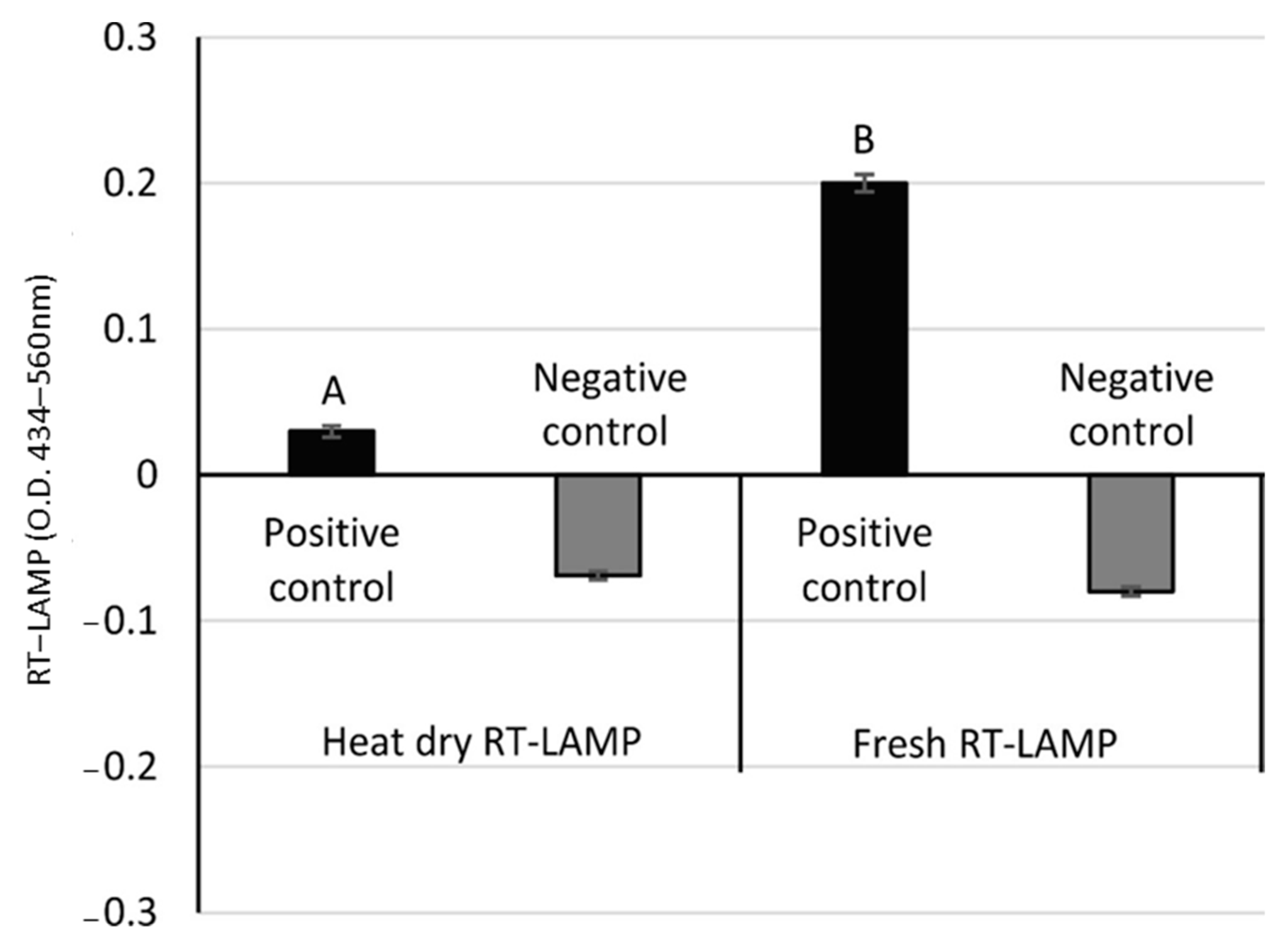
3.2. Stabilizing Dry RT-LAMP Temperature

In previous studies, lyophilization and vacuum centrifugation have been employed to dry and preserve RT-LAMP reaction mixtures for pathogen detection [28,29]. However, the use of heat as a drying method for RT- LAMP reaction mixtures has not been extensively explored. This study aimed to fill this gap by evaluating the effectiveness of various heating temperatures on the RT-LAMP process for detecting Botrytis cinerea as a model pathogen. The experiment involved heating primer mix and WarmStart® LAMP mixtures at room temperature, 37 °C, or 43 °C, with corresponding drying times of 18 h, 10 h, and 2 h and 15 min. After drying, the residue was reconstituted in sterile water containing the target DNA and subsequently incubated at 65 °C. It was observed that mixtures dried at room temperature or 37 °C exhibited no visible color change, suggesting that amplification for either 100 or 210 min at 65 °C did not lead to successful amplification (Table S1). The likely cause of this reduced activity might be attributed to the denaturation or loss of enzyme functionality [35]. Figure 2 demonstrates a significant difference between the positive and negative controls in both the heat-dried RT-LAMP assay and the fresh mixture. However, the heat-dried RT-LAMP assay was significantly less effective than the fresh mixture. This difference suggests that the heating process might lead to the degradation/denaturation of components in the RT-LAMP mixture, aligning with observations from previous freeze-drying experiments, where significant differences were reported between fresh and lyophilized RT-LAMP mixtures [28]. Such degradation in the heat-dried samples might be caused by the breakdown of sensitive components, such as enzymes and primers. These findings highlight the critical need for optimizing drying conditions to preserve the integrity of RT-LAMP reaction components and suggest that further research is necessary to explore alternative drying methods. Such methods would ideally maintain the functionality of the reaction mixture while also providing the practical benefits of dry-state storage and transportation for field applications.

3.3. Optimizing Trehalose Concentration and Incubation Time in Heat-Dried LAMP Assays for Enhanced Latent Botrytis cinerea Detection

Additives are commonly integrated into RT-LAMP assays for pathogen detection to function as protectants, enhance detection accuracy, and ensure longer preservation. Figure 3 illustrates the impact of using trehalose as a protectant in a heat-dried RT-LAMP reaction. In the absence of trehalose, maximum amplification was reached at 1 h and 10 min, reducing the likelihood of false amplification on the plate (Figure 3A). However, at an extended incubation time of 1 h and 40 min, false amplification was observed (Figure 3C) in this assay without trehalose (Figure 3C). This study found that adding more trehalose slowed the amplification process and led to weaker amplification signals (Figure 3). This effect can be explained by trehalose’s ability to interact with enzymes during the drying process, preserving their native structure and preventing degradation/denaturation. Such interactions also disrupt the correct functionality of enzymes, which, in turn, prolongs the amplification time and reduces amplification efficiency [36,37]. Additionally, the supplementation of trehalose was found to protect DNA polymerase, reverse transcriptase enzymes, and other components of the RT-LAMP mixture from degradation, further highlighting its crucial role in maintaining the integrity and functionality of this assay’s components [10,29]. Indeed, Figure 3 demonstrates that an increase in trehalose concentration led to longer reaction times and lower optical density (O.D.) values, indicating a decrease in signal strength. The concentrations of trehalose were chosen to optimize amplification efficiency while minimizing the risk of mixture evaporation during the heat-drying process. The absence of trehalose in the negative control resulted in a false-positive outcome at 1 h and 40 min and a diminished pink coloration at 1 h and 20 min, impacting its O.D. measurement (Figure 3). The observed response could be attributed to increased evaporation and reduced thermal stability of enzymes, leading to non-specific reactions. Without trehalose, this assay becomes more susceptible to environmental factors, impacting reaction kinetics and leading to inaccurate results.
Among the various trehalose concentrations tested, no visible differences in amplification efficiency were observed between 5% (w/v) and 10% (w/v) across all reaction times examined. Therefore, considering the intensity of the positive signal and the distinction between negative and positive responses, 5% (w/v) trehalose concentration and an incubation duration of 1 h and 40 min were identified as optimal. This decision was influenced by the comprehension that mRNA, constituting approximately 1–5% of the total RNA extracted, may necessitate longer amplification times when present in lower quantities. This timeframe also produced optimal outcomes without risking the evaporation of the reaction mixture at 65 °C before amplification. Establishing an ideal equilibrium between reaction time and mixture stability. This analysis underscores that while trehalose plays a crucial role in safeguarding the components of the reaction, a careful calibration of its concentration and the reaction duration is essential to ensure optimal amplification efficiency without compromising the reaction mixture’s integrity.

3.4. Sensitivity Analysis Using Synthesized and Biological Targets

The sensitivity of the heat-dried RT-LAMP assay was assessed to establish its minimum detectable limit. Figure 4 demonstrates a direct correlation between the amount of PLF target present and the level of positive amplification, with increasing values noted from 0.001 pg up to 50 pg. This assay effectively identified a minimum of 0.01 pg (10 fg) of the Botrytis PLF target, establishing this as the lowest amount for successful detection with positive amplification, while concentrations lower than 0.01 pg (10 fg) did not produce positive results in this assay. This detection limit aligns with previous findings, where a similar threshold of 10 fg was observed in RT-PCR for detecting Indian citrus ringworm [26] and in the detection of influenza A virus using a fresh reaction mixture with an HNB indicator [38]. It is noteworthy that, despite the heat-drying process applied to the RT-LAMP mixture in this study, this assay maintained a detection limit of 10 fg with an incubation period of 1 h and 40 min. This finding is significant as it demonstrates the assay’s robustness and effectiveness even after the heat-drying step, which could potentially influence the sensitivity of the reaction. The ability to detect such low concentrations of target DNA/RNA highlights the potential of this heat-dried RT-LAMP assay for sensitive and accurate pathogen detection, making it a valuable tool in molecular diagnostics, not only for the detection of B. cinerea that served as a model.

3.5. Specificity

This study phase was dedicated to assessing the assay’s capability to distinguish between the markers of B. cinerea and A. alternata, utilizing primers specifically designed for B. cinerea detection. As illustrated in Figure 5, this assay successfully responded to B. cinerea without any response to the A. alternata or generation of false positives in the negative control, thereby demonstrating its specificity for the PLF marker of B. cinerea and avoiding cross-reactivity with the HPM marker of A. alternata.
Further validation was conducted through gel electrophoresis of the amplicon products, which confirmed the absence of amplicons for A. alternata and the negative control, in contrast to the clear positive amplification bands for B. cinerea. These findings emphasize the assay’s high specificity and its ability to differentiate effectively between the two fungi without cross-reactivity or false positives, particularly in relation to A. alternata. The precision in identifying B. cinerea underscores the developed assay’s reliability and its potential utility in targeted pathogen detection, reinforcing its value in the fields of plant pathology and agricultural research.

3.6. Validation Using Infected Biological Samples

In this phase of this study, tomatoes were inoculated with spores of B. cinerea, A. alternata, and Penicillium expansum; samples were collected after 3 days, and total RNA was extracted. Following this, a specially prepared RT-LAMP mixture, enriched with trehalose, underwent a drying and rehydration process, ready to be combined with RNA from the inoculated fruits. The mixture was then incubated at 65 °C, targeting the amplification of specific genetic markers that pinpoint the presence of latent B. cinerea. Figure 6 demonstrates the test’s success in accurately identifying B. cinerea in real samples extracted from tomato fruit, where all tests for B. cinerea came back positive, including positive control. In contrast, tests for negative control, A. alternata, and Penicillium expansum inoculated tomato fruit showed negative results, demonstrating the test’s precision in detecting B. cinerea specifically.
This validation shows that the test can accurately find latent B. cinerea in crops, making it a key tool for improving food safety and reducing waste. The assay’s successful performance with infected agricultural produce underlines its potential in practical applications in real-world agricultural settings.

4. Conclusions

In conclusion, this study introduces a novel desiccation of RT-LAMP assay with a heat-dry method utilizing trehalose as a protective agent for the reaction. This research successfully presents the first heat-dry RT-LAMP assay for the rapid and efficient detection of Botrytis cinerea, addressing the challenges of limited access to lyophilization equipment in certain regions and making it more accessible. Following improvements, the enhanced method exhibited impressive sensitivity, capable of detecting RNA quantities as small as 10 fg, and demonstrated specific detection of B. cinerea in both laboratory-prepared and actual samples, highlighting its accuracy and reliability. The assay’s evaluation with B. cinerea-infected tomato fruit displays its simplicity through visible color change monitoring, eliminating the need for complex equipment. This procedure requires only basic tools and does not need expert technical personnel, thus making it accessible to a wider range of users. Future endeavors will focus on the detection of natural infection in field conditions and achieving long-term storage at room temperature. This development is essential in enabling true point-of-care testing (POCT) under field conditions, enabling the on-field applicability of the heat-dry RT-LAMP assay for latent Botrytis cinerea detection and potentially other pathogen-related applications.

Supplementary Materials

The following supporting information can be downloaded at https://www.mdpi.com/article/10.3390/applmicrobiol4040110/s1, Table S1: Heat-dry RT-LAMP reaction at various temperatures, observational, and analytical results.

Author Contributions

K.A., conceptualization, methodology, investigation, data curation, formal analysis, writing—original draft; D.D.-A., methodology, formal analysis; N.A., conceptualization, methodology, supervision, writing—reviewing and editing; E.E., conceptualization, methodology, resources, supervision, funding acquisition, writing—review and editing. All authors have read and agreed to the published version of the manuscript.

Funding

This research was funded by the Jewish Charitable Association (grant no. 430- 0812).

Data Availability Statement

The study’s original contributions are included in the article, further inquiries can be directed to the corresponding author.

Conflicts of Interest

The authors declare no conflicts of interest.

References

  1. Alkan, N.; Fortes, A.M. Insights into molecular and metabolic events associated with fruit response to post-harvest fungal pathogens. Front. Plant Sci. 2015, 6, 889. [Google Scholar] [CrossRef] [PubMed]
  2. Wenneker, M.; Thomma, B.P.H.J. Latent postharvest pathogens of pome fruit and their management: From single measures to a systems intervention approach. Eur. J. Plant Pathol. 2020, 156, 663–681. [Google Scholar] [CrossRef]
  3. Williamson, B.; Tudzynski, B.; Tudzynski, P.; Van Kan, J.A.L. Botrytis cinerea: The cause of grey mould disease. Mol. Plant Pathol. 2007, 8, 561–580. [Google Scholar] [CrossRef] [PubMed]
  4. Dugan, F.M.; Lupien, S.L.; Grove, G.G. Incidence, aggressiveness and In Planta interactions of Botrytis cinerea and other filamentous fungi quiescent in grape berries and dormant buds in central Washington State. J. Phytopathol. 2002, 150, 375–381. [Google Scholar] [CrossRef]
  5. Edwards, S.; Seddon, B. Selective media for the specific isolation and enumeration of Botrytis cinerea conidia. Lett. Appl. Microbiol. 2001, 32, 63–66. [Google Scholar] [CrossRef]
  6. Wahab, H.A.; Younis, R.A.A. Early detection of gray mold in grape using conventional and molecular methods. Afr. J. Biotechnol. 2012, 11, 15251–15257. [Google Scholar] [CrossRef]
  7. Calvo-Garrido, C.; Usall, J.; Viñas, I.; Elmer, P.A.; Cases, E.; Teixidó, N. Potential secondary inoculum sources of Botrytis cinerea and their influence on bunch rot development in dry Mediterranean climate vineyards. Pest. Manag. Sci. 2014, 70, 922–930. [Google Scholar] [CrossRef] [PubMed]
  8. Jaspers, M.V.; Seyb, A.M.; Trought, M.C.T.; Balasubramaniam, R. Overwintering grapevine debris as an important source of Botrytis cinerea inoculum. Plant Pathol. 2013, 62, 130–138. [Google Scholar] [CrossRef]
  9. Celik, M.; Kalpulov, T.; Zutahy, Y.; Ish-Shalom, S.; Lurie, S.; Lichter, A. Quantitative and qualitative analysis of Botrytis inoculated on table grapes by qPCR and antibodies. Postharvest Biol. Technol. 2009, 52, 235–239. [Google Scholar] [CrossRef]
  10. Tomlinson, J.A.; Boonham, N.; Hughes, K.J.D.; Griffin, R.L.; Barker, I. On-site DNA extraction and real-time PCR for detection of Phytophthora ramorum in the field. Appl. Environ. Microbiol. 2005, 71, 6702–6710. [Google Scholar] [CrossRef]
  11. Lin, Y.-H.; Lin, Y.-J.; Chang, T.-D.; Hong, L.-L.; Chen, T.-Y.; Chang, P.-F.L. Development of a taqman probe-based insulated isothermal polymerase chain reaction (iiPCR) assay for detection of Fusarium oxysporum f. sp. cubense race 4. PLoS ONE 2016, 11, e0159681. [Google Scholar] [CrossRef] [PubMed]
  12. Yan, L.; Zhou, J.; Zheng, Y.; Gamson, A.S.; Roembke, B.T.; Nakayama, S.; Sintim, H.O. Isothermal amplified detection of DNA and RNA. Mol. Biosyst. 2014, 10, 970–1003. [Google Scholar] [CrossRef] [PubMed]
  13. Notomi, T.; Okayama, H.; Masubuchi, H.; Yonekawa, T.; Watanabe, K.; Amino, N.; Hase, T. Loop-mediated isothermal amplification of DNA. Nucleic Acids Res. 2000, 28, e63. [Google Scholar] [CrossRef]
  14. Saiki, R.K.; Scharf, S.; Faloona, F.; Mullis, K.B.; Horn, G.T.; Erlich, H.A.; Arnheim, N. Enzymatic amplification of beta-globin genomic sequences and restriction site analysis for diagnosis of sickle cell anemia. 1985. Biotechnology 1992, 24, 476–480. [Google Scholar] [PubMed]
  15. Mohammadniaei, M.; Zhang, M.; Ashley, J.; Christensen, U.B.; Friis-Hansen, L.J.; Gregersen, R.; Lisby, J.G.; Benfield, T.L.; Nielsen, F.E.; Rasmussen, J.H.; et al. A non-enzymatic, isothermal strand displacement and amplification assay for rapid detection of SARS-CoV-2 RNA. Nat. Commun. 2021, 12, 5089. [Google Scholar] [CrossRef] [PubMed]
  16. Walker, G.T.; Fraiser, M.S.; Schram, J.L.; Little, M.C.; Nadeau, J.G.; Malinowski, D.P. Strand displacement amplification—An isothermal, in vitro DNA amplification technique. Nucleic Acids Res. 1992, 20, 1691–1696. [Google Scholar] [CrossRef] [PubMed]
  17. Fang, R.; Li, X.; Hu, L.; You, Q.; Li, J.; Wu, J.; Xu, P.; Zhong, H.; Luo, Y.; Mei, J.; et al. Cross-priming amplification for rapid detection of Mycobacterium tuberculosis in sputum specimens. J. Clin. Microbiol. 2009, 47, 845–847. [Google Scholar] [CrossRef] [PubMed]
  18. Woźniakowski, G.; Niczyporuk, J.S.; Samorek-Salamonowicz, E.; Gaweł, A. The development and evaluation of cross-priming amplification for the detection of avian reovirus. J. Appl. Microbiol. 2015, 118, 528–536. [Google Scholar] [CrossRef]
  19. Christian, A.T.; Pattee, M.S.; Attix, C.M.; Reed, B.E.; Sorensen, K.J.; Tucker, J.D. Detection of DNA point mutations and mRNA expression levels by rolling circle amplification in individual cells. Proc. Natl. Acad. Sci. USA 2001, 98, 14238–14243. [Google Scholar] [CrossRef] [PubMed]
  20. Kaur, M.; Ayarnah, K.; Duanis-Assaf, D.; Alkan, N.; Eltzov, E. Rapid and simple colorimetric detection of quiescent Colletotrichum in harvested fruit using reverse transcriptional loop-mediated isothermal amplification (RT-LAMP) technology. Talanta 2023, 255, 124251. [Google Scholar] [CrossRef]
  21. Cardoso, T.C.; Ferrari, H.F.; Bregano, L.C.; Silva-Frade, C.; Rosa, A.C.G.; Andrade, A.L. Visual detection of turkey coronavirus RNA in tissues and feces by reverse-transcription loop-mediated isothermal amplification (RT-LAMP) with hydroxynaphthol blue dye. Mol. Cell. Probes 2010, 24, 415–417. [Google Scholar] [CrossRef]
  22. Martineau, R.L.; Murray, S.A.; Ci, S.; Gao, W.; Chao, S.-H.; Meldrum, D.R. Improved Performance of Loop-Mediated Isothermal Amplification Assays via Swarm Priming. Anal. Chem. 2017, 89, 625–632. [Google Scholar] [CrossRef] [PubMed]
  23. Tavakoli-Koopaei, R.; Javadi-Zarnaghi, F.; Aboutalebian, S.; Mirhendi, H. Malachite Green-Based Detection of SARS-CoV-2 by One-Step Reverse Transcription Loop-Mediated Isothermal Amplification. Iran. J. Sci. 2023, 47, 359–367. [Google Scholar] [CrossRef]
  24. Ge, A.; Liu, F.; Teng, X.; Cui, C.; Wu, F.; Liu, W.; Liu, Y.; Chen, X.; Xu, J.; Ma, B. A Palm Germ-Radar (PaGeR) for rapid and simple COVID-19 detection by reverse transcription loop-mediated isothermal amplification (RT-LAMP). Biosens. Bioelectron. 2022, 200, 113925. [Google Scholar] [CrossRef] [PubMed]
  25. Nzelu, C.O.; Gomez, E.A.; Cáceres, A.G.; Sakurai, T.; Martini-Robles, L.; Uezato, H.; Mimori, T.; Katakura, K.; Hashiguchi, Y.; Kato, H. Development of a loop-mediated isothermal amplification method for rapid mass-screening of sand flies for Leishmania infection. Acta Trop. 2014, 132, 1–6. [Google Scholar] [CrossRef] [PubMed]
  26. Kokane, A.D.; Kokane, S.B.; Warghane, A.J.; Gubyad, M.G.; Sharma, A.K.; Reddy, M.K.; Ghosh, D.K. A Rapid and sensitive reverse transcription–loop-mediated isothermal amplification (RT-LAMP) assay for the detection of indian citrus ringspot virus. Plant Dis. 2021, 105, 1346–1355. [Google Scholar] [CrossRef]
  27. García-Bernalt Diego, J.; Fernández-Soto, P.; Crego-Vicente, B.; Alonso-Castrillejo, S.; Febrer-Sendra, B.; Gómez-Sánchez, A.; Vicente, B.; López-Abán, J.; Muro, A. Progress in loop-mediated isothermal amplification assay for detection of Schistosoma mansoni DNA: Towards a ready-to-use test. Sci. Rep. 2019, 9, 14744. [Google Scholar] [CrossRef] [PubMed]
  28. Chen, H.-W.; Weissenberger, G.; Ching, W.-M. Development of lyophilized loop-mediated isothermal amplification reagents for the detection of leptospira. Mil. Med. 2016, 181, 227–231. [Google Scholar] [CrossRef] [PubMed]
  29. Song, X.; Coulter, F.J.; Yang, M.; Smith, J.L.; Tafesse, F.G.; Messer, W.B.; Reif, J.H. A lyophilized colorimetric RT-LAMP test kit for rapid, low-cost, at-home molecular testing of SARS-CoV-2 and other pathogens. Sci. Rep. 2022, 12, 7043. [Google Scholar] [CrossRef]
  30. Silva, C.J.; Adaskaveg, J.A.; Mesquida-Pesci, S.D.; Ortega-Salazar, I.B.; Pattathil, S.; Zhang, L.; Hahn, M.G.; van Kan, J.A.L.; Cantu, D.; Powell, A.L.T.; et al. Botrytis cinerea infection accelerates ripening and cell wall disassembly to promote disease in tomato fruit. Plant Physiol. 2023, 191, 575–590. [Google Scholar] [CrossRef] [PubMed]
  31. Francia, F.; Dezi, M.; Mallardi, A.; Palazzo, G.; Cordone, L.; Venturoli, G. Protein—Matrix Coupling/Uncoupling in ‘Dry’ Systems of Photosynthetic Reaction Center Embedded in Trehalose/Sucrose: The Origin of Trehalose Peculiarity. J. Am. Chem. Soc. 2008, 130, 10240–10246. [Google Scholar] [CrossRef]
  32. Cicerone, M.T.; Soles, C.L. Fast dynamics and stabilization of proteins: Binary glasses of trehalose and glycerol. Biophys. J. 2004, 86, 3836–3845. [Google Scholar] [CrossRef] [PubMed]
  33. Hardinge, P.; Murray, J.A.H. Reduced False Positives and Improved Reporting of Loop-Mediated Isothermal Amplification using Quenched Fluorescent Primers. Sci. Rep. 2019, 9, 7400. [Google Scholar] [CrossRef] [PubMed]
  34. Meagher, R.J.; Priye, A.; Light, Y.K.; Huang, C.; Wang, E. Impact of primer dimers and self-amplifying hairpins on reverse transcription loop-mediated isothermal amplification detection of viral RNA. Analyst 2018, 143, 1924–1933. [Google Scholar] [CrossRef] [PubMed]
  35. Oscorbin, I.; Filipenko, M. Bst polymerase—A humble relative of Taq polymerase. Comput. Struct. Biotechnol. J. 2023, 21, 4519–4535. [Google Scholar] [CrossRef] [PubMed]
  36. Tsai, A.M.; Udovic, T.J.; Neumann, D.A. The inverse relationship between protein dynamics and thermal stability. Biophys. J. 2001, 81, 2339–2343. [Google Scholar] [CrossRef]
  37. Mensink, M.A.; Frijlink, H.W.; Van Der Voort Maarschalk, K.; Hinrichs, W.L.J. How sugars protect proteins in the solid state and during drying (review): Mechanisms of stabilization in relation to stress conditions. Eur. J. Pharm. Biopharm. 2017, 114, 288–295. [Google Scholar] [CrossRef]
  38. Boora, S.; Khan, A.; Sharma, V.; Kaushik, S.; Mehta, P.K.; Singh, S.; Kaushik, S. RT-LAMP is a potential future molecular diagnostic tool for influenza A virus. Futur. Virol. 2023, 18, 165–175. [Google Scholar] [CrossRef]
Figure 1. Comparative Analysis of RT-LAMP Reaction Efficiency Using Different Heat Sources. (A) Performance comparison of RT-LAMP reactions at 65 °C using a wet bath, oven, and dry bath (heat chamber) for 30 min, 1 h and 15 min, and 45 min, respectively. (B) Detection of Botrytis cinerea in its latent stage using spectrophotometer kinetics. Positive control includes a 50 pg PLF marker. A statistically significant difference between treatments is denoted by different letters, p-value < 0.05, by one-way ANOVA.
Applmicrobiol 04 00110 g001
Figure 2. Stabilized (i.e., heat-dried) vs. Fresh RT-LAMP Assay Performance Comparison. A 50 pg sample of the latent PLF Botrytis cinerea marker was tested. This assay compared the performance of a 43 °C heat-dried RT-LAMP mixture against a fresh RT-LAMP mixture, both incubated in multiwell plates at 65 °C for 1 h and 40 min for the heat-dried and 45 min for the fresh mixture. A statistically significant difference between treatments is denoted by different letters, p-value < 0.05, by one-way ANOVA.
Applmicrobiol 04 00110 g002
Figure 3. Optimization of trehalose concentrations in heat-dry RT-LAMP reaction assay and its efficiency in detecting latent Botrytis cinerea. Three different amplification times were tested. (A) 1 h and 10 min, (B) 1 h and 20 min, (C) and 1 h and 40 min. A statistically significant difference between treatments is denoted by different letters, p-value < 0.05, by one-way ANOVA.
Applmicrobiol 04 00110 g003
Figure 4. Assessing the Sensitivity of the 43 °C Heat-Dried RT-LAMP Assay. This assay evaluated a range of synthesized DNA target concentrations, from 0.01 pg to 50 pg. A negative control without any DNA target was also incorporated into this assay.
Applmicrobiol 04 00110 g004
Figure 5. (A) Detection of specific targets: Botrytis cinerea PLF (50 pg) and Alternaria alternata (50 pg), alongside a negative control. (B) Results of RT-LAMP reactions shown through agarose gel electrophoresis, featuring positive targets for Botrytis cinerea in wells 2–4, no amplification for Alternaria alternata in wells 5–7, and negative control in wells 8–10, following heat drying at 43 °C.
Applmicrobiol 04 00110 g005
Figure 6. Detection of latent Botrytis cinerea in infected tomato fruit samples using heat-dry RT-LAMP. A 1 ng/µL of total RNA was used for detecting latent Botrytis cinerea, Alternaria alternata, and Penicillium expansum from inoculated tomato fruit. An amount of 50 pg of synthesized DNA (positive control) was used to detect Botrytis cinerea (PLF) target. The negative control contained no RNA or synthetic DNA of the target gene.
Applmicrobiol 04 00110 g006
Table 1. Primers for RT-LAMP assay.
SpeciesTargeted SequencePrimer
Name
Primer Sequences 5′-3′Final Concentration
Botrytis cinereaLatent marker (Polysaccharide Lyase family 1 protein, Table 2)F3CAAATTCCACGAGATTCTCAT100 µM
B3ACGCTCAGCAATAGCATC100 µM
FIPCGGGAGATCGATTCAAAAGATTGATTTTCACAACTCACTCAACTCAAG100 µM
BIPAATGAAGCTCTCCATTCTCTCCACTTTTACCTTCAGTTGGTGTTGG100 µM
Loop FAGAGCTGTCAACTTCGTTCTT100 µM
Loop BCGCAGTCCTTGCGCAGTT100 µM
Table 2. Latent markers for RT-LAMP assay.
DescriptionSequence
Latent marker of Botrytis cinerea
(Polysaccharide Lyase family 1 protein)
GTGAAGATTTGCACGGCTGCGGTTGTACTGACGTTGAATGGACTCAAATCTCGTTTGACGATATGGGGGTTGAACGAGAAATCAAAAGTATATAAAGGAGAACCAAATTCCACGAGATTCTCATTCTTCACAACTCACTCAACTCAAGGCAAAGAACGAAGTTGACAGCTCTACAATCAATCTTTTGAATCGATCTCCCGCAACGAACTTTTTGAATATCCAAAAAAAAATGAAGCTCTCCATTCTCTCCACAGGACTCGCAGTCCTTGCGCAGTTCGTCTCTGCTGCTCCAACACCAACTGAAGGTGATGCTATTGCTGAGCGTGCAAACATCGCTAAGAGAGCTACTATCACCGATGTTGCCACTACCGGCTTTGCAACCCA
Latent marker of Alternaria alternata
(Hypothetical protein partial mRNA (small ribosomal RNA unit)
ATGAAGTTCCTTGCCACCATCATCCTCCTTACCTTCCTCGCAGCCACGGTCATCGCAGGTGATTGCATGAAAGCACCTTTTCACTGCAATGGTATGTCTTTCACCTGTACCACCGCCATTCTCGTCACCCTCTTCACCACTACAGCTTCGGCGACTGAGTGTACAGTACCTGTTCTCTGCACCCGTAGCGTCGGTAAATCGCGGCTCATAGCAAACATACCCGGCTTTGAAGAATTCGGCGACTCTTACAGCAGCATCGAGTGTGCCCAGAGTCTTGTCGTGTCCTCTGTGATGGGGATGAAGACCTGCCCCGCTGTGCCATCGGTGTCTCACCCCTTCCCCCAGGTCGTCACGATATCGTCCACACCATACCCACCAACTACACGACCACCCATCTCAGCAAGAACAACACGCACCCACCCATACGGGTCGTCTGAACTGCGCGCTTACCAAGCTCCAGCGATGGTTTCTTTCTCCGCCGTCCTCATCCTAAGCTATCC
Disclaimer/Publisher’s Note: The statements, opinions and data contained in all publications are solely those of the individual author(s) and contributor(s) and not of MDPI and/or the editor(s). MDPI and/or the editor(s) disclaim responsibility for any injury to people or property resulting from any ideas, methods, instructions or products referred to in the content.

