

Reliability of Artificial Intelligence-Assisted Cephalometric Analysis. A Pilot Study


Abstract
:1. Introduction
2. Materials and Methods
2.1. Tracing Technique
2.2. Cephalometric Measurements
- −
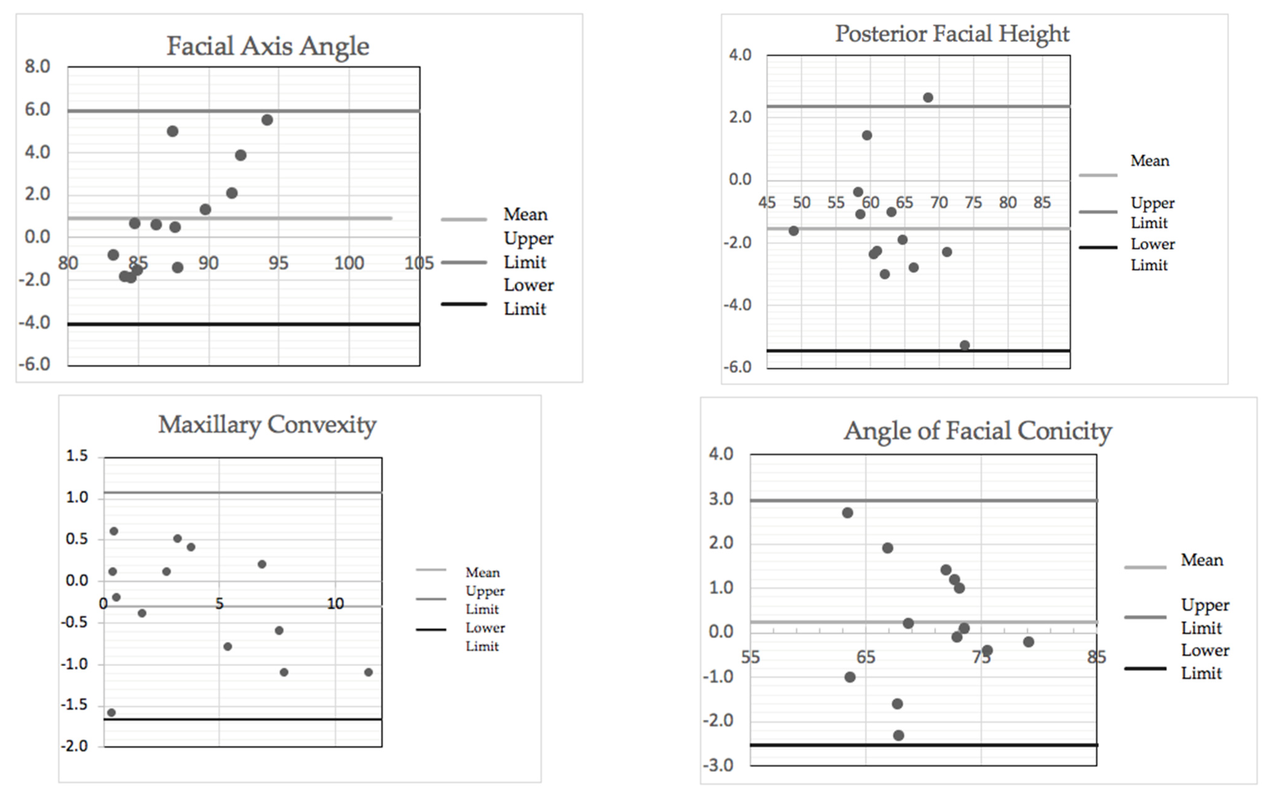
- Maxillary Convexity: determined by the distance of A point to the facial plane N-Pg. The normal value is 2 ± 2 mm. This reflects the sagittal protrusion of the maxillary part of the face compared to the facial profile. A reduced angle indicates a maxillary retrusion within a normal facial plane, a mandibular protrusion with normal or retruded maxillary projection, or a brachycephalic facial profile. An increased angle identifies a maxillary protrusion with a mandible within normal limit, a mandibular retrusion with a maxilla within normal limit, a maxillary protrusion with retrognathia, or a dolichocephalic facial profile [22,23].
- −
- Angle of Facial Conicity: the angle formed by facial plane N-Pg with the mandibular plane Go-Gn. The normal value is 68 ± 4°. This reflects the sagittal and vertical position of the chin as well as the direction of the facial growth. A reduced angle suggests a clockwise mandibular growth and a dolichocephalic facial type; an increased angle indicates the tendency for a counterclockwise mandibular growth and a brachycephalic facial type [22].
- −
- Facial Axis Angle: identifies the posterior angle constituted by the intersection of the extension of facial axis Pt-Gn with the basal plane Ba-N. The normal value is 90 ± 3°. This angle reflects the vertical mandibular growth: a reduced angle indicates a dolichocephalic growth or retrognathic profile; an increased angle indicates a brachycephalic growth [22].
- −
- Posterior Facial Height: identified by the linear measurement that connects S-Go [22].
- −
- Lower Facial Height: the angle formed by mandibular axis Xi-PM and the line that connects the mandibular centroid and SNA (Xi-SNA). The normal value is 47 ± 4°. It identifies the position and the direction of the mandibular growth and the spatial location of the maxilla. A reduced angle suggests a horizontal growth, a downward inclination of the maxilla, or a counterclockwise rotation of the mandible. An increased angle indicates a vertical growth, an upward inclination of the maxilla, or a clockwise rotation of the mandible [22].
2.3. Statistical Analysis
3. Results
3.1. Intra-Operator Measurements
3.2. Inter-Operator Measurements
4. Discussion
5. Conclusions
Author Contributions
Funding
Institutional Review Board Statement
Informed Consent Statement
Data Availability Statement
Conflicts of Interest
References
- Khanagar, S.B.; Al-Ehaideb, A.; Maganuretal, P.C. Developments, application, and performance of artificial intelligence in dentistry—A systematic review. J. Dent. Sci. 2021, 16, 508–522. [Google Scholar] [CrossRef]
- Subramanian, A.K.; Chen, Y.; Almalki, A.; Sivamurthy, G.; Kafle, D. Cephalometric Analysis in Orthodontics Using Artificial Intelligence-A Comprehensive Review. Biomed. Res. Int. 2022, 2022, 1880113. [Google Scholar] [CrossRef] [PubMed]
- Nguyen, T.T.; Larrivée, N.; Lee, A.; Bilaniuk, O.; Durand, R. Use of Artificial Intelligence in Dentistry: Current clinical trends and research advances. J. Can. Dent. Assoc. 2021, 87, 17. [Google Scholar] [CrossRef]
- Fatima, A.; Shafi, I.; Afzal, H.; Díez, I.D.L.T.; Lourdes, D.R.-S.M.; Breñosa, J.; Espinosa, J.C.M.; Ashraf, I. Advancements in Dentistry with Artificial Intelligence: Current clinical applications and future perspective. Healthcare 2022, 10, 2188. [Google Scholar] [CrossRef] [PubMed]
- Sangalli, L.; Savoldi, F.; Dalessandri, D.; Visconti, L.; Massetti, F.; Bonetti, S. Remote digital monitoring during the retention phase of orthodontic treatment: A prospective feasibility study. Kor. J. Orthod. 2022, 52, 123–130. [Google Scholar] [CrossRef]
- Sangalli, L.; Savoldi, F.; Dalessandri, D.; Bonetti, S.; Gu, M.; Signoroni, A.; Paganelli, C. Effects of remote digital monitoring on oral hygiene of orthodontic patients: A prospective study. BMC Oral Health 2021, 21, 435. [Google Scholar] [CrossRef]
- Menterubbianesi, R.; Tosco, V.; Vitiello, F.; Orilisi, G.; Fraccastoro, F.; Putignano, A.; Orsini, G. Augmented, virtual and mixed reality in dentristy: A narrative review on the existing platforms and future challenges. Appl. Sci. 2022, 12, 877. [Google Scholar] [CrossRef]
- Bichu, Y.M.; Hansa, I.; Bichu, A.Y.; Premjani, P.; Flores-Mir, C.; Vaid, N.R. Applications of artificial intelligence and machine learning in orthodontics: A scoping review. Progr. Orthod. 2021, 22, 18. [Google Scholar] [CrossRef]
- Kunz, F.; Stellzig-Eisenhauer, A.; Zeman, F.; Boldt, J. Artificial intelligence in orthodontics. J. Orofac. Orthop. Fortschr. Kieferorthopädie 2020, 81, 52–68. [Google Scholar] [CrossRef]
- Sandler, P.J. Reproducibility of cephalometric measurements. Brit. J. Orthod. 1988, 15, 105–110. [Google Scholar] [CrossRef]
- Sayinsu, K.; Isik, F.; Trakyali, G.; Arun, T. An evaluation of the errors in cephalometric measurements on scanned cephalometric images and conventional tracings. Eur. J. Orthod. 2007, 29, 105–108. [Google Scholar] [CrossRef] [PubMed] [Green Version]
- Dreyer, K.J.; Geis, J.R. When machines think: Radiology’s next frontier. Radiology 2017, 285, 713–718. [Google Scholar] [CrossRef] [PubMed] [Green Version]
- Melsen, B.; Baumrind, S.; Athanasiou, A. Clinical Research Application of Cephalometry, Orthodontic Cephalometry; Mosby-Wolfe: London, UK, 1995; pp. 181–202. [Google Scholar]
- Keim, R.G.; Gottlieb, E.L.; Vogels, I.; Vogels, P.B. 2014 JCO study of orthodontic diagnosis and treatment procedures, part 1: Results and trends. J. Clin. Orthod. 2014, 48, 607–630. [Google Scholar] [PubMed]
- Tsolakis, I.A.; Tsolakis, A.I.; Elshebiny, T.; Matthaios, S.; Palomo, J.M. Comparing a fully automated cephalometric tracing methods to a manual tracing method for orthodontic diagnosis. J. Clin. Med. 2022, 11, 6854. [Google Scholar] [CrossRef] [PubMed]
- Wang, S.; Li, H.; Li, J.; Zhang, Y.; Zou, B. Automatic analysis of lateral cephalograms based on multiresolution decision tree regression voting. J. Health Eng. 2018, 2018, 1797502. [Google Scholar] [CrossRef]
- Leonardi, R.; Giordano, D.; Maiorana, F.; Spampinato, C. Automatic cephalometric analysis. Angle Orhod. 2008, 78, 145–151. [Google Scholar] [CrossRef] [Green Version]
- Mahto, R.K.; Kafle, D.; Giri, A.; Luintel, S.; Karki, A. Evaluation of fully automated cephalometric measurements obtained from web-based artificial intelligence driven platform. BMC Oral Health 2022, 22, 132. [Google Scholar] [CrossRef]
- Durão, A.P.; Morosolli, A.; Pittayapat, P.; Bolstad, N.; Ferreira, A.P.; Jacobs, R. Cephalometric landmark variability among orthodontists and dentomaxillofacial radiologists: A comparative study. Imaging Sci. Dent. 2015, 45, 213–220. [Google Scholar] [CrossRef] [Green Version]
- AlBarakati, S.F.; Kula, K.S.; Ghoneima, A.A. The reliability and reproducibility of cephalometric measurements: A comparison of conventional and digital methods. Dentomaxillofac. Radiol. 2012, 41, 11–17. [Google Scholar] [CrossRef]
- Savoldi, F.; Del Re, F.; Tonni, I.; Gu, M.; Dalessandri, D.; Visconti, L. Appropriateness of standard cephalometric norms for the assessment of dentofacial characteristics in patients with cleidocranial dysplasia. Dentomaxillofac. Radiol. 2022, 51, 20210015. [Google Scholar] [CrossRef]
- Ricketts, R.M. A foundation for cephalometric communication. Am. J. Orthod. 1960, 46, 330–357. [Google Scholar] [CrossRef]
- Godt, A.; Muller, A.; Kalwitzki, M.; Goz, G. Angles of facial convexity in different skeletal Classes. Eur. J. Orthod. 2007, 29, 648–653. [Google Scholar] [CrossRef] [PubMed] [Green Version]
- Ellis, E.; McNamara, J.A. Cephalometric evaluation of incisor position. Angle Orthod. 1986, 56, 324–344. [Google Scholar] [PubMed]
- Tweed, C.H. Indications for the extraction of teeth in orthodontic procedure. Am. J. Orthod. Oral. Surg. 1944, 30, 405–428. [Google Scholar] [CrossRef]
- Heiser, W.; Niederwanger, A.; Bancher, B.; Bittermann, G.; Neunteufel, N.; Kulmer, S. Threedimensional dental arch and palatal form changes after extraction and nonextraction treatment, Part 1: Arch Length and Area. Am. J. Orthod. 2004, 126, 71–81. [Google Scholar] [CrossRef] [PubMed]
- Margolis, H. The axial inclination of the mandibular incisors. Am. J. Orthod. 1943, 29, 571–594. [Google Scholar] [CrossRef]
- Proffitt, W.R. The soft tissue paradigm in orthodontic diagnosis and treatment planning: A new view for a new century. J. Esthet. Dent. 2000, 12, 46–49. [Google Scholar]
- Sangalli, L.; Savoldi, F.; Dalessandri, D.; Visconti, L. Historical Development of the Planning of Incisal Position in Orthodontic Treatments: A Narrative Review of the Literature. Curr. Trends Dent. 2021, 1, 01. [Google Scholar]
- Park, J.H.; Hwang, H.W.; Moon, J.H.; Yu, Y.; Kim, H.; Her, S.B.; Srinivasan, G.; Aljanabi, M.N.; Donatelli, R.E.; Lee, S.J. Automated identification of cephalometric landmarks: Part 1—Comparisons between the latest deep-learning methods YOLOV3 and SS.D. Angle Orthod. 2019, 89, 903–909. [Google Scholar] [CrossRef] [Green Version]
- Li, C.-H.; Vandaele, R.; Mirzaalian, H.; Chen, C.; Li, W.-C.; Zheng, G.; Jodogne, S.; Chang, S.-W.; Wang, C.W.; Maree, R.; et al. Evaluation and comparison of anatomical landmark detection methods for cephalometric x-ray images: A grand challenge. IEEE Trans. Med. Imaging 2015, 34, 1890–1900. [Google Scholar]
- Nimkarn, Y.; Miles, P.G. Reliability of computer-generated cephalometrics. Int. J. Adult Orthodon. Orthognath. Surg. 1995, 10, 43–52. [Google Scholar] [PubMed]
- Mahto, R.K.; Kharbanda, O.P.; Duggal, R.; Sardana, H.K. A comparison of cephalometric measurements obtained from two computerized cephalometric softwares with manual tracings. J. Indian Orthod. Soc. 2016, 50, 162–170. [Google Scholar] [CrossRef]
- Lindner, C.; Wang, C.-W.; Huang, C.-T.; Li, C.-H.; Chang, S.-W.; Cootes, T.F. Fully Automatic System for Accurate Localisation and Analysis of Cephalometric Landmarks in Lateral Cephalograms. Sci. Rep. 2016, 6, 33581. [Google Scholar] [CrossRef] [PubMed]
- Anuwongnukroh, N.; Dewchkunakorn, S.; Damrongsri, S.; Nilwarat, C.; Pudpong, N.; Radomsutthisarn, W.; Kengern, S. Accuracy of Automatic Cephalometric Software on Landmark Identification. Mater. Sci. Eng. 2017, 265, 012028. [Google Scholar] [CrossRef]
- Meriç, P.; Naoumova, J. Web-based fully automated cephalometric analysis: Comparisons between app-aided, computerized, and manual tracings. Turk. J. Orthod. 2020, 33, 142–149. [Google Scholar] [CrossRef]
- Silva, T.P.; Hughes, M.M.; Menezes, L.D.; de Melo, M.D.; Takeshita, W.M.; Freitas, P.H. Artificial intelligence-based cephalometric landmark annotation and measurements according to Arnett’s analysis: Can we trust a bot to do that? Dentomaxillofac. Radiol. 2021, 51, 20200548. [Google Scholar] [CrossRef]
- Alqahtani, H. Evaluation of an online website-based platform for cephalometric analysis. J. Stomatol. Oral Maxillofac. Surg. 2020, 121, 53–57. [Google Scholar] [CrossRef]
- Broch, J.; Slagsvold, O.; Rosler, M. Error in landmark identification in lateral radiographic headplates. Eur. J. Orthod. 1981, 3, 9–13. [Google Scholar] [CrossRef]
- Baumrind, S.; Frantz, R.C. The reliability of head film measurements. Landmark identification. Am. J. Orthod. 1971, 60, 111–127. [Google Scholar] [CrossRef]
- Livas, C.; Delli, K.; Spijkervet, F.K.L.; Vissink, A.; Dijkstra, P.U. Concurrent validity and reliability of cephalometric analysis using smartphone apps and computer software. Angle Orhod. 2019, 89, 889–896. [Google Scholar] [CrossRef] [Green Version]
- Farooq, M.U.; Khan, M.; Imran, S.; Sameera, A.; Qureshi, A.; Ahmed, S.A.; Kumar, S.; Rahman, M.A. Assessing the Reliability of Digitalized Cephalometric Analysis in Comparison with Manual Cephalometric Analysis. J. Clin. Diagn. Res. 2016, 10, ZC20–ZC23. [Google Scholar] [CrossRef] [PubMed]
- Trpkova, B.; Major, P.; Prasad, N.; Nebbe, B. Cephalometric landmarks identification and reproducibility: A meta analysis. Am. J. Orthod. Dentofac. Orthop. 1997, 112, 165–170. [Google Scholar] [CrossRef] [PubMed]


| Parameters | Measurement 1 1 Mean (SD) | Measurement 2 2 Mean (SD) | Dahlberg | 95% CI of the Mean |
|---|---|---|---|---|
| Maxillary Convexity | 3.90 (3.43) | 4.15 (3.52) | 0.396 | −1.27, 0.78 |
| Angle of Facial Conicity | 70.63 (4.63) | 70.80 (5.02) | 0.504 | −1.09, 1.18 |
| Facial Axis Angle | 88.03 (4.53) | 87.98 (4.43) | 0.395 | −1.27, 0.78 |
| Posterior Facial Height | 61.98 (6.21) | 61.88 (6.71) | 0.807 | −2.22, 2.42 |
| Lower Facial Height | 42.15 (4.39) | 41.76 (4.65) | 0.667 | −1.37, 2.14 |
| Parameters | x1 1 Mean (SD) | x2 2 Mean (SD) | Dahlberg | 95% CI of the Mean |
|---|---|---|---|---|
| Maxillary Convexity | 3.90 (3.43) | 4.20 (3.68) | 0.519 | −1.67, 1.07 |
| Angle of Facial Conicity | 70.63 (4.63) | 70.41 (4.73) | 0.969 | −2.54, 2.98 |
| Facial Axis Angle | 88.03 (4.53) | 87.09 (2.62) | 1.854 | −4.06, 5.93 |
| Posterior Facial Height | 61.98 (6.21) | 63.51 (6.73) | 1.732 | −5.44, 2.38 |
| Lower Facial Height | 42.15 (4.39) | 43.14 (4.17) | 1.455 | −4.67, 2.69 |
Disclaimer/Publisher’s Note: The statements, opinions and data contained in all publications are solely those of the individual author(s) and contributor(s) and not of MDPI and/or the editor(s). MDPI and/or the editor(s) disclaim responsibility for any injury to people or property resulting from any ideas, methods, instructions or products referred to in the content. |
© 2023 by the authors. Licensee MDPI, Basel, Switzerland. This article is an open access article distributed under the terms and conditions of the Creative Commons Attribution (CC BY) license (https://creativecommons.org/licenses/by/4.0/).
Share and Cite
Alessandri-Bonetti, A.; Sangalli, L.; Salerno, M.; Gallenzi, P. Reliability of Artificial Intelligence-Assisted Cephalometric Analysis. A Pilot Study. BioMedInformatics 2023, 3, 44-53. https://doi.org/10.3390/biomedinformatics3010003
Alessandri-Bonetti A, Sangalli L, Salerno M, Gallenzi P. Reliability of Artificial Intelligence-Assisted Cephalometric Analysis. A Pilot Study. BioMedInformatics. 2023; 3(1):44-53. https://doi.org/10.3390/biomedinformatics3010003
Chicago/Turabian StyleAlessandri-Bonetti, Anna, Linda Sangalli, Martina Salerno, and Patrizia Gallenzi. 2023. "Reliability of Artificial Intelligence-Assisted Cephalometric Analysis. A Pilot Study" BioMedInformatics 3, no. 1: 44-53. https://doi.org/10.3390/biomedinformatics3010003
APA StyleAlessandri-Bonetti, A., Sangalli, L., Salerno, M., & Gallenzi, P. (2023). Reliability of Artificial Intelligence-Assisted Cephalometric Analysis. A Pilot Study. BioMedInformatics, 3(1), 44-53. https://doi.org/10.3390/biomedinformatics3010003