1. Introduction
Soil contamination with heavy metals such as lead (Pb) and manganese (Mn) represents a major environmental constraint for plant growth and ecosystem functioning. Pb, a non-essential element, is highly toxic to plants, disrupting membrane integrity, cell division, and photosynthetic processes, thereby causing growth inhibition and premature senescence [
1,
2,
3]. In contrast, Mn is an essential micronutrient involved in enzymatic activity and photosystem II function, but becomes toxic at elevated concentrations, leading to oxidative stress and disturbance of redox balance [
4,
5,
6,
7]. Due to their contrasting physiological roles, Pb and Mn induce fundamentally different toxicity mechanisms, necessitating distinct plant tolerance and detoxification strategies, particularly in contaminated environments where both metals may co-occur.
The majority of legume species (Fabaceae) engage in a specialized form of symbiosis with nitrogen-fixing bacteria, resulting in the formation of root nodules that facilitate the supply of reduced nitrogen compounds to the plant [
7]. These highly diverse soil bacteria, collectively referred to as rhizobia, initiate coordinated developmental changes in host roots through an intricate mutual signal exchange system [
8]. Rhizobial symbiosis holds immense ecological and agricultural significance, enabling wild legume species to thrive in nitrogen-deficient habitats. However, legume cultivation in cropping systems offers the potential to enhance soil fertility without the excessive application of nitrogen fertilizers [
9,
10].
Rhizobial symbiosis has also attracted attention in the remediation of degraded or polluted environments [
11,
12,
13], where it can improve plant tolerance, promote growth, and decrease contaminant mobility [
14,
15]. The high ecological and functional diversity of wild legume species, on the one hand, and the taxonomic diversity of rhizobial bacteria, on the other, present a wide range of opportunities for the identification of novel model systems for scientific research and subsequent potential practical applications.
Previous studies have demonstrated that
Anthyllis vulneraria L. encompasses metallicolous genotypes exhibiting remarkable tolerance to Zn, Pb, Cd, and other metals, rendering it an ideal system for investigating plant adaptation to extreme soil conditions. Research on soils contaminated with Zn–Pb reveals that
A. vulneraria is frequently associated with metal-tolerant
Mesorhizobium spp., particularly
Mesorhizobium metallidurans [
16,
17]. These rhizobia dominate in mine soils and constitute the core of the
A. vulneraria microsymbiont community, facilitating nitrogen fixation under extreme metal conditions. To date, more attention has been directed specifically towards elucidating the metal tolerance properties of the bacterial partner, including transcriptional responses to the presence of heavy metals [
18,
19,
20,
21].
A. vulneraria populations can successfully develop on highly contaminated mine tailings and exhibit genotype-specific differences in growth, morphology, and metal accumulation patterns under metal stress [
16]. This genotype variability provides a natural experimental framework for analyzing the contributions of both symbiotic partners to tolerance mechanisms. As rhizobial interactions influence plant physiological responses by regulating nutrient acquisition, stress signaling, and root architecture [
22,
23,
24,
25], understanding the interaction between plant genotype and rhizobacterial species is crucial for identifying plant–microbe combinations that optimize tolerance and metal immobilization.
An ecologically isolated population of
A. vulneraria, specifically distributed on coastal dunes of the Eastern Baltics, is considered to represent
A. vulneraria subsp.
maritima (Hagen) Corb. [
26] or even a separate species,
Anthyllis maritima Schweigg. [
27]. Recently, it was established that
A. vulneraria plants from coastal sand dunes exhibit relatively high tolerance to several heavy metals (Cd, Cu, Zn) in a non-symbiotic state [
28]. However, the plants were moderately susceptible to Pb and Mn. Importantly, phytoremediation potential for Cd, Pb, Mn, and Zn was also characterized as relatively high. Given the ecological importance of
A. vulneraria and the role of rhizobacterial symbiosis in mediating heavy metal tolerance, a targeted comparison of multiple genotypes inoculated with a single bacterial strain under Pb and Mn stress may provide a useful experimental approach to disentangle plant–microbe–metal interactions.
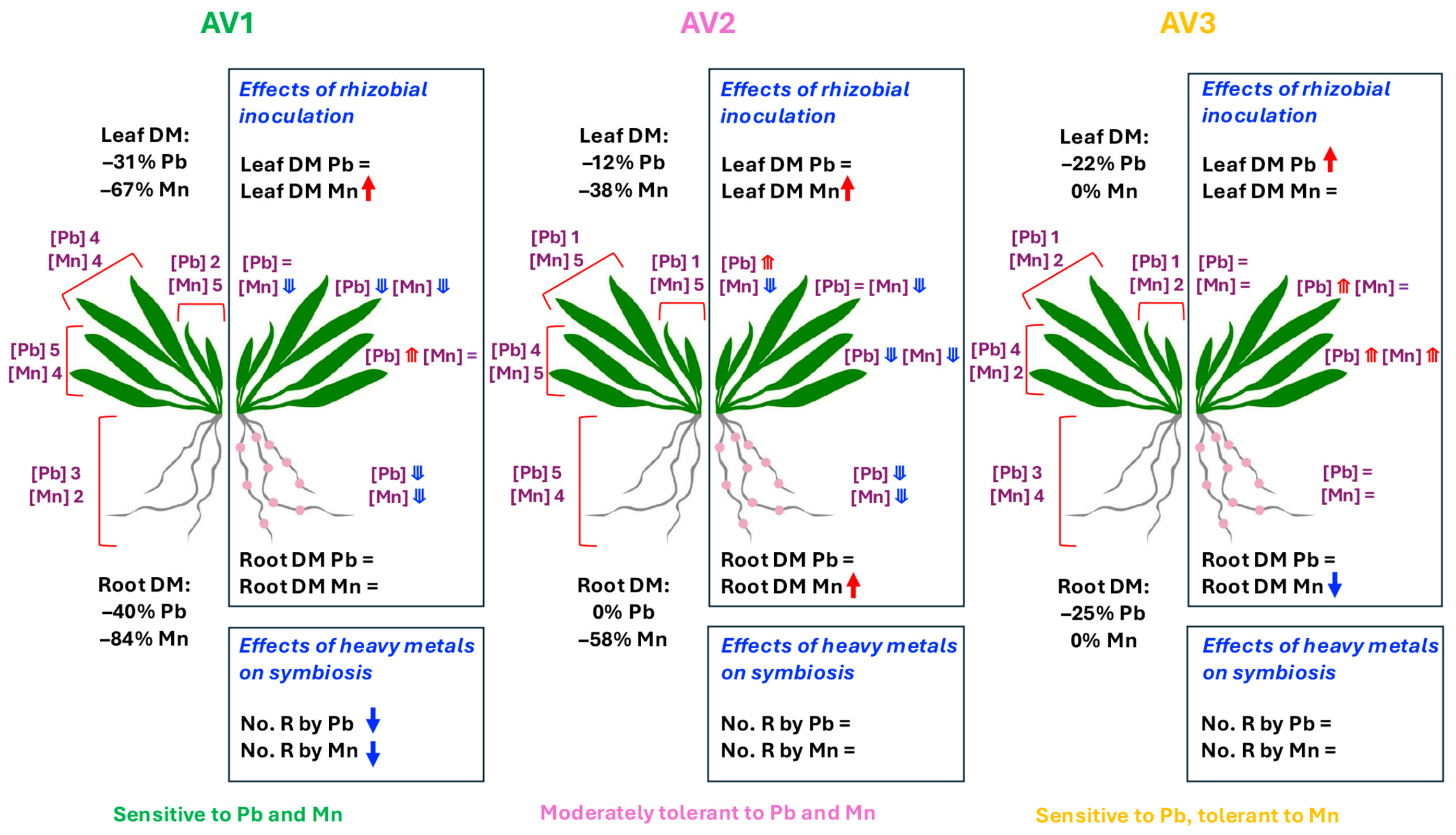
The objective of the present study was to compare the responses of three distinct A. vulneraria genotypes to Pb and Mn stress, with a particular emphasis on growth, physiological characteristics, and metal accumulation patterns. It was hypothesized that responses of contrasting A. vulneraria genotypes to Pb and Mn are influenced by inoculation with a specific rhizobial strain, resulting in genotype- and metal-specific differences in plant performance.
4. Materials and Methods
4.1. Plant and Microbiological Material
Three different genotypes of Anthyllis vulneraria were used in the present study. Seeds of Anthyllis vulneraria subsp. maritima (AV1) were collected on coastal dunes near Ovīši, Latvia (57°34′31.9″ N, 21°43′16.5″ E). Seeds of Anthyllis vulneraria (AV2) and Anthyllis vulneraria var. coccinea (AV3) were purchased from Jelitto Staudensamen GmbH (Schwarmstedt, Germany).
Rhizobia were obtained from wild-grown
A. vulneraria subsp.
maritima plants collected in November in the coastal dunes of Jūrmala in Lielupe, Latvia (57°00′16.7″ N, 23°55′21.1″ E). Bacterial inoculant for plant treatment was obtained according to the methodology described previously [
35]. The inoculum was taxonomically identified based on previous characterization [
35]. Culture purity was verified prior to use, and inoculum was produced under standard laboratory conditions.
4.2. Plant Propagation and Growth Conditions
The experiment was performed during the winter season in partially controlled conditions of an experimental greenhouse. Propagation of plant material for experiments and plant cultivation were carried out according to the procedures described earlier [
28,
35]. Briefly, seeds were surface sterilized using diluted household bleach and scarified, before sowing in sterile plastic tissue culture containers with sterilized commercial substrate (Garden Soil, Biolan, Eura, Finland). Containers were incubated in a growth cabinet at 23 °C in low light conditions. After appearance of the first true leaves, individual seedlings were transplanted to 200 mL plastic containers with a mixture (3:2,
v/
v) of Garden Soil and quartz sand (Saulkalne-S, Saulkalne, Latvia), placed in 48 L plastic boxes and accommodated to greenhouse conditions using gradual ventilation. After two weeks, plants were individually transplanted to 1.2 L containers with the same substrate.
Plants were cultivated in a greenhouse (MortiMaX, Maasdijk, The Netherlands) with supplemented light (photon flux density of photosynthetically active radiation 380 μmol m−2 s−1 at the plant level), for a 16 h photoperiod, at day/night temperature 23/16 °C, and a relative air humidity of 60 to 70%. Substrate water content was monitored daily with an HH2 moisture meter equipped with WET-2 sensor (Delta-T Devices, Burwell, UK) and maintained at 50 to 60% of volumetric water content using deionized water.
One week after the last treatment, the plants were fertilized with Yara Tera Kristalon Red and Yara Tera Calcinit fertilizers (Yara International, Oslo, Norway). A stock solution was prepared for each fertilizer (100 g L−1) and the working solution contained 25 mL of each per 10 L deionized water, used with a rate of 0.1 L per container.
4.3. Inoculation and Treatments
One week after the final transplanting, plants were assigned to six treatment groups, five replicates (individual containers) per treatment (
Table 6). After that, the respective plants were inoculated with a bacterial suspension (about 10
9 colony-forming units per mL; estimated by a standard plate count method), applied in six points (1 mL each) per container over the surface of the substrate. Treatment with heavy metals was carried out two separate times, three and four weeks after rhizobial inoculation. Two heavy metals, Pb and Mn, were selected for treatments, based on their pronounced negative effect on
A. vulneraria plants in similar experimental conditions [
28]. The lowest concentration that caused a negative effect on growth was used, 500 mg of the respective metal, Pb or Mn, per L of substrate (
Table 6). These concentrations were selected as experimental stress levels and may not directly correspond to natural field conditions. The indicated amount of respective salt was dissolved in deionized water and 0.1 L per container was applied to the substrate surface in each container. Plants were cultivated for four additional weeks after the full treatment with heavy metals. During this period, the morphological characteristics of visually assessed average plants from each treatment were photographed once a week.
4.4. Measurement of Chlorophyll a Fluorescence
The chlorophyll
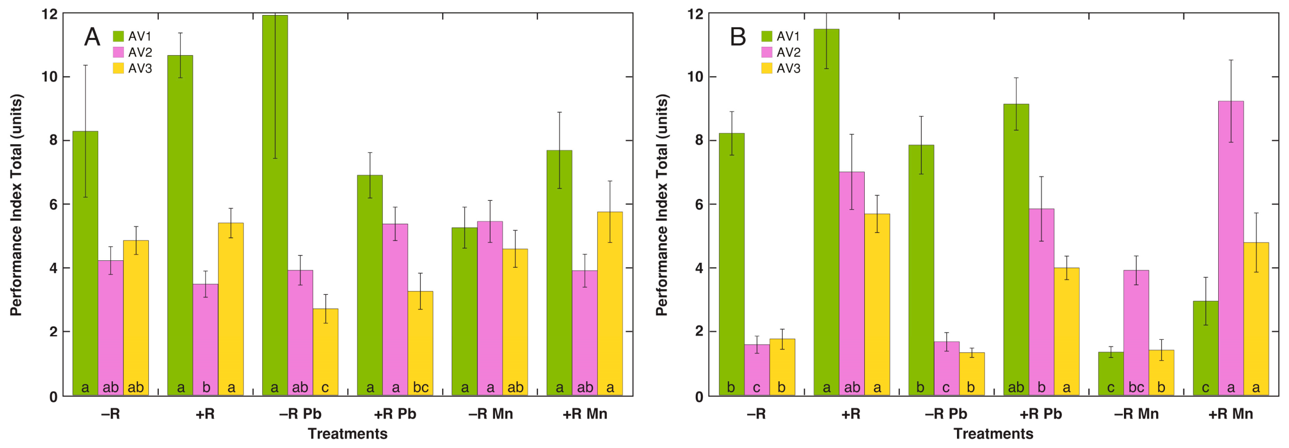
a fluorescence was measured in leaves dark-adapted for at least 20 min with a Handy PEA fluorometer (Hansatech Instruments, King’s Lynn, UK). Three measurements of fast fluorescence induction per individual plant were performed. Fluorescence data analysis was conducted utilizing PEA Plus software (version 3.11, Hansatech Instruments, King’s Lynn, UK). To characterize photochemical performance of photosynthesis, Performance Index Total was used. This complex parameter combines four function-related parameters (trapping of absorbed exciton, electron transport between the photosystems, reduction in end-electron acceptors, and the status of photosystem I) [
36].
4.5. Termination of the Experiment
At harvest, the plant leaves were classified into three age categories based on their position in the shoot, degree of expansion, and visual appearance. Young leaves were defined as recently emerged, upright, located in the center of the shoot, and not fully expanded leaves with light green coloration. Middle leaves were fully expanded, located to the edge of the new leaves, and uniformly dark green. Old leaves were basal leaves, usually horizontally oriented, exhibiting signs of senescence, including yellowing or chlorosis and reduced turgor. Leaves were counted and weighed separately. The roots were separated from the substrate and washed to remove any soil particles. The relative degree of nodule presence was evaluated according to the four point scale (0, no nodules; 1, a few nodules (1–5) at only one point; 2, small groups of nodules (<10) at several points on roots; 3, a large number of nodules (>10) throughout the root length). This measure was not intended as a quantitative assessment of symbiotic efficiency, and more detailed parameters (e.g., nodule number, biomass, or activity) were not evaluated in this study. All plants were included in the analysis irrespective of their nodulation status.
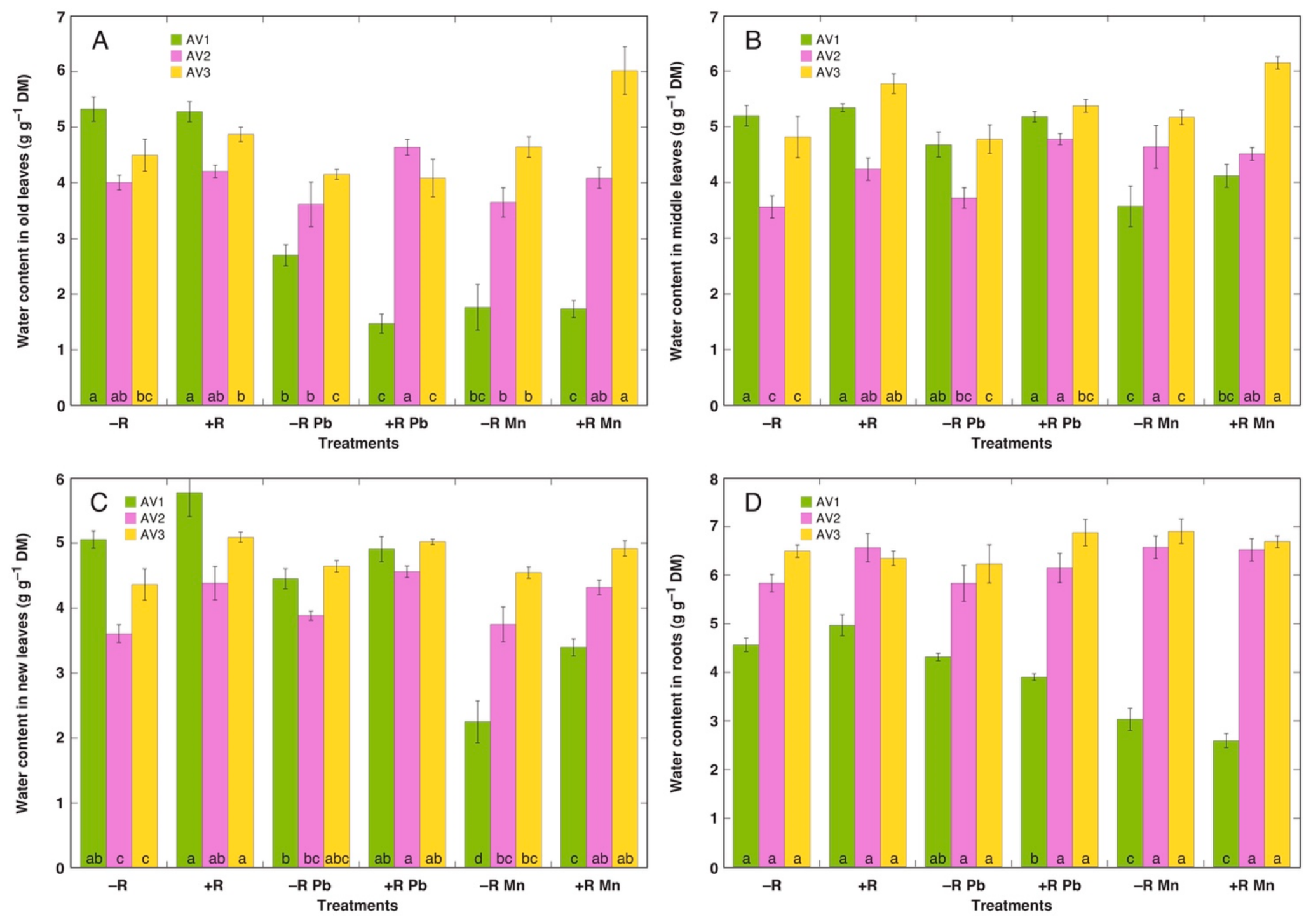
Plant material was weighed separately before and after drying in an oven at 60 °C for 72 h. The tissue water content was estimated as a mass of water in grams per gram of dry mass.
4.6. Analysis of Heavy Metals
Heavy metal concentrations in shoot and root samples were measured in triplicate as described in detail previously using a microwave plasma atomic emission spectrometer (4200 MP-AES, Agilent, Santa Clara, CA, USA) [
37]. From each treatment, three individual plants were randomly selected for analysis, which represents a limitation for interpretation of metal accumulation data.
4.7. Data Analysis
The data were analyzed by KaleidaGraph (v. 5.0, Synergy Software, Reading, PA, USA). All measured parameters satisfied the assumptions of normality and homogeneity of variance according to the Shapiro–Wilk and Levene tests. Statistical significance of differences for measured parameters between treatments was evaluated by two-way ANOVA followed by post hoc analysis by Tukey honestly significant difference test. The two factors considered were rhizobial inoculation and treatment with heavy metals. Analyses were performed separately for each A. vulneraria genotype. Significant differences were indicated at p < 0.05.




 ,
,