Article

Unleashing the Power of Fungi: Utilizing the Arbuscular Mycorrhizal Fungi Rhizophagus clarus to Mitigate Salinity Stress and Boost Cowpea Bean Productivity for Food Security

by
Davi Santos Tavares
1,
Bruno Francisco Sant’Anna-Santos
2 and
Marcelo Pedrosa Gomes
3,*
1
Environmental Biogeochemistry Laboratory, Institute of Agrarian and Technological Science, Federal University of Rondonópolis, Av. dos Estudantes, 5055, Cidade Universitária, Rondonópolis 78736-900, Mato Grosso, Brazil
2
Laboratory of Biomechanics and Plant Anatomy, Department of Botany, Sector of Biological Sciences, Federal University of Paraná, Av. Coronel Francisco H. dos Santos, 100, Centro Politécnico Jardim das Américas, P.C. 19031, Curitiba 81531-980, Paraná, Brazil
3
Laboratory of Plant Estress Physiology, Department of Botany, Biological Sciences Sector, Federal University of Paraná, Av. Coronel Francisco H. dos Santos, 100, Centro Politécnico Jardim das Américas, P.C. 19031, Curitiba 81531-980, Paraná, Brazil
*
Author to whom correspondence should be addressed.
Stresses 2024, 4(2), 393-410; https://doi.org/10.3390/stresses4020026
Submission received: 30 April 2024 / Revised: 6 June 2024 / Accepted: 11 June 2024 / Published: 19 June 2024
(This article belongs to the Collection Feature Papers in Plant and Photoautotrophic Stresses)

Abstract

:
The increasing demands for food, driven by shrinking arable land areas and a growing population, underscore the need for innovative agricultural practices to mitigate the effects of soil degradation due to salinity and promote food security, particularly in regions heavily impacted by salinity. In this study, we investigated the effects of inoculating the arbuscular mycorrhizal fungus (AMF) Rhizophagus clarus on the productivity of Vigna unguiculata cv. BRS Imponente plants in response to salinity (0, 25, 50, 75, and 100 mM). We found that NaCl concentrations ≥ 50 mM were phytotoxic, reducing plant growth and productivity. However, inoculation with AMF reduced plant oxidative stress (hydrogen peroxide concentration and lipid peroxidation) and ionic stress (Na+/K+ ratio). Inoculated plants exhibited increased antioxidant enzyme activity (ascorbate peroxidase and catalase), higher P and K concentrations, and lower Na concentrations in their leaves. As a result, salt did not interfere with grain production in the AMF-inoculated plants. For the first time, we demonstrate that inoculation with R. clarus can counteract the harmful effects of NaCl in V. unguiculata plants, ensuring their grain yields. Therefore, amid the escalating soil salinization globally, the AMF R. clarus emerges as a practical approach to ensure cowpea yields and enhance production in deteriorating agricultural lands, especially in saline areas. This can significantly contribute to promoting food security.

1. Introduction

The cowpea bean (Vigna unguiculata (L.) Walp.) stands as a pivotal crop for global food security, owing to its protein-rich grains and remarkable adaptability to diverse environmental conditions [1]. Cultivated extensively worldwide, cowpeas are a vital source of sustenance and income, particularly in regions vulnerable to water scarcity [2,3]. However, the escalating phenomenon of soil salinization poses a pervasive threat to agricultural productivity, impacting approximately one billion hectares of land [4]. Projections suggest that by 2050, half of the world’s agricultural regions will contend with salinity issues [5].
Salinity stress poses a multifaceted challenge to plant growth and development, exerting detrimental effects at various levels, from cellular physiology to whole-plant morphology. At the cellular level, salinity stress disrupts osmotic balance, leading to water deficit and reduced turgor pressure within plant cells [6]. This osmotic stress impairs nutrient uptake and transport, hindering essential metabolic processes such as photosynthesis and protein synthesis [6]. Additionally, the accumulation of toxic ions, primarily sodium (Na+) and chloride (Cl), disrupts ion homeostasis and interferes with enzyme activities, further compromising plant health [6].
Plants respond to salinity stress by modulating various physiological and biochemical processes. They undergo osmotic adjustment, which involves the accumulation of organic and inorganic solutes to reduce cellular osmotic potential and maintain turgor pressure [7]. This adjustment process includes the accumulation of compatible solutes like proline and soluble sugars, which protect plant cells against the adverse effects of salt stress and help in osmotic adjustment [8]. Additionally, plants may adjust their osmotic potential to maintain turgor pressure, which can exacerbate difficulties in measuring their relative water content (RWC) using classical methods [9]. At the morphological level, plants exhibit responses in their roots and leaves to reduce water loss and promote water use efficiency (WUE). Plants subjected to salinity stress often exhibit stunted growth, a reduced leaf area, and altered root architecture. In response to osmotic stress, plants may develop smaller leaves with reduced stomatal density to minimize water loss through transpiration [10]. Concurrently, roots may undergo extensive remodeling, with decreased root lengths and the proliferation of lateral roots in search of less-saline soil regions [10]. However, excessive salt accumulation in roots can lead to cellular damage and inhibit nutrient uptake, exacerbating the effects of salinity stress. Anatomically, salinity stress induces alterations in leaf structure and cellular organization. Leaves exposed to high salt concentrations may exhibit reduced leaf thickness, chloroplast disorganization, and impaired photosynthetic capacity [11,12]. Moreover, salinity stress can disrupt vascular tissue integrity, impairing nutrient and water transport within the plant. These anatomical changes contribute to reduced growth, photosynthesis, and overall plant productivity under saline conditions [6]. While certain plant species have evolved mechanisms to tolerate salinity, including salt exclusion, increased leaf succulence, and the redistribution of salt within tissues [7], the deleterious effects on productivity persist [13]. As a result, there is an urgent need to explore strategies that mitigate salt stress and enhance plant tolerance, thereby safeguarding agricultural sustainability [14].
In mitigating salt stress, arbuscular mycorrhizal fungi (AMF) are crucial in enhancing plant resilience to salinity. AMF form symbiotic associations with plant roots, facilitating nutrient uptake, particularly phosphorus (P), and improving plant water relations. Under saline conditions, AMF contribute to maintaining ion homeostasis by reducing Na+ uptake and enhancing the absorption of essential nutrients, such as potassium (K+) and calcium (Ca2+), which counteracts the adverse effects of salinity stress [15]. Additionally, AMF colonization promotes the synthesis of osmoprotectants and antioxidants in plants, mitigating oxidative stress and cellular damage induced by salt ions [15]. Furthermore, AMF-mediated alterations in root architecture, including increased root length and surface area, enhance the plant’s access to water and nutrients, improving plant performance under saline conditions.
Inoculation with symbiotic microorganisms, notably arbuscular mycorrhizal fungi (AMF), has emerged as a promising avenue of research to address salinity stress in plants. AMF play crucial roles in mitigating salt stress by modulating ion uptake, optimizing nutrient absorption, and bolstering plant defense mechanisms [15]. Moreover, AMF induce the activity of antioxidant systems and modulate phytohormone profiles, thereby minimizing the adverse effects of salts on plant growth and development [15].
Against this backdrop, this study investigates the interaction between salinity stress and plant responses, focusing on elucidating the mechanisms by which AMF mitigate ion toxicity. We examine the influence of Rhizophagus clarus AMF inoculation on cowpea bean plants cv. BRS Imponente under salinity stress conditions. This fungus has been reported to be tolerant to increased soil salinity, effectively forming a symbiosis with plants, such as maize, soybean, and cowpea (Vigna sinensis), under saline stress conditions and enhancing plant performance [16,17,18]. By elucidating the physiological and anatomical adaptations induced by saline stress and symbiosis with AMF, our study aims to underscore the potential of AMF inoculation in enhancing cowpea productivity in saline soils. Ultimately, our findings seek to contribute to developing sustainable agricultural practices that mitigate the adverse effects of salinization on crop production, thereby bolstering global food security.

2. Results

2.1. Mycorrhizal Colonization

In the absence of inoculation, no fungal structures were detected in the plant roots (Figure 1A). However, the percentage of fungal structures in root tissues (mycorrhizal colonization) decreased with increasing salinity (p < 0.05) in the inoculated plants (Figure 2).

2.2. Biochemical Evaluations

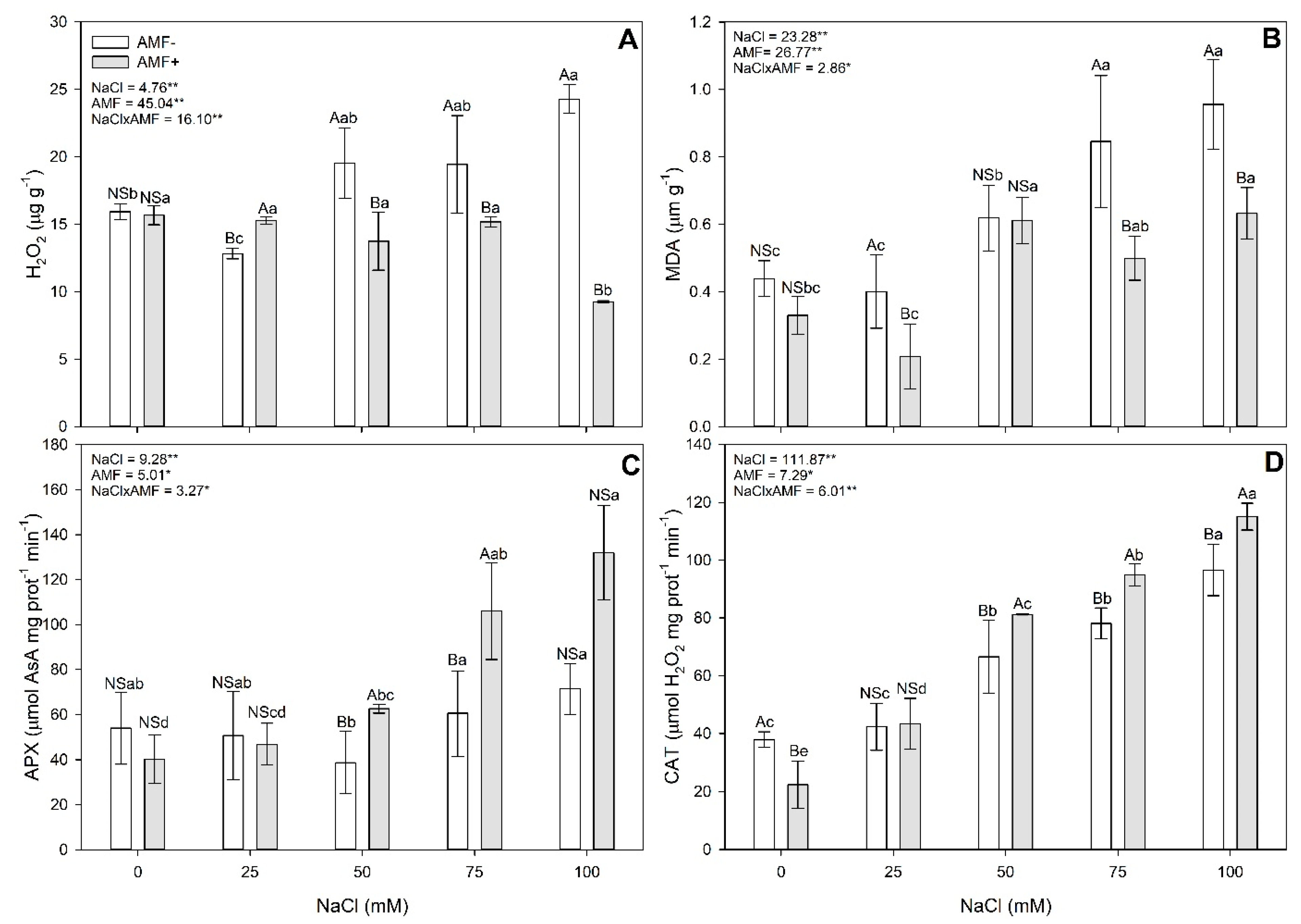
A significant interaction (p < 0.05) was observed between NaCl concentrations and AMF inoculation for all evaluated oxidative stress markers (Figure 3). In the non-inoculated plants, the concentrations of hydrogen peroxide (H2O2) in leaves increased significantly with the 100 mM NaCl treatment compared to the control (Figure 3A). Similarly, malondialdehyde (MDA) concentrations and catalase (CAT) activity increased in the leaves of plants exposed to NaCl concentrations ≥50 mM. In contrast, the inoculated plants exhibited increased MDA concentrations, ascorbate peroxidase (APX), and CAT activity in leaves with NaCl concentrations ≥50 mM. Regardless of the NaCl concentration, the CAT activity was significantly higher in inoculated plants treated with salt compared to the control. Moreover, lower H2O2 concentrations and greater levels of APX and CAT activity were observed in the inoculated plants compared to their respective controls without inoculation. MDA concentrations (indicative of lipid peroxidation) were lower in the leaves of inoculated plants compared to non-inoculated plants exposed to 25, 75, and 100 mM NaCl.
A significant interaction (p < 0.05) was observed between NaCl concentrations in the substrate and inoculation with AMF for all evaluated oxidative stress markers (H2O2, MDA, APX, and CAT) (Figure 3). In the non-inoculated plants, the concentrations of H2O2 in the leaves increased in plants treated with 100 mM NaCl relative to the control (Figure 3A), and the MDA concentrations (Figure 3B) and CAT activity (Figure 3D) increased in the leaves of plants exposed to NaCl concentrations ≥50 mM. Similarly, the inoculated plants showed increased MDA concentrations and increased activity of APX in the leaves (Figure 3C) of plants exposed to NaCl concentrations ≥50 mM (Figure 3). Regardless of the NaCl concentration, CAT activity was more significant in inoculated plants treated with salt compared to the control (Figure 3D). Lower H2O2 concentrations and greater levels of APX and CAT activity were observed for the treatments with inoculation (AMF+) compared to their respective controls (same NaCl concentration) without inoculation (AMF−) (Figure 3). MDA concentrations (lipid peroxidation) were lower in the leaves of inoculated plants compared to non-inoculated plants exposed to 25, 75, and 100 mM NaCl (Figure 3B).

2.3. Leaf Anatomy

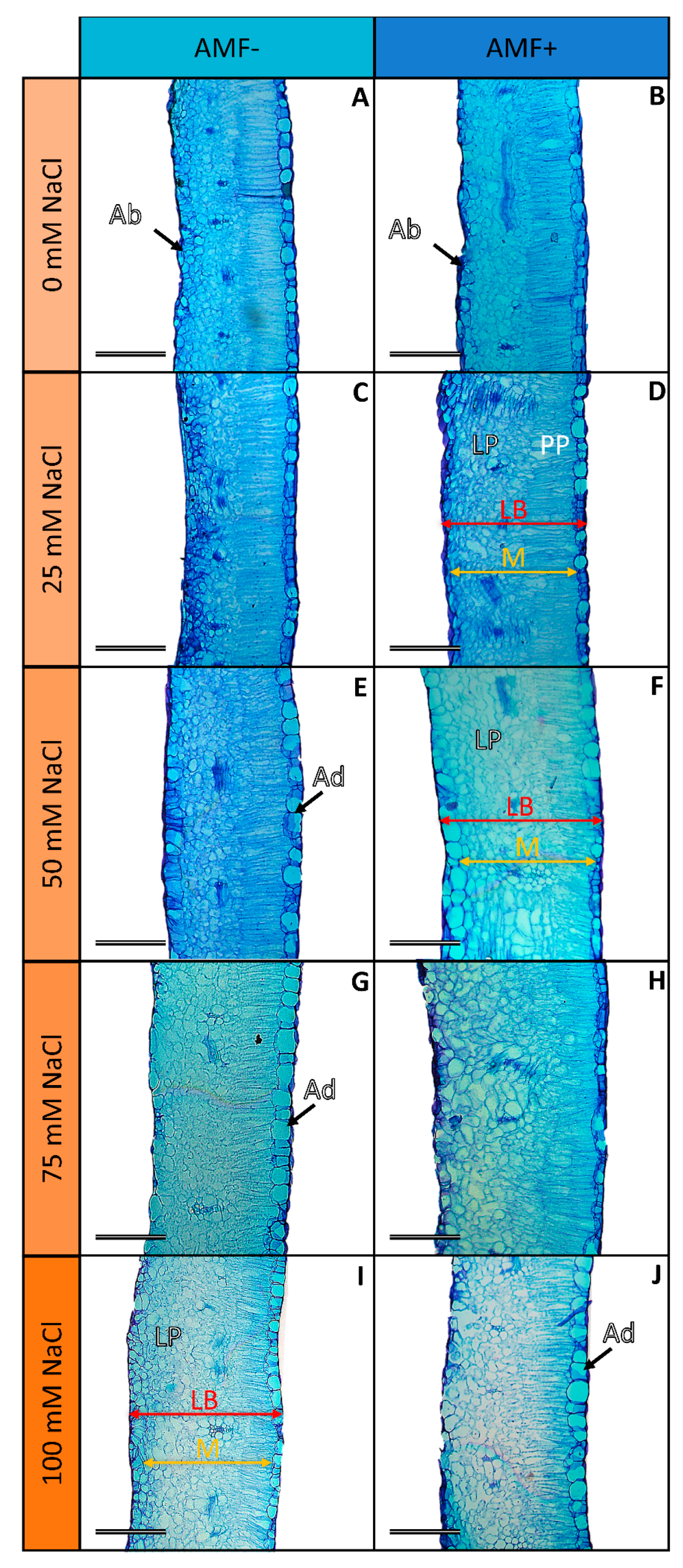
NaCl concentrations and mycorrhizal association significantly affected the thickness of various leaf tissues (p < 0.05; Figure 4 and Figure 5). In non-mycorrhizal plants, increased leaf blade, mesophyll, and lacunous parenchyma thicknesses were observed under the 75 and 100 mM NaCl treatments compared to those observed with the other concentrations. In contrast, the inoculated plants exhibited more excellent leaf blade and mesophyll thicknesses under the 50 and 75 mM NaCl treatments compared to those of the control and 100 mM NaCl concentrations. Additionally, mycorrhizal association led to increased thicknesses of the lacunous parenchyma and palisade parenchyma under specific NaCl concentrations.

2.4. Nutrient Concentration in Leaves

A significant interaction (p < 0.05) between the NaCl concentrations and AMF inoculation was observed for the concentration of all macronutrients and micronutrients except sulfur (S) (Table S1). Both the AMF+ and AMF− plants exhibited reductions in potassium (K), calcium (Ca), and sulfur (S) concentrations under specific NaCl concentrations. However, the AMF+ plants showed higher potassium concentrations under the 75 and 100 mM NaCl treatments. Mycorrhizal association also promoted increased phosphorus (P) concentrations under NaCl concentrations ≥ 50 mM. Additionally, inoculated plants exhibited lower magnesium (Mg) concentrations in leaves at all saline concentrations (Table 1).

2.5. Ion Stress Markers

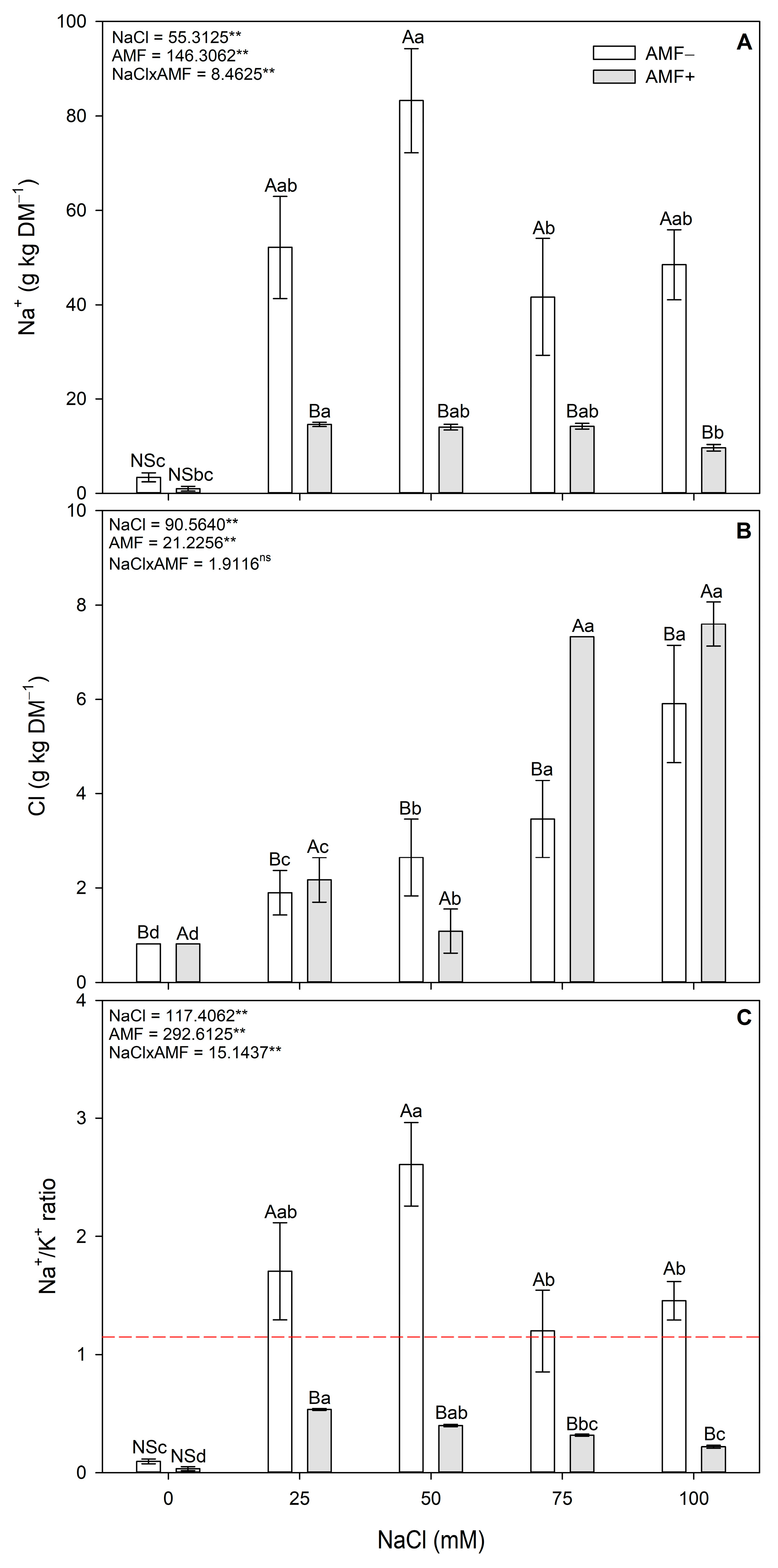
Increasing the salinity resulted in a significant accumulation of sodium (Na) and chlorine (Cl) in the cowpea leaves, as well as an increase in the Na+/K+ ratio for both AMF− and AMF+ plants (Figure 6). Inoculation with R. clarus reduced the Na+ concentration and the Na+/K ratio compared to each respective control (AMF−) (p < 0.05; Figure 6A,C).

2.6. Grain and Biomass Yields

A significant interaction between factors was observed for shoot and root biomass production (Table 2; p < 0.05). In AMF− plants, the shoot fresh mass (FM) accumulation was higher at the 0 and 75 mM NaCl concentrations, while in AMF+ plants, the concentration of 25 mM promoted the highest FM accumulation in the shoots. Salt concentrations ≥ 50 mM significantly decreased the grain yield in both AMF− and AMF+ plants, although the inoculated plants exhibited greater grain yields than the non-inoculated plants, regardless of the NaCl concentration (Figure 7; p < 0.05).

3. Material and Methods

3.1. Experimental Conditions

The inoculum of Rhizophagus clarus ([T. H. Nicolson and N. C. Schenck] C. Walker and A. Schüßler, 2010) used was acquired from Embrapa Agrobiologia (Rio de Janeiro, Brazil). This species of AMF was chosen due to its high efficiency in colonizing V. unguiculata roots [19]. According to the manufacturer’s specifications, the inoculum consisted of 429 spores (3.5 g of inoculum) per pot. For the non-inoculated treatments (AMF−, 3.5 g of autoclaved inoculum (127 °C, 30 min) was used. The inoculum was applied and slightly homogenized to the topsoil before sowing. The seeds of Vigna unguiculata cv. BRS Imponente, a 2018 crop, were purchased from Cebal Agro (Paraná, Brazil). The seed lot showed 100% viability when evaluated using the tetrazolium test [20]. Five seeds were sown in each pot after surface disinfection with sodium hypochlorite (1.5%) for two minutes and washing with deionized water.
Sand and vermiculite (2:1, v:v), autoclaved, were used as the substrate for plant growth. This substrate was chosen due to its low cation exchange capacity (CEC), representing a scenario of the total availability of salt in the solution. Two kilograms of this substrate was placed in 5 L plastic pots, free of leakage, that had previously been disinfected with 70% alcohol. The pots received 1.4 L (corresponding to 100% field capacity) of their respective saline solution (0, 25, 50, 75, and 100 mM). The electrical conductivity of each solution was measured as follows: 0.02, 3.99, 7.98, 12.00, and 15.54 dS m−1, respectively. The solutions were prepared using HPLC-grade NaCl (Sigma-Aldrich, São Paulo, Brazil) and distilled water. The pots were kept in a greenhouse (25 °C), and when 70% of field capacity was reached (three days), fungal inoculation and sowing were carried out.
The pots were kept in a greenhouse (25 °C; 500 µmol photons m−2 s−1). After seedling emergence (seven days), thinning was performed, keeping three healthy and similar-height plants in each pot. Ten pots per treatment were used, each containing three plants, resulting in thirty plants per treatment. Each plant was considered a replicate. The substrate was maintained at 70% of field capacity, verified daily, and, if necessary, corrected by adding distilled water. Plant nutrition was supplied through a weekly application (300 mL) of a complete solution (Hoagland and Arnon 1950). In the initial stage of culture, in the first and second weeks, the solution was diluted to ¼ and ½ strength, respectively. The nutrient solution was applied at full strength from the third week onwards. The experiment was conducted until the complete pod-filling stage (R5) was reached.

3.2. Evaluations

3.2.1. Mycorrhizal Colonization and Biochemical, Anatomical, and Nutritional Evaluations

Mycorrhizal colonization and physiological and anatomical evaluations were performed at the physiological maturity stage (R5 stage) in ten replicates per treatments. Mycorrhizal colonization (%) was determined using the Giovannetti and Mosse method described in [21]. Colonization was determined under a microscope (CarlZeiss, Axiolab re, Oberkochen, Germany) with a 5 mm grid slide, and the following equation determined the percentage: (total colonized roots/total roots studied) × 100.
The third and fourth leaves (the first fully expanded leaves from the apex) were utilized for biochemical and anatomical evaluations. Samples for the biochemical evaluations were harvested and immediately frozen in liquid nitrogen and stored at −80 °C until analysis. The concentrations of hydrogen peroxide [22] and malondialdehyde (MDA) [23], as well as the activity of catalase (CAT—EC 1.11.1.6) [24] and ascorbate peroxidase (APX—EC 1.11.1.11) [25], were measured using 0.1 g of the leaf samples. The extraction of antioxidant enzymes from the leaves was carried out using 1000 µL of an extraction buffer containing 100 mM phosphate buffer (pH 7.8), 100 mM ethylenediaminetetraacetic acid (EDTA), 1 mM ascorbate, and 2% polyvinylpyrrolidone (PVP) [26]. The Bradford method was used to quantify the protein content [27].
Anatomical evaluations were performed following the method described by Sant’Anna-Santos [28]. The material was fixed in FAA (formaldehyde + acetic acid + 70% ethanol 5:5:90 v/v/v) for 72 h and then stored in 70% ethanol [29]. After dehydration in an ethyl/butyl series, the samples were embedded in histological histories. Transverse sections of 5 µm were made using a semi-automatic rotary microtome (Olympus CUT 4055). The sections were stained with toluidine blue, and the slides were mounted with synthetic Canada balsam. All slides were examined and photographed using a light microscope (Carl Zeiss Axiolab, São Paulo, Brazil) and a digital camera 7.2 Mpix (Sony, Tokyo, Japan)). The midrib was used as a reference to evaluate leaf characteristics. The thicknesses of the leaf blade, mesophyll, lacunous, and palisade parenchyma, as well as the thicknesses of the abaxial and adaxial epidermis, were evaluated. Evaluations were performed in three sections/repetition. Thickness data were obtained using Anati Qaunti 2.0 software [30], with 3 observations/section/repetition, comprising 27 measurements for each structure/treatment evaluated.
After collecting material for the anatomical and biochemical assessments, the remaining leaves were pooled for nutritional evaluation. The leaves were dried at 65 °C until a constant weight was obtained and ground in a portable mill. A 0.50 g amount of the dry material was digested in a muffle furnace at 500 °C for three hours. Then, 1 mL of 3 M HCl was added, and the samples were heated again at 500 °C for three hours, according to Martins and Reissmann [31]. After cooling to room temperature, mineral elements were extracted by adding 5 mL of 3 M HCl on a hot plate at 200 °C for 10 min. The samples were then filtered on a laboratory blue classification filter paper, and their volume was completed to 50 mL with distilled water. The determination of macronutrient (P, K, Ca, Mg, and S) and micronutrient (Cu, Fe, Mn, and Zn) concentrations was performed by inductively coupled plasma optical-emission spectroscopy (ICP-OES; Varian Inc. model 720-ES, Palo Alto, CA, USA). The sodium (Na) content was determined with atomic spectroscopy using a flame photometer (Dm-62; Digimed, São Paulo, Brazil). Reference material (Specsol) with known concentrations of elements and analytical blanks were used for quality control. The chloride (Cl) content was determined through titration, following the Mohr method [32], using 1 g of the leaf samples. Chloride was extracted in 0.085 M calcium nitrate (Ca(NO3)2 4H2O) and determined using 0.0282 M silver nitrate (AgNO3) in the presence of potassium chromate 0.258 M (K2CrO4) as an indicator.

3.2.2. Grain and Biomass Yields

At the end of the experimental period, 10 plants per treatment were harvested and divided into roots, leaves, and pods. The fresh and dry weights of the shoot (SFW and SDW) and root (RFW and RDW) biomass and the grain yield per hectare were determined based on a total of 250,000 plants ha−1. The dry weight of the plant material was obtained after drying at 65 °C in a forced-air oven.

3.3. Statistical Analyses

The experiment was randomized entirely with a double factorial composed of different salt concentrations (0, 25, 50, 75, and 100 mM of NaCl) × AMF (presence, absence). Statistical analyses were performed using JMP 13.2 software (SAS Inst. Inc, Cary, NC, USA.). The results were expressed as the mean of ten replicates. The data were tested for normality (Shapiro–Wilk) and homogeneity (Bartlett) and statistically evaluated using a two-way analysis of variance (ANOVA). The interactions between the NaCl addition (0, 25, 50, 75, and 100 mM) and the fungal inoculation (AMF− and AMF+) were included in the model. When the ANOVA detected differences, means were compared using the Tukey all-pairs HSD test (p < 0.05).4. Discussion
The expansion of saline areas globally, driven by anthropogenic factors and exacerbated by climate change-induced temperature rises, poses significant challenges to agricultural productivity and food security. Soil salinity-induced crop yield reductions threaten food vulnerability, particularly in regions where crops like the cowpea (Vigna unguiculata) serve as staple foods [15]. Addressing this challenge requires exploring sustainable solutions to ensure grain production and food safety in saline soils.
Our investigation focused on elucidating the potential of the mycorrhizal fungus Rhizophagus clarus to mitigate salt stress in BRS Imponente cowpea plants. Despite being moderately tolerant to soil salinity, cowpeas remain susceptible to the adverse effects of salt stress, including reduced biomass and grain productivity [33]. Our findings reveal that elevated sodium chloride (NaCl) concentrations (>50 mM) induce osmotic and ionic disturbances, leading to diminished grain yields, with reductions of up to 36% (Figure 7). Salinity-induced oxidative stress, as evidenced by the increased hydrogen peroxide (H2O2) levels (up to 52%) and malondialdehyde (MDA) concentrations (up to 117%) in our plant tissues (Figure 3), disrupts the balance of reactive oxygen species (ROS) and leads to lipid peroxidation, ultimately impeding plant growth and yield [20,34,35]. Although the plants exhibited increased activity levels of antioxidant enzymes such as ascorbate peroxidase (APX) and catalase (CAT) under salt stress, this was insufficient to prevent H2O2 accumulation and oxidative damage. The increase in the ROS concentration may result from salt interference in the electron transport chain in the chloroplasts and mitochondria, leading to the leakage of electrons and the production of ROS [20,34].
Salinity (NaCl concentrations ≥ 75 mM) also interfered with the uptake and transport of essential nutrients, resulting in deficiencies in P, K, and Ca, vital for various metabolic processes necessary for plant development [36]. Under saline conditions, P can precipitate with Ca and Mg, making these three nutrients unavailable for plants [36]. Due to their similar physicochemical characteristics, K and Na compete for root absorption sites [15], affecting nutrient absorption and plant development. Deficiencies in P, K, and Mg can damage processes critical for plant development, such as energy generation and transfer, carbon metabolism, photosynthesis, membrane synthesis, enzyme activation, and stomatal regulation [37]. In addition to interfering with metabolic processes, salinity is known to induce changes in the anatomy and morphology of plants [38]. Changes in plant anatomy and disturbances in different cellular structures, such as chloroplasts [39], can significantly impact biochemical reactions [38]. The dimensions of the leaf tissues were affected by salinity, and the increase in thickness observed in the tissues of our plants is a typical response to saline stress [40]. This may be related to an attempt to dilute Na+ and Cl ions in the cellular space [7], as well as a response to compensate for the loss of photosynthetic area due to stresses caused by the salt [41]. However, the increase in leaf thickness may represent a barrier to CO2 diffusion, which can hinder photosynthetic processes [40], resulting in lower growth and biomass production.
The examination of mycorrhizal colonization revealed a significant decrease (up to 40%) (Figure 1B,D) in fungal structures within the root tissues of the inoculated plants under high-salinity conditions, a typical response to salinity [42,43]. This decline in mycorrhizal colonization could be attributed to the adverse effects of salinity on both plant and fungal growth, as salinity stress can impede the establishment and proliferation of mycorrhizal associations. Indeed, salt can inhibit spore germination and mycelial growth, impairing the formation and performance of the symbiosis [44]. Despite the reduction in colonization, arbuscular mycorrhizal fungi (AMF) proved beneficial in alleviating salt-induced oxidative and ionic stress in the cowpea plants. AMF inoculation ensured grain yields even under high NaCl concentrations (Figure 7). However, the inoculated plants exposed to 75 and 100 mM NaCl exhibited lower root biomass production than the non-inoculated plants (Table 2). This reduction could be due to the fungal biomass competing with the plant for carbon resources, potentially reducing root biomass production. Nevertheless, the reduction in root growth did not result in decreased production; thus, inoculated plants may allocate carbon towards grain production, resulting in lower biomass accumulation in their roots.
The salinity levels and AMF inoculation demonstrated a significant interaction concerning biochemical evaluations, affecting oxidative stress markers. The AMF+ plants, similar to the AMF− plants, exhibited oxidative damage when exposed to NaCl concentrations of ≥50 mM, as evidenced by lipid peroxidation. However, the AMF+ plants had significantly less damage than the AMF− plants, with only a 46% increase in MDA concentrations compared to a 110% increase in the AMF− plants. The AMF+ plants also showed more significant activity of antioxidant systems, as evidenced by the increased APX and CAT activity, which reduced ROS accumulation induced by the salt. This observation aligns with previous studies indicating the role of AMF in promoting antioxidant enzyme activity, such as ascorbate peroxidase (APX) and catalase (CAT), which are essential for scavenging reactive oxygen species (ROS) and maintaining cellular redox homeostasis [45,46].
The symbiosis with R. clarus also enhanced the plants’ nutrient uptake, particularly phosphorus (P) and potassium (K), essential for synthesizing antioxidant enzymes and maintaining cellular homeostasis under stress conditions [47]. In contrast, when exposed to salt stress, the AMF+ plants exhibited lower Na concentrations in their leaves. The Na/K ratio is an essential indicator of plant responses to salt stress since a high ratio can lead to cell damage and oxidative stress [37]. By increasing K nutrition and reducing Na concentrations, AMF can reduce the Na/K ratio, minimizing the harmful effects of salt-induced ionic stress [44]. AMF have developed various ways to increase P uptake in plants, including releasing phosphatases, expressing high-affinity phosphate transporter genes, and accumulating polyphosphates in hyphae [15]. They also induce the expression of high-affinity K transporters, which compete with sodium for uptake by the plant [48]. By improving the availability and uptake of other nutrients, AMF can help mitigate the adverse effects of Na on plant growth and development. The symbiosis with R. clarus promoted more excellent absorptions of Cu (control), Fe (0 and 75 mM NaCl), and Zn (50 and 100 mM NaCl), but reduced the Mn concentrations in the leaves of the plants at all salt concentrations. This lower absorption of Mn may be related to lower root exudation when associated with AMF, as these exudates reduce Mn in soils, facilitating its absorption by plants [49]. Additionally, the AMF+ plants exhibited lower magnesium (Mg) concentrations in their leaves at all saline concentrations (Table 1). This reduction in Mg concentrations could be due to competitive uptake interactions between Mg and other cations such as Ca or K, which might have been facilitated by the AMF inoculation. Interestingly, the symbiosis was inefficient in reducing chloride (Cl) in the leaves of the cowpea plants. Chloride is not as harmful to plants as Na, and there is no described selective uptake mechanism for Cl in plants. Chloride is an essential anion for photosynthesis and other plant metabolic processes. Therefore, AMF do not have specific mechanisms to reduce Cl concentrations in plants under salt stress. Under high concentrations, however, chloride (Cl) can become toxic and interfere with the absorption of nitrates and transport of organic acids inside plant cells, which can affect plant growth [50]. It is unclear if the negative responses of plants exposed to NaCl are related to Cl accumulation in their leaves, and further investigation is needed on this topic. However, it is clear that Cl accumulation did not disrupt the production of grains in the cowpeas since it was found to accumulate in the leaves of the AMF+ plants, and no effect on grain yield was observed. The differential nutrient concentrations in the AMF+ plants underscore the role of AMF in modulating nutrient uptake and allocation, contributing to stress tolerance and crop resilience [38].
The leaf anatomy studies also revealed differential responses to salinity and mycorrhizal association. In non-mycorrhizal plants, increased salinity resulted in thicker leaf tissues, potentially as an adaptive response to mitigate water loss and osmotic stress, suggesting morphological adjustments to balance water conservation and photosynthetic efficiency under saline conditions [50]. However, mycorrhizal inoculation modulated these responses, with the AMF-inoculated plants exhibiting optimal tissue thickness at intermediate salinity levels. This suggests that AMF may regulate plant morphology to enhance stress tolerance while optimizing resource utilization.
The integration of AMF inoculation into agricultural practices offers promising implications for enhancing crop productivity and food security in saline soils. By mitigating the adverse effects of salt stress on cowpea plants, AMF-based interventions provide a sustainable strategy to ensure grain production and food safety in regions vulnerable to soil salinization. Therefore, our findings underscore the pivotal role of mycorrhizal symbiosis in mitigating salt stress-induced crop yield reductions, emphasizing the potential of AMF-based interventions as a sustainable strategy to enhance agricultural resilience and promote food security in the face of climate change and environmental degradation. Inoculating cowpea plants (cv. BRS Imponente) with the AMF R. clarus represents a promising agricultural practice for mitigating the detrimental effects of salt stress and ensuring crop yields in saline soils. With a typical saline solution density of 1.0 g mL−1, the fungi enabled cowpea grain production in soils with up to 0.59% NaCl (w/w), which falls within the range of moderately saline soils in Brazil and Africa. In the semi-arid regions of Brazil and Africa, cowpeas are a staple food for millions of people. In Brazil, cowpeas are widely grown in the northeast region, known as “feijão-de-Corda” or “macaçar”. It is an essential crop for small farmers, who use it for consumption and sell it in local markets. In Africa, the cowpea is a critical crop for small-scale farmers, particularly in West and Central Africa, where it is a significant source of protein and income. It is also used for animal feed, and its leaves and stems are used for human consumption. The cowpea is a resilient crop that can tolerate drought, heat, and poor soils, making it an essential crop for food security in regions prone to climate variability and environmental stress. Therefore, inoculating cowpea plants (cv. BRS Imponente) with the AMF R. clarus represents a promising agricultural practice that can ensure cowpea production in saline soils and should be implemented as a technology to enhance crop productivity for food security, particularly in low-income regions.

4. Conclusions

Our research elucidates the beneficial effects of arbuscular mycorrhizal fungi (AMF) on cowpea plants under salt stress, highlighting the potential of AMF inoculation as a sustainable strategy for enhancing crop productivity in saline soils. Even under high-salinity conditions, the significant improvement in grain yields observed in AMF-inoculated plants underscores the economic viability of AMF-based interventions for salt-affected agricultural systems. Moreover, the adoption of AMF inoculation holds promise for broader social benefits, particularly in regions vulnerable to soil salinization. By promoting food security, enhancing agricultural sustainability, and reducing reliance on chemical inputs, AMF-based interventions offer a promising solution to address the challenges of salinity stress in global food production systems. In conclusion, our study provides valuable insights into cowpea plants’ physiological and anatomical responses to salinity stress, emphasizing the pivotal role of AMF in mediating plant–fungi interactions under adverse environmental conditions. Integrating AMF inoculation into agricultural practices represents a sustainable approach to mitigating the detrimental effects of soil salinity and ensuring food security in the face of climate change and environmental degradation.
Given the urgent need to increase food production and distribution efficiency amidst population growth and its environmental impacts, the drastic increase in soil salinization worldwide necessitates innovative solutions. The utilization of AMF, such as R. clarus, emerges as a promising tool to ensure crop yields and stimulate production in declining agricultural lands, particularly saline areas. Through addressing soil salinity challenges, AMF-based interventions can contribute to the pursuit of food security, aligning with the sustainable development goals of the United Nations.

Supplementary Materials

The following supporting information can be downloaded at https://www.mdpi.com/article/10.3390/stresses4020026/s1: Table S1: ANOVA summary of all analyzed variables.

Author Contributions

D.S.T., B.F.S.-S. and M.P.G. conceived and designed the experiments. B.F.S.-S. and M.P.G. gave technical support and conceptual advice. D.S.T. and M.P.G. performed the experiments and analyses. D.S.T. and M.P.G. wrote the manuscript. All authors have read and agreed to the published version of the manuscript.

Funding

This research was financed in part by the Coordenação de Aperfeiçoamento de Pessoal de Nível Superior (CAPES, Brazil; Finance Code 001). In addition, M.P. Gomes received a research productivity grant from Conselho Nacional de Desenvolvimento Científico e Tecnológico (CNPq, Brazil; Finance Code 302226/2022-2).

Institutional Review Board Statement

Not applicable.

Data Availability Statement

The original contributions presented in the study are included in the article/Supplementary Materials, further inquiries can be directed to the corresponding author/s.

Conflicts of Interest

The authors declare that they have no known competing financial interests or personal relationships that could have influenced the work reported in this paper.

References

  1. FAO. Pulses for Food Security and Nutrition: How Can Their Full Potential Be Tapped? FAO: Rome, Italy, 2016; Volume 2. [Google Scholar]
  2. Gonçalves, A.; Goufo, P.; Barros, A.; Domínguez-perles, R.; Trindade, H.; Rosa, E.A.S.; Rodrigues, M. Cowpea (Vigna unguiculata L. Walp), a renewed multipurpose crop for a more sustainable agri-food system: Nutritional advantages and constraints. J. Sci. Food Agric. 2016, 96, 2941–2951. [Google Scholar] [CrossRef]
  3. Enyiukwu, D.; Amadioha, A.; Ononuju, C. Nutritional significance of cowpea leaves for human consumption. Greener Trends Food Sci. Nutr. 2018, 1, 1–10. [Google Scholar] [CrossRef]
  4. Omuto, C.; Vargas, R.; El Mobarak, A.; Mohamed, N.; Viatkin, K.; Yigini, Y. Mapping of Salt-Affected Soils—Technical Manual; FAO: Rome, Italy, 2020. [Google Scholar] [CrossRef]
  5. Jamil, A.; Riaz, S.; Ashraf, M.; Foolad, M.R. Gene expression profiling of plants under salt stress. CRC Crit. Rev. Plant Sci. 2011, 30, 435–458. [Google Scholar] [CrossRef]
  6. Balasubramaniam, T.; Shen, G.; Esmaeili, N.; Zhang, H. Plants’ response mechanisms to salinity stress. Plants 2023, 12, 2253. [Google Scholar] [CrossRef]
  7. Acosta-Motos, J.R.; Ortuño, M.F.; Bernal-vicente, A.; Diaz-Vivancos, P.; Sanchez-Blanco, M.J.; Hernandez, J.A. Plant responses to salt stress: Adaptive Mechanisms. Agronomy 2017, 7, 18. [Google Scholar] [CrossRef]
  8. Kumar, S.; Li, G.; Yang, J.; Huang, X.; Ji, Q.; Liu, Z.; Ke, W.; Hou, H. Effect of Salt Stress on Growth, Physiological Parameters, and Ionic concentration of water dropwort (Oenanthe javanica) Cultivars. Front. Plant Sci. 2021, 12, 660409. [Google Scholar] [CrossRef]
  9. Negrão, S.; Schmöckel, S.M.; Tester, M. Evaluating physiological responses of plants to salinity stress. Ann. Bot. 2017, 119, 1–11. [Google Scholar] [CrossRef]
  10. dos Santos, T.B.; Ribas, A.F.; de Souza, S.G.H.; Budzinski, I.G.F.; Domingues, D.S. Physiological responses to drought, salinity, and heat stress in plants: A review. Stresses 2022, 2, 113–135. [Google Scholar] [CrossRef]
  11. Yao, X.-C.; Meng, L.-F.; Zhao, W.-L.; Mao, G.-L. Changes in the morphology traits, anatomical structure of the leaves and transcriptome in Lycium barbarum L. under salt stress. Front. Plant Sci. 2023, 14, 1090366. [Google Scholar] [CrossRef]
  12. Akcin, A.; Yalçın, E. The effect of salinity on anatomical characteristics of two halophyte species from Turkey. Bot. Lett. 2023, 170, 581–590. [Google Scholar] [CrossRef]
  13. Tagliaferre, C.; Guimarães, D.U.G.; Gonçalves, L.J.; Amorim, C.H.F.; Matsumoto, S.N.; D’arêde, L.O. Produtividade e tolerância do feijão caupi ao estresse salino. Irriga 2018, 23, 168–179. [Google Scholar] [CrossRef]
  14. Manaf, H.H.; Zayed, M.S. Productivity of cowpea as affected by salt stress in presence of endomycorrhizae and Pseudomonas fluorescens. Ann. Agric. Sci. 2015, 60, 219–226. [Google Scholar] [CrossRef]
  15. Kapoor, R.; Evelin, H.; Devi, T.S.; Gupta, S. Mitigation of salinity stress in plants by arbuscular mycorrhizal symbiosis: Current understanding and new challenges. Front. Plant Sci. 2019, 10, 450967. [Google Scholar] [CrossRef]
  16. Bisht, A.; Sharma, V.; Garg, N. Deciphering the role of arbuscular mycorrhizal fungi in mitigating the negative effects of abiotic stresses in legume crops. In Arbuscular Mycorrhizal Fungi in Sustainable Agriculture: Nutrient and Crop Management; Springer Nature: Singapore, 2024; pp. 337–361. [Google Scholar]
  17. Rabie, G.H.; Aboul-Nasr, M.B.; Al-Humiany, A. Increased salinity tolerance of cowpea plants by dual inoculation of an arbuscular mycorrhizal fungus Glomus clarum and a nitrogen-fixer Azospirillum brasilense. Mycobiology 2005, 33, 51. [Google Scholar] [CrossRef]
  18. de Carvalho Neta, S.J.; Araújo, V.L.V.P.; Fracetto, F.J.C.; da Silva, C.C.G.; de Souza, E.R.; Silva, W.R.; Lumini, E.; Fracetto, G.G.M. Growth-promoting bacteria and arbuscular mycorrhizal fungus enhance maize tolerance to saline stress. Microbiol. Res. 2024, 284, 127708. [Google Scholar] [CrossRef]
  19. Silva, G.A.E.; Siqueira, J.O.; Stürmer, S.L.; Moreira, F.M.S. Effectiveness of arbuscular mycorrhizal fungal isolates from the land uses of Amazon region in symbiosis with cowpea. An. Acad. Bras. Cienc. 2018, 90, 357–371. [Google Scholar] [CrossRef]
  20. Tavares, D.S.; Fernandes, T.E.K.; Rita, Y.L.; Rocha, D.C.; Sant’Anna-Santos, B.F.; Gomes, M.P. Germinative metabolism and seedling growth of cowpea (Vigna unguiculata) under salt and osmotic stress. S. Afr. J. Bot. 2021, 139, 399–408. [Google Scholar] [CrossRef]
  21. Dionísio, J.A.; Pimentel, I.C.; Signor, D.; de Paula, A.M.; Maceda, A.; Mattana, A.N.A.L. Guia Prático de Biologia do Solo, 1st ed.; de Lima, M.R., Neri, A., Grange, L., Genú, A.M., Moreira, A., Eds.; Sociedade Brasileira de Ciência do Solo/Núcleo Estadual Paraná: Curitiba, Brazil, 2016; ISBN 9788569146001. [Google Scholar]
  22. Velikova, V.; Yordanov, I.; Edreva, A. Oxidative stress and some antioxidant systems in acid rain-treated bean plants: Protective role of exogenous polyamines. Plant Sci. 2000, 151, 59–66. [Google Scholar] [CrossRef]
  23. Hodges, D.M.; DeLong, J.M.; Forney, C.F.; Prange, R.K. Improving the thiobarbituric acid-reactive-substances assay for estimating lipid peroxidation in plant tissues containing anthocyanin and other interfering compounds. Planta 1999, 207, 604–611. [Google Scholar] [CrossRef]
  24. Aebi, H. Catalase in vitro. Methods Enzymol. 1984, 105, 121–126. [Google Scholar]
  25. Nakano, Y.; Asada, K. Hydrogen peroxide is scavenged by ascorbate-specific peroxidase in spinach chloroplasts. Plant Cell Physiol. 1981, 22, 867–880. [Google Scholar]
  26. Gomes, M.P.; Gonçalves, C.A.; Brito, J.C.M.; Souza, A.M.; Cruz, F.V.S.C.S.; Bicalho, E.M.; Figueredo, C.C.; Garcia, Q.S. Ciprofloxacin induces oxidative stress in duckweed (Lemna minor L.): Implications for energy metabolism and antibiotic-uptake ability. J. Hazard. Mater. 2017, 328, 140–149. [Google Scholar] [CrossRef]
  27. Bradford, M.M. A rapid and sensitive method for the quantitation of microgram quantities of protein utilizing the principle of protein-dye binding. Anal. Biochem. 1976, 72, 248–254. [Google Scholar] [CrossRef]
  28. Sant’Anna-Santos, B.F.; dos Santos, S.A.; Nunes, E.L.P.; Francino, D.M.T.; Carvalho Júnior, W.G.O. Does leaf anatomy aid in species identification of Butia (Arecaceae)? AoB Plants 2018, 10, ply046. [Google Scholar] [CrossRef]
  29. Johansen, D.A. Plant Microtechnique; McGraw-Hill Book Company, Inc.: New York, NY, USA, 1940. [Google Scholar]
  30. Aguiar, T.V.; Sant’anna-Santos, B.F.; Azevedo, A.A.; Ferreira, R.S. ANATI QUANTI: Software de análises quantitativas para estudos em anatomia vegetal. Planta Daninha 2007, 25, 649–659. [Google Scholar] [CrossRef]
  31. Martins, A.P.L.; Reissmann, C.B. Material vegetal e as rotinas laboratoriais nos procedimentos químico-analíticos. Sci. Agrar. 2007, 8, 1–17. [Google Scholar] [CrossRef]
  32. Silva, E.B.; Nogueira, F.D.; Guimarães, P.T.G. Análise de Cloreto em Tecido Vegetal; UFLA: Lavras, Brazil, 1991; pp. 5–20. [Google Scholar]
  33. Mini, M.L.; Sathya, M.; Essa, M.M.; Al-sadi, A.M.; Jayachandran, K.S.; Anusuyadevi, M. Identification of salt-tolerant cowpea genotypes using ISSR markers and proteome analysis. Front. Biosci. 2019, 11, 130–149. [Google Scholar]
  34. Hameed, A.; Ahmed, M.Z.; Hussain, T.; Aziz, I.; Ahmad, N.; Gul, B.; Nielsen, B.L. Effects of salinity stress on chloroplast structure and function. Cells 2021, 10, 2023. [Google Scholar] [CrossRef]
  35. Gomes, M.P.; Le Manac’h, S.G.; Hénault-Ethier, L.; Labrecque, M.; Lucotte, M.; Juneau, P. Glyphosate-dependent inhibition of photosynthesis in willow. Front. Plant Sci. 2017, 8, 207. [Google Scholar] [CrossRef]
  36. Crutchik, D.; Sánchez, A.; Garrido, J.M. Simulation and experimental validation of multiple phosphate precipitates in a saline industrial wastewater. Sep. Purif. Technol. 2013, 118, 81–88. [Google Scholar] [CrossRef]
  37. Marschner, H. Marschner’s Mineral Nutrition of Higher Plants, 3rd ed.; Academic Press: San Diego, CA, USA, 2012. [Google Scholar]
  38. Miransari, M. Arbuscular mycorrhizal fungi and soil salinity. In Mycorrhizal Mediation of Soil; Elsevier Inc.: Amsterdam, The Netherlands, 2017; pp. 263–277. ISBN 9780128043127. [Google Scholar] [CrossRef]
  39. Suo, J.; Zhao, Q.; David, L.; Chen, S.; Dai, S. Salinity response in chloroplasts: Insights from gene characterization. Int. J. Mol. Sci. 2017, 18, 1011. [Google Scholar] [CrossRef]
  40. Longstreth, D.J.; Nobel, P.S. Salinity effects on leaf anatomy: Consequences for photosynthesis. Plant Physiol. 1979, 63, 700–703. [Google Scholar] [CrossRef]
  41. Tuffi Santos, L.D.; Sant’Anna-Santos, B.F.; Meira, R.M.S.A.; Ferreira, F.A.; Tiburcio, R.A.S.; Machado, A.F.L. Leaf anatomy and morphometry in three eucalypt clones treated with glyphosate. Braz. J. Biol. 2009, 69, 129–136. [Google Scholar] [CrossRef]
  42. Abdel-Fattah, G.M.; Rabie, G.H.; Lamis, D.S.; Rabab, A.M. The impact of the arbuscular mycorrhizal fungi on growth and physiological parameters of cowpea plants grown under salt stress conditions. Int. J. Appl. Sci. Biotechnol. 2016, 4, 372–379. [Google Scholar] [CrossRef]
  43. Abeer, H.; Abd_Allah, E.F.; Alqarawi, A.A.; Egamberdieva, D. Induction of salt stress tolerance in cowpea [Vigna unguiculata (L.) Walp.] by arbuscular mycorrhizal fungi. Legum. Res. 2015, 38, 579–588. [Google Scholar] [CrossRef]
  44. Evelin, H.; Kapoor, R.; Giri, B. Arbuscular mycorrhizal fungi in alleviation of salt stress: A review. Ann. Bot. 2009, 104, 1263–1280. [Google Scholar] [CrossRef]
  45. Gomes, M.P.; Marques, R.Z.; Nascentes, C.C.; Scotti, M.R. Synergistic effects between arbuscular mycorrhizal fungi and rhizobium isolated from As-contaminated soils on the As-phytoremediation capacity of the tropical woody legume Anadenanthera peregrina. Int. J. Phytoremediation 2020, 22, 1362–1371. [Google Scholar] [CrossRef]
  46. Bárzana, G.; Aroca, R.; Ruiz-LOZANO, J.M. Localized and non-localized effects of arbuscular mycorrhizal symbiosis on accumulation of osmolytes and aquaporins and on antioxidant systems in maize plants subjected to total or partial root drying. Plant. Cell Environ. 2015, 38, 1613–1627. [Google Scholar] [CrossRef]
  47. Dias, A.S.; Lima, G.S.D.; Gheyi, H.R.; Melo, A.S.D.; Silva, P.C.C.; Soares, L.A.D.A.; Paiva, F.J.D.S.; Silva, S.S.D. Effect of combined potassium-phosphorus fertilization on gas exchange, antioxidant activity and fruit production of West Indian cherry under salt stress. Arid L. Res. Manag. 2022, 36, 163–180. [Google Scholar] [CrossRef]
  48. Porcel, R.; Aroca, R.; Azcon, R.; Ruiz-lozano, J.M. Regulation of cation transporter genes by the arbuscular mycorrhizal symbiosis in rice plants subjected to salinity suggests improved salt tolerance due to reduced Na + root-to-shoot distribution. Mycorrhiza 2016, 26, 673–684. [Google Scholar] [CrossRef]
  49. Posta, K.; Marschner, H.; Römheld, V. Manganese reduction in the rhizosphere of mycorrhizal and nonmycorrhizal maize. Mycorrhiza 1994, 5, 119–124. [Google Scholar] [CrossRef]
  50. Gheyi, H.R.; da Dias, N.S.; de Lacerda, C.F.; Filho, E.G. Manejo da Salinidade Na Agricultura: Estudos Básicos e Aplicados [Internet]. 2016. Available online: https://ppgea.ufc.br/wp-content/uploads/2018/04/manejo-da-salinidade-na-agricultura.pdf (accessed on 30 April 2024).
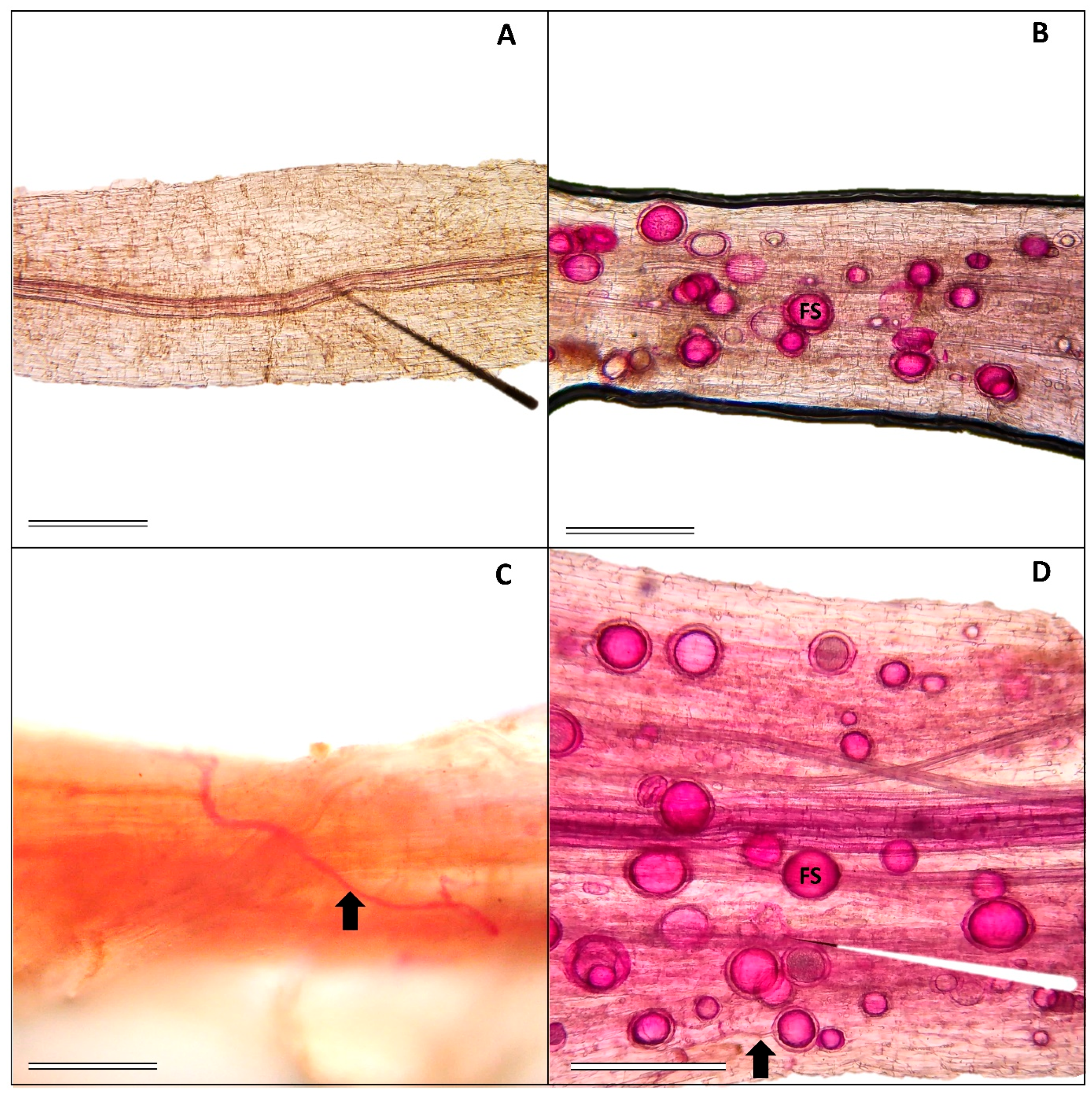
Figure 1. Fungal structures (arrows indicate hyphae; FS—fungal spore) inside roots of V. unguiculata cv. BRS Imponente inoculated with R. clarus. (A)—Control treatment (0 mM NaCl + AMF−); (B) 0 mM NaCl + AMF+; (C,D) 100 mM NaCl + AMF+; Scale bar: 100 µm.
Stresses 04 00026 g001
Figure 2. Mycorrhizal colonization (%) in V. unguiculata cv. BRS Imponente plants exposed to increasing concentrations of NaCl with (AMF+) and without (AMF−) inoculation by R. clarus. Letters indicate differences between the NaCl concentrations within the absence (AMF−) and presence (AMF+) of inoculation according to the Tukey all-pairs HSD test (p < 0.05). The bars represent the mean ± SD of ten repetitions. ** denotes significance at 1%. The F value and the significance of the NaCl factor are indicated in the upper left corner of the graph.
Stresses 04 00026 g002
Figure 3. Hydrogen peroxide (A) and malondialdehyde (B) concentrations and the activity of ascorbate peroxidase (C) and catalase (D) in V. unguiculata cv. BRS Imponente plants exposed to increasing concentrations of NaCl with (AMF+) and without (AMF−) inoculation by R. clarus. Lowercase letters indicate significant differences between NaCl concentrations within the absence (AMF−) or presence (AMF+) of inoculation by R. clarus, while uppercase letters indicate differences between inoculation treatments (AMF− and AMF+) at the same NaCl concentrations (0, 25, 50, 75, and 100 mM) according to the Tukey all-pairs HSD test (p < 0.05). The bars represent the mean ± SD of ten replicates. * and ** denote significance at 5% and 1%, respectively, while NS indicates no significance. The F value and the significance of the isolated factors and their interactions are indicated in the upper left corner of each graph.
Stresses 04 00026 g003
Figure 4. Anatomical markers of leaves from V. unguiculata cv. BRS Imponente exposed to increasing concentrations of NaCl with and without inoculation by R. clarus. (A) —0 mM of NaCl–AMF; (B) 0 mM of NaCl + AMF; (C) 25 mM of NaCl – AMF; (D) 25 mM of NaCl + AMF; (E) 50 mM of NaCl–AMF; (F) 50 mM of NaCl + AMF; (G) 75 mM of NaCl–AMF; (H) 75 mM of NaCl + AMF; (I) 100 mM of NaCl–AMF; (J) 100 mM of NaCl + AMF; LB—leaf blade; M—mesophyll; LP—lacunous parenchyma; PP—palisade parenchyma; Ad—adaxial epidermis; and Ab—abaxial epidermis.
Stresses 04 00026 g004
Figure 5. The thickness of leaf tissues of V. unguiculata cv. BRS Imponente exposed to increasing concentrations of NaCl with and without inoculation by R. clarus. Leaf blade (A); mesophyll (B); lacunous parenchyma (C); palisade parenchyma (D); adaxial epidermis (E), and abaxial epidermis (F). Lowercase letters indicate significant differences between NaCl concentrations within the absence (AMF−) or presence (AMF+) of inoculation by R. clarus, while uppercase letters indicate differences between inoculation treatments (AMF− and AMF+) at the same NaCl concentrations (0, 25, 50, 75, and 100 mM) according to the Tukey all-pairs HSD test (p < 0.05). The bars represent the mean ± SD of ten replicates. * and ** denote significance at 5% and 1%, respectively, while NS indicates no significance. The F value and the significance of the isolated factors and their interactions are indicated in the upper left corner of each graph.
Stresses 04 00026 g005
Figure 6. Concentration of sodium (Na+) (A), chloride (Cl) (B), and Na+/K+ ratio (C) in leaf tissues of V. unguiculata cv. BRS Imponente exposed to increasing concentrations of NaCl with and without inoculation by R. clarus. Lowercase letters indicate significant differences between NaCl concentrations within the absence (AMF−) or presence (AMF+) of inoculation by R. clarus, while uppercase letters indicate differences between inoculation treatments (AMF− and AMF+) at the same NaCl concentrations (0, 25, 50, 75, and 100 mM) according to the Tukey all-pairs HSD test (p < 0.05). The bars represent the mean ± SD of ten replicates. ** denote significance at 5% and 1%, respectively, while NS indicates no significance.
Stresses 04 00026 g006
Figure 7. Grain yields of V. unguiculata cv. BRS Imponente exposed to increasing concentrations of NaCl with and without inoculation by R. clarus. Lowercase letters indicate significant differences between NaCl concentrations within the absence (AMF−) or presence (AMF+) of inoculation by R. clarus, while uppercase letters indicate differences between inoculation treatments (AMF− and AMF+) at the same NaCl concentrations (0, 25, 50, 75, and 100 mM) according to the Tukey all-pairs HSD test (p < 0.05). The bars represent the mean ± SD of ten replicates. * and ** denote significance at 5% and 0.1%, respectively, while ns/NS indicates no significance. The F value and the significance of the isolated factors and their interactions are indicated in the upper left corner of each graph.
Stresses 04 00026 g007
Table 1. Nutrient concentrations (g kg−1) in leaves of V. unguiculata cv. BRS Imponente exposed to different concentrations of NaCl, with (+) and without (-) inoculation with R. clarus.
NaCl
(mM)
PKCaMgSCuFeMnZn
AMF−AMF+AMF−AMF+AMF−AMF+AMF−AMF+AMF−AMF+AMF−AMF+AMF−AMF+AMF−AMF+AMF−AMF+
05.65
Bab
8.50
Aa
35.43
Aa
30.06
Bc
16.14
NSa
16.31
NSa
17.29
Aab
14.56
Bab
3.40
NSa
3.57
NSa
0.003
Bc
0.012
Aa
0.15
Bab
1.23
Aa
0.084
NScd
0.075
NSab
0.039
NSb
0.034
NSc
256.39
NSa
6.33
NSbc
30.80
Ab
27.42
Bc
14.61
NSab
14.02
NSb
16.59
Ab
12.93
Bbc
3.45
NSa
3.29
NSa
0.009
NSbc
0.006
NSc
0.15
Bb
0.25
Aa
0.135
Aa
0.082
Ba
0.034
NSbc
0.038
NSb
505.58
NSab
6.69
NSbc
31.91
Bb
35.44
Ab
14.6
NSab
15.87
NSa
18.66
Aa
11.86
Bc
2.36
NSb
2.40
NSb
0.009
NSab
0.009
NSb
0.15
NSab
0.15
NSb
0.109
Ab
0.044
Bbc
0.031
Bc
0.038
Ab
755.10
Bab
7.58
Aab
34.65
Ba
44.98
Aa
13.70
NSb
12.90
NSb
17.68
Aa
15.55
Ba
1.95
NSc
2.09
NSc
0.010
NSa
0.008
NSbc
0.15
NSb
0.18
NSab
0.102
Abc
0.057
Bb
0.053
Aa
0.033
Bc
1004.67
Bb
6.01
Ac
33.19
Bab
44.46
Aa
12.98
Ac
10.35
Bc
17.79
Aa
12.47
Bc
1.70
NSc
2.04
NSc
0.01
NSa
0.009
NSb
0.21
Aa
0.14
Bb
0.078
Ad
0.033
Bc
0.037
Bb
0.060
Aa
Lowercase letters indicate significant differences between NaCl concentrations within the absence (AMF−) or presence (AMF+) of inoculation by R. clarus, while uppercase letters indicate differences between inoculation treatments (AMF− and AMF+) at the same NaCl concentrations (0, 25, 50, 75, and 100 mM) according to the Tukey all-pairs HSD test (p < 0.05). Mean ± SD of ten repetitions. NS indicates no significance.
Table 2. Biomass production (g plant−1) of V. unguiculata cv. BRS Imponente exposed to different concentrations of NaCl, with and without inoculation with R. clarus.
NaCl
(mM)
SFMSDMRFMRDM
AMF−AMF+AMF−AMF+AMF−AMF+AMF−AMF+
014.49NSa14.21NSc2.14 NSns2.01 NSab10.56Ab4.87Bab1.35Aab0.41Bb
2513.74Bab21.25 Aa2.13 NSns3.35 NSa15.54NSa11.41NSa1.49NSab1.36NSa
5013.30Bbc17.20 Aab1.96 NSns2.32 NSa14.20NSab12.19NSa1.36NSab1.38NSa
7514.48Aa11.96 Bc2.18 NSns1.43 NSb16.77Aa8.50Ba1.78Aa0.76Bab
10013.20Abc11.61 Bc2.06 NSns1.53NSb10.79Ab3.59Bb1.10NSb0.32NSb
SFM = shoot fresh mass; SDM = shoot dry mass; RFM = root fresh mass, and RDM = root dry mass. Lowercase letters indicate significant differences between NaCl concentrations within the absence (AMF−) or presence (AMF+) of inoculation by R. clarus, while uppercase letters indicate differences between inoculation treatments (AMF− and AMF+) at the same NaCl concentrations (0, 25, 50, 75, and 100 mM) according to the Tukey all-pairs HSD test (p < 0.05). Mean ± SD of ten repetitions. ns/NS indicates no significance.
Disclaimer/Publisher’s Note: The statements, opinions and data contained in all publications are solely those of the individual author(s) and contributor(s) and not of MDPI and/or the editor(s). MDPI and/or the editor(s) disclaim responsibility for any injury to people or property resulting from any ideas, methods, instructions or products referred to in the content.