
Article

Tactile Sensory Deprivation Impairs Spatial but Not Motor Behavior in Freely Moving Previsual Rat Pups

by
Mariya Odrinskaya
1,
Elizaveta Strelkova
2,
Anastasia Rebik
1,
Pavel Aleksandrov
1 and
Inna Midzyanovskaya
1,*
1
Institute of Higher Nervous Activity and Neurophysiology, Russian Academy of Sciences, 117465 Moscow, Russia
2
Biology Faculty, Moscow State University, 119234 Moscow, Russia
*
Author to whom correspondence should be addressed.
Biomechanics 2026, 6(2), 32; https://doi.org/10.3390/biomechanics6020032
Submission received: 23 January 2026 / Revised: 5 March 2026 / Accepted: 16 March 2026 / Published: 1 April 2026
(This article belongs to the Section Neuromechanics)

Abstract

Background/Objectives: The vibrotactile system, which is essential for guiding behavior in nocturnal rodents such as mice and rats, provides critical sensory input. To investigate the role of vibrotactile sensory inflow in neonatal locomotion, we used previsual rat pups that underwent bilateral vibrissectomy. Subsequently, their motor behavior was evaluated in an open field test. Methods: A total of 42 previsual pups from four litters were assigned to either bilateral vibrissectomy or sham surgery groups on postnatal days (PND) 9–12, with group allocation balanced across litters. Results: Open-field testing on PND 13 revealed that while vibrissectomy (VE) did not affect gross locomotor activity—such as distance traveled, speed, acceleration, or freezing episodes (all >0.05)—it significantly altered spatial behavior. To quantify spatial patterns of curvy tracks, we analyzed trajectorial compaction within the central zone, lacking the tactile guidance of the walls: trajectories were smoothed using virtual coatings scaled to the vibrissal length (16 mm). For each track, an individual linearized reference path was generated and subjected to identical smoothing. The compaction ratio—calculated as the coated area of the smoothed linearized reference divided by the coated area of the experimental track—was significantly greater in VE pups than in sham controls (p = 0.03). This effect was not attributable to differences in the path length traveled within the central zone. The increased compaction persisted when the smoothing scale was increased 2–3 fold (32–64 mm radii, approximating the pups’ mean body size), but not at smaller scales (2–4 mm). Conclusions: These results demonstrate that tactile input specifically modulates the spatial, rather than locomotor, components of nonvisual navigation. Consequently, the track compaction may serve as a sensitive marker for assessing vibrotactile function in developing laboratory rodents.

1. Introduction

Sensory deprivation represents a classic experimental approach in modeling developmental neurobiology and neurorehabilitation [1,2,3,4]. The first studies were conducted on the visual system of developing kittens [5], where complete or partial sensory deprivation during critical periods led to irreversible functional impairments of visual perception. Unlike the visual or auditory systems, the rodent somatosensory system allows for non-invasive sensory deprivation through whisker trimming. This approach avoids tissue damage and enables the effective study of deprivation effects without systemic disturbances [6].
Sensorimotor integration, in both normal and pathological conditions, is most often assessed by motor patterns that are easiest to observe. In developing rat pups, locomotor activity emerges before the maturation of the auditory and visual systems [7]. The initial acts of locomotion are driven by olfactory and tactile stimuli. Spatial exploration begins with spontaneous departures from the nest on postnatal days (PND) 10–12 [8]. Immature movement patterns (propulsion by the forelimbs and pivoting) transition to quadrupedal locomotion by PND 10–11, and movement on four limbs is registered from PND 14–15 [9,10,11]. Thus, prior to the functional involvement of visual afferentation, the role of the tactile system in spatial behavior can be assessed by experimentally modulating sensory input from the whiskers in experimental animals.
The movement trajectories of organisms possess complex characteristics due to measurement noise, micromovements, and essential path fractality [12,13,14], raising practical questions about track smoothing and the quantitative assessment of their nonlinear features. By the end of the second postnatal week, the locomotor patterns of rat pups include both rapid progression and lingering phases [15,16,17,18], the latter being associated with environmental scanning (lateral head movements, air sniffing, and small-step displacements). Consequently, the effects of sensory deprivation should be more evident in behavior related to orientation than during rapid movement, although quantifying such intermittent behavior remains a challenging task [19,20].
The precisely organized somatotopic structure of the rodent vibrotactile system provides unique opportunities for studying critical developmental periods and neuronal plasticity [21,22]. The barrel cortex—the primary somatosensory representation of whiskers—derives its name from its characteristic cylindrical morphofunctional units. Each “barrel” receives tactile input exclusively from a single whisker, creating a precise topographic map in the somatosensory cortex that mirrors the arrangement of whiskers on the snout [23,24,25]. Whisking represents one of the earliest active behavioral patterns for exploring the environment [23], beginning its functional maturation during postnatal days 8–9 [24]. Numerous studies have characterized both the neural mechanisms underlying whisking behavior and the anatomical consequences of whisker removal. Neonatal whisker trimming (vibrisectomy, VE) in rat pups does not prevent barrel formation but leads to expanded excitatory and reduced inhibitory receptive fields [24,25]. The affected cortical areas demonstrate an increase in area, with spiny stellate neurons showing increased dendritic branching, greater spine density [6], and altered patterns of intracortical connectivity [26] in layer IV of the somatosensory cortex in adulthood.
Despite extensive data on the anatomical and electrophysiological consequences of early vibrisectomy in laboratory rodents, simple quantitative measures of immediate behavioral responses to vibrisectomy during spontaneous behavior are still lacking. Vibrisectomy from PND 0 to PND 3 resulted in impaired whisker-specific tactile function at PND 30–35 [6] and altered behavioral strategies in gap-crossing tests [27]. Vibrisectomy impaired nipple attachment and huddling behavior when performed at PND 3–5 [22]. Early postnatal whisker removal significantly reduced exploratory activity [28], while prolonged vibrisectomy (PND 9–20) altered the development of defensive behavior, including threat response patterns [29]. Neonatal whisker ablation caused a persistent tactile discrimination deficit, with adult rats demonstrating serious impairments in texture differentiation [30]. However, in pups, the vibrotactile and olfactory systems should also support spatial behavior before visual cues become functionally available. We hypothesized that early tactile deprivation in functionally blind rat pups would likewise lead to a noticeable alteration in locomotor patterns, which could serve as a simple biomarker for sensorimotor integration. At PND 13, the whiskers of rat pups are already capable of bilateral movement [23] but visual cues are still unavailable. We hypothesized that vibrisectomy, by partially restricting available sensory input from the novel environment, would alter spatial locomotor patterns at the whisker-length scale.

2. Materials and Methods

2.1. Animal Housing

Outbred Wistar rats were obtained from the Stolbovaya breedery and mated at the Animal Chapter of IHNA&NPh, RAS. Gravid dams were individually housed in standard polycarbonate cages (dimensions: 40 × 60 × 19 cm) with wood saw bedding and additional nesting material (shredded paper towels). The housing facility maintained a 12:12 hr light/dark cycle (lights on at 08.00 A.M), controlled temperature (22 ± 2 °C), and humidity (50 ± 10%). Food (standard pellet chow) and water were provided ad libitum. Cage beddings were changed twice weekly to ensure hygiene while minimizing stress. Dams were monitored daily for pups’ appearance and signs of distress. To reduce disturbance near parturition, cages were left undisturbed from postnatal day 1 (PND) 1. The birth date was designated PND 0; litters were left intact until PND 9.

2.2. Vibrissectomy

Before handling the rat pups, the dams were removed from the home cage. Immediately before the procedure, the pups were extracted from the home cage and housed temporarily in a separate cage. On PND 9, pups from each litter were assigned to either an experimental group or a control group. In the experimental group (n = 21), all vibrissae on both sides of the snout were trimmed to the fur level daily from PND 9 to 12. In the control group (n = 21), a sham procedure was performed during the same period by gently applying scissors to the vibrissae bases without cutting. During the procedure, pups were restrained by hand to limit movement. Both procedures (vibrissae trimming and sham) lasted 60–120 s per pup. After processing, pups were weighed and marked, then returned to the home cage. The dams were reintroduced afterward.

2.3. Open Field Test

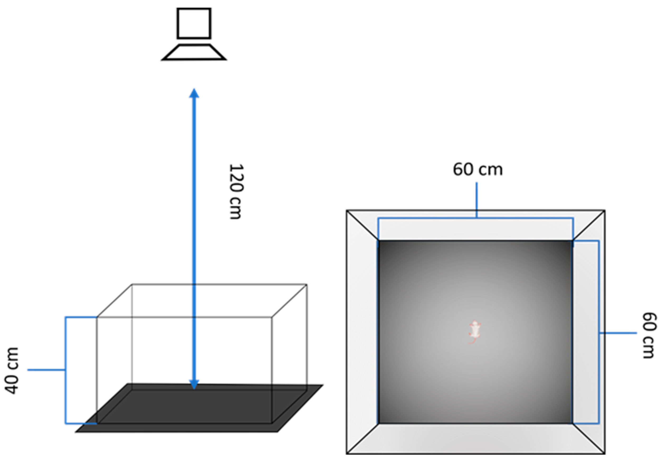
The behavioral testing protocol was performed as described earlier [17] with minor modifications. To minimize stress, dams were separated from their litters only after voluntarily leaving the nest. Pups were then allowed to remain undisturbed in their huddles for 30 min to achieve stable baseline behavioral states prior to testing. Each pup was tested only once. Individual pups were carefully grasped from the periphery of the huddle and transported to the adjacent testing room. Each trial began immediately upon placement of the pup in the center of the arena. The open field apparatus consisted of two detachable pieces: 60 cm × 60 cm vertical walls, constructed of plywood and painted with black, and replaceable plywood floor plate, covered by black plastic (Figure 1).
The design ensured the absence of possible dust and olfactory cues kept in the corners and gaps. Between trials, the arena was cleaned with 50% ethanol solution and dried with lint-free wipes to eliminate residual olfactory cues that could influence subsequent testing sessions. A video camera was placed 1 m above. The arena had a lighted center (98–102 Lx) and dimmed corners (32–38 Lx). A standard spatial orientation of the landed pup was with its head away from the experimenter, directed to the middle of the opposite wall. At the pup’s placement, videotracking started and lasted for 120 s.
The tracker (ToxTrac 25.1.1 [31,32]) determined the body position by calculating the white mass center against the black background every 400 ms and digitized the trajectories. After the test, the pups were sexed, weighed, and numbered; the information is summarized in Table 1. The eye status was checked individually.
This standardized protocol ensured consistent testing conditions while minimizing potential confounds from handling stress or environmental contamination. All experimental sessions were video-recorded using a digital camera synchronized with a computer located in the next room.

2.4. Trajectory Analysis

2.4.1. Offline Tracking

Offline tracking of the animals’ movements throughout all trials was conducted utilizing the open-source software ToxTrac (v 25.1.1), Umeå, Sweden [31,32], which calculated the centroid of the white body mass against the black background at 400 ms intervals, thereby digitizing the motion trajectories. Automated measurement included the cumulative distances, and the incidence and duration of freezing episodes.

2.4.2. Trajectory Coating

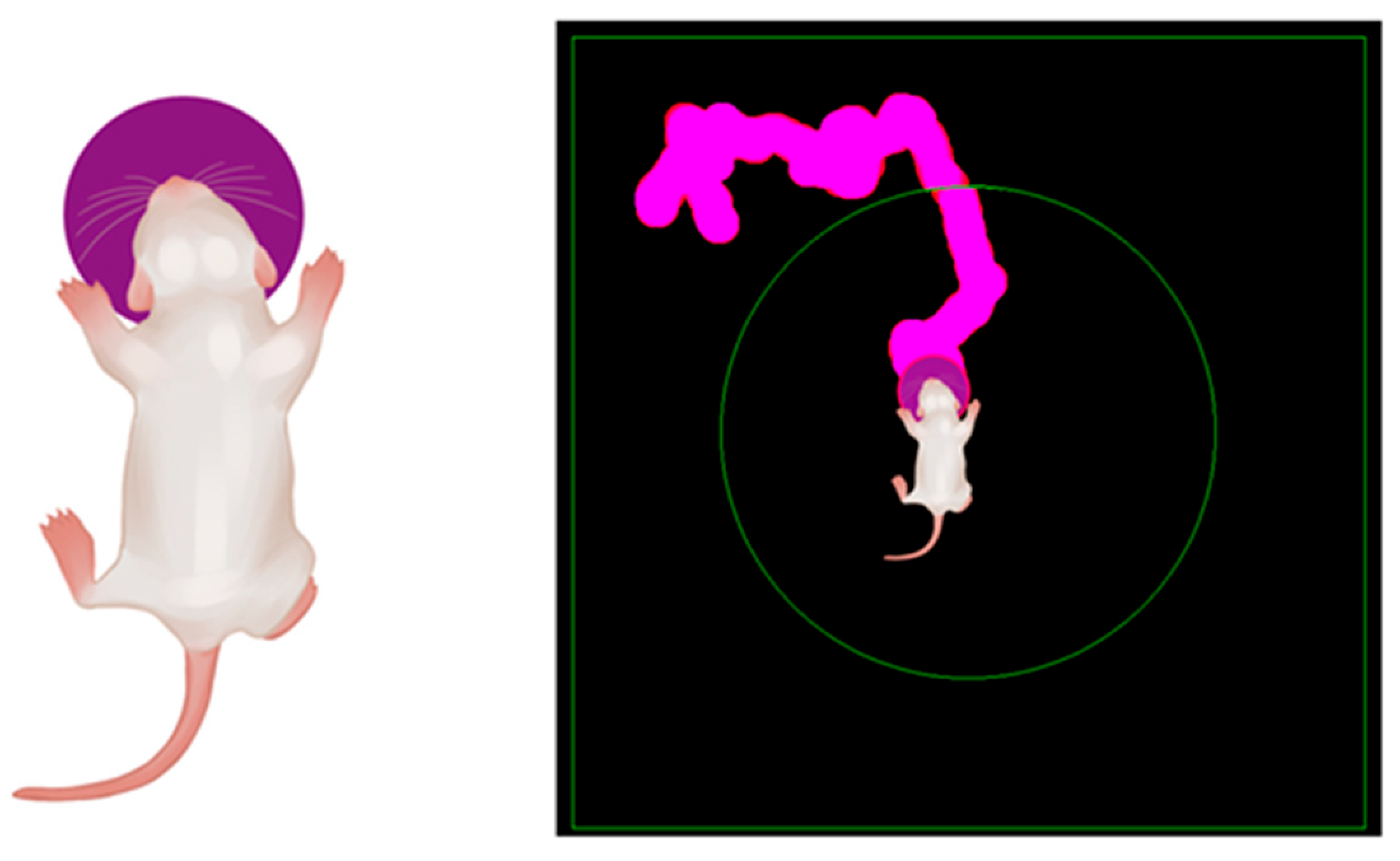
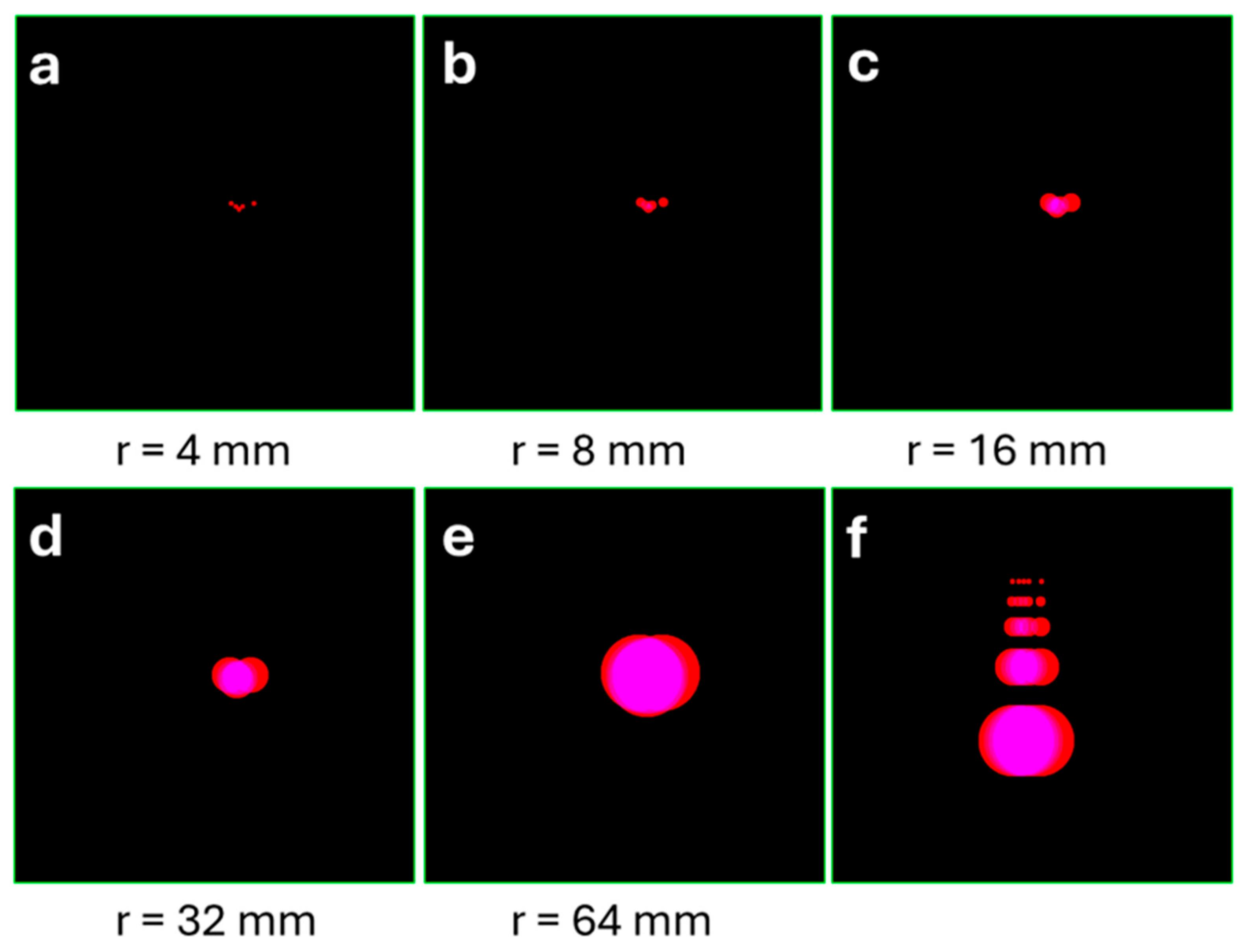
To quantify the area covered by experimental trajectories, while minimizing graphical noise, we developed a simple method of track coating (Figure 2). Each detected point of the original trajectory was overlaid with a virtual circle (Rv-circle) whose radius matched the mean vibrissal length (Rv) at the given age (PND 13; Rv = 16 mm according to the laboratory data). The circle was chosen as the coating element for its geometric simplicity and symmetry: it requires no calculation of trajectory normals at individual points and offers inherent robustness to tracking noise. This coating process smoothed the trajectory with a characteristic spatial scale of Rv (Figure 2). The total area of all superimposed circles was calculated in pixels. An individual reference for each experimental trajectory was constructed by applying the same procedure to the corresponding linearized trajectory. These linearized trajectories preserved the precise spacing between each pair of consecutive points while constraining them to a geometrically straight line (Figure 3a,f). The degree of trajectorial self-attraction (understood as partial or complete returns to the self-track or its vicinity) was quantified using the track compaction ratio:
Cr = S_coated_linearized_track/S_coated_original_track
Trajectory coating and compaction analysis were performed using a custom Python version 3.12.1 script implementing the following algorithm. First, a digital canvas corresponding to the dimensions of the open field was created. All tracked points of the animal’s movement were plotted on this canvas, with each point represented as a circle of a specified radius. The total area covered by all circles was then calculated in pixel units, accounting for overlapping regions only once.
To generate a linearized reference for each trajectory, a second canvas was created. The tracked points were then placed consecutively along a straight line, preserving the exact inter-point distances from the original trajectory. Each point was again represented as a circle of the same radius, and the total covered area was calculated, with overlaps counted only once. Overlap between immediately neighboring circles was preserved by design, while sensitivity to trajectory curvature arose from the difference in additional overlaps among third-order, fourth-order, and more distant circle pairs.
The compaction ratio (Cr) was defined as the ratio of the area covered by the linearized trajectory to that covered by the original trajectory. This metric quantifies the degree of trajectorial self-overlap, or “track compaction,” with higher values indicating more spatially confined exploration.
To differentiate between behaviors with and without haptic support from the arena walls, we established two virtual zones: (1) a central region (20 cm diameter) and (2) the periphery (remaining arena area). All trajectory coating and subsequent analyses focused exclusively on the central zone to isolate locomotor parameters independent of thigmotactic behavior.

2.5. Statistics

The data were analyzed using a general linear model (GLM) ANOVA, with ToxTrac-derived locomotor parameters as dependent variables, group and litter as categorical predictors, and of the session number within a litter as a continuous covariate. Prior to analysis, the normality of data distribution was assessed using the Shapiro–Wilk test. ANOVA was followed by post hoc Tukey tests. In addition, the Cr values, calculated per pup for a range of coating radii R (see above), were analyzed using a repeated measures GLM ANOVA. Statistical significance was set at an alpha level of 0.05. Statistical significance was set at an alpha level of 0.05. To account for multiple comparisons across the different coating radii, p-values were adjusted using the Benjamini–Hochberg (BH) false discovery rate correction.

2.6. Use of AI Tools Declaration

The authors utilized DeepSeek=V3.2 AI solely for language polishing. All scientific content, analysis, and conclusions remain the responsibility of the authors.

3. Results

3.1. Gross Locomotor Parameters

Inter-litter variability emerged as the primary factor influencing locomotor parameter variation. The experimental manipulation (vibrissectomy) did not significantly alter gross locomotor measures, including total distance traveled, average speed, or freezing duration (Table 1). The experimental and control pups demonstrated very similar distances travelled within the central zone (VE-group: 1392 ± 690 mm; Sham-group: 1242 ± 723 mm, p > 0.05).

3.2. Compaction of Trajectories Within the Central Zone

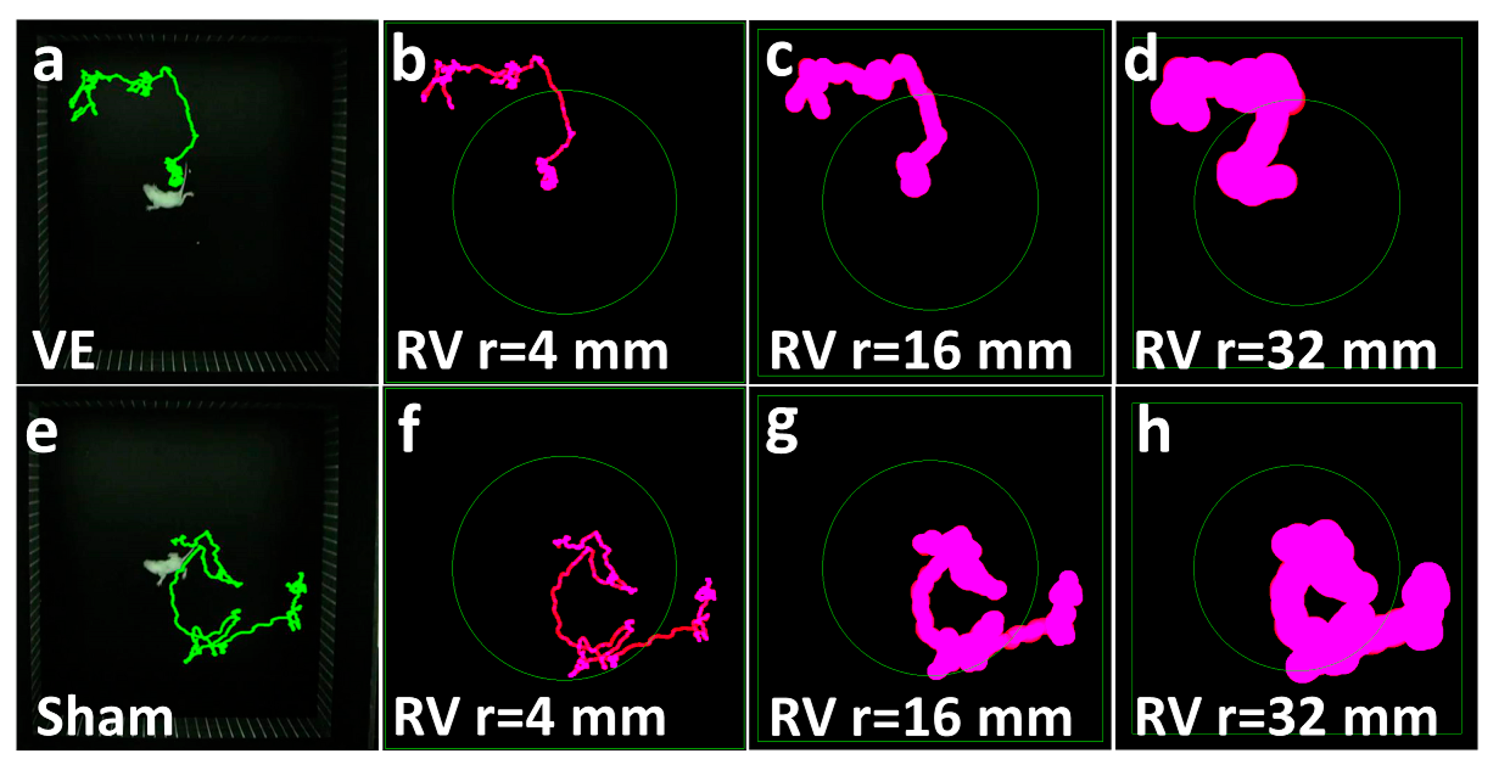
Spatial characteristics were assessed by virtually coating the trajectories with circles of specified radii. This approach was chosen for its simplicity and relevance to the pup’s body scale (see Method Section 2.4.2.). Sets of compaction ratios (Cr) were generated by comparing the area covered by the original trajectories with the area of their linearized references, both coated with circles of radii Rv, Rv/4, Rv/2, 2 Rv, and 4 Rv (Table 2, Figure 3 and Figure 4).
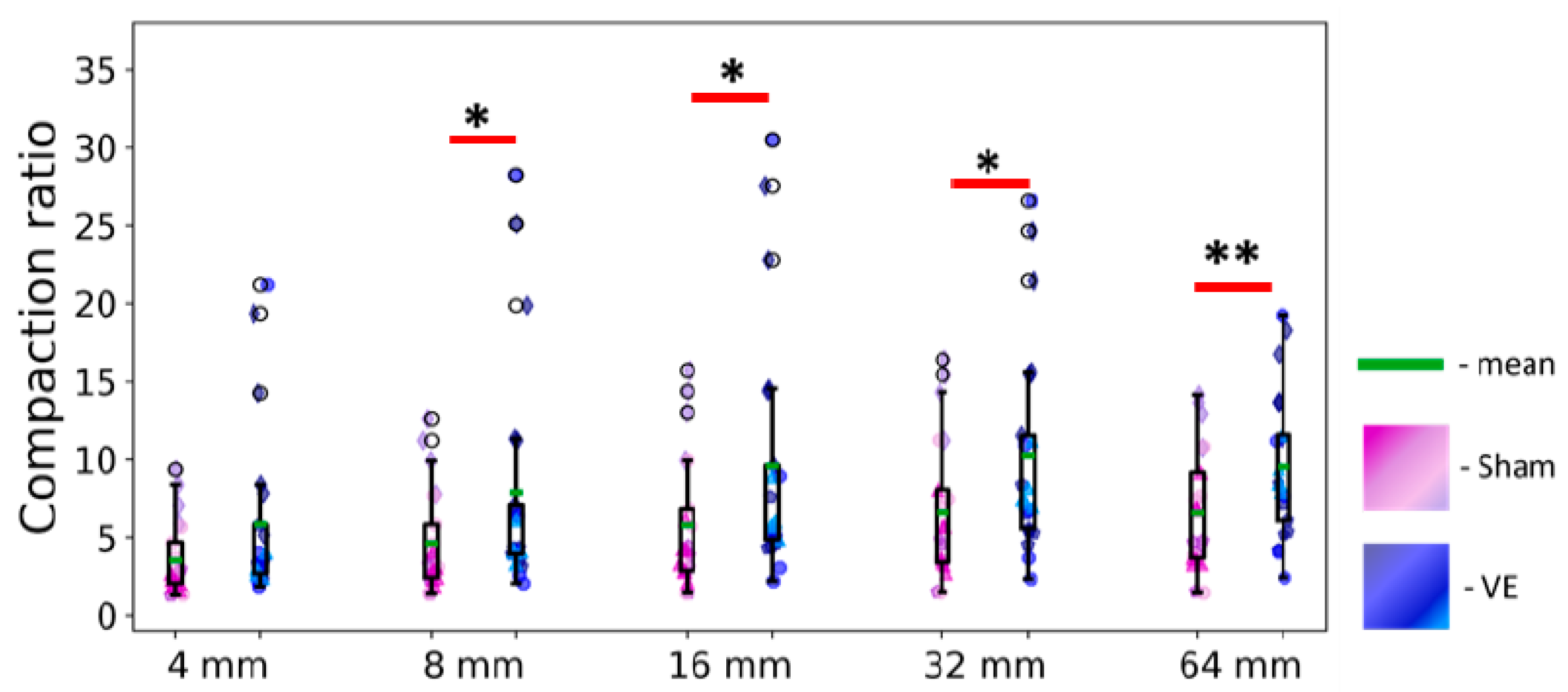
The between-litter variability showed the strongest effect on track compaction (Table 2), suggesting potential sensitivity of these parameters to mother-offspring interactions. Beyond this primary familial difference, the vibrissectomy (VE) effect became apparent starting at R = 8 mm and remained significant for R = 16 mm, 32 mm, and 64 mm (Figure 5), with no significant differences observed between these three largest radii conditions (as confirmed by repeated measures ANOVA: R1, {F(2,66) = 1.03, p = 0.35}; R1*VE interaction, {F(2,66) = 1.08, p = 0.34}). This indicates that these compaction ratios remained remarkably stable across the 16–64 mm scale, with all significant comparisons surviving Benjamini–Hochberg correction for multiple testing. The Cr values showed that VE pup trajectories were approximately 10 times more compact than their linearized references, compared to about 6 times for control pups. Notably, the vibrissectomy effect was consistent across litters—in each litter, VE pups developed more compact trajectories than their sham-operated mates.

4. Discussion

Although the anatomical and electrophysiological consequences of early whisker deprivation have been extensively characterized, the literature lacks simple quantitative measures of its immediate impact on spontaneous behavior in developing rodents. Such measures are essential, as they could serve as non-invasive biomarkers for sensorimotor integration in both normal and compromised early development. The present study addressed this gap by examining the spatial properties of pup trajectories at body-part-relevant scales, thereby partly recovering information lost by reducing an animal to a virtual moving centroid.
We demonstrated that the spatial locomotor pattern is significantly altered in rat pups subjected to early tactile deprivation. These changes are specific: the deprived animals exhibit markedly higher track compaction compared to controls (Table 1 and Table 2, Figure 5). This finding suggests that the loss of vibrissal input causes pups to remain closer to their previous trajectories, while sham-operated controls distribute their trajectories more broadly across the available environment—a phenomenon not previously reported in the literature.
Notably, vibrissectomy did not affect standard motion parameters in the open field, which aligns with observations in a rat model of epilepsy [33,34] and is consistent with reports that even spinal cord injury fails to disrupt gross locomotor activity in mouse pups [35]. This remarkable stability of fundamental motor patterns highlights the need for more sensitive, coupled behavioral measures.
Several analytical challenges arise when studying such movement patterns. The inherent complexity of natural trajectories, compounded by measurement artifacts, can mask underlying locomotor strategies [14]. Modern tracking systems (e.g., DeepLabCut, ToxTrac) inevitably combine: (1) whole-body translations, (2) pause-associated scanning behaviors [19,20,36], and (3) postural micro-adjustments [37,38]. To address these challenges, we adapted our recently described space potential method [17], which differentiates pivoting/scanning from directed movement, to create a simplified compaction metric (Cr). This ratio compares R-processed original trajectories to their individual linearized references, accounting for natural speed. Our prior work established that previsual pups typically exhibit imprecise path returns, detectable through velocity autocorrelation analysis [17]. Compared to existing approaches, the proposed “track coating” method is significantly simpler and necessitates less experimental data for analysis.
The Cr values showed surprising consistency across different spatial scales (16–64 mm), with vibrissectomized (VE) pups maintaining ratios near 10 compared to approximately 6 for control animals. This scale-invariant effect, persisting even beyond vibrissal length, suggests contributions from additional factors. Presumably, proprioceptive mechanisms related to body size (interpaw distance: 4–8 cm at this age) may be involved [39,40,41], although contributions from plantar tactile sensitivity remain poorly understood [42]. Future studies employing local plantar anesthesia could help clarify this point.
Alternative interpretations also warrant consideration. The increased compaction observed in vibrissectomized (VE) pups may stem not only from a sensory deficit but also from heightened maternal dependence [28]. This could manifest as a tendency to “stay in place” in anticipation of maternal retrieval [43]. Thus, although our data clearly established enhanced path compaction following vibrissectomy (Table 2, Figure 5), the precise mechanistic balance between sensory and affective factors requires further investigation.
Our results emphasize the crucial contribution of somatosensory input to spatial behavior in the absence of vision, which may inform research on neurodevelopmental/traumatic disorders involving impaired sensory integration. Furthermore, the compaction ratio (Cr) emerges as a sensitive and reliable metric for identifying the persistent, moderate behavioral alterations resulting from vibrissal deprivation.

5. Limitations

An unavoidable limitation of this study is the potential stress associated with daily handling during the vibrissectomy or sham procedure, as well as during separation from the dam and transport to the open field. Although both groups underwent identical handling protocols, we cannot entirely rule out that stress-related effects may have interacted with the sensory processing.
Another limitation concerns the reliability of our measurements, as the camera setup and tracking software were not subjected to a formal test–retest assessment. However, the observed effect sizes do not require the pixel-level precision. The smallest coating radius used in our analysis (4 mm) and the primary radius of interest (16 mm) are substantially larger than the expected tracking error. Therefore, minor inter-session variations in camera alignment or tracker precision are unlikely to have meaningfully influenced the compaction ratio. Moreover, the tracking method and experimental setup used here have been validated in a higher-resolution context aimed at resolving individual instantaneous velocities [15,16]. It supports the robustness of our approach, although this was not explicitly re-assessed in the present study.

6. Conclusions

We demonstrated that early sensory deprivation significantly increased trajectorial compaction during spontaneous locomotion without altering gross motor parameters. This finding underscores the sensitivity of the compaction ratio to tactile dysfunction, establishing it as a potential biomarker for developing vibrotactile function. Our quantitative methodology thus offers translational potential by providing a new objective behavioral metrics in neurodevelopmental and neurorehabilitation research.

Author Contributions

Conceptualization, I.M.; software, P.A.; validation, I.M. and A.R.; investigation, E.S.; data curation, P.A. and A.R.; writing—I.M. and M.O.; visualization, M.O., A.R. and P.A.; supervision, I.M. All authors have read and agreed to the published version of the manuscript.

Funding

This research received no external funding. The article was prepared within the state assignment of the National Ministry of Education and Science for IHNA & NPh RAS.

Institutional Review Board Statement

The animal study protocol was approved by the Ethics Committee of IHNA, the protocol N 04290520243.

Informed Consent Statement

Not applicable.

Data Availability Statement

The raw data are available upon reasonable request. The software developed to assess the trajectorial compaction: https://github.com/RebikAnastasiya/Program-for-animal-trajectory-coating- (accessed on 10 March 2026).

Acknowledgments

The authors are extremely grateful to V.V. Raevsky and O.A. Chichigina for valuable scientific discussions. The authors wish to thank the anonymous reviewers for their exceptionally thorough and insightful comments, which greatly enhanced the quality of this work.

Conflicts of Interest

The authors declare no conflicts of interest.

Abbreviations

The following abbreviations are used in this manuscript:
ANOVAAnalysis of Variance
CrCompaction ratio
GLMGeneral Linear Model
IHNA & NPh, RAS Institute of Higher Nervous Activity and Neurophysiology, Russian Academy of Sciences
PNDPostnatal Day
RvRadius of vibrissal length
VEVibrisectomy (whisker removal)

References

  1. Morrison, A. Changing Concepts of the Nervous System. In Proceedings of the First Institute of Neurological Sciences Symposium in Neurobiology; Elsevier: Amsterdam, The Netherlands, 2012. [Google Scholar] [CrossRef]
  2. Solomon, P.; Leiderman, P.H.; Mendelson, J.; Wexler, D. Sensory deprivation; a review. Am. J. Psychiatry 1957, 114, 357–363. [Google Scholar] [CrossRef] [PubMed]
  3. Kubzansky, P.E.; Leiderman, P.H. Sensory deprivation: An overview. In Sensory Deprivation; Harvard University Press: Cambridge, MA, USA, 2013; pp. 221–238. [Google Scholar]
  4. Caras, M.L.; Sanes, D.H. Sustained perceptual deficits from transient sensory deprivation. J. Neurosci. 2015, 35, 10831–10842. [Google Scholar] [CrossRef]
  5. Hubel, D.H.; Wiesel, T.N. Receptive fields, binocular interaction and functional architecture in the cat’s visual cortex. J. Physiol. 1962, 160, 106–154. [Google Scholar] [CrossRef]
  6. Lee, L.J.; Chen, W.J.; Chuang, Y.W.; Wang, Y.C. Neonatal whisker trimming causes long-lasting changes in structure and function of the somatosensory system. Exp. Neurol. 2009, 219, 524–532. [Google Scholar] [CrossRef]
  7. Eilam, D.; Golani, I. The ontogeny of exploratory behavior in the house rat (Rattus rattus): The mobility gradient. Dev. Psychobiol. 1988, 21, 679–710. [Google Scholar] [CrossRef]
  8. Renner, M.J.; Pierre, P.J. Development of exploration and investigation in the Norway rat (Rattus norvegicus). J. Gen. Psychol. 1998, 125, 270–291. [Google Scholar] [CrossRef]
  9. Clarac, F.; Brocard, F.; Vinay, L. The maturation of locomotor networks. In Progress in Brain Research; Elsevier: Amsterdam, The Netherlands, 2004; Volume 143, pp. 57–66. [Google Scholar]
  10. Jamon, M.; Clarac, F. Early walking in the neonatal rat: A kinematic study. Behav. Neurosci. 1998, 112, 1218–1228. [Google Scholar] [CrossRef]
  11. Swann, H.E.; Brumley, M.R. Locomotion and posture development in immature male and female rats (Rattus norvegicus): Comparison of sensory-enriched versus sensory-deprived testing environments. J. Comp. Psychol. 2019, 133, 183–196. [Google Scholar] [CrossRef]
  12. Turchin, P. Fractal analyses of animal movement: A critique. Ecology 1996, 77, 2086–2090. [Google Scholar] [CrossRef]
  13. Bartumeus, F.; Levin, S.A. Fractal reorientation clocks: Linking animal behavior to statistical patterns of search. Proc. Natl. Acad. Sci. USA 2008, 105, 19072–19077. [Google Scholar] [CrossRef]
  14. Benhamou, S. How to reliably estimate the tortuosity of an animal’s path: Straightness, sinuosity, or fractal dimension? J. Theor. Biol. 2004, 229, 209–220. [Google Scholar] [CrossRef]
  15. Midzyanovskaya, I.S.; Rebik, A.A.; Idzhilova, O.S.; Smyk, F.S.; Strelkov, V.V.; Komarova, N.L.; Chichigina, O.A. Two modes in the absolute velocity statistics in cautious walks of laboratory rodents. Biophys. J. 2025, 124, 2708–2730. [Google Scholar] [CrossRef]
  16. Idzhilova, O.S.; Midzyanovskaya, I.S.; Strelkov, V.V.; Komarova, N.L.; Chichigina, O.A. Pros and cons of group averaging in studies of stochastic rodent locomotion. Chaos Solitons Fractals 2025, 201, 117381. [Google Scholar] [CrossRef]
  17. Midzyanovskaya, I.S.; Strelkov, V.V. Measuring locomotor strategies of freely moving previsual rat pups. Behav. Process. 2022, 203, 104780. [Google Scholar] [CrossRef]
  18. Golani, I. The developmental dynamics of behavioral growth processes in rodent egocentric and allocentric space. Behav. Brain Res. 2012, 231, 309–316. [Google Scholar] [CrossRef]
  19. Golani, I.; Benjamini, Y.; Eilam, D. Stopping behavior: Constraints on exploration in rats (Rattus norvegicus). Behav. Brain Res. 1993, 53, 21–33. [Google Scholar] [CrossRef]
  20. Drai, D.; Benjamini, Y.; Golani, I. Statistical discrimination of natural modes of motion in rat exploratory behavior. J. Neurosci. Methods 2000, 96, 119–131. [Google Scholar] [CrossRef]
  21. Fox, K. A critical period for experience-dependent synaptic plasticity in rat barrel cortex. J. Neurosci. 1992, 12, 1826–1838. [Google Scholar] [CrossRef] [PubMed]
  22. Erzurumlu, R.S.; Kind, P.C. Neural activity: Sculptor of ‘barrels’ in the neocortex. Trends Neurosci. 2001, 24, 589–595. [Google Scholar] [CrossRef] [PubMed]
  23. Sullivan, R.M.; Landers, M.S.; Flemming, J.; Vaught, C.; Young, T.A.; Polan, H.J. Characterizing the functional significance of the neonatal rat vibrissae prior to the onset of whisking. Somatosens. Mot. Res. 2003, 20, 157–162. [Google Scholar] [CrossRef]
  24. Grant, R.A.; Mitchinson, B.; Prescott, T.J. The development of whisker control in rats in relation to locomotion. Dev. Psychobiol. 2012, 54, 151–168. [Google Scholar] [CrossRef]
  25. Simons, D.J.; Land, P.W. Early experience of tactile stimulation influences organization of somatic sensory cortex. Nature 1987, 326, 694–697. [Google Scholar] [CrossRef] [PubMed]
  26. Shoykhet, M.; Land, P.W.; Simons, D.J. Whisker trimming begun at birth or on postnatal day 12 affects excitatory and inhibitory receptive fields of layer IV barrel neurons. J. Neurophysiol. 2005, 94, 3987–3995. [Google Scholar] [CrossRef]
  27. Papaioannou, S.; Brigham, L.; Krieger, P. Sensory deprivation during early development causes an increased exploratory behavior in a whisker-dependent decision task. Brain Behav. 2013, 3, 24–34. [Google Scholar] [CrossRef]
  28. Smirnov, K.; Tsvetaeva, D.; Sitnikova, E. Neonatal whisker trimming in WAG/Rij rat pups causes developmental delay, encourages maternal care and affects exploratory activity in adulthood. Brain Res. Bull. 2018, 140, 120–131. [Google Scholar] [CrossRef]
  29. Shishelova, A.Y. Effect of whisker removal on defensive behavior in rats during early ontogenesis. Neurosci. Behav. Physiol. 2006, 36, 883–888. [Google Scholar] [CrossRef]
  30. Carvell, G.; Simons, D. Abnormal tactile experience early in life disrupts active touch. J. Neurosci. 1996, 16, 2750–2757. [Google Scholar] [CrossRef]
  31. Rodriguez, A.; Zhang, H.; Klaminder, J.; Brodin, T.; Andersson, P.L. ToxTrac: A fast and robust software for tracking organisms. Methods Ecol. Evol. 2018, 9, 460–464. [Google Scholar] [CrossRef]
  32. Rodriguez, A.; Zhang, H.; Klaminder, J.; Brodin, T.; Andersson, P.L. ToxId: An efficient algorithm to solve occlusions when tracking multiple animals. Sci. Rep. 2017, 7, 14774. [Google Scholar] [CrossRef] [PubMed]
  33. Smirnov, K.; Sitnikova, E. Developmental milestones and behavior of infant rats: The role of sensory input from whiskers. Behav. Brain Res. 2019, 374, 112143. [Google Scholar] [CrossRef]
  34. Shishelova, A.Y.; Raevskii, V.V. Effects of vibrissectomy during early postnatal ontogenesis in rat pups on behavioral development. Neurosci. Behav. Physiol. 2010, 40, 671–677. [Google Scholar] [CrossRef]
  35. Martes, A.C.; Bozeman, A.L.; Doell, J.; Dunn, A.C.; Reiserer, R.S.; Malkova, L.; Flynn, M.R.; Cramer, C.P. Developmental changes in locomotion and sensorimotor reflexes following spinal cord transection. Dev. Psychobiol. 2024, 66, e22558. [Google Scholar] [CrossRef]
  36. Drai, D.; Kafkafi, N.; Benjamini, Y.; Elmer, G.; Golani, I. Rats and mice share common ethologically relevant parameters of exploratory behavior. Behav. Brain Res. 2001, 125, 133–140. [Google Scholar] [CrossRef] [PubMed]
  37. Abbas, W.; Masip Rodo, D. Computer methods for automatic locomotion and gesture tracking in mice and small animals for neuroscience applications: A survey. Sensors 2019, 19, 3274. [Google Scholar] [CrossRef]
  38. Barrett, J.M.; Martin, M.E.; Shepherd, G.M.G. Manipulation-specific cortical activity as mice handle food. Curr. Biol. 2022, 32, 4842–4853. [Google Scholar] [CrossRef]
  39. Altman, J.; Sudarshan, K. Postnatal development of locomotion in the laboratory rat. Anim. Behav. 1975, 23, 896–920. [Google Scholar] [CrossRef]
  40. Clarac, F.; Vinay, L.; Cazalets, J.R.; Fady, J.C.; Jamon, M. Role of gravity in the development of posture and locomotion in the neonatal rat. Brain Res. Rev. 1998, 28, 35–43. [Google Scholar] [CrossRef]
  41. Ni, Z.; Neifert, C.; Rosete, A.; Alcover, D.C.; Chen, Y.; Jones, L.; Ginty, D.D. Tactile mechanisms and afferents underlying the rat pup transport response. Curr. Biol. 2024, 34, 5595–5601. [Google Scholar] [CrossRef]
  42. Varejão, A.S.P.; Filipe, V.M. Contribution of cutaneous inputs from the hindpaw to the control of locomotion in rats. Behav. Brain Res. 2007, 176, 193–201. [Google Scholar] [CrossRef]
  43. Wilson, C.; Nungaray, K.; Garza, M.; Sanchez, M. Sit down and stay here! Transport response elicitation modulates subsequent activity in rat pups. Behav. Process. 2008, 77, 131–134. [Google Scholar] [CrossRef]
Figure 1. The experimental setup consisted of an open field arena with removable walls constructed from black-painted plywood and a removable plywood floor covered with black plastic. This detachable design prevented the accumulation of potential odor cues in the arena’s crevices. A video camera was positioned directly above the center of the arena and connected to a computer in adjacent room.
Biomechanics 06 00032 g001
Figure 2. Schematic representation of (left panel) a previsual rat pup with virtual Rv-circle of vibrotactile sensing range, and (right panel) an individual trajectory smoothed through Rv-circle coating at each detection point.
Biomechanics 06 00032 g002
Figure 3. (ae) Five consecutive points from an original trajectory, each surrounded by R-circles of different radius. (f) The same five sequential trajectory points aligned linearly and coated with corresponding R-circles. Circle radius: 4, 8, 16, 32, 64 mm (given below the panels). To estimate compactness, we calculated the surface area ratio (Cr) between the two coated tracks.
Biomechanics 06 00032 g003
Figure 4. Example trajectories from a vibrisectomized (VE) and a sham-operated (Sham) pup. (a) Raw trajectory of a VE pup; (bd) the same trajectory after coating with circles of 4 mm, 16 and 32 mm radius, respectively. (e) Raw trajectory of a Sham pup; (fh) the corresponding trajectory coated with circles of 4 mm, 16 and 32 mm radius, respectively. Green lines: the original trajectories. Purple figures: the coated trajectories. The coating procedure visually illustrates the extent of exploration at different spatial scales.
Biomechanics 06 00032 g004
Figure 5. Compaction ratios for the trajectories of the experimental (vibrisectomized) and control (sham) groups, calculated using different coating radii (Rs: 4, 8, 16, 32, and 64 mm; shown left to right). The parameter demonstrates robustness across a wide range of radii. Data are presented as mean (green line) and interquartile range (boxplots); individual values are shown as blue (VE) and lilac (Sham) dots. Single and double asterisks denote significance levels of p < 0.05 and p < 0.01, respectively.
Biomechanics 06 00032 g005
Table 1. Major locomotor parameters of vibrissectomized (VE) and control (Sham) groups in the open field test. Values represent mean ± SEM. Statistical effects of between-litter variability (“Litter”), vibrissectomy (“VE”), and their interaction (“Litter × VE”) were assessed by ANOVA. Significance threshold was set at p = 0.05, with 0.05 < p < 0.10 considered marginal trends. “ns” denotes non-significant effects.
VE (N = 21)Sham (N = 21)EffectFp-Level
Total distance, mm1507 ± 1441829 ± 189litterF(3,41) = 18.8p < 0.001
VEns
litter × VEns
Average speed, mm/s11.3 ± 1.113.9 ± 1.5litterF(3,41) = 18.0p < 0.001
VEns
litter × VEns
Average acceleration, mm/s213.5 ± 1.215.4 ± 1.4litterF(3,41) = 15.6p < 0.001
VEns
litter × VEns
Frozen time, s12.7 ± 4.68.3 ± 2.8litterF(3,41) = 6.5p < 0.001
VEns
litter × VEns
The experimental and control groups showed comparable body mass (VE: 22.7 ± 1.5 g vs. control: 23.2 ± 2 g; p > 0.10), confirming that observed behavioral differences were not attributable to developmental or nutritional factors.
Table 2. Compaction ratio (Cr) values for vibrissectomized (VE) and control (Sham) groups, calculated across different coating radii (R, left column). Data are presented as mean ± SEM. ANOVA results show effects of between-litter variability (“litter”), vibrissectomy (VE), and their interaction (litter × VE). Statistical significance was set at an alpha level of 0.05.
R, mmCr (VE)Cr (Sham)EffectFp-LevelEta-SquaredBH Correction
Mean ± SEMMean ± SEM
45.9 ± 1.23.6 ± 0.5litterF(3,41) = 6.00.0020.35
VEns
litter × VEns
87.9 ± 1.65.6 ± 0.7litterF(3,41) = 6.50.0010.37
VEF(1,41) = 4.40.040.11
litter × VE ns 0.04
169.6 ± 1.75.8 ± 0.9litterF(3,41) = 8.70.00020.44
VEF(1,41) = 5.20.030.140.03
litter × VE ns
3210.3 ± 1.56.6 ± 1.0litterF(3,41) = 11.60.00020.51
VEF(1,41) = 6.60.020.170.02
litter × VE ns
649.6 ± 1.06.6 ± 0.9litterF(3,41) = 13.00.000010.54
VEF(1,41) = 8.00.0080.190.01
litter × VE ns
The Benjamini–Hochberg (BH) procedure was applied to account for multiple comparisons across the five coating radii, with the adjusted p-values shown on the right. All effects remained significant after correction.
Disclaimer/Publisher’s Note: The statements, opinions and data contained in all publications are solely those of the individual author(s) and contributor(s) and not of MDPI and/or the editor(s). MDPI and/or the editor(s) disclaim responsibility for any injury to people or property resulting from any ideas, methods, instructions or products referred to in the content.