1. Introduction
Hip exoskeletons have gained significant attention in recent years as assistive technologies aimed at reducing muscular effort during locomotion, enhancing mobility in individuals with impairments, and supporting physical function in occupational or rehabilitation settings [
1,
2,
3]. While their effectiveness during walking has been well-documented [
4,
5], less is known about their potential to assist other common and physically demanding movements such as the sit-to-stand transition.
The sit-to-stand (STS) transition is a frequent and essential activity of daily living that requires substantial lower limb joint moments, particularly at the knees but also the hips and the ankles [
6,
7]. These joint demands, especially at the hip and knee, can be significantly influenced by foot placement: positioning the feet posterior to the chair reduces the required hip extension moment, whereas placing them more anteriorly increases it [
8]. Yoshioka et al. [
9] revealed in simulation that the peak joint moments at the hip and knee exhibited a complementary relationship and that their combined magnitude needed to exceed 1.53 Nm/kg to complete the sit-to-stand transition effectively. Also, increased transition speed will increase the moment requirements for all lower limb joints [
7]. However, the sit-to-stand transition not only requires momentum generation but also requires postural stabilization. In older adults or individuals with impaired strength or neuromuscular control, this transition is often associated with increased effort and reduced safety due to a higher risk of falling [
10].
Exoskeletons capable of supporting sit-to-stand transitions [
11] hold significant potential for functional assistance beyond locomotion. Biomechanical analyses reveal that while both flexion and extension moments occur at the hip and ankle during STS, the knee predominantly experiences extension moments [
6,
7]. Therefore, from a purely biomechanical perspective, any lower limb exoskeleton capable of providing extension or flexion assistance at these joints is most likely able to assist during the STS transition.
Numerous exoskeleton concepts have been proposed to assist sit-to-stand transitions, primarily targeting the knees or combining assistance at both the hips and knees. However, many of these studies have prioritized device design over biomechanical evaluation, often providing limited or no assessment of user benefits and relying on small sample sizes [
11]. When biomechanical comparisons are conducted, they are typically made either against a condition without the exoskeleton (No Exo) or with the exoskeleton worn but inactive (Exo Off). While comparisons to the No Exo condition can highlight the overall effectiveness of the device, they may conflate mechanical assistance with the effects of added mass, inertia, or misalignment, whereas the Exo Off condition better isolates the impact of active assistance but may underestimate the device’s real-world utility.
Several knee-focused exoskeleton studies have reported reductions in vastus medialis (VM), vastus lateralis (VL), and rectus femoris (RF) activity ranging from 18% to 58% in 3–5 participants, but all of these comparisons were made relative to Exo Off [
12,
13,
14], and measurable benefits relative to No Exo were not demonstrated. Devices combining hip and knee support also used Exo Off comparisons and found smaller reductions in gluteus maximus (GLM) or VL activity (11–48%) across 1–4 participants [
15,
16,
17]. Only a few studies have evaluated No Exo comparisons: one hip-only exoskeleton reported a 45% reduction in GLM activity compared to No Exo [
18], and an exoskeleton supporting hip, knee, and ankle joints showed no significant effect in transparent mode for 15 participants relative to No Exo [
19]. Beyond able-bodied participants, several studies have explored coordinated hip and knee assistance to enable STS in paraplegic users, demonstrating feasibility but requiring highly controlled support strategies [
20,
21].
Hip exoskeletons, such as the GuroX, have been developed and shown to reduce muscular effort during walking [
22,
23]. When worn for this purpose, it is also desirable to unload the additional weight burden or even augment other daily tasks, as demonstrated for cycling [
24]. Within this study we investigate the biomechanical and neuromuscular effects of the GuroX hip exoskeleton during sit-to-stand transitions.
Three conditions were evaluated: without the exoskeleton (No Exo), with the exoskeleton worn but inactive (Exo Off), and with active hip extension assistance (Exo On). We hypothesized that wearing the exoskeleton would increase muscle activity due to the added mass and that hip extension assistance would partially mitigate this increase, especially in the gluteus maximus, a key hip extensor muscle. To test this, we analyzed surface electromyography (EMG), transition timing, vertical ground reaction forces, and lower limb joint angles across conditions.
2. Materials and Methods
2.1. Participant Information
Eleven participants (six male, five female; age years; mass kg; height m), with no history of movement disorders, were recruited for this study. The institutional review board of TU Darmstadt approved the study protocol (EK 89/2024). All subjects gave written informed consent in accordance with the Declaration of Helsinki after the nature and possible consequences of the studies were explained.
2.2. Experimental Setup
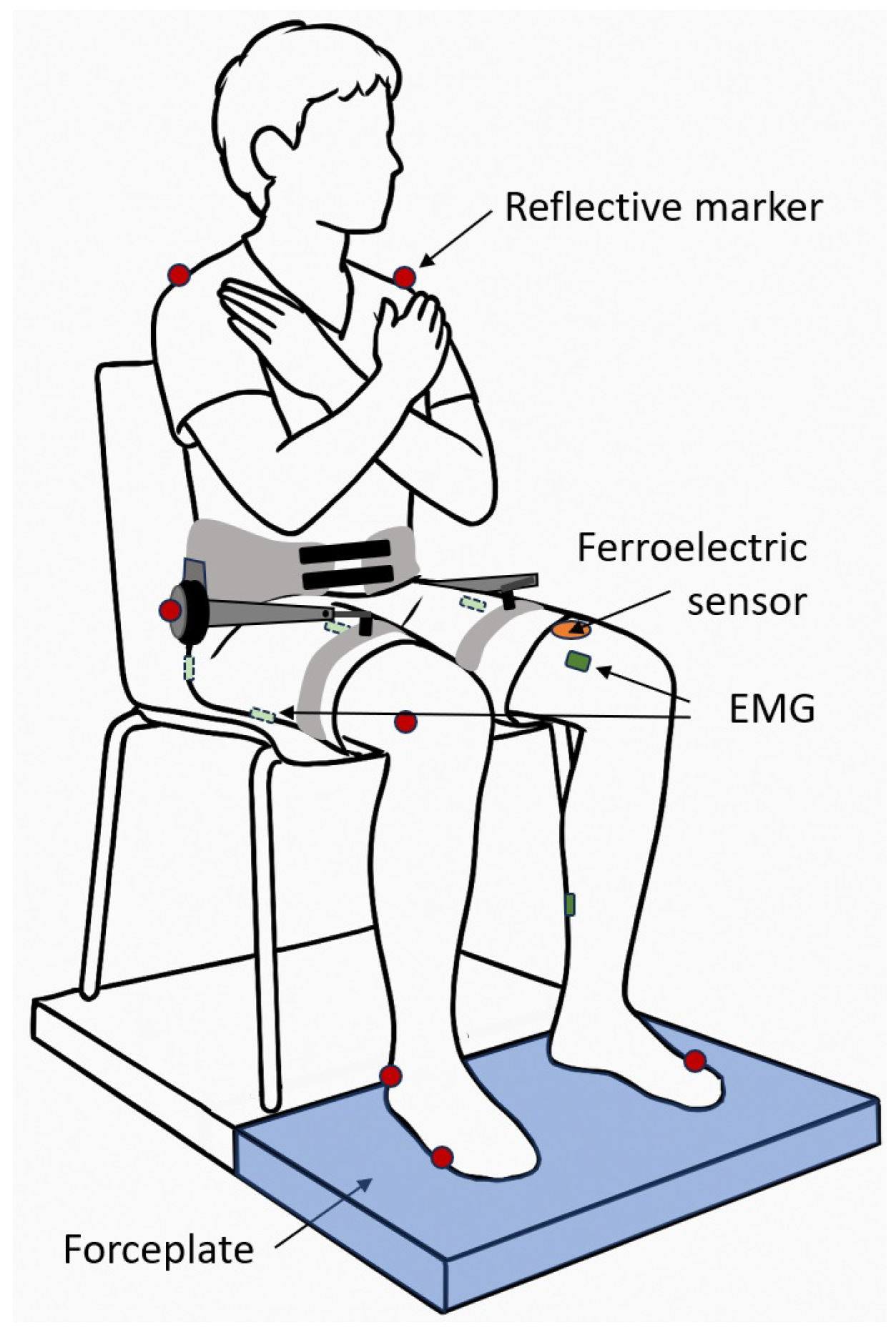
The experimental setup (
Figure 1) included one forceplate (Kistler Instrumente AG9, 260AA6, Winterthur, Switzerland), a motion capture system with twelve cameras (Qualisys, Oqus, Göteborg, Sweden), ten surface EMG sensors to measure muscle activation and kinematics using the integrated inertial measurement units (Delsys Incorporated, Trigno Research+ System, Natick, MA, USA), and the GuroX hip exoskeleton [
24]. Ground reaction forces and exoskeleton data were measured at 1000 Hz, motion capture data at 200 Hz, and EMG at 1259 Hz. All the measurements were synchronized using a trigger signal at the beginning of each sit-to-stand trial.
The forceplate was placed in front of a regular chair (height 0.46 m). Reflective motion capture markers were placed at prominent landmarks, including the shoulder (acromion), hip (trochanter major or on top of exoskeleton motor), knee (condylus lateralis femoris), ankle (malleolus lateralis), and forefoot (articulationes metatarsophalangeae V) of both limbs. EMG was recorded from the gluteus maximus (GLM), biceps femoris (BF), rectus femoris (RF), vastus medialis (VM), and soleus (SOL) of both limbs. EMG electrodes were placed according to the recommendations of SENIAM (seniam.org). Before fixing the EMG sensors, hair was removed, and the skin was cleaned with alcohol. To minimize the risk of sensor detachment due to movement and sweating, the sensors were secured with adhesive non-woven fabric tape (Rudavlies).
The GuroX hip exoskeleton (3.1 kg) features two onboard quasi-direct drive BLDC motors (AK10-9, CubeMars, Nanchang, China) that provide flexion and extension torques at each hip joint [
22,
23,
24]. It was controlled in real time via an off-board computer running MATLAB Simulink xPC (MATLAB R2018a, MathWorks, Natick, MA, USA). To trigger the sit-to-stand assistance, a ferroelectric sensor [
25,
26] was placed on the lower part of the left rectus femoris and used in conjunction with the motor encoder used to track the hip angle. Based on participant feedback during pretesting, early assistance caused discomfort. Therefore, assistance was provided only during the second half of the sit-to-stand transition, often referred to as the extension phase, starting after seat-off [
27]. Two criteria had to be met to initiate assistive extension torque. First, the ferroelectric signal had to exceed an individual threshold, determined for each participant during pretesting (8 to 20 mV), and second, the hip angle, which decreases and then increases during the transition to standing, had to exceed the rest angle measured while sitting. A maximum hip extension torque of approximately 1 Nm/kg, based on values from [
7,
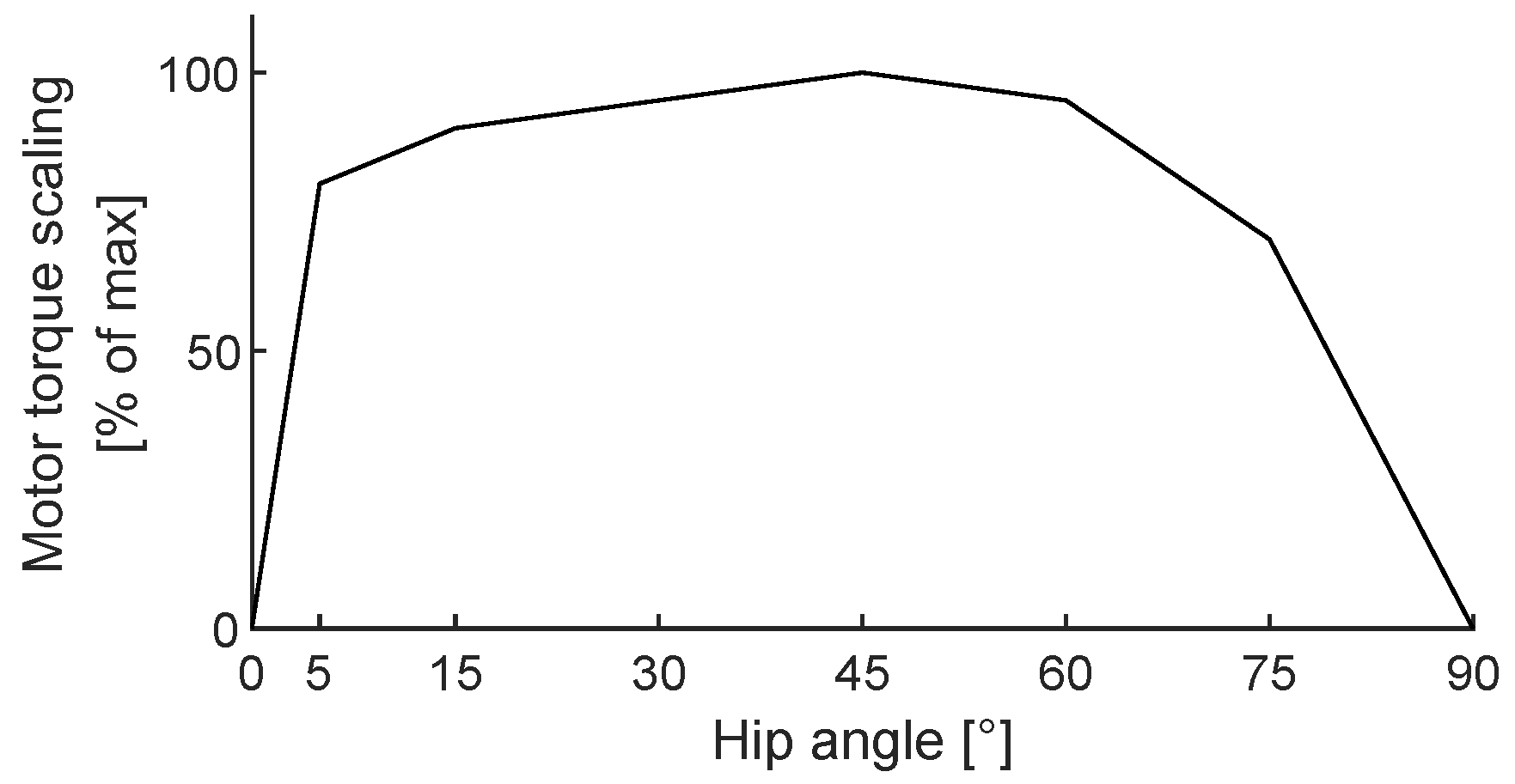
28], was used as a reference for peak extension assistance, with the exoskeleton control designed to provide about 10% of this maximum. In addition to scaling the assistance torque to participant mass, the extension torque was additionally scaled based on the hip angle to allow for smoother biologically inspired transitions, with the peak torque provided at the 100% level (
Figure 2). Torque control was implemented using a PD controller to achieve the desired assistance level. For evaluation, the motor torque was determined by multiplying the motor current with the linear torque constant of 0.0905 Nm/A and the gear ratio of 9 [
29].
2.3. Experimental Protocol
After providing informed consent, participants were equipped with EMG sensors and motion capture markers. They were then briefed on the experimental procedure: Upon receiving a verbal cue from the research team, participants performed a sit-to-stand transition at their preferred speed. After a brief period of standing, they returned to a seated position and waited for the next command. Each trial had a fixed duration of 15 s for data acquisition. Following several familiarization trials, participants completed ten sit-to-stand trials without wearing the exoskeleton (No Exo), with foot placement and seating position standardized using floor and seat markings to ensure consistent positioning across conditions and trials. Also, the individual trigger threshold from the ferroelectric sensor was fixed. Afterwards, the exoskeleton was fitted, and the ferroelectric sensor was attached near the distal end of the left rectus femoris. Participants then performed additional familiarization trials with the exoskeleton, both with and without active assistance. Subsequently, in randomized order, they completed ten sit-to-stand trials with the exoskeleton worn but inactive (Exo Off) and ten trials with extension assistance enabled (Exo On).
2.4. Analyses
All data were exported and analyzed using MATLAB (R2023b, MathWorks, Natick, MA, USA). Motion capture data were filtered using a zero-lag, second-order Butterworth filter with a cut-off frequency of 6 Hz. Subsequently, sagittal plane joint angles were calculated for the right limb. The hip angle was derived from the coordinates of the shoulder, hip, and knee markers; the knee angle from the hip, knee, and ankle markers; and the ankle angle from the knee, ankle, and forefoot markers. To ensure consistent comparisons across participants, average joint angles were offset to zero at the start of the sit-to-stand transition.
For the EMG data, the baseline offset was first removed by subtracting the mean of the signal. A zero-lag, fourth-order bandpass filter (10–450 Hz) was then applied. The signal was subsequently rectified and smoothed using a zero-lag, fourth-order Butterworth low-pass filter with a 6 Hz cut-off frequency. To enhance data quality, outlier detection was performed for each participant, muscle, condition, and trial based on maximum amplitude. Following common statistical practice for outlier detection in univariate data, trials with peak values exceeding three standard deviations from the average of the trial maxima were considered outliers and excluded from further analysis. This approach is based on the empirical 3-sigma rule covering 99.7% of data commonly used to identify unlikely values under a normal assumption of variability in time-series data. In total, approximately 5% of trials were removed. EMG signals from the left and right limbs were normalized to each participant’s peak muscle activation, defined as the average peak across all No Exo condition trials. This scaling set the peak activation to 100%, enabling comparison across muscles and participants.
Inertial measurement unit (IMU) angular velocity was extracted for the thigh based on the sensors placed at the right and left VM, RF, and BF. To ensure directional consistency, BF signals were inverted (multiplied by −1), aligning their sagittal plane angular velocity direction with those of the VM and RF sensors. A combined thigh angular velocity signal was then computed for each trial by averaging the IMU signals across all three sensors and both limbs.
To extract the sit-to-stand transition, the vertical ground reaction force (GRF) was used to identify the start, and the combined thigh angular velocity from the IMUs was used to determine the end. Baseline adjustment was performed by subtracting the mean of the first 1000 frames during quiet sitting to support signal comparability. For detecting the transition onset, the vertical GRF and IMU data were filtered using a zero-lag, second-order Butterworth filter with a 4 Hz cut-off frequency. The start of the flexion moment phase, as defined by Chen et al. [
27], was detected when the filtered GRF dropped below zero. A threshold of −12 N was applied to account for signal noise and small postural adjustments during sitting. The transition was considered complete when the filtered thigh angular velocity exceeded −8°/s, indicating near full leg extension and the end of the dynamic leg extension.
Based on these two events, all relevant data were extracted for each participant, condition, and trial.
Integrated EMG (iEMG) was calculated for each trial by computing the area under the rectified EMG curve using the trapezoidal integration method and dividing the result by the EMG sampling frequency to normalize for signal duration. Integrated EMG was normalized for each muscle and condition by setting the average of each participant and muscle to 100% for the No Exo condition. The iEMG values for the Exo Off and Exo On conditions were then adapted relative to the No Exo baseline, with the Exo Off and Exo On conditions expressed as a percentage of the No Exo iEMG.
Following this, joint angles (hip, knee, ankle), vertical GRF, motor torque, and EMG signals from five muscles were time-normalized to 1000 frames, representing 100% of the sit-to-stand transition, to allow for consistent temporal alignment across participants. For the EMG, data from the right and left limbs were combined and averaged. With that data, participant means were determined for all variables. Finally, group means and standard deviations were computed.
Some data could not be analyzed due to technical issues with exoskeleton data storage. Additional trials were excluded due to occluded or loose motion capture markers or because participants did not follow the instructed movement protocol. On average, each participant contributed 9.3 valid trials for the No Exo, 8.7 for Exo Off, and 8.2 for Exo On conditions.
Average values and their corresponding standard deviations are provided as
Supplementary Material in a MATLAB (.mat, MathWorks, Natick, MA, USA) file.
2.5. Statistics
Electromyography (EMG) data were analyzed across three experimental conditions, No Exo, Exo Off, and Exo On, for the iEMG of the five muscles, average across both limbs (GLM, BF, RF, VM, and SOL). For each muscle average, a non-parametric Friedman test was first used to assess whether there were overall differences between the conditions. The Friedman test was selected to account for the repeated-measures design and potential non-normality of the data. A significance threshold of = 0.05 was applied. If the Friedman test for a given muscle yielded a significant result (p < 0.05), post hoc pairwise comparisons were conducted using the Wilcoxon signed-rank test for the following condition pairs: No Exo vs. Exo Off, No Exo vs. Exo On, and Exo Off vs. Exo On. The resulting p-values from these pairwise tests were then subjected to a Benjamini–Hochberg False Discovery Rate (FDR) correction to control for multiple comparisons at q = 0.05. Only comparisons from muscles with significant Friedman results were included in the FDR correction. The Benjamini–Hochberg procedure compares each raw p-value to an adaptive critical value, without modifying the p-values themselves. Statistical significance after correction was determined by whether the p-value was below its corresponding BH critical value.
A similar test was performed for the average execution time of the three conditions. All statistical tests were implemented in MATLAB (MathWorks Inc., Natick, MA, USA), and results were reported as raw p-values along with their BH critical thresholds and FDR significance status.
3. Results
Participants performed sit-to-stand transitions in an average time of 1.88 ± 0.15 s under the No Exo condition, 2.06 ± 0.28 s with Exo Off, and 1.99 ± 0.32 s with Exo On. A statistically significant increase in transition time was observed for Exo Off compared to No Exo (p = 0.0186, BH critical = 0.025). The difference between No Exo and Exo On was not statistically significant, but approached significance (p = 0.0674, BH critical = 0.0375). No significant difference was found between Exo Off and Exo On (p = 0.1748, BH critical = 0.0458).
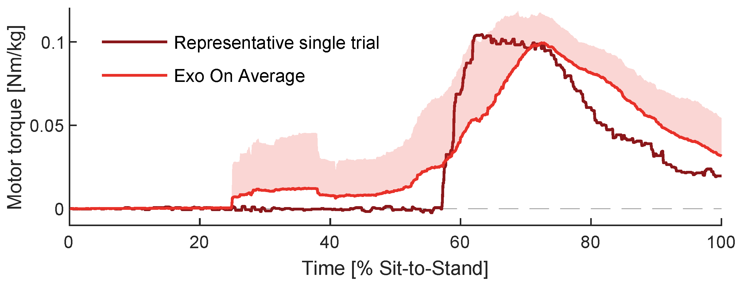
Within the Exo On condition, on average a peak extension torque of 0.12 ± 0.02 Nm/kg was provided. Due to averaging multiple subjects and trials, the average assistance extension torque shows a relatively smooth increase. In contrast, individual assistance extension torque showed a rapid increase (
Figure 3), as intended by the scaling (
Figure 2). For the majority of participants and trials, the exoskeleton assistance started to increase at close to 60% of the transition.
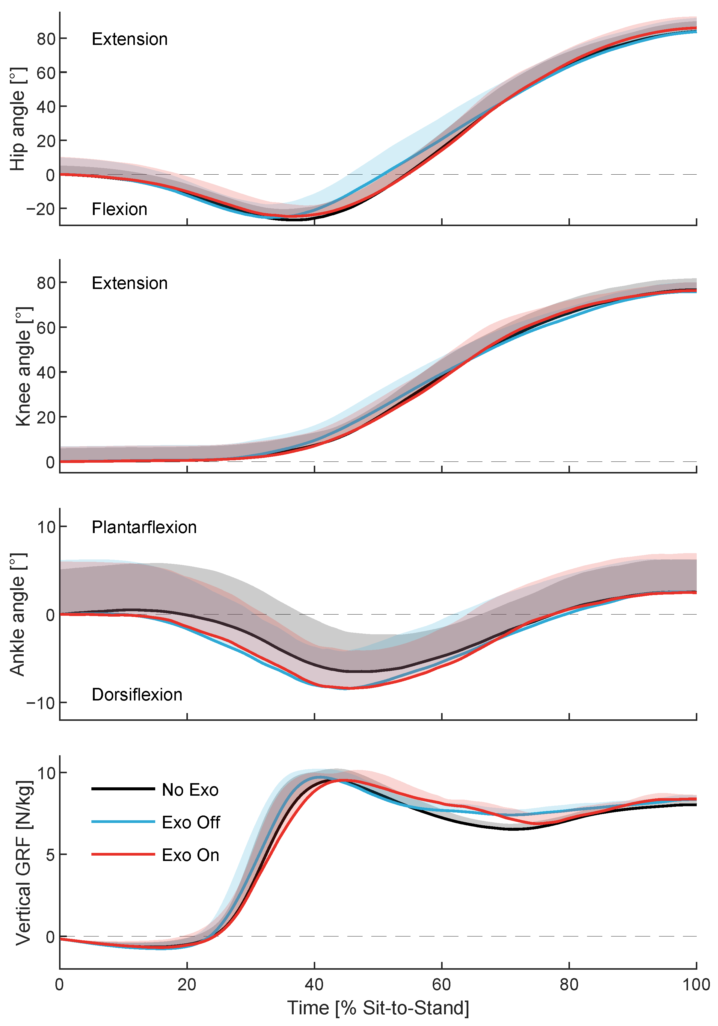
Differences in joint angles and vertical ground reaction forces across conditions did not exceed the between-subject standard deviation, suggesting consistent movement strategies (
Figure 4).
Time-normalized EMG data (
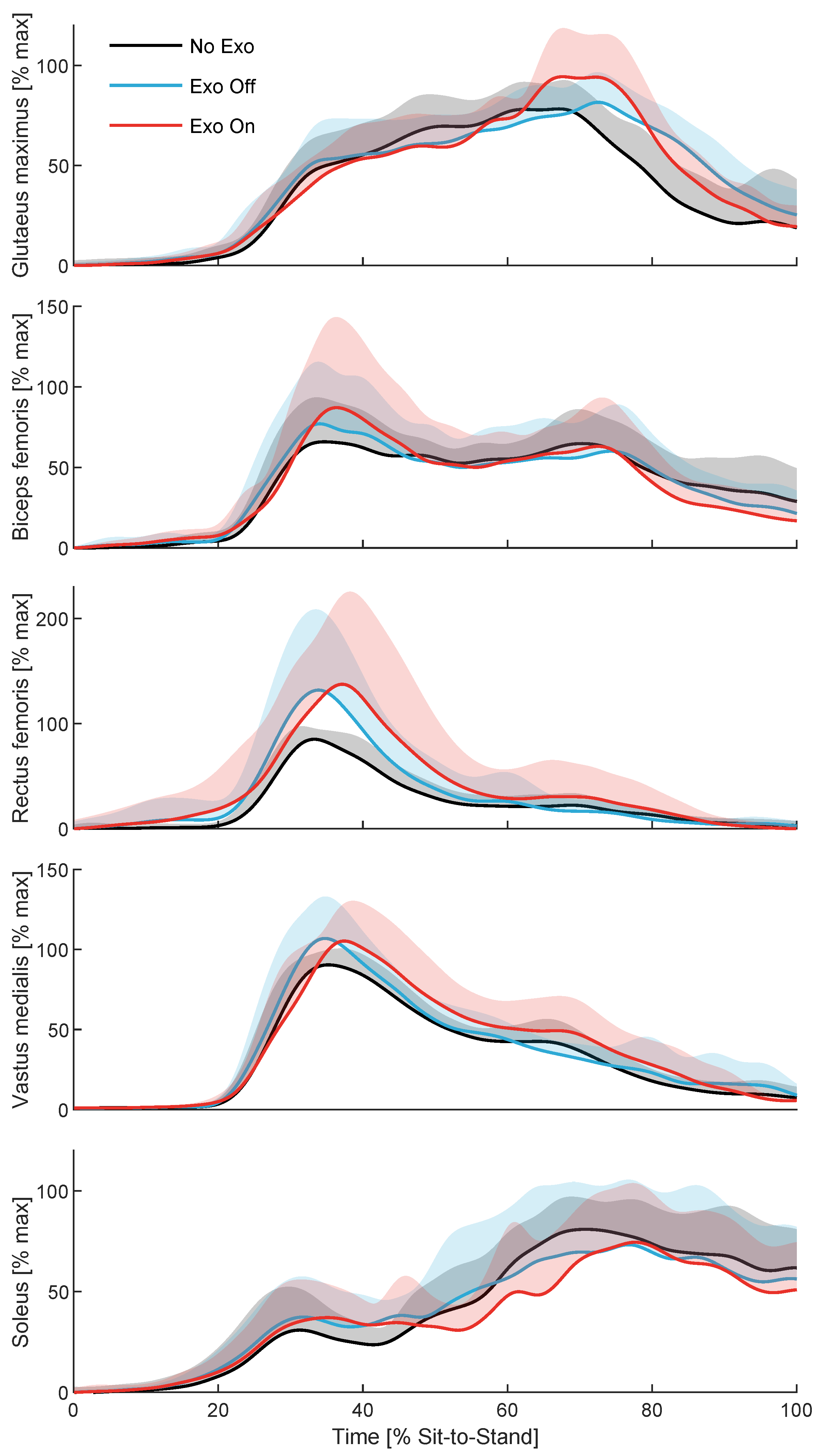
Figure 5) show 30–50% increases in RF, VM, and BF activity in both Exo conditions compared to No Exo. Additionally, GLM activity increased after 60% of the transition, while BF and SOL activity were reduced during the same phase.
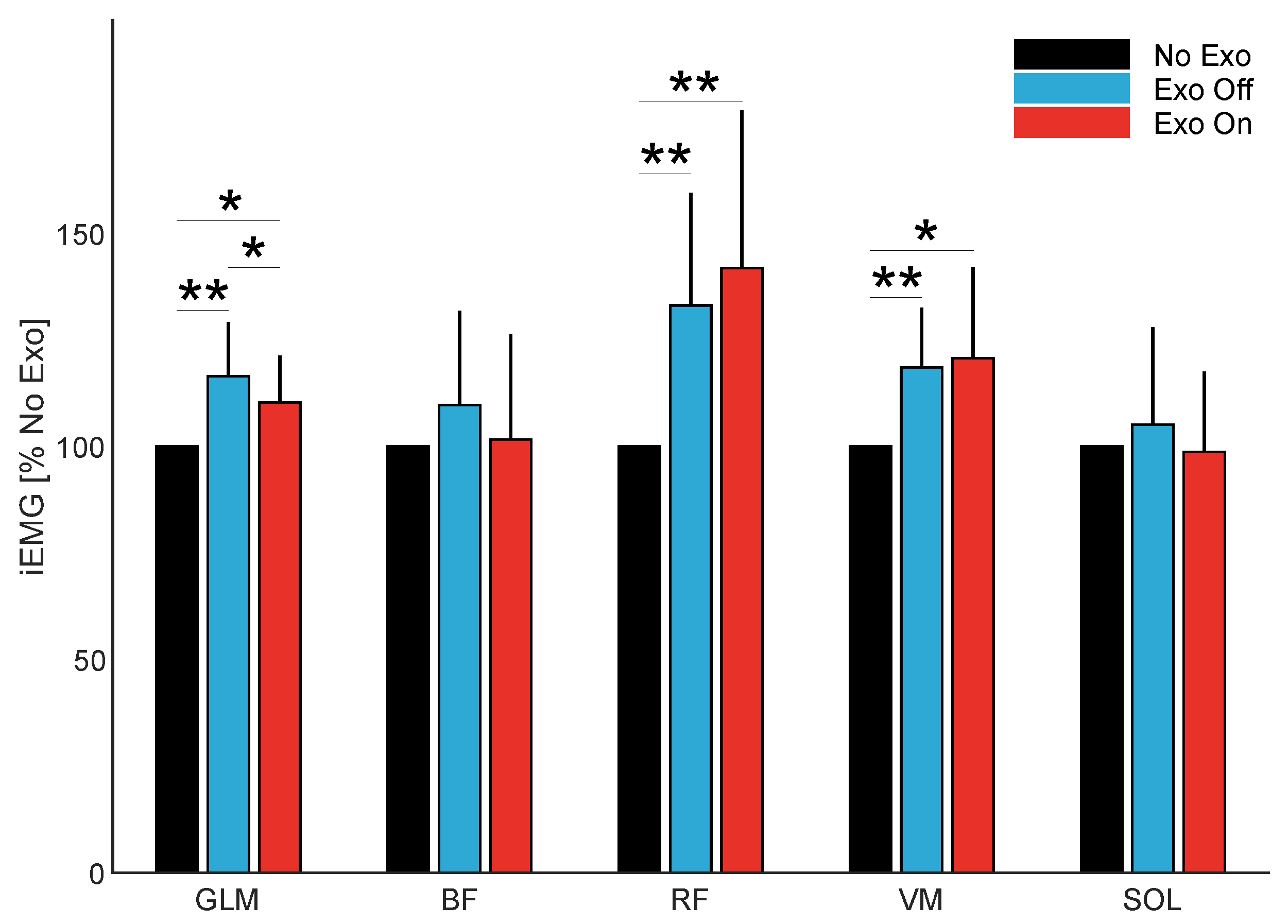
The statistical analysis of integrated EMG (iEMG) revealed significant effects of the conditions for the gluteus maximus (GLM), rectus femoris (RF), and vastus medialis (VM) muscles (Friedman test). Post hoc pairwise comparisons showed increased iEMG in the Exo conditions compared to No Exo for these muscles, with all differences remaining significant after FDR correction (
Table 1 and
Table 2,
Figure 6). Specifically, compared to the No Exo condition, iEMG increased by 16% and 10% for the GLM, 33% and 42% for the RF, and 19% and 21% for the VM in the Exo Off and Exo On conditions, respectively (
Table 2). In addition, a significant reduction of 6% in gluteus maximus iEMG was observed in the Exo On condition compared to Exo Off. No significant effects in iEMG were observed for the biceps femoris (BF) or soleus (SOL).
4. Discussion
In this study, we compared the sit-to-stand transition across three conditions: without the hip exoskeleton (No Exo), with the hip exoskeleton but no assistance (Exo Off), and with the hip exoskeleton providing extension assistance (Exo On). We hypothesized that wearing the exoskeleton would increase muscle effort during the transition and that some of this increase could be mitigated when assistance was provided.
EMG analysis revealed increased muscle activity in the leg extensors, rectus femoris (RF), and vastus medialis (VM) during the early phase of the transition, and in the gluteus maximus (GLM) during the late phase, in both exoskeleton conditions compared to the No Exo condition. As sit-to-stand kinematics and kinetics remained within the range of inter-subject variability and showed no substantial differences across conditions, the observed increases in muscle activity are unlikely to reflect altered joint-level movement patterns. The iEMG analysis confirmed that differences were statistically significant.
In addition, iEMG showed reductions in the GLM for the Exo On condition compared to Exo Off. However, this effect is not clearly visible in
Figure 5. This is likely because the execution time for the sit-to-stand transition was, on average, 0.07 s shorter in the Exo On condition than in Exo Off, masking the underlying difference in muscle activity. Furthermore, although just statistically significant for Exo Off, both exoskeleton conditions exhibited longer transition times compared to No Exo. The slower execution may be due to the additional physical effort required to counteract the exoskeleton’s weight and required postural control. Typical transition times were 1.2 s, 1.6 s, and 2.5 for fast, natural, and slow movement speed in [
7], respectively, which are in the range of our results, with a maximum of 2.06 s for the Exo Off condition. Also, while not significant, small reductions in iEMG were found for the BF and SOL for Exo On compared to Exo Off, with Exo On levels close to No Exo. In summary, reductions in GLM, BF, and SOL iEMG, along with slightly shorter execution time in the Exo On condition, suggest that hip extension assistance may have partially compensated for the exoskeleton’s weight and reduced the physical demands of the sit-to-stand transition. Nevertheless, these findings should be interpreted with caution given the limited sample size, which may restrict the generalizability of the observed effects.
Notably, increased co-activation of the RF and biceps femoris (BF) was observed during the early phase of the transition. The co-activation found is consistent with previous findings highlighting their role in maintaining postural balance through biarticular coordination [
30,
31]. In addition, increased BF activity was found to be a likely contributor to increased transition times in older adults with knee osteoarthritis [
32]. Such co-contraction contributes to dynamic stability in both sagittal and frontal planes [
33] and may serve as a neuromuscular strategy to manage the altered demands imposed by the exoskeleton.
Based on participant feedback during early testing, the active exoskeleton was perceived as disruptive when assistance was applied too early during the sit-to-stand transition. To minimize interference with natural movement, assistance was therefore restricted to the second half of the transition in this initial analysis. Previous studies show that hip extensor torque begins around the point when vertical center of mass velocity increases above zero. This moment corresponds with a rise in vertical ground reaction force, occurring at approximately 24% of the transition, according to
Figure 4. EMG data from earlier work [
6] and from our own recordings indicate that leg extensor activation, including that of the gluteus maximus (GLM), begins even earlier. Starting assistance closer to this point might have led to greater reductions in extensor activity or different transition dynamics. For comparison, Choi et al. [
14] observed a U-shaped relationship between knee torque peak timing and EMG activity in the VM; the greatest reduction occurred when peak torque was applied after a knee angle change of approximately 30° from sitting. This corresponds to about 55% of the sit-to-stand transition in our data and is thus on average earlier compared to our timing. However, the assumption that exoskeleton assistance must be applied earlier solely because EMG activity precedes the applied torques is not straightforward. EMG typically precedes joint moments by 10–130 ms due to electromechanical delay [
34], and joint moments themselves generally precede joint power, as peak power is influenced by joint velocity, which reaches its maximum later in the movement. For instance, Seven et al. [
35] reported peak hip extension moments during sit-to-stand at approximately 56% of the transition, whereas peak extension power occurred later at around 70%, with the respective increases starting near 37% and 45%. This temporal separation between EMG, moments, and power has practical implications for assistance timing. In our previous work [
36], we compared a moment-inspired control strategy with earlier assistance to a power-inspired strategy with later assistance during walking, and both strategies achieved comparable levels of mechanical assistance and effort reduction. Taken together, these considerations suggest that our participant-guided timing approach is more closely aligned with the power-based strategy.
The applied peak hip extension torque of 0.12 Nm/kg corresponded to approximately 10% to 24% of the typical per-limb peak torque observed during sit-to-stand transitions, which ranges from 0.5 Nm/kg to 1.0 Nm/kg depending on the study [
6,
7,
28]. Despite this low to moderate assistance level, GLM iEMG was reduced by only 6%. Since the added exoskeleton mass increased body weight by only 5%, a reduction in GLM activity below No Exo values might have been expected. Larger reductions of 45% in the mean absolute value (MAV) of the GLM were reported for another exoskeleton design assisting hip extension as well [
18]. Compared to our design, the system employed a fully textile-based human–machine interface and lightweight pneumatic actuators. However, the power source was not integrated, reducing the overall system mass and, consequently, the physical burden on the user. Additionally, foot and seating positions were not fixed, which is critical, as variations in these positions have been shown to significantly alter peak hip extension moments by a factor of 4.5 [
8], which can substantially affect lower limb muscle activity. Moreover, kinematic and temporal aspects of the sit-to-stand transition were not reported, despite the use of EMG MAV, a metric that does not account for time-dependent variations. Finally, the assistance strategy, including the timing and magnitude of support, was not clearly described. Maximum applied forces evaluated during the actuator characterization were about 22 N. Assuming a lever arm of 0.1 m, this would result in an assistance of 2 Nm per limb, which is about 25% of what we applied. Taken together, the limited methodological and quantitative information provided in [
18] constrains the ability to make a direct comparison between the two systems and does not fully explain the substantially larger reductions in muscle activity reported. However, the markedly lower system mass of the exosuit, 0.32 kg compared with 3.1 kg of our exoskeleton, alone could plausibly account for part of the observed advantages relative to the No Exo condition.
When compared with prior sit-to-stand exoskeleton studies assisting knee extension [
12,
13,
14,
16] or combined knee and hip extension [
15] that reported user benefits, the magnitude of EMG reductions observed in the present study is notably smaller. Previous knee-focused designs reported reductions in knee extensor muscle activity ranging from 12% to 58%. However, these systems only compared assisted conditions to Exo Off and did not demonstrate reductions in muscle activity relative to a No Exo condition. The modest reductions, compared to Exo Off, observed in our work may be attributed either to an insufficient assistance strategy or to a limited potential for unloading the hip extensors during sit-to-stand. Prior work has shown that transition speed [
7] and seating configuration [
8] strongly influence the relative contributions of the knee and hip joints, with slower transitions and more vertical or posterior foot placements favoring larger knee extension moments. Under such conditions, assistive strategies may preferentially benefit knee extension. In contrast, a study in children reported higher joint moments and power at the hip than at the knee during sit-to-stand [
35], highlighting that joint contributions might be also population-dependent. Among adult studies employing multiarticular designs, Schmidt et al. [
15] reported reductions of up to 48% in gluteus maximus activity (calculated by us from the original data) while providing peak assistive torques corresponding to approximately 26% of the nominal hip and knee extensor moments (22 Nm), with peak assistance occurring at around 50% of the transition. Although assistance was provided at both the hip and knee, these results suggest that higher relative assistance levels and earlier torque application can enhance sit-to-stand support. However, similarly to the second work including hip assistance [
18], fixation of seating position and transition timing were not reported, limiting comparability with the present study.
To summarize, the limited reductions in EMG activity observed in this study are likely attributable to several interacting factors, including the timing and magnitude of assistance, increased postural demands arising from the device mass and the application of parallel external torques, potential exoskeleton-related issues such as mechanical friction, inertia, or joint axis misalignment, and limited user adaptation and familiarization with the exoskeleton during a non-cyclic movement. In contrast to exoskeleton-assisted walking, where users can adapt over many continuous strides and where training across multiple sessions or days has been shown to enhance assistance benefits [
37], exposure to the sit-to-stand task in the present study was limited. Participants completed ten repetitions per condition, which may not have been sufficient to allow substantial neuromuscular adaptation to the applied assistance. As a result, the observed reductions in muscle activity may underestimate the potential benefits achievable with extended familiarization or repeated training. This study was also limited by a small sample size and relatively low peak assistance torque, which may have constrained the observable effects. Despite these limitations, this study introduces several key innovations: we evaluated a hip exoskeleton designed to assist walking for assisting hip extension during the sit-to-stand transition and systematically compared outcomes to both a No Exo and an Exo Off condition. Importantly, we observed statistically significant effects on transition time and gluteus maximus activity, demonstrating that even moderate, well-timed assistance can partially mitigate the physical demands imposed by the device. These findings provide a foundation for optimizing hip exoskeleton support during functional tasks beyond walking.
5. Conclusions
Hip exoskeletons have been shown to reduce muscular effort during daily walking. When worn for this purpose, it is equally important to minimize the additional physical burden they impose during other functional movements, such as sit-to-stand transitions.
Our findings show that wearing the exoskeleton, independent of assistance, increased lower limb extensor activity, primarily due to the added mass of the device. When hip extension assistance was applied during the second half of the transition, gluteus maximus (GLM) iEMG was reduced and execution time was shorter compared to the Exo Off condition, while no significant difference was observed relative to the No Exo condition. Together, these results indicate that appropriately timed assistance can partially compensate for the additional effort associated with the exoskeleton’s weight.
Although the level of assistance provided was relatively low, the observed reduction in GLM activity demonstrates the potential for even moderate support to reduce muscular demand during sit-to-stand transitions. The absence of broader reductions in muscle activity, particularly relative to the No Exo condition, likely reflects a combination of suboptimal assistance, limited user adaptation, increased postural demands, and mechanical or ergonomic constraints of the exoskeleton.
Overall, these findings highlight both the potential and the current limitations of hip exoskeletons for assisting non-cyclic functional movements such as sit-to-stand. Future work should systematically investigate assistance strategies, user adaptation, and human–machine interaction to better align support with users’ neuromechanical requirements. Beyond compensating for device weight during secondary tasks, future studies may also explore assistance strategies that enable performance gains beyond the No Exo condition, thereby maximizing the functional benefits of daily exoskeleton use.



 ,
,