Article

Evaluating Lumbar Biomechanics for Work-Related Musculoskeletal Disorders at Varying Working Heights During Wall Construction Tasks

by
Md. Sumon Rahman
1,*,
Tatsuru Yazaki
1,
Takanori Chihara
2 and
Jiro Sakamoto
1
1
Division of Transdisciplinary Sciences, Kanazawa University, Kanazawa 920-1192, Japan
2
Institute of Science and Engineering, Kanazawa University, Kanazawa 920-1192, Japan
*
Author to whom correspondence should be addressed.
Biomechanics 2025, 5(3), 58; https://doi.org/10.3390/biomechanics5030058 (registering DOI)
Submission received: 30 May 2025 / Revised: 7 July 2025 / Accepted: 14 July 2025 / Published: 3 August 2025
(This article belongs to the Section Tissue and Vascular Biomechanics)

Abstract

Objectives: The aim of this study was to evaluate the impact of four working heights on lumbar biomechanics during wall construction tasks, focusing on work-related musculoskeletal disorders (WMSDs). Methods: Fifteen young male participants performed simulated mortar-spreading and bricklaying tasks while actual body movements were recorded using Inertial Measurement Unit (IMU) sensors. Muscle activities of the lumbar erector spinae (ES), quadratus lumborum (QL), multifidus (MF), gluteus maximus (GM), and iliopsoas (IL) were estimated using a 3D musculoskeletal (MSK) model and measured via surface electromyography (sEMG). The analysis of variance (ANOVA) test was conducted to identify the significant differences in muscle activities across four working heights (i.e., foot, knee, waist, and shoulder). Results: Findings showed that working at foot-level height resulted in the highest muscle activity (7.6% to 40.6% increase), particularly in the ES and QL muscles, indicating an increased risk of WMSDs. The activities of the ES, MF, and GM muscles were statistically significant across both tasks and all working heights (p < 0.01). Conclusions: Both MSK and sEMG analyses indicated significantly lower muscle activities at knee and waist heights, suggesting these as the best working positions (47 cm to 107 cm) for minimizing the risk of WMSDs. Conversely, working at foot and shoulder heights was identified as a significant risk factor for WMSDs. Additionally, the similar trends observed between MSK simulations and sEMG data suggest that MSK modeling can effectively substitute for sEMG in future studies. These findings provide valuable insights into ergonomic work positioning to reduce WMSD risks among wall construction workers.

1. Introduction

In developing countries, the construction industry is rapidly growing. However, due to the lack of a skilled workforce and modern technology [1], most construction tasks are performed manually [2]. Consequently, workers in this sector are at a high risk of developing work-related musculoskeletal disorders (WMSDs) [3]. WMSDs refer to disorders, injuries or pain in the joints, muscles, ligament, nerve, and tendons due to the nature of work-related features [4]. The prevalence of WMSDs among construction workers has increased in the last decade for developing countries, such as being at 57% in Bangladesh [5], 89% in Pakistan, 66.7% in Malaysia, 82.5% in Sri Lanka, and 80% in India, respectively [6,7].
The occurrence of WMSDs among workers not only reduces productivity and financial benefits but also increases the cost of health care, disability, compensation, and absenteeism [8]. Strategies to prevent WMSDs not only improves workers’ health and safety but also reduces the negative effect of WMSDs. There are various risk factors which are responsible for the development of WMSD risk in construction workers. Risk factors refer to any actions or conditions that contribute to causing WMSDs [9]. Therefore, it is essential to identify the risk factors associated with WMSDs to develop appropriate interventions to mitigate these issues.
Employees in the construction sector are subject to a number of risk factors, particularly during wall construction tasks. Sterud [10] and Andersen et al. [11] found that both psychological (i.e., high job demands and low level of leadership) and physical factors (i.e., physical exposure and the number of exposures) are associated with WMSDs. Individual factors such as age, race, and gender have also been identified as risks for developing WMSDs [12,13]. Albers and Hudock [14] identified load moment, hand force, peak shear force, and peak trunk velocity as significant risk factors for developing lower-back WMSDs. In their study, only specific body parts and postures were analyzed. Conversely, workers in the construction industry often experience multiple body injuries resulting from repetitive body movement [14]. Awkward posture is one of the most critical risk factors for WMSDs because such posture requires muscles to sustain force over extended periods [15]. Repetitive and physically demanding activities, especially when prolonged, cause muscle fatigue and pain, inevitably increasing the risk of WMSDs. In addition, in wall construction tasks, unavoidable physical demands such as repetitive movements, awkward body postures, and manual material handling expose workers to extensive biomechanical stress, contributing to the development of WMSDs [15,16,17,18]. Forward flexion, lateral bending, and twisting of the trunk muscles are the common biomechanical movements involved in manual handling tasks, which exert compressive forces and increase moments on the lumbar spine [3]. High mechanical loads on the lumbar spine have been considered significant risk factors for the development of WMSDs [3]. Another significant risk factor such as abnormal working height plays an important role for the development of WMSDs. For example, if the working height is too low, workers have to bend their trunk forward and work in this position for long periods, which puts extra stress on their spine [19]. On the other hand, if the working height is too high, the workers have to work in a twisted position, which can cause damage to the shoulders, neck and upper arms [20]. Therefore, it is essential to determine the relationship between different working heights on lumbar biomechanics during wall construction tasks.
Many studies have used musculoskeletal (MSK) models to evaluate the biomechanical risk factors, which have been developed recently [21,22,23]. The MSK model is a computer-based simulation used to estimate various biomechanical factors, e.g., muscle activity and joint loads in the body. It relies on motion data and body geometry to calculate internal biomechanics during task performance. This model helps researchers to study human body mechanics without invasive measurements. For instance, Sumon et al. [21] used an MSK model for evaluating the relationship between lower-back muscle activity and trunk flexion angle during construction tasks. Skals et al. [22] estimated the joint kinetics during manual material handling using inertial motion capture and an MSK model. Zadon et al. [24] measured the musculoskeletal load among the workers to predict the body mass index (BMI) using the MSK model. Takeshita et al. [25] conducted a musculoskeletal model simulation to evaluate the effect of knee flexion angles on muscle and joint forces during exercise. Therefore, the MSK model can be used to evaluate the biomechanical risk factors in terms of muscle activity for wall construction workers.
Several surface electromyography (sEMG) studies have been conducted to analyze the biomechanical risk of WMSDs in various sectors [26,27,28,29]. sEMG records electrical signals from muscles using sensors placed on the skin. It shows when and how muscles are activated during body movement. sEMG is widely used to assess muscle effort, amplitude, fatigue, and coordination during real-time activities. For example, Bangaru et al. [26] used portable sEMG and IMU sensors placed on the forearm to continuously monitor fatigue in construction workers. Antwi-Afari et al. [28] evaluated biomechanical risk factors for WMSDs for construction workers during repetitive lifting and manual handling tasks. Kong et al. [29] showed that while performing bolting tasks in the automobile assembly line at the height of 60 cm and 85 cm, wearing a passive lower-limb exoskeleton decreased the sEMG amplitude of the lower-limb muscles. Kang and Mirka [27] developed sEMG-assisted biomechanical models and showed that compression and anterior–posterior forces of the L4/L5 disk may increase due to the transfer of load from active to passive tissues during a fully flexed trunk position. Thus, sEMG sensors have numerous advantages, such as being non-invasive, portable, convenient to use, and reusable, etc. However, the main disadvantage of this sensor is that it can measure the properties of muscles close to the skin surface rather than deep muscles.
Conversely, with the aid of three-dimensional (3D) musculoskeletal (MSK) models, both the skin surface and deep muscle loading can be estimated to evaluate the risk of WMSDs more comprehensively [30]. Several studies on biomechanical analysis have been conducted, and the 3D MSK model has been used in sports science [31], the automobile industry [32], medical treatment [33], and the furniture industry [34]. Despite these efforts, no study has combined 3D MSK modeling and sEMG to analyze the effect of different working heights on lumbar biomechanics (i.e., muscle amplitude and activity) in wall construction tasks.
In addition, a previous study investigated the effect of different working heights on body postures bending and twisting during wall construction tasks and found a significant impact of varying working heights on bending and twisting postures [35]. Another study was conducted employing an MSK model to examine the relationship between muscle activity and trunk bending posture while performing construction tasks at four working heights. The findings showed a strong positive correlation between trunk bending and muscle activity [21]. Therefore, evaluating the effect of varying working heights on lumbar biomechanics—in terms of muscle activity and the accuracy and reliability of MSK analysis with an extended sample size—is an important research issue in wall construction tasks.
Despite various working heights having a significant impact on WMSDs in the lower back, the effect of different working heights on lower-back muscle during wall construction tasks has largely been overlooked. In this study, the authors hypothesized that muscle activation decreases as the working height increases during wall construction tasks. Therefore, the main aim of this study was to evaluate the effect of different working heights on lumbar biomechanics in terms of muscle activity and muscle load during wall construction tasks based on the simulated (MSK analysis) and experimental (sEMG analysis) assessments. Additionally, this study examines the potential for substituting MSK analysis for EMG analysis.

2. Materials and Methods

2.1. Participants

In this study, fifteen healthy young university male students (age: 22.00 ± 1.20 years; height: 172.02 ± 5.26 cm; and weight: 64.36 ± 8.97 kg) participated. All participants in the experiment did so willingly and they had no history of physical discomfort. Before starting the experiment, participants provided written consent and submitted personal information. This study was approved by the Ethics Committee of the Institute of Science and Technology, Kanazawa University, Japan (Approval number: 2023-8).

2.2. Experimental Setup and Process

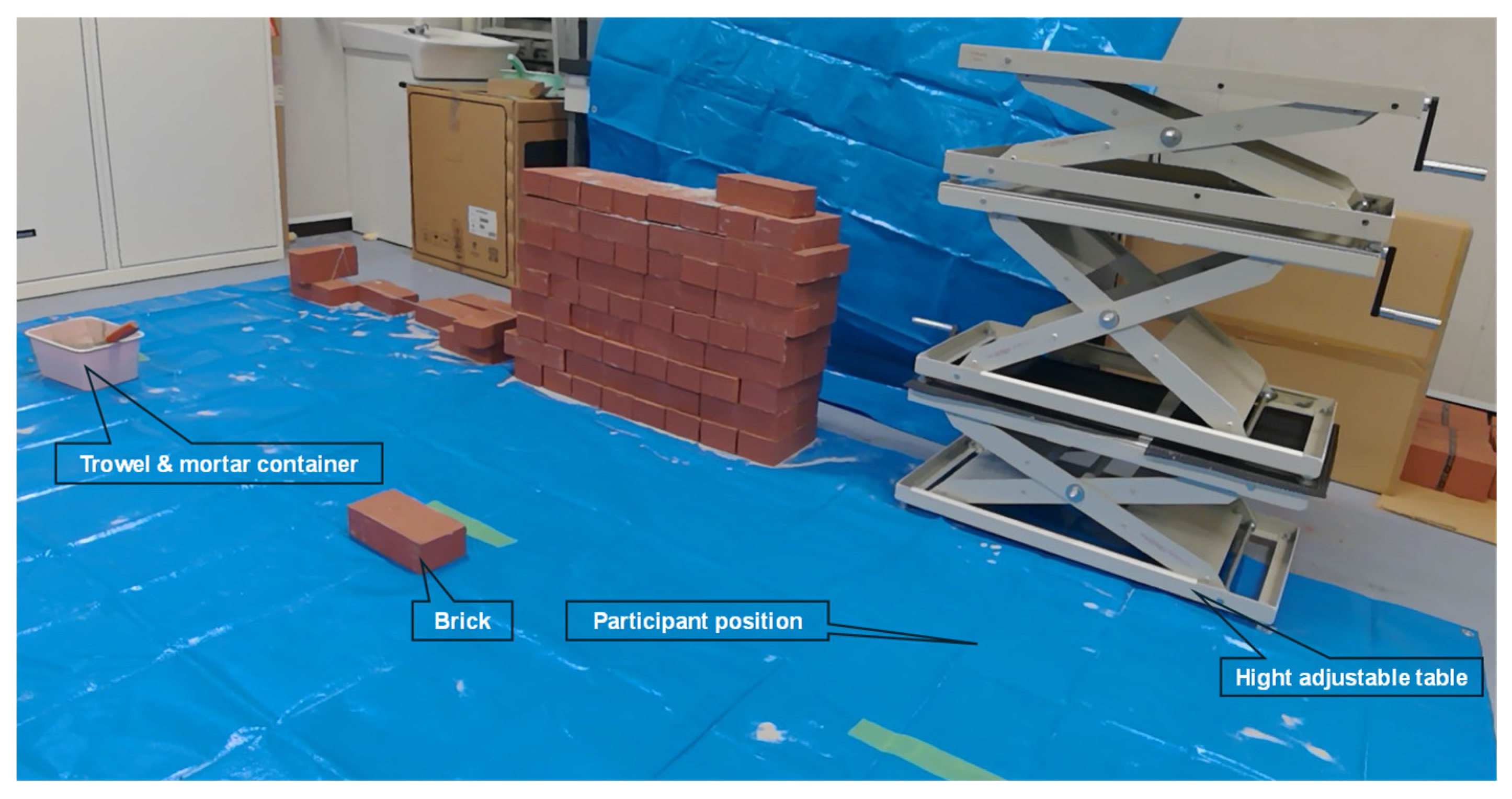
An imitation of wall construction tasks was created in a laboratory environment (Figure 1). The experimental tasks were a manual wall construction task. Participants performed wall construction activity with two different tasks: task-A, mortar spreading, and task-B, bricklaying.
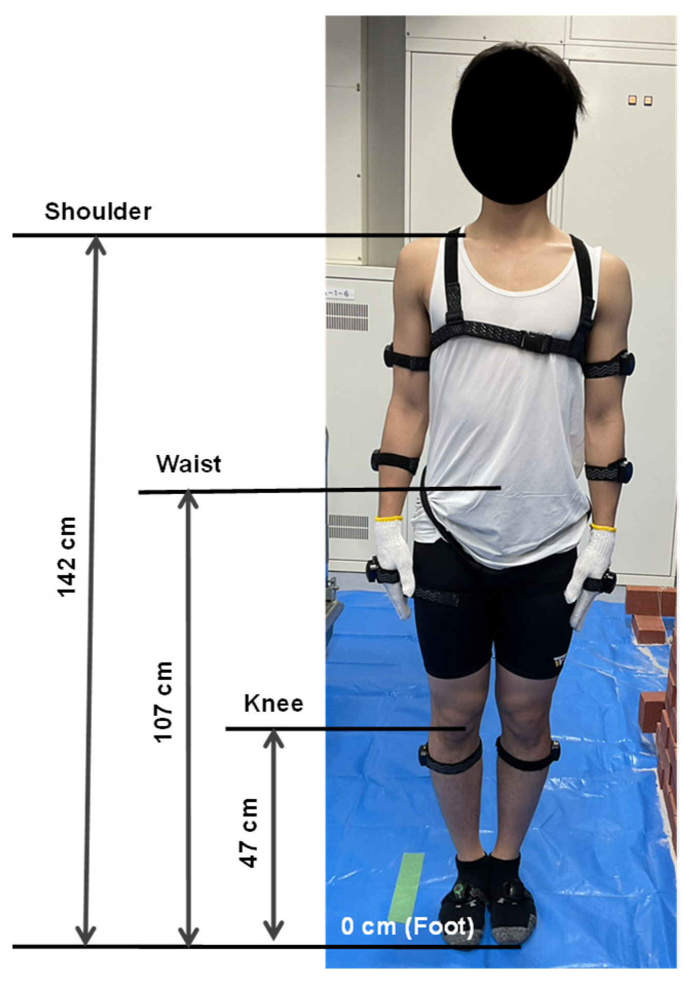
In task-A, participants scooped mortar from foot level (0 cm) on their right side and spread it at four different heights on their left side: foot (0 cm), knee (47 cm), waist (107 cm), and shoulder heights (142 cm). Here, the knee, waist, and shoulder-level heights were determined as the average values based on the participants’ measurements (Figure 2).
Similarly, in task B, participants picked up bricks from foot level on their right side and laid them on the spread mortar at the same four heights on their left side. The weight of the tool with the mortar was 0.6 kg, and the weight of the brick was 2.5 kg.

2.3. Motion Data Collection

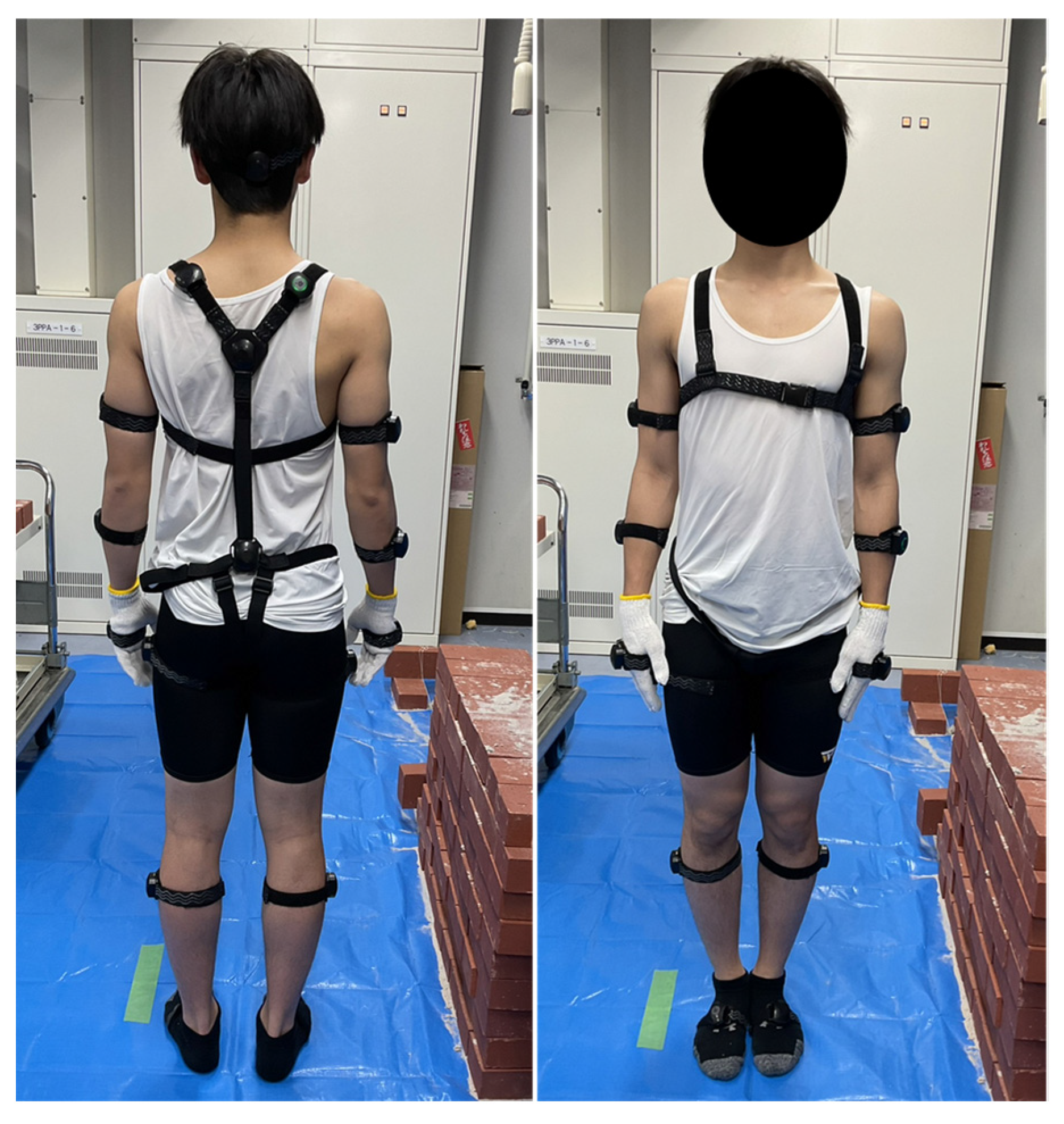
During the experiment, participants performed one task in one condition three times. The researchers recorded the more accurate task movement data among the three movements through the motion capture sensors. In this study, participants wore 17 Inertial Measurement Unit (IMU) sensors (Perception Neuron Studio, Noitom Ltd., Beijing, China) to capture the three-dimensional (3D) task movement data. The IMU sensors were positioned on the participant’s body in accordance with the manufacturer’s recommendations [36] (Figure 3). These sensors measure the movement of each body part and record the motion data as a bio-vision hierarchy (BVH) file using the dedicated software, i.e., Axis Studio, version 2.11.13128.2330. Data were acquired synchronously at a sampling frequency of 100 Hz. Previous studies [37,38] have confirmed the validity of this motion capture system.

2.4. Three-Dimensional Musculoskeletal Modeling Simulation

To analyze the muscle activity, a three-dimensional full-body musculoskeletal model (MSK) was created using the Anybody Managed Model Repository (AMMR) from the AnyBody Modeling System software (AnyBody Technology A/S version 7.4, Aalborg, Denmark). The accuracy of the Anybody technology system has been verified by previous studies [39,40,41].
The 3D MSK model was rescaled according to the participants’ anthropometry. In this study, inverse dynamics simulations were performed on motion capture data to compute muscle activity (Figure 4). In the AnyBody Modeling System, inverse dynamics simulations were performed using the polynomial muscle recruitment criterion defined by the following Equation (1) [39].
Minimize   G = i = 1 N ( F i F i , m a x 3 , 0 F i F i , m a x , C f = r
Here, G represents the objective function; i denotes the number of muscles; N is the total number of muscles; Fi is the muscle force; Fi,max represents the maximum isometric muscle force; f is the vector of joint and muscle forces; C is the coefficient matrix; and r denotes known vector of inertial and external forces.
The simulation of output muscle activity refers to the percentage of maximum muscle activation as estimated based on the MSK model. The lumbar biomechanics risk factors in terms of muscle activities of the five lower-back muscles, erector spinae (ES), quadratus lumborum (QL), multifidus (MF), gluteus maximus (GM), and iliopsoas (IL), were analyzed during wall construction tasks at four different working positions. These muscles were selected because they are associated with the risk of developing WMSDs in the lower back [42,43].

2.5. Measurement by Surface Electromyography (sEMG)

In this study, two lower-back muscles (on both the left and right sides), namely the erector spinae (ES) and multifidus (MF), were selected for sEMG analysis. These muscles were selected to compare the simulation results with experimental results during wall construction tasks. To evaluate the muscle load, two pairs of sEMG sensors (SX230-1000, Biometrics Ltd., Newport, UK) were attached to the lower back of the participants in accordance with a past study [44]. Prior to attaching the sEMG sensors, the skin was cleaned by alcohol. The data sampling frequency was 1000 Hz. The raw sEMG signals data were full-wave rectified and a fourth-order Butterworth filter was used as a low-pass filter with a cutoff frequency of 2 Hz [45]. The filtered sEMG amplitude data were used as the average value of the left- and right-side muscles. Figure 5 illustrates the process of measuring muscle load using sEMG sensors. The peak amplitude during work at foot height was set as the baseline. The peak sEMG amplitude is associated with the risk of WMSDs [46]. This baseline was used to normalize the amplitude at knee, waist, and shoulder heights.

2.6. Statistical Analysis

In the current study, simulated muscle activities during wall construction tasks were calculated. The average muscle activity of the right and left sides was determined and the average values for the fifteen participants were analyzed. The Shapiro–Wilk test was conducted to check the normality of the data. One-way repeated-measures analysis of variance (ANOVA) with post–hoc Bonferroni adjustment was performed to determine the effect of different working heights on lower-back muscle activity. Additionally, eta squared (η2) was used to report the effect sizes, where 0.01, 0.06, and 0.14 were interpreted as small, medium, and large effect, respectively [47]. A paired t-test was conducted to find the relationship between the amplitudes of sEMG signals (ES and MF muscles) during work at the foot-level height. To perform all statistical analyses, the authors used the Statistical Package for the Social Sciences (SPSS) software version 29.0.2.0 (International Business Machines (IBM) Corporation, New York, NY, USA). A p-value of less than 0.05 was considered as statistically significant.

3. Results

3.1. Analysis of Simulated Results from the MSK Model

The dominant working hand, body movements, weight of brick, and weight of mortar were different for the two construction tasks mentioned. Therefore, in this study two types of wall construction task (task-A and task-B) movements were collected and analyzed.

3.1.1. Effect of Working Heights on Muscle Activity During Mortar-Spreading Task (Task-A)

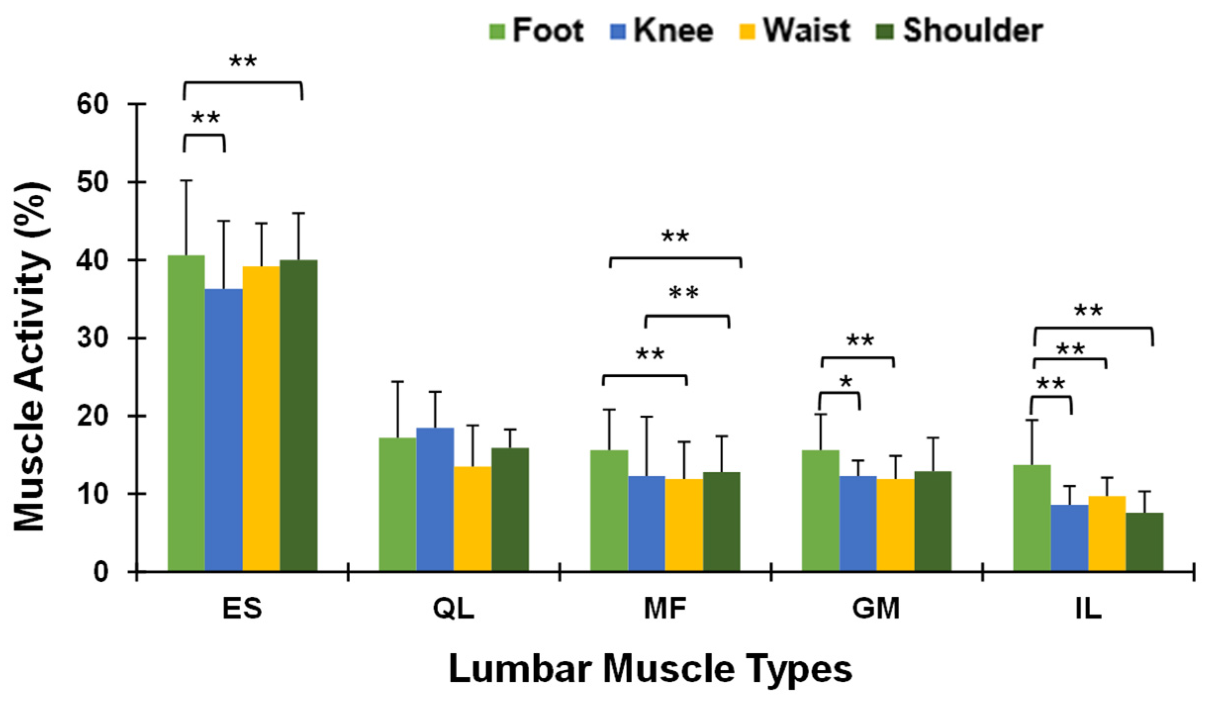
Table 1 presents the means, standard deviation, and ANOVA results for muscle activity during task-A. Statistically significant differences were observed in all muscles during work at different heights (p < 0.01), except for the QL muscle (p > 0.05). Working at foot level showed the highest activity for all muscles compared to other working heights, which decreased up to waist height, but not at shoulder height. The ES muscle displayed the highest muscle activity (i.e., 40.6%), while the IL showed the lowest muscle activity (13.7%) at foot-level height. The ES muscle activity was the greatest, followed by QL, GM, MF, and IL muscles.
The post hoc comparisons of the selected lower-back muscle activity at four working heights are shown in Figure 6. The activity for ES, MF, and IL muscles showed significant differences (p < 0.01) between the foot- and shoulder-level working heights. For the GM muscle, significant differences were found only between the foot and knee, and foot- and waist-level working heights. However, no significant differences were found in QL muscle activity across all working heights.

3.1.2. Effect of Working Heights on Muscle Activity During Bricklaying Task (Task-B)

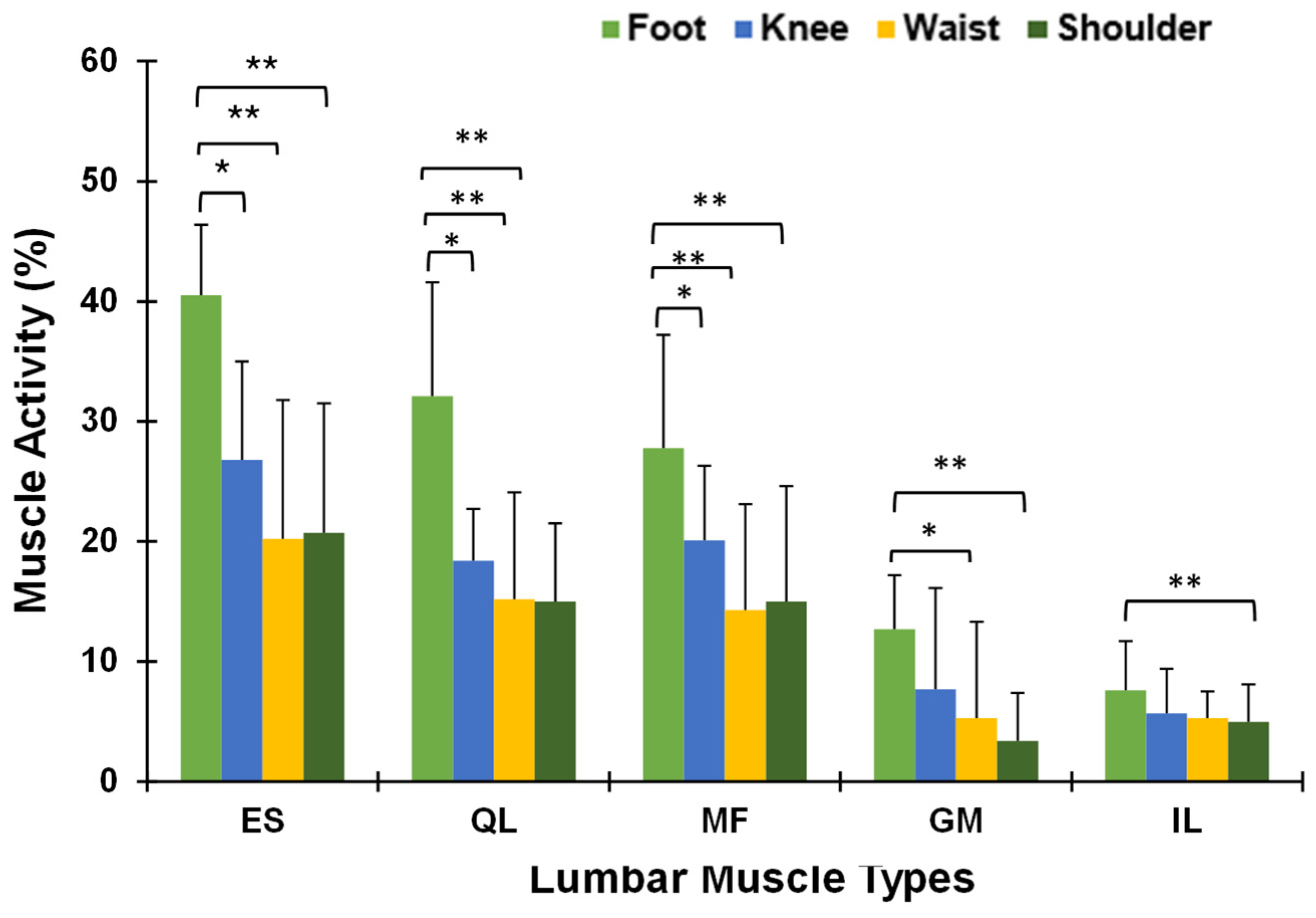
The summary of the mean and standard deviation of muscle activity (%) and ANOVA results for the bricklaying task is shown in Table 2. The ANOVA results indicated significant differences in muscle activity for all selected muscles across all working heights (p < 0.01). The activity of the selected lower-back muscles significantly decreased from the foot-level to waist-level working heights, but no significant reduction was found between the waist- and shoulder-level heights. The highest mean activity was observed in the ES (40.5%), followed by 32.1%, 27.8%, 12.7%, and 7.6% for the QL, MF, GM, and IL muscles, respectively. The eta-square value (0.23–0.47) also showed a large effect of working height on muscle activity.
For the bricklaying task, post hoc analysis showed that the mean muscle activities were significantly different between foot- and knee-level, foot- and waist-level, and foot- and shoulder-level working heights for the ES, QL, and MF muscles, respectively (Figure 7). For the GM muscles, the mean muscle activities showed significant differences between foot- and waist-level, and foot- and shoulder-level working heights. Conversely, for the IL muscle, a significant difference was only found between the mean activity of foot- and shoulder-level heights.

3.2. Analysis of Experimental Results from the sEMG Sensors

Figure 8 shows the peak sEMG amplitudes of the ES and MF muscles during task-A and task-B at foot-level height. The results of the t-test indicated that the amplitudes of the ES and MF muscles were statistically significant (p < 0.01) for both tasks. These sEMG amplitudes were considered the baseline values for comparison with the other three working heights.
To understand the data trend of the simulated MSK modeling results with experimental results, the peak sEMG amplitudes of the ES and MF muscles during task-A and task-B at foot-level height were considered as the base values for comparison with other three working heights. The sEMG amplitudes of the knee, waist, and shoulder height were normalized based on foot level base values. The normalized sEMG amplitudes of the ES and MF muscles during task-A and task-B are shown in Figure 9.
The sEMG amplitude for the ES muscle was higher than that for the MF muscle across all working heights. The lowest amplitude for the ES muscle (80.5%) was observed during task-A at waist-level height. In contrast, the lowest value for the MF muscle was 61.8% during task-B at the same height. For both muscles, the sEMG amplitudes were lower at knee- and waist-level height than at shoulder height in both task-A and task-B.

4. Discussion

This study used wearable inertial measurement unit (IMU) sensors, sEMG and a three-dimensional musculoskeletal model to assess the effect of different working heights on lumbar spinal biomechanics, focusing on muscle activity and muscle load during simulated wall construction tasks in a laboratory environment. The study results showed that different working heights have varying effects on the lower-back muscles. Additionally, the results revealed that the activity of the studied lower-back muscles significantly decreased in wall construction tasks as the working height increased from foot-level to waist-level height.

4.1. Effect of Working Heights on Lower-Back Muscles During Mortar-Spreading Task (Task-A)

The findings of this study indicated that working height significantly impacts lumbar muscle activity, particularly for the ES, GM, MF, and IL muscles, with lower working heights generally resulting in higher muscle activation (Table 1). The F-ratios and p-values showed that working height had a significant effect on these muscles, except for the QL muscle. Furthermore, the ES muscle exhibited the greatest activity, with a value of 40.6% at foot height. The highest muscle activity was observed in the ES muscle, followed by the QL, GM, MF, and IL muscles, while working at foot height. These results indicate that working at foot-level height may place greater demands on the ES muscle to maintain forward bending posture. Higher muscle activity may lead to a higher risk of WMSDs [48]. As a result, the risk of developing WMSDs in the lower extremities increased when mortar spreading was performed at foot level [49]. Skals et al. [50], Ojha et al. [51], and Kong et al. [29] reported similar findings among workers performing drilling, construction, and bolting tasks. In addition, working at knee- and waist-level heights showed the lowest muscle activity, while working at shoulder height exhibited an increasing trend in muscle activity. The elevated arm at shoulder height increases the biomechanical load on the muscle and increases the risk of WMSDs [52,53].
The activity of all studied muscles (except QL muscle) showed significant differences (p < 0.05, p < 0.01) across the four working heights (Figure 6). Among the four working heights, the foot-level condition elicited the highest muscle activity across all examined muscles, likely due to increased trunk flexion and the greater postural demands associated with performing tasks near the ground. This aligns with the simulation findings, where the estimated muscle load similarly indicated greater loading on the ES muscle compared to the MF, suggesting that the erector spinae play a more dominant role in maintaining posture during this specific wall construction activity (Figure 8). Statistically significant differences in muscle activity were found between foot-level and knee-level, foot-level and waist-level, and foot-level and shoulder-level heights; in contrast, no significant difference was found between knee- and waist-level heights. Working at knee- and waist-level heights, the ES and MF muscle amplitudes also decreased by 18.3%, 19.6%, 24.8%, and 34.99% respectively (Figure 9). Brandt et al. [54] found similar findings for masons’ workers. At these heights, participants minimized the forward bending of the trunk, which helped reduce the loads on the spine and the risk of WMSDs. Hayashi et al. [55] reported that a lesser forward tilt can decrease low-back compression and WMSD problems during manual material-handling tasks. However, no significant reduction was found while working at shoulder height.

4.2. Effect of Working Heights on Lower-Back Muscles During Bricklaying Task (Task-B)

In the bricklaying task, the muscle activity of the examined muscles showed similar results (Table 2) to those observed in the mortar-spreading task. However, significant differences in mean muscle activities were observed across the foot-, knee-, waist-, and shoulder-level working heights for all selected lower-back muscles. The highest muscle activity was found in the ES muscle compared to the other muscles across all the heights.
Dominant hand movement, brick weight, and twisting trunk movement are possible reasons for this result. During bricklaying at foot height, muscle activity was affected by a steady increase in working heights, which may lead to muscle weakness and the risk of WMSDs [56]. Researchers [21] showed that higher trunk flexion angles while working at foot level are responsible for the higher muscle activity in construction workers. Antwi-Afari et al. [28] reported similar findings during repetitive lifting work. Kozlenia and Kochan-Jachec [57] reported that improper body postures and low-quality movement patterns contribute to a higher frequency of WMSDs. Significant differences in muscle activity were found between foot-level and knee-level, foot- and waist-level, and foot- and shoulder-level heights for ES, QL, and MF muscles (Figure 7). sEMG analyzed results also showed a significant difference between the ES and MF muscle amplitudes during bricklaying at foot-level height (Figure 8). Kong et al. [29] showed that lower working postures had greater back flexion angles, resulting in large muscle loads on lumbar muscles. Therefore, the findings of this study indicate that working heights significantly affect muscle activity, with lower positions resulting in higher muscle activity, thereby increasing the biomechanical load on the lumbar spine. Working at knee and waist positions exhibited relatively lower muscle activity compared to foot- and shoulder-level heights. The possible reason behind this result is the more ergonomically favorable postures that reduce trunk forward bending and muscle demand during task performance. This supports prior findings by Lee and Hong [58], where a similar biomechanical load was revealed during lifting and lowering tasks due to the ergonomic advantage of mid-range working heights.
As expected from Figure 9, the sEMG amplitude of the MF muscle was significantly decreased, by 30.75% and 38.15% at knee- and waist-level heights, respectively, during task-B. In addition, the ES muscle amplitude was reduced by only 13.89% at knee height. In contrast, no significant reduction was observed for the ES and MF muscles at shoulder-level height. Working at shoulder-level height is considered an awkward body position [59], which may be the possible reason for this result. By taking it all together, it can be concluded that both lower- and higher-level working heights may increase the risk of developing WMSDs. On the other hand, working at knee- and waist-level heights may help reduce the risk of WMSDs. From a practical standpoint, these findings highlight the importance of optimizing workstation height in construction to reduce muscle strain and improve efficiency.

4.3. Implications and Limitations

In the current study, muscle activities of five groups of lower-back muscles during wall construction tasks were analyzed using the MSK model. In addition, two groups of lower-back muscles were analyzed by sEMG sensors. In the case of the MSK model analysis, the muscle activity of all muscles was highest at foot height, while muscle activity decreased at the working height of knee- and waist-level heights. Among the selected muscles, the ES muscle showed the highest activity across both tasks at foot height. In the case of sEMG analysis, the tendency of the ES muscle amplitude was similar to in the MSK model analysis. On the other hand, workers in wall construction tasks are at a high risk of developing WMSDs during work at foot height. Shin and Yoo reported that performing assembly work below knee height contributes to the development of WMSD risk [60]. So, workers are recommended to avoid working at foot height for extended periods. In this case, periodic rest, teamwork or the use of mechanical aid, and the use of some assistive devices (e.g., exoskeleton, back support belt) are recommended to mitigate the risks of WMSDs.
This study compared the sEMG amplitude of two lower-back muscles (ES and MF) during wall construction tasks at foot-, knee-, waist-, and shoulder-level heights. The lowest sEMG amplitudes were observed at knee- and waist-level heights. The muscle activity analyzed by the MSK model also revealed similar trends at these heights.
This study suggests that working at knee- and waist-level (47 cm to 107 cm) heights is the best compromise in terms of minimizing the risk of WMSDs. Additionally, the study results showed that muscle activity and sEMG amplitude increased at shoulder-level height, so it is recommended for workers to avoid working at this height.
In addition, our previous studies focused on understanding how working heights affect workers’ body postures and muscle activity during wall construction tasks. Specifically, one study examined postural variations, emphasizing bending and twisting movements across working heights [35], while another analyzed the relationship between muscle activity and trunk flexion [21]. However, these studies primarily addressed postural conditions and muscle activity without validating the MSK model analysis. The current study extends this line of research with an extended sample size by evaluating lumbar biomechanics (e.g., muscle activity, muscle load) at different working heights, integrating MSK modeling and sEMG analysis. By linking postural data with lumbar biomechanical loading, this study provides a deeper understanding of the risk factors contributing to WMSDs in the wall construction sector more precisely than in previous studies. Consequently, the current MSK model can serve as a substitute for sEMG in biomechanical analysis. Thus, the current study contributes novel insights beyond our past research and fills a critical gap in the ergonomic evaluation of wall construction tasks.
Although this study suggests the effectiveness of potential implementation to reduce the risk of WMSDs, some limitations also exist. First, the participants in this study were young male university students, so future studies could involve actual professional and skilled workers. Second, the current study was conducted in a laboratory environment; future studies could be conducted on real construction sites. Third, only the bricklaying tasks were analyzed in this study; other construction tasks, such as plastering and concrete laying could be included in the next study. Fourth, the sample size (fifteen) was limited, so future studies can be designed with a large sample, including experienced workers. Fifth, the current study only analyzed five groups of lower-back muscles using the MSK model and two muscles using sEMG; therefore, other lower-back muscles, leg muscles, and upper-arm muscles can be analyzed for future study. Six, the authors did not use any back support equipment (e.g., exoskeletons); thus, future studies can consider some back support equipment. Finally, this study did not consider the participant heat rate as a physiological parameter; therefore, future study could monitor heat rate for a more reliable and economical analysis of biomechanical risk factors.

5. Conclusions

Lower-back muscle activity and amplitudes as risk factors for developing WMSDs were studied during bricklaying tasks at four working heights using the MSK model and sEMG sensors. Different working heights during mortar-spreading and bricklaying tasks significantly affected the lower-back muscles. The muscle activity and sEMG amplitude of the ES muscle were significantly greater than those of the other examined muscles for both tasks and all working heights as per both analyses. Overall, the activity and sEMG amplitudes in the studied muscles were significantly higher at foot-level height, while they were significantly lower when working at knee- and waist-level height, although this was not the case for the shoulder position. These findings suggest that wall construction at foot and shoulder heights may increase the risk of WMSDs. Conversely, working at knee- and waist-level heights showed the best compromise regarding muscle activity and sEMG amplitude. Therefore, wall construction work at knee-to-waist level (47 cm to 107 cm) heights are recommended as the best working heights, which may help to prevent the risk of WMSDs among workers. In addition, since muscle activity analyses by the MSK model and the muscle sEMG amplitude showed similar patterns, it seems that the MSK model can be an additional option for analyzing and measuring biomechanical load in the body.

Author Contributions

Conceptualization, M.S.R. and J.S.; methodology, M.S.R. and T.Y.; software, M.S.R. and T.Y.; validation, M.S.R., T.C. and J.S.; formal analysis, M.S.R. and T.Y.; investigation, J.S.; resources, M.S.R. and J.S.; data curation, M.S.R. and T.Y.; writing—original draft preparation, M.S.R.; writing—review and editing, T.C. and J.S.; visualization, M.S.R.; supervision, J.S.; project administration, J.S.; funding acquisition, J.S. All authors have read and agreed to the published version of the manuscript.

Funding

This research received no external funding.

Institutional Review Board Statement

The study was conducted in accordance with the Declaration of Helsinki and approved by the Ethics Committee of the Institute of Science and Technology, Kanazawa University, Japan (Approval number: 2023-8).

Informed Consent Statement

Informed consent was obtained from all subjects involved in the study.

Data Availability Statement

The data analyzed in this study are available on request from the corresponding author.

Acknowledgments

The authors would like to thank all participants for their contributions in this study.

Conflicts of Interest

The authors declare no conflicts of interest.

References

  1. Yap, J.B.H.; Lam, C.G.Y.; Skitmore, M.; Talebian, N. Barriers to the Adoption of New Safety Technologies in Construction: A Developing Country Context. J. Civ. Eng. Manag. 2022, 28, 120–133. [Google Scholar] [CrossRef]
  2. Boadu, E.F.; Wang, C.C.; Sunindijo, R.Y. Characteristics of the Construction Industry in Developing Countries and Its Implications for Health and Safety: An Exploratory Study in Ghana. Int. J. Environ. Res. Public Health 2020, 17, 4110. [Google Scholar] [CrossRef] [PubMed]
  3. Antwi-Afari, M.F.; Li, H.; Anwer, S.; Li, D.; Yu, Y.; Mi, H.-Y.; Wuni, I.Y. Assessment of a Passive Exoskeleton System on Spinal Biomechanics and Subjective Responses during Manual Repetitive Handling Tasks among Construction Workers. Saf. Sci. 2021, 142, 105382. [Google Scholar] [CrossRef]
  4. Valero, E.; Sivanathan, A.; Bosche, F.; Abdel-Wahab, M. Musculoskeletal Disorders in Construction: A Review and a Novel System for Activity Tracking with Body Area Network. Appl. Ergon. 2016, 54, 120–130. [Google Scholar] [CrossRef] [PubMed]
  5. Mamin, F.A.; Dey, G.; Das, S.K. Health and Safety Issues among Construction Workers in Bangladesh. Int. J. Occup. Saf. Health 2019, 9, 13–18. [Google Scholar] [CrossRef]
  6. Rahman, M.S.; Sakamoto, J. The Risk Factors for the Prevalence of Work-Related Musculoskeletal Disorders among Construction Workers: A Review. J. Appl. Res. Ind. Eng. 2024, 11, 155–165. [Google Scholar] [CrossRef]
  7. Jeong, S.; Lee, B.-H. The Moderating Effect of Work-Related Musculoskeletal Disorders in Relation to Occupational Stress and Health-Related Quality of Life of Construction Workers: A Cross-Sectional Research. BMC Musculoskelet. Disord. 2024, 25, 147. [Google Scholar] [CrossRef]
  8. Chen, J.; Qiu, J.; Ahn, C. Construction Worker’s Awkward Posture Recognition through Supervised Motion Tensor Decomposition. Autom. Constr. 2017, 77, 67–81. [Google Scholar] [CrossRef]
  9. Jaffar, N.; Abdul-Tharim, A.H.; Mohd-Kamar, I.F.; Lop, N.S. A Literature Review of Ergonomics Risk Factors in Construction Industry. Procedia Eng. 2011, 20, 89–97. [Google Scholar] [CrossRef]
  10. Sterud, T.; Johannessen, H.A.; Tynes, T. Work-Related Psychosocial and Mechanical Risk Factors for Neck/Shoulder Pain: A 3-Year Follow-up Study of the General Working Population in Norway. Int. Arch. Occup. Environ. Health 2014, 87, 471–481. [Google Scholar] [CrossRef] [PubMed]
  11. Andersen, L.L.; Fallentin, N.; Thorsen, S.V.; Holtermann, A. Physical Workload and Risk of Long-Term Sickness Absence in the General Working Population and among Blue-Collar Workers: Prospective Cohort Study with Register Follow-Up. Occup. Environ. Med. 2016, 73, 246–253. [Google Scholar] [CrossRef] [PubMed]
  12. Wilkie, R.; Pransky, G. Improving Work Participation for Adults with Musculoskeletal Conditions. Best Pract. Res. Clin. Rheumatol. 2012, 26, 733–742. [Google Scholar] [CrossRef] [PubMed]
  13. Soares, C.O.; Pereira, B.F.; Pereira Gomes, M.V.; Marcondes, L.P.; de Campos Gomes, F.; de Melo-Neto, J.S. Preventive Factors against Work-Related Musculoskeletal Disorders: Narrative Review. Rev. Bras. Med. Trab. Publicacao Assoc. Nac. Med. Trab.—ANAMT 2019, 17, 415–430. [Google Scholar] [CrossRef]
  14. Albers, J.T.; Hudock, S.D. Biomechanical Assessment of Three Rebar Tying Techniques. Int. J. Occup. Saf. Ergon. 2007, 13, 279–289. [Google Scholar] [CrossRef] [PubMed]
  15. Grzywinski, W.; Wandycz, A.; Tomczak, A.; Jelonek, T. The Prevalence of Self-Reported Musculoskeletal Symptoms among Loggers in Poland. Int. J. Ind. Ergon. 2016, 52, 12–17. [Google Scholar] [CrossRef]
  16. Wang, D.; Dai, F.; Ning, X. Risk Assessment of Work-Related Musculoskeletal Disorders in Construction: State-of-the-Art Review. J. Constr. Eng. Manag. 2015, 141, 04015008. [Google Scholar] [CrossRef]
  17. Hess, J.; Weinstein, M.; Welch, L. Ergonomic Best Practices in Masonry: Regional Differences, Benefits, Barriers, and Recommendations for Dissemination. J. Occup. Environ. Hyg. 2010, 7, 446–455. [Google Scholar] [CrossRef] [PubMed]
  18. Lewis, C.; Wahlstrom, J.; Mukka, S.; Liv, P.; Jarvholm, B.; Jackson, J.A. Surgery for Subacromial Impingement Syndrome and Occupational Biomechanical Risk Factors in a 16-Year Prospective Study among Male Construction Workers. Scand. J. Work. Environ. Health 2023, 49, 156–163. [Google Scholar] [CrossRef] [PubMed]
  19. Rybski, M.F.; Juckett, L. Posture. In Kinesiology for Occupational Therapy; Routledge: New York, NY, USA, 2019; ISBN 978-1-003-52472-4. [Google Scholar]
  20. Debora, S.; Suryadi, A. Analysis of Cleaning Service Work Postures to Reduce Musculoskeletal Injuries with the Posture Evaluation Index Approach. J. Multiapp 2025, 6, 12–22. [Google Scholar] [CrossRef]
  21. Sumon, R.M.; Yazaki, T.; Chihara, T.; Sakamoto, J. Correlation between Lower Back Muscle Activity and Trunk Flexion during Wall Construction Tasks. J. Biomech. Sci. Eng. 2025, 20, 24-00392. [Google Scholar] [CrossRef]
  22. Skals, S.; De Zee, M.; Skipper Andersen, M. Estimation of Joint Kinetics During Manual Material Handling Using Inertial Motion Capture: A Follow-Up Study. J. Biomech. Eng. 2025, 147, 021003. [Google Scholar] [CrossRef] [PubMed]
  23. Hahnemann, Y.; Weiss, M.; Bernek, M.; Boblan, I.; Gotz, S. Advancing Biomechanical Simulations: A Novel Pseudo-Rigid-Body Model for Flexible Beam Analysis. Biomechanics 2024, 4, 566–584. [Google Scholar] [CrossRef]
  24. Zadon, H.; Michnik, R.; Nowakowska-Lipiec, K. Assessment of Musculoskeletal Loads among Office Workers Due to Predicted BMI Increase. Appl. Sci. 2024, 14, 8928. [Google Scholar] [CrossRef]
  25. Takeshita, Y.; Kawada, M.; Miyazaki, T.; Nakai, Y.; Araki, S.; Nakatsuji, S.; Matsuzawa, Y.; Nakashima, S.; Kiyama, R. Effects of Knee Flexion Angles on the Joint Force and Muscle Force during Bridging Exercise: A Musculoskeletal Model Simulation. J. Healthc. Eng. 2022, 2022, 7975827. [Google Scholar] [CrossRef] [PubMed]
  26. Bangaru, S.S.; Wang, C.; Aghazadeh, F. Automated and Continuous Fatigue Monitoring in Construction Workers Using Forearm EMG and IMU Wearable Sensors and Recurrent Neural Network. Sensors 2022, 22, 9729. [Google Scholar] [CrossRef] [PubMed]
  27. Kang, S.H.; Mirka, G.A. Load Transfer between Active and Passive Lumbar Tissues and Its Implications in Time-Dependent EMG-Assisted Biomechanical Modeling. J. Biomech. 2025, 183, 112600. [Google Scholar] [CrossRef] [PubMed]
  28. Antwi-Afari, M.F.; Li, H.; Edwards, D.J.; Pärn, E.A.; Seo, J.; Wong, A.Y.L. Biomechanical Analysis of Risk Factors for Work-Related Musculoskeletal Disorders during Repetitive Lifting Task in Construction Workers. Autom. Constr. 2017, 83, 41–47. [Google Scholar] [CrossRef]
  29. Kong, Y.-K.; Choi, K.-H.; Cho, M.-U.; Kim, S.-Y.; Kim, M.-J.; Shim, J.-W.; Park, S.-S.; Kim, K.-R.; Seo, M.-T.; Chae, H.-S.; et al. Ergonomic Assessment of a Lower-Limb Exoskeleton through Electromyography and Anybody Modeling System. Int. J. Environ. Res. Public Health 2022, 19, 8088. [Google Scholar] [CrossRef] [PubMed]
  30. Harrington, M.S.; Burkhart, T.A. Validation of a Musculoskeletal Model to Investigate Hip Joint Mechanics in Response to Dynamic Multiplanar Tasks. J. Biomech. 2023, 158, 111767. [Google Scholar] [CrossRef] [PubMed]
  31. Ye, B.; Liu, G.; He, Z.; Xu, J.; Pan, H.; Zhu, H. Biomechanical Mechanisms of Anterior Cruciate Ligament Injury in the Jerk Dip Phase of Clean and Jerk: A Case Study of an Injury Event Captured on-Site. Heliyon 2024, 10, e31390. [Google Scholar] [CrossRef]
  32. Zhang, W.; Yang, H.; Li, Y.; Zhao, Y.; Xu, H. An Optimization Model of Tractor Clutch Pedal Design Parameters Based on Lower Limb Biomechanical Characteristics. Int. J. Ind. Ergon. 2023, 98, 103519. [Google Scholar] [CrossRef]
  33. Theodorakos, I.; Andersen, M.S. Influence of the Initial Guess on the Estimation of Knee Ligament Parameters via Optimization Procedures. Bioengineering 2024, 11, 1183. [Google Scholar] [CrossRef] [PubMed]
  34. Acar, M.; Erdil, Y.Z.; Ozcan, C. Computer-Aided Ergonomic Analysis of Primary School Furniture Dimensions. Ergonomics 2025, 68, 1–18. [Google Scholar] [CrossRef] [PubMed]
  35. Rahman, S.; Yazaki, T.; Chihara, T.; Sakamoto, J. Effect of Working Heights on Body Postures During Wall Construction Tasks: Investigation of Bending and Twisting Movements. In Proceedings of the 15th International Conference on Industrial Engineering and Operations Management, Singapore, 18–20 February 2025. [Google Scholar]
  36. Perception Neuron 3 Hardware List. Available online: https://support.neuronmocap.com/hc/en-us/articles/4401815074203-Perception-Neuron-3-Hardware-List (accessed on 28 March 2025).
  37. Choo, C.Z.Y.; Chow, J.Y.; Komar, J. Validation of the Perception Neuron System for Full-Body Motion Capture. PLoS ONE 2022, 17, e0262730. [Google Scholar] [CrossRef] [PubMed]
  38. Wu, Y.; Tao, K.; Chen, Q.; Tian, Y.; Sun, L. A Comprehensive Analysis of the Validity and Reliability of the Perception Neuron Studio for Upper-Body Motion Capture. Sensors 2022, 22, 6954. [Google Scholar] [CrossRef] [PubMed]
  39. Damsgaard, M.; Rasmussen, J.; Christensen, S.T.; Surma, E.; De Zee, M. Analysis of Musculoskeletal Systems in the AnyBody Modeling System. Simul. Model. Pract. Theory 2006, 14, 1100–1111. [Google Scholar] [CrossRef]
  40. Bassani, T.; Stucovitz, E.; Qian, Z.; Briguglio, M.; Galbusera, F. Validation of the AnyBody Full Body Musculoskeletal Model in Computing Lumbar Spine Loads at L4L5 Level. J. Biomech. 2017, 58, 89–96. [Google Scholar] [CrossRef] [PubMed]
  41. Daroudi, S.; Arjmand, N.; Mohseni, M.; El-Rich, M.; Parnianpour, M. Evaluation of Ground Reaction Forces and Centers of Pressure Predicted by AnyBody Modeling System during Load Reaching/Handling Activities and Effects of the Prediction Errors on Model-Estimated Spinal Loads. J. Biomech. 2024, 164, 111974. [Google Scholar] [CrossRef] [PubMed]
  42. Singhvi, P.M.; Bharnuke, J.K. A Cross-Sectional Study on Association of Iliopsoas Muscle Length with Lumbar Lordosis Among Desk Job Workers. Indian J. Occup. Environ. Med. 2024, 28, 235–238. [Google Scholar] [CrossRef] [PubMed]
  43. Liu, Y.; Yuan, L.; Zeng, Y.; Ni, J.; Yan, S. The Difference in Paraspinal Muscle Parameters and the Correlation with Health-Related Quality of Life among Healthy Individuals, Patients with Degenerative Lumbar Scoliosis and Lumbar Spinal Stenosis. J. Pers. Med. 2023, 13, 1438. [Google Scholar] [CrossRef] [PubMed]
  44. Hermens, H.J.; Freriks, B.; Disselhorst-Klug, C.; Rau, G. Development of Recommendations for SEMG Sensors and Sensor Placement Procedures. J. Electromyogr. Kinesiol. 2000, 10, 361–374. [Google Scholar] [CrossRef] [PubMed]
  45. Hambly, M.J.; de Sousa, A.C.C.; Pizzolato, C. Comparison of Filtering Methods for Real-Time Extraction of the Volitional EMG Component in Electrically Stimulated Muscles. Biomed. Signal Process. Control 2024, 87, 105471. [Google Scholar] [CrossRef]
  46. Yousif, H.A.; Zakaria, A.; Rahim, N.A.; Salleh, A.F.B.; Mahmood, M.; Alfarhan, K.A.; Kamarudin, L.M.; Mamduh, S.M.; Hasan, A.M.; Hussain, M.K. Assessment of Muscles Fatigue Based on Surface EMG Signals Using Machine Learning and Statistical Approaches: A Review. IOP Conf. Ser. Mater. Sci. Eng. 2019, 705, 012010. [Google Scholar] [CrossRef]
  47. Cohen, J. Statistical Power Analysis for the Behavioral Sciences; Routledge: New York, NY, USA, 2013; ISBN 978-1-134-74270-7. [Google Scholar]
  48. Afolabi, A.; Yusuf, A.; Akanmu, A. Effect of a Passive Shoulder-Support Exoskeleton on Muscle Activity, Range of Motion, Discomfort, and Exertion during Painting Tasks. J. Constr. Eng. Manag. 2025, 151, 04025027. [Google Scholar] [CrossRef]
  49. Trafimow, J.H.; Schipplein, O.D.; Novak, G.J.; Andersson, G.B.J. The Effects of Quadriceps Fatigue on the Technique of Lifting. Spine 1993, 18, 364–367. [Google Scholar] [CrossRef] [PubMed]
  50. Skals, S.; Blafoss, R.; De Zee, M.; Andersen, L.L.; Andersen, M.S. Effects of Load Mass and Position on the Dynamic Loading of the Knees, Shoulders and Lumbar Spine during Lifting: A Musculoskeletal Modelling Approach. Appl. Ergon. 2021, 96, 103491. [Google Scholar] [CrossRef] [PubMed]
  51. Ojha, A.; Guo, H.; Jebelli, H.; Martin, A.; Akanmu, A. Assessing the Impact of Active Back Support Exoskeletons on Muscular Activity during Construction Tasks: Insights from Physiological Sensing. In Proceedings of the Computing in Civil Engineering 2023, Corvallis, OR, USA, 25–28 June 2023; pp. 340–347. [Google Scholar]
  52. Jensen, B.R.; Pilegaard, M.; Sjogaard, G. Motor Unit Recruitment and Rate Coding in Response to Fatiguing Shoulder Abductions and Subsequent Recovery. Eur. J. Appl. Physiol. 2000, 83, 190–199. [Google Scholar] [CrossRef] [PubMed]
  53. Wærsted, M.; Koch, M.; Veiersted, K.B. Work above Shoulder Level and Shoulder Complaints: A Systematic Review. Int. Arch. Occup. Environ. Health 2020, 93, 925–954. [Google Scholar] [CrossRef] [PubMed]
  54. Brandt, M.; Blafoss, R.; Jakobsen, M.D.; Samani, A.; Ajslev, J.Z.N.; Madeleine, P.; Andersen, L.L. Influence of Brick Laying Height on Biomechanical Load in Masons: Cross-Sectional Field Study with Technical Measurements. Work 2024, 79, 459–470. [Google Scholar] [CrossRef] [PubMed]
  55. Hayashi, S.; Katsuhira, J.; Matsudaira, K.; Maruyama, H. Effect of Pelvic Forward Tilt on Low Back Compressive and Shear Forces during a Manual Lifting Task. J. Phys. Ther. Sci. 2016, 28, 802–806. [Google Scholar] [CrossRef] [PubMed]
  56. Fallentin, N.; Jorgensen, K.; Simonsen, E.B. Motor Unit Recruitment during Prolonged Isometric Contractions. Eur. J. Appl. Physiol. 1993, 67, 335–341. [Google Scholar] [CrossRef] [PubMed]
  57. Kozlenia, D.; Kochan-Jachec, K. The Impact of Interaction between Body Posture and Movement Pattern Quality on Injuries in Amateur Athletes. J. Clin. Med. 2024, 13, 1456. [Google Scholar] [CrossRef] [PubMed]
  58. Lee, H.; Hong, J.H. Comparison of Trunk Muscle Activities in Lifting and Lowering Tasks at Various Heights. J. Phys. Ther. Sci. 2016, 28, 585–588. [Google Scholar] [CrossRef] [PubMed]
  59. Barthelme, J.; Sauter, M.; Mueller, C.; Liebers, F. Association between Working in Awkward Postures, in Particular Overhead Work, and Pain in the Shoulder Region in the Context of the 2018 BIBB/BAuA Employment Survey. BMC Musculoskelet. Disord. 2021, 22, 624. [Google Scholar] [CrossRef] [PubMed]
  60. Shin, S.; Yoo, W. Effects of Below-Knee Assembly Work at Different Reach Distances on Upper-Extremity Muscle Activity. J. Phys. Ther. Sci. 2014, 26, 1277–1278. [Google Scholar] [CrossRef] [PubMed]
Figure 1. Experimental setup in a laboratory environment.
Biomechanics 05 00058 g001
Figure 2. Four different working heights.
Biomechanics 05 00058 g002
Figure 3. Attachment of IMU sensors on the participant’s body.
Biomechanics 05 00058 g003
Figure 4. Muscle activity assessment flowchart.
Biomechanics 05 00058 g004
Figure 5. Workflow of sEMG data collection and analysis process.
Biomechanics 05 00058 g005
Figure 6. Simulated muscle activity comparison during task-A at four working heights. Note: * indicates significant differences by p < 0.05 (*), and p < 0.01 (**).
Biomechanics 05 00058 g006
Figure 7. Simulated muscle activity comparison during task-B at four different heights. Note: ** and * represent significant differences indicated by p < 0.01 and p < 0.05 respectively.
Biomechanics 05 00058 g007
Figure 8. Peak sEMG amplitude during task-A and task-B at foot level height. Note: ** indicates significant at p < 0.01 in paired t-test.
Biomechanics 05 00058 g008
Figure 9. Normalizing sEMG amplitude (%) during task-A (a) and task-B (b) at knee-, waist-, and shoulder-level height.
Biomechanics 05 00058 g009
Table 1. ANOVA analysis results for the effect of different working heights on muscle activity during task-A.
Lumbar MuscleWorking Heights (cm)F Ratiop-Valueη2
FootKneeWaistShoulder
ES40.6 (9.6)36.3 (8.7)39.2 (5.5)40.0 (6.0)10.30.001 **0.15
QL17.2 (7.2)18.5 (4.6)13.5 (5.3)15.9 (2.4)2.20.0930.08
MF13.9 (5.2)12.2 (7.6)11.6 (4.8)10.8 (4.6)3.8<0.011 **0.09
GM15.6 (4.6)12.3 (2.0)11.9 (3.0)12.8 (4.3)3.2<0.003 **0.15
IL13.7 (5.8)8.6 (2.4)9.7 (2.4)7.6 (2.7)8.30.001 **0.3
Note: ** indicates significant differences by p < 0.01.
Table 2. ANOVA analysis results for the effect of different working heights on muscle activity during task-B.
Lumbar
Muscle
Working Heights (cm)F Ratiop-Valueη2
FootKneeWaistShoulder
ES40.5 (5.9)26.8 (8.2)20.2 (11.6)20.7 (10.8)27<0.001 **0.44
QL32.1 (9.5)18.4 (4.3)15.2 (8.9)15.0 (6.5)30.7<0.001 **0.47
MF27.8 (9.4)20.1 (6.2)14.3 (8.8)15.0 (9.6)14.4<0.001 **0.29
GM12.7 (4.5)7.7 (8.4)5.3 (8.0)3.4 (4.0)10.3<0.001 **0.23
IL7.6 (4.1)5.7 (3.7)5.3 (2.2)5.0 (3.1)3.20.0250.08
Note: ** indicates significant differences by p < 0.01.
Disclaimer/Publisher’s Note: The statements, opinions and data contained in all publications are solely those of the individual author(s) and contributor(s) and not of MDPI and/or the editor(s). MDPI and/or the editor(s) disclaim responsibility for any injury to people or property resulting from any ideas, methods, instructions or products referred to in the content.