Review

Redox Biology of Capsaicin: ROS Signaling, Mitochondrial Regulation, and Ferroptosis

AgroBioTech Research Centre, Slovak University of Agriculture in Nitra, Tr. A. Hlinku 2, 949 76 Nitra, Slovakia
*
Author to whom correspondence should be addressed.
Compounds 2026, 6(3), 41; https://doi.org/10.3390/compounds6030041
Submission received: 26 May 2026 / Revised: 29 June 2026 / Accepted: 6 July 2026 / Published: 8 July 2026
(This article belongs to the Special Issue Organic Compounds with Biological Activity (2nd Edition))

Abstract

Capsaicin, the main pungent capsaicinoid of Capsicum species, is often described as either an antioxidant or a pro-oxidant compound. This binary view is useful but does not fully explain its effects on cellular redox homeostasis. The response to capsaicin depends on dose, exposure time, cell type, metabolic state, mitochondrial function, antioxidant capacity, and TRPV1 expression. Capsaicin can modulate reactive oxygen species (ROS) production through TRPV1-dependent calcium signaling, but also through TRPV1-independent effects on plasma and mitochondrial membranes. These mechanisms influence mitochondrial bioenergetics, membrane potential, lipid peroxidation, and redox-sensitive signaling. Moderate ROS formation may support adaptive responses, including Nrf2 activation, mitochondrial quality control, and cellular stress tolerance. In contrast, persistent or excessive ROS accumulation may promote mitochondrial dysfunction, apoptosis, and oxidative cell death. Evidence for capsaicin-associated ferroptosis is emerging, particularly through changes in lipid peroxidation, glutathione availability, GPX4 activity, and SLC7A11 expression or activity, but remains incomplete in many models. This review summarizes current evidence on capsaicin-mediated ROS regulation, mitochondrial stress, TRPV1-dependent and TRPV1-independent mechanisms, ferroptosis-related pathways, and methodological challenges in oxidative stress assessment.

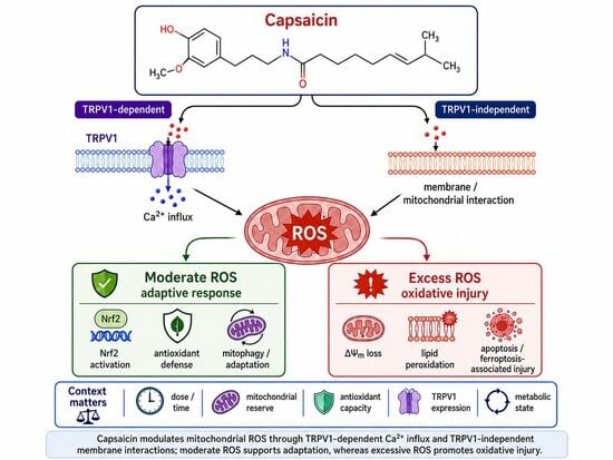
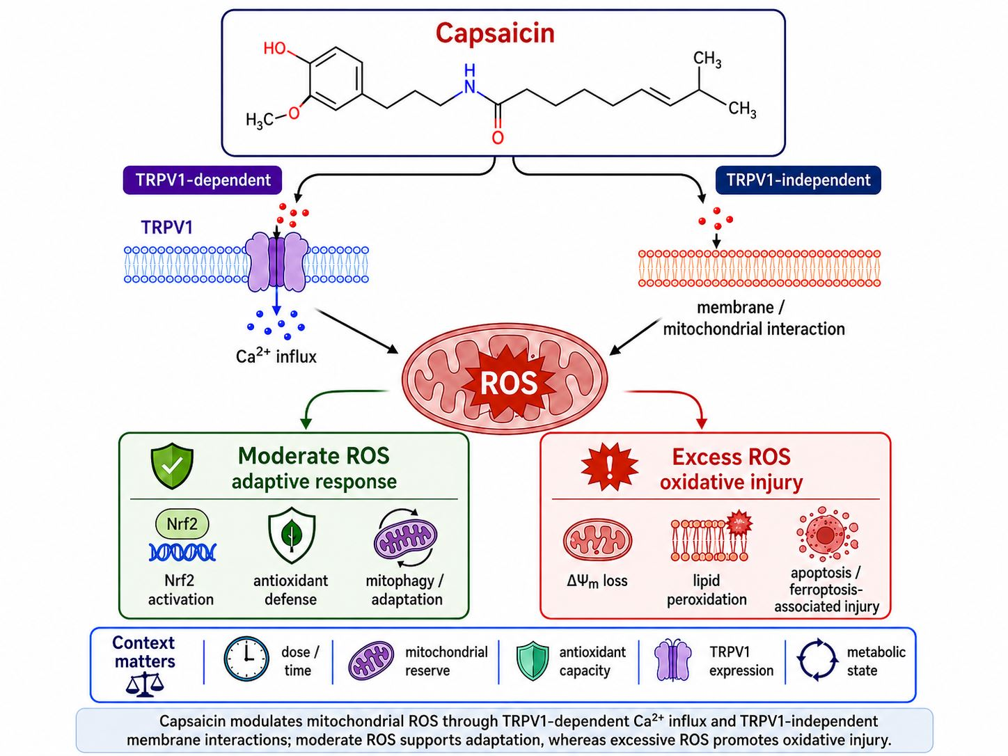
Graphical Abstract

1. Introduction

Capsaicin (trans-8-methyl-N-vanillyl-6-nonenamide), the principal pungent capsaicinoid of Capsicum species, affects cellular processes linked to oxidative stress, inflammation, metabolism, mitochondrial function, and regulated cell death [1,2,3]. Although its activity is often attributed to activation of transient receptor potential vanilloid 1 (TRPV1) [4], capsaicin can also act independently of TRPV1 through membrane interactions and effects on mitochondrial bioenergetics [5].
Reactive oxygen species (ROS) are central to many capsaicin-induced responses. Depending on concentration, exposure duration, and cellular metabolic state, capsaicin may support adaptive redox signaling or induce oxidative injury. Moderate ROS generation can activate antioxidant and cytoprotective pathways, including Nrf2-dependent signaling, whereas excessive ROS accumulation is associated with mitochondrial dysfunction, lipid peroxidation, calcium dysregulation, ferroptosis, and apoptosis [6,7,8].
Capsaicin has often been classified as either an antioxidant or a pro-oxidant compound [6,9]. This dichotomy is too narrow. The biological consequence of capsaicin exposure depends on dose, mitochondrial function, antioxidant defense capacity, TRPV1 expression, and disease-specific cellular context [6,10,11,12,13,14].
This review examines capsaicin-mediated ROS signaling, mitochondrial regulation, TRPV1-dependent and TRPV1-independent mechanisms, ferroptosis-associated pathways, and mitophagy. It also addresses ROS compartmentalization, methodological limitations in ROS assessment, and the translational relevance of exposure conditions used in experimental models.

2. Literature Search Strategy

A structured literature search was conducted in PubMed/MEDLINE, Scopus, and Web of Science databases. The following primary search terms were used individually and in Boolean combination: “capsaicin,” “capsaicinoid,” “TRPV1,” “reactive oxygen species,” “oxidative stress,” “mitochondrial dysfunction,” “ferroptosis,” “Nrf2,” “lipid peroxidation,” “apoptosis,” “redox signaling,” “antioxidant enzyme,” “mitophagy,” and “cancer.” Secondary search strings included “capsaicin pharmacokinetics,” “capsaicin bioavailability,” “capsaicin ROS,” “capsaicin ferroptosis,” “capsaicin Nrf2,” “capsaicin inflammation,” and “ferrostatin-1.” The search covered publications from January 2000 to May 2026 with no language restriction. Reference lists of identified articles were manually searched for additional relevant publications. Studies were included if they reported primary experimental data on capsaicin-mediated redox effects in cell culture or in vivo models, or provided methodologically relevant data on ROS detection, ferroptosis validation, or capsaicin pharmacokinetics. Review articles were included where they provided synthesis of primary evidence or methodological guidance. Case reports and studies focused exclusively on sensory physiology without oxidative stress endpoints were excluded.

3. Chemical Properties, Pharmacokinetics, and Biological Availability of Capsaicin

After oral administration, capsaicin is absorbed predominantly in the gastrointestinal tract, primarily via passive diffusion; however, systemic bioavailability remains relatively low because of rapid clearance and extensive first-pass hepatic metabolism [15,16]. Phase I biotransformation of capsaicin is mediated predominantly by cytochrome P450–dependent pathways, leading to the formation of hydroxylated, demethylated, and dehydrogenated metabolites, including hydroxycapsaicin derivatives and metabolites related to dehydrocapsaicin. Recent metabolomic evidence further indicates that capsaicin metabolism is modulated by tissue-specific factors and by formulation characteristics. These variables complicate direct extrapolation across experimental models and may limit the relevance of preclinical exposure conditions to concentrations achievable in clinical settings [17,18]. Such pharmacokinetic variability may substantially affect intracellular ROS dynamics and may partly account for the heterogeneous biological responses reported across experimental systems. The marked lipophilicity of capsaicin is particularly relevant in the context of cellular redox regulation, as preferential accumulation within lipid-rich intracellular compartments may alter membrane fluidity, calcium homeostasis, oxidative phosphorylation, and electron transport chain activity [5,19]. In parallel with TRPV1-mediated signaling, capsaicin may also exert receptor-independent effects through direct membrane-associated interactions that modulate ion transport, membrane excitability, and cellular bioenergetics [5].
These physicochemical and pharmacokinetic properties help explain the concentration-dependent redox effects of capsaicin. Low to moderate intracellular exposure may support antioxidant signaling and redox hormesis, whereas prolonged exposure or higher concentrations are more often associated with ROS amplification, lipid peroxidation, bioenergetic dysfunction, and apoptotic signaling [2,6,10,20]. The biological response is therefore shaped by intracellular accumulation, formulation, exposure duration, and metabolic context. Delivery platforms such as liposomes, nanoemulsions, polymeric nanoparticles, and nanostructured lipid carriers have been developed to improve solubility, stability, release profile, and tissue targeting [21]. These formulations may also change intracellular uptake, subcellular localization, ROS kinetics, and mitochondrial exposure; carrier-related toxicity should therefore be assessed separately.
Although capsaicin lipophilicity facilitates membrane interactions and intracellular access, rapid metabolism and limited systemic exposure remain important barriers to therapeutic translation [2,5].

4. Reactive Oxygen Species and Redox Homeostasis

Not all ROS behave the same way, and this distinction matters for interpreting capsaicin studies. Superoxide (O2), produced by electron leakage from mitochondrial Complex I and Complex III, is the primary mitochondrial radical in capsaicin-exposed cells; it is detectable with MitoSOX Red using HPLC to isolate the superoxide-specific product 2-OH-Mito-E+, or by EPR spin trapping [22,23]. SOD2 (mitochondrial) and SOD1 (cytosolic) rapidly convert O2 to H2O2, which acts primarily as a signaling molecule at low concentrations and is measured selectively by genetically encoded probes such as HyPer7 [24,25].
Downstream of H2O2, Fenton-type reactions with free ferrous iron (Fe2+) generate hydroxyl radicals (•OH), which are highly reactive and non-selective—they initiate the phospholipid peroxidation chain reactions that drive ferroptosis. Lipid peroxyl radicals (LOO•), propagated by ACSL4-mediated remodeling of arachidonoyl-containing phospholipids, are the proximal membrane-damaging species in this process and can be followed with C11-BODIPY or 4-HNE/MDA assays [26]. Peroxynitrite (ONOO), formed when O2 reacts with nitric oxide, is relevant primarily in inflammatory macrophage contexts. Where the original studies report “ROS” without identifying the species, we retain generic terminology but note the limitation; where specific species were measured with validated probes, we name them explicitly [27].
ROS occupy a dual role in capsaicin-exposed cells: at controlled concentrations and in metabolically competent systems, they function as transient second messengers activating stress-adaptive programs, whereas at excessive or sustained levels they become drivers of mitochondrial dysfunction and oxidative cell death [2,28]. Mitochondria represent one of the principal intracellular sources of ROS in this process, particularly through electron leakage from complexes I and III of the electron transport chain [29]. In addition to mitochondrial ROS production, oxidative responses may also involve NADPH oxidases, cytochrome P450 enzymes, and membrane-associated redox signaling pathways [30].
ROS signaling is compartmentalized and tightly linked to mitochondrial bioenergetics, calcium homeostasis, and membrane organization. This is relevant for capsaicin because its lipophilicity allows partitioning into membrane-rich compartments, where it may affect mitochondria through TRPV1-dependent calcium signaling and direct membrane-associated mechanisms [5,31]. The biological response depends on ROS localization, exposure duration, and antioxidant buffering capacity. Moderate ROS elevations may activate Nrf2-associated antioxidant signaling, mitochondrial quality control, and mitophagy, whereas sustained ROS accumulation can disrupt membrane potential, impair oxidative phosphorylation, promote lipid peroxidation, and trigger apoptotic or ferroptotic cell death [29,32,33,34,35].
This redox framework is essential for interpreting the apparently contradictory biological effects of capsaicin reported across different experimental systems. Rather than reflecting a fixed antioxidant or pro-oxidant profile, capsaicin-induced responses appear to depend on whether ROS generation remains within an adaptive hormetic range or exceeds the mitochondrial and antioxidant buffering capacity of a given cell type under specific exposure conditions [28,29].

5. Molecular Mechanisms of Capsaicin-Mediated ROS Regulation

5.1. Mitochondrial Dysfunction and ROS Generation

Mitochondria represent major intracellular targets of capsaicin-induced oxidative stress and play a central role in ROS-mediated cellular responses. Experimental evidence indicates that capsaicin can disrupt mitochondrial bioenergetics by altering electron transport chain activity, reducing mitochondrial membrane potential, and inhibiting complex I and complex III enzymatic activity, thereby promoting mitochondrial superoxide and hydrogen peroxide generation [6,29]. Increased mitochondrial ROS production subsequently contributes to cytochrome c release, ATP depletion, caspase activation, and oxidative stress-associated apoptosis [36,37].
The susceptibility of cancer cells to capsaicin-induced mitochondrial dysfunction appears closely related to their elevated basal oxidative stress, metabolic reprogramming, and impaired antioxidant buffering capacity [12,38]. Under these conditions, additional ROS accumulation may exceed the cellular oxidative threshold, leading to mitochondrial depolarization, permeability transition pore opening, and apoptotic cell death [37].
Mitochondrial metabolic state also appears to influence the downstream effects of capsaicin-induced oxidative stress. SIRT3-dependent mitochondrial deacetylation has been linked to preservation of mitochondrial integrity and attenuation of ferroptosis-associated injury through SLC7A11 and mitophagy-related mechanisms, although direct evidence in capsaicin-specific redox models remains limited [39,40]. More broadly, mitochondrial quality control and metabolic adaptability are key determinants of ROS sensitivity and cell fate [41,42].

5.2. TRPV1-Dependent ROS Signaling

TRPV1 is a polymodal, calcium-permeant cation channel that responds to structurally diverse stimuli—including capsaicin, noxious heat (>43 °C), protons, and endogenous lipid mediators such as anandamide—making it a convergence point for multiple sensory and redox signaling inputs [43,44]. Activation of TRPV1 represents one of the principal mechanisms linking capsaicin exposure to calcium dysregulation, mitochondrial stress, and ROS generation.
The structural basis of capsaicin binding to TRPV1 has been characterized in detail by cryo-EM and computational studies. Capsaicin (MW 305.41 g/mol; log P ≈ 3.0) binds within the intracellular vanilloid-binding pocket, formed by transmembrane helices S3, S4, and the S4–S5 linker of one subunit, together with contributions from S5 and S6 of a neighboring subunit [45,46,47]. The bound capsaicin adopts a “tail-up, head-down” configuration: the vanillyl head group and amide moiety form specific polar interactions that anchor the molecule, while the hydrophobic aliphatic tail occupies a conformationally flexible position within the hydrophobic core [47]. Key binding residues identified by mutagenesis include Y511 in the S3 helix and T550 in the S4 helix; their substitution substantially reduces capsaicin binding affinity [45,46]. The vanillyl group stabilizes TRPV1 channel opening through “pull-and-contact” interactions with the S4–S5 linker [47]. Capsaicin accesses its binding site via a membrane-mediated route, partitioning preferentially into the outer-leaflet lipid–water interface before translocating to the intracellular pocket [45,48]. The regulatory pocket of TRPV1 also accommodates endogenous phosphoinositide lipids, whose competitive displacement is a prerequisite for channel activation [49,50].
Binding of capsaicin to TRPV1 promotes rapid calcium influx, which subsequently enhances mitochondrial calcium uptake and amplifies ROS production through increased metabolic demand and destabilization of electron transport chain activity [37,51]. Moderate activation of this pathway may support adaptive antioxidant signaling and mitochondrial stress responses, whereas sustained calcium overload is more frequently associated with mitochondrial depolarization, oxidative injury, and apoptotic cell death [51,52].
ROS generated following TRPV1 activation have been linked to modulation of MAPK, NF-κB, and PI3K/Akt signaling cascades, thereby influencing cytokine production, cell survival, mitochondrial adaptation, and apoptotic cell death [38,53].
TRPV1 activity is also shaped by the surrounding membrane environment and cellular metabolic state. Changes in lipid composition, mitochondrial integrity, and oxidative stress may alter TRPV1 sensitivity and downstream calcium signaling [5].
The relative contribution of TRPV1-dependent signaling remains incompletely resolved. Many studies reporting capsaicin-induced ROS generation do not clearly separate receptor activation from direct membrane-associated or mitochondrial effects [5,19]. Future work should combine selective TRPV1 inhibition or genetic deletion with mitochondrial bioenergetics and compartment-specific redox imaging. Such designs would better distinguish receptor-dependent calcium signaling from receptor-independent membrane and mitochondrial effects.

5.3. TRPV1-Independent Mechanisms

Although TRPV1 activation is a major mechanism of capsaicin action, oxidative and mitochondrial effects can also occur independently of receptor activation [5]. This distinction matters because many studies reporting ROS amplification or mitochondrial dysfunction do not directly test TRPV1 dependency with antagonists or genetic loss-of-function models.
Capsaicin’s high lipophilicity (log P ≈ 3.0) favors concentration-dependent partitioning into membrane bilayers. This can perturb bilayer packing, modify ion-channel behavior, and alter mitochondrial ultrastructure without requiring receptor-mediated Ca2+ entry [1,19]. Membrane-associated interactions with mitochondrial structures may also disturb respiratory supercomplex organization, impair oxidative phosphorylation, and increase electron leakage [12,54].
Such receptor-independent mechanisms may partly explain why capsaicin-induced oxidative responses have been observed in cellular systems exhibiting low, variable, or poorly characterized TRPV1 expression [55,56]. In several experimental models, apoptosis induced by capsaicin appears to involve direct mitochondrial injury, oxidative membrane damage, and bioenergetic collapse rather than canonical TRPV1 signaling alone [56].
Membrane-associated oxidative responses may involve mitochondrial contact sites, lipid remodeling, and redox-sensitive microdomains that regulate calcium flux, lipid metabolism, and stress-responsive cell fate [52,57]. Definitive separation of TRPV1-dependent and TRPV1-independent effects remains limited because few studies measure receptor expression, calcium dynamics, mitochondrial bioenergetics, and compartment-specific ROS signaling in the same model.
An important but underexplored question is whether the TRPV1-independent membrane effects of capsaicin modulate the function of TRPV1 itself, rather than operating as a strictly parallel pathway. Computational evidence from molecular dynamics (MD) simulations suggests that capsaicin accesses its intracellular vanilloid binding site within the S1–S4 transmembrane domain via a membrane-mediated route: it localizes at the lipid–water interface of the outer leaflet and subsequently flips to the inner leaflet before accessing the binding pocket [45,48]. Critically, the regulatory pocket of TRPV1 that accommodates capsaicin also binds endogenous phosphoinositide lipids, whose displacement is a prerequisite for channel activation [49]. Changes in membrane lipid composition or bilayer packing—including those induced by capsaicin’s own membrane-partitioning effects—may therefore alter the occupancy, conformation, or ejection kinetics of these endogenous lipids from the regulatory pocket, thereby modulating TRPV1 gating efficiency or the affinity of subsequent capsaicin binding. This creates a plausible mechanism by which the TRPV1-independent membrane perturbation caused by capsaicin acts as an amplifier or contextual modulator of TRPV1-dependent signaling, rather than a fully independent parallel pathway. It should be noted that this model is currently supported by computational rather than direct experimental evidence, and future studies combining membrane lipidomics, site-directed mutagenesis of the regulatory lipid-binding pocket, and compartment-specific calcium imaging would be needed to test it directly.
Future studies using TRPV1 knockdown, knockout, or CRISPR/Cas9 deletion, together with mitochondrial lipidomics and compartment-resolved redox imaging, would substantially improve mechanistic resolution.
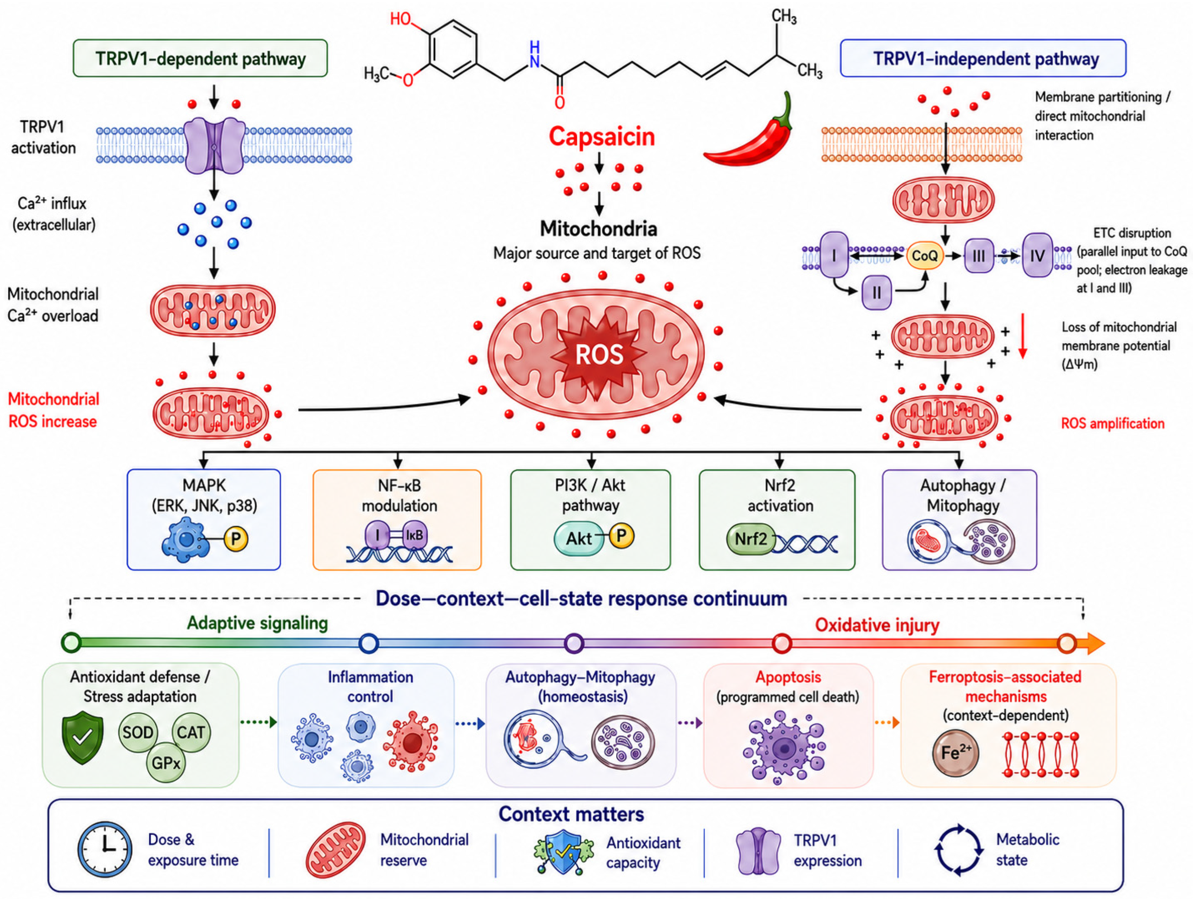
The principal TRPV1-dependent and TRPV1-independent routes linking capsaicin exposure to mitochondrial ROS generation and redox-sensitive signaling are summarized in Figure 1.

5.4. Redox-Sensitive Signaling Pathways

Capsaicin-induced ROS generation affects MAPK, NF-κB, PI3K/Akt, Nrf2, and autophagy-related pathways. These pathways should not be viewed as isolated cascades; they operate as connected redox-response modules whose outcomes depend on mitochondrial function and ROS localization [30,52].
MAPK signaling represents one of the best-characterized pathways linking capsaicin-induced ROS generation to apoptosis. In cancer models, capsaicin has been associated with activation of stress-responsive p38 and JNK pathways, mitochondrial depolarization, caspase activation, and apoptotic cell death [37,58]. In renal carcinoma cells, capsaicin-induced apoptosis was linked to p38/JNK activation, and pharmacological inhibition of these kinases attenuated caspase activation, supporting a functional role for MAPK signaling in capsaicin-mediated cytotoxicity [58]. Similar ROS-dependent activation of JNK and p38 MAPK has been reported in hepatocellular carcinoma models, where antioxidant treatment with N-acetylcysteine reduced capsaicin-induced MAPK phosphorylation and apoptosis, further supporting a mitochondrial ROS-dependent mechanism [59,60].
NF-κB signaling is particularly relevant in inflammatory models. Capsaicin has been reported to suppress LPS-induced inflammatory responses in macrophages through inhibition of MAPK and NF-κB signaling, accompanied by reduced production of inflammatory mediators such as TNF-α, IL-1β, IL-6, and nitric oxide [61]. In a bleomycin-induced pulmonary fibrosis model, capsaicin reduced expression of TNF-α, IL-6, IL-1β, NF-κB, COX-2, and TGF-β1 while simultaneously increasing Nrf2 and PPAR-γ signaling, suggesting coordinated regulation of oxidative stress and inflammatory pathways [62].
PI3K/Akt-related signaling also appears to participate in capsaicin-mediated redox adaptation, although its role varies according to cell type and metabolic context. In prostate cancer cells, capsaicin-induced ROS accumulation was associated with inhibition of PI3K/Akt/mTOR signaling and disruption of autophagic flux; antioxidant treatment partially reversed these effects, supporting a ROS-dependent mechanism [63]. In contrast, in non-malignant cardiovascular and neurohumoral models of salt-sensitive hypertension, capsaicin has been linked to activation of AMPK/Akt/Nrf2-associated antioxidant pathways, reduced NOX2/NOX4 expression, and increased HO-1 activity [64]. These observations suggest that PI3K/Akt signaling may contribute either to adaptive stress resistance or oxidative cytotoxicity depending on mitochondrial reserve capacity and ROS burden [28,65].
Autophagy and mitochondrial quality-control pathways are similarly interconnected with capsaicin-mediated oxidative signaling. Moderate mitochondrial stress may activate adaptive autophagy and mitophagy, thereby facilitating removal of dysfunctional mitochondria and preservation of redox homeostasis [42,66]. Under conditions of sustained oxidative stress, however, impaired autophagic flux and mitochondrial dysfunction may amplify apoptotic and ferroptosis-associated injury [67]. This distinction may partly explain why malignant models frequently demonstrate ROS-associated autophagy disruption and apoptosis, whereas non-malignant systems more commonly exhibit antioxidant pathway activation and mitochondrial adaptation.
Overall, these signaling pathways appear to determine whether capsaicin-induced ROS remain within an adaptive range or exceed mitochondrial and antioxidant buffering capacity [32,68].

Capsaicin-Induced Nrf2 Activation: Mechanisms and Experimental Evidence

Nrf2 is the principal transcription factor linking capsaicin-induced ROS to adaptive antioxidant gene expression. Under basal conditions, Keap1 sequesters Nrf2 in the cytoplasm and targets it for proteasomal degradation [33]. Oxidative modification of Cys151, Cys273, and Cys288 in Keap1 disrupts this interaction, releasing Nrf2 to enter the nucleus and bind antioxidant response elements (ARE) in the promoters of cytoprotective genes [33].
Molecular evidence for a direct interaction between capsaicin and Keap1 has recently been reported. Using biolayer interferometry (BLI), cellular thermal shift assay (CETSA), pull-down, co-immunoprecipitation, and hydrogen–deuterium exchange mass spectrometry (HDX-MS), capsaicin was shown to bind directly to Keap1 and disrupt the Keap1–Nrf2 protein–protein interaction in gastric epithelial GES-1 cells, facilitating Nrf2 nuclear translocation and upregulation of HMOX1 (HO-1), NQO1, thioredoxin (TXN), and glutathione synthetase (GSS) [69]. These findings were first available as a reviewed preprint and have since been published in eLife; independent replication remains needed, but they provide evidence that capsaicin may act as a non-covalent Keap1 inhibitor, in contrast to established electrophilic Nrf2 inducers that covalently modify Keap1 cysteine residues.
In neuronal models, capsaicin-induced Nrf2 activation has been demonstrated through TRPV1-dependent mechanisms. In dorsal root ganglion (DRG) neurons and Schwann cells, capsaicin upregulated Nrf2, HO-1, NQO1, and catalase in a TRPV1-dependent manner [70]. This indicates that TRPV1-mediated calcium influx can act as an upstream signal coupling capsaicin receptor activation to Nrf2 pathway engagement.
In non-neuronal systems, capsaicin-induced Nrf2 activation has been documented in cardiovascular, pulmonary, and metabolic disease models. In salt-sensitive hypertension models, capsaicin activated AMPK/Akt/Nrf2 signaling in the hypothalamic paraventricular nucleus, reducing NOX2/NOX4 expression and increasing HO-1 activity [64]. In a bleomycin-induced pulmonary fibrosis model, capsaicin increased Nrf2 and PPAR-γ while suppressing NF-κB/TGF-β1 [62]. In neurodegenerative disease models, capsaicin has been associated with Nrf2 upregulation contributing to neuroprotection [13].
The main downstream targets of Nrf2 induced by capsaicin include HO-1, NQO1, glutamate-cysteine ligase (GCL), thioredoxin reductase, and ferritin—enzymes that replenish glutathione, detoxify lipid peroxides, and limit iron availability for Fenton reactions. In cancer cells with constitutively active Nrf2 arising from somatic Keap1 mutations or oncogene-driven Nrf2 stabilization, however, this same pathway may confer resistance to oxidative stress-based therapies and reduce capsaicin’s cytotoxic efficacy [71,72]. Whether capsaicin-induced Nrf2 activation is cytoprotective or counterproductive therefore depends on the pre-existing Nrf2 status of the target cell.

5.5. Ferroptosis, Mitophagy, and Redox Compartmentalization

Autophagy and mitophagy are closely related but mechanistically distinct processes. Autophagy (macroautophagy) is a degradation pathway in which cytoplasmic contents are engulfed by double-membrane autophagosomes and delivered to lysosomes [42,66]. Mitophagy is a selective form of autophagy specifically targeting damaged mitochondria. While mitophagy uses the core autophagosome machinery, it requires a priming step to label damaged mitochondria for selective recognition. The best-characterized pathway is the PINK1/Parkin axis: loss of mitochondrial membrane potential stabilizes PINK1 on the outer mitochondrial membrane (OMM), which phosphorylates ubiquitin and activates Parkin, leading to ubiquitination of OMM proteins (VDAC1, MFN1/2, TOM20) and recruitment of autophagy receptors (OPTN, NDP52) that bridge the cargo to LC3-II on the autophagosome [42,66]. Alternative receptor-mediated pathways operate through BNIP3, NIX (BNIP3L), and FUNDC1, which interact directly with LC3 via LIR motifs and can be activated independently of PINK1/Parkin [42].
In the context of capsaicin exposure, moderate mitochondrial stress may activate adaptive PINK1/Parkin-dependent mitophagy, selectively removing depolarized mitochondria before they amplify ROS production [42,66]. In contrast, capsaicin at higher concentrations may impair autophagic flux and disrupt mitochondrial quality control, as observed in prostate cancer models where capsaicin disrupted autophagosome–lysosome fusion [63]. Direct mechanistic evidence for PINK1/Parkin-mediated mitophagy in capsaicin-specific contexts remains limited; most evidence is inferred from LC3-II accumulation or electron microscopy rather than pathway-specific assays (PINK1 stabilization, Parkin recruitment, mt-Keima, bafilomycin A1 sensitivity).
Ferroptosis is an iron-catalyzed form of regulated cell death driven by phospholipid hydroperoxide accumulation, glutathione depletion, loss of GPX4-mediated peroxide reduction, and lipid-derived oxidants [67,73]. Several capsaicin-induced changes overlap with this biology, including mitochondrial ROS generation, glutathione depletion, lipid peroxidation, and altered GPX4/SLC7A11 signaling [34,74].
Ferrostatin-1 (Fer-1; ethyl 3-amino-4-(cyclohexylamino)benzoate; CAS 347174-05-4; MW 262.35 g/mol) is a synthetic small-molecule radical-trapping antioxidant (RTA) that was identified as the first specific inhibitor of ferroptosis [73]. Fer-1 scavenges lipid peroxyl radicals (LOO•) within the membrane bilayer, interrupting the oxidative chain reaction that drives phospholipid peroxidation, without chelating iron directly or inhibiting GPX4 activity; EC50 ≈ 60 nM in HT-1080 fibrosarcoma cells [73]. Liproxstatin-1 (Lip-1; CAS 950455-15-9; MW 340.85 g/mol) is a structurally distinct spiroquinoxalinamine ferroptosis inhibitor that also acts as a lipid peroxyl radical scavenger, with greater in vivo stability than Fer-1; IC50 ≈ 22 nM in cell-based assays [75]. Both compounds represent the principal pharmacological tools for ferroptosis rescue experiments; however, because both act as radical-trapping antioxidants rather than specific ferroptosis pathway inhibitors, their protective effect alone does not distinguish ferroptosis from other forms of lipid peroxidation-dependent cell death.
Direct evidence for capsaicin-induced ferroptosis remains limited to selected models and requires rigorous methodological evaluation. In non-small cell lung cancer cells (A549 adenocarcinoma and NCI-H23) [76], in glioblastoma cells (U87-MG, U251) [77], and in tongue squamous cell carcinoma (TSCC) cells (HN6 and CAL-27) [78], capsaicin was reported to suppress SLC7A11/GPX4 signaling and induce ferroptosis-like cell death. While mechanistically suggestive, ferrostatin-1 (Fer-1) is a radical-trapping antioxidant that inhibits lipid peroxidation-driven cell death and may also attenuate certain forms of necroptosis independently of iron-dependent phospholipid peroxidation [79]. Importantly, necrostatin-1 has been demonstrated to prevent ferroptosis in a RIPK1- and IDO-independent manner in hepatocellular carcinoma cells, likely through upregulation of xCT (SLC7A11) expression rather than through RIPK1 inhibition, underscoring the pharmacological overlap between these death modalities at the level of lipid peroxidation [80]. Since capsaicin is well established as an inducer of apoptosis in multiple cancer models, definitive assignment of ferroptosis as the dominant death modality requires concurrent pharmacological exclusion of apoptosis (e.g., Z-VAD-FMK) and necroptosis (e.g., necrostatin-1) under identical experimental conditions, with Fer-1 demonstrating substantially greater protection than these alternatives [74]. The absence of such parallel exclusion experiments is a prevalent and significant limitation in published studies reporting capsaicin-induced ferroptosis.
In many studies, ferroptosis is inferred from ROS accumulation, lipid peroxidation, or glutathione depletion alone. These markers support oxidative stress but do not establish ferroptosis unless iron dependence, lipid-peroxide accumulation, pathway markers, and ferroptosis-specific rescue are demonstrated.
Mitochondrial quality-control pathways also appear to contribute substantially to cellular responses following capsaicin exposure. Moderate mitochondrial stress may activate adaptive mitophagy, thereby facilitating removal of dysfunctional mitochondria and limiting excessive ROS accumulation [42,66]. Conversely, persistent oxidative injury may impair mitochondrial quality control and amplify oxidative stress signaling.
Redox compartmentalization is also important. Mitochondrial, lipid-associated, and cytosolic ROS pools may have different consequences depending on localization, iron availability, and metabolic state [81].
With respect to iron metabolism, capsaicin exposure in NSCLC cells (A549, NCI-H23) was associated with increased total intracellular iron levels and elevated ferrous ion (Fe2+) concentration [76]. This iron accumulation most likely reflects impaired iron handling secondary to glutathione depletion and SLC7A11 suppression, which reduce intracellular glutathione-dependent iron buffering and ferritin-mediated iron sequestration. There is currently no published evidence that capsaicin directly chelates iron through its chemical structure—its hydroxyl and carbonyl groups are potential iron-coordinating moieties in principle, but direct capsaicin–iron complex formation has not been demonstrated in cellular or biochemical models. Future studies should address this question directly using iron speciation analysis.
Taken together, capsaicin-mediated oxidative responses reflect interactions among mitochondrial dysfunction, ROS compartmentalization, membrane dynamics, calcium signaling, and antioxidant capacity. Ferroptosis-associated pathways may contribute in specific contexts, but their role in capsaicin-induced cell death requires stricter validation.

6. Capsaicin-Induced ROS Regulation in Disease Contexts

The apparently contradictory effects of capsaicin—promoting apoptosis and ferroptosis in cancer models, while exhibiting antioxidant and anti-inflammatory properties in non-malignant systems—can be unified within a coherent mechanistic framework centered on basal redox state and antioxidant buffering capacity. Cancer cells characteristically operate at elevated basal ROS levels due to oncogene-driven metabolic reprogramming, mitochondrial dysfunction, and increased NADPH oxidase activity [38]. To survive this chronic pro-oxidant state, cancer cells upregulate compensatory antioxidant systems, including Nrf2-driven gene expression, glutathione biosynthesis, and thioredoxin reductase activity [71,72]. However, this adaptation leaves cancer cells functioning near their oxidative damage threshold. Capsaicin-driven ROS augmentation, even at concentrations well tolerated by normal cells, may therefore be sufficient to exceed this threshold, triggering lipid peroxidation cascades, GPX4 dysfunction, mitochondrial depolarization, and ultimately apoptotic or ferroptotic cell death. In contrast, non-malignant cells—including inflammatory macrophages, neurons, and metabolically stressed but otherwise intact tissues—typically maintain a more resilient redox buffering system. In these cells, the same ROS increment fails to breach the oxidative damage threshold, instead activating the Keap1-Nrf2 axis, inducing cytoprotective gene expression, suppressing NF-κB-driven inflammatory signaling, and stimulating mitochondrial quality control through mitophagy [33,66]. This “basal redox state—antioxidant buffering threshold” framework unifies the apparent contradiction within a single dose-context-cell state determinism model [38].

6.1. Cancer

Cancer cells frequently exhibit elevated basal ROS production, mitochondrial dysfunction, and altered antioxidant capacity, which may increase their vulnerability to additional oxidative stress. In several tumor models, capsaicin has been reported to induce ROS generation, mitochondrial dysfunction, caspase activation, and apoptosis. In pancreatic cancer models, capsaicin-induced apoptosis has been linked to ROS generation and mitochondrial death signaling.
Specifically, in prostate cancer PC-3 cells (androgen-insensitive, p53-null) and LNCaP cells, capsaicin inhibited cell growth and induced apoptosis [36,63]. In pancreatic cancer BxPC-3 and AsPC-1 cells, capsaicin-induced apoptosis was linked to ROS generation and mitochondrial death signaling [6,12]. In hepatocellular carcinoma SMMC-7721 cells, capsaicin activated JNK and p38 MAPK through a ROS-dependent mechanism [59].
Recent evidence also suggests that capsaicin may promote ferroptosis-associated cell death in selected cancer systems. In non-small cell lung cancer cells, capsaicin inhibited proliferation of A549 and NCI-H23 cells and was reported to induce ferroptosis through suppression of the SLC7A11/GPX4 pathway; ferrostatin-1 rescue supported ferroptosis involvement in that model [76]. However, these findings should be interpreted cautiously, because most cancer studies use high micromolar capsaicin concentrations that may exceed clinically achievable systemic exposure. Thus, capsaicin-mediated anticancer effects are best viewed as mechanistically relevant but translationally dependent on dose, formulation, local exposure, and tumor-specific oxidative vulnerability. In glioblastoma cells (U87-MG, U251), capsaicin induced redox imbalance and ferroptosis through ACSL4/GPX4 signaling [77]. In tongue squamous cell carcinoma (HN6 and CAL-27), capsaicin induced ferroptosis via AMPK-mediated suppression of SLC7A11 activity and upregulation of ACSL4 [78].
Across these cancer models, capsaicin-induced mitochondrial ROS generation appears to be the proximal event. What follows—apoptosis or ferroptosis—depends on what the cell can absorb: its glutathione reserves, GPX4 expression, and iron availability. Where antioxidant buffering holds, caspase-dependent apoptosis tends to dominate; where GPX4 is limiting and free iron is present, ferroptotic phospholipid peroxidation can proceed. This has a direct clinical implication: a blanket capsaicin dose is unlikely to produce a predictable outcome across tumor types. Selecting which cancers might respond—and at what exposure—will probably require prior characterisation of basal GSH status, GPX4 levels, and mitochondrial reserve capacity in the target tumour.

6.2. Inflammation

In inflammatory models, capsaicin more commonly shows antioxidant and anti-inflammatory effects rather than direct cytotoxicity. In macrophage-based systems, capsaicin reduced LPS-induced secretion of inflammatory mediators, including TNF-α, interleukins, nitric oxide, and iNOS-related responses, partly through inhibition of NF-κB and MAPK signaling [61]. These findings support the view that capsaicin can suppress redox-sensitive inflammatory amplification under conditions where ROS are linked to cytokine production and immune activation.
Protective effects have also been reported in in vivo inflammatory injury models. In LPS-induced acute lung injury, capsaicin reduced oxidative stress, inflammation, and apoptosis, with involvement of HMGB1/NF-κB and PI3K/Akt/mTOR signaling pathways [82]. These observations indicate that in inflammatory contexts, capsaicin-induced redox modulation may contribute to tissue protection when exposure remains within an adaptive or anti-inflammatory range.
These anti-inflammatory observations are consistent with the unified redox framework outlined in Section 6: at concentrations achievable through dietary exposure or local application, capsaicin-induced ROS remain within the adaptive signaling range for non-malignant immune cells, activating cytoprotective pathways rather than triggering cytotoxic cascades. This mechanistic distinction—between adaptive ROS signaling in non-malignant cells and cytotoxic ROS accumulation in cancer cells—should guide route-of-administration and formulation decisions in future clinical development programs.

6.3. Neurodegeneration

The role of capsaicin in neurodegenerative models is strongly dependent on TRPV1-mediated calcium signaling, mitochondrial function, and neuroinflammatory status. Moderate TRPV1 activation may support neuroprotective responses by reducing glial activation, oxidative stress, and inflammatory injury. In an MPTP model of Parkinson’s disease, capsaicin protected dopaminergic neurons by inhibiting glial activation-mediated oxidative stress and neuroinflammation [83].
Additional evidence suggests that capsaicin may preserve mitochondrial-associated endoplasmic reticulum membrane integrity and improve mitochondrial dysfunction in chronic cerebral hypoperfusion models [84]. However, excessive or prolonged TRPV1 activation may increase calcium influx, mitochondrial depolarization, and oxidative injury. Therefore, capsaicin should not be interpreted as uniformly neuroprotective; its effects depend on exposure intensity, neuronal vulnerability, calcium handling, and mitochondrial stress tolerance.

6.4. Metabolic Disorders

Metabolic disorders such as obesity, insulin resistance, and fatty liver disease are characterized by chronic low-grade inflammation, mitochondrial dysfunction, and oxidative stress. In diet-induced metabolic models, capsaicinoid intake has been associated with improved metabolic parameters, reduced oxidative damage, and modulation of NADPH oxidase- and Nrf2-related pathways [85]. Dietary capsaicin has also been reported to reduce obesity-induced insulin resistance and hepatic steatosis in obese mice, supporting a role for capsaicin-sensitive pathways in metabolic adaptation [86].
In high-fat diet and NAFLD-related models, capsaicin effects appear to involve interactions among lipid metabolism, gut-related mechanisms, inflammation, and oxidative stress [85,87]. Nevertheless, translational interpretation remains limited by variability in diet composition, route of administration, formulation, dose, and species-specific metabolism. These limitations should be considered before extrapolating animal data to long-term clinical metabolic outcomes.

7. Experimental Approaches for ROS Detection and Oxidative Stress Assessment

A fundamental challenge in capsaicin-related oxidative stress research is that individual probes detect different ROS, and these species are not interchangeable in their biological significance. H2DCFDA (DCF) detects general oxidant activity—including H2O2, peroxynitrite (ONOO), and •OH—but not superoxide (O2) directly, and is subject to significant non-ROS artifacts including photo-oxidation and peroxidase-mediated activation [88]. Dihydroethidium (DHE) and MitoSOX Red generate an O2-specific product (2-hydroxyethidium, 2-OH-E+; 2-OH-Mito-E+) but also non-specific oxidation products (ethidium, Mito-E+) that overlap spectrally; reliable O2 detection requires chromatographic separation by HPLC or LC-MS [22,88]. Genetically encoded probes (HyPer7, roGFP2-Tsa2) specifically detect H2O2 in defined subcellular compartments [24,25,89]. C11-BODIPY and MDA/4-HNE assays detect lipid peroxyl radical (LOO•) products relevant to ferroptosis [26]. EPR/ESR spin trapping can detect O2 and •OH directly but requires specialized equipment [23]. These distinctions are critical for interpreting capsaicin studies: an increase in DCF fluorescence does not establish superoxide generation, and an increase in MitoSOX fluorescence without HPLC validation does not establish mitochondrial superoxide production. The probes used in each study, and the ROS species they are designed to detect, are specified in Table 1.
Reliable assessment of ROS generation is essential for interpreting capsaicin-mediated redox effects, but ROS detection remains technically challenging. Individual oxidants differ in reactivity, half-life, intracellular localization, diffusion capacity, and biological targets. Therefore, increased fluorescence from a single probe should not be interpreted as direct evidence for a specific ROS or subcellular source. This is particularly important in capsaicin studies, because capsaicin may simultaneously affect mitochondrial membrane potential, membrane organization, calcium influx, respiratory chain activity, and cell viability, all of which can alter probe uptake, oxidation kinetics, and fluorescence intensity.
Commonly used probes such as H2DCFDA, DHE, and MitoSOX provide useful screening information but have substantial limitations. H2DCFDA reflects a general oxidant-sensitive signal rather than a specific H2O2 readout, whereas DHE- and MitoSOX-derived red fluorescence requires product-specific separation to distinguish superoxide-specific products from non-specific oxidation products. Methodological reviews strongly caution that fluorescence-based ROS assays require appropriate controls, product validation, and orthogonal confirmation before mechanistic conclusions are made [22,88].
Mitochondrial membrane potential probes such as JC-1, TMRM, and TMRE are useful for assessing mitochondrial depolarization, but they do not directly measure ROS. Their interpretation is affected by dye loading, mitochondrial mass, plasma membrane potential, efflux pumps, and the choice of quenching or non-quenching conditions. Therefore, loss of mitochondrial membrane potential after capsaicin exposure should be interpreted as a functional mitochondrial change rather than direct proof of ROS production [90].
More specific approaches, including genetically encoded H2O2 sensors, peroxiredoxin-based redox probes, EPR/ESR spin trapping, GSH/GSSG analysis, lipid peroxidation assays, and mitochondrial bioenergetic profiling, can substantially improve mechanistic resolution when combined with appropriate controls. In studies proposing ferroptosis, lipid peroxidation markers such as C11-BODIPY, MDA, or 4-HNE should be interpreted together with GPX4, SLC7A11, ACSL4, GSH status, iron dependency, and rescue by ferroptosis inhibitors such as ferrostatin-1 or liproxstatin-1. Ferroptosis is defined by iron-dependent phospholipid peroxidation and cannot be established from ROS accumulation or lipid peroxidation alone [74].
The main methodological limitations, essential controls, and interpretation criteria for ROS probes, mitochondrial functional assays, lipid peroxidation readouts, and ferroptosis-associated markers used in capsaicin studies are summarized in Table 1.

8. Therapeutic Challenges

Systemic Toxicity and Risks Associated with Long-Term Capsaicin Use

Alongside its therapeutic potential, capsaicin carries a well-characterized dose-dependent toxicological profile. Gastrointestinal toxicity is the most commonly reported adverse effect of oral capsaicin. At acute high doses, capsaicin irritates the mucosal lining from the oropharynx to the rectum, producing burning pain, nausea, vomiting, diarrhea, and gastric cramping [16]. Chronic high-dose intake has been associated with increased gastric cancer risk in epidemiological studies, in contrast to the gastroprotective effects documented at lower dietary doses [92]. Individuals with pre-existing gastritis, peptic ulcer disease, or irritable bowel syndrome are particularly vulnerable [16].
Hepatic effects of capsaicin are dose-dependent and bidirectional. At dietary concentrations, capsaicin activates Nrf2/antioxidant pathways and may reduce hepatic lipid accumulation [86,87]. At supraphysiological doses, hepatic CYP450 biotransformation generates reactive metabolites that can deplete glutathione and cause hepatocyte oxidative injury [17]. This dual profile mirrors the hormesis-like pattern described for capsaicin’s cellular redox effects.
Cardiovascular effects are similarly complex. TRPV1 activation in endothelial cells and sensory neurons releases calcitonin gene-related peptide (CGRP), inducing vasodilation and potentially lowering blood pressure—effects that may be beneficial in hypertension [4]. However, high acute doses have been associated with tachycardia and elevated circulating blood volume [81]. The long-term cardiovascular consequences of sustained TRPV1 activation remain incompletely characterized.
From a clinical safety perspective, the Qutenza 8% topical patch represents the best-characterized human exposure model, with systemic capsaicin concentrations in the low nanomolar range (mean Cmax~1.86 ng/mL after 60 min application) [93], at which systemic adverse effects are not attributable to systemic absorption. For any proposed systemic therapeutic application, establishing a human-equivalent NOAEL through rigorous pharmacokinetic modeling will be an essential prerequisite.
Most in vitro cancer studies use capsaicin at 50–300 µM, but this concentration range is rarely achievable in human tissues through conventional oral or topical exposure. The only published human pharmacokinetic study, which followed ingestion of approximately 26.6 mg pure capsaicin equivalent, recorded a peak plasma concentration of 2.47 ± 0.13 ng/mL—roughly 8 nM—with a half-life of about 25 min and complete elimination within 90 min [94]. Topical application of the 8% Qutenza patch produces similarly low systemic exposure (mean Cmax~1.86 ng/mL) [93]. The gap between ~8 nM in plasma and 50–300 µM in culture dishes is approximately four orders of magnitude—large enough to raise a direct question: which of the mechanisms described in this review can actually operate at concentrations achievable in human tissues without targeted local delivery? Ferroptosis induction, Complex I/III inhibition, and GPX4 suppression at 50–150 µM are unlikely to be triggered systemically. This does not diminish their mechanistic interest, but it does shift the translational question toward route and formulation: intratumoral injection or targeted nanoparticle delivery may be the only realistic routes for bringing these concentrations within reach in a clinical context [21,95].
Despite extensive experimental evidence that capsaicin modulates oxidative stress, mitochondrial function, inflammation, and regulated cell death, its clinical translation remains limited by exposure-related constraints. A major challenge is the frequent use of high micromolar concentrations in in vitro cancer studies, whereas systemic exposure after conventional oral or topical administration is substantially lower and strongly influenced by absorption, first-pass metabolism, tissue distribution, and formulation. Capsaicin bioavailability is therefore highly context dependent, and direct extrapolation from cell culture concentrations to systemic therapeutic effects is often inappropriate [95,96].
Because capsaicin exposure differs across in vitro, in vivo, dietary, topical, nanoformulated, and ex vivo models, interpretation of biological outcomes requires consideration of route of administration, formulation, exposure duration, tissue compartment, and local versus systemic availability. The translational relevance and limitations of commonly used capsaicin exposure models are summarized in Table 2.
An additional challenge is the narrow boundary between adaptive redox signaling and oxidative cytotoxicity. Cellular outcomes depend on mitochondrial functional state, antioxidant reserve capacity, iron metabolism, ROS compartmentalization, and disease-specific metabolic context. These variables likely explain why capsaicin can promote apoptosis or ferroptosis-associated injury in some cancer models, while supporting anti-inflammatory or adaptive antioxidant responses in non-malignant systems.
Delivery strategies such as liposomes, nanoemulsions, polymeric nanoparticles, solid-dispersion systems, and lipid-based carriers may improve solubility, stability, local retention, or intracellular uptake. However, these systems may also alter subcellular distribution, mitochondrial exposure, and ROS-generation kinetics relative to free capsaicin, requiring independent pharmacokinetic and toxicity assessment. Future translational studies should therefore prioritize physiologically relevant exposure models, standardized formulations, integrated pharmacokinetic analysis, and clear separation of TRPV1-dependent, TRPV1-independent, and formulation-driven effects.

9. Current Controversies and Future Directions

Several mechanistic and translational questions remain unresolved. First, classifying capsaicin as either antioxidant or pro-oxidant is too simplistic. In redox biology, low and spatially controlled oxidant production can support signaling, whereas excessive or poorly buffered oxidant generation can cause oxidative damage. Capsaicin outcomes are therefore determined by dose, exposure duration, mitochondrial function, antioxidant reserve capacity, and disease-specific context [28].
A second unresolved issue is the translational relevance of commonly used experimental exposure conditions. Many in vitro studies use high micromolar capsaicin concentrations that are useful for mechanistic stress models but difficult to extrapolate directly to systemic clinical exposure. Differences in formulation, local versus systemic availability, metabolism, cell type, TRPV1 expression, and mitochondrial reserve capacity likely contribute to the heterogeneous outcomes reported across experimental systems. This limitation should be addressed by integrating pharmacokinetic data, physiologically relevant exposure ranges, and standardized formulation reporting into future studies.
The relative contribution of TRPV1-dependent and TRPV1-independent mechanisms also remains incompletely defined. TRPV1 activation clearly links capsaicin to Ca2+ influx, mitochondrial calcium loading, mitochondrial depolarization, and ROS generation in several models. However, capsaicin can also act independently of TRPV1 by modifying membrane properties, ion transport, and possibly mitochondrial bioenergetics. This distinction is important because many studies report capsaicin-induced ROS generation without directly confirming TRPV1 expression or using receptor antagonism and genetic loss-of-function approaches [5,19,98].
Future studies should therefore combine pharmacological inhibition with genetic models, including TRPV1 knockdown, knockout, or CRISPR/Cas9-mediated deletion, together with mitochondrial bioenergetic analysis and compartment-specific ROS imaging. Such designs would help distinguish receptor-mediated Ca2+ signaling from direct membrane-associated or mitochondrial effects. This is particularly important in non-neuronal and cancer models, where TRPV1 expression can be variable and where capsaicin may influence cell fate through multiple overlapping mechanisms.
Another major area requiring clarification is ROS compartmentalization. Mitochondrial, cytosolic, membrane-associated, and lipid-derived ROS pools can have different biological effects depending on where, when, and how strongly they are produced. Bulk ROS measurements using non-specific fluorescent probes are therefore insufficient to define mechanism. Future work should increasingly use genetically encoded redox sensors, product-specific analytical validation of DHE/MitoSOX oxidation products, mitochondrial respiratory profiling, redox lipidomics, and spatially resolved live-cell imaging [81].
Ferroptosis-associated mechanisms represent another promising but still incompletely resolved area. Several features of capsaicin-mediated oxidative stress overlap with ferroptosis biology, including lipid peroxidation, glutathione depletion, GPX4 dysfunction, SLC7A11 modulation, iron sensitivity, and mitochondrial redox remodeling. However, ferroptosis cannot be concluded from ROS accumulation or lipid peroxidation alone. Future studies should include ferroptosis-specific rescue experiments, such as ferrostatin-1, liproxstatin-1, or iron chelation, together with assessment of GPX4, SLC7A11, ACSL4, GSH/GSSG status, and exclusion of apoptosis or necroptosis where appropriate [99,100].
Future research should therefore define the exposure conditions, cellular context, subcellular ROS source, and death-pathway specificity that determine the biological outcome of capsaicin exposure.

10. Conclusions

The evidence reviewed here supports repositioning capsaicin from a binary antioxidant/pro-oxidant classification to a context-resolved redox modulator whose biological outcome is principally governed by mitochondrial buffering capacity, antioxidant reserve, TRPV1 status, and local exposure conditions. Across experimental models, its impact depends on exposure level, duration, formulation, tissue environment, mitochondrial capacity, antioxidant buffering, and TRPV1 status.
The current literature supports a model in which capsaicin can shift cells along a redox-response continuum. At moderate or locally controlled exposure, it may support adaptive signaling, inflammatory resolution, mitochondrial quality control, or antioxidant defense. Under conditions of high oxidative burden, limited redox reserve, or supraphysiological exposure, the same compound may amplify mitochondrial stress, lipid peroxidation, and regulated cell death.
This distinction is particularly relevant for cancer studies, where tumor cells with elevated basal ROS may be more susceptible to capsaicin-induced oxidative injury. However, many anticancer findings remain difficult to translate directly because experimental concentrations often exceed expected systemic exposure. Conversely, dietary, topical, gastrointestinal, or formulation-enhanced contexts may provide more plausible settings for biologically relevant capsaicin activity.
Ferroptosis-associated mechanisms, mitophagy, and compartment-specific ROS signaling add important mechanistic depth to this field, but they also require stricter validation. Future studies should avoid relying on single-probe ROS measurements or isolated pathway markers and should instead combine bioenergetics, redox imaging, lipid peroxidation assays, pharmacokinetics, and rescue-based cell death validation.
Overall, capsaicin-mediated redox modulation represents a biologically plausible but context-sensitive mechanism. Its therapeutic relevance will depend on defining the exposure conditions, cellular vulnerabilities, and subcellular redox events that separate adaptive signaling from oxidative injury.

Author Contributions

Conceptualization, L.K.; methodology, L.K. and H.Ď.; investigation, L.K. and H.Ď.; resources, L.K.; writing—original draft preparation, L.K.; writing—review and editing, L.K. and H.Ď.; project administration, L.K. and H.Ď.; funding acquisition, L.K. All authors have read and agreed to the published version of the manuscript.

Funding

This research was funded by the EU NextGenerationEU through the Recovery and Resilience Plan for Slovakia under project No. 09I03-03-V04-00381. The APC was funded by the same project.

Data Availability Statement

No new data were created or analyzed in this study. Data sharing is not applicable to this article.

Acknowledgments

During the preparation of this manuscript, the authors used ChatGPT by OpenAI, GPT-4 version to support language editing and improvement of readability. Its use was limited to refining existing text and did not involve the generation of new scientific content, data, analyses, interpretations, or conclusions. All AI-assisted output was critically reviewed and edited by the authors, who take full responsibility for the final content of the publication.

Conflicts of Interest

The authors declare no conflicts of interest.

Abbreviations

The following abbreviations are used in this manuscript:
4-HNE4-Hydroxynonenal
ACSL4Acyl-CoA synthetase long-chain family member 4
AMPKAMP-activated protein kinase
ATPAdenosine triphosphate
CATCatalase
CCCPCarbonyl cyanide m-chlorophenyl hydrazone
COX-2Cyclooxygenase-2
CoQCoenzyme Q
CRISPRClustered regularly interspaced short palindromic repeats
Cas9CRISPR-associated protein 9
DCF2′,7′-Dichlorofluorescein
DHEDihydroethidium
DTTDithiothreitol
EPRElectron paramagnetic resonance
EREndoplasmic reticulum
ESRElectron spin resonance
ETCElectron transport chain
FCCPCarbonyl cyanide-4-(trifluoromethoxy)phenylhydrazone
GPX4Glutathione peroxidase 4
GPxGlutathione peroxidase
GSHReduced glutathione
GSSGOxidized glutathione
HMGB1High-mobility group box 1
HO-1Heme oxygenase-1
HPLCHigh-performance liquid chromatography
H2DCFDA2′,7′-Dichlorodihydrofluorescein diacetate
IL-1βInterleukin-1 beta
IL-6Interleukin-6
JC-15,5′,6,6′-Tetrachloro-1,1′,3,3′-tetraethylbenzimidazolyl carbocyanine
JNKc-Jun N-terminal kinase
LC-MSLiquid chromatography–mass spectrometry
LPSLipopolysaccharide
MAPKMitogen-activated protein kinase
MDAMalondialdehyde
MitoSOXMitochondria-targeted hydroethidine probe
MitoTEMPOMitochondria-targeted TEMPO antioxidant
MPTP1-Methyl-4-phenyl-1,2,3,6-tetrahydropyridine
NADPHNicotinamide adenine dinucleotide phosphate
NAFLDNon-alcoholic fatty liver disease
NEMN-Ethylmaleimide
NF-κB Nuclear factor kappa B
NONitric oxide
NOXNADPH oxidase
NOX2NADPH oxidase 2
NOX4NADPH oxidase 4
NSCLCNon-small cell lung cancer
Nrf2Nuclear factor erythroid 2-related factor 2
P450Cytochrome P450 enzymes
PI3KPhosphoinositide 3-kinase
PKPharmacokinetics
PPAR-γPeroxisome proliferator-activated receptor gamma
PrxPeroxiredoxin
RNSReactive nitrogen species
ROSReactive oxygen species
RSL3RAS-selective lethal 3
SIRT3Sirtuin 3
SLC7A11Solute carrier family 7 member 11
SODSuperoxide dismutase
SOD2Superoxide dismutase 2
SypHerpH-sensitive HyPer control sensor
TBARSThiobarbituric acid reactive substances
TGF-β1Transforming growth factor beta 1
TMRETetramethylrhodamine ethyl ester
TMRMTetramethylrhodamine methyl ester
TNF-αTumor necrosis factor alpha
TRPV1Transient receptor potential vanilloid 1
iNOSInducible nitric oxide synthase
i.p.Intraperitoneal
log POctanol–water partition coefficient
mTORMammalian target of rapamycin
p.o.Per os, oral administration
p38 MAPKp38 mitogen-activated protein kinase
ΔΨmMitochondrial membrane potential

References

  1. Reyes-Escogido, M.d.L.; Gonzalez-Mondragon, E.G.; Vazquez-Tzompantzi, E. Chemical and Pharmacological Aspects of Capsaicin. Molecules 2011, 16, 1253–1270. [Google Scholar] [CrossRef] [PubMed]
  2. Basith, S.; Cui, M.; Hong, S.; Choi, S. Harnessing the Therapeutic Potential of Capsaicin and Its Analogues in Pain and Other Diseases. Molecules 2016, 21, 966. [Google Scholar] [CrossRef] [PubMed]
  3. Petran, E.M.; Periferakis, A.; Troumpata, L.; Periferakis, A.-T.; Scheau, A.-E.; Badarau, I.A.; Periferakis, K.; Caruntu, A.; Savulescu-Fiedler, I.; Sima, R.-M.; et al. Capsaicin: Emerging Pharmacological and Therapeutic Insights. Curr. Issues Mol. Biol. 2024, 46, 7895–7943. [Google Scholar] [CrossRef] [PubMed]
  4. Yang, D.; Luo, Z.; Ma, S.; Wong, W.T.; Ma, L.; Zhong, J.; He, H.; Zhao, Z.; Cao, T.; Yan, Z.; et al. Activation of TRPV1 by Dietary Capsaicin Improves Endothelium-Dependent Vasorelaxation and Prevents Hypertension. Cell Metab. 2010, 12, 130–141. [Google Scholar] [CrossRef] [PubMed]
  5. Oz, M.; Lorke, D.E.; Howarth, F.C. Transient Receptor Potential Vanilloid 1 (TRPV1)-Independent Actions of Capsaicin on Cellular Excitability and Ion Transport. Med. Res. Rev. 2023, 43, 1038–1067. [Google Scholar] [CrossRef] [PubMed]
  6. Pramanik, K.C.; Boreddy, S.R.; Srivastava, S.K. Role of Mitochondrial Electron Transport Chain Complexes in Capsaicin Mediated Oxidative Stress. PLoS ONE 2011, 6, e20151. [Google Scholar] [CrossRef] [PubMed]
  7. NavaneethaKrishnan, S.; Rosales, J.L.; Lee, K.-Y. ROS-Mediated Cancer Cell Killing through Dietary Phytochemicals. Oxid. Med. Cell. Longev. 2019, 2019, 9051542. [Google Scholar] [CrossRef] [PubMed]
  8. Adetunji, T.L.; Olawale, F.; Olisah, C.; Adetunji, A.E.; Aremu, A.O. Capsaicin: A Two-Decade Systematic Review of Global Research Output and Recent Advances Against Human Cancer. Front. Oncol. 2022, 12, 908487. [Google Scholar] [CrossRef] [PubMed]
  9. Gangabhagirathi, R.; Joshi, R. Antioxidant Activity of Capsaicin on Radiation-Induced Oxidation of Murine Hepatic Mitochondrial Membrane Preparation. Res. Rep. Biochem. 2015, 5, 163–171. [Google Scholar] [CrossRef]
  10. Shibata, M.; Kayama, Y.; Takizawa, T.; Ibata, K.; Shimizu, T.; Yuzaki, M.; Suzuki, N.; Nakahara, J. Resilience to Capsaicin-Induced Mitochondrial Damage in Trigeminal Ganglion Neurons. Mol. Pain 2020, 16, 1744806920960856. [Google Scholar] [CrossRef] [PubMed]
  11. Yang, K.M.; Pyo, J.O.; Kim, G.-Y.; Yu, R.; Han, I.S.; Ju, S.A.; Kim, W.H.; Kim, B.-S. Capsaicin Induces Apoptosis by Generating Reactive Oxygen Species and Disrupting Mitochondrial Transmembrane Potential in Human Colon Cancer Cell Lines. Cell. Mol. Biol. Lett. 2009, 14, 497–510. [Google Scholar] [CrossRef] [PubMed]
  12. Zhang, R.; Humphreys, I.; Sahu, R.P.; Shi, Y.; Srivastava, S.K. In Vitro and In Vivo Induction of Apoptosis by Capsaicin in Pancreatic Cancer Cells Is Mediated through ROS Generation and Mitochondrial Death Pathway. Apoptosis 2008, 13, 1465–1478. [Google Scholar] [CrossRef] [PubMed]
  13. Varshney, V.; Kumar, A.; Parashar, V.; Kumar, A.; Goyal, A.; Garabadu, D. Therapeutic Potential of Capsaicin in Neurodegenerative Diseases with Focus on Nrf2 Signaling. CNS Neurol. Disord. Drug Targets 2024, 25, 1693–1707. [Google Scholar] [CrossRef] [PubMed]
  14. Li, L.; Chen, C.; Chiang, C.; Xiao, T.; Chen, Y.; Zhao, Y.; Zheng, D. The Impact of TRPV1 on Cancer Pathogenesis and Therapy. Int. J. Biol. Sci. 2021, 17, 2034–2049. [Google Scholar] [CrossRef] [PubMed]
  15. Kawada, T.; Suzuki, T.; Takahashi, M.; Iwai, K. Gastrointestinal Absorption and Metabolism of Capsaicin and Dihydrocapsaicin. Toxicol. Appl. Pharmacol. 1984, 72, 449–456. [Google Scholar] [CrossRef] [PubMed]
  16. Zhang, W.; Zhang, Y.; Fan, J.; Feng, Z.; Song, X. Pharmacological Activity of Capsaicin: Mechanisms and Controversies. Mol. Med. Rep. 2024, 29, 38. [Google Scholar] [CrossRef] [PubMed]
  17. Reilly, C.A.; Ehlhardt, W.J.; Jackson, D.A.; Kulanthaivel, P.; Mutlib, A.E.; Espina, R.J.; Moody, D.E.; Crouch, D.J.; Yost, G.S. Metabolism of Capsaicin by Cytochrome P450 Produces Novel Dehydrogenated Metabolites and Decreases Cytotoxicity to Lung and Liver Cells. Chem. Res. Toxicol. 2003, 16, 336–349. [Google Scholar] [CrossRef] [PubMed]
  18. Peng, Z.; Zhang, W.; Zhang, X.; Mao, J.; Zhang, Q.; Zhao, W.; Zhang, S.; Xie, J. Recent Advances in Analysis of Capsaicin and Its Effects on Metabolic Pathways. Front. Nutr. 2023, 10, 1227517. [Google Scholar] [CrossRef] [PubMed]
  19. Juárez-Contreras, R.; Mota-Carrillo, E.; Piedra-Ramírez, A.; Farías-Sánchez, D.; González-Ramírez, R.; Morales-Lázaro, S.L. Capsaicin: Beyond TRPV1. Front. Nutr. 2025, 12, 1594742. [Google Scholar] [CrossRef] [PubMed]
  20. Ristow, M. Unraveling the Truth About Antioxidants: Mitohormesis Explains ROS-Induced Health Benefits. Nat. Med. 2014, 20, 709–711. [Google Scholar] [CrossRef] [PubMed]
  21. Maharjan, A.; Vasamsetti, B.; Park, J.A. Comprehensive Review of Capsaicin: Biosynthesis, Industrial Productions, Processing to Applications, and Clinical Uses. Heliyon 2024, 10, e39721. [Google Scholar] [CrossRef] [PubMed]
  22. Zielonka, J.; Kalyanaraman, B. Hydroethidine- and MitoSOX-Derived Red Fluorescence Is Not a Reliable Indicator of Intracellular Superoxide Formation: Another Inconvenient Truth. Free Radic. Biol. Med. 2010, 48, 983–1001. [Google Scholar] [CrossRef] [PubMed]
  23. Dikalov, S.I.; Harrison, D.G. Methods for Detection of Mitochondrial and Cellular Reactive Oxygen Species. Antioxid. Redox Signal. 2014, 20, 372–382. [Google Scholar] [CrossRef] [PubMed]
  24. Pak, V.V.; Ezeriņa, D.; Lyublinskaya, O.G.; Pedre, B.; Tyurin-Kuzmin, P.A.; Mishina, N.M.; Thauvin, M.; Young, D.; Wahni, K.; Martínez Gache, S.A.; et al. Ultrasensitive Genetically Encoded Indicator for Hydrogen Peroxide Identifies Roles for the Oxidant in Cell Migration and Mitochondrial Function. Cell Metab. 2020, 31, 642–653. [Google Scholar] [CrossRef] [PubMed]
  25. Bilan, D.S.; Pase, L.; Joosen, L.; Gorokhovatsky, A.Y.; Ermakova, Y.G.; Gadella, T.W.J.; Grabher, C.; Schultz, C.; Lukyanov, S.; Belousov, V.V. HyPer-3: A Genetically Encoded H2O2 Probe with Improved Performance for Ratiometric and Fluorescence Lifetime Imaging. ACS Chem. Biol. 2013, 8, 535–542. [Google Scholar] [CrossRef] [PubMed]
  26. Meagher, E.A.; FitzGerald, G.A. Indices of Lipid Peroxidation In Vivo: Strengths and Limitations. Free Radic. Biol. Med. 2000, 28, 1745–1750. [Google Scholar] [CrossRef] [PubMed]
  27. Zielonka, J.; Zielonka, M.; VerPlank, L.; Cheng, G.; Hardy, M.; Ouari, O.; Ayhan, M.M.; Podsiadły, R.; Sikora, A.; Lambeth, J.D. Mitigation of NADPH Oxidase 2 Activity as a Strategy to Inhibit Peroxynitrite Formation. J. Biol. Chem. 2016, 291, 7029–7044. [Google Scholar] [CrossRef] [PubMed]
  28. Sies, H.; Jones, D.P. Reactive Oxygen Species (ROS) as Pleiotropic Physiological Signalling Agents. Nat. Rev. Mol. Cell Biol. 2020, 21, 363–383. [Google Scholar] [CrossRef] [PubMed]
  29. Murphy, M.P. How Mitochondria Produce Reactive Oxygen Species. Biochem. J. 2009, 417, 1–13. [Google Scholar] [CrossRef] [PubMed]
  30. Schieber, M.; Chandel, N.S. ROS Function in Redox Signaling and Oxidative Stress. Curr. Biol. 2014, 24, R453–R462. [Google Scholar] [CrossRef] [PubMed]
  31. Forman, H.J.; Zhang, H. Targeting Oxidative Stress in Disease: Promise and Limitations of Antioxidant Therapy. Nat. Rev. Drug Discov. 2021, 20, 689–709. [Google Scholar] [CrossRef] [PubMed]
  32. Holmström, K.M.; Finkel, T. Cellular Mechanisms and Physiological Consequences of Redox-Dependent Signalling. Nat. Rev. Mol. Cell Biol. 2014, 15, 411–421. [Google Scholar] [CrossRef] [PubMed]
  33. Baird, L.; Yamamoto, M. The Molecular Mechanisms Regulating the KEAP1-NRF2 Pathway. Mol. Cell. Biol. 2020, 40, e00099-20. [Google Scholar] [CrossRef] [PubMed]
  34. Stockwell, B.R.; Friedmann Angeli, J.P.; Bayir, H.; Bush, A.I.; Conrad, M.; Dixon, S.J.; Fulda, S.; Gascón, S.; Hatzios, S.K.; Kagan, V.E.; et al. Ferroptosis: A Regulated Cell Death Nexus Linking Metabolism, Redox Biology, and Disease. Cell 2017, 171, 273–285. [Google Scholar] [CrossRef] [PubMed]
  35. Dixon, S.J.; Pratt, D.A. Ferroptosis: A Flexible Constellation of Related Biochemical Mechanisms. Mol. Cell 2023, 83, 1030–1042. [Google Scholar] [CrossRef] [PubMed]
  36. Sánchez, A.M.; Sánchez, M.G.; Malagarie-Cazenave, S.; Olea, N.; Díaz-Laviada, I. Industion of Apoptosis in Prostate Tumor PC-3 Cells by Capsaicin. Apoptosis 2006, 11, 89–99. [Google Scholar] [CrossRef] [PubMed]
  37. Amantini, C.; Mosca, M.; Nabissi, M.; Lucciarini, R.; Caprodossi, S.; Arcella, A.; Giangaspero, F.; Santoni, G. Capsaicin-Induced Apoptosis of Glioma Cells Is Mediated by TRPV1 Vanilloid Receptor and Requires p38 MAPK Activation. J. Neurochem. 2007, 102, 977–990. [Google Scholar] [CrossRef] [PubMed]
  38. Trachootham, D.; Alexandre, J.; Huang, P. Targeting Cancer Cells by ROS-Mediated Mechanisms: A Radical Therapeutic Approach? Nat. Rev. Drug Discov. 2009, 8, 579–591. [Google Scholar] [CrossRef] [PubMed]
  39. Li, X.; Zhang, W.; Xing, Z.; Hu, S.; Zhang, G.; Wang, T.; Wang, T.; Fan, Q.; Chen, G.; Cheng, J.; et al. Targeting SIRT3 Sensitizes Glioblastoma to Ferroptosis. Cell Death Dis. 2024, 15, 168. [Google Scholar] [CrossRef] [PubMed]
  40. Lin, J.; Ou, H.; Luo, B.; Ling, M.; Lin, F.; Cen, L.; Hu, Z.; Ye, L.; Pan, L. Capsaicin Mitigates Ventilator-Induced Lung Injury by Suppressing Ferroptosis via SIRT3. Mol. Med. 2024, 30, 148. [Google Scholar] [CrossRef] [PubMed]
  41. Sena, L.A.; Chandel, N.S. Physiological Roles of Mitochondrial Reactive Oxygen Species. Mol. Cell 2012, 48, 158–167. [Google Scholar] [CrossRef] [PubMed]
  42. Pickles, S.; Vigié, P.; Youle, R.J. Mitophagy and Quality Control Mechanisms in Mitochondrial Maintenance. Curr. Biol. 2018, 28, R170–R185. [Google Scholar] [CrossRef] [PubMed]
  43. Caterina, M.J.; Schumacher, M.A.; Tominaga, M.; Rosen, T.A.; Levine, J.D.; Julius, D. The Capsaicin Receptor: A Heat-Activated Ion Channel in the Pain Pathway. Nature 1997, 389, 816–824. [Google Scholar] [CrossRef] [PubMed]
  44. Moran, M.M.; Szallasi, A. Targeting Nociceptive Transient Receptor Potential Channels to Treat Chronic Pain: Current State of the Field. Br. J. Pharmacol. 2018, 175, 2185–2203. [Google Scholar] [CrossRef] [PubMed]
  45. Domene, C.; Darré, L.; Oakes, V.; Gonzalez-Resines, S. A Potential Route of Capsaicin to Its Binding Site in the TRPV1 Ion Channel. J. Chem. Inf. Model. 2022, 62, 2481–2489. [Google Scholar] [CrossRef] [PubMed]
  46. Liao, M.; Cao, E.; Julius, D.; Cheng, Y. Structure of the TRPV1 Ion Channel Determined by Electron Cryo-Microscopy. Nature 2013, 504, 107–112. [Google Scholar] [CrossRef] [PubMed]
  47. Yang, F.; Xiao, X.; Cheng, W.; Yang, W.; Yu, P.; Song, Z.; Yarov-Yarovoy, V.; Zheng, J. Structural Mechanism Underlying Capsaicin Binding and Activation of the TRPV1 Ion Channel. Nat. Chem. Biol. 2015, 11, 518–524. [Google Scholar] [CrossRef] [PubMed]
  48. Hanson, S.M.; Newstead, S.; Swartz, K.J.; Sansom, M.S.P. Capsaicin Interaction with TRPV1 Channels in a Lipid Bilayer: Molecular Dynamics Simulation. Biophys. J. 2015, 108, 1425–1434. [Google Scholar] [CrossRef] [PubMed]
  49. Arnold, W.R.; Mancino, A.; Moss, F.R.; Frost, A.; Julius, D.; Cheng, Y. Structural Basis of TRPV1 Modulation by Endogenous Bioactive Lipids. Nat. Struct. Mol. Biol. 2024, 31, 1377–1385. [Google Scholar] [CrossRef] [PubMed]
  50. Poblete, H.; Oyarzún, I.; Olivero, P.; Comer, J.; Zuñiga, M.; Sepulveda, R.V.; Báez-Nieto, D.; González Leon, C.; González-Nilo, F.; Latorre, R. Molecular Determinants of Phosphatidylinositol 4,5-Bisphosphate Binding to TRPV1. J. Biol. Chem. 2015, 290, 2086–2098. [Google Scholar] [CrossRef] [PubMed]
  51. Brookes, P.S.; Yoon, Y.; Robotham, J.L.; Anders, M.W.; Sheu, S.-S. Calcium, ATP, and ROS: A Mitochondrial Love-Hate Triangle. Am. J. Physiol. Cell Physiol. 2004, 287, C817–C833. [Google Scholar] [CrossRef] [PubMed]
  52. Giorgi, C.; Marchi, S.; Pinton, P. The Machineries, Regulation and Cellular Functions of Mitochondrial Calcium. Nat. Rev. Mol. Cell Biol. 2018, 19, 713–730. [Google Scholar] [CrossRef] [PubMed]
  53. Reuter, S.; Gupta, S.C.; Chaturvedi, M.M.; Aggarwal, B.B. Oxidative Stress, Inflammation, and Cancer: How Are They Linked? Free Radic. Biol. Med. 2010, 49, 1603–1616. [Google Scholar] [CrossRef] [PubMed]
  54. Lenaz, G.; Genova, M.L. Structure and Organization of Mitochondrial Respiratory Complexes: A New Understanding of an Old Subject. Antioxid. Redox Signal. 2010, 12, 961–1008. [Google Scholar] [CrossRef] [PubMed]
  55. Díaz-Laviada, I.; Rodríguez-Henche, N. The Potential Antitumor Effects of Capsaicin; Progress in Drug Research; Springer: Basel, Switzerland, 2014; Volume 68, pp. 181–208. [Google Scholar] [CrossRef] [PubMed]
  56. Athanasiou, A.; Smith, P.A.; Vakilpour, S.; Kumaran, N.M.; Turner, A.E.; Bagiokou, D.; Layfield, R.; Ray, D.E.; Westwell, A.D.; Alexander, S.P.H.; et al. Vanilloid Receptor Agonists and Antagonists Are Mitochondrial Inhibitors. Biochem. Biophys. Res. Commun. 2007, 354, 50–55. [Google Scholar] [CrossRef] [PubMed]
  57. Sassano, M.L.; Felipe-Abrio, B.; Agostinis, P. ER-Mitochondria Contact Sites for Ca2+ Signaling and Lipid Transport. Front. Cell Dev. Biol. 2022, 10, 988014. [Google Scholar] [CrossRef] [PubMed]
  58. Liu, T.; Wang, G.; Tao, H.; Yang, Z.; Wang, Y.; Meng, Z.; Cao, R.; Xiao, Y.; Wang, X.; Zhou, J. Capsaicin Mediates Caspase-Independent Apoptosis in Human Renal Carcinoma Cells via p38 and JNK MAPK Pathways. BMC Cancer 2016, 16, 790. [Google Scholar] [CrossRef] [PubMed]
  59. Bu, H.Q.; Cai, K.; Shen, F.; Bao, X.D.; Xu, Y.; Yu, F.; Pan, H.Q.; Chen, C.H.; Du, Z.J.; Cui, J.H. Induction of Apoptosis by Capsaicin in Hepatocellular Carcinoma Cell Line SMMC-7721. Neoplasma 2015, 62, 582–591. [Google Scholar] [CrossRef] [PubMed]
  60. Clark, R.; Lee, S.-H. Anticancer Properties of Capsaicin Against Human Cancer. Anticancer Res. 2016, 36, 837–843. [Google Scholar] [PubMed]
  61. Li, J.; Wang, H.; Zhang, L.; An, N.; Ni, W.; Gao, Q.; Yu, Y. Capsaicin Affects Macrophage Anti-Inflammatory Activity via MAPK and NF-κB. Int. J. Vitam. Nutr. Res. 2023, 93, 289–297. [Google Scholar] [CrossRef] [PubMed]
  62. Abdulaal, W.H.; Asfour, H.Z.; Helmi, N.; Al Sadoun, H.; Eldakhakhny, B.; Alhakamy, N.A.; Alqarni, H.M.; Alzahrani, S.A.M.; El-Moselhy, M.A.; Sharkawi, S.S.; et al. Capsaicin Ameliorate Pulmonary Fibrosis via Antioxidant Nrf-2/PPAR-γ Pathway Activation and Inflammatory TGF-β1/NF-κB/COX II Pathway Inhibition. Front. Pharmacol. 2024, 15, 1333715. [Google Scholar] [CrossRef] [PubMed]
  63. Ramos-Torres, Á.; Bort, A.; Morell, C.; Rodríguez-Henche, N.; Díaz-Laviada, I. The pepper’s natural ingredient capsaicin induces autophagy blockage in prostate cancer cells. Oncotarget 2016, 7, 1569–1583. [Google Scholar] [CrossRef] [PubMed]
  64. Jia, X.-Y.; Yang, Y.; Jia, X.-T.; Jiang, D.-L.; Fu, L.-Y.; Tian, H.; Yang, X.-Y.; Zhao, X.-Y.; Liu, K.-L.; Kang, Y.-M.; et al. Capsaicin Pretreatment Attenuates Salt-Sensitive Hypertension by Alleviating AMPK/Akt/Nrf2 Pathway in Hypothalamic Paraventricular Nucleus. Front. Neurosci. 2024, 18, 1416522. [Google Scholar] [CrossRef] [PubMed]
  65. Manning, B.D.; Toker, A. AKT/PKB Signaling: Navigating the Network. Cell 2017, 169, 381–405. [Google Scholar] [CrossRef] [PubMed]
  66. Onishi, M.; Yamano, K.; Sato, M.; Matsuda, N.; Okamoto, K. Molecular Mechanisms and Physiological Functions of Mitophagy. EMBO J. 2021, 40, e104705. [Google Scholar] [CrossRef] [PubMed]
  67. Dixon, S.J.; Olzmann, J.A. The Cell Biology of Ferroptosis. Nat. Rev. Mol. Cell Biol. 2024, 25, 424–442. [Google Scholar] [CrossRef] [PubMed]
  68. Murphy, M.P.; Holmgren, A.; Larsson, N.-G.; Halliwell, B.; Chang, C.J.; Kalyanaraman, B.; Rhee, S.G.; Thornalley, P.J.; Partridge, L.; Gems, D.; et al. Unraveling the Biological Roles of Reactive Oxygen Species. Cell Metab. 2011, 13, 361–366. [Google Scholar] [CrossRef] [PubMed]
  69. Gao, X.; Guo, W.; Liu, P.; Yuwen, M.; Ren, H.; Hu, S.; Liu, Z.; Tan, R.; Liu, K.; Yang, Z.; et al. Capsaicin Acts as a Novel NRF2 Agonist to Suppress Ethanol-Induced Gastric Mucosa Oxidative Damage by Directly Disrupting the KEAP1-NRF2 Interaction. eLife 2026, 13, RP97632. [Google Scholar] [CrossRef] [PubMed]
  70. Sarpong, B.K.; Rilke, N.; Joswig, L.; Specht, F.; Shaygan Tabar, M.; Blusch, A.; Meichsner, A.; Renk, P.; Pedreiturria, X.; Grüter, T.; et al. TRPV1 Agonist Capsaicin Enhances Oxidative-Stress Resistance and Regeneration in Dorsal Root Ganglia and Schwann Cells. Cells 2026, 15, 1142. [Google Scholar] [CrossRef]
  71. Lignitto, L.; LeBoeuf, S.E.; Homer, H.; Jiang, S.; Askenazi, M.; Karakousi, T.R.; Pass, H.I.; Bhutkar, A.J.; Tsirigos, A.; Ueberheide, B.; et al. Nrf2 Activation Promotes Lung Cancer Metastasis by Inhibiting the Degradation of Bach1. Cell 2019, 178, 316–329. [Google Scholar] [CrossRef] [PubMed]
  72. Sainz, R.M.; Lombo, F.; Mayo, J.C. Radical Decisions in Cancer: Redox Control of Cell Growth and Death. Cancers 2012, 4, 442–474. [Google Scholar] [CrossRef] [PubMed]
  73. Dixon, S.J.; Lemberg, K.M.; Lamprecht, M.R.; Skouta, R.; Zaitsev, E.M.; Gleason, C.E.; Patel, D.N.; Bauer, A.J.; Cantley, A.M.; Yang, W.S.; et al. Ferroptosis: An Iron-Dependent Form of Nonapoptotic Cell Death. Cell 2012, 149, 1060–1072. [Google Scholar] [CrossRef] [PubMed]
  74. Jiang, X.; Stockwell, B.R.; Conrad, M. Ferroptosis: Mechanisms, Biology and Role in Disease. Nat. Rev. Mol. Cell Biol. 2021, 22, 266–282. [Google Scholar] [CrossRef] [PubMed]
  75. Friedmann Angeli, J.P.; Schneider, M.; Proneth, B.; Tyurina, Y.Y.; Tyurin, V.A.; Hammond, V.J.; Herbach, N.; Aichler, M.; Walch, A.; Eggenhofer, E.; et al. Inactivation of the Ferroptosis Regulator Gpx4 Triggers Acute Renal Failure in Mice. Nat. Cell Biol. 2014, 16, 1180–1191. [Google Scholar] [CrossRef] [PubMed]
  76. Liu, X.Y.; Wei, D.G.; Li, R.S. Capsaicin Induces Ferroptosis of NSCLC by Regulating SLC7A11/GPX4. Sci. Rep. 2022, 12, 11996. [Google Scholar] [CrossRef] [PubMed]
  77. Hacioglu, C.; Kar, F. Capsaicin Induces Redox Imbalance and Ferroptosis through ACSL4/GPx4 Signaling Pathways in U87-MG and U251 Glioblastoma Cells. Metab. Brain Dis. 2023, 38, 393–408. [Google Scholar] [CrossRef] [PubMed]
  78. Zhao, Q.; Wang, Y.; Ding, L.; Li, Z.; Wang, M.; Huang, Y.; Cao, Q.; Sun, Y.; Guo, X. Capsaicin Induces Ferroptosis via Suppression of SLC7A11 Activity and Upregulation of ACSL4 Mediated by AMPK in Tongue Squamous Cell Carcinoma. Front. Oncol. 2025, 15, 1532555. [Google Scholar] [CrossRef] [PubMed]
  79. Scarpellini, C.; Klejborowska, G.; Lanthier, C.; Hassannia, B.; Vanden Berghe, T.; Augustyns, K. Beyond Ferrostatin-1: A Comprehensive Review of Ferroptosis Inhibitors. Trends Pharmacol. Sci. 2023, 44, 778–794. [Google Scholar] [CrossRef] [PubMed]
  80. Yuk, H.; Abdullah, M.; Kim, D.H.; Lee, H.; Lee, S.J. Necrostatin-1 Prevents Ferroptosis in a RIPK1- and IDO-Independent Manner in Hepatocellular Carcinoma. Antioxidants 2021, 10, 1347. [Google Scholar] [CrossRef] [PubMed]
  81. Kaludercic, N.; Deshwal, S.; Di Lisa, F. Reactive Oxygen Species and Redox Compartmentalization. Front. Physiol. 2014, 5, 285. [Google Scholar] [CrossRef] [PubMed]
  82. Chen, H.; Li, N.; Zhan, X.; Zheng, T.; Huang, X.; Chen, Q.; Song, Z.; Yang, F.; Nie, H.; Zhang, Y.; et al. Capsaicin Protects Against Lipopolysaccharide-Induced Acute Lung Injury Through the HMGB1/NF-κB and PI3K/AKT/mTOR Pathways. J. Inflamm. Res. 2021, 14, 5291–5304. [Google Scholar] [CrossRef] [PubMed]
  83. Chung, Y.C.; Baek, J.Y.; Kim, S.R.; Ko, H.W.; Bok, E.; Shin, W.-H.; Won, S.-Y.; Jin, B.K. Capsaicin Prevents Degeneration of Dopamine Neurons by Inhibiting Glial Activation and Oxidative Stress in the MPTP Model of Parkinson’s Disease. Exp. Mol. Med. 2017, 49, e298. [Google Scholar] [CrossRef] [PubMed]
  84. Ouyang, M.; Zhang, Q.; Shu, J.; Wang, Z.; Fan, J.; Yu, K.; Lei, L.; Li, Y.; Wang, Q. Capsaicin Ameliorates Loosening of Mitochondria-Associated Endoplasmic Reticulum Membranes in Chronic Cerebral Hypoperfusion. Front. Cell. Neurosci. 2022, 16, 822702. [Google Scholar] [CrossRef] [PubMed]
  85. Sahin, K.; Orhan, C.; Tuzcu, M.; Sahin, N.; Ozdemir, O.; Juturu, V. Capsaicinoids Prevent Diet-Induced Obesity by Modulating Nuclear Transcription Factors and NADPH Oxidase. J. Inflamm. Res. 2017, 10, 161–168. [Google Scholar] [CrossRef] [PubMed]
  86. Kang, J.-H.; Tsuyoshi, G.; Han, I.-S.; Kawada, T.; Kim, Y.M.; Yu, R. Dietary Capsaicin Reduces Obesity-Induced Insulin Resistance and Hepatic Steatosis in Obese Mice Fed a High-Fat Diet. Obesity 2010, 18, 780–787. [Google Scholar] [CrossRef] [PubMed]
  87. Gong, T.; Wang, H.; Liu, S.; Zhang, M.; Liu, X. Capsaicin Regulates Lipid Metabolism via Bile Acid/Gut Microbiota. Food Nutr. Res. 2022, 66, 8289. [Google Scholar] [CrossRef] [PubMed]
  88. Kalyanaraman, B.; Darley-Usmar, V.; Davies, K.J.A.; Dennery, P.A.; Forman, H.J.; Grisham, M.B.; Mann, G.E.; Moore, K.; Roberts, L.J., II; Ischiropoulos, H. Measuring Reactive Oxygen and Nitrogen Species with Fluorescent Probes: Challenges and Limitations. Free Radic. Biol. Med. 2012, 52, 1–6. [Google Scholar] [CrossRef] [PubMed]
  89. Morgan, B.; Van Laer, K.; Owusu, T.N.E.; Ezeriņa, D.; Pastor-Flores, D.; Amponsah, P.S.; Tursch, A.; Dick, T.P. Real-Time Monitoring of Basal H2O2 Levels with Peroxiredoxin-Based Probes. Nat. Chem. Biol. 2016, 12, 437–443. [Google Scholar] [CrossRef] [PubMed]
  90. Perry, S.W.; Norman, J.P.; Barbieri, J.; Brown, E.B.; Gelbard, H.A. Mitochondrial Membrane Potential Probes and the Proton Gradient: A Practical Usage Guide. Biotechniques 2011, 50, 98–115. [Google Scholar] [CrossRef] [PubMed]
  91. Giustarini, D.; Tsikas, D.; Colombo, G.; Milzani, A.; Dalle-Donne, I.; Fanti, P.; Rossi, R. Pitfalls in the Analysis of the Physiological Antioxidant Glutathione (GSH) and Its Disulfide (GSSG) in Biological Samples: An Elephant in the Room. J. Chromatogr. B Anal. Technol. Biomed. Life Sci. 2016, 1019, 21–28. [Google Scholar] [CrossRef] [PubMed]
  92. López-Carrillo, L.; Hernández Avila, M.; Dubrow, R. Chili Pepper Consumption and Gastric Cancer in Mexico: A Case-Control Study. Am. J. Epidemiol. 1994, 139, 263–271. [Google Scholar] [CrossRef] [PubMed]
  93. Babbar, S.; Marier, J.-F.; Mouksassi, M.-S.; Beliveau, M.; Vanhove, G.F.; Chanda, S.; Bley, K. Pharmacokinetic Analysis of Capsaicin after Topical Administration of a High-Concentration Capsaicin Patch to Patients with Peripheral Neuropathic Pain. Ther. Drug Monit. 2009, 31, 502–510. [Google Scholar] [CrossRef] [PubMed]
  94. Chaiyasit, K.; Khovidhunkit, W.; Wittayalertpanya, S. Pharmacokinetic and the Effect of Capsaicin in Capsicum frutescens on Decreasing Plasma Glucose Level. J. Med. Assoc. Thai. 2009, 92, 108–113. [Google Scholar] [PubMed]
  95. Rollyson, W.D.; Stover, C.A.; Brown, K.C.; Perry, H.E.; Stevenson, C.D.; McNees, C.A.; Ball, J.G.; Valentovic, M.A.; Dasgupta, P. Bioavailability of Capsaicin and Its Implications for Drug Delivery. J. Control. Release 2014, 196, 96–105. [Google Scholar] [CrossRef] [PubMed]
  96. Anand, P.; Bley, K. Topical Capsaicin for Pain Management: Therapeutic Potential and Mechanisms of Action of the New High-Concentration Capsaicin 8% Patch. Br. J. Anaesth. 2011, 107, 490–502. [Google Scholar] [CrossRef] [PubMed]
  97. Hu, J.; Luo, H.; Jiang, Y.; Chen, P. Dietary Capsaicin and Antibiotics Act Synergistically to Reduce Non-Alcoholic Fatty Liver Disease Induced by High-Fat Diet in Mice. Oncotarget 2017, 8, 38161–38175. [Google Scholar] [CrossRef] [PubMed]
  98. Ramírez-Barrantes, R.; Córdova, C.; Gatica, S.; Rodriguez, B.; Lozano, C.; Marchant, I.; Echeverria, C.; Simon, F.; Olivero, P. TRPV1 Expression Mediates Capsaicin-Induced Cell Death. Front. Physiol. 2018, 9, 682. [Google Scholar] [CrossRef] [PubMed]
  99. Gan, B. Mitochondrial Regulation of Ferroptosis. J. Cell Biol. 2021, 220, e202105043. [Google Scholar] [CrossRef] [PubMed]
  100. Endale, H.T.; Tesfaye, W.; Mengstie, T.A. ROS Induced Lipid Peroxidation and Their Role in Ferroptosis. Front. Cell Dev. Biol. 2023, 11, 1226044. [Google Scholar] [CrossRef] [PubMed]
Figure 1. Mechanisms of capsaicin-mediated ROS regulation and cellular outcomes. Capsaicin activates TRPV1-dependent Ca2+ influx, leading to mitochondrial Ca2+ overload and ROS generation. In parallel, capsaicin may act through TRPV1-independent membrane partitioning and direct mitochondrial interactions, contributing to electron transport chain disruption, electron leakage, mitochondrial membrane potential loss, and ROS amplification. Both pathways converge on mitochondrial ROS production and redox-sensitive signaling, including MAPK, NF-κB, PI3K/Akt, Nrf2, and autophagy/mitophagy pathways. These mechanisms do not operate as discrete, mutually exclusive alternatives but form a dose- and context-dependent continuum of cellular outcomes. At low to moderate ROS levels and in metabolically competent cells, adaptive responses predominate—including Nrf2-mediated antioxidant defense, inflammatory resolution, and autophagy–mitophagy-based mitochondrial quality control. As ROS burden increases or antioxidant buffering capacity becomes limiting, the balance progressively shifts toward apoptotic programs and, in specific cellular and metabolic contexts, toward ferroptosis-associated mechanisms. The same core signaling events thus produce qualitatively different outcomes depending on intensity, duration, and cellular redox reserve—consistent with a dose–context–cell state continuum rather than a selection among discrete pathways.
Compounds 06 00041 g001
Table 1. Methodological limitations and recommended controls for ROS, mitochondrial function, and lipid peroxidation assessment in capsaicin studies.
Method/ProbePrimary ReadoutCompartment/LocalizationMain LimitationsEssential ControlsInterpretation in Capsaicin StudiesRef.
DCF/H2DCFDAGeneral oxidant-sensitive fluorescence; not a specific H2O2, O2Mainly cytosolic/non-compartment-specific after intracellular de-esterification.Non-specific oxidation by peroxidases, metal-dependent reactions, photo-oxidation, RNS and secondary radicals; cannot identify ROS or subcellular source.Unstained and dye-only controls; H2O2 positive control; catalase or PEG-catalase where appropriate; vehicle control; orthogonal validation with another assay.Increased DCF fluorescence should be reported as a general oxidant-sensitive signal, not as proof of mitochondrial ROS or a specific ROS.[88]
DHE/hydroethidineSuperoxide-associated oxidation; 2-hydroxyethidium (2-OH-E+) is the superoxide-specific product.Mostly cytosolic/nuclear fluorescence after oxidation products intercalate with DNA.Total red fluorescence is not specific for O2; ethidium and 2-OH-E+ overlap spectrally; fluorescence microscopy alone is insufficient for reliable superoxide quantification.HPLC or LC-MS separation of 2-OH-E+ and E+; SOD or SOD-mimetic competition; positive superoxide-generating control; normalization to cell number/protein.Use cautiously because capsaicin-induced cell death, altered permeability or DNA accessibility can affect fluorescence independently of superoxide generation.[22,88]
MitoSOX RedMitochondria-targeted hydroethidine probe; 2-OH-Mito-E+ is the superoxide-specific product.Mitochondria-enriched signal, dependent on mitochondrial accumulation.Bulk red fluorescence reflects mixed oxidation products; uptake depends on mitochondrial membrane potential; signal may change if capsaicin depolarizes mitochondria; HPLC/LC-MS is needed for specificity.Antimycin A positive control; MitoTEMPO or SOD2 modulation; FCCP/CCCP control for ΔΨm-dependent uptake; HPLC/LC-MS verification of 2-OH-Mito-E+.Without chromatographic validation, report as MitoSOX-derived fluorescence, not definitive mitochondrial superoxide production.[22,90]
HyPer family/HyPer7Genetically encoded H2O2-sensitive ratiometric fluorescence.Cytosol, mitochondria, ER, nucleus or other compartments depending on targeting sequence.Earlier HyPer variants are pH-sensitive; HyPer7 is more pH-stable but still requires ratiometric imaging, expression optimization and localization validation; genetic delivery may alter cell physiology.Ratiometric 488/405 nm imaging; SypHer or pH-control sensor for older HyPer variants; exogenous low-dose H2O2 positive control; catalase/peroxiredoxin modulation; localization validation.Useful for compartment-specific H2O2 dynamics after capsaicin exposure, especially to distinguish cytosolic versus mitochondrial redox changes.[24,25]
roGFP2-Tsa2/peroxiredoxin-based sensorsHighly sensitive peroxiredoxin-relay readout of basal H2O2 redox state.Cytosol or mitochondria depending on targeting.Measures H2O2 through a redox relay, not free H2O2 directly; possible saturation at high oxidative stress; requires calibration and expression controls.DTT-reduced and H2O2- or diamide-oxidized calibration; localization validation; expression-level control; parallel viability control.Strong option for low-level redox signaling and compartmentalized H2O2 responses to low/moderate capsaicin exposure.[89]
JC-1/TMRM/TMREMitochondrial membrane potential (ΔΨm); functional mitochondrial readout, not ROS.Mitochondrial inner membrane potential-dependent accumulation.Not a direct ROS assay; JC-1 aggregation depends on dye concentration/loading and mitochondrial mass; TMRM/TMRE depend on dye concentration, quenching mode, plasma membrane potential and efflux pumps.FCCP or CCCP depolarization control; oligomycin/rotenone/antimycin A where appropriate; mitochondrial mass control; cell viability and protein/DNA normalization.Useful to test whether capsaicin causes mitochondrial depolarization, but ΔΨm loss should not be interpreted as ROS generation without parallel ROS/redox assays.[90]
EPR/ESR spin trappingDirect detection of radical species depending on spin trap or probe.Cell-free systems, isolated mitochondria, homogenates or intact cells depending on protocol.Technically demanding; short radical half-life; spin-trap specificity and artifacts; lower throughput than fluorescence assays.Spin trap alone; positive radical-generating system; SOD/catalase competition; metal chelator controls; cell-free artifact controls.Most rigorous method for confirming radical formation, but it requires specialized instrumentation and careful controls.[23]
GSH/GSSG ratioCellular glutathione redox state; indirect redox-buffering readout.Whole-cell lysate unless compartment-specific methods are used.Highly sensitive to sample handling; artificial GSH oxidation can overestimate GSSG; does not localize redox changes to mitochondria, cytosol or ER.Rapid quenching; NEM or appropriate alkylation to prevent artificial oxidation; internal standards; protein/cell number normalization; parallel GPX4 or peroxide readouts.Relevant for capsaicin-ferroptosis studies, but GSH depletion alone does not prove ferroptosis.[91]
4-HNE/MDA/TBARSLipid peroxidation-associated aldehydes; indirect oxidative damage biomarkers.Membranes, lipid-rich compartments, tissue homogenates or lysates.MDA/TBARS are non-specific and artifact-prone; 4-HNE is biologically relevant but not ferroptosis-specific; bulk assays lack compartment resolution.Include antioxidant or lipid-peroxidation inhibitor controls; use LC-MS or more specific assays where possible; normalize to protein/lipid content; combine with C11-BODIPY or GPX4/SLC7A11 readouts.Supports lipid peroxidation after capsaicin exposure, but cannot distinguish ferroptosis from general oxidative membrane damage alone.[26]
C11-BODIPY 581/591Live-cell lipid peroxidation-sensitive fluorescence; often used as supportive ferroptosis readout.Cellular lipid membranes.Not specific for ferroptosis by itself; signal depends on membrane composition, dye loading, imaging/flow settings and oxidative environment.Ferrostatin-1 or liproxstatin-1 rescue; deferoxamine or iron chelation; RSL3/erastin positive controls; GPX4/SLC7A11/ACSL4 assessment; apoptosis/necroptosis exclusion where needed.Strong supportive assay for capsaicin-induced lipid oxidation, but ferroptosis requires rescue and pathway validation.[34,74]
GPX4/SLC7A11/ACSL4 protein or mRNA assessmentFerroptosis-associated pathway status, not direct ROS measurement.Whole-cell or compartment-enriched fractions depending on method.Expression changes alone do not prove ferroptosis; mRNA and protein may diverge; pathway markers are context-dependent.Ferrostatin-1/liproxstatin-1 rescue; RSL3 or erastin positive control; GSH/GSSG; lipid ROS assay; viability/cell death assay; caspase inhibitor where apoptosis is considered.Useful only when combined with lipid peroxidation and ferroptosis-specific rescue.[34,74]
Abbreviations: ACSL4, acyl-CoA synthetase long-chain family member 4; CCCP, carbonyl cyanide m-chlorophenyl hydrazone; C11-BODIPY, BODIPY 581/591 C11; DCF, 2′,7′-dichlorofluorescein; DHE, dihydroethidium; DTT, dithiothreitol; EPR, electron paramagnetic resonance; ER, endoplasmic reticulum; ESR, electron spin resonance; FCCP, carbonyl cyanide-4-(trifluoromethoxy)phenylhydrazone; GPX4, glutathione peroxidase 4; GSH, reduced glutathione; GSSG, oxidized glutathione; H2DCFDA, 2′,7′-dichlorodihydrofluorescein diacetate; HE, hydroethidine; HPLC, high-performance liquid chromatography; LC-MS, liquid chromatography-mass spectrometry; MDA, malondialdehyde; Prx, peroxiredoxin; RNS, reactive nitrogen species; ROS, reactive oxygen species; SLC7A11, solute carrier family 7 member 11; SOD, superoxide dismutase; TBARS, thiobarbituric acid reactive substances; TMRM, tetramethylrhodamine methyl ester; TMRE, tetramethylrhodamine ethyl ester; ΔΨm, mitochondrial membrane potential.
Table 2. Translational relevance and limitations of capsaicin exposure models used in redox, mitochondrial, and regulated cell death studies.
Experimental ModelExposure/Dose/RouteMain Redox-Related OutcomeTranslational InterpretationEvidence LevelRef.
In vitro—pancreatic cancer cells
BxPC-3, AsPC-1; HPDE-6 comparator
~150 µM; short-term to 24 h exposureIncreased mitochondrial ROS, including superoxide and H2O2; inhibition of mitochondrial complex I and III; ATP reduction; mitochondrial oxidative stress and apoptosis.Mechanistically strong model for mitochondrial ROS generation, but supraphysiological relative to systemic exposure after conventional oral dosing.Direct primary mechanistic evidence[6,12]
In vitro—NSCLC cells
A549, NCI-H23
50–300 µM; 24–48 hReduced viability; increased iron and Fe2+; decreased GSH; downregulation of SLC7A11 and GPX4; ferrostatin-1 rescue supports ferroptosis involvement.Strong in vitro evidence for ferroptosis-associated cell death in NSCLC, but concentrations are high and not directly translatable to systemic oral exposure.Direct primary ferroptosis evidence[76]
In vitro—prostate cancer cells
PC-3, LNCaP, DU145
Low-to-high micromolar ranges; exact dose varies by endpoint and cell lineGrowth inhibition, apoptosis or autophagy-related effects; reported mechanisms include ROS generation, ER stress, ceramide signaling, NF-κB inhibition and autophagy blockade.Relevant as a cancer redox-stress model, but not all prostate lines respond identically; avoid implying one universal dose–response profile.Direct primary evidence, model-dependent[63]
In vitro—hepatocellular carcinoma models
SMMC-7721, HepG2, or related hepatocellular carcinoma models
Approx. 50–200 µM in many cancer-cell studies; exact exposure depends on modelROS accumulation, JNK and p38 MAPK activation, apoptosis; in HepG2, capsaicin has also been linked to fatty acid synthase inhibition and apoptosis.Useful for mechanistic cancer biology, but high-dose in vitro findings require cautious interpretation for systemic therapy.Direct primary evidence for liver cancer redox/MAPK effects; cell-line dependent[59]
In vitro—inflammatory macrophage models
RAW 264.7 or related macrophage systems stimulated with LPS
Usually low-to-mid micromolar; exact concentration varies among studiesReduced NO and inflammatory cytokine production; inhibition of NF-κB and MAPK signaling; reduced LPS-induced inflammatory response.More relevant to local or tissue-level anti-inflammatory mechanisms than to systemic high-dose cytotoxicity; dose and cytotoxicity controls are essential.Direct primary anti-inflammatory evidence[61]
In vivo—diet-induced obesity/metabolic oxidative stress models
Rats or mice fed high-fat/high-carbohydrate diets with capsaicinoids
Dietary capsaicinoid supplementation; dose depends on diet formulationReduced hyperglycemia and hyperlipidemia; reduced serum and liver MDA; increased antioxidant capacity; modulation of NADPH oxidase and Nrf2-related pathways.Translationally relevant for dietary/metabolic modulation, but not directly comparable with acute µM concentrations used in cell culture.Direct in vivo metabolic redox evidence[85]
In vivo—high-fat diet/NAFLD-related modelsDietary, topical, or oral capsaicin depending on study; dose and route varyReduced hepatic lipid accumulation; some studies report changes in inflammatory and oxidative stress-related endpoints.Useful for metabolic disease context, but avoid claiming a universal 10–20 mg/kg p.o. effect unless the exact study used that dose and route.Direct disease-model evidence, route-dependent[97]
In vivo—LPS-induced acute lung injury
Mouse models
Capsaicin pretreatment/treatment; dose and route vary among studiesReduced lung inflammation, oxidative injury, apoptosis or autophagy-related injury; reported involvement of HMGB1/NF-κB, PI3K/Akt/mTOR or TRPV1/Akt depending on study.Relevant for inflammatory lung injury, but route-specific. Intraperitoneal delivery bypasses first-pass metabolism and is not equivalent to oral exposure.Direct in vivo inflammatory disease evidence[82]
In vitro/in vivo—nanoformulated capsaicin
Liposomes, nanoemulsions, polymeric nanoparticles, lipid-based carriers
Variable; carrier-dependent; dose cannot be directly compared with free capsaicinImproved solubility, altered uptake, modified release kinetics, changed subcellular distribution and potentially altered ROS kinetics.Nanocarrier exposure is not interchangeable with free capsaicin; carrier toxicity and altered mitochondrial exposure require separate controls.Formulation-dependent PK/delivery evidence[21,95]
Clinical—capsaicin 8% patch/Qutenza
Topical high-concentration patch
Topical 8% patch; usually 30–60 min application; systemic exposure in ng/mL rangeAnalgesia through TRPV1-expressing nociceptor defunctionalization/desensitization; systemic ROS effects are not expected at such low plasma exposure.Clinically approved local exposure model; not a systemic redox-modulation model.Clinical pharmacokinetic and therapeutic evidence[93,96]
Abbreviations: Akt, protein kinase B; Cmax, maximum plasma concentration; ETC, electron transport chain; Fe2+, ferrous iron; GPX4, glutathione peroxidase 4; GSH, reduced glutathione; i.p., intraperitoneal; JNK, c-Jun N-terminal kinase; LPS, lipopolysaccharide; MAPK, mitogen-activated protein kinase; MDA, malondialdehyde; NAFLD, non-alcoholic fatty liver disease; NF-κB, nuclear factor kappa B; Nrf2, nuclear factor erythroid 2-related factor 2; NSCLC, non-small cell lung cancer; p.o., per os; ROS, reactive oxygen species; SLC7A11, solute carrier family 7 member 11; TRPV1, transient receptor potential vanilloid 1.
Disclaimer/Publisher’s Note: The statements, opinions and data contained in all publications are solely those of the individual author(s) and contributor(s) and not of MDPI and/or the editor(s). MDPI and/or the editor(s) disclaim responsibility for any injury to people or property resulting from any ideas, methods, instructions or products referred to in the content.