1. Introduction
Clonorchiasis is a food-borne trematodiasis caused by
Clonorchis sinensis, predominantly endemic to China, Korea, and Vietnam, with increasing detection in non-endemic regions due to globalization and migration patterns [
1,
2]. Transmission occurs through ingestion of undercooked or raw freshwater fish containing infective metacercariae [
3]. Once ingested, the larvae migrate retrograde via the ampulla of Vater to the intrahepatic bile ducts, where they mature into adult worms and release eggs excreted in stool [
3].
Most infections remain asymptomatic, with the development of symptoms related to parasite burden. In the setting of chronic infection with high parasite loads, hepatobiliary complications can arise, including cholangiocarcinoma, cholelithiasis, biliary fibrosis, pancreatitis, and recurrent pyogenic cholangitis (RPC) [
4,
5]. RPC is a known sequela of chronic liver fluke infection, marked by biliary obstruction, stone formation, and hepatic atrophy.
The mainstay of diagnosis remains microscopic stool examination for eggs; however, direct diagnosis is limited by the low sensitivity of stool microscopy, especially in chronic or low-burden infections [
6]. Imaging modalities such as ultrasound, contrast-enhanced CT, or MRI can support diagnosis by identifying characteristic features such as peripheral intrahepatic ductal dilation and periductal enhancement [
7].
We herein present a case of RPC in a patient with imaging findings compatible with chronic clonorchiasis (or less likely opisthorchiasis) in whom empiric treatment with praziquantel led to clinical resolution. Clinicians in non-endemic areas should be aware of clonorchiasis and opisthorchiasis as potential contributory drivers of RPC in migrants from Southeast Asia.
2. Case Presentation
A woman in her late 30s, originally from the Philippines and who had immigrated to Canada two years prior, was referred to a Liver Clinic due to a persistent elevation of liver enzymes in a cholestatic pattern. Her past medical history included treated Graves’ disease. She also reported a single episode of acute self-limited pruritus and jaundice with negative viral hepatitis serologies while in the Philippines three years prior. Her Graves’ disease was initially managed with methimazole before elevation in her alkaline phosphatase (ALP) levels prompted the discontinuation of therapy. She was on no other medications.
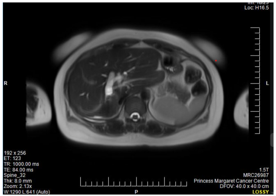
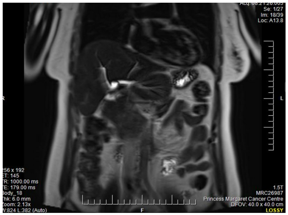
Following referral to Hepatology, abdominal ultrasound demonstrated moderate bilateral intrahepatic biliary dilation. Follow-up MRI demonstrated multiple intrahepatic stones, chronic hepatic atrophy (right lobe), and periductal enhancement suggestive of RPC (see
Figure 1 and
Figure 2). No extrahepatic ductal obstruction was seen. Her epidemiologic background raised suspicion for
C. sinensis infection, and she was, therefore, referred to our Tropical Disease Unit.
At initial evaluation in our unit, she reported intermittent right upper quadrant pain over two to three years, worsened by fatty meals, and unintentional weight loss of six kilograms over six months. She denied jaundice, fever, pruritus, nausea, vomiting, diarrhea, constipation, and dyspepsia at that time. The review of systems was otherwise negative. An examination revealed mild right upper quadrant tenderness without signs of chronic liver disease.
She denied freshwater exposure but reported consuming raw fish (sushi) while living in the Philippines. She had no history of alcohol, smoking, or drug use. Her family history included unspecified liver disease in a sibling.
Laboratory findings were notable for alanine transaminase (ALT) levels of 151 U/L (reference range, 7–40 U/L), aspartate transaminase (AST) levels of 112 U/L (reference range, 5–34 U/L), ALP 532 U/L (reference range, 40–150 U/L), and gamma-glutamyl transferase (GGT) levels of 749 U/L (reference range 8–33 U/L), reflecting a 3-5-fold elevation across liver function tests. The complete blood count was normal with an absolute eosinophil count of 0.2 × 10⁹/L (reference range, <0.4 × 10⁹/L). Stool ova and parasite (O&P) testing with microscopic examination of iron hematoxylin-stained smears and formalin ethyl acetate concentrates was repeatedly negative. Serologies for Strongyloides, Schistosoma, and hepatitis A were negative. IgE was elevated at 408. QuantiFERON-TB Gold was positive, and she was, therefore, referred to a TB clinic.
Due to imaging findings of RPC, a compatible exposure history related to frequent raw fish consumption in the Philippines, and persistent symptoms, empiric treatment with praziquantel (25 mg/kg TID × 2 days) was initiated. The medication was well tolerated except for transient dizziness.
Two months post-treatment, her right upper quadrant abdominal pain had improved and liver enzymes demonstrated some improvement (ALT 71 U/L, AST 61 U/L, ALP 504 U/L). Follow-up MRI demonstrated moderate bilateral intrahepatic biliary dilatation, multiple intrahepatic pigmented stones, periductal enhancement and fibrosis consistent with chronic biliary inflammation, and right hepatic lobar atrophy. No extrahepatic ductal obstruction was identified. These findings are characteristic of recurrent pyogenic cholangitis. There was no evidence of malignancy. Surveillance with Hepatology continued for her chronic hepatobiliary disease (
Table 1).
3. Discussion
Clonorchis sinensis infects an estimated 15 million people globally, with the majority of cases occurring in East Asia [
1]. Infection occurs through consumption of raw or undercooked freshwater fish, leading to intrahepatic colonization and chronic inflammation [
3]. Prolonged infection can lead to biliary strictures, pigmented stones, and complications such as cholangiocarcinoma [
5,
8].
The differential diagnosis of RPC and related symptoms in a patient with raw freshwater fish exposure in Southeast Asia should prioritize the hepatobiliary trematodes. As outlined by Chan and colleagues in their framework of parasitic liver disease in travelers and migrants, liver flukes of the family Opisthorchiidae are among the most clinically consequential parasitic causes of cholestatic liver disease in this population [
9].
Fasciola spp., while also trematodes, are transmitted via contaminated aquatic vegetation rather than fish and typically produce pronounced eosinophilia and migratory hepatic lesions—features absent in our patient [
9].
Ascaris lumbricoides may rarely cause biliary obstruction but would not account for the chronic intrahepatic ductal changes seen here [
9].
Clonorchis sinensis and the
Opisthorchis species, therefore, represent the most plausible parasitic etiologies.
Among the Opisthorchiidae,
C. sinensis,
O. viverrini, and
O. felineus share a similar biology, clinical syndrome, and treatment response but differ in geographic distribution.
C. sinensis predominates in East Asia,
O. viverrini in mainland Southeast Asia, and
O. felineus across Eastern Europe and Siberia. Both
C. sinensis and
O. viverrini are International Agency for Research on Cancer (IARC) Group 1 carcinogens, given their association with cholangiocarcinoma. Species-level differentiation on clinical or imaging grounds alone is not possible, and praziquantel is effective against all three, rendering the precise species of secondary importance to clinical management, while still being of public health relevance [
10].
Non-parasitic causes of RPC and biliary structuring should also be considered in the differential. IgG4-related cholangiopathy can produce biliary strictures and cholestatic liver tests, but it is typically associated with elevated serum IgG4 levels and diffuse biliary thickening on imaging; our patient had no serologic or imaging features to support this. Primary sclerosing cholangitis produces multifocal intra- and extrahepatic biliary strictures with beading on cholangiography, and it is commonly associated with inflammatory bowel disease—neither feature was present. Post-infectious benign biliary strictures from prior bacterial cholangitis may be considered, though the patient had no documented cholangitis episodes. Congenital biliary anomalies such as Caroli disease produce segmental cystic dilatation of intrahepatic ducts, a pattern distinct from the diffuse periductal changes seen here. Ultimately, the absence of these features alongside the epidemiological profile and treatment response favored a parasitic etiology.
This case highlights diagnostic challenges in non-endemic regions, where there remains a lack of provider awareness and poor sensitivity of available reference-level diagnostics. Stool microscopy, the standard diagnostic tool, can yield false negatives in chronic or obstructive cases due to low egg excretion [
6]. Sensitivity may fall below 30% in such contexts [
6]. Advanced diagnostics such as circulating antigen assays or PCR on bile specimens offer greater sensitivity but are invasive and typically unavailable outside of research settings [
11,
12]. Imaging, thus, becomes central to diagnosis in the appropriate epidemiological context. Characteristic MRI findings of clonorchiasis include intrahepatic ductal dilation, pigmented stones, periductal enhancement, and hepatic atrophy [
7]. Our patient’s imaging findings, including microabscesses and right lobe atrophy, pointed to long-standing biliary inflammation due to likely clonorchiasis.
Our patient had multiple negative stool tests yet demonstrated typical imaging features and an exposure history. Despite the absence of confirmatory microbiologic testing, given the appropriate epidemiology, compatible imaging findings, and potential for complications such as cholangiocarcinoma, empiric treatment was provided. A limitation of the present case is that species-level confirmation was not possible; while the patient’s Philippine origin increases the probability of C. sinesis, co-infection with or alternative infection by Opisthorchis spp. cannot be excluded on clinical or radiographic grounds alone.
Praziquantel remains the treatment of choice, with the WHO recommending a dose of 25 mg/kg three times daily for two days [
13]. This regimen has shown near 100% cure rates in trials [
14]. Side effects are typically mild, including nausea, dizziness, and fatigue [
10,
15,
16].
4. Public Health Implications
Physicians in non-endemic regions must consider neglected parasitic infections like clonorchiasis in at-risk individuals, especially in migrants from endemic areas. This case underscores three public health priorities:
Dietary history is crucial in risk assessment. It is important to have a high clinical index of suspicion despite negative diagnostics, if the epidemiological context and imaging are appropriate.
Long-term surveillance is warranted given the elevated lifetime risk of cholangiocarcinoma and progressive biliary damage in chronic hepatobiliary trematodiasis [
5].
In non-endemic settings, public health efforts should include enhancing provider awareness and scaling up diagnostic capacity. Reference laboratories should maintain validated serological or molecular tests for rare helminthiases.
5. Conclusions
In non-endemic settings, recurrent pyogenic cholangitis should prompt evaluation for parasitic infections such as clonorchiasis, especially in individuals from endemic regions. Despite negative stool O&P tests, our patient responded to empiric praziquantel therapy for suspected hepatobiliary trematodiasis, underscoring the value of a syndromic approach incorporating imaging and epidemiologic history. Early treatment may mitigate long-term hepatobiliary morbidity and cancer risk.
Author Contributions
J.T.: literature review and synthesis; writing—original drafting, review, and critical revision of the manuscript. K.A.: literature review and synthesis; writing—original drafting, review and critical revision of the manuscript. G.D.H.: literature review and synthesis; writing—original drafting, review, and critical revision of the manuscript. A.K.B.: conceptualization of the report; literature review and synthesis; writing—original drafting, review and critical revision of the manuscript; overall project oversight. All authors serve as guarantors of the work. All authors have read and agreed to the published version of the manuscript.
Funding
Dr. Boggild is supported as a Clinician Scientist by the Departments of Medicine at the University Health Network and University of Toronto. Dr. Hawley is supported by an Ontario Graduate Scholarship and a Canada Graduate Research Scholarship.
Institutional Review Board Statement
The study was conducted in accordance with the Declara-tion of Helsinki. Our institutions do not require Research Ethics Board or Institutional Review Board approval for publication of case reports where patient informed consent has been obtained for communication of their de-identified personal health information. Case reports are not considered research requiring human subjects consideration by our institutions. Verbal informed consent for publication following discussion of risks and benefits was obtained and documented in the electronic medical records in accordance with PHIPA.
Informed Consent Statement
Informed consent to publish de-identified personal health information was obtained from the patient described in the case report and documented in the patient’s medical record in accordance with PHIPA. Verbal consent was obtained rather than written because the creation and storage of additional paper copies of identifying consent forms that link individual cases to case reports does not conform to PHIPA.
Data Availability Statement
All available data are contained in this report.
Conflicts of Interest
Dr. Boggild oversees the Tropical Disease Fund for Excellence at the University Health Network Foundation, which has received an unrestricted educational grant from Seegene Canada. Neither Seegene nor UHN contributed to the conception of the work; the collection, analysis, or interpretation of the literature; the writing of the manuscript; or the decision to publish the paper.
References
- Yoo, W.G.; Sohn, W.M.; Na, B.K. Current status of Clonorchis sinensis and clonorchiasis in Korea: Epidemiological perspectives integrating data from human and intermediate hosts. Parasitology 2022, 149, 1296–1305. [Google Scholar] [CrossRef] [PubMed]
- Qian, M.B.; Chen, Y.D.; Liang, S.; Yang, G.-J.; Zhou, X.-N. The global epidemiology of clonorchiasis and its relation with cholangiocarcinoma. Infect. Dis. Poverty 2012, 1, 4. [Google Scholar] [CrossRef] [PubMed]
- Li, X.; Chen, Y.; Huang, G.; Sun, X.; Mo, G.; Peng, X. Epidemiology and risk factors of Clonorchis sinensis infection in Guangxi. Parasitol. Res. 2025, 124, 26. [Google Scholar] [CrossRef] [PubMed]
- Locke, V.; Kusnik, A.; Richardson, M.S. Clonorchis sinensis. In StatPearls; StatPearls Publishing: St. Petersburg, FL, USA, 2022. [Google Scholar]
- Tang, Z.L.; Huang, Y.; Yu, X.B. Current status and perspectives of Clonorchis sinensis and clonorchiasis. Infect. Dis. Poverty 2016, 5, 71. [Google Scholar] [CrossRef] [PubMed]
- Kim, T.S.; Pak, J.H.; Kim, J.B.; Bahk, Y.Y. Clonorchis sinensis as a human biological agent of cholangiocarcinoma. BMB Rep. 2016, 49, 590–597. [Google Scholar] [CrossRef] [PubMed]
- Choi, D.; Hong, S.T. Imaging diagnosis of clonorchiasis. Korean J. Parasitol. 2007, 45, 77–85. [Google Scholar] [CrossRef] [PubMed]
- Dong, H.; Zhao, L.; Sun, H.; Shang, M.; Lv, G.; Yu, X.; Hu, B.; Huang, Y. Coinfection of Clonorchis sinensis and hepatitis B virus: Clinical liver indices and interaction in hepatic cell models. Parasit. Vectors 2022, 15, 460. [Google Scholar] [CrossRef] [PubMed]
- Chan, W.W.; Showler, A.; Boggild, A.K. Parasitic liver disease in travelers. Infect. Dis. Clin. N. Am. 2012, 26, 755–780. [Google Scholar] [CrossRef]
- Qian, M.B.; Patel, C.; Palmeirim, M.S.; Wang, X.; Schindler, C.; Utzinger, J.; Zhou, X.-N.; Keiser, J. Efficacy of drugs against clonorchiasis and opisthorchiasis: A systematic review and network meta-analysis. Lancet Microbe 2022, 3, e616–e624. [Google Scholar] [CrossRef] [PubMed]
- Nie, G.; Wang, T.; Lu, S.; Liu, W.; Li, Y.; Lei, J. Detection of Clonorchis sinensis circulating antigen in sera from Chinese patients by immunomagnetic bead ELISA based on IgY. PLoS ONE 2014, 9, e113208. [Google Scholar] [CrossRef] [PubMed]
- Liao, G.; Ruan, H.; Peng, P.; Liu, S.; Qin, J.; Liang, Z.; Tang, G.; Qin, M.; Huang, J. Clonorchiasis in Patients with Biliary and Pancreatic Diseases: Diagnosis and Risk Factors. Biomed Res. Int. 2020, 2020, 2946541. [Google Scholar] [CrossRef] [PubMed]
- WHO Study Group. Control of Foodborne Trematode Infections: Report of a WHO Study Group; World Health Organization: Geneva, Switzerland, 1995. [Google Scholar]
- Soh, C.T. Clonorchis sinensis: Experimental and clinical studies with praziquantel in Korea. Arzneimittelforschung 1984, 34, 1156–1159. [Google Scholar] [PubMed]
- Xu, L.L.; Jiang, B.; Duan, J.H.; Zhuang, S.F.; Liu, Y.C.; Zhu, S.Q.; Zhang, L.P.; Zhang, H.B.; Xiao, S.H.; Zhou, X.N. Efficacy and safety of praziquantel, tribendimidine, mebendazole, and albendazole against Clonorchis sinensis. PLoS Negl. Trop. Dis. 2014, 8, e3046. [Google Scholar] [CrossRef] [PubMed][Green Version]
- Yangco, B.G.; De Lerma, C.; Lyman, G.H.; Price, D.L. Clinical study evaluating efficacy of praziquantel in clonorchiasis. Antimicrob. Agents Chemother. 1987, 31, 135–138. [Google Scholar] [CrossRef] [PubMed]
| Disclaimer/Publisher’s Note: The statements, opinions and data contained in all publications are solely those of the individual author(s) and contributor(s) and not of MDPI and/or the editor(s). MDPI and/or the editor(s) disclaim responsibility for any injury to people or property resulting from any ideas, methods, instructions or products referred to in the content. |





 ,
,