
Case Report

Case Report of a Mixed Plasmodium ovale and Plasmodium malariae Malaria Infection in a Returning Patient from Cameroon to Greece with False Negative Malaria Rapid Diagnostic Test

by
Eleni V. Patsoula
1,*,
Anastasia Bimpa
1,
Nikolaos Tegos
1,
Anastasia Panagopoulou
1,
Ilias Karaiskos
2,
Argyro Triantafyllou
3 and
Eleni Papadogeorgaki
3
1
Unit of Parasitic and Tropical Infections, Laboratory for the Surveillance of Tropical Diseases, Department of Public Health Policy, School of Public Health, University of West Attica, 11521 Athens, Greece
2
1st Department of Internal Medicine-Infectious Diseases, Hygeia General Hospital, 15123 Athens, Greece
3
Central Laboratory, Hygeia General Hospital, 15123 Athens, Greece
*
Author to whom correspondence should be addressed.
Parasitologia 2026, 6(3), 26; https://doi.org/10.3390/parasitologia6030026
Submission received: 3 April 2026 / Revised: 9 May 2026 / Accepted: 15 May 2026 / Published: 18 May 2026

Abstract

Malaria in a Greek citizen with prior malaria history residing and working in Cameroon returning in his home country is a medical emergency warranting prompt and accurate diagnosis and effective treatment. We describe a mixed malaria case of a febrile patient, a professional returning to Greece from a malaria-endemic country whose initial diagnosis was a false-negative malaria rapid diagnostic test. Subsequent alternative rapid diagnostic test, malaria thin-film blood examination and molecular diagnosis revealed mixed malaria infection from Plasmodium ovale and Plasmodium malariae. The patient was successfully treated and achieved complete clinical recovery. The case described here highlights important points regarding prompt and accurate malaria diagnosis in returning travelers in non-endemic countries, emphasizing the importance of revealing cryptic mixed malaria cases and providing molecular approaches to malaria diagnosis in combination with the gold-standard microscopy.

1. Introduction

Malaria is a life-threatening parasitic infection, the most important vector-borne disease worldwide. It remains a leading global public-health emergency, with the vast majority of human cases being recorded in sub-Saharan Africa [1]. Surveillance data show that most malaria cases reported in Europe are travel-related, underscoring the ongoing risk posed by returning travelers and migrants [2]. Malaria in humans is mainly caused by Plasmodium falciparum (P. falciparum), related to high mortality and morbidity, P. vivax, P. ovale and P. malariae, as well as P. knowlesi, which occurs only in rare cases. Plasmodium species present different geographical distributions; however, significant overlap is noted [1]. P. ovale spp. and P. malariae are increasingly documented by molecular surveys and case series across Africa and are responsible for a number of detected infections. These species frequently present with lower parasite densities and atypical clinical presentations, and P. ovale can cause relapses due to hypnozoites in the liver. Therefore, prompt diagnosis and appropriate treatment are of major importance [3,4,5,6].
Mixed malaria infections are quite uncommon in travelers; however, they can involve P. ovale and P. malariae. They are often underestimated due to the diagnostic challenges they pose related to low-density infections and microscopic differentiations between similar appearing parasitic forms [7,8,9,10]. Molecular diagnostic approaches, being more sensitive, are performed to accurately detect mixed Plasmodium sp. infections that can be missed by microscopy or rapid diagnostic tests (RDTs) [11].
RDTs have become a useful tool in point-of-care malaria diagnosis as they are rapid, require minimal training, and produce satisfactory results regarding the detection of P. falciparum through HRP2 antigen recognition. However, RDT performance is variable for non-falciparum infections and can be undermined as HRP2/3 gene deletions in P. falciparum primarily lead to false-negative HRP2-based tests, while antigen persistence after treatment and its cross-reactivity with other pathogens are mainly associated with false-positive results and low parasitemia is a classical cause of false-negative results. Consequently, relying on RDTs alone may lead to misdiagnosis and inappropriate treatment, particularly in returning travelers with non-falciparum or mixed infections [12,13,14,15,16].
Here, we describe a case of mixed P. ovale/P. malariae infection in a patient returning to Europe from Cameroon and the emerging diagnostic challenges, as several RDTs performed were negative and only subsequent microscopy and molecular testing succeeded in detecting the mixed infection. Cameroon is a malaria-endemic country and the disease is transmitted in all areas. The main species is P. falciparum, while P. malariae, P. ovale, and P. vivax are less commonly detected [17]. This case report highlights the diagnostic difficulties posed by non-falciparum mixed Plasmodium infections in travelers and emphasizes the need for Plasmodium species confirmation in non-endemic settings to ensure appropriate patient management and public-health response [18].

2. Case Report

In April 2025, an adult patient with a past medical history of hyperlipidemia residing and working in a malaria-endemic region presented in Greece with a 10-day history of fever up to 38.5 °C, accompanied by rigors and night sweats. The patient had a previous hospitalization approximately one year earlier, in 2024, for persistent fever, during which Plasmodium falciparum malaria was diagnosed in the endemic country and treated with artemether/lumefantrine. Despite appropriate therapy, febrile episodes persisted, leading to hospitalization after return to Greece. During that admission, high-grade fever and gastrointestinal symptoms occurred and enterocolitis due to Shigella spp. was diagnosed. Although antimicrobial therapy was administered, fever persisted. Laboratory evaluation demonstrated pancytopenia and markedly elevated serum ferritin levels (>16,000 ng/mL; reference range: 22–322 ng/mL).
Bone marrow examination revealed hemophagocytosis, establishing the diagnosis of hemophagocytic lymphohistiocytosis (HLH). High-dose corticosteroid therapy was initiated, resulting in clinical and laboratory remission.
In April 2025, the patient again presented with fever, leukocytosis, and elevated C-reactive protein (CRP), requiring a new hospitalization lasting several days. Initial laboratory tests at admission demonstrated: WBC 9.97 ×10³/μL [normal: 4–10 × 103/μL]; Hb 14.2 g/dL [normal: 13–17 g/dL]; Hct 43% [normal: 40–50%]; Platelets 106 ×103/μL [normal: 150–400 × 103/μL].
No malaria chemoprophylaxis had been used due to long-term residence in the endemic region, and no consistent personal protective measures against mosquito exposure were reported. The patient recalled multiple mosquito bites prior to symptom onset.
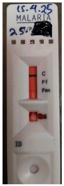
Three (3) malaria rapid diagnostic tests (RDTs) performed in Cameroon (product details not available), as well as additional testing in the hospital in Greece with the Abbot-Bioline™ Malaria Ag P.f/Pan (Abbott Diagnostics Korea, Gyeonggi-do, Republic of Korea) targeting P. falciparum-specific histidine-rich protein II (HRP-2) antigen and common Plasmodium lactate dehydrogenase (pLDH) of Plasmodium species antigen (Pan), were negative. His whole-blood sample was sent from the hospital to the Malaria Reference Center (MRC) in the Unit of Parasitic and Tropical infections of the Department of Public Health Policy, University of West Attica, Athens, Greece, where the Healgen Malaria P.F./Pan Ag RDT cassette whole-blood (Healgen Scientific LLC, Houston, TX, USA) was performed to detect and differentiate HRP-2 and pLDH and the results were positive for the Pan line region (Figure 1).
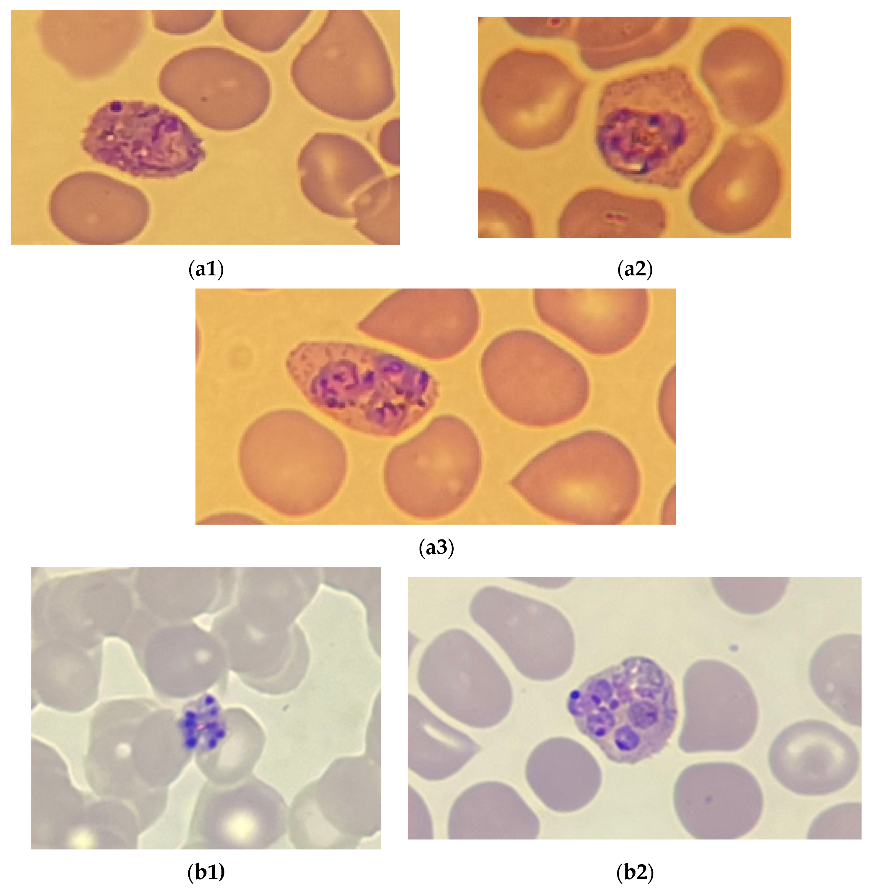
Furthermore, an MRC thin film microscopic examination of peripheral blood smears was performed by two independent expert microscopists. Thin blood smears were stained with Giemsa and viewed under a ×100 oil immersion objective using a Zeiss Primo Star microscope (Carl Zeiss Microscopy GmbH, Jena, Germany), revealing the presence of both Plasmodium ovale (trophozoite forms) and Plasmodium malariae (mature and immature schizont forms) (Figure 2). Parasitemias were counted as percentage of parasitized erythrocytes by two expert microscopists and the average estimated parasitemias was <0.5% for P. ovale and <0.1% for P. malariae, respectively. In the Malaria Reference Center in Greece, we routinely perform a thick blood smear after a positive RDT and a negative thin blood smear. This approach increases sensitivity, as blood is concentrated, allowing for the detection of low-level parasitemias, the identification of Plasmodium sp. presence and density calculations, but is not specific enough for species identification, as recommended by the WHO and CDC [1].
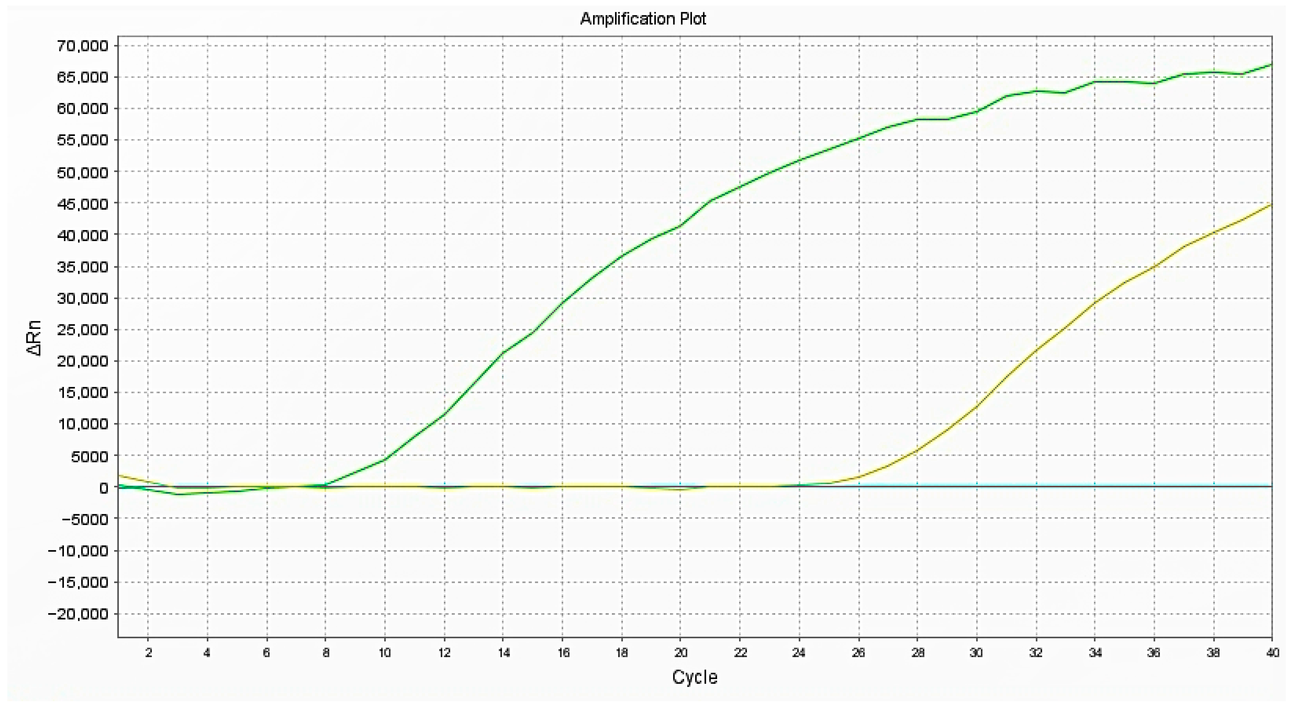
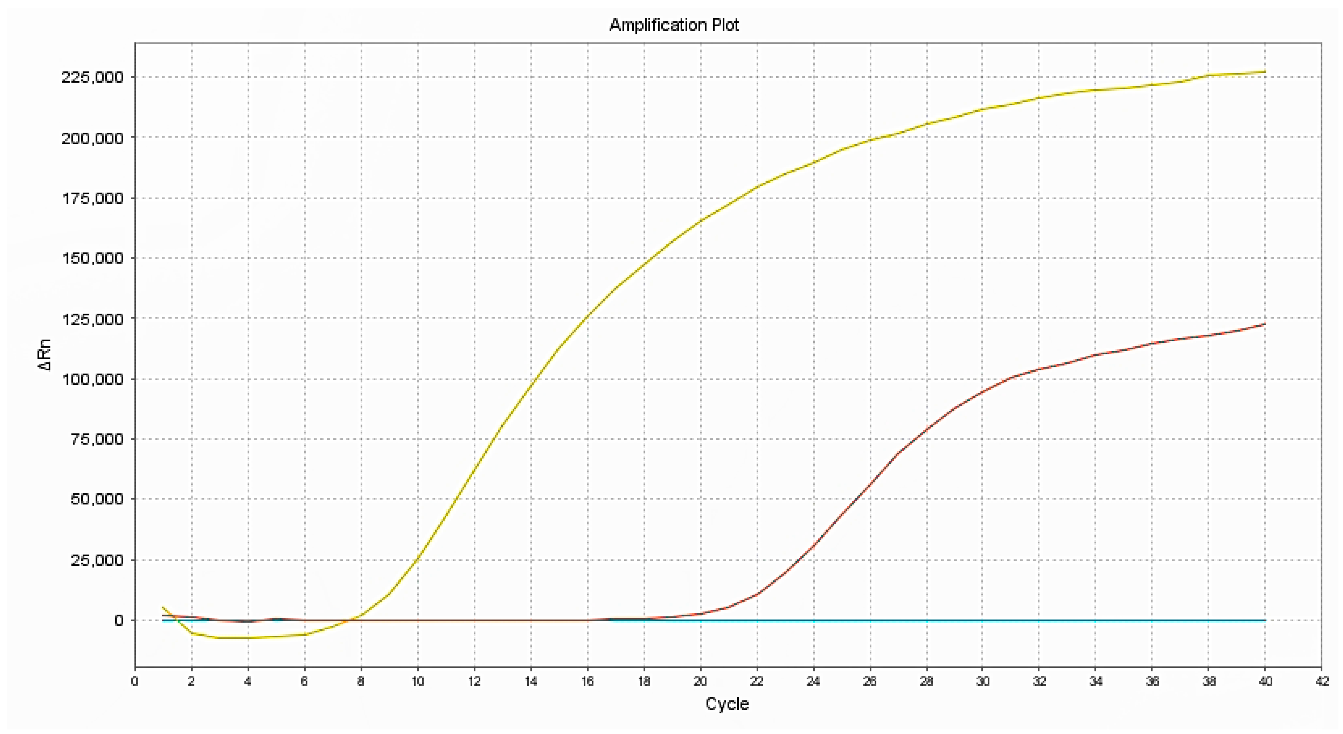
An in-house multiplex PCR protocol, targeting P. falciparum and P. vivax species in the same reaction, was implemented and the results were negative [19,20]. Given that P. ovale and P. malariae were detected by microscopy targeted to the species level, real-time PCR protocols were implemented [21]. In the real-time PCR tests performed for P.ovale and P. malariae, the specific protocols described by Rougemont et al., 2004 [21] were used. Species-specific probes for P. malariae (minor groove binder probe) labeled with 5′FAM (Malaprobe: 5′FAM-CTATCTAAAA GAAACACTCAT), and P. ovale (minor groove binder probe) labeled with 5′VIC (Ovaprobe: 5′VIC-CGAAAGGAATTTTCTTATT) were used, along with forward Plasmo primer and species-specific reverse primers [21]. Dual infection was confirmed, corroborating the respective microscopy result (Figure 3 and Figure 4). For P. ovale, a Ct value of 22.22 versus Ct: 7.1 for positive control was observed, while for P. malariae, Ct value was 16.14 versus Ct: 7.7 for positive control.
The patient received intravenous artesunate (three doses), followed by a full course of oral artemether/lumefantrine (24 tablets in total), and a 20-day course of primaquine to target liver hypnozoites and prevent relapse.
Following treatment completion, the patient achieved complete clinical recovery. Repeat microscopic evaluation showed no evidence of parasitaemia, and all laboratory parameters returned to normal limits.

3. Discussion

Fever in the returning traveler is a common clinical scenario encountered in emergency and outpatient clinics, warranting immediate exclusion of life-threatening infections such as malaria. This case represents a typical mixed-species malaria infection acquired in Central Africa, where multiple Plasmodium species co-circulate. Cameroon reports approximately 2.5–3 million malaria cases annually, with P. falciparum predominating (>90% of cases), but significant co-circulation of P. ovale (2–5%) and P. malariae (1–3%) (WHO [1] is also reported. Mixed infections, while underestimated by conventional diagnostics, occur in 5–15% of symptomatic cases in Central–West Africa when assessed by molecular methods [16,22].
The false negative RDT result in this case highlights the critical limitations of current rapid diagnostic tests for detecting non-falciparum malaria. The Abbot-Bioline™ RDT Malaria test used in this case detects P. falciparum-specific HRP2 and pan-Plasmodium lactate dehydrogenase. The negative result occurred despite the combined presence of the two Plasmodium species, which should theoretically be detectable by pan-lactate dehydrogenase detection. Several recent publications discuss the underperformance of the Abbot-Bioline™ Ag P.f/P.v malaria antigen test and its failure to detect microscopically confirmed P. vivax malaria cases [14,15].
Several factors may contribute to the poor RDT performance for P. ovale and P. malariae. These two species often have lower parasite densities in the blood compared to P. falciparum, which leads to lower pLDH concentrations and reduced sensitivity of pLDH-based RDTs. Studies suggest that the performance of the several pLDH-based RDTs for P. ovale detection was not optimal, with the low parasite densities and pLDH concentration contributing to the failure of RDTs for P. ovale [23]. Studies report detection rates of P. falciparum ranging from 96.2% to 98.7%, while no significant differences were noted between sensitivities to P. vivax by pan LDH or pvLDH (81.1% to 94.6%). Some of the RDTs missed most of P. ovale and P. malariae, with sensitivities ranging from 4.5% to 81.8% and 14.3% to 95.2% respectively [24]. The performance of RDTs for P. ovale malaria in Japanese travelers was reported to be very low, as the overall sensitivity of RDTs to P. ovale malaria and P. vivax malaria was 22.2% and 94.1%, respectively [25]. In mixed infections, the situation is further complicated, as Pan-bands in the RDTs may be positive due to the presence of any non-falciparum Plasmodium species, but attribution to specific species is impossible without microscopy or molecular methods.
Overall, thin- and thick-film microscopy offer sub-optimal sensitivity for detecting mixed P. ovale and P. malariae co-infections, as both species typically present with low parasitemias, combined with subtle and sometimes overlapping morphological characteristics, frequently leading to misidentification or missed diagnoses, especially when microscopic diagnosis is not performed by experts and trained microscopists [6,26]. RDTs targeting HRP-2 and pLDH show limited sensitivity and specificity for both species compared to P. falciparum, often failing to detect P. malariae and P. ovale, especially at low parasitemias, leading to false negatives [22,24], whereas real-time PCR with species-specific primers and probes achieves high sensitivity (>95%) and specificity (>99%), enabling the detection and differentiation of both species, making it indispensable for the accurate diagnosis of mixed P. ovale/P. malariae infections and submicroscopic parasite densities [27,28].
The patient was diagnosed with a mixed P. ovale/P. malariae malaria infection. While the most reasonable explanation regarding a traveler returning from Central Africa is that this is a new, simultaneously acquired mixed-species infection, the clinical timeline and host context could also include aspects of pathophysiological interpretation. P. ovale is able to form dormant parasite hepatic hypnozoite stages that can recrudesce several months to years after the primary infection. If the patient was simultaneously exposed to P. ovale and P. falciparum during the original 2024 episode, the treatment regimen used for P. falciparum would have eradicated blood-stage P. ovale parasites if present but left hepatic hypnozoites intact; therefore, the current P. ovale detection could represent a relapse rather than a new infection [29]. Furthermore, the suppression and unmasking hypothesis could also be considered, as both P. malariae and P. ovale not only usually display low-density parasitemias but also are capable of sustaining cryptic, sub-microscopic blood-stage parasitemias for extended periods. In the case of a low-level co-infection being originally present, along with a high-density parasitemia of P. falciparum, this could constitute competitive suppression and lead to re-emergence once the dominant species was treated [30]. Another factor further supporting the plausibility of this scenario is the patient’s immunosuppression condition resulting from high-dose corticosteroid treatment, a mechanism that could be responsible for constraining non-falciparum parasite densities to undetectable levels, permitting residual P. ovale and/or P. malariae erythrocytic stages that are present to later multiply and result in a patent and symptomatic infection [31]. The one-year interval is generally consistent with the relapse timing of P. ovale and the simultaneous appearance of P. malariae, which does not form hypnozoites but can persist for a long time as a low-grade infection can support the unmasking scenario for P. malariae, unless it is genuinely a new infection. Distinguishing definitively between these scenarios would require molecular typing and a comparison between samples from the initial 2024 episode, which were not available, with current blood samples.
This case underscores the irreplaceable role of expert microscopy in malaria diagnosis, particularly in non-endemic settings where imported malaria may involve any of the five human-infecting Plasmodium species. Microscopy provides several advantages, such as Plasmodium species identification, parasitemia assessment, the detection of mixed infections, and treatment monitoring and, for these reasons, the World Health Organization states that microscopy by trained technicians remains the gold standard for malaria diagnosis, with RDTs serving as a valuable adjunct when expert microscopy is unavailable or delayed [1]. In developed countries with low malaria caseloads, maintaining microscopy expertise is challenging but essential for accurate diagnosis.
Molecular methods, particularly multiplex, nested and quantitative PCRs targeting the 18S rRNA gene, represent the most sensitive approach for malaria diagnosis and species identification. PCR detects mixed infections at higher rates than microscopy due to its ability to detect low-level parasitemias displaying detection frequencies of 3% by qPCR, 1.4% by microscopy and 0.4% by RDT [32].
Regarding treatment, the regimen employed (intravenous artesunate, then artemether/lumefantrine followed by primaquine) represents the standard therapy for P. ovale and P. malariae infections acquired in most endemic areas.
This case highlights several important public health issues, such as pre-travel counseling and adherence to chemoprophylaxis, along with the importance of personal protective measures against mosquito bites.

4. Conclusions

We report a successfully treated case of mixed P. ovale and P. malariae infection with false negative rapid diagnostic test results in a returning traveler from Cameroon. In conclusion, the diagnostic evaluation of mixed P. malariae and P. ovale infections reveals the importance of a well-featured malaria diagnosis performance, with real-time PCR demonstrating superior sensitivity and specificity over microscopy, which is significantly limited by low parasitemias and RDTs, which are limited by the absence of species-specific antigenic targets, rendering them unable to reliably detect these two Plasmodium species. The patient achieved complete clinical and parasitological cure through appropriate treatment guided by a comprehensive diagnostic evaluation, including microscopy and molecular confirmation, and was discharged in stable condition with full recovery. This case emphasizes the importance of a comprehensive malaria diagnosis that requires the integration of clinical judgment, appropriate diagnostic technology including both microscopy and molecular methods, and expert interpretation. When these aspects are combined with appropriate species-specific treatment, patients achieve excellent outcomes with complete cure, as demonstrated in this case.

Author Contributions

Conceptualization, E.V.P. and E.P.; methodology, E.V.P., A.B., N.T. and A.P.; investigation, I.K., A.T. and E.P.; writing—original draft preparation, E.V.P.; writing—review and editing, E.V.P., A.B., N.T. and E.P.; visualization, N.T. and A.B.; supervision, E.V.P.; funding acquisition, E.V.P. All authors have read and agreed to the published version of the manuscript.

Funding

The Malaria Reference Center is funded by the National Public Health organization regarding malaria–Plasmodium typing laboratory procedures.

Institutional Review Board Statement

The study was conducted in accordance with the Declaration of Helsinki. Ethical review and approval were waived for this study due to the fact that clinical samples were coded and patients’ anonymity and personal data protection was assured.

Informed Consent Statement

Informed consent was obtained and was ensured by the referring hospital.

Data Availability Statement

The original contributions presented in the study are included in the article, further inquiries can be directed to the corresponding author.

Conflicts of Interest

The authors declare no conflicts of interest.

References

  1. WHO. World Malaria Report 2025: Addressing the Threat of Antimalarial Drug Resistance; World Health Organization: Geneva, Switzerland, 2025. [Google Scholar]
  2. European Centre for Disease Prevention and Control. Malaria. In Annual Epidemiological Report for 2022; ECDC: Stockholm, Sweden, 2024. [Google Scholar]
  3. Sendor, R.; Banek, K.; Kashamuka, M.M.; Mvuama, N.; Bala, J.A.; Nkalani, M.; Kihuma, G.; Atibu, J.; Thwai, K.L.; Svec, W.M.; et al. Epidemiology of Plasmodium malariae and Plasmodium ovale spp. in Kinshasa Province, Democratic Republic of Congo. Nat. Commun. 2023, 14, 6618. [Google Scholar] [CrossRef] [Scilit]
  4. Mueller, I.; Zimmerman, P.A.; Reeder, J.C. Plasmodium malariae and Plasmodium ovale—The “bashful” malaria parasites. Trends Parasitol. 2007, 23, 278–283. [Google Scholar] [CrossRef] [Scilit] [PubMed] [PubMed Central]
  5. Bichara, C.; Flahaut, P.; Costa, D.; Bienvenu, A.-L.; Picot, S.; Gargala, G. Cryptic Plasmodium ovale concurrent with mixed Plasmodium falciparum and Plasmodium malariae infection in two children from Central African Republic. Malar. J. 2017, 16, 339. [Google Scholar] [CrossRef] [Scilit] [PubMed] [PubMed Central]
  6. Kotepui, M.; Kotepui, K.U.; De Jesus Milanez, G.; Masangkay, F.R. Plasmodium spp. mixed infection leading to severe malaria: A systematic review and meta-analysis. Sci. Rep. 2020, 10, 11068. [Google Scholar] [CrossRef] [Scilit] [PubMed] [PubMed Central]
  7. Senn, H.; Alattas, N.; Boggild, A.K.; Morris, S.K. Mixed-species Plasmodium falciparum and Plasmodium ovale malaria in a paediatric returned traveller. Malar. J. 2014, 13, 78. [Google Scholar] [CrossRef] [Scilit] [PubMed] [PubMed Central]
  8. Tournoy, T.K.; Rosanas-Urgell, A.; Van Esbroeck, M.; Bottieau, E.; Huits, R. Plasmodium malariae after successful treatment of P. falciparum malaria with artemether-lumefantrine. Int. J. Infect. Dis. 2022, 119, 56–58. [Google Scholar] [CrossRef] [Scilit] [PubMed]
  9. Kim, G.; Hong, H.L.; Kim, S.Y.; Lee, H.R.; Kim, D.G.; Park, S.; Shin, H.-S.; Chin, B.S.; Kim, Y. Mixed Infection with Plasmodium falciparum and Plasmodium ovale in a Returned Traveller: The First Case in Korea. J. Korean Med. Sci. 2019, 34, e23. [Google Scholar] [CrossRef] [Scilit] [PubMed] [PubMed Central]
  10. Nguyen, H.T.T.; Romano, F.; Wampfler, R.; Mühlethaler, K.; Tannich, E.; Oberli, A. Case Report: Diagnostic Challenges in the Detection of a Mixed Plasmodium vivax/ovale Infection in a Non-Endemic Setting. Am. J. Trop. Med. Hyg. 2020, 103, 1085–1087. [Google Scholar] [CrossRef] [Scilit] [PubMed] [PubMed Central]
  11. Ikegbunam, M.; Maurer, M.; Abone, H.; Ezeagwuna, D.; Sandri, T.L.; Esimone, C.; Ojurongbe, O.; Woldearegai, T.G.; Kreidenweiss, A.; Held, J.; et al. Evaluating Malaria Rapid Diagnostic Tests and Microscopy for Detecting Plasmodium Infection and Status of Plasmodium falciparum Histidine-Rich Protein 2/3 Gene Deletions in Southeastern Nigeria. Am. J. Trop. Med. Hyg. 2024, 110, 902–909. [Google Scholar] [CrossRef] [Scilit] [PubMed] [PubMed Central]
  12. Ojeniyi, F.D.; Ayoola, A.O.; Ibitoye, O.; Opaleye, O.O.; Olowe, O.A.; Ehigie, L.O.; Thomas, B.N.; Ojurongbe, O. Performance and challenges of malaria rapid diagnostic tests in endemic regions of Africa. Sci. Rep. 2025, 15, 43663. [Google Scholar] [CrossRef] [Scilit] [PubMed] [PubMed Central]
  13. Ismail, M.; Mukhtar, A.; Odetokun, I.A.; Ibrahim, M.A.; Salman, A.A.; Bawa-Sani, H.G.; Nass, S.S.; Goni, B.W.; Nuhu, A.; Zhang, X.; et al. Performance of rapid diagnostic tests for malaria diagnosis in Nigeria: A systematic review and meta-analysis. Malar. J. 2025, 25, 17. [Google Scholar] [CrossRef] [Scilit] [PubMed] [PubMed Central]
  14. Thein, K.S.; Soe, K.; Tun, S.K.S.; Kuipers, R.; Janssen, S.; Tarning, J.; Htun, Y.; Day, N.P.; Kyaw, T.T.; Dondorp, A.M.; et al. The underperforming Abbott-Bioline Malaria Ag P.f/P.v rapid diagnostic test: A whiter shade of pale-where the truth is not plain to see. Malar. J. 2025, 24, 441. [Google Scholar] [CrossRef] [Scilit] [PubMed] [PubMed Central]
  15. Aung, M.T.T.; Thu, A.M.; Phyo, A.P.; Aung, W.L.; Pateekhum, C.; Proux, S.; Yotyingaphiram, W.; Archusuksan, L.; Wongaeka, M.; White, N.J.; et al. Investigating the Abbott-Bioline™ malaria antigen Pf/Pv rapid diagnostic test. Malar. J. 2025, 24, 343. [Google Scholar] [CrossRef] [Scilit] [PubMed] [PubMed Central]
  16. Missoup, V.O.; Suh, P.F.; Ndjebakal, C.N.; Tagne, D.; Poumachu, Y.; Joko, S.; Kouamendjouo Djilla, A.; Njiokou, F.; Mbacham, W.; Wondji, C.; et al. Malaria infection prevalence and diagnostic performance of the Abbott Bioline Malaria Ag P.f/Pan rapid test in rural populations of Central Cameroon. Sci. Rep. 2025, 15, 27930. [Google Scholar] [CrossRef] [Scilit] [PubMed] [PubMed Central]
  17. Ridpath, A.D.; Wallender, E. Malaria. In CDC Yellow Book: Health Information for International Travel; Oxford Academic: New York, NY, USA, 2026. [Google Scholar]
  18. Mortazavi, S.E.; Lugaajju, A.; Ivarsson, A.-C.; Karlsson Söbirk, S.; Norrgren, H.; Persson, K.E.M. High rate of false positive malaria rapid diagnostic tests in a district hospital in Uganda. Front. Malar. 2025, 3, 1545825. [Google Scholar] [CrossRef] [Scilit]
  19. Patsoula, E.; Spanakos, G.; Sofianatou, D.; Parara, M.; Vakalis, N.C. A single-step, PCR-based method for the detection and differentiation of Plasmodium vivax and P. falciparum. Ann. Trop. Med. Parasitol. 2003, 97, 15–21. [Google Scholar] [CrossRef] [Scilit]
  20. Tegos, N.; Goumenopoulos, C.; Mpimpa, A.; Papavasilopoulos, V.; Beleri, S.; Patsoula, E. Sensitivity Assessment of a Multiplex and Real-Time PCR Protocols for the Detection of Malaria in External Quality Control Samples in the Malaria Reference Center in Greece. Parasitologia 2024, 4, 150–161. [Google Scholar] [CrossRef] [Scilit]
  21. Rougemont, M.; Van Saanen, M.; Sahli, R.; Hinrikson, H.P.; Bille, J.; Jaton, K. Detection of four Plasmodium species in blood from humans by 18S rRNA gene subunit-based and species-specific real-time PCR assays. J. Clin. Microbiol. 2004, 42, 5636–5643. [Google Scholar] [CrossRef] [Scilit] [PubMed]
  22. Oguike, M.C.; Betson, M.; Burke, M.; Nolder, D.; Stothard, J.R.; Kleinschmidt, I.; Proietti, C.; Bousema, T.; Ndounga, M.; Tanabe, K.; et al. Plasmodium ovale curtisi and Plasmodium ovale wallikeri circulate simultaneously in African communities. Int. J. Parasitol. 2011, 41, 677–683. [Google Scholar] [CrossRef] [Scilit] [PubMed]
  23. Tang, J.; Tang, F.; Zhu, H.; Lu, F.; Xu, S.; Cao, Y.; Gu, Y.; He, X.; Zhou, H.; Zhu, G.; et al. Assessment of false negative rates of lactate dehydrogenase-based malaria rapid diagnostic tests for Plasmodium ovale detection. PLoS Neglected Trop. Dis. 2019, 13, e0007254. [Google Scholar] [CrossRef] [Scilit] [PubMed] [PubMed Central]
  24. Gendrot, M.; Madamet, M.; Fonta, I.; Benoit, N.; Amalvict, R.; Mosnier, J.; French National Reference Centre for Imported Malaria Study Group; Pradines, B. Comparative Assessment of the Sensitivity of Ten Commercial Rapid Diagnostic Test Kits for the Detection of Plasmodium. Diagnostics 2022, 12, 2240. [Google Scholar] [CrossRef] [Scilit] [PubMed] [PubMed Central]
  25. Tanizaki, R.; Kato, Y.; Iwagami, M.; Kutsuna, S.; Ujiie, M.; Takeshita, N.; Hayakawa, K.; Kanagawa, S.; Kano, S.; Ohmagari, N. Performance of Rapid Diagnostic Tests for Plasmodium ovale Malaria in Japanese Travellers. Trop. Med. Health 2014, 42, 149–153. [Google Scholar] [CrossRef] [Scilit] [PubMed] [PubMed Central]
  26. Obare, P.; Ogutu, B.; Adams, M.; Odera, J.S.; Lilley, K.; Dosoo, D.; Adhiambo, C.; Owusu-Agyei, S.; Binka, F.; Wanja, E.; et al. Misclassification of Plasmodium infections by conventional microscopy and the impact of remedial training on the proficiency of laboratory technicians in species identification. Malar. J. 2013, 12, 113. [Google Scholar] [CrossRef] [Scilit] [PubMed] [PubMed Central]
  27. Lazrek, Y.; Florimond, C.; Volney, B.; Discours, M.; Mosnier, E.; Houzé, S.; Pelleau, S.; Musset, L. Molecular detection of human Plasmodium species using a multiplex real time PCR. Sci. Rep. 2023, 13, 11388. [Google Scholar] [CrossRef] [Scilit] [PubMed] [PubMed Central]
  28. Phuong, M.; Lau, R.; Ralevski, F.; Boggild, A.K. Sequence-based optimization of a quantitative real-time PCR assay for detection of Plasmodium ovale and Plasmodium malariae. J. Clin. Microbiol. 2014, 52, 1068–1073. [Google Scholar] [CrossRef] [Scilit] [PubMed] [PubMed Central]
  29. Guerra, R.I.; Ore, M.; Valdivia, H.O.; Bishop, D.K.; Ramos, M.; Mores, C.N.; Campbell, W.R. A cluster of the first reported Plasmodium ovale spp. infections in Peru occuring among returning UN peace-keepers, a review of epidemiology, prevention and diagnostic challenges in nonendemic regions. Malar. J. 2019, 18, 176. [Google Scholar] [CrossRef] [Scilit] [PubMed] [PubMed Central]
  30. Shankar, H.; Kumar, G.; Ahmed, N.; Florentin, A. Plasmodium malariae is an overlooked malaria parasite with emerging challenges. Commun. Med. 2026, 6, 95. [Google Scholar] [CrossRef] [Scilit] [PubMed] [PubMed Central]
  31. Calle, C.L.; Mordmüller, B.; Singh, A. Immunosuppression in Malaria: Do Plasmodium falciparum Parasites Hijack the Host? Pathogens. Pathogens 2021, 10, 1277. [Google Scholar] [CrossRef] [Scilit] [PubMed] [PubMed Central]
  32. Rios-Orrego, A.; Lopera-Mesa, T.M.; Cifuentes, V.S.; Zuluaga-Idárraga, L. Performance of microscopy and rapid test for the diagnosis of mixed malaria in an endemic area of Colombia. Diagn. Microbiol. Infect. Dis. 2025, 112, 116799. [Google Scholar] [CrossRef] [Scilit] [PubMed]
Figure 1. Healgen Malaria P.F./Pan Ag RDT showing positive result for the Pan line region.
Parasitologia 06 00026 g001
Figure 2. Peripheral blood smears (15 April 2025) were microscopically examined at ×100 magnification by two independent expert microscopists. Microscopic examination under a ×100 oil-immersion objective of Giemsa-stained peripheral thin blood smears revealed trophozoites of Plasmodium ovale (a1a3), with the characteristic oval shape in the (a1,a2) images, fimbriated erythrocytes in the first image and the slight “amoeboid” shape of the erythrocytes in the second image. All three images (a1a3) show slightly enlarged red blood cells, compact trophozoites and prominent Schüffner’s dots. Microscopic examination of mature (b1) and immature (b2) schizonts of Plasmodium malariae revealed up to eight merozoites and the characteristic rosette pattern, which is more prominent in the mature (b1) image, while in the immature (b2) image the merozoites are slightly bigger and the rosette pattern less well-defined.
Parasitologia 06 00026 g002
Figure 3. Real-time PCR protocol results for Plasmodium ovale (from left to right: positive control in green color Ct: 7.1; clinical sample in yellow color Ct: 22.22, negative control in blue color).
Parasitologia 06 00026 g003
Figure 4. Real-time PCR protocol results for Plasmodium malariae (from left to right: positive control in yellow color Ct: 7.7; clinical sample in red color Ct: 16.14, negative control in blue color).
Parasitologia 06 00026 g004
Disclaimer/Publisher’s Note: The statements, opinions and data contained in all publications are solely those of the individual author(s) and contributor(s) and not of MDPI and/or the editor(s). MDPI and/or the editor(s) disclaim responsibility for any injury to people or property resulting from any ideas, methods, instructions or products referred to in the content.